Embed Size (px)
Citation preview

九州大学学術情報リポジトリKyushu University Institutional Repository
Ultrastructure of Postembryonic Development ofthe Pectoral Muscles in the Japanese LesserHorseshoe Bat, Rhinolophus cornutus cornutusfrom the Standpoint of Adaptation for Flight
Yokoyama, KeiichiZoological Laboratory, Faculty of Agriculture, Kyushu University
Uchida, TeruakiZoological Laboratory, Faculty of Agriculture, Kyushu University
https://doi.org/10.5109/23695
出版情報:九州大学大学院農学研究院紀要. 24 (1), pp.49-63, 1979-08. 九州大学農学部バージョン:権利関係:

J. Fat. Agr., Kyushu Univ., 24 (1). 49-63 (1979)
Ultrastructure of Postembryonic Development of thePectoral Muscles in the Japanese Lesser Horseshoe
Bat, Rhinolophus cornutus cornutus fromthe Standpoint of Adaptation for Flight
Keiichi Yokoyama and Teru Aki Uchida
Zoological Laboratory, Faculty of Agriculture,Kyushu University 46-06, Fukuoka 812
(Received May 2, 1979)
From the viewpoint of adaptation for flight in bats, this report presented electronmicroscopic observations on the pectoral muscles during the growth period fromthe late embryo to the adult stage in the Japanese Lesser horseshoe bat, Rhino-Zophus covnutus corm&s. Evidence obtained by examining the muscle fibers at differ-en t growth s tages s t rongly indica ted tha t the i r number was de termined beforebirth, and that they remarkably increased in size to the flying stage probably be-cause of the longitudinal splitting of the myofibrils when they attained a certainsize. Fur thermore , in the la te embryo and newborn s tages , a smal l number oflongitudinally oriented T tubules occurred along the margin of the A-band althoughthe predominant or ien ta t ion of T tubules was t ransverse . In the f ly ing s tagewhich gave the first indication of differentiation into the adult muscle fiber types,however, almost all the T tubules ran in transverse direction near the A-I junc-tion. Judging from the facts that the completion in orientation of the T tubulesand the earliest sign of muscle-type differentiation were recognized in the flyingstage, it was ultrastructurally concluded that the flying stage was the most impor-tant stage of the postnatal period in the bat from the standpoint of survival po-ten t i a l .
I N T R O D U C T I O N
It is well known that the ability of the Japanese Lesser horseshoe bat,Rhinolophus cornutus cornutus (Rhinolophidae) to execute quick turns and to flyat low speed through dense vegetation during foraging time is remarkable.Recently, the authors (Yokoyama et al., 1975; Yokoyama and Uchida, 1979)reported the correlation between morphological changes on growth of thewings and ecological aspects in the bat from the standpoint of adaptation forflight. Moreover, the relationship between ecological information on growthand biochemical properties of the pectoral muscles with the growth in the batalso was discussed (Yokoyama et al., 1979) : i.e., it was revealed that therewas a remarkable increase in B subunit containing isozymes in pectoral mus-cle LDH during the transition period from the flapping (and eye-open) to fly-ing stage, which was regarded as an adaptation for a highly manoeuvrableflight at the isozyme level.
49

50 K. Yokoyama and T. A. Uchida
Consequently, it seems very important to investigate ontogenetically howthe above-mentioned biochemical adaptation of the pectoral muscles during thepostnatal period is reflected in the ultrastructure of their postembryonic de-velopment. In this paper, thus, we deal with the ultrastructural changes ofthe pectoral muscles from the late embryo to the adult stage, focusing ourattention on the fine structure of the muscles at the fiying stage which marksan important epoch in the mode of life during the postnatal period.
MATERIALS AND METHODS
From late June to early September a maternity colony, nursery colonies(including mother colony, infant colony, flapping colony and flying colony)and a young colony are formed in a cave in the vicinity of TTcno City, IwatePrefecture in northern Japan.
The whole pectoral muscles of two embryos, four newborn young, twoinfants in each of the colony-forming and flying stages, and an adult pregnantfemale collected at the cave from June through July of 1976-7 were carefullydissected. The muscles were first fixed in 4 % paraformaldehyde with 0.05 Mphosphate buffer (pH 7.4). Postfixiation was achieved with 1% osmium tetrox-ide, after which the tissue was dehydrated in acetone and embedded in Epon812. Thin sections for electron microscopy were cut with glass knives on aPorter-Blum MT-l microtome. After staining with uranyle and lead acetate,the sections were examined with a Hitachi HS-9 electron microscope.
RESULTS
Late embryo stage (about one week before birth)The forearm length and body weight are 14.0 mm and 2. Og, respectively.
At this stage the myotube vanishes already. As shown in Fig. 1, the myo-nuclei of the pectoral muscles in the late embryo have already migrated to theperiphery of the cells and have taken their position beneath the sarcolemmawith caveolae. In each of the embryo muscle fibers a few myofibrils are seenwithin an individual muscle fiber, and the fiber is accordingly small in diam-eter. The interstitial space between the muscle fibers is considerably broad.
As seen in Fig. 2, although the T tubules usually run in transverse direc-tion near the A-I junction, a small number of longitudinally oriented T tubulesalso are observed at the margin of the A-band facing to the interfibrillarsarcoplasm. The cristae of mitochondria with a less dense matrix are lessclosely packed, and darkly stained glycogen particles are abundant in the in-terfibrillar sarcoplasm. The sarcoplasmic reticulum network of diverging andconverging tubules forms a loosely woven lacework around the myofibril thatextends throughout each sarcomere. But, differentiation of the network atthe M-line does not occur at this stage.
As indicated in Fig, 1, on the other hand, the satellite cells are conspicu-ous, and usually rest on the surface of the muscle fibers without forming a
Ultrastructure of Pectoral Muscle in Bat 51
Fig. 1. Cross-section of the pectoral muscle fibers in the late embryostage, showing a satellite cell (S) present on the surface of a musclefiber. The fiber is small in diameter and the interstitial space (IS) be-tween the muscle fibers is considerably broad. Ca, caveola ; Mf, myofibril ;Mt, mitochondrion ; N, nucleus. x 10,000.
convex toward them, being unique in their position since they lie betweenthe basement membrane of the muscle fiber and the fiber plasma membrane.The interspace between the cell and the muscle fiber is narrow and the in-terface does not parallel each other. The cytoplasm of each satellite cell isvery sparse in comparison with the nucleoplasm, of which the greater part ofthe condensed chromatin is disposed along the outer rim of the nucleus, someof which being also scattered throughout the central regions of the nucleus.Furthermore, the transitional cell from the undifferentiated cell to the satel-lite cell, which is usually enclosed by a common basement membrane of themuscle fiber, is rarely found associated with the muscle fibers. The apposedmembranes of the cell and muscle fiber, however, are separated by a clear
gap.In addition, as shown in Fig. 3a, there are a large number of morpholog-
ically undifferentiated cells lying free in the interstitial space between themuscle fibers. Not uncommonly the pseudopodium-like structures of the un-differentiated cells make close membrane contact with each other. Occasion-ally, the undifferentiated cells are longitudinally arrayed parallel to themuscle fibers. Centriole and kinetosome are present close to the Golgi ap-paratus, together with numerous free ribosomes, rough endoplasmic reticulaand mitochondria (Fig. 3b).
Newborn stage (at birth)The forearm measures 19.5 mm and the body weight weighs 3. Og. As
seen in Fig. 4, many undifferentiated cells are still observed in the interstitial

52 K. Yokoyama and T. A. Uchida
Fig. 2. Longitudinal section of the pectoral muscle fiber in the late em-bryo s tage , showing the longitudinally oriented T tubules (T) seen atthe margin of the A-band facing to the interfibrilar sarcoplasm. T h ecristae of mitochondria (Mt) are loosely packed in a clearly less densemat r ix . A, A-band; G, g lycogen par t ic le ; I, I -band; ML, M-l ine ; Z, Z-line. x 30,000.
Fig. 3. Longitudinal sections of the pectoral muscle fibers in the lateembryo s tage , showing the undi f ferent ia ted ce l l s ly ing be tween twoneighboring muscle fibers. a) The longitudinal arrangements of the un-di f ferent ia ted ce l l s (U). L, l ip id drople t ; N. nuc leus . ~ 4 , 0 0 0 . b ) T h eGolgi apparatus (GA), centriole (C) and kinetosome (K) in the cytoplasmof the cell. fR, f r e e r i b o s o m e ; rER, rough endoplasmic re t icu lum. x40,000.

Ultrastructure of Pectoral Muscle in Bat
Fig. 4. Cross-section of the pectoral muscle fibers in the newborn stageshowing a satellite cell. The satellite cell (S) with large pseudopodium-l ike s t ruc tures c lose ly appl ies to the sur face of a musc le f iber . Mf ,myofibril ; Mt, mitochondrion ; N, nuc leus ; U, undi f fe ren t ia ted ce l l . x10,000.Fig. 5. Cross-section of the pectoral muscle fibers in the colony-form-ing stage, showing a remarkable increase in s ize of the musc le f iber(cf. Fig. 4). The interstitial space between the muscle fibers cannot beobserved. Abbreviations as in Fig. 4. x 10,000.

54 K. Yokoyama and T. A. Uchida
space between the muscle fibers as well as in the preceding stage. The in-terstitial space, however, becomes narrower and the satellite cells with largepseudopodium-like structures also are occasionally found in the space. Thecharacteristic features of such cytoplasmic organellae of the muscle fibers atthis stage as myofibrils, T tubules and mitochondria, the fiber diameter andthe status of the satellite and undifferentiated cells are similar to those inthe late embryo stage. The longitudinal arrangements of the undifferentiatedcells, however, cannot be found in this stage.
Colony-forming stage (about 7 days after birth)The forearm length and body weight are 20.1 mm and 4. Og, respectively.
As indicated in Fig. 5, one of the most striking changes that occur duringthe transition period from the newborn to this stage is a growth in numberof the myofibrils within an individual muscle fiber. As the result, the musclefibers increase considerably in size during this period and the interstitialspace between the muscle fibers can hardly be distinguished. Furthermore,another remarkable change that occurs during the period was a substantialincrease in number of the mitochondsia with a conspicuously dense matrix.As shown in Fig. 6 at high magnification, the complicated cristae of mito-chondria are disposed in longitudinal rows between myofibrils.
On the other hand, Fig. 6 shows also a myofibril that is apparently justcommencing to split in two part at only one Z-line. The myofibril splittingoccurs frequently in this stage. On rare occasions, however, the Z-lines aredivided into three parts, A number of glycogen particles are present in thespIitting portion. There is a much higher incidence of splitting in large mus-cle fibers, while no splitting is seen in the small muscle fibers. At this stage,although the longitudinally oriented T tubules decrease in number, a fewsuch T tubules still occur, and the satellite cells also are frequently found(Fig. 5), although the satellite cell and muscle fiber are in close connectionwith each other and the inter face between them becomes smooth. Theundifferentiated cells also are present in the interstitial space between themuscle fibers. One can rarely find the features which show indications of themembranous fusion between both the plasma membranes of the satellite celland muscle fiber.
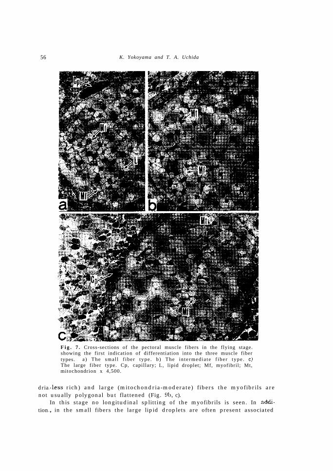
Flying stage (about 20 days after birth)The forearm measures 35.6mm and the body weight weighs 6. Og. In
this stage, as shown in Fig. 7, both of the number of myofibrils within anindividual muscle fiber and the diameter of each muscle fiber increase remark-ably, and the first indication of differentiation into three muscle fiber types,I.e., the small (Fig. 7a), intermediate (Fig. 7b) and large (Fig. 7c) fiber typescorresponding to those of the adult is discernible. The shape of myofibrilsin all types is polygonal in cross section with straight sides. The myofibrilsize in the small and intermediate fibers is smaller than those of the largefibers. In the small fibers a number of lipid droplets are frequently observedassociated with the mitochondria, while in the intermediate and large fibersthe lipid droplets are absent. As seen in Fig. 8, the density of matrix and

Ultrastructure of Pectoral Muscle in Bat 55
Fig. 6. Longitudinal section of the pectoral muscle fiber in the colony-forming stage, showing a myofibril splitting (an arrow) seen at the Z-l ine (Z) . The c r i s tae of mi tochondr ia (Mt) wi th a dense mat r ix a remuch compl ica ted in s t ruc ture . A, A-band; G, glycogen particle ; I, I-band; L, l ipid droplet; ML, M-line; T, T tubule; Tr. Triad. ~60,000.
morphological features of cristae in the mitochondria bear a striking resem-blance to those in the preceding stage (cf. Fig. 6).
On the other hand, in this stage the longitudinally oriented T tubules arealready absent and almost all the T tubules exhibit a transverse orientationnear the A-I junction. The sarcoplasmic reticulum has developed into itsmature form of well organized tubules, and differentiation of the network atthe M-l ine seen in a mature muscle f iber i s evident (F ig . 8 inset ) . Thelongitudinal splitting of the myofibrils is still found frequently. The numberof the satellite and undifferentiated cells in developing muscles of this stagebecomes fewer than those of the preceding stages.
Adult stage
The forearm length and body weight are 40.5mm and 8.7g, respectively.At this stage, the distinctive features of the three fiber types of which thefirst sign was indicated at the flying stage become established. Although themyofibrils in the small fibers (mitochondria-more rich) are polygonal in crosssection (Fig. 9a) as well as in the flying stage, in the intermediate (mitochon-
56 K. Yokoyama and T. A. Uchida
drianot
tion
Fig. 7. Cross-sections of the pectoral muscle fibers in the flying stage.showing the first indication of differentiation into the three muscle fibertypes. a ) The smal l f iber type . b ) The in te rmedia te f iber type . c)The large fiber type. Cp, capillary; L, lipid droplet; Mf, myofibril; Mt,mitochondrion x 4,500.
!-less rich) and large (mitochondria-moderate) fibers the myofibrils areusually polygonal but flattened (Fig. 9b, c).In this stage no longitudinal splitting of the myofibrils is seen. In addi-
L, in the small fibers the large lipid droplets are often present associated

Ultrastructure of Pectoral Muscle in Bat
Fig. 8. Longitudinal sections of the pectoral muscle fibers in the flyingstage, showing typical T tubules and sarcoplasmic reticulum. The Ttubules (T) exhibit a transverse orientation near the A-I junction. Dif-ferentiation of the network of the sarcoplasmic reticulum (SR) at theM-line (ML) is visible (inset). Abbreviations as in Fig. 6. ~30,000.
with the large mitochondria as well as in the preceding stage, whereas inthe intermediate fibers the lipid droplets become first recognized in thisstage. The glycogen particles are abundant in the interfibrillar sarcoplasmof the small and intermediate fibers. In the large fibers, however, the lipiddroplets and glycogen particles are considerably fewer and the mitochondriaare very small in diameter (Fig. 9c). But, as indicated in Fig. 9d, the cristaeof mitochondria become consisted of regularly parallel plates, compared withthe complicated cristae in the colony-forming and flying stages.
DISCUSSION
Although considerable researches on muscle growth from various anglesin Chiroptera have added to our knowledge of this aspect, few ultrastructuralinvestigations have been made except for the report on the myogenesis ofthe web muscles of the fruit-bat by Church (1969). In our previous papers(Yokoyama et al., 1975; Yokoyama et al., 1979; Yokoyama and Uchida, 1979),we reported the information on growth of R. c. comutus from the standpointof morphological, biochemical and ecological aspects, and indicated that theflying stage of the bat was the most important stage in the mode of lifeduring the postnatal period. In this study, we will discuss here the ultrastruc-tural changes with growth of the pectoral muscles in the bat from the lateembryo to adult stage, and especially accumulate attention on the flying stageat which the first sign of differentiation into the three muscle fiber typesseen in the adult stage occurs.

58 K. Yokoyama and T. A. Uchida
1.Muscle
In
Fig. 9. Sections of the pectoral muscle fibers in the adult stage, show-ing the three muscle fiber types (cross-sections) and typical mitochon-dria (a longitudinal section). a) The myofibrils of mitochondria-morer ich f iber . b ) The myof ibr i l s of mi tochondr ia - less r ich f iber . c ) Themyofibrils of mitochondria-moderate fiber. x 20,000. d) The mitochon-dria (Mt) with closely packed lamellar cristae. Abbreviations as in Fig.6. x 40,000.
Ultrastructural changes in the pectoral muscle fibers during growth periodjiber size and fate of the satellite cell
t h e l a t e e m b r y o a n d n e w b o r n s t a g e o f t h e b a t , t h e p e c t o r a l m u s c l e

Ultrastructure of Pectoral Muscle in Bat 59
tissues mainly consisted of immature muscle fibers accompanied with thesatellite cells. Although the myofibrils in the immature muscle fibers weresmall in number, they were quite compact and myonuclei migrated to the sub-sarcolemmal position; even at the late embryo stage the myotubes could notbe observed. Therefore, it is reasonable that the number of the pectoralmuscle fibers is fixed before birth, and consequently does not increase duringthe postnatal growth. Furthermore, it is well known that the muscle fibersincrease in size considerably during the growth period. The reasons are dueto the increase in the amount of myofibrillar material within an individualmuscle fiber. With respect to this, it is of interest to note that the longitu-dinal splitting of myofibrils is frequently observed at the Z-lines during theperiod from the late embryo to flying stage. Recently, it has been suggestedthat in the hind limb muscle of the mouse embryo (Platzer, 1978) and infantmouse (Goldspink, 1972), the increase in muscle fiber size is due to the prolif-eration of myofibrils, which is the result of longitudinal splitting in themyofibrils when they attain a certain size.
As to the status of satellite cells, in the colony-forming stage we couldfind indications of membranous fusion between both the plasma membranes ofthe satellite cell and muscle fiber. After that, in the flying stage, the numberof the satellite cells became fewer than those of the preceding stages. Thus,it seems that the decrease in number of the satellite cells is due to the above-mentioned membranous fusion by a process of which the cytoplasms of thesatellite cell and muscle fiber are confluent.
In this connection, the fate of the satellite cells in growing muscles hasbeen investigated by several workers. In the rat (Enesco and Puddy, 1964;Moss and Leblond, 1971) and domestic fowl (Moss, 1968) during the postnatallife of the animals, the myonuclei considerably increased in number as devel-opment proceeds, whereas the satellite cells diminished gradually in number.Similar phenomena have been known also in the skeletal muscles of thehuman fetus (Ishikawa, 1966) and the mouse fetus (Schultz, 1976). Particular-ly, Moss and Leblond (1971) reported autoradiographically that after a mitoticdivision, one or both satellite-daughter cells of the rat might fuse with musclefiber and thereby added to the myonuclear population. Furthermore, usingthe electron microscope, Schultz (1976) demonstrated a satellite cell whichappeared to be in the process of fusion with a muscle fiber in the mouse.
On the other hand, recent and detailed morphological studies (Schultz,1976 etc.) on the satellite cells of adult mice have supported earlier sugges-tions (Mauro, 1961; Ishikawa, 1966; Church, 1969) that the satellite cells rep-resent dormant myoblasts.
Differentiation of the muscle fiber types and change in orientation of the T tubules
We indicated that in the pectoral muscles of the bat an early evidence ofdifferentiation into the three muscle fiber types corresponding to the small(mitochondria-more rich), intermediate (mitochondria-less rich) and large(mitochondria-moderate) fibers of the adult appeared in the flying stage.
It is well known that adult skeletal muscles are heterogeneous, being

60 K. Yokoyama and T. A. Uchida
composed of different fiber types, of which at least three are recognized asred, intermediate and white fibers with the aid of histochemical methods(Edgerton and Simpson, 1969). Furthermore, on the bases of morphologicaland biochemical properties, Ishikawa (1975) stated that red muscle containinga large number of mitochondria was composed predominantly of smaller darkfibers with high capacity for oxidative metabolism, while white muscle con-taining a small number of mitochondria consisted principally of larger palefibers with high glycolytic enzyme activities.
It is very interesting to determine when the first indication of characteris-tic differentiation into the three muscle fiber types appears during the growthperiod. From this point of view, the histochemical studies of developingmuscle have been carried out by many workers. According to recent papers,it has become clear that there is a striking difference in maturation of skele-tal muscles in various mammals. Namely, in the rat and mouse (Dubowitz,1965), pig (Cooper et al., 19701, cat (Cody and Richardson, 1976a, b) and rabbit(Lobley et al., 1977), the differentiation of skeletal muscle fiber types could notbe discerned at birth. Especially, in the pig (Cooper et nl., 1970) when theinfants are weaned from their mothers (28-35 days after birth), and in therabbit (Lobley et al., 1977) by the time the infants have opened their eyes andare beginning to show increased mobility (8-12 days after birth), the adultpattern of fiber types is established. In contrast to these, even in the fetusof rhesus monkey (Beatty et al., 1967). in the newborn young of the guineapig (Dubowitz, 1965) and human (Fenichel, 1966), the skeletal muscles havealready shown full differentiation into adult fiber types. With respect to this,Dubowitz (1965) suggested that there was some correlation between the pres-ence of differentiation in skeletal muscles at birth and general maturity andmobility of animals, and also the length of gestation.
As shown clearly in this study and other works mentioned above, inmammals with no differentiation of muscle fiber types at birth, it is worthyof note that the first sign of differentiation into the adult fiber types appearsin such developmental epochs as flying (bats), eye-open (rabbit) and weaning(pig) stage during the postnatal growth period.
As to the orientation of T tubules, from our electron microscopical studyon the pectoral muscles of the bat, it became obvious that although the pre-dominant orientation of T tubules in the late embryo and newborn stage wastransverse near the A-I junction, a small number of longitudinally oriented Ttubules also were present at the margin of the A-band. In the flying stage,however, few longitudinally oriented T tubules were found and the orienta-tion of almost all the T tubules was transverse as seen in the adult stage.
The morphogenesis of the sarcoplasmic reticulum and triads in rat skeletalmuscle has been examined in detail by Walker and Schrodt (1968), Schiaffinoand Margreth (1969) and Edge (1970). Especially, from the observations onthe skeletal muscle fibers from the fetal and newborn to 35-day rats, Edge(1970) showed that the predominantly longitudinally oriented triads of imma-ture muscle fibers changed orientation, and that the change from predominant-ly longitudinally to transversely oriented triads was complete in fibers from

Ultrastructure oJ Pectoral Muscle in Bal 61
IO- to &day rats. In this connection, Chaplin et al. (1970) indicated, using therat skeletal muscles, that the time between the beginning of the action poten-tial and the beginning of the recorded development tention (excitation-con-traction latency) declined from 6 msec to 2.8 msec during the first lo-15 daysafter birth. From the above facts, as stated by Edge (1970), it is presumedthat the change in triad orientation might be related to the observed decreasein E-C latency. Taking the above into consideration, it seems likely that thecompletion in orientation of the T tubules in the flying stage of the bat isregarded as an adaptation to contractile mechanism of the flight muscles.
2. Relationship among ecological aspects, changes in morphology of thewings, and in biochemistry and ultrastructure of the pectoral muscles with growth
It is well known that R. c. cornutus having a relatively low wing loading(Kuramoto, 1972) and a predominance of anodal LDH isozymes of the pectoralmuscles (Kitahara et al., 1974; Yokoyama et al., 1979) is capable of doing highlymanoeuvrable flight with low speed, like a butterfly. In a previous paper(Yokoyama et al., 1975), we pointed out, on an analysis of the aspect ratio infive growth stages from the newborn to self-supporting (adult size) stage ofthe bat, that the characteristic of the short-broad wing-type was graduallycompleted with the lapse of time. Furthermore, it became clear that the batwas able to fly in a short period (about 22 days) after birth owing to the lowwing loading at the flying stage (Yokoyama and Uchida, 1979), although thewings in the stage did not yet attain to its adult size and synostosis did notoccur in the wing bones (Yokoyama et al., 1975). It seemed that the above-mentioned morphological features of the wings at the flying stage apply tomany members of the short-broad wing-type bats (Yokoyama and Uchida,1979). Recently, Yokoyama et al. (1979) reported that in the pectoral musclesof R. c. cornutus there was a reversal of activity toward more anodal side ofthe spectrum (cardiac muscle type) during the transition period from theflapping to flying stage, which was considered as an adaptation to metabolicrequirement at isozyme level of the pectoral muscles for an adult flying abil-ity of a slow and delicate flight in the species.
Besides such ecological, morphological and biochemical changes during thepostnatal period, another factor such as ultrastructural change in the flightmuscles must have an important bearing on the abilities of young bats to fly.With respect to this, this ultrastructural research on the pectoral muscles ofthe bat from the late embryo to the adult stage revealed that the changefrom longitudinally to transversely oriented T tubules was almost completedin the flying stage which gave the first indication of differentiation into thethree muscle fiber types in the adult, too. Accordingly, it is supposed thatthe above-mentioned phenomena seen in the pectoral muscles during thepostnatal period are regarded as an adaptation to the contractile and meta-bolic requirements at ultrastructural level of the pectoral muscles for an adultflight.
As stated above, when the bat infants reached the flying stage at about22 days of age, the area of wing membranes became adequate for their limit-

62 K. Yokoyama and T. A. Uchida
ed flight, and the LDH isozyme pattern of the pectoral muscles showed thecardiac muscle type as well as in the adult, and the earliest sign of muscle-type differentiation was recognized. It is thus concluded, from the viewpointof survival potential, that the flying stage marks an important epoch in themode of life during the postnatal period of the bat not only ecologically,morphologically and biochemically but also ultrastructurally, as a precursorstage of the self-supporting stage when the bat establishes the flying abilityand species specific Right mode, etc.
ACKNOWLEDGEMENT
The authors are much indebted to Mr. T. Mbri of the Zoological Labora-tory, Faculty of Agriculture, Kyushu University for his technical advices. Ourgratitude is expressed to Associate Professor S. Shiraishi and graduate stu-dents of the same Laboratory for their encouragement, and to Professor E.W. Jameson, Jr, of the University of CaliforniaScript.
for comments on the manu-
REFERENCES
Beatty. C. H., G. M. Basinger and R. M. Bocek 1967 Dif ferent ia t ion of red and whi tefibers in muscle from fetal, neonatal and infant rhesus monkeys. 1. Histochem. Cyto-chew. 15: 93-103
Chaplin, E. R., G. W. Nell and S. M. Walker 1970 Excitation-contraction latencies in post-natal rat skeletal muscle fibers. Exp. Neuvol.. 29: 142-151
Church , J. C. T. 1969 Satellite cells and myogenesis; a study in the fruit-bat web. 1. Anat..105 : 419-438
Cody, F. W. and H. C. Richardson 1976a Application of histological techniques to develop-ing and adult jaw muscles. 1. Pkysiol., Lond.. 257: 4
Cody, F. W. and H. C. Richardson 1976b Development of histochemical profiles in cat jawmuscles. J. Physiol., Lond., 258: 83-84
Cooper, C. C., R. G. Cassens, L. L. Kastenschmidt and E. J. Briskey 1970 Histochemicalcharacterization of muscle differentiation. Devl. Biol., 23: 169-184
Dubowitz, V. 1965 Enzyme histochemistry of skeletal muscle-1 and II.]. Neural. Neurosurg.Psychiat., 28 : 516-524
Edge, M. B. 1970 Development of apposed sarcoplasmic reticulum at the T system andsarcolemma and the change in orientation of triads in rat skeletal muscle. Devl. Biol..23 : 634-650
Edgerton. V. R. and D. R. Simpson 1969 The intermediate muscle fiber of rats and guineapigs. J. Histochem. Cytochem.. 17: 828-838
Enesco, M. and D. Puddy 1964 Increase in the number of nuclei and weight in skeletalmuscle of rats of various ages. Am. /. Anat., 114: 235-244
Fenichel, G. M. 1966 A histochemical study of developing human skeletal muscle. Neuro-logy, 16: 741-745
Goldspink, G. 1972 Postembryonic growth and differentiation of striated muscle. In “TheStructure and Function of Muscle,” Vol. I , Structure Part 1, ed. by G. H. Bourne.Academic Press, New York and London, pp. 179-236
Ishikawa, H. 1966 Electoron microscopic observations of satellite cells with special refer-

Ultrastructure OJ Pectoral Muscle in Bat 63
ence to the development of mammalian skeletal muscles. 2. Anat. EntwickZ.-Gesch., 125:43-63
Ishikawa, H. 1975 Skeletal muscle. I. Structure. Metabo. Dis., 12: 1‘243-1338 (in Japanese)Kitahara, H., T. A. Uchida and F. Hamajima 1974 LDH isozymes from the standpoint of
adaptation for flight in Chiroptera. Zool. i%fag., Tokyo, 83: IO-17 (in Japanese with Eng-lish abstract)
Kuramoto, T. 1972 Studies on bats at the Akiyoshi-dai Plateau, with special reference tothe ecological and phylogenic aspects. Bull. Akiyoshi-dai Sci. MU., (8) : 7-119 (in Jap-anese with English abstract)
Lobley. G. E., A. B. Wilson and A. S. Bruce 1977 An es t imat ion of the f iber type com-position of eleven skeletal muscles from New Zealand White rnhhits between wean-ing and early maturity. J. Amt., 123: 501-513
Mauro, A. 1961 Satellite cell of skeletal muscle fihers. /. Biophys. Biochem. Cytol. , 9: 4%495
M o s s , F. P. 1968 The relationships between the dimensions of the libers and the num-ber of nuclei during normal growth of skeletal muscle in the domestic fowl. A m . J .Anat.. 122: 555-563
Moss, F. P. and C. P. Leblond 1971 Satellite cells as the source of nuclei in muscles ofgrowing rats. Anat. Rec., 170: 421-436
Platzer. A. C. 1978 The ultrastructure of normal myogenesis in the limb of the mouse.Anat. Rec., 190: 639-658
Schiaffino, S. and A. M. Margreth 1969 Coordinated development of the sarcoplasmic re-ticulum and T system during postnatal differentiation of rat skeletal muscle. /. CellBiol., 41: 855-875
Schultz, E. 1976 Fine structure of satellite cells in growing skeletal muscle. Am. J. Anat.,147: 49-70
Walker, S. M. and G. R. Schrodt 1968 Triads in skeletal muscle fibers of 19-day fetalrats. J. Cell Biol., 37: 564-569
Yokoyama, K. and T. A. Ucida 1979 Functional morphology of wings from the standpointof adaptation for flight in Chiroptera. II. Growth and changes in mode of life duringthe young period in Rhinolophus con&us contutus. J. Fat. Agr., Kyushu Univ., 23: 185-198
Yokoyama, K., R. Ohtsu and T. A. Uchida 1979 Growth and LDH isozyme patterns in thepectoral and cardiac muscles of the Japanese Lesser horseshoe bat, Rhinolophus cornzltuscorn&us from the standpoint of adaptation for flight. /. 2001.. Lotzd., 187: 85-96
Yokoyama. K., T. A. Uchida and S. Shiraishi 1975 Functional morphology of wings fromthe standpoint of adaptation for flight in Chiroptera. I. Relative growth and ossifica-tion in forelimb, wing loading and aspect ratio. Zool. Aiag., Tok.vo, 84: 233-247 (in Jap-anese with English abstract)


![Practice For May: Cell Ultrastructure [114 marks]blogs.4j.lane.edu/.../2018/02/Cell-Ultrastructure-Test-1.pdfPractice For May: Cell Ultrastructure [114 marks]1. Which structure found](https://img.pdfslide.net/doc/110x75/5eda4db5b3745412b5711d9c/practice-for-may-cell-ultrastructure-114-marksblogs4jlaneedu201802cell-ultrastructure-test-1pdf.jpg)