Embed Size (px)
Citation preview

Université de Neuchâtel-Faculté des Sciences
Département d'Immunologie
Modulation de la réponse immunitaire de souris BALB/c infestées par la tique Ixodes ricinus, importance
de la salive pour l'induction d'une réponse Th2 in vitro
Par
Naceur Mejri
Thèse de Doctorat es Sciences 2001
IMPRIMATUR POUR LA THESE
Modulation de la réponse immunitaire de souris BALB/c infestées par la tique Ixodes ricinus,
importance de la salive pour l'induction d'une réponse Th2 in vitro
de M. Naceur Mejri
UNIVERSITE DE NEUCHATEL
FACULTE DES SCIENCES
La Faculté des sciences de l'Université de Neuchâtel sur le rapport des membres du jury,
MM. M. Brossard (directeur de thèse), B. Betschart, B. Rutti et D. Dobbelaere (Berne)
autorise l'impression de la présente thèse.
Neuchâtel, le 2 octobre 2001
Le doyen:
J.-P. Derendinger
Les publications citées ci-dessous représentent une forme réduite de la thèse pour l'obtention du grade de docteur es sciences de l'Université de Neuchâtel
La liste des publications
Immunosuppressive effects of Ixodes ricinus tick saliva or salivary gland
extracts on innate and acquired immune response of BALB/c mice.
{Parasitology Research in press)
Th2 polarization of the immune response of BALB/c mice to Ixodes ricinus
instars, importance of several antigens in activation of specific Th2
subpopulations. Parasite Immunol, 23:61-69.
Induction of Th2 cell differentiation in primary immune response in vitro and in
vivo: splenic dendritic cells incubated with Ixodes ricinus tick saliva prime naive
CD4+ T cells to secrete IL-4. (submitted in Immunology)
Le texte complet de la thèse est déposé à la bibliothèque de Biologie (Neuchâtel).
Résultat n°l
Immunosuppressive effects of Ixodes ricinus tick saliva or salivary gland extracts on innate and acquired immune response of BALB/c
mice. {Parasitology Research, in press)
Naceur Mejri, Bernard Rutti, Michel Brossard
Immunosuppressive effects of Ixodes ricinus tick saliva or salivary gland extracts on innate and acquired immune response of BALB/c mice.
Institute of Zoology, Rue Emile Argand 9, CH-2007 Neuchâtel
Running title : Immunosuppressive activities of tick antigens
Address for Correspondence : Prof. Michel Brossard, Institute of Zoology, Rue Emile Argand 9, CH-2007 Neuchâtel, Switzerland. Tel:+41-32-718 30 15 Fax:+41-32-718 30 11 E-mail address: Michel. [email protected]
ABSTRACT
Saliva and salivary gland extract (SGE) of Ixodes ricinus ticks have suppressive effects on
the innate immune response of BALB/c mice. Tick saliva prevents hemolysis of sheep red
blood cells (SRBC) by the human alternative pathway of complement. The adaptive immune
response is also modulated by tick antigens (saliva or SGE). When stimulated in vitro with
increasing doses of tick antigens, the proliferation and IL-4 production of draining lymph node
T cells of mice infested with nymphal ticks increase, peak and then decrease. These results
indicate that immunostimulative and immunosuppressive molecules have competing effects
in tick saliva or in SGE. I. ricinus saliva inhibits in a dose dependent manner splenic T cell
proliferation in response to concanavalin A (Con A). Tick SGE or saliva injected
intraperitoneal^ into BALB/c mice simultaneously with SRBC systemically immunosuppress
the anti-SRBC response as shown in vitro by the reduced responsiveness of sensitized
splenic T cells to restimulation with SRBC. In brief, some components of SGE or tick saliva
reduce the responsiveness of draining lymph node T cells and of sensitized splenic T cells in
vitro. The responsiveness of naive splenic T cells to Con A stimulation in vitro is also
decreased by tick saliva. Modulation of host responses by tick antigens may facilitate tick
feeding, transmission and the propagation of pathogens.
2
Introduction
Ixodid ticks attach to their hosts and feed for several days during which time they may
transmit various pathogens. Infested animals are immunologically tolerant or acquire
resistance against ticks. One would expect that tick saliva is produced to aid feeding, as well
as for the transmission and propagation of tick-borne pathogens. The few pharmacological
properties of saliva molecules that have been described are related to evasive mechanisms
which facilitate feeding and pathogens transmission (Ribeiro et al. 1985; Titus and Ribeiro
1990). Depending on the tick-host association, innate and adaptive immune responses have
different influences on tick feeding and pathogens transmission. Females BALB/c mice
repeatedly infested with pathogen-free Ixodes scapularis are tolerant to tick feeding but
resistant to the subsequent tick transmission of B. burgdorferi (Wikel et al. 1997). The partial
resistance of wild white-footed mice fPeromvscus leucopus) to repeatedly feeding ticks did
not prevent the transmission of Borrelia burgdorferi (Richter et al. 1998). In our model L
ricinus nymphs modulate the anti-tick immune response of BALB/c mice which failed to
acquire resistance against ticks (Mbow et al. 1994a). We have recently shown that some
chromatographic fractions of salivary gland extract (SGE) have either stimulative or
suppressive activities on the responsiveness of draining lymph node cells in BALB/c mice
infested with nymphal I. ricinus (Mejri et al. 2001). Saliva molecules would have competing
activities during infestation acting on several levels of the immune response. Ticks attach to
their hosts in varying densities in natural conditions. The amount of salivary secretion
injected into the skin could influence tick feeding as well as pathogens transmission. The
balance between the immunostimulative and immunosuppressive effects of tick saliva and of
SGE molecules has never been addressed.
The purpose of this work is to make an in vitro analyse of the effect of different
concentrations of I. ricinus saliva and of SGE (tick antigens) on the non specific innate or
specific acquired immunological responses of BALB/c mice. The non specific effects of tick
j
antigens were determined using the human alternative complement pathway and the
proliferation of naive spleen cells stimulated with concanavalin A (Con-A). The specific
effects of tick antigens were studied on lymphocyte proliferation and the secretion of IL-4 by
lymph node cells. Low concentrations were generally immunostimulative whereas high
concentrations were immunosuppressive. The influence of tick antigens was also estimated
using the systemic immune response against SRBC.
4
Materials and methods
Animals
Eight to 12 weeks old BALB/c female mice and male rabbits (New Zealand) weighing an
average of 3 kg were purchased from IFFA-CREDO (Arbresle, France) and from Elevage
des Dombes (Romans, France) respectively. Ticks were reared in our laboratory as
previously described (Graf 1978).
Infestations
Mice were infested with 15 I. ricinus nymphs each. These were placed into a small plastic
capsule glued to shaved skin at the site drained by the brachial and axillary lymph nodes of
the host shoulder using a mixture of one part beeswax and four parts colophonium (Mbow et
al. 1994b). Each experiment was done using a group of five mice. To prepare tick antigens,
adult female I. ricinus were applied to a rabbit's ears and allowed to feed for 5 days. They
were contained in a nylon bag stuck to the ear with an adhesive band. A collar was placed
around the rabbit's neck to prevent grooming.
Saliva preparation
Partially engorged female ticks were removed. Saliva was collected in a glass capillary tube
fitted over the mouth-parts of the tick. From 0.3 to 0.5 fil of saliva was collected per tick after
10-30 min. The saliva from 80 partially fed ticks was pooled, sterilized through a 0.22 jim
filter and stored at -20 0C until used.
Salivary gland extracts
An other group of partially fed I. ricinus females which had been attached to a rabbit's ears
for 5 days was used to prepare SGE as previously described (Rutti and Brossard. 1989).
Eighty pairs of the salivary glands were dissected out and homogenised in 1 ml of ice cold
extraction buffer consisting of 50 mM phosphate-buffered saline (PBS) pH 7.4 supplemented
with 1 mM phenylmethylsulphonyl fluoride (PMSF) and 5 mM ethylene diaminetetraacetic
acid (EDTA). The homogenate was centrifuged at 16 000 g for 30 min at 4 0C. The
5
supernatant was dialysed overnight in 10 mM (PBS) pH 7.4 in a cellulose ester membrane
tube with a molecular weight cut off of 100 Da (Spectrum, Socochim, Switzerland). Dialysate
was sterilised through a 0.22 jam filter and stored at-20 0C until used. The protein
concentration of the saliva and the SGE was determined using a BCA protein Assay Kit
(Pierce, Socochim, Switzerland).
Hemolysis test
The hemolysis test was performed according to a modified method of Ribeiro (1987) using
human serum, sheep red biood cells (SRBC) (BioMérieux, Switzerland) and tick saliva.
Briefly, 400 ^l of SRBC at 50% (v/v) were washed twice and resuspended in 1.5 ml Veronal
buffer (VBS) pH 7.35 supplemented with 0.1% BSA1 2 mM MgCI2 and 5 mM EGTA (Sigma,
Switzerland). SRBC at 6% were then incubated with 10 jil rabbit anti-SRBC Ab (Nordic,
Netherlands) for 30 min at 37 0C. Serial dilution of the saliva protein (15, 30, and 45 ng/ml)
plus 10 U.1 of human serum were added to the VBS-BSA-Mg2+ buffer followed by 30 ^l
erythrocytes giving a final volume of 0.1 ml. The mixture was then incubated for 1 h at 37 0C.
The optical density of the supernatant was measured at 405 nm with a spectrophotometer
(Dynatech, Switzerland).
Proliferation of draining lymph node cells restimulated with tick antigens
Mice were killed 9 days after infestation and axillary and brachial lymph nodes were
removed. A total of 106 lymph node cells per well were cultivated in 200 jil culture medium
containing RPMI-1640 (Gibco, Basel, Switzerland), supplemented with 10% fetal calf serum
(v/v), 2 mM L-glutamin, 1 mM sodium pyruvate, 1 mM non-essential aminoacids (Sigma, St
Louis, Mo), 0.05 mM mercaptoethanol, 100 U/ml penicillin/streptomycin (Gibco) and 25^ig/ml
Fungizone (Gibco). Cells were stimulated for 96 h during incubation at 37 0C in a 5% CO2
saturated atmosphere, with serial dilutions of respectively dialysed and filtered SGE (1.56,
3.12, 6.25, 12.5,25,50, and 100 ng/ml) or saliva (1.09, 2.18, 4.37, 8.75 and 17.5 ng/ml). At
18-24 h before harvesting, they were pulsed with 1 fiCi/well of methyl [3H] thymidine (specific
6
activity 25 Ci/mmol) {Amersham, UK). Methyl [3H] thymidine incorporation was determined
using a liquid scintillation counter (MR-300 DPM, Kontron, Switzerland).
Quantification of interleukin-4
Similarly to the cell cultures described above, culture supernatants collected 96h after
stimulation with serial dilutions of SGE or saliva were used for IL-4 quantification using an
enzyme-linked immunosorbent assay (ELISA) {Ganapamo et al. 1995). Dilutions of rlL-4
ranging from 12.5 to 400 U/ml (Pharmingen, Germany) were used to construct a standard
curve. The assay for IL-4 has a sensitivity of 10 U/ml.
Con A stimulation of spleen cells
Spleen cell suspension was obtained by teasing the spleen from naive BALB/c mice with the
large striated end of forceps and red blood cells were removed by incubation in hypotonic
lysis buffer (0.15M NH4CI, 1mM KHCO3, 0,1 mM EDTA). Spleen cells were plated into 96-
well culture plates (Falcon) at 106 cells per well in RPMI-1640 supplemented with 10% fetal
calf serum (v/v), 2 mM L-glutamin, 1 mM sodium pyruvate, 1 mM non-essential amino acids,
0.05 mM mercaptoethanol, 100 U/ml penicillin/streptomycin and 25 jig/ml Fungizone. The
cells were then incubated with or without different protein concentrations of saliva ( 0.6, 1.2,
6 and 30 ng/ml ) for 2h at 370C and 5% CO2 before being stimulated with con A (1 tag/ml) in a
total volume of 200 nl according to the modified procedure of Urioste et al (1994). Cells were
pulsed 24 h later for 18-24h with I^Ci/well of methyl [ 3H ] thymidine. The degree of
proliferation was determined by scintillation counting.
In vitro restimulation of SRBC-sensitized spleen T cells
SRBC were washed twice in 50 mM PBS containing 0.1% BSA before intraperitoneal
injection into three BALB/c mice. The mice were injected with 107 SRBC1 a mixture of 107
SRBC and 25 fig of SGE or 5 ng of saliva respectively in 2 ml total volume of PBS pH 7.3.
After 1 week, the spleens of the three treated mice were removed and 7 x 10⁵ spleen
cells/well were plated onto 96 well plates. Sensitized splenic T cells from each mouse were
then restimulated in vitro with the following numbers of SRBC (5 x 10⁴, 105, 5 x 10⁵ and 106)
7
1 * "
according to the method of Titus (1998). Cells were incubated for 96h at 37 0C in a 5% CO2
saturated atmosphere. One ^iCi/well of methyl [3H] thymidine was added 18-24 h before
harvesting the cells. Methyl [3H] thymidine incorporation was determined by liquid scintillation
counting.
Results
Anti-complement activity of tick saliva
To examine whether the human alternative pathway of complement was affected by tick
saliva, we performed a hemolysis test using different protein concentrations of saliva. The full
activity of the human alternative complement pathway, corresponding to 0% of the SRBC
hemolysis inhibition, was obtained in the absence of tick saliva in the culture medium. The
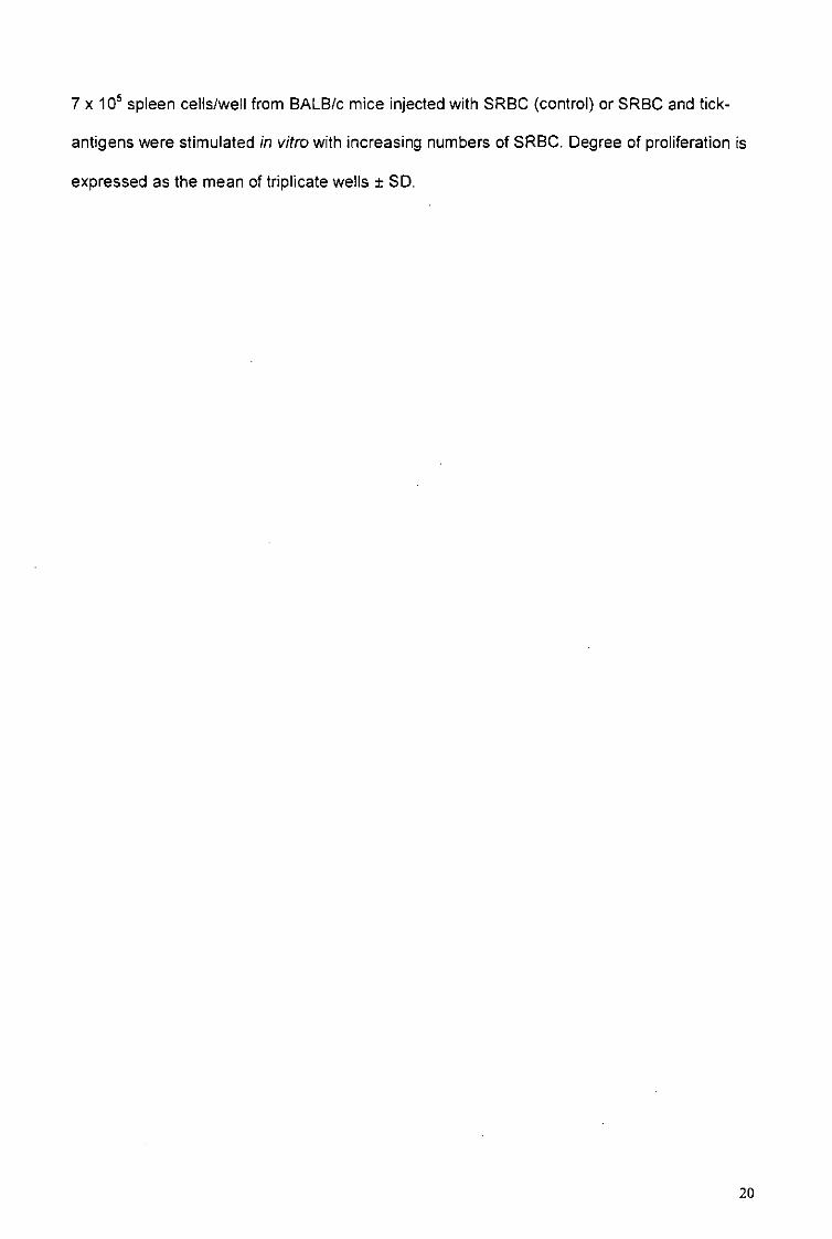
presence of tick saliva inhibited the SRBC hemolysis in a dose-dependent manner (fig. 1).
Anti-complement activity increased with increasing concentration of saliva. The optimum of
anti-complement activity was reached with saliva protein concentration of 30 ng/ml. This
inhibite about 70% of the SRBC lysis.
In vitro proliferation of tick-sensitized lymph node cells
To study the influence of tick saliva and SGE on the adaptive immune response, we
stimulated tick-specific lymph node cells in vitro with increasing doses of tick antigens. The
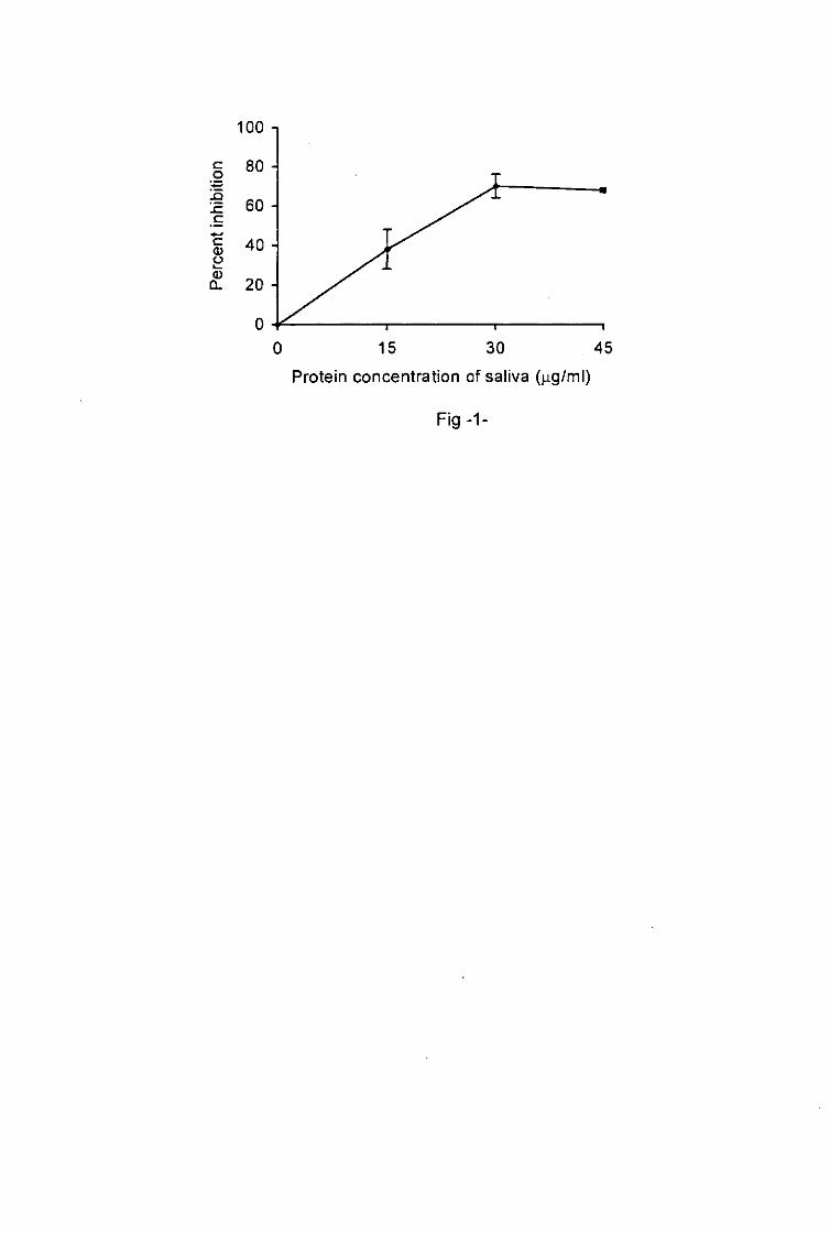
proliferation of primed lymph node cells was influenced by the concentration of tick saliva
and SGE applied in the cell cultures. This proliferation increased with the protein
concentrations of the saliva and SGE ranged from 1.09 to 2.18 ng/ml and from 1.56 to 12.5
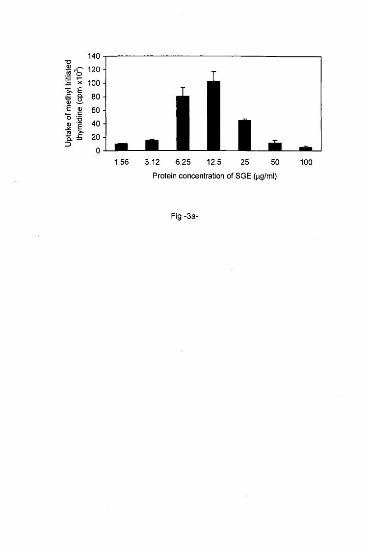
jig/ml respectively (Figs. 2a, 3). With higher protein concentrations of saliva and of SGE1
ranging from 2.18 to 17.5fig/ml and from 12.5 to 100 jig/ml respectively, the proliferation of
primed T cells decreased. Low doses of tick antigens were immunostimulative whereas
higher doses were immunosuppressive.
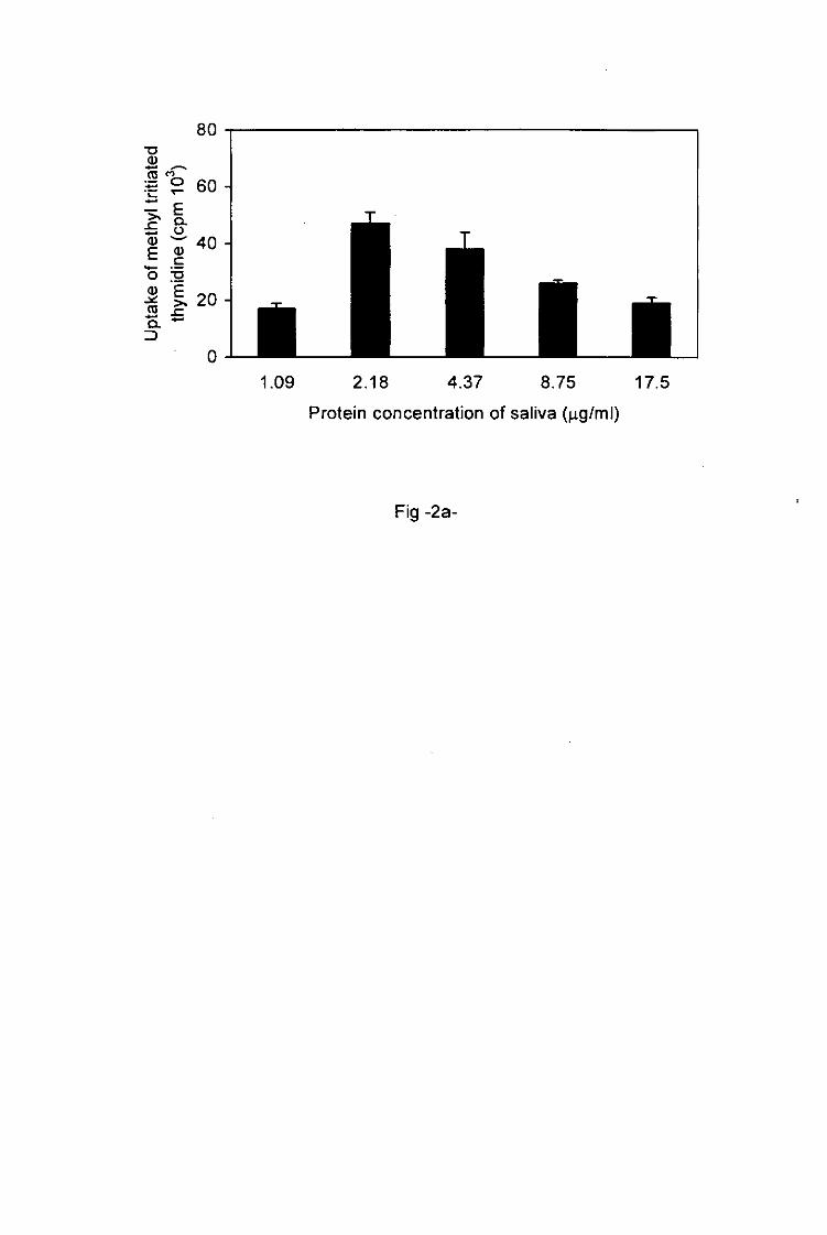
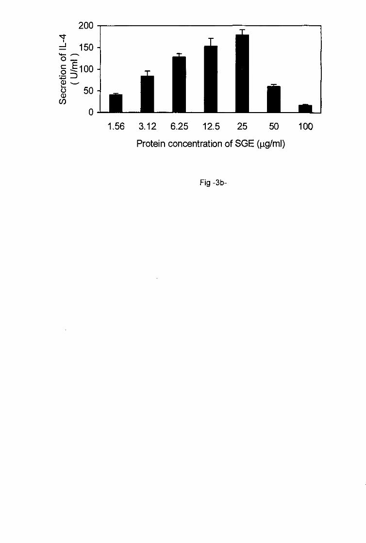
In vitro IL-4 production
IL-4 secretion also depended on the protein concentrations of the tick saliva as well as of the
SGE used to restimulate primed lymph node cells in vitro. Increasing protein concentrations
of saliva (1.09-4.37 jig/ml) and SGE (1.56-25 jig/ml) induced increased production of IL-4
which reached a maximum at a concentration of 4.37 ng/ml for saliva and 25 ^g/ml for SGE
9
(Figs. 2b, 3). Thereafter increasing protein concentrations of saliva (4.37-17.5 fig/ml) and of
SGE (25-100 jig/ml) triggered the decrease of IL-4 production by primed T cells.
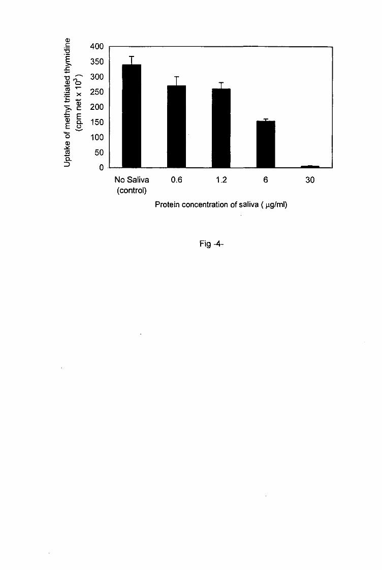
I. ricinus saliva inhibits splenic T cell proliferation to Con-A
To show the non-specific immunosuppressive activity of tick saliva, naive spleen cells were
preincubated in vitro for 2h with different protein concentrations of I. ricinus saliva ranging
between 0.6 and 30 jig/ml. Thereafter, they were stimulated in vitro with Con-A. A high
reduction (>90%) of cell proliferation in response to Con-A was observed with 30 t*g/ml of
saliva proteins (fig. 4). The inhibitory effect of tick saliva was dose-dependent. It diminished
with decreasing saliva concentrations but was still evident at a concentration of 0.6 ng/ml of
salivary proteins. Spleen cells untreated with saliva and stimulated with Con-A acted as
controls.
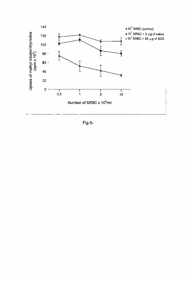
Systemic immunosuppressive effect of tick antigens
To investigate the systemic immunosuppressive activity of tick antigens we injected a mixture
of saliva or SGE and 107 SRBC intraperitoneal^. At 1 week post injection, mice elicited an
anti-SRBC response as shown by the proliferation of splenic T cells stimulated with SRBC in
vitro. Spleen cells from the control mouse, which was only injected with 107 SRBC, showed a
high proliferative response of T cells when restimulated in wfrawith 0.5 x 10s-10 x 10s
SRBC1 whereas splenic T cells from the mice injected with a mixture of saliva or SGE and
107 SRBC showed a reduction in proliferation when they were restimulated in vitro with
SRBC (fig. 5). The impairment in proliferation of splenic T cells after stimulation with SRBC
was more pronounced when mice were treated with SGE than with saliva.
10
Discussion
Ixodid ticks need several days to feed. During this time hosts acquire resistance or develop
tolerance to the ectoparasites. In contrast to resistant hosts, for example rabbits infested with
!.ricinus adults (Bowessidjaou et al. 1977), no difference was observed in the percentage of
attachment or the weight of engorged larvae or nymphs of I. ricinus during reinfestations of
BALB/c mice (Christe et al. 1998). In order to feed, as well as to transmit pathogens
successfully, ticks must maintain blood flow and control the immune response of their host.
They must develop countermeasures to antagonize the mechanisms of the innate as well as
the adaptive immune response. Some bioactive molecules confer anti-hemostatic, anti
inflammatory and immunosuppressive properties to tick saliva (Ribeiro et al. 1985). The
innate immunity constitutes the first line of defense against noxious or innocuous antigens. It
is followed by the specific immune response (Fearon and Locksley 1996). Activation of the
alternative pathway of complement is important in the innate defense and in the development
of some tissue inflammation. We showed that the saliva of I. ricinus inhibits the hemolysis of
SRBC by the human alternative pathway of complement. Accordingly, the saliva of Ixodes
dammini (= Ixodes scapularis) also has an anti-complement activity on human serum
(Ribeiro 1987). Impairment of the innate immune system, either by the inhibition of the
alternative pathway of complement or by downregulation of the activities of cellular
components such as NK cytotoxicity and macrophage killing of pathogens (Kopecky et al.
1999), would contribute to the reduction of skin inflammation and protection against ticks and
favour the transmission and propagation of tick-borne pathogens.
BALB/c mice infested with I. ricinus ticks develop a Th2 immune response characterized by
CD4+ T cells secreting IL-4, IL-5 and IL-10 (Ganapamo et al. 1995; 1996). SDS-PAGE
analysis showed some similarities in polypeptide profiles between tick saliva and SGE (Mejri
et al. 2001 ). Therefore, both of these were used to study the dose dependent influence of tick
antigens on the responsiveness of tick-sensitized lymph node T cells. The
immunosuppressive effects of saliva and SGE from I. ricinus were obtained with high
l l
concentrations of tick saliva or SGE. BALB/c mice tolerated frequent feedings by pathogens-
free larval or nymphal I. ricinus. The two instars seem to engorge more effectively on
repeatedly exposed mice (Christe et al. 1998). In nature, I. ricinus larvae and nymphs feed
abundantly on yellow-necked mice (Apodemus flavicollis) and black-striped mice (Apodemus
agrarius). These mice infect more ticks with B. burgdorferi than do other rodents (Matuschka
et al. 1991; 1992). Both species fully tolerate repeated experimental feedings (Dizij and
Kurtenbach. 1995). The susceptibility of either laboratory or wild mice to larval or nymphal I
ricinus feeding or to pathogens transmission may be due to the prolonged and massive
exposure to these ectoparasites which deposit a high amount of salivary secretions at the
site of their attachments. We suggest that the immunosuppressive activities overcome the
competitively immunostimulative activités in injected saliva resulting in the reduction of
primed lymph node T cells responsiveness. In vivo effector lymphocytes formed in the
draining lymph nodes of infested BALB/c mice circulate and infiltrate the skin at the site of
tick attachment. Numerous T cells have been observed in skin infested with nymphal tick.
CD4+T cells outnumbered CD8+T cells from a primary to a tertiary infestation (Mbow et
al. 1994b). These cells could be activated by locally salivary antigens to proliferate and
produce higher level of IL-4 than IFN-y. In situ hybridization of skin sections showed a
positive signal for IFN-y mRNA in some infiltrating mononuclear cells in the dermis near the
tick hypostome and chelecerae beside a few cells positive for IL-4 mRNA (Mbow et al.
1994c). The weak activity of locally primed Th2 cells could be due to the high amounts of
salivary secretion deposited at the site of attachment of nymphal ticks. This would be in
agreement with our finding showing a reduced activity of primed lymph node T cells
stimulated in vitro with higher protein concentrations of saliva or SGE.
To test whether tick saliva inhibits the development of an immune response non specifically,
we stimulated naive splenic T cells in vitro with Con-A. This T cell mitogen mimics the action
of antigens on primed T cells (Sharon 1983). We demonstrated that I. ricinus saliva inhibited
the proliferation of splenic naive T cells to Con-A in a dose-dependent manner. Accordingly L
12
scapularis saliva inhibited T cell proliferation to Con-A (Un'oste et al. 1994). SGE from
Dermacentor andersoni fed for 9 days also suppressed Con-A stimulated T cell proliferation
in vitro (Ramachandra and Wikel 1992). Few immunosuppressive molecules have been
described in the salivary glands of ixodid ticks. We previously showed that some
chromatographic fractions in the SGE of I. ricinus inhibit tick-sensitized lymph node T cells
proliferation (Mejri et al. 2001). Prostaglandin E2, detected in the saliva of I. ricinus females
(unpublished result), has a suppressive effect on Th1 cytokines elaboration (Betz and Fox
1991). A recombinant protein derived from a subtractive cDNA library of the salivary gland of
female I .ricinus was also found to modulate T lymphocyte and macrophage responsiveness
by inducing a Th2 response and by inhibiting the production of pro-inflammatory cytokines
(Leboulle et al. 2001 and unpublished results). A calreticulin protein secreted in Amblvomma
americanum saliva (Jaworski et al. 1995) as well as a protein of 36 kDa isolated from salivary
glands of D. andersoni ticks (Bergman et al. 2000) also have immunosuppressive properties.
The hypostome and the chelecerae of a nymphal tick penetrates deeply into the dermis
causing skin damage (Mbow et al. 1994a). Tick saliva carried with the blood stream could
stimulate leucocytes in the deep lymphoid organs such as the spleen. We therefore
examined the effect of tick saliva and of SGE on the priming of splenic T cells to SRBC. The
results showed that both of them have a systemic immunosuppressive effect on SRBC
sensitized splenic T cells for at least 1 week after intraperitoneal injection. A weak effect was
already evident after 4 days of culture (unpublished result). The immune response of guinea-
pigs infested with D. andersoni was also suppressed systemicalty (Wikel 1982). The
intraperitoneal injection of sand fly salivary glands lysate induced systemic
immunosuppression in C57BL/6 and BALB/c mice (Titus 1998). As with sand flies and ticks,
the salivary glands of other arthropods such as black flies (Cross et al. 1994a) and
mosquitoes (Cross et al. 1994b) contain immunomodulatory molecules. The
immunosuppressive effect of I. ricinus saliva probably influences the transmission of tick-
borne pathogens. It has been reported that the immunomodulator properties of vector saliva
13
may be required for the successful transmission and establishment of host infection (Titus
and Ribeiro 1990).
In conclusion, the impairment of the innate and acquired immune system allows BALB/c mice
to tolerate tick feeding and might facilitate the transmission of tick-borne pathogens. The
identification of immunomodulatory molecules in I, ricinus tick saliva and SGE requires
further investigations. This could be usefull in the conception of a vaccine against tick feeding
and pathogens transmission.
14
ACKNOWLEDGEMENTS
This work is part of the PhD thesis of Naceur Mejri and was supported by the Swiss National Science Foundation, grant number 31-56836. 99.
References
Bergman DK, Palmer MJ1 Caimano MJ1 Radolf JD Wikel SK (2000) Isolation and molecular
cloning of a secreted immunosuppressant protein from Dermacentor andersoni
salivary gland. J Parasitol 86: 516-525
Betz M1 Fox BS (1991) Prostaglandin E2 inhibits production of Th1 lymphokines but not of
Th2 lymphokines. J Immunol 146: 108 -113
Bowessidjaou J1 Brossard M, Aeschlimann A (1977) Effects and duration of resistance
acquired by rabbits on feeding and egg laying in Ixodes ricinus. L Experientia 33:
548-550
Christe M, Rutti B, Brossard M (1998) Susceptibility of BALB/c mice to nymphs and larvae of
Ixodes ricinus after modulation of IgE production with anti-interleukin-4 or anti-
interferon-y monoclonal antibodies. Parasitol Res 84: 388-393
Cross ML, Cupp EW, Enriquez FJ (1994a) Modulation of murine cellular immune responses
and cytokines by salivary gland extract of the black fly Simulium vittatum.
Ann Trop Med Parasitol 45: 119-124
Cross ML1 Cupp EW1 Enriquez FJ (1994b) Differential modulation of murine cellular immune
responses by salivary gland extract of Aedes aegypti. Am J Trop Med Hyg 51: 690-696.
Dizij A, Kurtenbach K (1995) Ciethrionomys gfareolus, but not Apodemus flavicoilis, acquires
resistance to Ixodes ricinus L., the main European vector of Borrelia burgdorferi. Parasite
Immunol 17:177-183
Fearon DT, Locksley RM (1996) The instructive role of innate immunity in the acquired
immune response. Science 272: 50-54
Ganapamo F, Rutti B, Brossard M (1995) In vitro production of interleukin-4 and interferon-y
by lymph node cells from BALB/c mice infested with nymphal Ixodes ricinus ticks.
Immunology 85:120-124
Ganapamo F1 Rutti B1 Brossard M (1996) Immunosuppression and cytokine production in
mice infested with Ixodes ricinus ticks: a possible role of laminin and interleukin-10
16
on the in vitro responsiveness of lymphocytes to mitogens. Immunology 87: 259-263
Graf J-F (1978) Copulation, nutrition et ponte chez Ixodes ricinus L. (Ixodoidea: Ixodidae).
Bull Soc Entomol Suisse 51: 89-97
Jaworski DC1 Simmen FA1 Lamoreaux W, Coons LB1 Müller MT, Needham GR (1995) A
secreted calreticulin protein in ixodid tick (Amblyomma americanum) saliva.
J Insect Physiol 41: 369-375
Kopecky J, Kuthejlova M, Pechova J (1999) Salivary gland extract from Ixodes ricinus ticks
inhibits production of interferon-gamma by the upregulation of interleukin-10.
Parasite Immunol 21:351-356
Leboulle G, Rochez C, Louahed J1 Rutti B1 Brossard M1 Bollen A1 Godfroid E
(2001) Isolation of Ixodes ricinus salivary gland mRNAs encoding factors induced during
the blood feeding process. Am J Trop Med Hyg (in press)
Matuschka FR1 Fischer P1 Musgrave K1 Richter D, Spielman A (1991) Hosts on which
nymphal Ixodes ricinus most abundantly feed. Am J Trop Med Hyg 44:100-107
Matuschka FR1 Fischer P1 Heiler M1 Richter D, Spielman A (1992) Capacity of European
animals as reservoir hosts for the Lyme disease spirochete. J Infect Dis 165:479-483
Mbow ML1 Christe M, Rutti B, Brossard M (1994a) Absence of acquired resistance to
nymphal Ixodes ricinus ticks in BALB/c mice developing cutaneous reactions. J Parasitol
80: 81-87
Mbow ML1 Rutti B, Brossard M (1994b) Infiltration of CD4+ CD8+ T cells, and expression of
ICAM-1, Ia antigens, IL-Ia and TNF-ct in the skin lesion of BALB/c mice undergoing
repeated infestations with nymphal Ixodes ricinus ticks. Immunology 82: 596-602.
Mbow ML, Rutti B1 Brossard M (1994c) IFN-y, IL-2, and IL-4 mRNA expression in the skin
and draining lymph nodes of BALB/c mice repeatedly infested with nymphal Ixodes ricinus
ticks. Cell Immunol 156: 254-261
Mejri N, Franscini N, Rutti B1 Brossard M (2001) Th2 polarization of immune response of
BALB/c mice to Ixodes, ricinus instars, importance of several antigens in activation of
specific Th2 subpopulations. Parasite Immunol 23: 61-69
17
Ramachandra RN1 Wikel SK (1992) Modulation of host immune responses by ticks (Acari:
Ixodidae): effect of salivary gland extracts on host macrophages and lymphocyte
cytokine production. J Med Entomol 29: 818-826
Ribeiro JM (1987) Ixodes dammini salivary Anti-complement activity. Exp Parasitol 64:
347-353
Ribeiro JM1 Makoul GT, Levine J, Robinson DR1 Spielman A (1985) Antihemostatic,
antiinflammatory, and immunosuppressive properties of the saliva of a tick,
Ixodes dammini. J Exp Med 161: 332-344
Richter D1 Spielman A, Matuschka FR (1998) Effect of prior exposure to noninfected ticks on
susceptibility of mice to Lyme disease spirochetes. Appi Environ Microbiol 64:4596-4599
Rutti B, Brossard M (1989) Repetitive detection by immunoblotting of a 25 kDa antigen in
Ixodes ricinus and a 20 kDA antigen in Rhipicephalus appendiculatus with sera of
pluriinfested mice and rabbits. Parasitol Res 75: 325-329
Sharon N (1983) Lectin receptors as lymphocyte surface markers. Adv Immunol 34: 213-298.
Titus RG (1998) Salivary gland lysate from the sand fly Lutzomyia longipalpis suppresses the
immune response of mice to sheep red blood cells in vivo and Concanavalin A in
vitro. Exp Parasitol 89: 133-136
Titus RG, Ribeiro JMC (1990) The role of vector saliva in transmission of arthropod-borne
disease. Parasitol Today 6: 157-160
Urioste S, Laurie R. Hall LR, Telford SR , Titus RG (1994) Saliva of the Lyme disease vector,
Ixodes dammini, blocks cell activation by a nonprostaglandin E2-dependent
mechanism. J Exp Med 180: 1077-1085
Wikel SK (1982) Influence of Dermacentor andersoni infestation on lymphocyte
responsiveness to mitogens. Ann Trop Med Parasit 76: 627-632
Wikel SK, Ramachandra RN, Bergman DK, Burkot TR1 Piesman J (1997) Infestation with
pathogen-free nymphs of the tick Ixodes scapularis induces host resistance to
transmission of Borrelia burgdorferi by ticks. Infect Immun 65: 335-338
I8
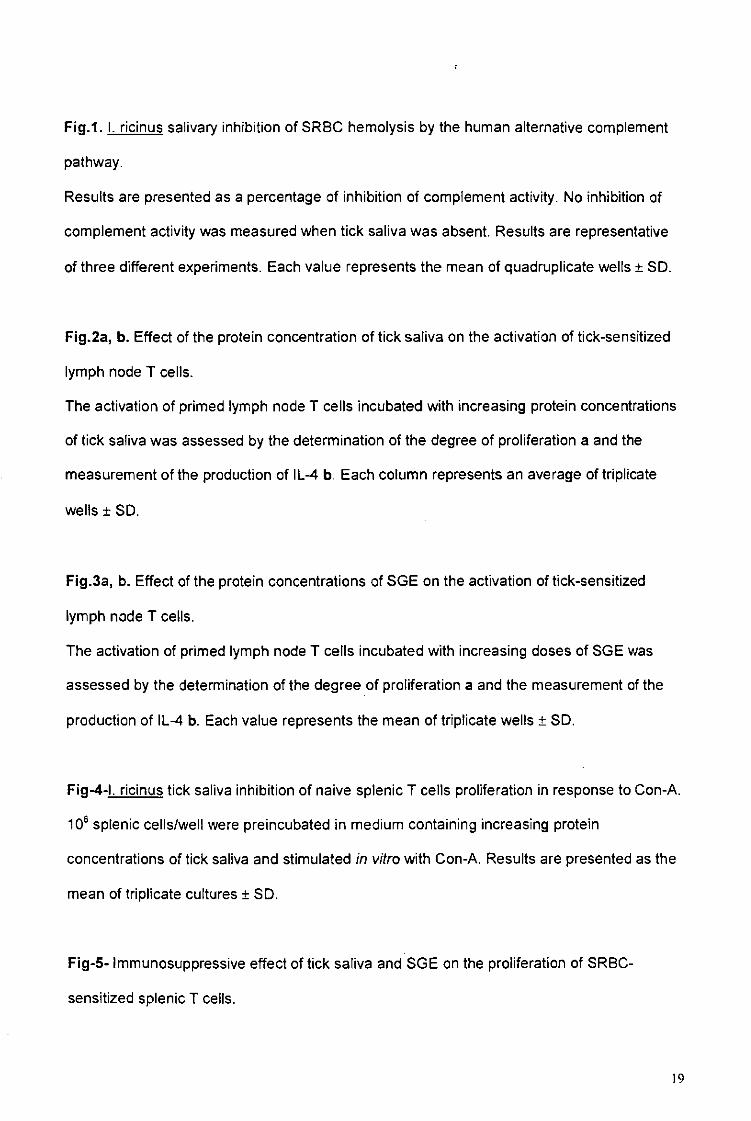
Fig.1. I. ricinus salivary inhibition of SRBC hemolysis by the human alternative complement
pathway.
Results are presented as a percentage of inhibition of complement activity. No inhibition of
complement activity was measured when tick saliva was absent. Results are representative
of three different experiments. Each value represents the mean of quadruplicate wells ± SD.
Fig.2a, b. Effect of the protein concentration of tick saliva on the activation of tick-sensitized
lymph node T cells.
The activation of primed lymph node T cells incubated with increasing protein concentrations
of tick saliva was assessed by the determination of the degree of proliferation a and the
measurement of the production of IL-4 b. Each column represents an average of triplicate
wells ± SD.
Fig.3a, b. Effect of the protein concentrations of SGE on the activation of tick-sensitized
lymph node T cells.
The activation of primed lymph node T cells incubated with increasing doses of SGE was
assessed by the determination of the degree of proliferation a and the measurement of the
production of IL-4 b. Each value represents the mean of triplicate wells ± SD.
Fig-4-l. ricinus tick saliva inhibition of naive splenic T cells proliferation in response to Con-A.
106 splenic cells/well were preincubated in medium containing increasing protein
concentrations of tick saliva and stimulated in vitro with Con-A. Results are presented as the
mean of triplicate cultures ± SD.
Fig-5- Immunosuppressive effect of tick saliva and SGE on the proliferation of SRBC-
sensitized splenic T ceils.
19
7 x 10⁵ spleen cells/well from BALB/c mice injected with SRBC (control) or SRBC and tick-
antigens were stimulated in vitro with increasing numbers of SRBC. Degree of proliferation is
expressed as the mean of triplicate wells ± SD.
20
100 n
Protein concentration of saliva (jig/ml)
Fig - 1 -
80
• i i . . 1.09 2.18 4.37 8.75 17.5
Protein concentration of saliva (^g/ml)
Fig -2a-
250
1.09 2.18 4.37 8.75 17.5
Protein concentration of saliva ( ng/ml )
Fig -2b-
1.56 3.12 6.25 12.5 25 50
Protein concentration of SGE (ng/ml)
100
Fig -3a-
200
d 150-1 O —
§-£l00H
ö 50 -\ co
1.56 3.12 6.25 12.5 25 50 100
Protein concentration of SGE (jig/ml)
Fig -3b-
No Saliva 0.6 1.2 6 30 (control)
Protein concentration of saliva ( ng/ml)
Fig-4-
a> e
"E
•a Q) —>
ro o
O)S E
TO "a.
140
120
100
80 -
60 -
40
20 •
0 - T
• 107 SRBC (control)
• 107SRBC + 5 ng of saliva * 107 SRBC+ 25 ^g of SGE
0.5 1 5
Number of SRBC x 105/ml
10
Fig-5-
Résultat n°2
Th2 polarization of the immune response of BALB/c mice to Ixodes ricinus instars, importance of several antigens in activation of specific
Th2 subpopulations. Parasite Immunol, 23:61-69
Parasite Immunology, 2001: 23: 61-69
Th2 polarization of the immune response of BALB/c mice to Ixodes
ricinus instars, importance of several antigens in activation of specific
Th2 subpopulations
NACEUR MEJRI. NICOLA FRANSCINI, BERNARD RUTTI & MICHEL BROSSARD
Institute of Zoology, Neuchâtel. Switzerland
SUMMARY INTRODUCTION
BALB/c mice were infested with Ixodes ricinus larvae, nymphs or adults. Expression of IL-4 and IFN-y mRNA in axillary and brachial draining lymph node cells were measured by competitive quantitative reverse transcrip-tion-polymerase chain reaction 9 days after the beginning of primary-infestation. IL-4 mRNA was always higher than that of IFN-y mRNA for all tick instars. Moreover, IL-4 mRNA expression progressively increased during nymphal primary-infestation with a high burst of expression 7 days after the beginning of infestation. No evolution of IFN-y mRNA expression was detected. Draining lymph node cells of infested BALB/c produced higher level of IL-4 than IFN-y following in vitro restimulation with adult tick saliva, salivary gland extract (SCE) or with five selected different chromatographic fractions of SGE. Anti-tick IgGi antibodies but no IgG2a were detected in BALB/c pluri-infested with I. ricinus nymphs, which confirmed the Th2 polarization of the immune response.
Keywords Ixodes ricinus, tick instars, BALB/c, antigens, saliva, cytokines. IL-4, IFN-y, Th2
Correspondence: Professor Michel Brossard, Institute of Zoology, Rue
Emile Argand 9. CH-2007 Neuchâtel. Switzerland
Received: 2 May 2000
Accepted far publication: 20 September 2000
In recent years, several studies have shown that helper T cell clones of CD4+ phenotype can be separated into two subsets designated ThI and Th2. These subsets can be distinguished on the basis of their pattern of cytokine secretion following stimulation with mitogens or antigens (1). ThI cells produce EL2, IFN-7 and lymphotoxin and promote cell mediated immune responses which are important for the destruction of intracellular pathogens, such as some bacteria or protozoa (2). Th2 cells secrete IL-4, IL-5, ILIO and IL-13. IL-4 and IL-13 are effective in providing help for the expression of IgE and IgGl in mice (3). Th2 cells are predominant in helminth infections or atopic diseases (4,5). It has also been reported that IL-4 and IFN-7 reciprocally regulate each other, with IFN-7 inhibiting Th2 lymphocytes, IL-4 and IL-IO downregulat-ing ThI cells (6). Priming of naive CD4+ T cells in presence of IL-4 causes the development of Th2 effector cells while IL-12 yields ThI effector cells (7,8). Other factors are implicated directly or indirectly in the orientation of the immune response such as the type of antigen presenting cells (APC) and the molecular environment of the immune induction site. In our model, it has been previously demonstrated that mice of different haplotypes develop Th2 immune responses after larval or nymphal Ixodes ricinus tick infestations (9). Pl uri-infested mice produced high levels of IgE and draining lymph node cells from these animals produced high level of IL-4 and low level of IFN--J» after being stimulated in vitro with ConA. Moreover, when ticks are infected with Borrelia burgdorferi, the antispirochete immune response is also biased toward Th2 (10).
To characterize the primary immune response of primary-infested mice with I. ricinus tick, we have measured IL-4 and IFN-7 mRNA in draining lymph node cells using a competitive quantitative reverse transcription-polymerase
© 2001 Blackwell Science Ltd 61
iV.Mcjri et al.
chain reaction (RT-PCR) (II). After secondary in vitro stimulation of T cells with tick antigens, enzyme-linked immunosorbent assay (ELISA) was used to measure IL-4 and IFN-7 proteins. Despite cytokines being consumed in the culture medium, this test is sensitive enough to detect the effects of tick saliva, salivary gland extract (SGE) and SGE chromatographic fractions on cytokine production. Furthermore, specific IgGl and IgG2a antitick antibodies, characteristic of Th2 or ThI responses, have been analysed in mice infested four times with nymphal ticks.
Briefly, the purpose of this work was to establish the polarization of the immune response after exposure of BALB/c mice to adult and immature tick I. ricinus instars, and to follow in vivo the cytokine profile of draining lymph node cells in primary-infestation and in vitro, after restimulaiion of draining lymph node cells with different tick antigens.
MATERIALS AND METHODS
Animals
Eight to I2-week-old BALB/c female mice and male rabbits (New Zealand), weighing an average of 3 kg, were purchased, respectively, from IFFA-CREDO (Arbresle, France) and from Elevage des Dombes (Romans, France). !. ricinus larval, nymphal and adult ticks were reared in our laboratory as previously described (12).
Infestations
Mice were infested with 40 I. ricinus larvae, 15 nymphs or one pair of adult ticks. They were placed in a small plastic capsule (15 mm in diameter) glued onto the shoulders of the mice with a mixture of one part beeswax and four parts colophonium, at the site drained by brachial and axillary lymph nodes (13). Each experiment was done on a group of four mice. To detect antitick antibodies, four successive nymphal infestations were interspaced by 14 days. Mice flanks were alternated during these repeated infestations. To prepare tick antigens, adults I. ricinus were applied and allowed to feed for 5 days on rabbit's ears. They were contained by a nylon bag. An Elizabethan collar prevented the host from grooming.
RNA extraction
Total RNA was extracted from 5 x 10⁵ axillary and brachial draining lymph node cells of mice infested with tick larvae, nymphs or adults using the tripure isolation kit (Boehringer Mannheim. Germany). The addition of chloroform to the solution before centrifugation allowed the
62
Parasite Immunology
formation of three phases. Total RNA in the upper phase was then precipitated with cold isopropanol (molecular grade). The pellet washed twice in 75% ethanol was then dissolved in 20 p.1 sterile distilled water (RNAase free). Two p.1 containing 01-0-5 u.g of total RNA were used as template for the reverse transcriptase reaction.
Competitive quantitative reverse transcription-polymerase chain reaction (CQ RT-PCR)
The first-strand cDNA synthesis kit (Boehringer Mannheim) was used. The semiquantitative competitive PCR was carried out using a competitor construct (pPQRS) containing sequences for multiple cytokines including E--4 and IFN-'Y and for hypoxanthine guanine phosphoribosyl transferase gene (HPRT) (14). Primers for IL-4 were: 5 ' - C A T C G G C A T T T T G A A C G A G G T C A - S ' (sense) and 5'-GCTACGGACCTAAGTAGCTATTC-3' (antisense), for IFN-y 5'-CATTGAAAGCCTAGAAAGTCTG-3' (sense) and 5'-CTCATGAATGCATCCTTTTTCG-3' (antisense) and for HPRT: 5'-GTTGGATACAGGCCAGACTTTGTTG-3' (sense) and 5'-GAGGGTAGGCCTATAGGCT-3' (anti-sense). Sense and antisense primers were chosen on different exons separated by large intronic sequences which enables unambiguous differentiation of cDNA from contaminating genomic DNA amplification products. cDNA synthesis using RNA extracted from draining lymph node cells from mice under different infestation conditions, were used as templates. The thermal cycling conditions were: 94°C for 40 s. 6O0C for 20 s, 72°C for 40 s, followed by a final incubation at 72°C for 10 min. The number of cycles varied between 33 and 36. The simultaneous amplification of the cytokine gene in the first strand cDNA reaction mixture and of an eight-fold serial dilution of competitor of known concentration allowed the determination of the level of HPRT, IL-4 or IFN-7 specific transcript. The point of equivalence in intensity between the competitor (upper band) and the cDNA (lower band) indicates the relative concentration of mRNA. The ratio of the relative concentration of the gene of interest (IL-4 or IFN-7) to the relative concentration of HPRT was then calculated. Results were expressed as the fold of increase in IL-4 or IFN-*y mRNA expression in mice infested with nymphal I. ricinus ticks compared to non-infested mice. The formula: IL-4 or IFN-7 ;,/HPRT /„: IL-4 or IFN-7 /c/HPRT f0l which was used to calculate the fold of increase in IL-4 or IFN-7 mRNA expression, emanated from the work of Reiner et at. ( 14) who calculated the HPRT mRNA concentration to control the varying efficiencies of the RT step among different experimental groups. The different concentrations of mRNA have been measured at /0 and /x
which represent different times points, respectively, 0 h in
© 2001 Blackwell Science Ltd. Parasite Immunology, 23. 61-69
Voliiiuf 2.'. \iiinber 2, February 2001
lymph node cells of non-infested mice or 12 h, I, 3, 5. and 7 days in lymph node cells of postnymphal infested mice.
Tick antigens
To collect saliva, adult I. ricinus ticks were allowed to feed for 5 days on rabbit's ears. Partially engorged female ticks were removed. To activate the salivation one drop of 5% (wt/vol) solution of pilocarpine in absolute methanol was applied to their dorsum previously scratched by abrasive paper. A finely drawn capillary tube was fitted over the mouthparts of each tick which was allowed to salivate for 10-30 min (15). The volume provided by each tick was 0-5 jil in average. Saliva from 100 to 200 partially fed ticks was pooled, sterilized through a 0-22-jjLm filter and stored at - 200C until use. I. ricinus females fed for 5 days on rabbit's ears were used to prepare SGE as previously described (16). The salivary glands were dissected and homogenized in ice cold extraction buffer consisting of 50 IHM phosphate-buffered saline (PBS) pH 7-4 supplemented with 1 HIM phenylmethylsulphonyl fluoride (PMSF) and 5 mM ethylene diaminetetraacetic acid (EDTA). The homogenate was centrifuged at 16 000^ for 30 min at 40C. The supernatant was dialysed in cellulose ester membrane tube with a molecular weight cut-off of 100 Da (Spectrum, Socochim, Switzerland) overnight in 10 mM (PBS) pH 7-4. Dialysate was sterilized through a 0-22-u.m filter and stored at - 20°C until use. Soluble proteins from SGE were fractionated by FPLC (Pharmacia, Switzerland). The extract was desalted on a Fast desalting Column HR 10/10 using 10 mM Tris-buffer. pH 7-5 and 50 mM NaCI as eluant. The peak containing proteins was then applied onto an anion exchange MonoQ HR 5/5 column. Bound proteins were eluted with a 50-600 mM NaCl linear gradient in 10 mM Tris-buffer pH 7-5. The main parameters were the gradient volume (20 ml), the salt concentration change/ml (25 mM/ml). The volume of each fraction was fixed at 0-5 ml. They were all dialysed against 10 mM PBS. pH 7-5 during 48 h at 4°C and stored at — 200C until use. Protein concentration of saliva, SGE or chromatographic fractions were determined using BCA Protein Assay Kit (Pierce, Socochim, Switzerland).
SDS-page
Sodium dodecy! sulphate Polyacrylamide gel electrophoresis (SDS-PAGE) was used with a 12% separation gel and a 6 ^ stacking gel (17). Chromatographic fractions. SLIIiva, and SGE from partially fed tick females were boiled lor 4 min in 5% SDS buffer before loading on the gel. The proteins separated on SDS-gel were electrophoretically
© 2(X)I Blackwell Science Lid, Parasite Immunol«^ 23. 61-69
Tick antigens in mice ThI polarization
transferred onto a nitrocellulose sheet. The strips of different samples were rinsed in PBS and incubated in PBS-Tween 20 (0-3%) at 370C for 30 min. Blots were then washed three times during 5 min with PBS-Tween 20 (0-3%). The strips were incubated in colloidal gold solution until optimal band visualization was obtained.
Western blot
Free binding sites on the strips were blocked by 5% (w/v) milk powder in PBS pH 7-5 during 1 h. They were then washed four times during 5 min with 1% milk powder in PBS. For the specific detection of both IgGl or IgG2a, the strips corresponding to SGE from partially fed females were incubated with diluted pooled sera (1:5) from mice infested four times overnight at room temperature. The membranes were washed four times in PBS and incubated for I h with diluted (1 : 1000) rat antimouse IgGl or IgG2a (Pharmingen, Germany) coupled to alkaline phosphatase. Negative controls were performed with non-infested mice sera (1 : 5). All strips were then treated with the Immun-Star chemiluminescent protein detection system (BioRad, Hercules, CA, USA) for few minutes before film exposure (BioMax, Kodak.New Haven, CT, USA).
Preparation, culture and quantification of proliferation of draining lymph node cells stimulated with tick antigens
Mice were killed 9 days after infestation and axillary and brachial lymph nodes were removed. 106 draining lymph node cells per well were cultivated in 200 JJ.1 total volume of complete culture medium containing RPMI-I640 (Gibco, Basel, Switzerland), supplemented with 10% fetal calf serum (v/v), 2 m.M L-glutamin. 1 mM sodium pyruvate, 1 mM nonessential aminoacids (Sigma, St Louis, MO, USA), 005 mM mercaptoethanol, 100 U/ml penicillin/ streptomycin (Gibco) and 25 fig/ml fungizone (Gibco). After 96 h incubation at 37°C in saturated atmosphere with 5% COJ, with or without 40 u.l/well of dialysed and filtrated chromatographic fractions of SGE. 1 u-Ci/well of methyl [3H] thymidine (specific activity 25 Ci/mmol) (Amersham. Bucks, UK) was added 18-24 h before harvesting the cells. Tritiated thymidine incorporation was determined by liquid scintillation counting.
Quantification of IL-4 and IFN-7 in supernatants of tick antigens-stimulated draining lymph node cells
For culture supernatant collection, 10h draining lymph node cells in 200 |xl total volume of complete culture medium were incubated with SGE (20 jig/ml), saliva (4 u.g/ml) or
63
N.Mejrì ci al.
dialysed and filtrated chromatographic tractions of SGE (40 jil/well) at 37°C in saturated atmosphere with 5% CO;. The volume applied for each fraction is kept constant at 40 (xl/well to respect the proportionality of antigens concentrations present in the SGE cocktail. Based on recent published data (!8). and preliminary assays showing that no cytokines were detected 24 or 48 h following incubation with tick antigens, supernatants of draining lymph node celts were removed after 96 h and stored at - 8O0C until used for IL-4 and IFN-7 determination. ELISA cytokine tests were performed as previously reported (19). Dilutions of rIL-4 (12-5-400 U/ml) or rIFN--y (4-125 U/ml) (Pharmingen, Germany) were used as positive test controls and for the construction of standard curves.
RESULTS
In vivo expression of IL-4 and IFN-7 mRNA during a primary infestation
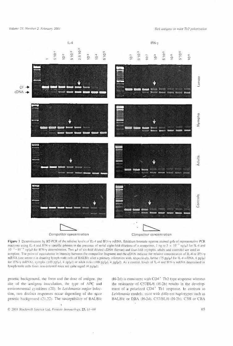
BALB/c were infested with 40 larvae, 15 nymphs or I pair of I. ricinus adults. A suspension of 5 x 10⁵ cells prepared from the brachial and axillary draining lymph nodes was used for RNA extraction. IL-4 and IFN-7 mRNA levels were determined by competitive quantitative RT-PCR nine days after infestation with larvae, nymphs or adult ticks (Figure 1). The IL-4 and IFN-7 mRNA concentrations were, respectively, IL-4 mRNA (75 pg/uJ) > IFN-7 mRNA (6 pg/|xl), IL-4 mRNA (100 pg/u.1) > IFN-7 mRNA (4 pg/ JJLI) and IL-4 mRNA (100 pg/u.1) > IFN-7 mRNA (4 pg/u,l). In all cases, we observed a higher dose of IL-4 mRNA expression compared to IFN-7 mRNA. The concentrations of IL-4 and IFN--y mRNA in the lymph node cells of non-infested mice were equally low.
Kinetic of IL-4 and IFN-7 mRNA expression
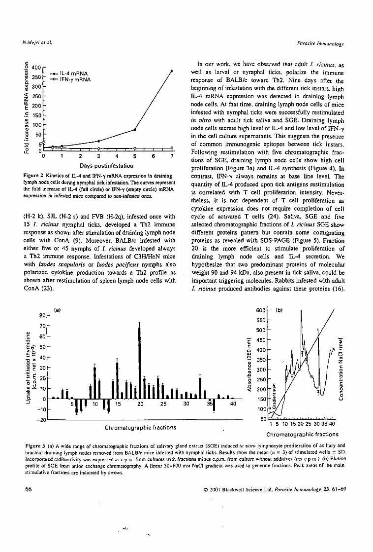
BALB/c were infested with 15 nymphs of /. ricinus. This was followed by the removal of draining lymph nodes after half a day or at 1, 3, 5 and 7 days post primo-infestation. A cell suspension was prepared and used for RNA extraction. The time course of IL-4 mRNA and IFN-7 expression in draining lymph node cells from infested mice is shown (Figure 2). There is a slight and regular increase of IL-4 mRNA expression during the first five days (2-5, 11-5 and 71-fold increase, respectively) followed by a high burst of expression at day 7 (> 375-fold). In contrast, IFN-7 mRNA always stay low during the corresponding days.
64
Parasite Immunology
Specific T cell proliferation with SGE chromatographically defined fractions
Nine days after being infested with 15 nymphal I. ricinus ticks, cells from axillary and brachial lymph nodes draining the site of nymphs fixation respond to a wide range of chromatographic fractions of SGE. Among them fractions 10, 15, 20, 24 and 33 display a higher effect on T cell proliferation (Figure 3a), each of them corresponding to peaks in the chromatogram (Figure 3b).
In vitro IL-4 and IFN-7 production
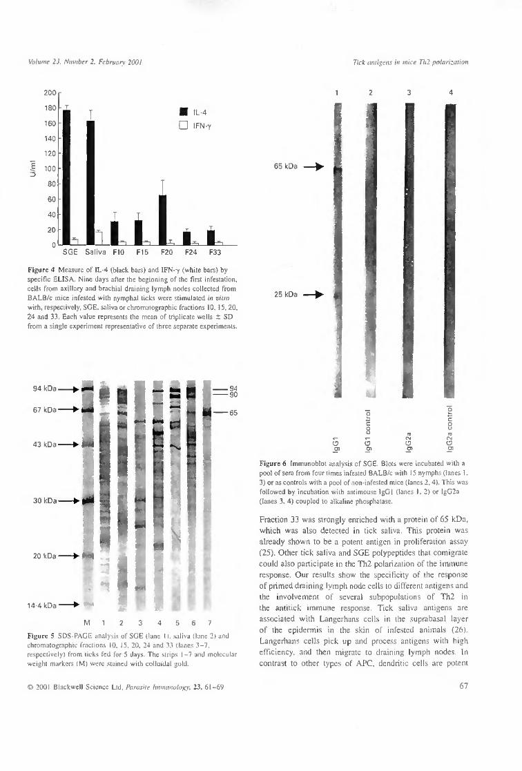
Primed T cells of axillary and brachial lymph nodes collected from BALB/c mice infested with nymphal ticks produced high level of IL-4 (> 150 U/ml) when stimulated with either SGE or saliva and > 20 U/ml when stimulated, respectively, with fractions 10, 15,20,24 and 33 (Figure 4). In contrast we detected a low dose of IFN-7 in the supernatant of the same cell culture. Cells incubated without any antigen did not produce IL-4 or IFN-7.
SDS-PAGE analysis of SGE, saliva and chromatographic fractions
SGE, saliva or the five chromatographic fractions used in antigen-specific T cell proliferation and cytokines detection show different protein patterns (Figure 5). The pattern of SGE (lane 1) is more complex than that of saliva (lane 2) and of each chromatographic fraction (ianes 3-7). Several proteins of tick saliva, SGE and chromatographic fractions comigrate, as for example proteins of 90 and 94 kDa in F20 and F24 (lanes 5 and 6) or of 65 kDa which is enriched in F33 (lane 7). FlO and FI5 (lanes 3 and 4) show also some comigrating polypeptides with saliva and SGE among a relative complex pattern of proteins.
Specific IgGl and IgG2a antibodies produced by pluri-infested BALB/c
The immunoblot analysis (Figure 6) shows two bands representing the reactivity of two SGE proteins (25 and 65 kDa) with BALB/c mice IgGl (lane 1) infested four times with 15 nymphal I. ricinus (see arrows). No specific IgG2a antibodies were detected (lane 3). Controls were performed with non-infested mice sera (lanes 2 and 4) which did not show any reactivity with tick antigens.
DISCUSSION
It has been reported that many factors are implicated in the regulation of ThI and Th2 subsets in mice such as the
© 2001 Blackwell Science Lid. Parasite Immunology. 23, 61-69
Volume 23. Number 2. February 2001 Tick antigens in mue Tli2 polarization
IL-4 IFIM-Y
O T™ O
T-
O
"~ i n
T-
<N O
«-O
«-
CF cDNA
a E > z
c
c o
U
Competitor concentration Competitor concentration
Figuri- 1 Determination by RT-PCR of the relative levels of IL-4 and IFN--y mRNA. Eihidium bromide agarose stained gels of representative PCR reactions using IL-4 and IFN-v specific primers in the presence of serial eight-fold dilutions of a competitor. I ng to 5 x 10~5 ng/u.1 for IL-4 and 10"*—10"° ng/u.1 for IFN-Y determination. Two u.1 of six-fold diluted cDNA (larvae) and four-fold (nymphs, adults and controls) are used as template. The point of equivalence in intensity between the competitor fragment and the cDNA indicate the relative concentration of IL-4 or IFN-Y mRNA (see arrows) m draining lymph node cells of BALB/c after a primary infestation with, respectively, larvae (75 pg/u.1 for IL-4 mRNA. 6 pg/u.1 for IFN-Y mRNA). nymphs (100 pg/u.1. 4 pg/u.1) or adult ticks ( KK) pg/u.1. 4 pg/u.1) As a control, levels of IL-4 and IFN-Y mRNA determined in lymph node cells from non-infested mice are quite equal (4 pg/u.1).
genetic background, the form and the dose of antigen, the
site of the antigens inoculation, the type of APC and
environmental cytokines (20). In Leishmania major infec
tion, two distinct responses occur depending of the mice
genetic background (21.22). The susceptibility of BALB/c
(H-2d) is consistent with CD4+ Th2 type response whereas
the resistance of-C57BL/6 (H-2b) results in the develop
ment of a polarized CD4+ ThI response. In contrast to
Leishmania models, mice with different haplotypes such as
BALB/c or DBA (H-2d). C57/BL/6 (H-2b). C3H or CBA
C 2001 Blackwell Science Ltd. Parasite Immunology, 23, dl-o9 65
N.Mejri et al. Parasite immunology
.9 400 W
S 350 x 300 < 250
| 200
.£ 150
£ 100 (U
o 50
IL-4 mRNA IFN-Y mRNA
Days postinfestation
Figure 2 Kinetics of IL-4 and IFN-7 mRNA expression in draining lymph node cells during nymphal tick infestation. The curves represent the fold increase of 0--4 (full circle) or IFN-7 (empty circle) mRNA expression in infested mice compared to non-infested ones.
(H-2 k), SJL (H-2 s) and FVB (H-2q), infested once with 15 I. ricinus nymphal ticks, developed a Th2 immune response as shown after stimulation of draining lymph node cells with ConA (9). Moreover, BALB/c infested with either five or 45 nymphs of I. ricinus developed always a Th2 immune response. Infestations of C3H/HeN mice with Ixodes scapularis or Ixodes pacißcus nymphs also polarized cytokine production towards a Th2 profile as shown after restimulation of spleen lymph node cells with ConA (23).
In our work, we have observed that adult I. ricinus, as well as larval or nymphal ticks, polarize the immune response of BALB/c toward Th2. Nine days after the beginning of infestation with the different tick instars, high IL-4 mRNA expression was detected in draining lymph node cells. At that time, draining lymph node cells of mice infested with nymphal ticks were successfully restimulated in vitro with adult tick saliva and SGE. Draining lymph node cells secrete high level of IL-4 and low level of IFN-7 in the cell culture supernatants. This suggests the presence of common immunogenic epitopes between tick instars. Following restimulation with five chromatographic fractions of SGE, draining lymph node cells show high cell proliferation (Figure 3a) and IL-4 synthesis (Figure 4). In contrast, IFN--, always remains at base line level. The quantity of IL-4 produced upon tick antigens restimulation is correlated with T cell proliferation intensity. Nevertheless, it is not dependent of T cell proliferation as cytokine expression does not require completion of cell cycle of activated T cells (24). Saliva, SGE and five selected chromatographic fractions of I. ricinus SGE show different proteins pattern but contain some comigrating proteins as revealed with SDS-PAGE (Figure 5). Fraction 20 is the more efficient to stimulate proliferation of draining lymph node cells and IL-4 secretion. We hypothesize that two predominant proteins of molecular weight 90 and 94 kDa, also present in tick saliva, could be important triggering molecules. Rabbits infested with adult 1. ricinus produced antibodies against these proteins (16).
TJ
"D X U *-
% E
Q .
80
70
60
•50
40
30
20
10
0
-10
-20
(a)
IL IT 10 ÏÏ
Il llili li.iiill 15 20 25 30 35I
j . i . 40
Chromatographic fractions 1 5 10 15 20 25 30 35 40
Chromatographic fractions
Figure 3 (a) A wide range of chromatographic fractions of salivary gland extract (SGE) induced //1 vil m lymphocyte proliferation of axillary and brachial draining lymph nodes removed from BALB/c mice infested with nymphal ticks. Results show the mean (n = 3) of stimulated wells ± SD. incorporated radioactivity was expressed as c.p.m. from cultures with fractions minus c.p.m. from culture without additives (net c.p.m.). (b) Elution pronte of SCE from anion exchange chromatography. A linear 50-600 IHM NaCl gradient was used to generate fractions. Peak areas of the main stimulative fractions are indicated by arrows.
66 © 2001 Blackwell Science Ltd. Parasite Immunology. 23. 61-69
Volume 23. Number 2. February 2001 Tick antigens in mice 77(2 polarization
200
180
160
140
120
100
80
60
40
20
0
• IL-4
D IFN-Y
SGE Saliva F10
Figure 4 Measure of IL-4 (black bars) and IFN-y (white bars) by specific ELISA. Nine days after the beginning of the first infestation, cells from axillary and brachial draining lymph nodes collected from BALB/c mice infested with nymphal ticks were stimulated in vitro with, respectively, SGE, saliva or chromatographic fractions 10, 15, 20. 24 and 33. Each value represents the mean of triplicate wells ± SD from a single experiment representative of three separate experiments.
«I -M 43kDa
3OkDa
2OkDa
14-4 kDa
.94 •90
•65
«h»
M 1 2 3 4 5 6 7
Figure 5 SDS-PAGE analysis of SGE (lane I). saliva (lane 2) and chromatographic frictions K). 15. 20, 24 and 33 (lanes 3-7. respectively) from ticks fed for 5 days. The strips 1-7 and molecular weight markers (M) were Stained with colloidal gold.
65 kDa
25 kDa
,— (D O)
<— (D en
CM
(D O)
CSI
(D 01
Figure 6 Immunoblot analysis of SGE. Blots were incubated with a pool of sera from four times infested BALB/c with 15 nymphs (lanes 1. 3) or as controls with a pool of non-infested mice (lanes 2,4). This was followed by incubation with antimouse IgGl (lanes I. 2) or IgG2a (lanes 3. 4) coupled to alkaline phosphatase.
Fraction 33 was strongly enriched with a protein of 65 kDa,
which was also detected in tick saliva. This protein was already shown to be a potent antigen in proliferation assay (25). Other tick saliva and SGE polypeptides that comigrate could also participate in the Th2 polarization of the immune response. Our results show the specificity of the response of primed draining lymph node cells to different antigens and the involvement of several subpopulations of Th2 in the antitick immune response. Tick saliva antigens are associated with Langerhans cells in the suprabasal layer of the epidermis in the skin of infested animals (26). Langerhans cells pick up and process antigens with high efficiency, and then migrate to draining lymph nodes. In contrast to other types of APC. dendritic cells are potent
© 2001 Blackwell Science Ltd. Parasite Immunology. 23. 61-69 67
N.Mcjri et al. Parasite fmmuno!og\
activators of naive Th cells (27). The differential development of naive Th into functionally distinct effector Th cells depends upon microenvironmental factors. Only dendritic cells which have been activated by exogenous IL-12 inducing factors such as bacteria or their constituents can direct ThI development through the release of IL-12. Increasing IFN-7 production by activated naive T cells directs their development toward the ThI phenotype (27). PGE2 is also an important molecule in the regulation of the immune response (28). With a colourimetric assay, PGE2
has been detected in the saliva of 5 days engorged I. ricinus female ticks (05 p.g of PGE2AnI; unpublished results^ It is also present in the saliva of other tick species such as /. scapularis (15). In the skin, this molecule may down regulate the expression of IL-12 by dendritic cells (28). This effect which is stable in vitro for at least 48 h could contribute to the development of Th2 responses in draining lymph nodes.
The I. ricinus rostrum penetrates deeply into the dermis (13) so that saliva molecules could be drained directly into lymph nodes. High local PGE2 concentration should act synergistically with IL-4 on uncommitted B cells to direct isotype switching to IgE and IgGl (2930). The antibody isotypes produced in BALB/c pluri-infested with I. ricinus ticks are IgGl (Figure 6) and IgE (31). The use of IL-4 deficient mice or the treatment of mice with anti-IL-4 monoclonal antibodies inhibited the production of IgE during successive infestations (31). IL-4 is an indispensable factor for the differential development of naive T cells into Th2 (8). A burst of IL-4 mRNA has been observed in the popliteal draining lymph node cells of BALB/c 16 h after subcutaneous injection of L major promastigotes into the hind footpads (32). In this model IL-4 mRNA returned to baseline level by 48 h. For these authors, the early burst of IL-4 mRNA should play an essential role in the development of the second wave of IL-4 mRNA expressed from day 5 onwards. This new wave reflects the differentiation of parasite-specific CD4* T cells toward the Th2 functional phenotype. In tick infested BALB/c the IL-4 mRNA expression did not show an early peak but a progressive increase of IL-4 mRNA from 12 h onwards with a drastic increase at day 7 post infestation. This observation suggests that an early burst of IL-4 secretion in draining lymph node cells is not imperious but that sufficient quantities of IL-4 are required to the development of mature Th2 cells.
In conclusion, tick saliva molecules could have different functions in relation with the Th2 polarization of the immune response. Some of them could act already at the site of tick attachment. Dendritic cells migrating from the skin to draining lymph nodes mature, present some specific antigens to naive T cells and prime them to generate effector Th2 cells.
68
ACKNOWLEDGEMENTS
This work is part of the PhD thesis of Naceur Mejri and was supported by the Swiss National Science Foundation, grant number 31-46862.96 and 31-56836.99.
REFERENCES
! Mosmann TR. Cherwinski H. Bond MW, Giedlin MA, Coffman RL. Two types of murine helper T cell clone. I. Definition according to profiles of Iymphokines activities and secreted proteins. J Immunol 1986: 136: 2348-2357.
2 Lahesmaa R. Yssel H. Batsford S et al. Yersinia enterocolitica activates a T helper type 1-like T cell subset in reactive arthritis. J Immunol 1992: 148: 3079-3085.
3 Snapper CM. Finkelman FD, Paul WE. Differential regulation of [gGI and IgE synthesis by interleukin 4. J Exp Med 1988; 167: 183-196.
4 Del Prete CF, De Carli M, D'Elios MM et al. Allergen exposure induces the activation of allergen-specific Th2 cells in the airway mucosa of patients with allergic respiratory disorders. Eur J Immunol 1993: 23: 1445-1449.
5 Mahanty S, Abrams JS, King CL. Limaye AP, Nutman TB. Parallel regulation of IL-4 and IL-5 in human helminth infections. J Immunol 1992: 148: 3567-3571.
6 Seder RA, Paul WE. Acquisition of lymphokine-producing phenotype by CD4* T cells. Annu Rev Immunol 1994; 12: 635-673.
7 Hsieh CS, Macatonia SE. Tripp CS, Wolf SF, O'Garra A, Murphy KM. Development of THl,CD4+ Tcells through IL-12 produced by Listeria-induced macrophages. Science 1993; 260: 547-549.
8 Swain SL, Weinberg AD, English M, Huston G. IL-4 directs the development of Th2-like helper effectors. J Immunol 1990; 145: 3796-3806.
9 Christe M, Rutti B, Brassard M. Influence of genetic background and parasite load of mice on immune response developed against nymphs of Ixodes ricinus. Parasitai Res 1999: 85: 557-561,
10 Christe M, Rutti B. Brassard M. Cytokines (D_-4 and IFN--y) and antibodies (IgE and IgG2a) produced in mice infected with Borrelia burgdorferi sensu stricto via nymphs of Ixodes ricinus ticks or syringe inoculations. Parasitol Res 2000: 86: 491-496.
11 Mbow ML. Bleyenberg JA, Hall LR. Titus RG. Phlebotomus papatasi sand fly salivary gland lysate down-regulates a ThI. but up-regulates a Th2, response in mice infected with Leishmania major. J Immunol 1998; 161: 5571-5577.
12 Graf J-F. Copulation, nutrition et ponte chez Ixodes ricinus L. (Ixodoidea: Ixodidae) - le panie. Bull Soc E/uomol Suisse 1978: Sl: 89-97.
13 Mbow ML. Christe M, Rutti B. Brassard M. Absence of acquired resistance to nymphal Ixodes ricinus ticks in BALB/c mice developing cutaneous reactions. J Parasitol 1994: 80: 81-87.
14 Reiner SL. Zheng S. Corry DB. Locksley RM. Constructing polycompetitor cDNAs for quantitative PCR. J Immunol Methods 1993: 165: 37-46.
!5 Ribeiro JM. Makoul GT. Levine J. Robinson DR. Spielman A. Antihemosiatic. antiinflammatory, and immunosuppressive properties of the saliva of a tick, Ixodes dammini. J Exp Med 19S5; 161: 332-344.
16 Rutti B, Brossiird M. Repetitive detection by immunoblotting of a 25 kDa antigen in Ixodes ricinus and a 20 kDa antigen in
© 2001 Blackwell Science Ltd, Parasite Immunology. 23, 61-69
Volume 23. Number 2. February 2001 Tick antigens in mice Th2 polarization
Rhipicephalus oppendiculatus with sera of pi uri infested mice and rabbits. Parasitai Res 19S9: 75: 325-329.
17 Laemmti UK. Cleavage of structural proteins during the assembly of the head of bacteriophage T4. Nature 1970; 227: 680-685.
18 Chen YL. Simons FER. Peng ZK. A mouse model of mosquito allergy for study of antigen-specific IgE and IgG subclass responses, lymphocyte proliferation, and IL-4 and IFN-f production. Im Arch Allergy Immunol 1998; U6: 269-277.
19 Oanapamo F. Rutti B. Brossard M. In vitro production of interleukin-4 and interferon-gamma by lymph node cells from BALB/c mice infested with nymphal Ixodes ricinus ticks. Immunology 1995; 85: 120-124.
20 Sedltk C, Deriaud E. Ledere C. Lack of ThI or Th2 polarization of CD4"*" T cell response induced by particulate antigen targeted to phagocytic cells. Int Immunol 1997; 9: 91-103.
21 Heinzel FP. Sadick MD, Mutha SS. Locksley RM. Production of interferon gamma, interleukin 2. interleukin 4, and interleukin 10 by CD4* lymphocytes in vivo during healing and progressive murine leishmaniasis. Proc Natl Acad Sci USA 1991; 88: 7011-7015.
22 Holaday BJ, Sadick MD. Wang Z-E et al. Reconstitution of Leishmania immunity in severe combined immunodeficient mice using ThI- and Th2-like cell lines. J Immunol 1991; 147: 1653-1658.
23 Schoeler GB. Manweiler SA, Wikel SK. Cytokine responses of C3H/HeN mice infested with Ixodes scapularis or Ixodespaeißcus nymphs. Parasite Immunol 2000; 22: 31-40.
24 Richter A. Lohning M. Radbruch A. Instruction for cytokine expression in T helper lymphocytes in relation to proliferation and cell cycle progression. J Exp Med 1999; 190: 1439-1450.
25 Ganapamo F. Rutti B. Brossard M. Identification of an Ixodes ricinus salivary gland fraction through its ability to stimulate CD4 T cells present in BALB/c mice lymph nodes draining the tick fixation site. Parasitology 1997; IIS: 91-96.
26 Allen JR. Khali! HM. Wikel SK. Langerhans cells trap tick salivary gland antigens in tick-resistant guinea pigs. J Immunol 1979; 122: 563-565.
27 Hilkens CMU. Kalinski P. De Boer M. Kapsenberg ML. Human dendritic cells require exogenous interleukin-12-inducing factors to direct the development of naive T-helper cells toward the ThI phenotype. Blood 1997; 90: 1920-1926.
28 Kalinski P. Hilkens CMU, Snijders A, Snijdewint FGM. Kapsenberg ML. IL-12-deficient dendritic cells, generated in the presence of prostaglandin E2. promote type 2 cytokine production in maturing human naive T helper cells. J Immunol 1997; 159: 2 8 -35.
29 Roper RL. Brown DM, Phipps RP. Prostaglandin E2 promotes B lymphocyte Ig isotype switching to IgE. J Immunol 1995; 154: 162-170.
30 Kalinski P, Hilkens CMU. Wierenga EA. Kapsenberg ML. T-cell priming by type-1 and type-2 polarized dendritic cells: the concept of a third signal. Immunol Today 1999; 20: 561-567.
31 Christe M. Rutti B. Brossard M. Susceptibility of BALB/c mice to nymphs and larvae of Ixodes ricinus after modulation of IgE production with anti-interleukin-4 or anti-interferon-gamma monoclonal antibodies. Parasiiol Res 1998; 84: 388-393.
32 Launois P. Ohteki T, Swihart K, MacDonald HR. Louis JA. In susceptible mice, Leishmania major induce very rapid interleukin-4 production by CD4+ T cells which are NKl.1". Eur J Immunol 1995: 25: 3298-3307.
© 2001 Blackwell Science Ltd. Parasite Immunology. 23. 61-69 69
Résultat n°3
Induction of Th2 cell differentiation in primary immune response in vitro and in vivo: splenic dendritic cells incubated with Ixodes ricinus
tick saliva prime naive CD4+ T cells to secrete IL-4. (Submitted in Immunology)
Induction of Th2 cell differentiation in primary immune response in vitro and in vivo: splenic dendritic cells incubated with Ixodes ricinus tick saliva prime naive CD4+ T cells to secrete IL-4
Naceur Mejri, Bernard Rutti, Michel Brossard Institute of Zoology, Department of Immunology, University of Neuchâtel, Switzerland
Running title : CD4+ T cells primed into Th2 in vitro or in vivo.
Correspondence : Prof. Michel Brossard, Institute of Zoology, Rue Emile Argand 9, CH-2007 Neuchâtel, Switzerland Tel:+41-32-718 30 15 Fax:+41-32-718 30 11 E-mail address: [email protected]
l
Summary
Dendritic cells (DCs) are the most potent antigen presenting cells (APC) in priming naive T cells.
In this report we showed that DCs incubated overnight with Ixodes ricinus tick saliva prime CD4+
T cells to secrete IL-4 and IL-10 in vitro. Tick saliva pulsed DCs injected subcutaneously into
BALB/c mice elicit also a Th2 immune response. This finding brings out the importance of the
molecular nature of tick saliva for the induction of the DCs type that generate either in vitro or in
vivo a Th2 immune response. Early during the interaction of tick saliva pulsed DCs with bulk
CD4+ T cells, the presence of IL-4 in the cell culture environment is of great importance to
initiate the Th2 differentiation of CD4+ T cells. This is monitored indirectly by neutralizing IL-4
with specific monoclonal antibodies which blocked the differentiation of CD4+ T cells into Th2.
This finding suggests that among bulk CD4+ T cells used in the culture certain populations such
as memory Th2 cells, NK 1.17 NK 1.1' CD4+ T cells, conventional CD4+otß T cells themselves
are a potential source of IL-4. CD4+ T cells, differentiated into Th2 cells following stimulation in
vitro with tick saliva pulsed DCs, proliferate only when IL-1ß was added early in the culture. The
early presence of IL-1 ß is required for the proliferation but not for the differentiation. Our priming
in vitro system was restricted to major factors implicated in the anti-tick immune response such
as naive CD4+ T cells, APCs and tick saliva. It provides the possibility to delimit further the
antigenic molecule(s) and type of CD4+ T cells involved in the Th2 polarization of the immune
response and to manipulate this response.
Key words: Ixodes ricinus saliva, cellular activation, priming in vitro, priming in vivo, dendritic
cells, Thin~h2, IL-1ß, IL-4, IL-10.
2
Introduction
Cytokines produced by CD4+ T cells polarize the immune response in vivo or in vitro in
response to antigen challenge. TM cells are characterized by the predominance of IFN-y
production while Th2 cells produce predominantly IL-4 (1). This dichotomy in T helper activity is
reported to be associated to susceptibility or resistance to pathogens. A well studied example is
experimental murine cutaneous leishmaniasis in which selective activation of Th1 cells is
associated with resolution of the disease whereas selective activation of Th2 cells is associated
with disease progression (2). Ectoparasites such as tick or sand fly inject pathogens into skin
along with saliva. The old world sand fly Phlebotomus papatasi salivary gland lysate
exacerbated lesion size and enhanced Leishmania major burden in disease-resistant CBA mice.
This observation was correlated with inhibition of the production of Th1 cytokines and
enhancement of Th2 ones (3). Likewise BALB/c mice infested by Borrelia burgdorferi infected
nymphal /. ricinus ticks enhance Th2 immune response by reducing anti-borrelial lgG2a antibody
production (4). These examples revealed the promoting effect of ectoparasite saliva toward a
Th2 immune response. Pathogen free tick instars salivary secretions elicit a Th2 immune
response in infested BALB/c mice (5). Many factors had been reported to be implicated directly
or indirectly and may act independently or together to polarize the immune response in vivo (6).
In our model of BALB/c mice infested with nymphal ticks, neither the genetic background nor the
antigen doses are implicated in the orientation of the immune response (7). Other factors such
as the molecular nature of tick saliva and the environment at the immune induction site may
selectively differentiate naive CD4+ T cells into Th1 or Th2 cells. The activation of CD4+ T cells
occurs often in secondary lymphoid organs and requires the involvement of antigen presenting
cells. Regarding their wide distribution in the body of the host and their efficiency as antigen
presenting cells (APCs) dendritic cells (DCs) are the most important cells implicated in the
activation of CD4+ T cells (8). In peripheral tissues such as skin DCs trap antigens. They
migrate to draining lymph nodes where they present antigen to T cells (9). The original
3
microenvironment of DCs including cytokines and antigens may influence their antigen
presentation properties and the induction of naive CD4+ T cells to differentiate along either a
TM orTh2 pathway.
An in vitro priming system has been set up to reproduce the leukocyte reaction in vivo and bring
out the importance of the molecular nature of tick saliva for the polarization of the immune
response. DCs pulsed in vitro with tick saliva induced the Th2 differentiation of naive CD4+ T
cells. We also showed the ability of DCs pulsed in vitro with tick saliva and injected
subcutaneously to BALB/c to generate a Th2 immune response in vivo. To investigate whether
endogenous IL-4 is present early in the environment of the culture and if its presence is
indispensable to direct the immune response toward Th2, we added neutralizing anti-IL-4
monoclonal antibodies to the primary in vitro (PIV) culture. Results showed that this
neutralization inhibits the differentiation of CD4+T cells into Th2. Moreover we demonstrated that
the early presence of IL-Ip is indispensable as costimulator for the proliferation but not for the
differentiation of Th2 cells. In this study we showed that the nature of tick saliva influenced DCs
to promote Th2 immune response in vivo. Pulsed DCs synergize in vitro with induced
endogenous IL-4 to prime naive CD4+ T cells to secrete IL-4 and IL-10.
4
MATERIALS AND METHODS
Animals
Male rabbits (New Zealand) weighing an average 3 kg and BALB/c female mice 8 to 12 weeks
old were purchased from Elevage des Dombes (Romans, France) and IFFA-CREDO (Arbresle,
France) respectively. I. ricinus adult female ticks were reared in our laboratory as previously
described (10).
Tick saliva preparation
To collect saliva, adult /. ricinus ticks were allowed to feed for 5 days and 12 hours on rabbit's
ears. Partially engorged female ticks were removed and fixed by their dorsum on a sticky paper.
To enhance the salivation, 5 ^l of dopamine solution 0.2% (w/v) (Fluka, Buchs, Switzerland) in
50 mM phosphate-buffered saline (PBS) pH 7.4 were injected into the hemocoele of the tick
using a 0.3 x 13 mm needle (Becton Dickinson, Switzerland) (11). A finely drawn capillary tube
of 20 nl of countenance was fitted over the mouthparts of each tick which was allowed to
salivate for 20 to 40 min. The volume provided by each tick was 3 (il in average. Saliva from 100
partially fed ticks was pooled and stored at -20 0C until use. Protein concentration of saliva was
determined using BCA Protein Assay Kit (Pierce, Socochim, Switzerland).
Purification of cell populations
Dendritic cells separation
A cellular suspension was obtained by teasing BALB/c mice spleens with a large striated
forceps. The red blood cells were removed by incubation in ACK hypotonic lysis buffer (0.15M
NH4CI, 1mM KHCO3, 0.1 mM Na2EDTA) during 5 to 10 s. Briefly, for DCs enrichment, 5 x 10⁷
cells I spleen were washed and suspended in the isolation buffer containing PBS 50 mM, pH 7.4
supplemented with 5% FCS, 5% NRS (normal rat serum) and 1 mM EDTA. DCs were then
separated by a negative selection system, using the StemSep biotinylated antibody cocktail
(StemCell, Basel, Switzerland) in combination with StemSep magnetic colloid, according to the
manufacturer's instructions. Intact DCs which flow through the column were washed and
5
suspended in culture medium containing RPMI-1640 (Gibco, Basel, Switzerland), supplemented
with 10% fetal calf serum (v/v), 2 mM L-glutamin, 1 mM sodium pyruvate, 1 mM non-essential
aminoacids (Sigma, St Louis, MO, USA), 0.05 mM mercaptoethanol, 100 U/ml
penicillin/streptomycin (Gibco) and 25 ug/ml fungizone (Gibco).
CD4+ T cells separation
Red blood cells depleted spleen cell suspension prepared from BALB/c mice was washed twice
in HBSS and suspended in the isolation buffer without EDTA. CD4+ T cells were then separated
using the StemSep biotinylated antibody cocktail (StemCell, Basel, Switzerland) for CD4+ T cells
in combination with StemSep magnetic colloid, according to the manufacturer's instructions.
Highly purified CD4* T cells collected in the column flowthrough were suspended in complete
RPMI-1640.
CD4+ T cells priming in vitro
5 x 10⁵ purified splenic DCs I well were pulsed or not (control) in vitro with 15 ^l of I. ricinus tick
saliva (1 mg/ml) in 500 p.\ of complete RPMI-1640 during 24 hours. Thereafter 5 x 10⁶ purified
CD4+ T cells /well were added in 500 u.l of complete RPMI-1640 alone or modified medium with
adjunction of 10 ng/well of IL-Ip or 50 ^g of neutralizing anti-IL-4 monoclonal antibodies (Becton
Dickinson, Switzerland). IL-1ß and anti-IL-4 were added at the beginning of cultures. CD4+ T
cells primed in absence or in presence of IL-1ß were designated respectively pCD4+ T and piL_ip
CD4+ T. In all cultures cells were incubated in 1 ml total volume of complete RPMI-1640 in 24-
well plates (Falcon, Becton Dickinson, Switzerland) during 7 days at 37 0C in saturated
atmosphere and 5% CO2.
Competitive PCR analysis of cytokine messenger RNA production
RNA extraction
Total RNA was extracted from 5x10⁵ CD4* T cells primary stimulated in vitro with tick saliva
pulsed DCs or DCs alone either in presence or absence of anti-IL-4. The prescript isolation kit
6
(Gentra Systems, Minneapolis USA) was used according to manufacturer's instructions. Briefly
following CD4+ T cell lysis step a protein-DNA precipitation solution was added to the cell lysate.
After centrifugation the supernatant containing the RNA was removed from the precipitated
protein-DNA pellet. Total RNA was then precipitated with cold pure isopropanol. The pellet was
washed in 70% ethanol, air dried, suspended in 20 jil of water treated with diethyl pyrocarbonate
(DEPC) 1% (Sigma, Switzerland) and stored at -800C until use. 1 ^l containing 0.1 to 0.5 ^g of
total RNA were used as template for the reverse transcriptase reaction.
cDNA synthesis
Total RNA was heated to 57 0C for 10 min, cooled on ice for 5 min and used as template for the
cDNA synthesis. 1 ^g of total RNA contained in 5 ^l of diethylpyrocarbonate-treated H2O was
added to the master mix (kit Omniscript Qiagen) including 2 ^l of reverse transcription buffer
(100 mM tris, 50OmM KCl, pH 8.3), 2 ^l of mixed dNTP, 2 ^l of oligo (dT)15, 1 ^l of omniscript
reverse transcriptase and 8 ^l of sterile H2O. The reaction mixture was incubated 60 min at 37
°C, heated to 93 0C for 5 min and chilled on ice for 5 min. The cDNA contained in 20 ^l total
volume was stored at -20 °C until use.
Competitive PCR
The first-strand cDNA synthesis kit (HotStarTaq, Qiagen) was used in this competitive PCR. The
semi-quantitative competitive PCR was carried out using a competitor construct (pPQRS)
containing sequences for multiple cytokines including IL-4, IL-10, IFN-y and IL-2. Primers for IL-4
were: 5'-CATCGGCATTTTGAACGAGGTCA-3' (sense) and
5'-GCTACGGACCTAAGTAGCTATTC-3'(antisense),
for IL-10: 5'-CCAGTTTTACCTGGTAGAAGTGATG-3' (sense) and
5'-AACTCAGACGACCTGAGGTCCTGGATCTGT-3'(antisense),
for IFN-y: 5'-CATTGAAAGCCTAGAAAGTCTG-3' (sense)
and5'-CTCATGAATGCATCCTTTTTCG-3'(antisense)and
7
for IL-2: 5' - TCCACTTCAAGCTCTACAG - 3' (sense) and 5' - CCGTACAAGACCTAAACTGAG
- 3' (antisense). Sense and antisense primers were chosen on different exons separated by
large intronic sequences which enables unambiguous differentiation of cDNA from
contaminating genomic DNA amplification products as described by Reiner et al (12). 40 cycles
were performed in this PCR. The thermal cycling conditions were: 94 0C for 40 s, 60 0C for 20 s,
72 0C for 40s, followed by a final incubation at 72 0C for 10 min. The simultaneous amplification
of the cytokine gene in the first strand cDNA and of a serial dilution of competitor of known
concentration allowed the determination of the level of IL-4, IL-10, IFN-y and IL-2 specific
transcripts. The point of equivalence in intensity between the competitor (upper band) and the
cDNA (lower band) indicates the relative concentration of mRNA.
Secondary stimulation of primed CD4+ T cells in vitro
2 x 10⁴ purified splenic DCs / well were pulsed or not with 4 jil of tick saliva overnight in vitro in
100 [x\ of complete RPMI-1640 and cultivated with 2 x 10⁵ of homologous piL.ip CD4+ T cells or p
CD4+ T cells in vitro I well in the presence or absence of 2 ng / well of IL-Ip. Cells were
incubated in 96-well plates (Falcon, Becton Dickinson, Switzerland) in 200 ^l total volume of
complete RPMI-1640 during 96 h at 37 0C and saturated atmosphere and 5% CO2. Cultures
were pulsed each with 1 jiCi/well of methyl [3H] thymidine (specific activity 25 Ci/mmol)
(Amersham, Bucks.UK) 24 h before harvesting the cells. Tritiated thymidine incorporation was
determined by liquid scintillation counting.
Sub-cutaneous injection of tick saliva pulsed DCs
2 x 10⁵ purified splenic DCs were pulsed or not with 15 jig of tick saliva in vitro overnight in 500
P-I of complete RPMI-1640. Cells washed and suspended in 1 ml of PBS (50 mM, pH 7.4) were
then injected intradermal^ at the level of the shoulder in one flank of BALB/c mice. Nine days
after cells inoculation mice were killed and the axillary and brachial draining lymph nodes were
removed. The draining lymph node cell suspensions were prepared and used in secondary
8
Stimulation in vitro with tick saliva. Results were assessed by the quantification of T cells
proliferation and ELISA cytokine assay.
Secondary stimulation of draining lymph node T cells with tick saliva in vitro
In vitro proliferation
106 draining lymph node cells I well prepared from mice injected with DCs pulsed or not with tick
saliva were stimulated with 2, 4 and 8 fig of tick saliva. Cells were cultivated in 200 jil total
volume of complete RPM1-1640 during 96 h at 37 0C and saturated atmosphere and 5% CO2.
24h before harvesting cells, 1 |iCi/weII of methyl [3H] thymidine was added. Methyl tritiated
thymidine incorporation was determined by liquid scintillation counting.
In vitro IL-4 and IFN-y production
106 draining lymph node cells I well prepared from mice injected with pulsed DCs or DCs alone
were stimulated with 4 \xg of tick saliva. Culture supernatants were collected and used for IL-4
and IFN-y quantification using an Enzyme-linked immunosorbent assay (ELISA) as previously
reported (13). Dilutions of rlL-4 (12.5-400 U/ml) or rIFN-y (4-125 U/ml) (Pharmingen, Germany)
were used for construction of standard curves.
9
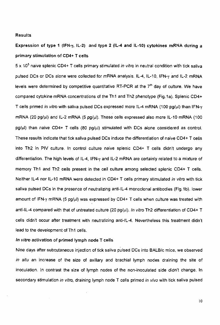
Results
Expression of type 1 (IFN-y, IL-2) and type 2 (IL-4 and IL-10) cytokines mRNA during a
primary stimulation of CD4+ T cells
5 x 10⁵ naive splenic CD4+ T cells primary stimulated in vitro in neutral condition with tick saliva
pulsed DCs or DCs alone were collected for mRNA analysis. IL-4, IL-10, IFN-y and IL-2 mRNA
levels were determined by competitive quantitative RT-PCR at the 7th day of culture. We have
compared cytokine mRNA concentrations of the Th1 and Th2 phenotype (Fig.1a). Splenic CD4+
T cells primed in vitro with saliva pulsed DCs expressed more IL-4 mRNA (100 pg/jil) than IFN-y
mRNA (20 pg/jil) and IL-2 mRNA (5 pg/jil). These cells expressed also more IL-10 mRNA (100
pg/|j.l) than naive CD4+ T cells (80 pg/jil) stimulated with DCs alone considered as control.
These results indicate that tick saliva pulsed DCs induce the differentiation of naive CD4+ T cells
into Th2 in PIV culture. In control culture naive splenic CD4+ T cells didn't undergo any
differentiation. The high levels of IL-4, IFN-y and IL-2 mRNA are certainly related to a mixture of
memory Th1 and Th2 cells present in the cell culture among selected splenic CD4+ T cells.
Neither IL-4 nor IL-10 mRNA were detected in CD4+ T cells primary stimulated in vitro with tick
saliva pulsed DCs in the presence of neutralizing anti-IL-4 monoclonal antibodies (Fig.1b). lower
amount of IFN-y mRNA (5 pg/jil) was expressed by CD4+ T cells when culture was treated with
anti-IL-4 compared with that of untreated culture (20 pg/p.l). In vitro Th2 differentiation of CD4+ T
cells didn't occur after treatment with neutralizing anti-IL-4. Nevertheless this treatment didn't
lead to the development of Th1 cells.
in vitro activation of primed lymph node T cells
Nine days after subcutaneous injection of tick saliva pulsed DCs into BALB/c mice, we observed
in situ an increase of the size of axillary and brachial lymph nodes draining the site of
inoculation, in contrast the size of lymph nodes of the non-inoculated side didn't change. In
secondary stimulation in vitro, draining lymph node T cells primed in vivo with tick saliva pulsed
10
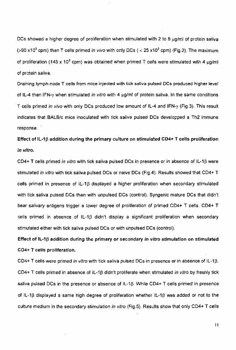
DCs showed a higher degree of proliferation when stimulated with 2 to 8 fag/ml of protein saliva
(>90 x103 cpm) than T cells primed in vivo with only DCs ( < 25 x103 cpm) (Fig.2). The maximum
of proliferation (145 x 103 cpm) was obtained when primed T cells were stimulated with 4 ng/ml
of protein saliva.
Draining lymph node T cells from mice injected with tick saliva pulsed DCs produced higher level
of IL-4 than IFN-y when stimulated in vitro with 4 |ig/ml of protein saliva. In the same conditions
T cells primed in vivo with only DCs produced low amount of IL-4 and IFN-y (Fig.3). This result
indicates that BALB/c mice inoculated with tick saliva pulsed DCs developped a Th2 immune
response.
Effect of lL-1ß addition during the primary culture on stimulated CD4+ T cells proliferation
in vitro.
CD4+ T cells primed in vitro with tick saliva pulsed DCs in presence or in absence of IL-1ß were
stimulated in vitro with tick saliva pulsed DCs or naive DCs (Fig.4). Results showed that CD4+ T
cells primed in presence of IL-1ß displayed a higher proliferation when secondary stimulated
with tick saliva pulsed DCs than with unpulsed DCs (control). Syngenic mature DCs that didn't
bear salivary antigens trigger a lower degree of proliferation of primed CD4+ T cells. CD4+ T
cells primed in absence of IL-1ß didn't display a significant proliferation when secondary
stimulated either with tick saliva pulsed DCs or with unpulsed DCs (control).
Effect of IL-1ß addition during the primary or secondary in vitro stimulation on stimulated
CD4+ T cells proliferation.
CD4+ T cells were primed in vitro with tick saliva pulsed DCs in presence or in absence of IL-1 ß.
CD4+ T cells primed in absence of lL-1ß didn't proliferate when stimulated in vitro by freshly tick
saliva pulsed DCs in the presence or absence of IL-1 p. While CD4+ T cells primed in presence
of IL-1 ß displayed a same high degree of proliferation whether IL-1 ß was added or not to the
culture medium in the secondary stimulation in vitro (Fig.5). Results show that only CD4+ T cells
I i

primed in vitro in presence of IL-1 ß proliferate in secondary stimulation . This finding indicates
that the presence of IL-1 ß is required early during primary stimulation but not during secondary
stimulation to generate proliferation of Th2 cells primed in vitro.
12
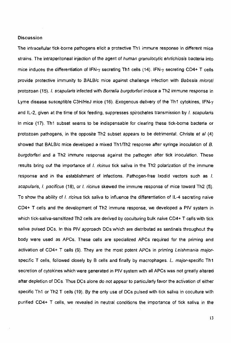
Discussion
The intracellular tick-borne pathogens elicit a protective Th1 immune response in different mice
strains. The intraperitoneal injection of the agent of human granulocytic ehrlichiosis bacteria into
mice induces the differentiation of IFN-y secreting TM cells (14). IFN-y secreting CD4+ T cells
provide protective immunity to BALB/c mice against challenge infection with Babesia microti
protozoan (15). I. scapufaris infected with Borrelia burgdorferi induce a Th2 immune response in
Lyme disease susceptible C3H/HeJ mice (16). Exogenous delivery of the Th1 cytokines, IFN-y
and IL-2, given at the time of tick feeding, suppresses spirochetes transmission by I. scapufaris
in mice (17). Th1 subset seems to be indispensable for clearing these tick-borne bacteria or
protozoan pathogens, in the opposite Th2 subset appears to be detrimental. Christe et al (4)
showed that BALB/c mice developed a mixed Th1/Th2 response after syringe inoculation of S.
burgdorferi and a Th2 immune response against the pathogen after tick inoculation. These
results bring out the importance of I. ricinus tick saliva in the Th2 polarization of the immune
response and in the establishment of infections. Pathogen-free Ixodid vectors such as /.
scapularis, I. pacificus (18), or I. ricinus skewed the immune response of mice toward Th2 (5).
To show the ability of I. ricinus tick saliva to influence the differentiation of IL-4 secreting naive
CD4+ T cells and the development of Th2 immune response, we developed a PIV system in
which tick-saliva-sensitized Th2 cells are derived by coculturing bulk naive CD4+ T cells with tick
saliva pulsed DCs. In this PIV approach DCs which are distributed as sentinels throughout the
body were used as APCs. These cells are specialized APCs required for the priming and
activation of CD4+ T cells (9). They are the most potent APCs in priming Leishmania major-
specific T cells, followed closely by B cells and finally by macrophages. L ma/or-specific Th1
secretion of cytokines which were generated in PIV system with all APCs was not greatly altered
after depletion of DCs. Thus DCs alone do not appear to particularly favor the activation of either
specific Th1 or Th2 T cells (19). By the only use of DCs pulsed with tick saliva in coculture with
purified CD4+ T cells, we revealed in neutral conditions the importance of tick saliva in the
13
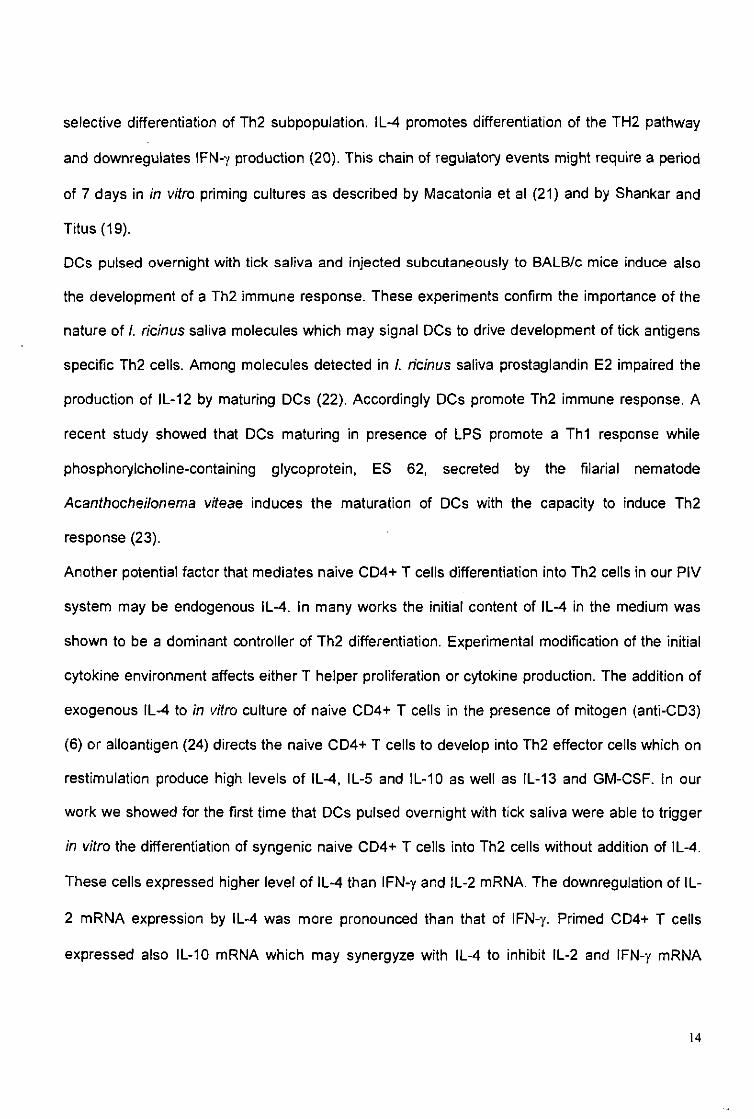
selective differentiation of Th2 subpopulation. IL-4 promotes differentiation of the TH2 pathway
and downregulates IFN-y production (20). This chain of regulatory events might require a period
of 7 days in in vitro priming cultures as described by Macatonia et al (21) and by Shankar and
Titus (19).
DCs pulsed overnight with tick saliva and injected subcutaneously to BALB/c mice induce also
the development of a Th2 immune response. These experiments confirm the importance of the
nature of f. ricinus saliva molecules which may signal DCs to drive development of tick antigens
specific Th2 cells. Among molecules detected in /. ricinus saliva prostaglandin E2 impaired the
production of IL-12 by maturing DCs (22). Accordingly DCs promote Th2 immune response. A
recent study showed that DCs maturing in presence of LPS promote a Th1 response while
phosphorylcholine-containing glycoprotein, ES 62, secreted by the filarial nematode
Acanthocheilonema viteae induces the maturation of DCs with the capacity to induce Th2
response (23).
Another potential factor that mediates naive CD4+ T cells differentiation into Th2 cells in our PIV
system may be endogenous IL-4. In many works the initial content of IL-4 in the medium was
shown to be a dominant controller of Th2 differentiation. Experimental modification of the initial
cytokine environment affects either T helper proliferation or cytokine production. The addition of
exogenous IL-4 to in vitro culture of naive CD4+ T cells in the presence of mitogen (anti-CD3)
(6) or alloantigen (24) directs the naive CD4+ T cells to develop into Th2 effector cells which on
restimulation produce high levels of IL-4, IL-5 and !L-10 as well as IL-13 and GM-CSF. In our
work we showed for the first time that DCs pulsed overnight with tick saliva were able to trigger
in vitro the differentiation of syngenic naive CD4+ T cells into Th2 cells without addition of IL-4.
These cells expressed higher level of IL-4 than IFN-y and tL-2 mRNA. The downregulation of IL-
2 mRNA expression by IL-4 was more pronounced than that of IFN-y. Primed CD4+ T cells
expressed also IL-10 mRNA which may synergyze with IL-4 to inhibit IL-2 and IFN-y mRNA
14
expression. In accordance with our observations IL-10 inhibits the production of IFN-y by CD4+
T cells stimulated in vitro by allogenic DCs (25). It suppressed also the production of IL-2 by
CD4+T cells inducing the inhibition of their proliferation (26).
The early treatment of PIV culture with anti-IL-4 provides a useful tool to determine whether
endogenous IL-4 was early secreted in sufficient quantity to initiate the anti-tick Th2 polarization
of the immune response. Indeed neutralizing anti-IL-4 monoclonal antibodies applied in the
culture inhibit the development of IL-4 secreting Th2 cells. Subsequently no more expression of
IFN-y mRNA by CD4+ T cells occurred. In contrast the addition of anti-IL-4 in order to neutralize
any endogenous IL-4 during primary culture of T cells from TCR transgenic mice specific for
ovalbumine (OVA) with various types of APC and OVA resulted in the appearance of T cells that
produced IFN-y and IL-2 upon stimulation (27). Our experiments suggest that IL-4 dependent
signal pathway is indispensable to bias strongly the cytokine secretion pattern toward Th2
phenotype. However in our PIV assay the neutralization of the endogenous IL-4 didn't lead to
the development of IFN-y secreting Th1 cells. The endogenous IL-4 may be secreted by minor
population of splenic CD4+ T cells present in the culture such as memory Th2 cells, NK 1.1* /
NK 1.1* CD4+ T cells or naive aß CD4+ T cells themselves. The composition of salivary
secretion is complex (5). Memory Th2 cells could cross-react with some salivary antigens and
provide IL-4 necessary to trigger IL-4 expression by naive Th cells. NK 1.1* and NK 1,1" CD4+
splenic T cells may also produce enough IL-4 following stimulation by salivary antigens. NK 1.1*
CD4+ T cells are responsible for the rapid (30 to 90 min) and transient IL-4 production after in
vivo challenge with anti-CD3 or staphylococcal enterotoxin B (28). NK 1.1" CD4+ T cells express
a high level of IL-4 transcripts 90 min after intravenous injection of L major (29). The early
inducing IL-4 may be produced by naive aß CD4+ T cells themselves. Small percentage of
transgenic aß CD4+ T cells from lL-4Ra"'" mice can acquire lL-4-producing capacity when primed
in the absence of IFN-y and IL-12. This finding demonstrates that there is an IL-4-independent
15
pathway of IL-4 secretion by conventional aß CD4+ T cells. The amount of IL-4 secreted by
these cells may be sufficient to cause a certain degree of polarization (30). This endogenous IL-
4 could be sufficient to initiate the differentiation of naive CD4+ T cells into Th2.
Tick saliva specific Th2 differentiated in vitro didn't display any proliferation neither in a primary
stimulation nor in a secondary in vitro stimulation with tick saliva pulsed DCs, but strongly
proliferate when 1L-1 was added as costimulator in the PIV culture (Fig.4). In accordance with
our finding, Chang et ai (31) showed that the optimal proliferation of Th2 clones in response to
IL-4 required the addition of IL-1. In contrast Th1 clones secrete IL-2 and proliferate without any
requirement for IL-1. Exogenous IL-1 is needed because IL-4 secreted by Th2 cells inhibits the
transcription and secretion of IL-1 (32). In a TCR transgenic T cell in vitro priming system,
neither IL-1 receptor antagonist (IL-1 Ra) nor antibodies to IL-1 diminished priming for IL-4
production mediated by antigen, APC and IL-4 (27; 33). If IL-1 ß is not required for the
differentiation of murine CD4+ T cells into Th2 clones, it is for the proliferation of some Th2
clones in response to IL-4. Proliferative responses and lymphokine production by Th2 cells have
distinct signaling requirements. Moreover we showed that IL-1 ß is not required in secondary
stimulation of differentiated Th2 cells neither when CD4+ T cells were primed in presence of IL-
1ß nor in its absence (Fig.5). This suggested that proliferation of differentiated Th2 cells require
an early IL-1ß-dependent signaling pathway during primary in vitro culture only.
Our PIV system is useful to analyze further the salivary antigens and the cells implicated in the
anti-tick Th2 response. Neutralization of Th2 triggering factors could invert the response and
confer resistance against tick and tick borne pathogens.
16

ACKNOWLEDGEMENTS
This work is part of the PhD thesis of Naceur Mejri and was supported by the Swiss National
Science Foundation, grant number 31-56836. 99.
17
References
1 Mosmann T R1 Sad S. The expanding universe of T cell subsets: Th1, Th2 and more.
Immunol Today 1996; 17:138-46.
2 Locksley R M, Scott P. Helper T-cell subsets in mouse leishmaniasis: induction, expansion,
and effector function. Immunol Today 1991; 12:58-61.
3 Mbow M L, Bleyenberg J A, Hall L R, Titus RG. Phlebotomus papatasi sand fly salivary
gland lysate down-regulates a T M , but up-regulates a Th2, response in mice infected with
Leishmania major. J Immunol 1998; 161:5571-7.
4 Christe M, Rutti B1 Brossard M. Cytokines (IL-4 and IFN-y) and antibodies (IgE and lgG2a)
produced in mice infected with Borrelia burgdorferi sensu stricto via lymphs of Ixodes ricinus
ticks or syringe inoculations. Parasitol Res 2000; 86:491-6.
5 Mejri N, Franscini N, Rutti B, Brossard M. Th2 polarization of immune response of BALB/c
mice to Ixodes ricinus instars, importance of several antigens in activation of specific Th2
subpopulations. Parasite Immunol 2001; 23:61-9.
6 Seder R A, Paul WE. Acquisition of lymphokine-producing phenotype by CD4+ T cells. Annu
Rev Immunol 1994; 12:635-73.
7 Christe M, Rutti B, Brossard M. Influence of the genetic background and parasite load of
mice on the immune response developed against nymphs of Ixodes ricinus. Parasitol Res
1999;85:557-61.
8 Steinman R M. The dendritic cell system and its role in immunogenicity. Annu Rev Immunol
1991;9:271-96.
9 Banchereau J, Steinman R M. Dendritic cells and the control of immunity. Nature 1998;
392:245-52.
10 Graf J F. Copulation, nutrition et ponte chez Ixodes ricinus L (Ixodoidea: Ixodidae) - 1 e
partie. Bull Soc Entomol Suisse 1978; 51:89-97.
18
11 Ferreira B R, Silva J S. Saliva of Rhipicephalus sanguineus tick impairs T cell proliferation
and IFN-y-induced macrophage microbicidal activity. Vet Immunol lmmunopathol 1998;
64:279-93.
12 Reiner S L1 Zheng S, Corry D B1 Locksley R M. Constructing polycompetitor cDNAs for
quantitative PCR. J Immunol Methods 1993; 165:37-46.
13 Ganapamo F, Rutti B, Brossard M. In vitro production of interleukin-4 and interferon-y by
lymph node cells from BALB/c mice infested with nymphal Ixodes n'cinus ticks.
Immunology 1995; 85:120-4.
14 Akkoyunlu M, Fikrig E. Gamma interferon dominates the murine cytokine response to the
agent of human granulocytic ehrlichiosis and helps to control the degree of early
rickettsemia. Infect Immun 2000; 68:1827-33. '
15 lgarashi I1 Suzuki R, Waki S et al. Roles of CD4(+) T cells and gamma interferon in
protective immunity against Babesia microti infection in mice. Infect Immun 1999; 67:4143-8.
16 Zeidner N1 Mbow M L, Dolan M, Massung R, Baca E1 Piesman J. Effects of Ixodes
scapularis and Borrelia burgdorferi on modulation of the host immune response: induction of
a TH2 cytokine response in Lyme disease-susceptible (C3H/HeJ) mice but not in disease-
resistant (BALB/c) mice. Infect Immun 1997; 65:3100-6.
17 Zeidner N, Dreitz M1 Belasco D1 Fish D. Suppression of acute Ixodes scapty/ar/s-induced
Borrelia burgdorferi infection using tumor necrosis factor-alpha, interleukin-2, and interferon-
gamma. J Infect Dis 1996; 173:187-95.
18 Schoeler G B1 Manweiler S A1 Wikel S K. Cytokine responses of C3H/HeN mice infested
with Ixodes scapularis or Ixodes pacificus nymphs. Parasite Immunol 2000; 22:31-40.
19 Shankar A H1 Titus R G. The influence of antigen-presenting cell type and interferon-y on
priming and cytokine secretion of Leishmania mayor-specific T cells. J Infect Dis 1997;
175:151-7.
19
20 Swain S L, Weinberg A D, English M, Huston G. IL-4 directs the development of Th2-like
helper effectors. J Immunol 1990; 145:3796-806.
21 Macatonia S E, Hsieh C S, Murphy K M1 O'Garra A. Dendritic cells and macrophages are
required for Th1 development of CD4+ T cells from alpha beta TCR transgenic mice: IL-12
substitution for macrophages to stimulate IFN-gamma production is IFN-gamma-dependent
lnt Immunol 1993;5:1119-28.
22 Kalinski P, Hilkens C M, Snijders A, Snijdewint F G, Kapsenberg M L Dendritic cells,
obtained from peripheral blood precursors in the presence of PGE2, promote Th2
responses. Adv Exp Med Biol 1997; 417:363-7.
23 Whelan M1 Harnett M M, Houston K M1 Patel V, Harnett W, Rigley K P. A filarial nematode-
secreted product signals dendritic cells to acquire a phenotype that drives development of
Th2 cells. J Immunol 2000; 164:6453-60.
24 Swain S L1 Weinberg A D, English M. CD4+ T cell subsets. Lymphogne secretion of memory
cells and of effector cells that develop from precursors in vitro. J Immunol 1990; 144:1788-
99.
25 Macatonia S E, Doherty T M1 Knight S C, O'Garra A. Differential effect of IL-10 on dendritic
cell-induced T cell proliferation and IFN-y production. J Immunol 1993; 150:3755-65.
26 De Waal Malefyt R, Yssel H, de Vries J E. Direct effects of IL-10 on subsets of human CD4+
T cell clones and resting T cells. Specific inhibition of IL-2 production and proliferation. J
Immunol 1993; 150:4754-65.
27 Hsieh C S1 Heimberger A B1 Gold J S, O'Garra A1 Murphy K M. Differential regulation of T
helper phenotype development by interleukins 4 and 10 in an alpha beta T-cell-receptor
transgenic system. Proc Natl Acad Sci U S A 1992; 89:6065-9.
28 Yoshimoto T, Bendelac A, Watson C, Hu-Li J, Paul W E. Role of NK1.1+ T cells in a Th2
response and in immunoglobulin E production. Science 1995; 270:1845-7.
20
29 Launois P, Ohteki T, Swihart K, MacDonald H R, Louis J A. In susceptible mice, Leishmania
major induce very rapid interleukin-4 production by CD4+ T cells which are NK1.1-. Eur J
Immunol 1995; 25:3298-307.
30 Noben-Trauth N, Hu-Li J1 Paul W E. Conventional, naive CD4+ T cells provide an initial
source of IL-4 during Th2 differentiation. J Immunol 2000; 165:3620-5.
31 Chang T L, Shea C M, Urioste S, Thompson R C, Boom W H1 Abbas A K. Heterogeneity of
helper/inducer T lymphocytes. III. Responses of IL-2- and IL-4-producing (TM and Th2)
clones to antigens presented by different accessory cells. J Immunol 1990; 145:2803-8.
32 Hart P H, Cooper R L, Finlay-Jones J J. IL-4 suppresses IU-Ip1 TNF-cc and PGE2 production
by human peritoneal macrophages. Immunology 1991; 72:344-9.
33 Seder R A, Paul W E, Davis M M, Fazekas D S, Groth B. The presence of interleukin-4
during in vitro priming determines the lymphokine-producing potential of CD4+ T cells from T
cell receptor transgenic mice. J Exp Med 1992; 176:1091-8. .
21
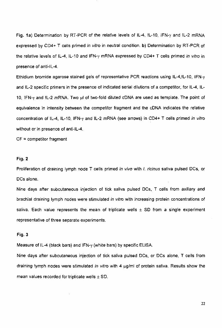
Fig. 1a) Determination by RT-PCR of the relative levels of IL-4, IL-10, IFN-y and IL-2 mRNA
expressed by CD4+ T cells primed in vitro in neutral condition, b) Determination by RT-PCR of
the relative levels of IL-4, IL-10 and IFN-y mRNA expressed by CD4+ T cells primed in vitro in
presence of anti-lL-4.
Ethidium bromide agarose stained gels of representative PCR reactions using IL-4.IL-10, IFN-y
and IL-2 specific primers in the presence of indicated serial dilutions of a competitor, for IL-4, IL-
10, IFN-y and IL-2 mRNA. Two ^l of two-fold diluted cDNA are used as template. The point of
equivalence in intensity between the competitor fragment and the cDNA indicates the relative
concentration of IL-4, IL-10, IFN-y and IL-2 mRNA (see arrows) in CD4+ T cells primed in vitro
without or in presence of anti-IL-4.
CF = competitor fragment
Fig. 2
Proliferation of draining lymph node T cells primed in vivo with i. ricinus saliva pulsed DCs1 or
DCs alone.
Nine days after subcutaneous injection of tick saliva pulsed DCs, T cells from axillary and
brachial draining lymph nodes were stimulated in varo with increasing protein concentrations of
saliva. Each value represents the mean of triplicate wells ± SD from a single experiment
representative of three separate experiments.
Fig. 3
Measure of IL-4 (black bars) and IFN-y (white bars) by specific ELISA.
Nine days after subcutaneous injection of tick saliva pulsed DCs, or DCs alone, T cells from
draining lymph nodes were stimulated in vitro with 4 ng/ml of protein saliva. Results show the
mean values recorded for triplicate wells ± SD.
22
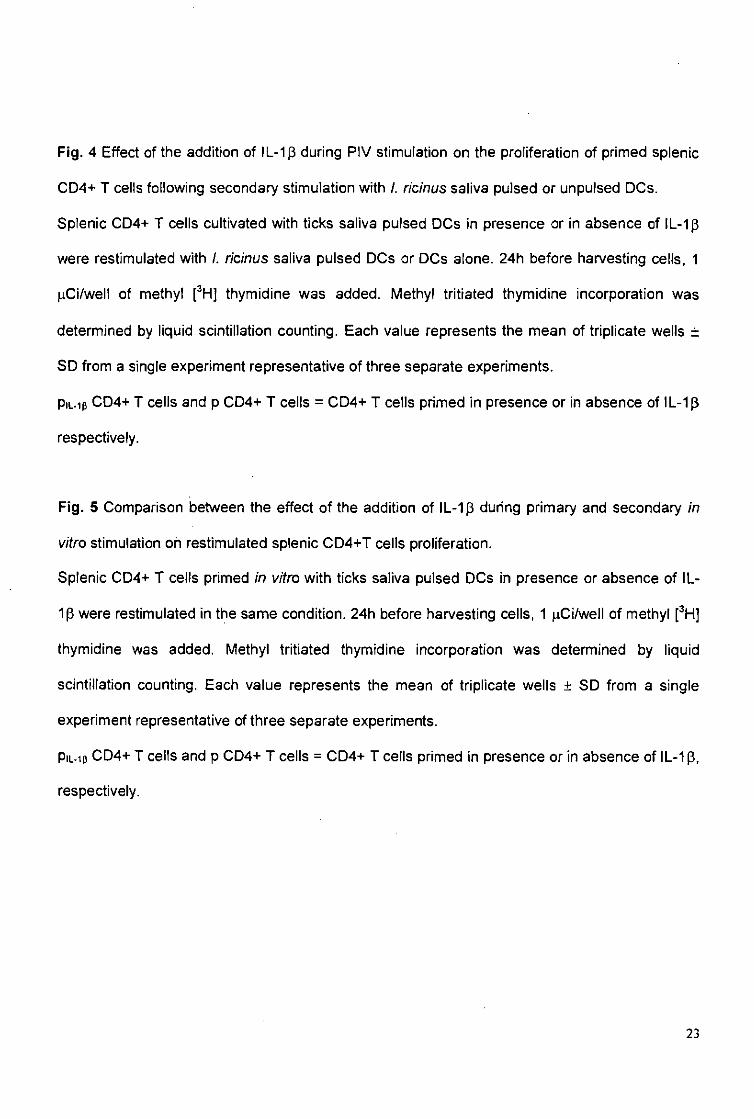
Fig. 4 Effect of the addition of IL-1ß during PIV stimulation on the proliferation of primed splenic
CD4+ T cells following secondary stimulation with I. ricinus saliva pulsed or unpulsed DCs.
Splenic CD4+ T cells cultivated with ticks saliva pulsed DCs in presence or in absence of IL-Ip
were restimulated with I. ricinus saliva pulsed DCs or DCs alone. 24h before harvesting cells, 1
(iCi/well of methyl [3H] thymidine was added. Methyl tritiated thymidine incorporation was
determined by liquid scintillation counting. Each value represents the mean of triplicate wells ±
SD from a single experiment representative of three separate experiments.
PiL-ip CD4+ T cells and p CD4+ T cells = CD4+ T cells primed in presence or in absence of 1L-1 ß
respectively.
Fig. 5 Comparison between the effect of the addition of IL-1ß during primary and secondary in
vitro stimulation oh restimulated splenic CD4+T cells proliferation.
Splenic CD4+ T cells primed in vitro with ticks saliva pulsed DCs in presence or absence of IL-
tß were restimulated in the same condition. 24h before harvesting cells, 1 jiCi/well of methyl [3H]
thymidine was added. Methyl tritiated thymidine incorporation was determined by liquid
scintillation counting. Each value represents the mean of triplicate wells ± SD from a single
experiment representative of three separate experiments.
Pinp CD4+ T cells and p CD4+ T cells = CD4+ T cells primed in presence or in absence of IL-1 ß,
respectively.
23
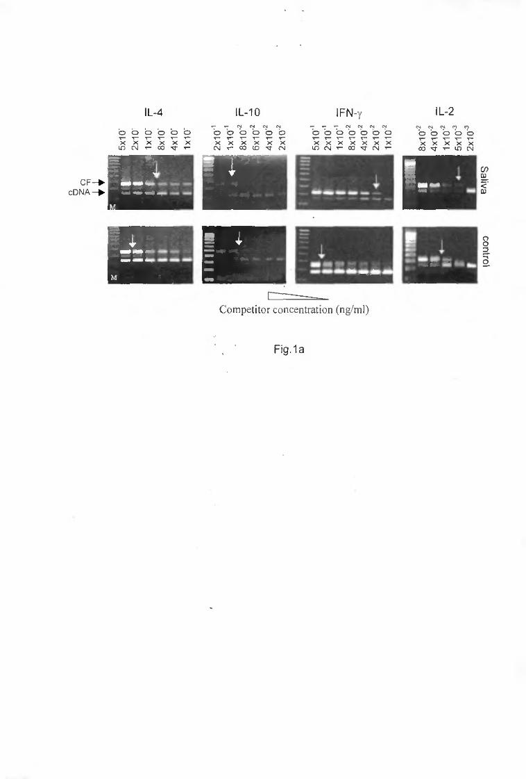
IL-4 IL-10 IFN-y IL-2
Competitor concentration (ng/ml)
Fig.1a
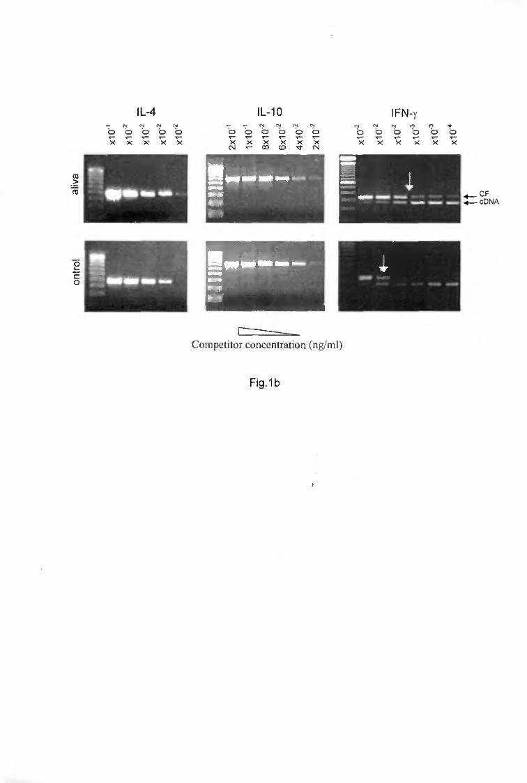
IL-4 IL-10 IFN-y
CF cDNA
Competitor concentration (ng/ml)
FmAb
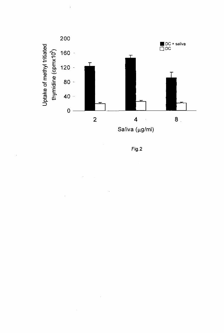
200 "O BOC + saliva S ~ DDC m ^ 160
• t i ^ -
r I t I 120 -(D ^
E ^ * £ 80 -O ^ 0 E
IS i? 40 4 8
Saliva (|ag/ml)
Fig.2
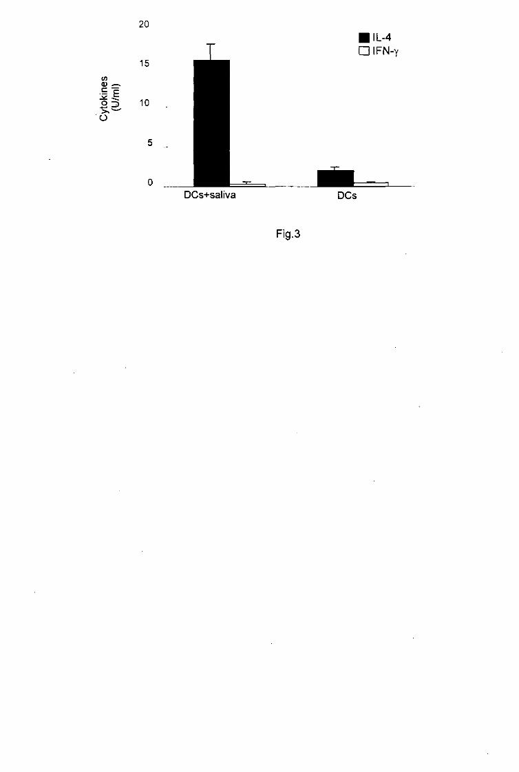
(L) ^
11 O
20
15
10
• IL-4 DIFN-y
DCs+saliva DCs
Fig.3
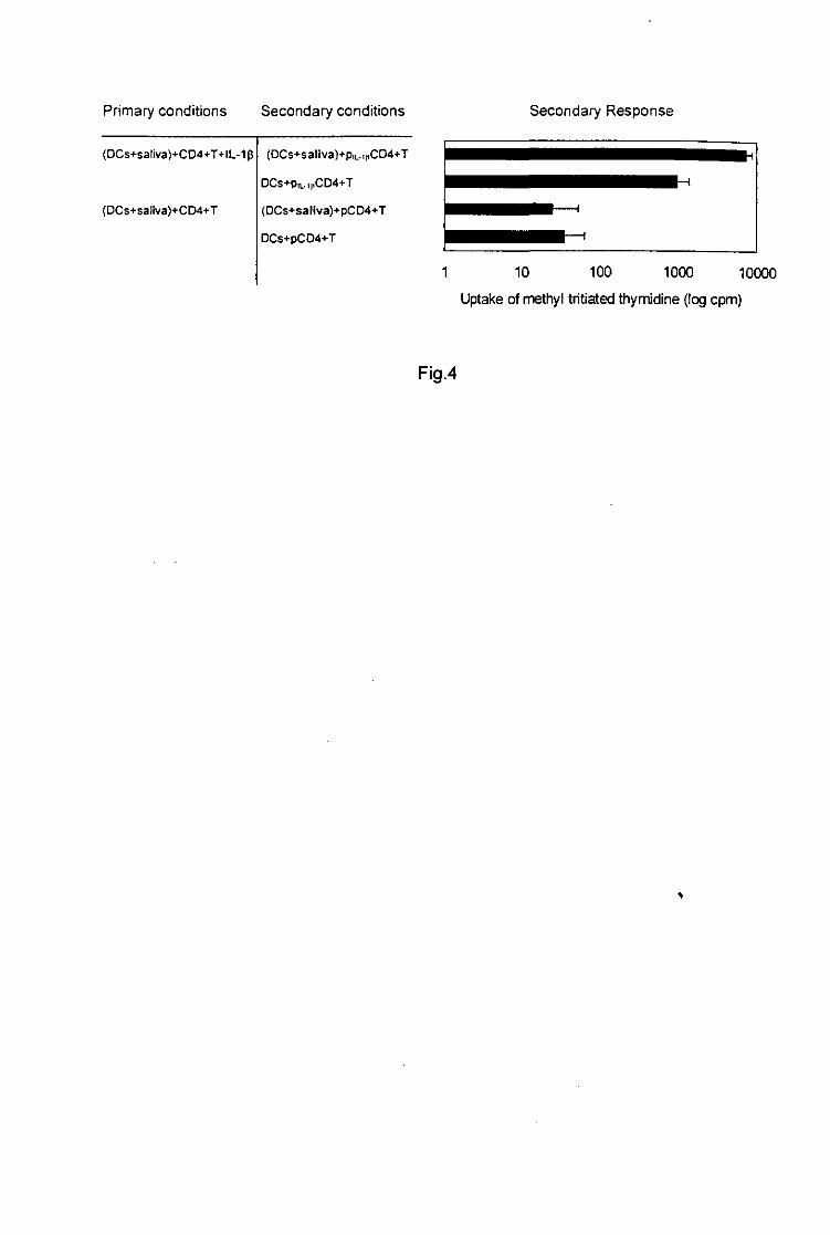
Primary conditions Secondary conditions Secondary Response
(DCs+saliva)+CD4+T+IL-1ß
{DCs+saliva)+CD4+T
(DCs+saltva)+piL-inCD4+T
DCs+pn..t|tCD4+T
(DCs+saliva)+pCD4+T
DCs+pCD4+T
1 10 100 1000 10000
Uptake of methyl tritiated thymidine (log cpm)
Fig.4
^
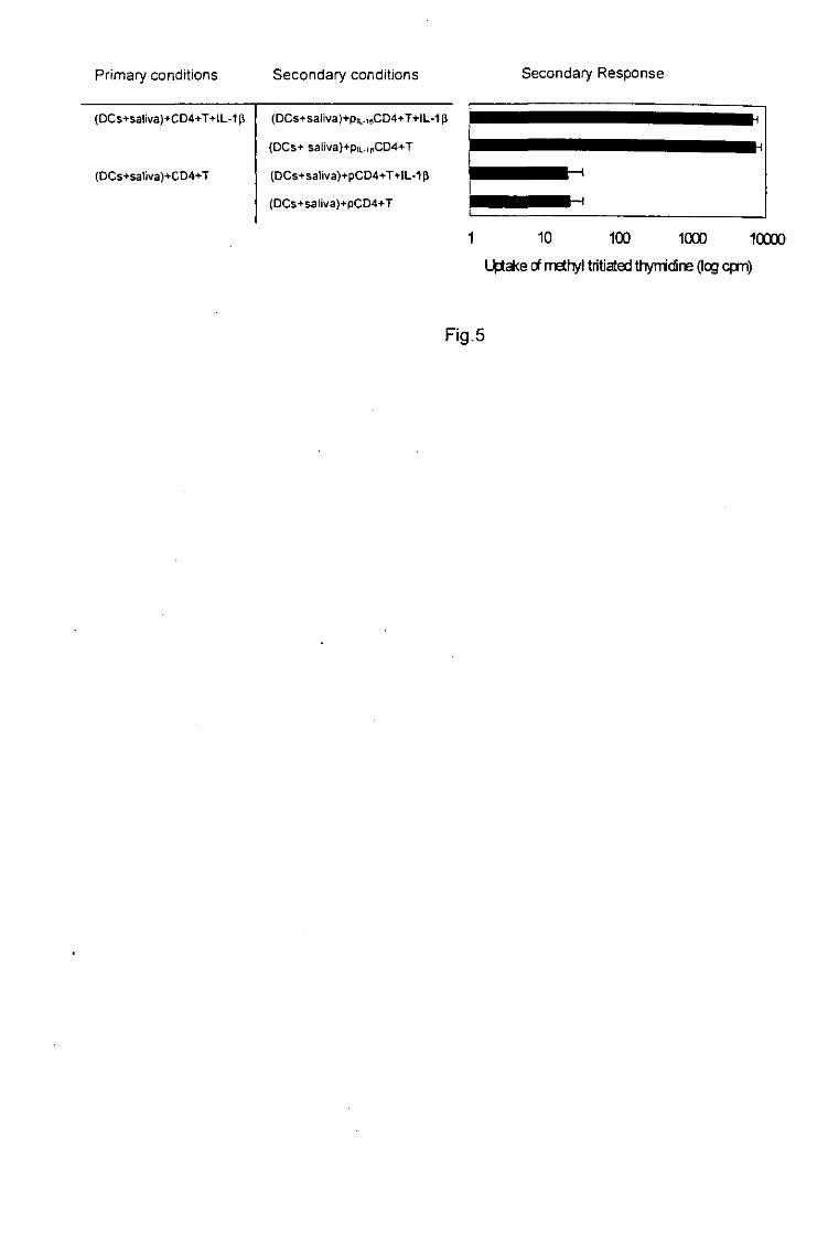
Primary conditions Secondary conditions Secondary Response
(DCs+sa!iva)+CD4+T+lL-1 ß
(DCs+saliva)+CD4+T
(DCs+saliva)+pn..ipCD4+T+IL-1 ß
(DCs+ satrva)+piL.,|,CD4+T
(DCs+saliva)+pCD4+T+IL-1 ß
(DCs+saliva)+pCD4+T
*
1 10 100 1000 10000
Uptake of methyl tritiated thymidine (log cpm)
Fig.5