Embed Size (px)
Citation preview

Citation for published version:Johnson, EK, Chen, L, Kubiak, PS, McDonald, SF, Adams, DJ & Cameron, PJ 2013, 'Surface nucleated growthof dipeptide fibres', Chemical Communications, vol. 49, no. 77, pp. 8698-8700.https://doi.org/10.1039/c3cc44738c
DOI:10.1039/c3cc44738c
Publication date:2013
Document VersionPeer reviewed version
Link to publication
University of Bath
General rightsCopyright and moral rights for the publications made accessible in the public portal are retained by the authors and/or other copyright ownersand it is a condition of accessing publications that users recognise and abide by the legal requirements associated with these rights.
Take down policyIf you believe that this document breaches copyright please contact us providing details, and we will remove access to the work immediatelyand investigate your claim.
Download date: 13. Oct. 2019

Received (in XXX, XXX) Xth
5
10
15
20
25
30
35
40
45
Chemical Communications
Cite this: DOI: 10.1039/c0xx00000x
www.rsc.org/xxxxxx
This journal is
Surface
Eleanor K. Johnson, Cameron
*a
Received (in XXX, XXX) Xth
DOI: 10.1039/b
We report the surface
dipeptide films
with a globular structure.
with dipeptide led to growth of
enzymes were
growth solution
Low molecular weight hydrogelators (LMWGs) are small
molecules that can self
structures, trapping water inside the matrix to give hydrogels
LMW hydrogels are currently being investigated as energy
transfer materials
wound healing
gelator. A range of aromatic functionalised dipeptides have been
shown to self-assemble into
ordinate into fibres. Fibr
based LMWG systems enzymatically
changing solvent composition
gelators that assemble at surfaces due to electrostatic interactions
or ligand-receptor interactions have also been reported.
2010, we showed that ultra
be grown by inducing a localised pH drop at the surface of an
electrode.17 The pH was decreased by the release of protons that
accompanied the electrochemical oxidation of 1,4
to 1,4-benzoquinone.
Fmoc-LG (Figure
drops below the pK16). In this communication, we report the nucleated growth of
fibrous N
(Fmoc-LG) gel
grown seeding layer
the seeding layer was placed in contact with a solution of Fmoc
LG at pH 7 (Fig 1S in the ESI shows photos of a seeding layer
and the thicker layer formed after 4
(‘seeding layer’, thickness
the surface of a gold electrode by electrochemically generating a
surface localised pH drop following protocols described
previously (see
thick layer was invisible to the naked eye, but its presence was
confirmed by surface plasmon resonance spectroscopy (SPR,
Figure 1S) and transmission electron microscopy (TEM
2). TEM showed a continuous film that containe
spherical aggregates which were only a few nanometres in
diameter (Figure 1(a)). The seeding layer was gently rinsed with
Chemical Communications
Cite this: DOI: 10.1039/c0xx00000x
www.rsc.org/xxxxxx
is © The Royal Society of Chemistry
Surface nucleated
Eleanor K. Johnson, a Lin Chen,
Received (in XXX, XXX) Xth
DOI: 10.1039/b000000x
We report the surface nucleated
dipeptide films. The seeding
with a globular structure. Placing the
dipeptide led to growth of
enzymes were incorporated into the gel by
growth solution.
Low molecular weight hydrogelators (LMWGs) are small
molecules that can self-assemble into complex hierarchical
rapping water inside the matrix to give hydrogels
LMW hydrogels are currently being investigated as energy
transfer materials5, 6 and as 3
wound healing7-9. One subset of the LMWG is the dipeptide
gelator. A range of aromatic functionalised dipeptides have been
assemble into β-
ordinate into fibres. Fibril formation can be induced in dipeptide
based LMWG systems enzymatically
changing solvent composition
gelators that assemble at surfaces due to electrostatic interactions
receptor interactions have also been reported.
2010, we showed that ultra-thin gel fi
be grown by inducing a localised pH drop at the surface of an
The pH was decreased by the release of protons that
accompanied the electrochemical oxidation of 1,4
benzoquinone.
Figure 1) is a LMW
drops below the pKa of the terminal carboxylic acid (pK
In this communication, we report the nucleated growth of
N-(9-Fluorenylmethoxycarbonyl)
gels on top of an
grown seeding layers. The thicker gel layer grew over 48 hours if
the seeding layer was placed in contact with a solution of Fmoc
(Fig 1S in the ESI shows photos of a seeding layer
and the thicker layer formed after 4
(‘seeding layer’, thickness 80
the surface of a gold electrode by electrochemically generating a
surface localised pH drop following protocols described
previously (see ESI for full experiment
thick layer was invisible to the naked eye, but its presence was
confirmed by surface plasmon resonance spectroscopy (SPR,
Figure 1S) and transmission electron microscopy (TEM
TEM showed a continuous film that containe
spherical aggregates which were only a few nanometres in
diameter (Figure 1(a)). The seeding layer was gently rinsed with
Chemical Communications
Cite this: DOI: 10.1039/c0xx00000x
Royal Society of Chemistry
nucleated growth of dipeptide fibres
Lin Chen, b
XXXXXXXXX 20XX, Accepted Xth XXXXXXXXX 20XX
nucleated growth of
. The seeding-layer was a thin dipeptide film
Placing the seeding
dipeptide led to growth of fibres
incorporated into the gel by
Low molecular weight hydrogelators (LMWGs) are small
assemble into complex hierarchical
rapping water inside the matrix to give hydrogels
LMW hydrogels are currently being investigated as energy
and as 3-D scaffolds for cell growth and
. One subset of the LMWG is the dipeptide
gelator. A range of aromatic functionalised dipeptides have been
-sheet like structures that further co
il formation can be induced in dipeptide
based LMWG systems enzymatically10-
changing solvent composition14, or via pH drop.
gelators that assemble at surfaces due to electrostatic interactions
receptor interactions have also been reported.
thin gel films and membranes could
be grown by inducing a localised pH drop at the surface of an
The pH was decreased by the release of protons that
accompanied the electrochemical oxidation of 1,4
is a LMWG that forms gels when the pH
of the terminal carboxylic acid (pK
In this communication, we report the nucleated growth of
Fluorenylmethoxycarbonyl)
80-100 nm thick electrochemically
The thicker gel layer grew over 48 hours if
the seeding layer was placed in contact with a solution of Fmoc
(Fig 1S in the ESI shows photos of a seeding layer
and the thicker layer formed after 48 hours). T
80-100 nm; Figure
the surface of a gold electrode by electrochemically generating a
surface localised pH drop following protocols described
experimental detail).
thick layer was invisible to the naked eye, but its presence was
confirmed by surface plasmon resonance spectroscopy (SPR,
Figure 1S) and transmission electron microscopy (TEM
TEM showed a continuous film that containe
spherical aggregates which were only a few nanometres in
diameter (Figure 1(a)). The seeding layer was gently rinsed with
Chemical Communications
Cite this: DOI: 10.1039/c0xx00000x
Royal Society of Chemistry [year]
growth of dipeptide fibres
Peter S. Kubiak,
XXXXXXXXX 20XX, Accepted Xth XXXXXXXXX 20XX
growth of self-assembled
was a thin dipeptide film
seeding-layer in contact
fibres overnight. Active
incorporated into the gel by adding them to the
Low molecular weight hydrogelators (LMWGs) are small
assemble into complex hierarchical
rapping water inside the matrix to give hydrogels
LMW hydrogels are currently being investigated as energy
D scaffolds for cell growth and
. One subset of the LMWG is the dipeptide
gelator. A range of aromatic functionalised dipeptides have been
sheet like structures that further co
il formation can be induced in dipeptide12, thermally,13
, or via pH drop. 15-21
gelators that assemble at surfaces due to electrostatic interactions
receptor interactions have also been reported.22,
lms and membranes could
be grown by inducing a localised pH drop at the surface of an
The pH was decreased by the release of protons that
accompanied the electrochemical oxidation of 1,4-hydroquinone
G that forms gels when the pH
of the terminal carboxylic acid (pKa
In this communication, we report the nucleated growth of
Fluorenylmethoxycarbonyl)-L-leucine-glycine
nm thick electrochemically
The thicker gel layer grew over 48 hours if
the seeding layer was placed in contact with a solution of Fmoc
(Fig 1S in the ESI shows photos of a seeding layer
. The initial gel lay
nm; Figure 2S) was grown on
the surface of a gold electrode by electrochemically generating a
surface localised pH drop following protocols described
al detail). The nanometre
thick layer was invisible to the naked eye, but its presence was
confirmed by surface plasmon resonance spectroscopy (SPR,
Figure 1S) and transmission electron microscopy (TEM, Figure
TEM showed a continuous film that contained numerous
spherical aggregates which were only a few nanometres in
diameter (Figure 1(a)). The seeding layer was gently rinsed with
Chemical Communications
growth of dipeptide fibres
Peter S. Kubiak, a Shane F. McDonald,
XXXXXXXXX 20XX, Accepted Xth XXXXXXXXX 20XX
assembled
was a thin dipeptide film
in contact
Active
adding them to the
Low molecular weight hydrogelators (LMWGs) are small
assemble into complex hierarchical
rapping water inside the matrix to give hydrogels1-4.
LMW hydrogels are currently being investigated as energy
D scaffolds for cell growth and
. One subset of the LMWG is the dipeptide
gelator. A range of aromatic functionalised dipeptides have been
sheet like structures that further co-
il formation can be induced in dipeptide-
, thermally,13 by
LMW
gelators that assemble at surfaces due to electrostatic interactions , 23 In
lms and membranes could
be grown by inducing a localised pH drop at the surface of an
The pH was decreased by the release of protons that
hydroquinone
G that forms gels when the pH
a = 5.8
In this communication, we report the nucleated growth of a
glycine
nm thick electrochemically
The thicker gel layer grew over 48 hours if
the seeding layer was placed in contact with a solution of Fmoc-
(Fig 1S in the ESI shows photos of a seeding layer
he initial gel layer
S) was grown on
the surface of a gold electrode by electrochemically generating a
surface localised pH drop following protocols described
The nanometre
thick layer was invisible to the naked eye, but its presence was
confirmed by surface plasmon resonance spectroscopy (SPR,
, Figure
d numerous
spherical aggregates which were only a few nanometres in
diameter (Figure 1(a)). The seeding layer was gently rinsed with
NaCl (0.098 mol dm
cm70
buffered saline both at pH7) for 48
of any further current.
60
65
Fig 1
film85
75
80
110
electrochemically onto the TEM grid (the grid was used as the electrode);
(b) shows the development of fibrous structures after the seeding layer
has been left in contact with Fmoc
scale bar on the left hand image is 20 nm and the scale bar on the right
115
The seeding layer was not removed by
that was checked by both SPR and TEM.
gel layer formed on top
showed that fibres were now present in the gel. It is important to
note that the scale bar in Figure 115
growth of dipeptide fibres
Shane F. McDonald,
XXXXXXXXX 20XX, Accepted Xth XXXXXXXXX 20XX
NaCl (0.098 mol dm
cm-3 of Fmoc-LG (2.4 mmol dm
buffered saline both at pH7) for 48
of any further current.
Fig 1. (a) Schematic showing the surface initiated growth of an Fmoc
film, (b) the structure of Fmoc
layer after 48 hours in solution (
delineated by the circular o
Fig. 2 (a) shows a TEM image of the seeding layer grown
electrochemically onto the TEM grid (the grid was used as the electrode);
(b) shows the development of fibrous structures after the seeding layer
has been left in contact with Fmoc
scale bar on the left hand image is 20 nm and the scale bar on the right
The seeding layer was not removed by
that was checked by both SPR and TEM.
gel layer formed on top
showed that fibres were now present in the gel. It is important to
note that the scale bar in Figure
(a)
Dynamic Article Links
Communication
[journal]
Shane F. McDonald, a Dave J. Adams,
NaCl (0.098 mol dm-3, at pH 7) and then left in contact with ~ 0.5
LG (2.4 mmol dm
buffered saline both at pH7) for 48
of any further current.
Schematic showing the surface initiated growth of an Fmoc
the structure of Fmoc-LG, (c) a photo of a surface initiated gel
layer after 48 hours in solution (The gel was
delineated by the circular o-ring of an electrochemical SPR cell
(a) shows a TEM image of the seeding layer grown
electrochemically onto the TEM grid (the grid was used as the electrode);
(b) shows the development of fibrous structures after the seeding layer
has been left in contact with Fmoc-LG
scale bar on the left hand image is 20 nm and the scale bar on the right
hand image is 200 nm.
The seeding layer was not removed by
that was checked by both SPR and TEM.
gel layer formed on top of the seeding layer, TEM (Figure
showed that fibres were now present in the gel. It is important to
note that the scale bar in Figure
Dynamic Article Links
Communication
[journal], [year],
Dave J. Adams,b
and then left in contact with ~ 0.5
LG (2.4 mmol dm-3, either in NaCl or phosph
buffered saline both at pH7) for 48 hours without the application
Schematic showing the surface initiated growth of an Fmoc
, (c) a photo of a surface initiated gel
The gel was formed within an area
ring of an electrochemical SPR cell
(a) shows a TEM image of the seeding layer grown
electrochemically onto the TEM grid (the grid was used as the electrode);
(b) shows the development of fibrous structures after the seeding layer
LG-OH in solution for 48
scale bar on the left hand image is 20 nm and the scale bar on the right
hand image is 200 nm.
The seeding layer was not removed by very gentle
that was checked by both SPR and TEM. After 48 hours a thick
of the seeding layer, TEM (Figure
showed that fibres were now present in the gel. It is important to
note that the scale bar in Figure 2(a) is 20 nm and in Figure
(b)
Dynamic Article Links
Communication
, [vol], 00–00
and Petra J.
and then left in contact with ~ 0.5
, either in NaCl or phosph
hours without the application
Schematic showing the surface initiated growth of an Fmoc-
, (c) a photo of a surface initiated gel
formed within an area
ring of an electrochemical SPR cell).
(a) shows a TEM image of the seeding layer grown
electrochemically onto the TEM grid (the grid was used as the electrode);
(b) shows the development of fibrous structures after the seeding layer
OH in solution for 48 hours. The
scale bar on the left hand image is 20 nm and the scale bar on the right
very gentle rinsing, a fact
After 48 hours a thick
of the seeding layer, TEM (Figure 2(b))
showed that fibres were now present in the gel. It is important to
(a) is 20 nm and in Figure 2
►
Communication
00 | 1
and Petra J.
and then left in contact with ~ 0.5
, either in NaCl or phosphate
hours without the application
-LG
, (c) a photo of a surface initiated gel
formed within an area
electrochemically onto the TEM grid (the grid was used as the electrode);
(b) shows the development of fibrous structures after the seeding layer
hours. The
scale bar on the left hand image is 20 nm and the scale bar on the right
rinsing, a fact
After 48 hours a thick
(b))
showed that fibres were now present in the gel. It is important to
2(b)

2
is 200nm
spherical
(tens of nanometres
of diameters, which were up to several microns in length.
5
10
15
20
nucleated and grew on the pre
represents a steady increase in
The pH of the grow
remained at pH 7. The pH of the 25
with both pH paper and by inserting a pH probe designed for gels
into the film) was 6.5 after 48 hours. Importantly, control
experiments showed that
seeding layer. In a second set of control experiments, all of the
experimental steps (electrochemistry, rinsing and 48 hour 30
monitoring) were carried out in the absence of hydroquinone.
Hydroquinone is necessary to g
drop that induces formation of the seeding layer. Without
hydroquinone, no seeding layer formed and no thicker gel layer
grew on the surface.35
spontaneous assembly of
using SPR, a technique which can be used to measures changes in
refractive index on top of a metal surface.
wave of the surface plasmon only extends ~ 200 nm into the 40
material above the metal,
thickness of gel layers above 200 nm. SPR c
to prove the existence of the seeding layer
used to measure changes in the gel density as hierarchical
structures developed during the 48 hour growth period. The real 45
time change in
shown in Figure
related to an increase in structure density, e.g. the formation of
aggregates or fibres at the surface.
50
gel layer at the metal surface remained unchanged, followed by
44 hours where the
steadily with time.
1.348
44 hours55
film remained s
no changes were occurring.
| Journal Name
is 200nm. The
spherical aggregates,
tens of nanometres
of diameters, which were up to several microns in length.
Fig. 3 The real time change in
nucleated and grew on the pre
represents a steady increase in
corresponding shift in minimum angle as the thick gel layer formed.
The pH of the grow
remained at pH 7. The pH of the
with both pH paper and by inserting a pH probe designed for gels
into the film) was 6.5 after 48 hours. Importantly, control
experiments showed that
seeding layer. In a second set of control experiments, all of the
experimental steps (electrochemistry, rinsing and 48 hour
monitoring) were carried out in the absence of hydroquinone.
Hydroquinone is necessary to g
drop that induces formation of the seeding layer. Without
hydroquinone, no seeding layer formed and no thicker gel layer
grew on the surface.
Both the growth of the seeding layer and the subsequent
spontaneous assembly of
using SPR, a technique which can be used to measures changes in
refractive index on top of a metal surface.
wave of the surface plasmon only extends ~ 200 nm into the
material above the metal,
thickness of gel layers above 200 nm. SPR c
to prove the existence of the seeding layer
used to measure changes in the gel density as hierarchical
structures developed during the 48 hour growth period. The real
time change in gel refractive index measured over
shown in Figure
related to an increase in structure density, e.g. the formation of
aggregates or fibres at the surface.
Interestingly there was a four
gel layer at the metal surface remained unchanged, followed by
44 hours where the
steadily with time.
1.348 which correspond
44 hours (ESI). The
film remained static during this time, just that close to the surface
no changes were occurring.
Journal Name, [year], [vol]
he thicker film (Fig. 2(b))
aggregates, although the diameters were
tens of nanometres). The film also contained fibres with a range
of diameters, which were up to several microns in length.
The real time change in refractive index
nucleated and grew on the pre-seeded surface. The
represents a steady increase in gel
corresponding shift in minimum angle as the thick gel layer formed.
The pH of the growth solution was measured after 48 hours and
remained at pH 7. The pH of the
with both pH paper and by inserting a pH probe designed for gels
into the film) was 6.5 after 48 hours. Importantly, control
experiments showed that no gel layer grew in the absence of the
seeding layer. In a second set of control experiments, all of the
experimental steps (electrochemistry, rinsing and 48 hour
monitoring) were carried out in the absence of hydroquinone.
Hydroquinone is necessary to g
drop that induces formation of the seeding layer. Without
hydroquinone, no seeding layer formed and no thicker gel layer
grew on the surface.
Both the growth of the seeding layer and the subsequent
spontaneous assembly of a thicker layer were followed in
using SPR, a technique which can be used to measures changes in
refractive index on top of a metal surface.
wave of the surface plasmon only extends ~ 200 nm into the
material above the metal, SPR cannot be used to measure the
thickness of gel layers above 200 nm. SPR c
to prove the existence of the seeding layer
used to measure changes in the gel density as hierarchical
structures developed during the 48 hour growth period. The real
gel refractive index measured over
shown in Figure 3. The increase in
related to an increase in structure density, e.g. the formation of
aggregates or fibres at the surface.
Interestingly there was a four
gel layer at the metal surface remained unchanged, followed by
44 hours where the amount of structure in the gel increased
steadily with time. The refractive index increased from 1.338 to
which corresponds to a fivefold increase in gel density over
The time lag does not necessarily indicate that the
tatic during this time, just that close to the surface
no changes were occurring.
[vol], 00–00
thicker film (Fig. 2(b)) still contained some
although the diameters were
. The film also contained fibres with a range
of diameters, which were up to several microns in length.
refractive index as the thick gel layer
seeded surface. The refractive index
density with time. Insert shows the
corresponding shift in minimum angle as the thick gel layer formed.
th solution was measured after 48 hours and
remained at pH 7. The pH of the 1-2mm thick gel film (measured
with both pH paper and by inserting a pH probe designed for gels
into the film) was 6.5 after 48 hours. Importantly, control
no gel layer grew in the absence of the
seeding layer. In a second set of control experiments, all of the
experimental steps (electrochemistry, rinsing and 48 hour
monitoring) were carried out in the absence of hydroquinone.
Hydroquinone is necessary to generate the surface localised pH
drop that induces formation of the seeding layer. Without
hydroquinone, no seeding layer formed and no thicker gel layer
Both the growth of the seeding layer and the subsequent
a thicker layer were followed in
using SPR, a technique which can be used to measures changes in
refractive index on top of a metal surface.24-26
wave of the surface plasmon only extends ~ 200 nm into the
SPR cannot be used to measure the
thickness of gel layers above 200 nm. SPR can
to prove the existence of the seeding layer (Fig.
used to measure changes in the gel density as hierarchical
structures developed during the 48 hour growth period. The real
gel refractive index measured over
he increase in refractive index
related to an increase in structure density, e.g. the formation of
aggregates or fibres at the surface.
Interestingly there was a four-hour time lag during which the
gel layer at the metal surface remained unchanged, followed by
amount of structure in the gel increased
The refractive index increased from 1.338 to
fivefold increase in gel density over
time lag does not necessarily indicate that the
tatic during this time, just that close to the surface
still contained some
although the diameters were much larger
. The film also contained fibres with a range
of diameters, which were up to several microns in length.
as the thick gel layer
refractive index shift
density with time. Insert shows the
corresponding shift in minimum angle as the thick gel layer formed.
th solution was measured after 48 hours and
2mm thick gel film (measured
with both pH paper and by inserting a pH probe designed for gels
into the film) was 6.5 after 48 hours. Importantly, control
no gel layer grew in the absence of the
seeding layer. In a second set of control experiments, all of the
experimental steps (electrochemistry, rinsing and 48 hour
monitoring) were carried out in the absence of hydroquinone.
enerate the surface localised pH
drop that induces formation of the seeding layer. Without
hydroquinone, no seeding layer formed and no thicker gel layer
Both the growth of the seeding layer and the subsequent
a thicker layer were followed in-
using SPR, a technique which can be used to measures changes in
As the evanescent
wave of the surface plasmon only extends ~ 200 nm into the
SPR cannot be used to measure the
an, however, be used
(Fig. 2S). It was also
used to measure changes in the gel density as hierarchical
structures developed during the 48 hour growth period. The real
gel refractive index measured over 48 hours is
refractive index is directly
related to an increase in structure density, e.g. the formation of
hour time lag during which the
gel layer at the metal surface remained unchanged, followed by
amount of structure in the gel increased
The refractive index increased from 1.338 to
fivefold increase in gel density over
time lag does not necessarily indicate that the
tatic during this time, just that close to the surface
This journal is © The Royal Society of Chemistry [year]
still contained some
much larger
. The film also contained fibres with a range
as the thick gel layer
shift
density with time. Insert shows the
corresponding shift in minimum angle as the thick gel layer formed.
th solution was measured after 48 hours and
2mm thick gel film (measured
with both pH paper and by inserting a pH probe designed for gels
into the film) was 6.5 after 48 hours. Importantly, control
no gel layer grew in the absence of the
seeding layer. In a second set of control experiments, all of the
experimental steps (electrochemistry, rinsing and 48 hour
monitoring) were carried out in the absence of hydroquinone.
enerate the surface localised pH
drop that induces formation of the seeding layer. Without
hydroquinone, no seeding layer formed and no thicker gel layer
Both the growth of the seeding layer and the subsequent
-situ
using SPR, a technique which can be used to measures changes in
As the evanescent
wave of the surface plasmon only extends ~ 200 nm into the
SPR cannot be used to measure the
, however, be used
. It was also
used to measure changes in the gel density as hierarchical
structures developed during the 48 hour growth period. The real
48 hours is
rectly
related to an increase in structure density, e.g. the formation of
hour time lag during which the
gel layer at the metal surface remained unchanged, followed by
amount of structure in the gel increased
The refractive index increased from 1.338 to
fivefold increase in gel density over
time lag does not necessarily indicate that the
tatic during this time, just that close to the surface
A nucleation and growth mechanism
previously been reported
a pH trigger, fibre growth was imaged, although the nucleation 140
step was below the temporal and spatial resolution of confocal
microscopy.
fibres that
sites. In amyloid plaque formation, a nucleation and growth type
mechanism is frequently suggested.145
formation show
phase in which the nuclei are extended into fibrillar structures.
The nuclei can be small oligomers, spherical aggregates or
micellar species. If pre
solution, there i150
follows.
proteins has also been induced by the presence of a mica
surface.
In the experi
mechanisms are contributing to the nucleated growth of the 230
thicker film. Firstly there may be enough protons remaining
inside the seeding layer to create a local pH close to the pK
the Fmoc
6.5, even though it had been sitting in a solution at pH 7 for 48
hours. This suggests that some protons remained trapped within 235
the film.
of dipeptides and other a
environment.
pKas considerably higher than the
been reported240
reported for a range of
surface bound molecules.
used to measure surface p
SAMs; it was found tha
functionalised monolayers shifted about 2 pH units more alkaline 245
when compared to the p
there was a relationship between surface roughness and apparent
pKa for 3
with shifts of up to 4 pH units relative to the solution p
possible.250
shifts of up to 2 pH units for benzoic acid covalently bound to
different carbon surfaces.
entropically controlled due to changes in the solvent ordering at
the surface upon ionisation. The hydrophobicity/hydrophilicity of
the surface also played a role.255
moldm
already slightly above the pK
take a small increase in the pK
nucleated growth to occur.
In a final experiment, surface160
used to incorporate horse
films, by
for experimental details
into diphenylalanine nanotubes and the authors found that
encapsulation enhanced the st165
and bacteria have also been incorporated in Fmoc
films.
layer was removed from the growth solution and rinsed.
incorporation of HRP
simple colorimetric assay, namely the conversion of o170
This journal is © The Royal Society of Chemistry [year]
A nucleation and growth mechanism
previously been reported
a pH trigger, fibre growth was imaged, although the nucleation
step was below the temporal and spatial resolution of confocal
microscopy.28 Spheres were initially formed and then replaced by
fibres that appeared to grow from a small number of nucleation
sites. In amyloid plaque formation, a nucleation and growth type
mechanism is frequently suggested.
formation show a time lag while nuclei are created, then a growth
phase in which the nuclei are extended into fibrillar structures.
The nuclei can be small oligomers, spherical aggregates or
micellar species. If pre
solution, there is no time
follows.30 Fibre formation by the mi
proteins has also been induced by the presence of a mica
surface.31
In the experiments described above it is likely that two
mechanisms are contributing to the nucleated growth of the
thicker film. Firstly there may be enough protons remaining
inside the seeding layer to create a local pH close to the pK
the Fmoc-LG. As mentioned a
6.5, even though it had been sitting in a solution at pH 7 for 48
hours. This suggests that some protons remained trapped within
the film. Secondly there is evidence in the literature that the pK
of dipeptides and other a
environment. Dipeptides with environment specific or ‘apparent’
s considerably higher than the
been reported. 32-35
reported for a range of
surface bound molecules.
used to measure surface p
SAMs; it was found tha
functionalised monolayers shifted about 2 pH units more alkaline
when compared to the p
there was a relationship between surface roughness and apparent
for 3-mercaptopropionic acid monolayers on gold electrodes
with shifts of up to 4 pH units relative to the solution p
possible.37 Abiman
shifts of up to 2 pH units for benzoic acid covalently bound to
different carbon surfaces.
entropically controlled due to changes in the solvent ordering at
the surface upon ionisation. The hydrophobicity/hydrophilicity of
the surface also played a role.
moldm-3 NaCl was measured to
already slightly above the pK
take a small increase in the pK
nucleated growth to occur.
In a final experiment, surface
used to incorporate horse
films, by simply adding it to the second growth solution (
for experimental details
into diphenylalanine nanotubes and the authors found that
encapsulation enhanced the st
and bacteria have also been incorporated in Fmoc
films.41 After 48 hours, a 1
layer was removed from the growth solution and rinsed.
incorporation of HRP
simple colorimetric assay, namely the conversion of o
This journal is © The Royal Society of Chemistry [year]
A nucleation and growth mechanism
previously been reported in mixed DMSO/water systems.
a pH trigger, fibre growth was imaged, although the nucleation
step was below the temporal and spatial resolution of confocal
Spheres were initially formed and then replaced by
appeared to grow from a small number of nucleation
sites. In amyloid plaque formation, a nucleation and growth type
mechanism is frequently suggested.
a time lag while nuclei are created, then a growth
phase in which the nuclei are extended into fibrillar structures.
The nuclei can be small oligomers, spherical aggregates or
micellar species. If pre-formed nuclei are added to a protein
s no time-lag and fibre formation rapidly
Fibre formation by the mi
proteins has also been induced by the presence of a mica
ments described above it is likely that two
mechanisms are contributing to the nucleated growth of the
thicker film. Firstly there may be enough protons remaining
inside the seeding layer to create a local pH close to the pK
As mentioned above the final pH of the gel was
6.5, even though it had been sitting in a solution at pH 7 for 48
hours. This suggests that some protons remained trapped within
Secondly there is evidence in the literature that the pK
of dipeptides and other acids may be strongly modified by their
Dipeptides with environment specific or ‘apparent’
s considerably higher than the
Surface induced p
reported for a range of self-assembled monolayers (SAMs) and
surface bound molecules.36-39 Impedance spectroscopy has been
used to measure surface pKa values for a range of functionalised
SAMs; it was found that the p
functionalised monolayers shifted about 2 pH units more alkaline
when compared to the pKa in solution.
there was a relationship between surface roughness and apparent
mercaptopropionic acid monolayers on gold electrodes
with shifts of up to 4 pH units relative to the solution p
Abiman et al. investigated the reasons behind p
shifts of up to 2 pH units for benzoic acid covalently bound to
different carbon surfaces.36 They concluded that the p
entropically controlled due to changes in the solvent ordering at
the surface upon ionisation. The hydrophobicity/hydrophilicity of
the surface also played a role. The pK
NaCl was measured to be 6.1 by a pH titration (Fig
already slightly above the pKa in pure water (5.8). It would only
take a small increase in the pKa of the molecules at the surface for
nucleated growth to occur.
In a final experiment, surface-
used to incorporate horse radish peroxidase (HRP) inside the gel
adding it to the second growth solution (
for experimental details). HRP has previously been incorporated
into diphenylalanine nanotubes and the authors found that
encapsulation enhanced the stability of the enzyme.
and bacteria have also been incorporated in Fmoc
After 48 hours, a 1-2 mm thick gel layer formed,
layer was removed from the growth solution and rinsed.
incorporation of HRP in the gel was proved by carry
simple colorimetric assay, namely the conversion of o
This journal is © The Royal Society of Chemistry [year]
A nucleation and growth mechanism for Fmoc
in mixed DMSO/water systems.
a pH trigger, fibre growth was imaged, although the nucleation
step was below the temporal and spatial resolution of confocal
Spheres were initially formed and then replaced by
appeared to grow from a small number of nucleation
sites. In amyloid plaque formation, a nucleation and growth type
mechanism is frequently suggested.29, 30 The kinetics of amyloid
a time lag while nuclei are created, then a growth
phase in which the nuclei are extended into fibrillar structures.
The nuclei can be small oligomers, spherical aggregates or
formed nuclei are added to a protein
lag and fibre formation rapidly
Fibre formation by the miss-folding of silk
proteins has also been induced by the presence of a mica
ments described above it is likely that two
mechanisms are contributing to the nucleated growth of the
thicker film. Firstly there may be enough protons remaining
inside the seeding layer to create a local pH close to the pK
bove the final pH of the gel was
6.5, even though it had been sitting in a solution at pH 7 for 48
hours. This suggests that some protons remained trapped within
Secondly there is evidence in the literature that the pK
cids may be strongly modified by their
Dipeptides with environment specific or ‘apparent’
s considerably higher than the pKa of the free dipeptide have
Surface induced pKa shifts have also been
assembled monolayers (SAMs) and
Impedance spectroscopy has been
values for a range of functionalised
t the pKa of both acid and amine
functionalised monolayers shifted about 2 pH units more alkaline
in solution.38 Burris et al.
there was a relationship between surface roughness and apparent
mercaptopropionic acid monolayers on gold electrodes
with shifts of up to 4 pH units relative to the solution p
investigated the reasons behind p
shifts of up to 2 pH units for benzoic acid covalently bound to
They concluded that the p
entropically controlled due to changes in the solvent ordering at
the surface upon ionisation. The hydrophobicity/hydrophilicity of
The pKa of Fmoc
be 6.1 by a pH titration (Fig
in pure water (5.8). It would only
of the molecules at the surface for
-templated gel formation was
radish peroxidase (HRP) inside the gel
adding it to the second growth solution (
). HRP has previously been incorporated
into diphenylalanine nanotubes and the authors found that
ability of the enzyme.
and bacteria have also been incorporated in Fmoc
2 mm thick gel layer formed,
layer was removed from the growth solution and rinsed.
was proved by carry
simple colorimetric assay, namely the conversion of o
This journal is © The Royal Society of Chemistry [year]
Fmoc-LG gels has
in mixed DMSO/water systems.27 Using
a pH trigger, fibre growth was imaged, although the nucleation
step was below the temporal and spatial resolution of confocal
Spheres were initially formed and then replaced by
appeared to grow from a small number of nucleation
sites. In amyloid plaque formation, a nucleation and growth type
The kinetics of amyloid
a time lag while nuclei are created, then a growth
phase in which the nuclei are extended into fibrillar structures.
The nuclei can be small oligomers, spherical aggregates or
formed nuclei are added to a protein
lag and fibre formation rapidly
folding of silk-like
proteins has also been induced by the presence of a mica
ments described above it is likely that two
mechanisms are contributing to the nucleated growth of the
thicker film. Firstly there may be enough protons remaining
inside the seeding layer to create a local pH close to the pKa of
bove the final pH of the gel was
6.5, even though it had been sitting in a solution at pH 7 for 48
hours. This suggests that some protons remained trapped within
Secondly there is evidence in the literature that the pK
cids may be strongly modified by their
Dipeptides with environment specific or ‘apparent’
of the free dipeptide have
shifts have also been
assembled monolayers (SAMs) and
Impedance spectroscopy has been
values for a range of functionalised
of both acid and amine
functionalised monolayers shifted about 2 pH units more alkaline
et al. showed that
there was a relationship between surface roughness and apparent
mercaptopropionic acid monolayers on gold electrodes
with shifts of up to 4 pH units relative to the solution pK
investigated the reasons behind pK
shifts of up to 2 pH units for benzoic acid covalently bound to
They concluded that the pKa shift was
entropically controlled due to changes in the solvent ordering at
the surface upon ionisation. The hydrophobicity/hydrophilicity of
of Fmoc-LG in 0.1
be 6.1 by a pH titration (Fig 3S),
in pure water (5.8). It would only
of the molecules at the surface for
templated gel formation was
radish peroxidase (HRP) inside the gel
adding it to the second growth solution (See ESI
). HRP has previously been incorporated
into diphenylalanine nanotubes and the authors found that
ability of the enzyme.40 Enzymes
and bacteria have also been incorporated in Fmoc-F/gelatin
2 mm thick gel layer formed, the
layer was removed from the growth solution and rinsed. The
was proved by carrying out a
simple colorimetric assay, namely the conversion of o
This journal is © The Royal Society of Chemistry [year]
has
Using
a pH trigger, fibre growth was imaged, although the nucleation
step was below the temporal and spatial resolution of confocal
Spheres were initially formed and then replaced by
appeared to grow from a small number of nucleation
sites. In amyloid plaque formation, a nucleation and growth type
The kinetics of amyloid
a time lag while nuclei are created, then a growth
phase in which the nuclei are extended into fibrillar structures.
The nuclei can be small oligomers, spherical aggregates or
formed nuclei are added to a protein
lag and fibre formation rapidly
like
proteins has also been induced by the presence of a mica
ments described above it is likely that two
mechanisms are contributing to the nucleated growth of the
thicker film. Firstly there may be enough protons remaining
of
bove the final pH of the gel was
6.5, even though it had been sitting in a solution at pH 7 for 48
hours. This suggests that some protons remained trapped within
Secondly there is evidence in the literature that the pKa
cids may be strongly modified by their
Dipeptides with environment specific or ‘apparent’
of the free dipeptide have
shifts have also been
assembled monolayers (SAMs) and
Impedance spectroscopy has been
values for a range of functionalised
of both acid and amine
functionalised monolayers shifted about 2 pH units more alkaline
showed that
there was a relationship between surface roughness and apparent
mercaptopropionic acid monolayers on gold electrodes
Ka
Ka
shifts of up to 2 pH units for benzoic acid covalently bound to
ift was
entropically controlled due to changes in the solvent ordering at
the surface upon ionisation. The hydrophobicity/hydrophilicity of
0.1
),
in pure water (5.8). It would only
of the molecules at the surface for
templated gel formation was
radish peroxidase (HRP) inside the gel
See ESI
). HRP has previously been incorporated
into diphenylalanine nanotubes and the authors found that
Enzymes
F/gelatin
the
he
ing out a
simple colorimetric assay, namely the conversion of o-
This journal is © The Royal Society of Chemistry [year] Journal Name, [year], [vol], 00–00 | 3
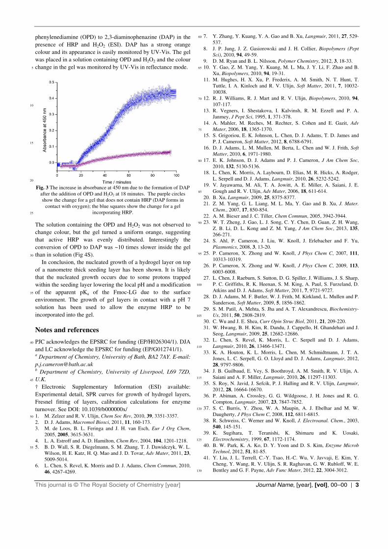
phenylenediamine (OPD) to 2,3-diaminophenazine (DAP) in the
presence of HRP and H2O2 (ESI). DAP has a strong orange
colour and its appearance is easily monitored by UV-Vis. The gel
was placed in a solution containing OPD and H2O2 and the colour
change in the gel was monitored by UV-Vis in reflectance mode. 5
10
15
20
Fig. 3 The increase in absorbance at 450 nm due to the formation of DAP
after the addition of OPD and H2O2 at 18 minutes. The purple circles
show the change for a gel that does not contain HRP (DAP forms in
contact with oxygen); the blue squares show the change for a gel
incorporating HRP. 25
The solution containing the OPD and H2O2 was not observed to
change colour, but the gel turned a uniform orange, suggesting
that active HRP was evenly distributed. Interestingly the
conversion of OPD to DAP was ~10 times slower inside the gel
than in solution (Fig 4S). 30
In conclusion, the nucleated growth of a hydrogel layer on top
of a nanometre thick seeding layer has been shown. It is likely
that the nucleated growth occurs due to some protons trapped
within the seeding layer lowering the local pH and a modification
of the apparent pKa of the Fmoc-LG due to the surface 35
environment. The growth of gel layers in contact with a pH 7
solution has been used to allow the enzyme HRP to be
incorporated into the gel.
Notes and references
PJC acknowledges the EPSRC for funding (EP/H026304/1). DJA 40
and LC acknowledge the EPSRC for funding (EP/G012741/1). a Department of Chemistry, University of Bath, BA2 7AY. E-mail:
[email protected] b Department of Chemistry, University of Liverpool, L69 7ZD,
U.K. 45
† Electronic Supplementary Information (ESI) available:
Experimental detail, SPR curves for growth of hydrogel layers,
Fresnel fitting of layers, calibration calculations for enzyme
turnover. See DOI: 10.1039/b000000x/ 1. M. Zelzer and R. V. Ulijn, Chem Soc Rev, 2010, 39, 3351-3357. 50
2. D. J. Adams, Macromol Biosci, 2011, 11, 160-173.
3. M. de Loos, B. L. Feringa and J. H. van Esch, Eur J Org Chem,
2005, 2005, 3615-3631.
4. L. A. Estroff and A. D. Hamilton, Chem Rev, 2004, 104, 1201-1218.
5. B. D. Wall, S. R. Diegelmann, S. M. Zhang, T. J. Dawidczyk, W. L. 55
Wilson, H. E. Katz, H. Q. Mao and J. D. Tovar, Adv Mater, 2011, 23,
5009-5014.
6. L. Chen, S. Revel, K. Morris and D. J. Adams, Chem Commun, 2010,
46, 4267-4269.
7. Y. Zhang, Y. Kuang, Y. A. Gao and B. Xu, Langmuir, 2011, 27, 529-60
537.
8. J. P. Jung, J. Z. Gasiorowski and J. H. Collier, Biopolymers (Pept
Sci), 2010, 94, 49-59.
9. D. M. Ryan and B. L. Nilsson, Polymer Chemistry, 2012, 3, 18-33.
10. Y. Gao, Z. M. Yang, Y. Kuang, M. L. Ma, J. Y. Li, F. Zhao and B. 65
Xu, Biopolymers, 2010, 94, 19-31.
11. M. Hughes, H. X. Xu, P. Frederix, A. M. Smith, N. T. Hunt, T.
Tuttle, I. A. Kinloch and R. V. Ulijn, Soft Matter, 2011, 7, 10032-
10038.
12. R. J. Williams, R. J. Mart and R. V. Ulijn, Biopolymers, 2010, 94, 70
107-117.
13. R. Vegners, I. Shestakova, I. Kalvinsh, R. M. Ezzell and P. A.
Janmey, J Pept Sci, 1995, 1, 371-378.
14. A. Mahler, M. Reches, M. Rechter, S. Cohen and E. Gazit, Adv
Mater, 2006, 18, 1365-1370. 75
15. S. Grigoriou, E. K. Johnson, L. Chen, D. J. Adams, T. D. James and
P. J. Cameron, Soft Matter, 2012, 8, 6788-6791.
16. D. J. Adams, L. M. Mullen, M. Berta, L. Chen and W. J. Frith, Soft
Matter, 2010, 6, 1971-1980.
17. E. K. Johnson, D. J. Adams and P. J. Cameron, J Am Chem Soc, 80
2010, 132, 5130-5136.
18. L. Chen, K. Morris, A. Laybourn, D. Elias, M. R. Hicks, A. Rodger,
L. Serpell and D. J. Adams, Langmuir, 2010, 26, 5232-5242.
19. V. Jayawarna, M. Ali, T. A. Jowitt, A. E. Miller, A. Saiani, J. E.
Gough and R. V. Ulijn, Adv Mater, 2006, 18, 611-614. 85
20. B. Xu, Langmuir, 2009, 25, 8375-8377.
21. Z. M. Yang, G. L. Liang, M. L. Ma, Y. Gao and B. Xu, J. Mater.
Chem., 2007, 17, 850-854.
22. A. M. Bieser and J. C. Tiller, Chem Commun, 2005, 3942-3944.
23. W. T. Zheng, J. Gao, L. J. Song, C. Y. Chen, D. Guan, Z. H. Wang, 90
Z. B. Li, D. L. Kong and Z. M. Yang, J Am Chem Soc, 2013, 135,
266-271.
24. S. Ahl, P. Cameron, J. Liu, W. Knoll, J. Erlebacher and F. Yu,
Plasmonics, 2008, 3, 13-20.
25. P. Cameron, X. Zhong and W. Knoll, J Phys Chem C, 2007, 111, 95
10313-10319.
26. P. Cameron, X. Zhong and W. Knoll, J Phys Chem C, 2009, 113,
6003-6008.
27. L. Chen, J. Raeburn, S. Sutton, D. G. Spiller, J. Williams, J. S. Sharp,
P. C. Griffiths, R. K. Heenan, S. M. King, A. Paul, S. Furzeland, D. 100
Atkins and D. J. Adams, Soft Matter, 2011, 7, 9721-9727.
28. D. J. Adams, M. F. Butler, W. J. Frith, M. Kirkland, L. Mullen and P.
Sanderson, Soft Matter, 2009, 5, 1856-1862.
29. S. M. Patil, A. Mehta, S. Jha and A. T. Alexandrescu, Biochemistry-
Us, 2011, 50, 2808-2819. 105
30. C. Wu and J. E. Shea, Curr Opin Struc Biol, 2011, 21, 209-220.
31. W. Hwang, B. H. Kim, R. Dandu, J. Cappello, H. Ghandehari and J.
Seog, Langmuir, 2009, 25, 12682-12686.
32. L. Chen, S. Revel, K. Morris, L. C. Serpell and D. J. Adams,
Langmuir, 2010, 26, 13466-13471. 110
33. K. A. Houton, K. L. Morris, L. Chen, M. Schmidtmann, J. T. A.
Jones, L. C. Serpell, G. O. Lloyd and D. J. Adams, Langmuir, 2012,
28, 9797-9806.
34. J. B. Guilbaud, E. Vey, S. Boothroyd, A. M. Smith, R. V. Ulijn, A.
Saiani and A. F. Miller, Langmuir, 2010, 26, 11297-11303. 115
35. S. Roy, N. Javid, J. Sefcik, P. J. Halling and R. V. Ulijn, Langmuir,
2012, 28, 16664-16670.
36. P. Abiman, A. Crossley, G. G. Wildgoose, J. H. Jones and R. G.
Compton, Langmuir, 2007, 23, 7847-7852.
37. S. C. Burris, Y. Zhou, W. A. Maupin, A. J. Ebelhar and M. W. 120
Daugherty, J Phys Chem C, 2008, 112, 6811-6815.
38. R. Schweiss, C. Werner and W. Knoll, J. Electroanal. Chem., 2003,
540, 145-151.
39. K. Sugihara, T. Teranishi, K. Shimazu and K. Uosaki,
Electrochemistry, 1999, 67, 1172-1174. 125
40. B. W. Park, K. A. Ko, D. Y. Yoon and D. S. Kim, Enzyme Microb
Technol, 2012, 51, 81-85.
41. Y. Liu, J. L. Terrell, C.-Y. Tsao, H.-C. Wu, V. Javvaji, E. Kim, Y.
Cheng, Y. Wang, R. V. Ulijn, S. R. Raghavan, G. W. Rubloff, W. E.
Bentley and G. F. Payne, Adv Func Mater, 2012, 22, 3004-3012. 130