Embed Size (px)
Citation preview

Urinary System Of GoatPresented To: Sir Zeeshan Akbar Sb.Presented By: 14-Arid-2030 14-Arid-2029 14-Arid-2028 14-Arid-2036
DVM 1st (Evening) Group (A)
HomeostasisThe urinary system maintains homeostasis
in several ways:Removal of urea (nitrogenous waste) from
the bloodstream.Control of water and salt balance in the
bloodstream.Involved in blood pressure regulation.

ReninRenin is an enzyme released by the kidneys
in response to a drop in blood pressure.Renin catalyzes the production of
angiotensin, a hormone that causes arterioles to constrict, raising blood pressure. This also causes water retention. How does this maintain homeostasis of blood pressure?
ErythropoietinA second response to low blood pressure is
the release of erythropoietin, another hormone.
Erythropoietin travels to the bone marrow and stimulates the production of new blood cells. How does this maintain homeostasis?

Amino acid metabolismAmino acids are the
building blocks of protein. If not needed for building protein, then can be metabolized for energy, or broken apart and the carbon chains used to make fat.
Metabolism requires removal of the amine unit (NH3).

Regulating waterAntidiuretic hormone (ADH, also called
vasopressin) is part of a negative feedback system that regulates water in the mammalian body.
ADH increases the permeability of the distal tubule, allowing greater water recovery.

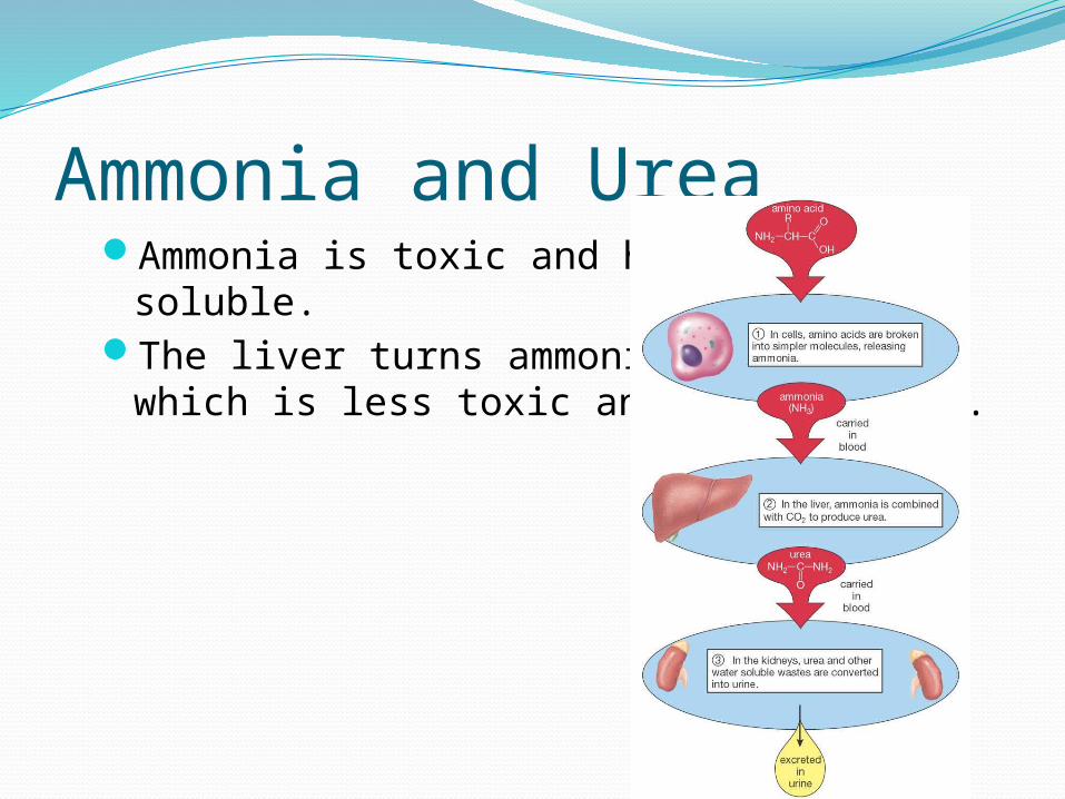
Ammonia and UreaAmmonia is toxic and highly water soluble.The liver turns ammonia into urea, which is
less toxic and less soluble.

Urinary system anatomyMain structures of the urinary system:kidneysuretersbladderurethra
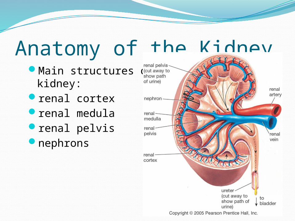
Anatomy of the KidneyMain structures of the mammalian kidney:renal cortexrenal medularenal pelvisnephrons

12
OutlineUriniferous tubule (anatomical unit for
forming urine)Nephron
Renal corpuscle (in cortex) Glomerulus (tuft of capillaries) Glomerular (Bowman’s) capsule
Tubular section Proximal convoluted tubule Loop of Henle Distal convoluted tubule
Collecting duct
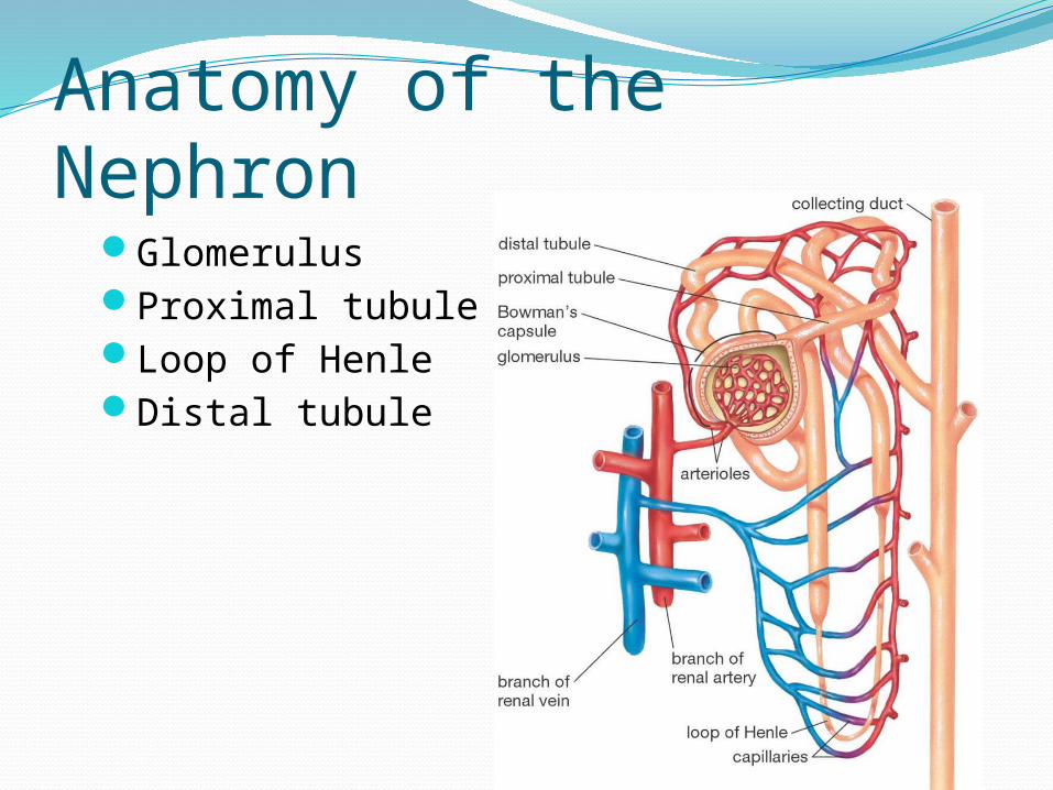
Anatomy of the NephronGlomerulusProximal tubuleLoop of HenleDistal tubule

GlomerulusThis is the only
place in the system where the blood is actually “filtered.”
Blood pressure is used to push plasma through capillary walls and into the Bowman’s capsule.

Proximal tubuleNutrients (salts, vitamins, etc.) are moved
out of the tubule through active transport.Water follows the nutrients by osmosis.

Loop of HenleTissue around
the Loop of Henle is salty, from active transport and diffusion of sodium chloride.
The salty conditions allow water to diffuse out of the loop.

Distal tubuleActive transport
is used to move more nutrients out of the concentrated urine.
Some ions, drugs, and toxins are actively pumped into the tubule.
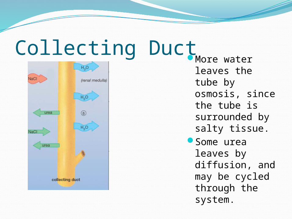
Collecting DuctMore water
leaves the tube by osmosis, since the tube is surrounded by salty tissue.
Some urea leaves by diffusion, and may be cycled through the system.

20
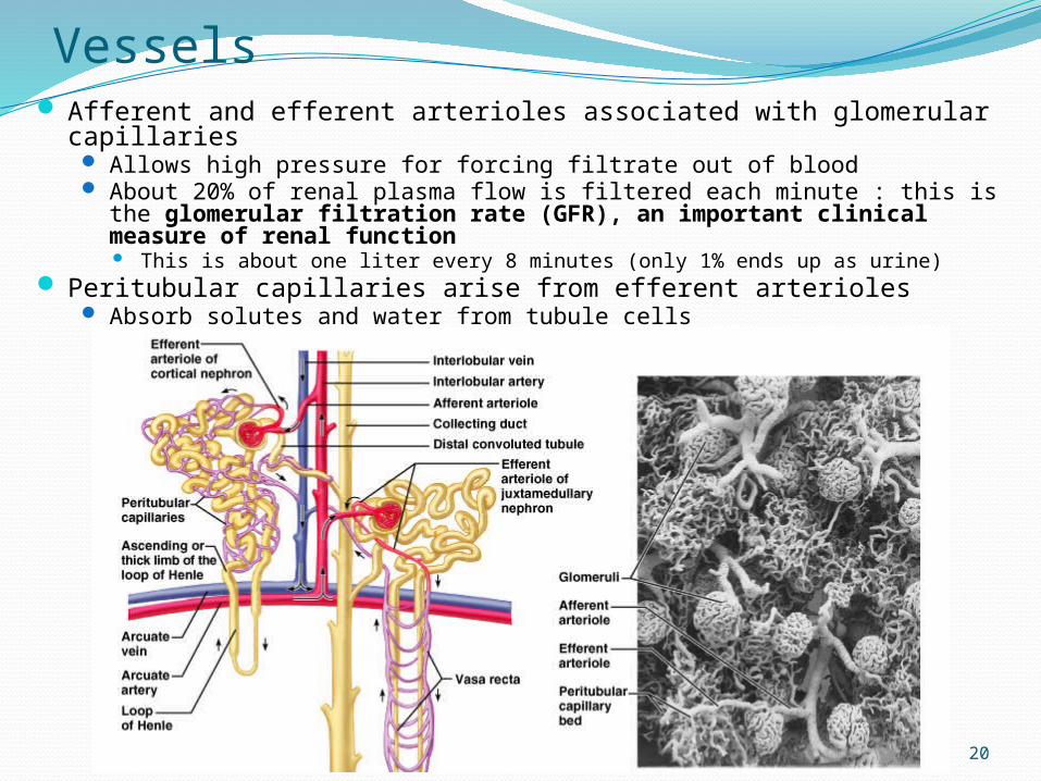
Vessels Afferent and efferent arterioles associated with glomerular capillaries
Allows high pressure for forcing filtrate out of blood About 20% of renal plasma flow is filtered each minute : this is the
glomerular filtration rate (GFR), an important clinical measure of renal function This is about one liter every 8 minutes (only 1% ends up as urine)
Peritubular capillaries arise from efferent arterioles Absorb solutes and water from tubule cells
21
22
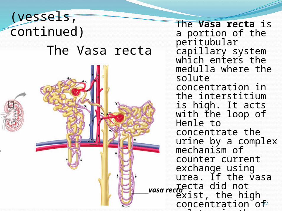
The Vasa recta is a portion of the peritubular capillary system which enters the medulla where the solute concentration in the interstitium is high. It acts with the loop of Henle to concentrate the urine by a complex mechanism of counter current exchange using urea. If the vasa recta did not exist, the high concentration of solutes in the medullary interstitium would be washed out.
____vasa recta
(vessels, continued)
The Vasa recta
23
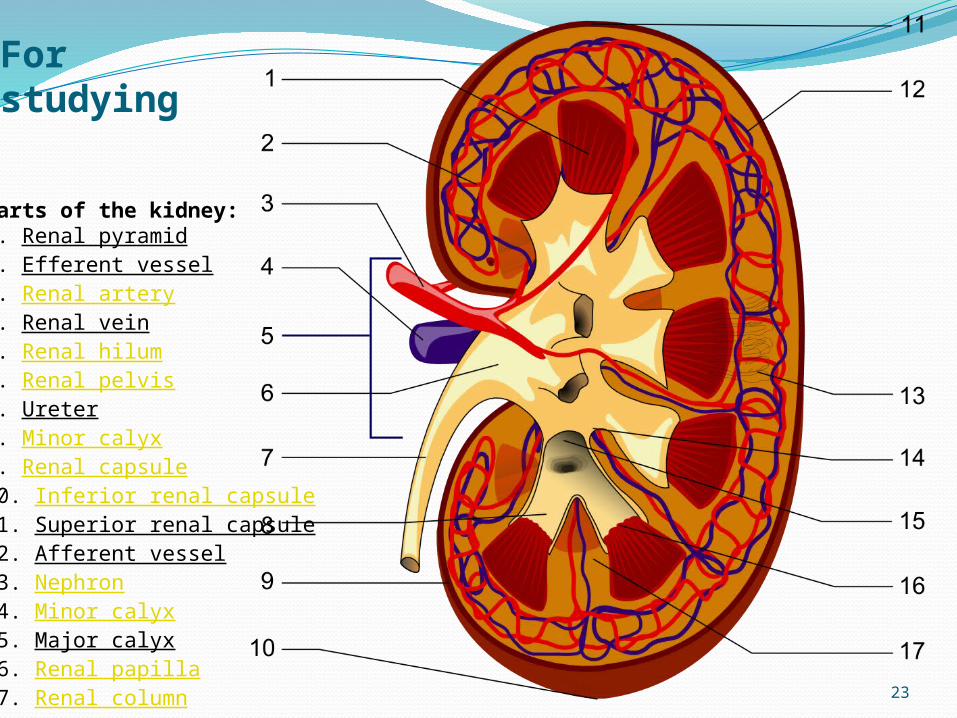
For studying
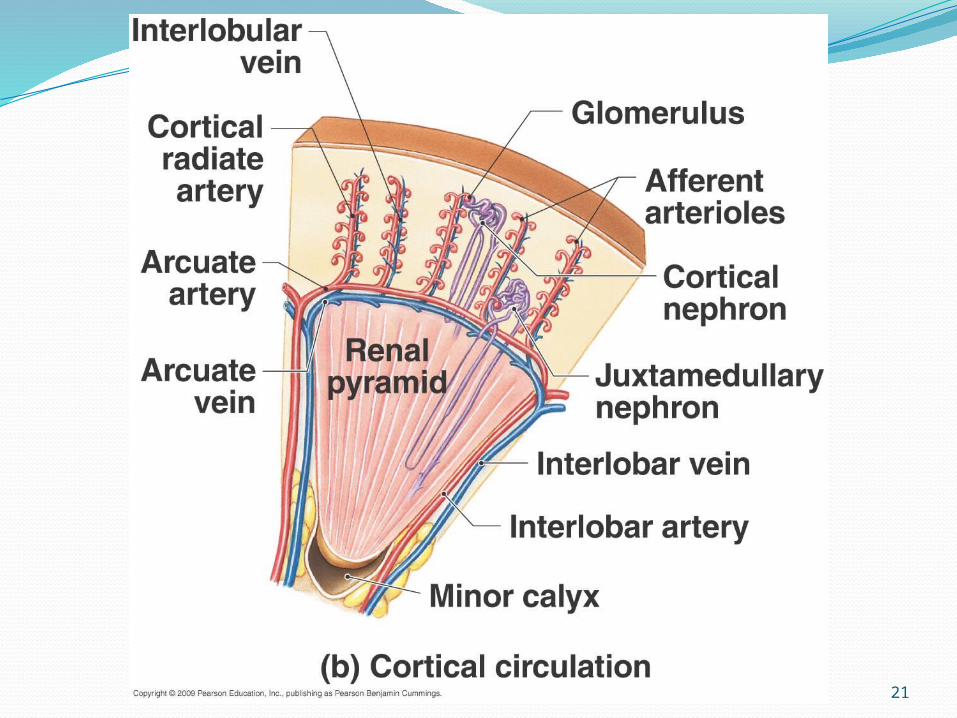
Parts of the kidney:1. Renal pyramid2. Efferent vessel3. Renal artery4. Renal vein5. Renal hilum6. Renal pelvis7. Ureter8. Minor calyx9. Renal capsule10. Inferior renal capsule11. Superior renal capsule12. Afferent vessel13. Nephron14. Minor calyx15. Major calyx16. Renal papilla17. Renal column

24
The UretersSlender tubes leaving
each renal pelvisOne for each kidney
carrying urine to the bladder
Run medially within posterior bladder wall before opening into interior
This oblique entry helps prevent backflow of urine
25
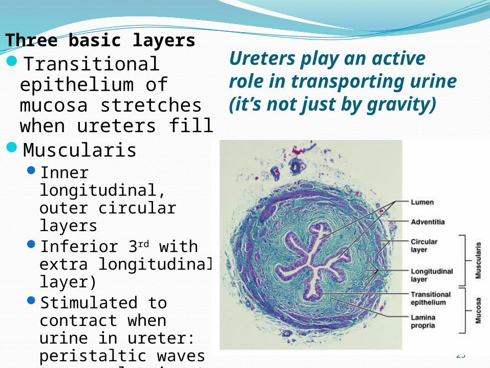
Ureters play an active role in transporting urine (it’s not just by gravity)
Three basic layersTransitional
epithelium of mucosa stretches when ureters fill
Muscularis Inner longitudinal,
outer circular layersInferior 3rd with
extra longitudinal layer)
Stimulated to contract when urine in ureter: peristaltic waves to propel urine to bladder
Adventitia (external)

26
Urinary Bladder Collapsible muscular sac
Stores and expels urine
When full it become ovoid.
In male it is dorsaly related to the rectum, ductusdeference and acessary sex glands
In female it is related with body of uterus and vagina.
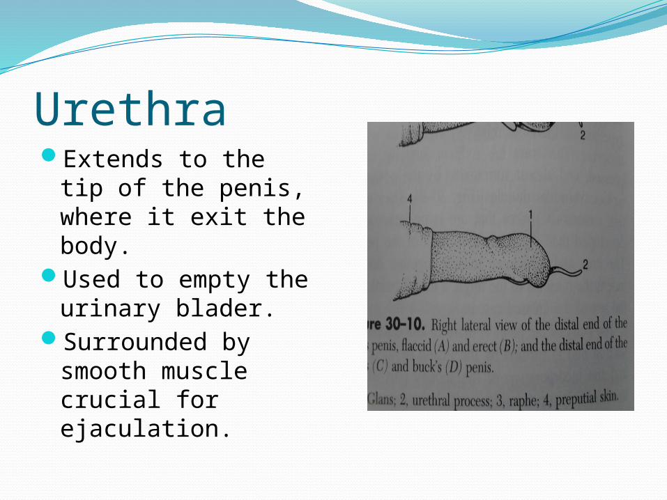
UrethraExtends to the tip of
the penis, where it exit the body.
Used to empty the urinary blader.
Surrounded by smooth muscle crucial for ejaculation.