Embed Size (px)
Citation preview

JOURNAL OF BACTERIOLOGY, Feb. 1976, p. 706-712Copyright © 1976 American Society for Microbiology
Vol. 125, No. 2Printed in U.S.A.
Utilization of Exogenous Thymidine by Chlamydia psittaciGrowing in Thymidine Kinase-Containing and Thymidine
Kinase-Deficient L CellsTHOMAS P. HATCH1
Department of Microbiology, University of Chicago, Chicago, Illinois 60637
Received for publication 29 July 1975
The incorporation of [3H]thymidine into the deoxyribonucleic acid (DNA) ofChlamydia psittaci (strain 6BC) growing in thymidine kinase (adenosine5'-triphosphate-thymidine 5'-phosphotransferase, EC 2.7.1.21)-containing Lcells, L(TK+), and thymidine kinase-deficient L cells, LM(TK-), was examinedby autoradiography. Label was detected over C. psittaci inclusions in L(TK+)but not LM(TK-) cells. No evidence for a chlamydia-specific thymidine kinaseactivity in either L(TK+) or LM(TK-) cells was obtained. Entry of [3H]thymi-dine into the DNA of C. psittaci growing in L(TK+) cells was quantitated bymeasuring label in purified C. psittaci. It was 265 times less efficient than entryinto infected host cell DNA. It is concluded that low levels of exogenous thymi-dine are incorporated into the DNA of C. psittaci and that this incorporation isdependent on a fully competent host thymidine kinase activity. Evidence alsois presented that L cells possess at least two thymidine kinase activities, both ofwhich are capable of supplying thymidylate precursors for nuclear DNA syn-thesis.
Members of the genus Chlamydia are obli-gately intracellular parasitic bacteria. The ob-servation that host-free chlamydiae are unableto generate adenosine 5'-triphosphate (23, 25)supports Moulder's (17) hypothesis that chla-mydiae are energy parasites dependent on theirhost cells for the energy intermediates requiredfor biosynthesis of macromolecules. Subse-quently, Hatch (10), using an equilibrium label-ing technique, demonstrated that Chlamydiapsittaci draws on the ribonucleoside triphos-phate pools of its L-cell host for precursors forribonucleic acid synthesis. It follows from thisobservation that an unusual mechanism mustexist for the transport of phosphorylated prod-ucts of host metabolism across the inclusionmembrane within which chlamydiae multiply,as well as across the cytoplasmic membrane ofthe parasite itself.
Several investigators (4, 8, 14, 19, 22; S. R.Pelc and T. T. Crocker, Biochem. J. 78:20p,1961) have observed that exogenously addedthymidine is either not incorporated or is poorlyincorporated into the deoxyribonucleic acid(DNA) of C. psittaci, although thymidine isreadily incorporated into the DNA of infectedhost cells. It has been concluded that thymidy-lic acid, which makes up about 30% of the base
I Present address: Department of Microbiology. Universityof Texas, Austin, Tex. 78712.
composition of chlamydial DNA (24), is synthe-sized by the parasite from deoxyuridine mono-phosphate by a host-independent pathway (8,14, 22). A corollary of this conclusion is that thethymidylate pools of the host cell are notavailable to the parasite as precursors for DNAsynthesis.
In light of the recent observation that hostribonucleotide pools are utilized by C. psittacifor precursors for ribonucleic acid synthesis(10), this study was conducted to reevaluatethe incorporation of exogenous thymidine intochlamydial DNA. It is concluded that exoge-nous thymidine is incorporated into C. psittaciDNA, although much less efficiently than it isincorporated into L-cell DNA, and that thislow level of incorporation is dependent on afully competent host thymidine kinase (adeno-sine 5'- triphosphate - thymidine - 5'- phospho-transferase, EC 2.7.1.21). Evidence also is pre-sented that L cells possess at least two thymi-dine kinase activities, both of which are capableof supplying thymidylate precursors f'or nuclearDNA synthesis.
MATERIALS AND METHODS
Growth of L cells and C. psittaci. Suspensioncultures of the Sb clone of L cells (mouse fibroblasts)
706
on April 30, 2019 by guest
http://jb.asm.org/
Dow
nloaded from
UTILIZATION OF THYMIDINE BY C. PSITTACI 70
were grown by procedures previously described (21) inEagle minimal essential medium with non-essentialamino acids (Grand Island Biological Co.) containing0.1% sodium bicarbonate (gas phase, 5% carbondioxide-95%. air), 200 Ag of streptomycin sulfate perml, and 10% heat-inactivated fetal calf serum (GrandIsland Biological Co.). Hereafter, the 5b clone of Lcells will be referred to as L(TK+) cells. In someexperiments, LM(TK-) cells, a subline of LM cells,were used. These cells, which are deficient in thymi-dine kinase activity (11), were obtained from Saul Kit(Baylor University College of Medicine, Houston,Tex.) and were grown in the same way as L(TK+)cells, except that 25 ug of bromodeoxyuridine per mlwas included in the growth medium. LM(TK-) cellswere incubated in bromodeoxyuridine-free mediumfor 48 h prior to the initiation of all experimentsreported. All experiments were done with the 6BCstrain of C. psittaci, grown as described elsewhere (9).Cell cultures were infected with 10 times the amountof C. psittaci required to infect 50% of the cells inculture, a multiplicity sufficient to infect more than95% of the host cells.
Autoradiographic experiments. C. psittaci-infected cells at a density of 200,000/ml were incu-bated as monolayers in Leighton tubes as previouslydescribed (9) in the presence of either 20 MCi of[8Hjthymidine per ml (20 uCi/nmol) or 20 MCi of[3H]thymidine per ml (12.5 MACi/Mmol). In some exper-iments, 5 Mg of cycloheximide per ml was added at thetime of infection. At 29 h postinfection, the mediumcontaining radioactive thymidine was decanted, andthe cells were washed five times with 1 volume ofgrowth medium containing 1.6 mM thymidine ai.dwere further incubated in growth medium containing1.6 mM thymidine. At :30 h after infection, the me-dium was decanted, and the monolayers were washedfive times with 5 volumes of' Dulbecco phosphate-buffered saline and f'ixed for 2 h with a 60:30:10(vol/vol/vol) solution of' 957 ethanol, chlorof'orm, andglacial acetic acid, with changes of f'ixative every15 min. The f'ixed monolayers were dried in air andexposed to Kodak AR-10 autoradiographic film foreither 4 or 21 days. In some experiments, f'ixed cellswere treated with 250 Mg of' pancreatic deoxyribo-nuclease per ml (Sigma Chemical Co.) f'or 2 h at37 C before exposure to f'ilm. The photographicemulsion was developed with Kodak D19 developerfor 5 min, f'ixed, and dried, and the cells in mono-layer were treated for 10 min with Giemsa stain(NA 0561, Allied Chemical Corp., Morristown, N.J.)containing 1 drop of concentrated ammonium hy-droxide per 10 ml of stain.
Analysis of thymidine in the growth medium.One volume of iced 20% trichloroacetic acid wasadded to growth medium in which C. psittaci-inf'ectedcells had been incubating in the presence of 20 MCi of[3H]thymidine per ml (12.5 gCi/,Mmol) for 29 h.Particulate material was removed by centrifugationat 10,000 x g for 30 min, and the trichloroacetic acidwas removed from the supernatant by extractingthree times with 1 volume of ethyl ether. Fivemicroliters of the aqueous phase was applied to thinlayers of poly(ethyleneimine)-cellulose (BrinkmannInstruments, Inc.), and thymidine was separated from
thymine by one-dimensional chromatography inwater. Radioactive spots were quantitated as previ-ously described (10).
Assay of thymidine kinase activity. Thymidinekinase activity was assayed by a method similar tothat described by Klemperer et al. (13). Cells (20 x106) were harvested by centrifugation at 1,000 x g for10 min at 4 C, washed two times with 25 ml of'Dulbecco phosphate-buffered saline, and resuspendedin 0.5 ml of lysis buffer consisting of 0.5%S NonidetP-40 (a gift from Shell Chemical Co.) in 20 mM2-(n-morpholino)ethanesulfonic acid (pH 7.0) (Calbi-ochem), 3 mM ethylenediaminetetraacetic acid, and 2mM dithiothreitol. Nuclei were removed by sedimen-tation at 1,000 x g for 10 min, and the supernatantwas assayed for thymidine kinase activity. The stan-dard reaction mixture consisted of cell extract, 5 mMadenosine 5'-triphosphate, 2.5 mM MgCl2, 50 MMthymidine, 2 mM dithiothreitol, 50 mM tris(hydrox-ymethyl)aminomethane-hydrochloride (pH 7.8), 1 mgof bovine serum albumin per ml, and 10 MCi of'[9H]thymidine, in a total volume of 100 Ml. After 20min at 37 or 60 C, the reaction was stopped by heatingto 90 C for 3 min and then chilled to 0 C. Samples (10Ml) of each reaction were applied to 1-cm squares ofWhatman diethylaminoethyl filter paper. The paperwas washed four times with 500 ml of 4 mM tris-(hydroxymethyl)aminomethane-hydrochloride (pH7.8). Radioactive nucleotides adherent to the paperwere eluted with 1 ml of 2 N NaCl and counted byliquid scintillation spectrometry. Protein was assayedby the method of Lowry et al. (16).
Purification of C. psittaci. C. psittaci-infectedL(TK+) cells (108) were incubated in suspensionculture in the presence of 5 MCi of [3H ]thymidine perml (0.5 MCi/nmol) for 48 h. Uninfected L(TK+) cells(7 x 10⁶), which had been incubated in the presence of'5 MCi of ['4C]thymidine per ml (55 mCi/mmol) for 48h, were added to the infected cells immediately beforethe parasite was harvested and purified as previouslydescribed (9). The nuclear fraction of the harvestprocedure was retained to analyze host cell DNA inthe infected culture. Labeled DNA in host nuclei andC. psittaci was precipitated in 10%7 trichloroaceticacid, collected on Whatman GF/C glass-fiber filters,and counted in a Packard Tri-Carb liquid scintillationspectrometer. The total DNA content of nuclei and C.psittaci was determined by the colorimetric assay of'Burton (6).
Isotopes. [methyl-3H]thymidine (11 Ci/mmol) waspurchased from Schwarz/Mann. [methyl- 14C ]thymi-dine (55 mCi/mmol) was purchased from New Eng-land Nuclear Corp.
RESULTS
Autoradiographic analysis of infected Lcells. L cells were infected with C. psittaci,incubated for 29 h in the presence of [3H ]thymi-dine, and prepared for autoradiographic analy-sis, as described in Materials and Methods, todetect the incorporation of exogenously addedthymidine into parasite DNA. When L(TK+)
VOL. 125, 1976
on April 30, 2019 by guest
http://jb.asm.org/
Dow
nloaded from

X08 HATCH
Sp
4,.*,_4
%*I&.
A
Ip1i_s.s f
C
C
B
~v
b
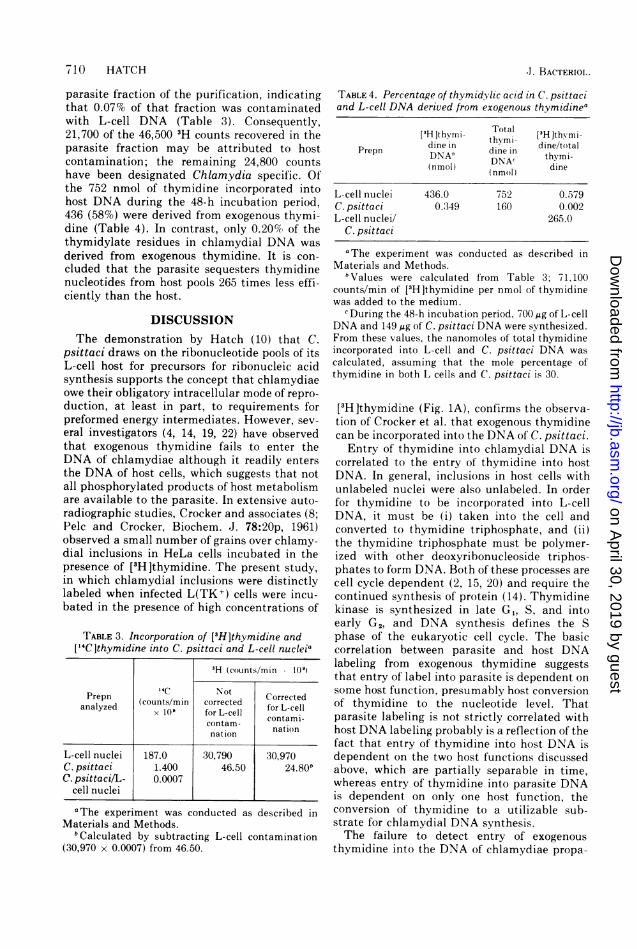
s ~~~~~~~~~~~~~~~~~~~~~~~~~.;FIG. 1. Autoradiographs of L(TKj) and LM(TK-) cells infected with C. psittaci and incubated for 29 h in the
presence of 20 4Ci [3H]thymidine per ml. Cells shown in (A), (C), and (D) were exposed to autoradiographicfilm for4 days; the cell shown in (B) was exposedfor21 days. (A) L(TK+) cell incubated with [3H]thymidine (20,uCi/nmol). (B) L(TK+) cell incubated with [3H]thymidine (12.5,uCi/,umol). Arrow points to labeled nucleus;other labeled bodies are inclusions containing C. psittaci. (C) L(TK ) cells incubated with [3H]thymidine (20gCi/nmol) and 5 ,g of cycloheximide per ml. The field includes: (a) a cell with a labeled nucleus and labeledinclusions; (b) a cell with an unlabeled nucleus and labeled inclusions; and (c) a cell with an unlabeled nucleusand unlabeled or lightly labeled inclusions. (D) LM(TK-) cell incubated with [3H]thymidine (20 ACi/nmol).Bar, 20 ,im.
cells were incubated in the presence of 20 ACi of[3H]thymidine per ml (20 4Ci/nmol), L-cellnuclei were completely blackened by incorpora-tion of the 3H label. Although significantly lesslabel was incorporated into the DNA of C.psittaci, numerous grains over cytoplasmic in-clusions containing chlamydiae were observed(Fig. IA). Detection of incorporation of[3H]thymidine into chlamydial DNA was de-pendent on the use of high levels of isotope;under comparable incubation and autoradio-graphic film exposure periods, 0.5 4Ci of[3H]thymidine per ml (20 ,Ci/nmol) in thegrowth medium labeled host nuclei but not
inclusions. The addition of 1.6 mM unlabeledthymidine to medium containing 20 ,Ci of[3H ]thymidine per ml reduced the incorpora-tion of label into both host and parasite DNA(Fig. 1B). The reduction in labeling of L-cellDNA was disproportionally greater than that ofchlamydial DNA, because the high concentra-tion of thymidine preferentially inhibits L-cellDNA synthesis (14). Grains over both nucleiand inclusions were eliminated by treatment offixed cells with deoxyribonuclease before expo-sure to autoradiographic film.About 21(l of the L cells failed to incorporate
[3H Ithymidine into their nuclei during the 29-h
J. BACTERIOL.
-10D
on April 30, 2019 by guest
http://jb.asm.org/
Dow
nloaded from

UTILIZATION OF THYMIDINE BY C. PSITTACI 09
incubation period. In general, label could not bedetected in the chlamydiae-containing inclu-sions of these cells, although labeled inclusionswere observed occasionally in cells with unla-beled nuclei and vice versa. Cycloheximideinhibits DNA synthesis in eukaryotic L cellswithout having any effect on DNA synthesis inprokaryotic C. psittaci (1). When cycloheximidewas added to the incubation medium, entry of[3H]thymidine into the DNA of L(TK+) cellswas restricted; most cells incorporated no labelin the presence of cycloheximide (Fig. 1C). Aswas the case for cells incubating in the absenceof cycloheximide, the intensity of grains overinclusions usually, but not always, correlatedwith the intensity of grains over the nucleus.
Considerably less exogenous thymidine wasincorporated into the nuclei of LM(TK-) cells,which are deficient in thymidine kinase (11),than was incorporated into the nuclei of L(TK +)cells incubating under the same conditions, andthere was no concentration of grains in thecytoplasm of LM(TK-) cells over chlamydialinclusions (Fig. 1D).The decrease in entry of [3H]thymidine into
the nuclei of LM(TK-) cells and the lack ofdetectable incorporation into chlamydial inclu-sions could be due to the breakdown of thymi-dine to a non-utilizable substrate, such asthymine, rather than to the deficiency of thymi-dine kinase in LM(TK-) cells. However, thepercentage of thymidine remaining in thegrowth medium after incubation of infectedLM(TK-) cells was about the same as thatremaining after incubation of L(TK+) cells(Table 1).Thymidine kinase activity in uninfected
and infected L cells. Thymidine kinase activitywas measured in extracts of uninfected and C.psittaci-infected L(TK+) and LM(TK-) cells inan attempt to detect a Chlamydia-specific thy-midine kinase activity. When assayed at 37 C,the specific activity of thymidine kinase inextracts of uninfected L(TK+) cells was about
TABLE 1. Breakdown of thymidine by L(TK+) andLM(TK-) cells infected with C. psittaci for 29 ha
3H counts(counts/min x 106) % Countsremain-
Host cell In ing inTotal in In h thymi-medium thymine ti- dine
dine
L(TK+) 8.86 0.352 8.51 96.0LM(TK-) 11.5 0.360 11.1 96.5
aThe experiment was conducted as described inMaterials and Methods.
10 times higher than that in extracts ofLM(TK-) cells; infection with C. psittaci didnot induce a significant increase in activity ineither cell line (Table 2). Consequently, aChlamydia-specific thymidine kinase, if presentat all, is present at a level significantly belowthat in both L(TK+) and LM(TK-) cells. Thisconclusion supports Lin's (14) suggestion thatC. psittaci is devoid of thymidine kinase activ-ity.When assayed at 60 C, the specific activity of
thymidine kinase in uninfected and infectedL(TK+) cells was between three and four timeshigher than when assayed at 37 C, whereasactivity was essentially abolished in LM(TK-)cell extracts assayed at the higher temperature(Table 2). These results suggest that L(TK+)cells possess at least two thymidine kinaseenzymes, one of which is heat labile and theother of which is heat stabile and more effec-tively assayed at elevated temperatures.LM(TK-) cells appear to be deficient in themajor, heat-stabile enzyme.Quantitation of the incorporation of exoge-
nous thymidine into C. psittaci and L-cellDNA. The autoradiographic studies reported inFig. 1 revealed that exogenously supplied thy-midine is incorporated into the nuclei of hostcells considerably more efficiently than into theDNA of C. psittaci. The following experimentwas designed to compare the entry of thymidineinto host and parasite DNA. L(TK+) cells wereinfected with C. psittaci and incubated in thepresence of [3H ]thymidine for 48 h. At thistime, uninfected ["4C ]thymidine-labeledL(TK+) cells were added to the infected culture,and C. psittaci was harvested and purified asdescribed in Materials and Methods. Nucleiwere retained from the harvesting procedure toanalyze the DNA content of the host cells. Ofthe 1.87 x 10⁶ 14C counts in the added unin-fected L cells, 1,400 were recovered in the
TABLE 2. Thymidine kinase activity in uninfected andC. psittaci-infected L(TK+) and LM(TK-) cellsa
Host-parasite Sp act ± SD'system 37 C 60 C
L(TK+) uninfected 21.6 ± 0 85.8 i 3.6LM(TK -) uninfected 2.04 i 0.06 0.276 ± 0.18L(TK+) infected 19.8 i 3.0 66.0 ± 3.0LM(TK-) infected 2.34 i 0.12 0.304 ± 0.010
aThe experiment was conducted as described inMaterials and Methods.
"Expressed as picomoles of [9H]thymidine con-verted to nucleotides per hour per micrograms ofprotein. SD, Standard deviation among four samples.
VOL. 125, 1976
on April 30, 2019 by guest
http://jb.asm.org/
Dow
nloaded from
/10 HATCH
parasite fraction of the purification, indicatingthat 0.07% of that fraction was contaminatedwith L-cell DNA (Table 3). Consequently,21,700 of the 46,500 3H counts recovered in theparasite fraction may be attributed to hostcontamination; the remaining 24,800 countshave been designated Chlamydia specif'ic. Ofthe 752 nmol of thymidine incorporated intohost DNA during the 48-h incubation period,436 (58%) were derived from exogenous thymi-dine (Table 4). In contrast, only 0.20% of thethymidylate residues in chlamydial DNA was
derived from exogenous thymidine. It is con-cluded that the parasite sequesters thymidinenucleotides from host pools 265 times less effi-ciently than the host.
DISCUSSIONThe demonstration by Hatch (10) that C.
psittaci draws on the ribonucleotide pools of itsL-cell host for precursors for ribonucleic acidsynthesis supports the concept that chlamydiaeowe their obligatory intracellular mode of repro-duction, at least in part, to requirements forpreformed energy intermediates. However, sev-eral investigators (4, 14, 19, 22) have observedthat exogenous thymidine fails to enter theDNA of chlamydiae although it readily entersthe DNA of host cells, which suggests that notall phosphorylated products of host metabolismare available to the parasite. In extensive auto-radiographic studies, Crocker and associates (8;Pelc and Crocker, Biochem. J. 78:20p, 1961)observed a small number of grains over chlamy-dial inclusions in HeLa cells incubated in thepresence of [3H]thymidine. The present study,in which chlamydial inclusions were distinctlylabeled when infected L(TK+) cells were incu-bated in the presence of high concentrations of
TABLE 3. Incorporation of [3H]thymidine and["4C]thymidine into C. psittaci and L-cell nucleia
3H (counts/min 109O
Prepn 1'C Not CorcePrepn (counts/min corrected CorrLcellx 10' for L-cell fortaL -c
contam - contain
nation nationnatilon
L-cell nuclei 187.0 30,790 30,970C. psittaci 1.400 46.50 24.80bC. psittaci/L- 0.0007
cell nuclei
a The experiment was conducted as described inMaterials and Methods.
bCalculated by subtracting L-cell contamination(30,970 x 0.0007) from 46.50.
TABLE 4. Percentage of thymidylic acid in C. psittaciand L-cell DNA derived from exogenous thymidinea
[3Hlthymni- Total 3H ]thymithymi- [3]t I-dine in dine dine/totalPrepn DNAb DNAc thymi-
(nmol) nl) dine
L-cell nuclei 436.0 752 0.579C. psittaci 0.349 160 0.002L-cell nuclei/ 265.0
C. psittaci
aThe experiment was conducted as described inMaterials and Methods.
bValues were calculated from Table 3; 71,100counts/min of [3H]thymidine per nmol of thymidinewas added to the medium.
c During the 48-h incubation period, 700 mg of L-cellDNA and 149 jig of C. psittaci DNA were synthesized.From these values, the nanomoles of total thymidineincorporated into L-cell and C. psittaci DNA wascalculated, assuming that the mole percentage ofthymidine in both L cells and C. psittaci is 30.
[3H ]thymidine (Fig. 1A), confirms the observa-tion of Crocker et al. that exogenous thymidinecan be incorporated into the DNA of C. psittaci.Entry of thymidine into chlamydial DNA is
correlated to the entry of' thymidine into hostDNA. In general, inclusions in host cells withunlabeled nuclei were also unlabeled. In orderf'or thymidine to be incorporated into L-cellDNA, it must be (i) taken into the cell andconverted to thymidine triphosphate, and (ii)the thymidine triphosphate must be polymer-ized with other deoxyribonucleoside triphos-phates to form DNA. Both of these processes arecell cycle dependent (2, 15, 20) and require thecontinued synthesis of protein (14). Thymidinekinase is synthesized in late G1, S, and intoearly G2, and DNA synthesis def'ines the Sphase of the eukaryotic cell cycle. The basiccorrelation between parasite and host DNAlabeling from exogenous thymidine suggeststhat entry of' label into parasite is dependent onsome host function, presumably host conversionof thymidine to the nucleotide level. Thatparasite labeling is not strictly correlated withhost DNA labeling probably is a reflection of thefact that entry of thymidine into host DNA isdependent on the two host functions discussedabove, which are partially separable in time,whereas entry of thymidine into parasite DNAis dependent on only one host function, theconversion of' thymidine to a utilizable sub-strate for chlamydial DNA synthesis.The failure to detect entry of exogenous
thymidine into the DNA of chlamydiae propa-
J. BACTERIOL.
on April 30, 2019 by guest
http://jb.asm.org/
Dow
nloaded from

UTILIZATION OF THYMIDINE BY C. PSITTACI 711
gated in LM(TK-) cells (Fig. 1D) and thefailure to detect a Chlamydia-specific thymi-dine kinase activity in infected cells (Table 2)confirm the supposition that entry of exoge-nous thymidine into parasite DNA is mediatedby the host's conversion of thymidine to thenucleotide level. An interesting sidelight of thisinvestigation was the discovery that L(TK+)cells possess at least two thymidine kinaseenzymes, one of which is heat stabile and theother of which is heat labile (Table 2). Kit andassociates (12) reported that L cells possess twothymidine kinase activities, one of which islocated in the cytosol and supplies precursors
for nuclear DNA synthesis. Berk and Clayton(3) have concluded that the cytosol enzyme alsocontributes some of the precursors for mito-chondrial DNA synthesis. The other thymidinekinase enzyme is located in the mitochondriaand is believed to supply precursors for mi-tochondrial DNA synthesis only (3, 12).LM(TK-) cells are reported to be deletionmutants which completely lack cytosol kinaseactivity (11, 12). Assuming this to be correct,the heat-stabile enzyme detected in L(TK+)cells, but not in LM(TK-) cells, is of cytosolorigin, and the heat-labile enzyme is of mito-chondrial origin. Since exogenous thymidine isincorporated to some extent into the nuclei ofLM(TK-) cells (Fig. ID), the mitochondrialenzyme may, despite earlier conclusions to thecontrary (3, 12), supply precursors for nuclearDNA synthesis.Entry of exogenous thymidine into host DNA
is approximately 265 times as efficient as entryinto parasite DNA (Table 4), although thecomposition of both C. psittaci and L-cell DNAis about 30% thymidylic acid. This result sup-
ports the suggestion that thymidylic acid inchlamydial DNA is derived primarily from theparasite's host-independent conversion of deox-yuridine monophosphate to thymidine mono-
phosphate (8, 14, 22). Nonetheless, the autora-diographic studies illustrated in Fig. 1 and thedetection of labeled thymidine in purified C.psittaci (Table 4) demonstrate that exogenous
thymidine can be incorporated into chlamydialDNA and that its entry into parasite DNA isdependent on host cytosol thymidine kinaseactivity.The efficient entry of host-synthesized ribo-
nucleotides into chlamydial ribonucleic acid(10) and the inefficient entry of host thymidyl-ate pools into chlamydial DNA suggest thatnot all host metabolic pools are equally availa-ble to the parasite multiplying within an inclu-sion vacuole in the host's cytoplasm. Althoughthe major thymidine kinase enzyme of mamma-
lian cells is located in the cytosol (20), Skoogand Bjursell (18) have demonstrated that thy-midine triphosphate pools of Chinese hamsterovary cells are concentrated primarily in thenucleus. Colby and Edlin (7) reported thatdeoxyribonucleotide pools in eukaryotic cellsare between 10 and 100 times smaller thancorresponding ribonucleotide pools. Concentra-tion of deoxyribonucleotides in the nucleus mayresult in an even greater disparity between theconcentration of deoxyribonucleotides and ribo-nucleotides in the cytoplasm. Hatch (9) hasdemonstrated that C. psittaci fails to multiplyin L cells when the intracellular pool of isoleu-cine is below the concentration required topromote L-cell multiplication, because the hostcell efficiently competes for growth-limitingconcentrations of this essential amino acid. Thefailure of chlamydiae to sequester host thymi-dylate pools efficiently may be due to thelocalization of thymidine triphosphate pools inthe nucleus of the host cell and to successfulhost competition for thymidine triphosphate.
ACKNOWLEDGMENTS
I thank James W. Moulder for his advice throughout thisstudy and N. Patrick Higgins for his assistance with theassays of thymidine kinase.
This investigation was supported by Public Health Serviceresearch grant AI-1594 from the National Institute of Allergyand Infectious Diseases and by a research grant from theLouis Block Fund of the University of Chicago.
LITERATURE CITED
1. Alexander, J. J. 1969. Effect of infection with the menin-gopneumonitis agent on deoxyribonucleic acid andprotein synthesis by its L-cell host. J. Bacteriol.97:653-657.
2. Bello, L. J. 1974. Regulation of thymidine kinase synthe-sis in human cells. Exp. Cell Res. 89:263-274.
3. Berk, A. J., and D. A. Clayton. 1973. A geneticallydistinct thymidine kinase in mammalian mitochon-dria. J. Biol. Chem. 248:2722-2729.
4. Bernkopf, H., P. Mashiah, and Y. Becker. 1962. Correla-tion between morphological and biochemical changesand the appearance of infectivity in FL cell culturesinfected with trachoma agent. Ann. N.Y. Acad. Sci.98:62-81.
5. Brown, R. L., and E. Stubblefield. 1974. Use of mutantcell lines to localize thymidine kinase in mammaliancell-free system. Exp. Cell Res. 87:400-402.
6. Burton, K. 1956. A study of the conditions and mecha-nism of the diphenylamine reaction for the colorimetricestimation of deoxyribonucleic acid. Biochem. .J.62:315-323.
7. Colby, C.. and G. Edlin. 1970. Nucleotide pool levels ingrowing, inhibited, and transformed chick fibroblastcells. Biochemistry 9:917-920.
8. Crocker, T. T., S. R. Pelc, B. I. Nielson. J. M. Eastwood,and J. Banks. 1965. Population dynamics and deoxyri-bonucleic acid synthesis in HeLa cells infected withornithosis agent. J1. Infect. Dis. 115:105-122.
9. Hatch, T. P. 1975. Competition between Chlamydiapsittaci and LJ cells for host isoleucine pools: a limitingfactor in chlamydial multiplication. Infect. Immun.12:211-220.
VOL. 125, 1976
on April 30, 2019 by guest
http://jb.asm.org/
Dow
nloaded from

712 HATCH
10. Hatch, T. P. 1975. Utilization of L-cell nucleoside tri-phosphates by Chlamydia psittaci for ribonucleic acidsynthesis. J. Bacteriol. 122:393-400.
11. Kit, S., D. R. Dubbs, L. J. Piekarski, and T. C. Hsu. 1963.Deletion of thymidine kinase activity from L cellsresistant to bromodeoxyuridine. Exp. Cell Res.31:297-312.
12. Kit, S., W.-C. Leung, and L. A. Kaplan. 1973. Distinctivemolecular forms of thymidine kinase in mitochondriaof normal and bromodeoxyuridine-resistant HeLa cells.Eur. J. Biochem. 39:43-48.
13. Klemperer, H. G., G. R. Haynes, W. I. H. Shedden, andD. H. Watson. 1967. A virus-specific thymidine kinasein BHK21 cells infected with herpes simplex virus.Virology 31:120-128.
14. Lin, H.-S. 1968. Inhibition of thymidine kinase activityand deoxyribonucleic acid synthesis in L cells infectedwith the meningopneumonitis agent. J. Bacteriol.96:2054-2065.
15. Littlefield, J. W. 1966. The periodic synthesis of thymi-dine kinase in mouse fibroblasts. Biochim. Biophys.Acta 114:398-403.
16. Lowry, 0. H., N. J. Rosebrough, A. L. Farr, and R. J4Randall. 1951. Protein measurement with the Folinphenol reagent. J. Biol. Chem. 193:265-275.
17. Moulder, J. W. 1962. The biochemistry of intracellularparasitism, p. 123. The University of Chicago Press,Chicago.
18. Skoog, L., and G. Bjursell. 1974. Nuclear and cytoplasmicpools of deoxyribonucleoside triphosphates in Chinesehamster ovary cells. J. Biol. Chem. 249:6434-6438.
19. Starr, T. J., and N. Sharon. 1963. Autoradiography withthe agents of psittacosis and trachoma. Proc. Soc. Exp.Biol. Med. 113:912-914.
20. Stubblefield, E., and S. Murphree. 1967. Synchronizedmammalian cell cultures. II. Thymidine kinase activityin colcemid synchronized fibroblasts. Exp. Cell Res.48:652-656.
21. Tribby, I. I. E. 1970. Cell wall synthesis by Chlamydiapsittaci growing in L cells. J. Bacteriol. 104:1176-1188.
22. Tribby, I. I. E., and J. W. Moulder. 1966. Availability ofbases and nucleosides as precursors of nucleic acids inL cells and in the agent of meningopneumonitis. J.Bacteriol. 91:2362-2367.
23. Weiss, E. 1965. Adenosine triphosphate and other re-quirements for the utilization of glucose by agents ofthe psittacosis-trachoma group. J. Bacteriol.90:243-253.
24. Weiss, E., S. Schramek, N. N. Wilson, and L. W.Newman. 1970. Deoxyribonucleic acid heterogeneity
-between human and murine strains of Chlamydiatrachomatis. Infect. Immun. 2:24-28.
25. Weiss, E., and N. N. Wilson. 1969. Role of exogenousadenosine triphosphate in catabolic and syntheticactivities of Chlamydia psittaci. J. Bacteriol.97:719-724.
J. BACTERIOL.
on April 30, 2019 by guest
http://jb.asm.org/
Dow
nloaded from