Embed Size (px)
Citation preview
7252019 Varianta 2 Articol Epr
httpslidepdfcomreaderfullvarianta-2-articol-epr 18
CLINICAL RESEARCH
Clinical evaluations of cast gold alloy machinable zirconia andsemiprecious alloy crowns A multicenter study
Ji-Man Park DDS PhDa Yong-Shin Hong DDSb Eun-Jin Park DDS PhDc Seong-Joo Heo DDS PhDd and
Namsik Oh DDS PhDe
Computer-aided design andcomputer-aided manufacture(CADCAM) technology whichuses a computer to design andprecisely mill dental prostheseshas been applied to multipleareas of dentistry including thedesign and fabrication of simpleinlays and crowns complex 1047297xed and removable dentalprostheses implants andcement- and screw-retained
implant-supported restora-tions1-9 In addition this tech-nology results in consistentmaterial properties by address-ing the deformation issues thatresult from the casting andsintering processes and by con-trolling the shapes and thick-nesses of the prosthesis and the space for cement1011
The increased cost of gold has resulted in the need fora replacement material and the use of a precious metalalloy that contains less gold Zirconia which is widely used clinically has been shown to be robust biocom-patible esthetically pleasing and applicable to the CADCAM systems12-17 Zirconia units cannot be connected
with soldering which makes it dif 1047297cult to fabricate long-span 1047297xed dental prostheses
To overcome the limitations of cast gold crowns andthe disadvantag es of zirconia machinable metals havebeen developed18 Titanium has been used as the mainmachinable metal1920 but bonding to a ceramic veneer isdif 1047297cult due to the thick oxide layer21-23 Thus instead of
This work was partially supported by the Medical Devices Comparative Clinical and Performance Evaluation Program of the Korea Medical Devices
Industrial Cooperation Association (KMDICA) and the Basic Science Research Program through the National Research Foundation of Korea (NRF)
funded by the Ministry of Science ICT and Future Planning (NRF-2013R1A1A1076022)aClinical Associate Professor Department of Prosthodontics and Dental Research Institute Seoul National University Gwanak Dental Hospital Seoul KoreabFormer resident Department of Prosthodontics School of Medicine Ewha Womans University Seoul Koreac
Professor Department of Prosthodontics School of Medicine Ewha Womans University Seoul Koread
Professor Department of Prosthodontics and Dental Research Institute Seoul National University Seoul Koreae
Professor Department of Dentistry School of Medicine Inha University Incheon Korea
ABSTRACTStatement of problem Few studies have compared the marginal and internal 1047297ts of crowns
fabricated from machinable palladium-silver-indium (Pd-Ag-In) semiprecious metal alloy
Purpose The purpose of this clinical study was to evaluate and compare the marginal and internal
1047297ts of machined Pd-Ag-In alloy zirconia and cast gold crowns
Material and methods A prospective clinical trial was performed on 35 participants and 52
abutment teeth at 2 centers Individuals requiring prosthetic restorations were treated with gold
alloy or zirconia crowns (2 control groups) or Pd-Ag-In alloy crowns (experimental group) A replica
technique was used to evaluate the marginal and internal 1047297ts The buccolingual and mesiodistal
cross-sections were measured and a noninferiority comparison was conducted
Results The mean marginal gaps were 682 mm for the gold crowns 754 mm for the zirconia
crowns and 769 mm for the Pd-Ag-In alloy crowns In the 5 cross-sections other than the distal
cross-section the 2-sided 95 con1047297dence limits for the differences between the Pd-Ag-In alloycrowns and the 2 control groups were not larger than the 25-mm noninferiority margin The
control groups displayed smaller internal gaps in the line angle and occlusal spaces compared
with the Pd-Ag-In crown group
Conclusion The marginal gaps of machinable Pd-Ag-In alloy crowns did not meet the
noninferiority criterion in the distal margin compared with zirconia and gold alloy crowns
Nonetheless all 3 crowns had clinically applicable precision (J Prosthet Dent 2015----)
THE JOURNAL OF PROSTHETIC DENTISTRY 1
7252019 Varianta 2 Articol Epr
httpslidepdfcomreaderfullvarianta-2-articol-epr 28
titanium alloy alloys of platinum group metals have beenused for 1047297xed prostheses and implant abutments
Although the platinum metal alloys have been used inconventional casting methods their speci1047297c composi-tions differ from those of the conventional alloys if they are manufactured for precision milling24-26 If the weightratio of palladium (Pd) and indium (In) is adjusted thealloy has a gold-like color excellent physical propertiesand adequate bond strength with ceramic The machin-able Pd-silver (Ag)-In alloy has a hardness of between185 and 330 VHN an elongation of 5 a modulus of
elasticity of 80 GPa and a bond strength with ceramic of 38 MPa27 Moreover it displays resistance to corrosionand discoloration that is higher than the criteria forclinical use
For long-term success prostheses made of a variety of materials require appropriate marginal and internaladaptation Inappropriate margins may cause restorationfailure due to the accumulation of plaque and secondary caries and the excess or lack of space for the cement canresult in fracture detachment or incomplete place-ment2829 Many in vitro and in vivo studies have beenconducted on marginal and internal gaps30-44 Although
a consensus has not yet been reached a marginal gap of 100 to 120 mm and an internal gap of 140 to 150 mm arerecommended as the clinical upper limits for completecrowns38-40
A representative method for identifying the in vivomarginal and internal 1047297ts is the replica technique which
was described by Molin et al in 199345 This technique which duplicates the relationship of the inner space of acrown and an abutment on a model with the registrationmaterial has a number of advantages A crown does notneed to be sectioned and the number of measurementsites can be decided without restriction In additionrepeated measurements are possible Consequently thismethod has been frequently used in in vitro and in vivostudies446-54
Most studies of the marginal accuracy of crowns haveexamined CADCAM prostheses or compared cast alloy crowns and CADCAM crowns55-59 Few comparativestudies of the 1047297ts of Pd-Ag-In semiprecious metalcrowns CADCAM zirconia crowns andor cast goldalloy crowns have been conducted Thus in this studyprostheses fabricated with gold alloy zirconia and Pd-
Ag-In alloy were provided for study participants andtheir marginal and internal 1047297ts were compared The
primary null hypothesis was that the marginal 1047297t of thesemiprecious metal alloy crown was statistically non-inferior to that of the zirconia and cast gold crowns Thesecondary null hypothesis was that the internal 1047297t of thePd-Ag-In alloy crown did not differ statistically fromthose of the control groups
MATERIAL AND METHODS
This study was approved by the Institutional Review Board of Inha University and Ewha Womans University Hospitals The study participants were adults ranging from 20 to 81 years of age who voluntarily consented toenroll in this clinical trial Individuals under the age of 20pregnant women or women suspected of being pregnantindividuals with alcohol addiction or mental illness andindividuals with abnormal clinical 1047297ndings that a study supervisor or investigator considered inappropriate forthis study were excluded
Prosthesis materials were gold alloy (DeguDent LTGDeguDent GmbH) zirconia (Lava 3M ESPE) and block-type Pd-Ag-In alloy (Innovium Ceragem Biosys Co) Thegold alloy and zirconia were fabricated as controls andthe Pd-Ag-In alloy was the experimental material Thecrown that was selected among the 3 by the participant
was cemented for the de1047297nitive prosthesisTo determine the necessary number of participants
differences in the marginal gaps of the matched pairs were set as the primary evaluation variable When theactual differences and standard deviations of the differ-ences in the marginal gaps of the Pd-Ag-In alloy crown
compared with the gold alloy and zirconia crowns of the 2control groups were set to 0 mm and 637 mm respectivelythe number of required participants was estimated to be52 from a noninferiority test with a noninferiority marginof 25 mm a 1-sided alpha level of 025 and a powerof 806061 Fifty-two teeth (31 molars 11 premolars2 canines and 8 anterior teeth 20 teeth in the maxilla and32 in the mandible) in 35 participants (16 men and 19
women average age 467 years) who needed metal orceramic complete crown restorations because of toothdamage such as dental caries or tooth fractures wereenrolled
A de1047297nitive cast was produced for the tooth to betreated according to the conventional restoration processthrough tooth preparation and impression making Thepreparation design was for a ceramic restoration withrounded line angles The design of an equally placedgingival 1047297nish line was the chamfer for the most part andthe deep chamfer for the esthetic areas The de1047297nitivestone cast underwent the die process and was thenscanned with a 3-dimensional model scanner (Dental
Wings 7Series Dental Wings Inc) to generate a virtualmodel The gold alloy crown was fabricated by the con-
ventional waxing investment wax elimination and
Clinical Implications The marginal and internal 1047297ttings of machinable
Pd-Ag-In alloy crowns were comparable to those of
conventional cast and zirconia crowns
2 Volume - Issue -
THE JOURNAL OF PROSTHETIC DENTISTRY Park et al
7252019 Varianta 2 Articol Epr
httpslidepdfcomreaderfullvarianta-2-articol-epr 38
casting processes For the fabrication of the zirconia andPd-Ag-In alloy crowns a double scanning method wasused The waxing that was made for the cast gold crown
was scanned 3 dimensionally and the scan was super-imposed on the existing cast and applied as the contourand morphology of the Pd-Ag-In alloy crown6263 Thismethod was used to exclude variables other than themarginal and internal 1047297t For crowns in the esthetic areaa veneering ceramic was applied on the labial surface
with a conventional layering technique The marginaland internal cement space parameters and the minimum
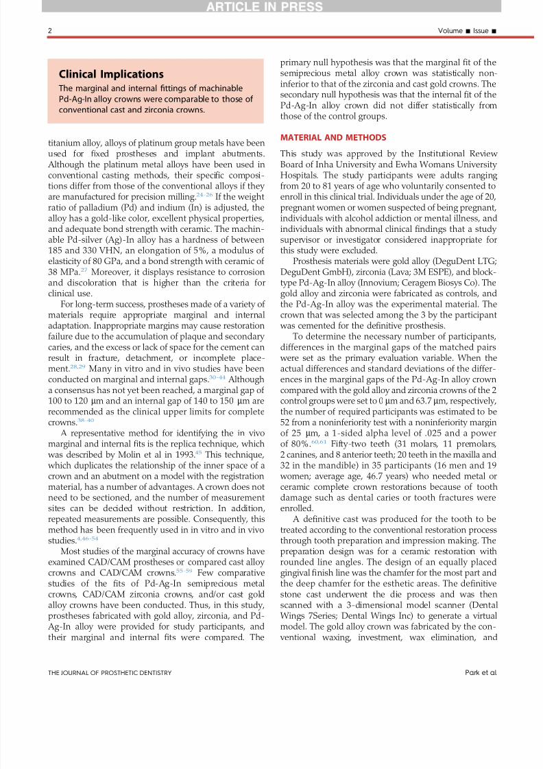
core thickness of both the Lava and Innovium crowns were set in the CAD program at 35 mm 70 mm and 05mm respectively ( Fig 1 ) Based on the completed designthe Innovium alloy and Lava zirconia blocks were pro-cessed and 1047297nished with a milling machine (Dento Mc-5AX Digiworks) and the Lava zirconia blocks wereprocessed with a Lava CNC 500 (3M)
The replica technique was used to examine the mar-ginal and internal 1047297t41 The 3 types of completed crowns
were evaluated separately on the abutment tooth Theproximal contact margin and occlusion were evaluated
After an initial adjustment the intaglio surface of the
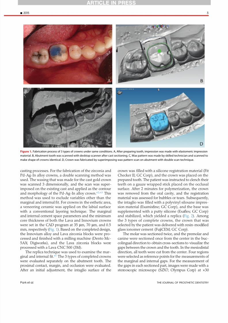
crown was 1047297lled with a silicone registration material (FitChecker II GC Corp) and the crown was placed on theprepared tooth The patient was instructed to clench theirteeth on a gauze wrapped stick placed on the occlusalsurface After 2 minutes for polymerization the crown
was removed from the oral cavity and the registrationmaterial was assessed for bubbles or tears Subsequentlythe intaglio was 1047297lled with a polyvinyl siloxane impres-sion material (Examix 1047297ne GC Corp) and the base wassupplemented with a putty silicone (Exa1047298ex GC Corp)and stabilized which yielded a replica ( Fig 2 ) Among
the 3 types of complete crowns the crown that wasselected by the patient was delivered with resin-modi1047297edglass ionomer cement (FujiCEM GC Corp)
The molar was sectioned twice and the premolar andcanine were sectioned once from the center in the buc-colingual direction to obtain cross-sections to visualize thegaps between the crown and the tooth In the mesiodistaldirection all teeth were cut from the center Four regions
were selected as reference points for the measurements of the marginal and internal gaps For the measurement of the gaps in each sectioned part images were made with astereoscopic microscope (SZX7 Olympus Corp) at times30
Figure 1 Fabrication process of 3 types of crowns under same conditions A After preparing tooth impression was made with elastomeric impression
material B Abutment tooth was scanned with desktop scanner after cast sectioning C Wax pattern was made by skilled technician and scanned to
make shape of crowns identical D Crown was fabricated by superimposing wax pattern scan on abutment with double scan technique
- 2015 3
Park et al THE JOURNAL OF PROSTHETIC DENTISTRY
7252019 Varianta 2 Articol Epr
httpslidepdfcomreaderfullvarianta-2-articol-epr 48
magni1047297cation and images were analyzed with image
analysis software (ImageJ version 145 software US Na-tional Institutes of Health) A laboratory technician whodid not participate in the clinical process performed themeasurement ( Fig 3 ) The dental laboratory processes
were evaluated by comparing the 1047297t on a cast shapereproducibility level of oxide layer formation bubblesduring the opaque application properties and deformationlevel of the surface deformation level after ceramic
veneering and satisfaction with ceramic shades relative toeach material
Measurements from the replica specimen wereanalyzed with statistical software (SAS v92 SAS Insti-
tute Inc) Comparisons of the noninferiority of themargins between the experimental group with crownsthat were prepared by milling the Pd-Ag-In alloy blockand the control groups of the cast and zirconia crowns
were performed as the primary end point If the upperlimit of the 95 con1047297dence interval (CI) of the gap dif-ference between the 2 groups was less than the pre-speci1047297ed noninferiority margin of 25 mm the Pd-Ag-Inalloy group was considered noninferior to the gold orzirconia group at a 1-sided alpha level of 025 The samemethod and criteria were applied for the values of 6sections from different locations The results of the survey of the evaluations of the laboratory procedure wereanalyzed by obtaining the average ranking from therankings for each of the respective items
RESULTS
The average marginal gap measured on the siliconereplica of the cast gold crowns was 682 m m 754 mm forthe zirconia crowns and 769 mm for the Pd-Ag-In alloy crowns The average internal gaps at the margin axial
wall line angle and occlusal space are shown inTable 1 In the 5 sections other than the distal section of the margin the differences in the gaps between the
Pd-Ag-In alloy crowns and the 2 control groups did not
exceed the noninferiority margin of 25 mm However inthe distal section the marginal gap was larger in the Pd-
Ag-In alloy group (845 mm) than those in the gold group(679 mm P =002) and the zirconia group (698 mm P =01) The difference in the marginal gap between theexperimental group and the gold group was 166 mm(95 con1047297dence limits [CL] 63 269 mm) and that be-tween the experimental group and the zirconia group
was 147 mm (95 CL 34 26 mm) Thus the criterion fornoninferiority was not met in the distal sections ( Fig 4 )
At the axial wall the gap difference between the goldalloy and the Pd-Ag-In alloy crowns was 119 mm (95
CL minus
24 262 mm) At the mesiobuccal section the gapdifferences were 26 mm (95 CL 102 417 mm) betweenthe gold and Pd-Ag-In crowns and 188 mm (95 CL19 357 mm) between the zirconia and Pd-Ag-In alloy crowns These differences did not meet the noninferiority criteria However in the remaining 4 sections the upperlimit of the CL did not exceed the noninferiority margin( Fig 5 ) For the line angle and occlusal space the size of the internal gaps of the experimental group did not meetthe noninferiority criteria in comparison with the goldalloy and zirconia crowns in all of the sections ( Figs 6-8 )In the evaluations of the fabrication procedures of thecrowns in the dental laboratory the zirconia crown dis-played the best overall results for all of the itemsincluding the model 1047297t and bubble formation during theopaque application (Table 2)
DISCUSSION
In an in vitro study that compared the 1047297t of differentcrowns fabricated with the CA DCAM system under thesame conditions Gonzalo et al35 reported that in 3-unitpartial 1047297xed dental prostheses made of Lava Procera zir-conia InCeram zirconia and metal ceramic the 1047297rst 3groups showed smaller marginal gaps than metal ceramic
Figure 2 Replica technique procedure to measure internal 1047297t of crowns A Internal gaps of crowns in 3 groups were made with silicone registration
material under same condition B Replica was prepared by stabilizing volume of registration material with silicone material with different color
4 Volume - Issue -
THE JOURNAL OF PROSTHETIC DENTISTRY Park et al
7252019 Varianta 2 Articol Epr
httpslidepdfcomreaderfullvarianta-2-articol-epr 58
In addition Baig et al55 reported that the marginal gaps were 664 mm for the Cercon system and 366 mm for theIPS Empress II system both made with the CADCAMmethod and 371 mm for a cast complete metal crown Inan in vivo study that was conducted on 19 participants
Reich et al50 reported that the marginal gaps of the Lavaand Cerec systems were both approximately 65 mm which
was not signi1047297cantly different from the 54 mm for themetal ceramic crown Wettstein et al51 reported that whenthe Cercon zirconia framework and conventional metalceramic framework were selectively fabricated for 25 par-ticipants the internal 1047297ts of the zirconia framework werecervical 1896 mm axial 1405 mm occlusal cusp tip 1213
Figure 3 Cross-section view of each area to measure marginal and internal gaps in silicone replica after mesiodistal and buccolingual sectioning (times30
magni1047297cation) A Margin B Axial wall C Line angle D Occlusal space
300
T w o - S
i d e d 9 5
C I o f G a p D i ff e r e n c e
a t M a r g i n ( micro m )
200
M
e s i o b u c c a l
M
e s i o l i n g u a l
D i s t o b u c c a l
D
i s t o l i n g u a l
M e s i a l
D i s t a l
100
00
ndash100
ndash200
ndash300
Mp - Gold
Mp - Mz
Location
Figure 4 Two-sided 95 CI of gap differences at margin Blue dotted
line denotes noninferiority margin (25 mm) MP machined group of Pd-
Ag-In alloy MZ machined group of zirconia
M e s i o b u c c a l
M e s i o l i n g u a l
D i s t o b u c c a l
D i s t o l i n g u a l
M e s i a l
D i s t a l
Mp - Gold
Mp - Mz
450
350
250
150
50
ndash50
ndash150
ndash250
ndash350
T w o - S
i d e d 9 5
C I o f G a p D i ff e r e n c e
a t A x i a l W a l l ( micro m )
Location
Figure 5 Two-sided 95 CI of gap differences at axial wall
- 2015 5
Park et al THE JOURNAL OF PROSTHETIC DENTISTRY
7252019 Varianta 2 Articol Epr
httpslidepdfcomreaderfullvarianta-2-articol-epr 68
mm and centro-occlusal 1920 mm these values weresigni1047297cantly greater than those of the metal ceramicframework which were 1186 mm 957 mm 1225 mm and1531 mm Thus in most studies the crowns fabricated
with the CADCAM method were comparable with metalceramic or complete-metal crowns that were made by casting and had marginal and internal 1047297ts that were withinthe clinically acceptable range21384452 In addition Tanet al59 compared the vertical margin openings of titaniumcrowns that were designed by CAD software milled tita-nium crowns that were waxed followed by double scan-ning with a 3-dimensional scanner and cast high noblecrowns The marginal discrepancies were 79 mm 73 mmand 24 mm respectively In addition the CADCAM
method with and without double scanning did not show signi1047297cantly different marginal gaps
In this in vivo study the 1047297rst null hypothesis wasrejected because the crown that was fabricated by milling the Pd-Ag-In alloy block showed a marginal 1047297ton the distal cross-section of the crown that was infe-rior to that of the cast crowns and zirconia crownsHowever the marginal gap of the Pd-Ag-In alloy crown
was not statistically inferior to those of the controlgroups on all of the cross-sections except for the distalsection The average marginal gaps of the cast crowns(682 mm) zirconia crowns (754 mm) and Pd-Ag-In
alloy crowns (769 mm) were generally greatercompared with the results of previous in vivo studies55-58
In contrast other in vivo studies have reported largermarginal gaps than those found in our study5051 Inaddition the marginal gap (845 mm) of the distal section
was statistically inferior to the 120 mm that was pro-posed as the clinically acceptable gap by Hung et al38
indicating that the Pd-Ag-In alloy crown had a clinically applicable 1047297t The differences in the internal gaps at theaxial wall between the Pd-Ag-In group and the controlgroups exceeded the noninferiority margin at the mesialsection (1008 mm) and mesiobuccal section (1031 mm)
However similar to the marginal gap all of the internalgaps obtained in this study showed values less than 140 to150 mm
For the line angle and occlusal space the second nullhypothesis was rejected because the Pd-Ag-In alloy crown showed larger internal gaps in all of the crosssections compared with the control groups Both the Pd-
Ag-In alloy and zirconia crowns had signi1047297cantly greaterinternal gaps in the occlusal space compared with thecast crowns This was likely a result of the characteristicsof the CADCAM fabrication method such as the limitedsize of the milling tool and tool compensation64 Inaddition some crowns have their internal spacer pa-rameters set to be bigger in the CAD system to avoid
early contact on the sharp edge of a preparation and toreduce the marginal gap Therefore the gap mightbecome larger in the CAD process compared with a castrestoration5665
The larger gap in the line angle and the occlusal spaceof the Pd-Ag-In alloy crown compared with the zirconiacrown from the same CADCAM method was thought toresult from differences in the gaps that might haveoccurred as a result of the parameters set during milling because of the difference between zirconia which has
volume reduction during the sintering process and thePd-Ag-In alloy which has no volume change For the
general procedures for crowns in the dental laboratory evaluations the zirconia crowns appeared to be theeasiest to fabricate However this was thought to bebecause of skill differences and the fabrication andplacement of the gold or Pd-Ag-In alloy crowns was notexceptionally dif 1047297cult
The methods that are used to evaluate the 1047297t of acrown include direct visualization under a microscope
visualization by the attachment of the crown to a die thatis followed by sectioning evaluation after impressionmaking and evaluation with an explorer and the nakedeye6667 This in vivo study used the replica technique
M e s i o b u c c a l
M e s i o l i n g u a l
D i s t o b u c c a l
D i s t o l i n g u a l
M e s i a l
D i s t a l
Mp - Gold
Mp - Mz
750
650
550
450
350
250
150
50
ndash50
ndash150
T w o - S
i d e d 9 5
C I o f G a p D i ff e r e n c e
a t
L i n e A n g l e ( micro m )
Location
Figure 6 Two-sided 95 CI of gap differences at line angles
M e s i o b u c c a l
M e s i o l i n g u a l
D i s t o b u c c a l
D i s t o l i n g u a l
M e s i a l
D i s t a l
Mp - Gold
Mp - Mz
ndash250
ndash50
150
350
550
750
950
T w o - S
i d e d 9 5
C I o f G a p D i ff e r e n c e
a t O c c l u s a l S p a c e ( micro m )
Location
Figure 7 Two-sided 95 CI of gap differences at occlusal spaces
6 Volume - Issue -
THE JOURNAL OF PROSTHETIC DENTISTRY Park et al
7252019 Varianta 2 Articol Epr
httpslidepdfcomreaderfullvarianta-2-articol-epr 78
which duplicated the state of the abutment becausedirectly cutting the patientrsquos tooth was not possible45 Nosigni1047297cant differences have been reported between the diesectioning method and replica technique and these results
were not affected by the type of silicone registration ma-terial5354 Although the replica was prepared using themaximum clenching force of a patient the replica may nothave re1047298ected the precise positions possibly increasing the standard deviation of the occlusal space
A deep chamfer 1047297nish line was prepared for the labialor buccal side of the incisors canines and premolars
while a conventional chamfer margin was prepared forthe remaining margin Syu et al68 suggested that the typeof 1047297nish line does not affect the marginal gap In addi-tion the preparation design may not result in signi1047297cant
differences between the 1047297
t of the cast crowns and CADCAM crowns6970
The analysis in this clinical study indicated that themarginal and internal 1047297ts of the crowns fabricated by milling machining Pd-Ag-In semiprecious metal alloys
were comparable with those of the conventionally usedcast crowns and zirconia crowns Although the Pd-Ag-Incrowns did not meet the noninferiority criterioncompared with the existing materials for the line angleand occlusal space their values were within the clinically acceptable range Furthermore most areas of the mar-ginal and axial locations met the noninferiority criterionHowever because this study had a small sample size anda short period for crown placement long-term clinicalstudies that involve a larger sample are required Inaddition studies on possible events in the oral cavitysuch as antagonistic tooth wear or fracture chipping of the ceramic veneer and methods of fabricating moreprecise crowns through improvements of the CADCAMtechnique are necessary
CONCLUSIONS
Within the limitations of this in vivo study the following conclusions may be drawn
1 The average plusmnstandard deviation marginal gaps were 682 plusmn248 mm for the cast gold crowns 754plusmn288 mm for the zirconia crowns and 769 plusmn270mm for the Pd-Ag-In alloy crowns The Pd-Ag-Inalloy crowns met the noninferiority criterion in allthe areas except for the distal cross-sectioncompared with the control groups
2 For the line angle and occlusal space the Pd-Ag-Inalloy crowns were inferior to the control groups forthe internal gaps
REFERENCES
1 Fasbinder DJ Clinical performance of chairside CADCAM restorations J AmDent Assoc 200613722S-31S
2 Mormann WH Brandestini M Lutz F Barbakow F Chairside computer-aided direct ceramic inlays Quintessence Int 198920329-39
3 Bindl A Mormann WH Fit of all-ceramic posterior 1047297xed partial dentureframeworks in vitro Int J Periodontics Restorative Dent 200727567-75
4 Tinschert J Natt G Mautsch W Spiekermann H Anusavice KJ Marginal 1047297tof alumina-and zirconia-based 1047297xed partial dentures produced by a CADCAM system Oper Dent 200126367-74
5 Williams RJ Bibb R Eggbeer D CADCAM in the fabrication of removablepartial denture frameworks a virtual method of surveying 3D scanned dentalcasts Quint J Dent Technol 20042268-76
6 Sun Y Lu P Wang Y Study on CADampRP for removable complete dentureComput Methods Programs Biomed 200993266-72
7 Williams RJ Bibb R Eggbeer D Collis J Use of CADCAM technology tofabricate a removable partial denture framework J Prosthet Dent 20069696-9
Figure 8 Representative silicone replicas of internal gaps of crowns
evaluated A Gold alloy group B Zirconia group C Pd-Ag-In alloy group
(times30 magni1047297cation)
- 2015 7
Park et al THE JOURNAL OF PROSTHETIC DENTISTRY
7252019 Varianta 2 Articol Epr
httpslidepdfcomreaderfullvarianta-2-articol-epr 88
8 Andersson M Razzoog ME Oden A Hegenbarth EA Lang BR Procera anew way to achieve an all-ceramic crown Quintessence Int 199829285-96
9 Kapos T Evans C CADCAM technology for implant abutments crownsand superstructures Int J Oral Maxillofac Implants 201429 Suppl117-36
10 Luthardt RG Sandkuhl O Herold V Walter MH Accuracy of mechanicaldigitizing with a CADCAM system for 1047297xed restorations Int J Prosthodont200114146-51
11 McLaren EA Terry DA CADCAM systems materials and clinical guidelinesfor all-ceramic crowns and 1047297xed partial dentures Compend Contin EducDent 200223637-41 44 46 passim quiz 54
12 Piconi C Maccauro G Zirconia as a ceramic biomaterial Biomaterials1999201-25
13 Tinschert J Zwez D Marx R Anusavice KJ Structural reliability of alumina-feldspar- leucite- mica- and zirconia-based ceramics J Dent 200028529-35
14 Ardlin BI Transformation-toughened zirconia for dental inlays crowns andbridges chemical stability and effect of low-temperature aging on 1047298exuralstrength and surface structure Dent Mater 200218590-5
15 Guazzato M Albakry M Ringer SP Swain MV Strength fracture toughnessand microstructure of a selection of all-ceramic materials Part II Zirconia-based dental ceramics Dent Mater 200420449-56
16 Raigrodski AJ Contemporary materials and technologies for all-ceramic 1047297xedpartial dentures a review of the literature J Prosthet Dent 200492557-62
17 Tinschert J Natt G Hassenp1047298ug S Spiekermann H Status of current CADCAM technology in dental medicine Int J Comput Dent 2004725-45
18 Lee DH Lee BJ Kim SH Lee KB Shear bond strength of porcelain to a new millable alloy and a conventional castable alloy J Prosthet Dent 2015113329-35
19 Abduo Fit of CADCAM implant frameworks a comprehensive review
J Oral Implantol 2014 Dec40(6)758-6620 Kapos T Ashy LM Gallucci GO Weber HP Wismeijer D Computer-aided
design and computer-assisted manufacturing in prosthetic implant dentistryInt J Oral Maxillofac Implants 200924110-7
21 Boening KW Walter MH Reppel PD Non-cast titanium restorations in 1047297xedprosthodontics J Oral Rehabil 199219281-7
22 Gilbert JL Covey DA Lautenschlager EP Bond characteristics of porcelainfused to milled titanium Dent Mater 199410134-40
23 Walter M Boning K Reppel PD Clinical performance of machined titaniumrestorations J Dent 199422346-8
24 Goodacre CJ Palladium-silver alloys a review of the literature J ProsthetDent 19896234-7
25 Huget EF Civjan S Status report on palladium-silver-based crown andbridge alloys J Am Dent Assoc 197489383-5
26 Kansu G Aydin AK Evaluation of the biocompatibility of various dentalalloys Part 2e Allergenical potentials Eur J Prosthodont Restor Dent 19964155-61
27 Hong JT Shin SY A comparative study on the bond strength of porcelain to
the millingable Pd-Ag alloy J Adv Prosthodont 20146372-828 Sorensen JA A rationale for comparison of plaque-retaining properties of crown systems J Prosthet Dent 198962264-9
29 Sorensen SE Larsen IB Jorgensen KD Gingival and alveolar bone reactionto marginal 1047297t of subgingival crown margins Scand J Dent Res 198694109-14
30 Assif D Rimer Y Aviv I The 1047298ow of zinc phosphate cement under a full-coverage restoration and its effect on marginal adaptation according to thelocation of cement application Quintessence Int 198718765-74
31 Bader JD Rozier RG McFall WT Jr Ramsey DL Effect of crown margins onperiodontal conditions in regularly attending patients J Prosthet Dent19916575-9
32 Belser UC MacEntee MI Richter WA Fit of three porcelain-fused-to-metalmarginal designs in vivo a scanning electron microscope study J ProsthetDent 19855324-9
33 Felton DA Kanoy BE Bayne SC Wirthman GP Effect of in vivo crownmargin discrepancies on periodontal health J Prosthet Dent 199165357-64
34 Fusayama T Ide K Hosoda H Relief of resistance of cement of full castcrowns J Prosthet Dent 19641495-106
35 Gonzalo E Suarez MJ Serrano B Lozano JF Marginal 1047297t of Zirconia pos-terior 1047297xed partial dentures Int J Prosthodont 200821398-9
36 Grasso JE Nalbandian J Sanford C Bailit H Effect of restoration quality onperiodontal health J Prosthet Dent 19855314-9
37 Grey NJ Piddock V Wilson MA In vitro comparison of conventional crownsand a new all-ceramic system J Dent 19932147-51
38 Hung SH Hung KS Eick JD Chappell RP Marginal 1047297t of porcelain-fused-to-metal and two types of ceramic crown J Prosthet Dent 19906326-31
39 McLean JW von Fraunhofer JA The estimation of cement 1047297lm thickness by an in vivo technique Br Dent J 1971131107-11
40 Palomo F Peden J Periodontal considerations of restorative procedures J Prosthet Dent 197636387-94
41 Passon C Lambert RH Lambert RL Newman S The effect of multiple layersof die-spacer on crown retention Oper Dent 19921742-9
42 Schwartz NL Whitsett LD Berry TG Stewart JL Unserviceable crowns and1047297xed partial dentures life-span and causes for loss of serviceability J AmDent Assoc 1970811395-401
43 Walton JN Gardner FM Agar JR A survey of crown and 1047297xed partial denturefailures length of service and reasons for replacement J Prosthet Dent198656416-21
44 Christensen GJ Marginal 1047297t of gold inlay castings J Prosthet Dent 196616297-305
45 Molin M Karlsson S The 1047297t of gold inlays and three ceramic inlay systems A clinical and in vitro study Acta Odontol Scand 199351201-6
46 Boening KW Wolf BH Schmidt AE Kastner K Walter MH Clinical 1047297t of Procera AllCeram crowns J Prosthet Dent 200084419-24
47 Coli P Karlsson S Fit of a new pressure-sintered zirconium dioxide coping
Int J Prosthodont 20041759-6448 Kohorst P Brinkmann H Dittmer MP Borchers L Stiesch M In1047298uence of
the veneering process on the marginal 1047297t of zirconia 1047297xed dental prostheses J Oral Rehabil 201037283-91
49 Reich S Kappe K Teschner H Schmitt J Clinical 1047297t of four-unit zirconiaposterior 1047297xed dental prostheses Eur J Oral Sci 2008116579-84
50 Reich S Wichmann M Nkenke E Proeschel P Clinical 1047297t of all-ceramicthree-unit 1047297xed partial dentures generated with three different CADCAMsystems Eur J Oral Sci 2005113174-9
51 Wettstein F Sailer I Roos M Hammerle CH Clinical study of the internalgaps of zirconia and metal frameworks for 1047297xed partial dentures Eur J OralSci 2008116272-9
52 Fransson B Oilo G Gjeitanger R The 1047297t of metal-ceramic crowns a clinicalstudy Dent Mater 19851197-9
53 Rahme HY Tehini GE Adib SM Ardo AS Rifai KT In vitro evaluation of theldquoreplica techniquerdquo in the measurement of the 1047297t of Procera crowns
J Contemp Dent Pract 2008925-3254 Laurent M Scheer P Dejou J Laborde G Clinical evaluation of the marginal
1047297t of cast crownse validation of the silicone replica method J Oral Rehabil200835116-22
55 Baig MR Tan KB Nicholls JI Evaluation of the marginal 1047297t of a zirconiaceramic computer-aided machined (CAM) crown system J Prosthet Dent2010104216-27
56 Bindl A Mormann WH Marginal and internal 1047297t of all-ceramic CADCAMcrown-copings on chamfer preparations J Oral Rehabil 200532441-7
57 Hertlein GHS Frank S Suttor D Marginal 1047297t of CADCAM manufactured allceramic prosthesis J Dent Res 20018042-4
58 May KB Russell MM Razzoog ME Lang BR Precision of 1047297t the Procera AllCeram c rown J Prosthet Dent 199880394-404
59 Tan PL Gratton DG Diaz-Arnold AM Holmes DC An in vitro comparisonof vertical marginal gaps of CADCAM titanium and conventional cast res-torations J Prosthodont 200817378-83
60 Kokubo Y Tsumita M Kano T Sakurai S Fukushima S Clinical marginal andinternal gaps of zirconia all-ceramic crowns J Prosthodont Res 20115540-3
61 Chow SC Shao J Wang H Sample size calculations in clinical research 2nded Boca Raton FL Chapman and HallCRC 2007
62 Beuer F Stimmelmayr M Gueth JF Edelhoff D Naumann M In vitro per-formance of full-contour zirconia single crowns Dent Mater 201228449-5663 Kollar A Huber S Mericske E Mericske-Stern R Zirconia for teeth and
implants a case series Int J Periodontics Restorative Dent 200828479-8764 Sturdevant JR Bayne SC Heymann HO Margin gap size of ceramic inlays
using second-generation CADCAM equipment J Esthet Dent 199911206-14
65 Bornemann G Lemelson S Luthardt R Innovative method for the analysisof the internal 3D 1047297tting accuracy of Cerec-3 crowns Int J Comput Dent20025177-82
66 Sorensen JA A standardized method for determination of crown margin 1047297-delity J Prosthet Dent 19906418-24
67 Holmes JR Bayne SC Holland GA Sulik WD Considerations in measure-ment of marginal 1047297t J Prosthet Dent 198962405-8
68 Syu JZ Byrne G Laub LW Land MF In1047298uence of 1047297nish-line geometry onthe 1047297t of crowns Int J Prosthodont 1993625-30
69 Tsitrou EA Northeast SE van Noort R Evaluation of the marginal 1047297t of threemargin designs of resin composite crowns using CADCAM J Dent 20073568-73
70 Ayad MF Effect of the crown preparation margin and die type on the mar-ginal accuracy of 1047297ber-reinforced composite crowns J Contemp Dent Pract200899-16
Corresponding author
Dr Namsik OhInha University 7-2063 Shinhung-dong Jung-guIncheon 400-711REPUBLIC OF KOREA Email onsdoinhaackr
AcknowledgmentsThe authors thank Dr Kyoung-Ae Kong for performing statistical analyses in thisstudy
Copyright copy 2015 by the Editorial Council for The Journal of Prosthetic Dentistry
8 Volume - Issue -
THE JOURNAL OF PROSTHETIC DENTISTRY Park et al
7252019 Varianta 2 Articol Epr
httpslidepdfcomreaderfullvarianta-2-articol-epr 28
titanium alloy alloys of platinum group metals have beenused for 1047297xed prostheses and implant abutments
Although the platinum metal alloys have been used inconventional casting methods their speci1047297c composi-tions differ from those of the conventional alloys if they are manufactured for precision milling24-26 If the weightratio of palladium (Pd) and indium (In) is adjusted thealloy has a gold-like color excellent physical propertiesand adequate bond strength with ceramic The machin-able Pd-silver (Ag)-In alloy has a hardness of between185 and 330 VHN an elongation of 5 a modulus of
elasticity of 80 GPa and a bond strength with ceramic of 38 MPa27 Moreover it displays resistance to corrosionand discoloration that is higher than the criteria forclinical use
For long-term success prostheses made of a variety of materials require appropriate marginal and internaladaptation Inappropriate margins may cause restorationfailure due to the accumulation of plaque and secondary caries and the excess or lack of space for the cement canresult in fracture detachment or incomplete place-ment2829 Many in vitro and in vivo studies have beenconducted on marginal and internal gaps30-44 Although
a consensus has not yet been reached a marginal gap of 100 to 120 mm and an internal gap of 140 to 150 mm arerecommended as the clinical upper limits for completecrowns38-40
A representative method for identifying the in vivomarginal and internal 1047297ts is the replica technique which
was described by Molin et al in 199345 This technique which duplicates the relationship of the inner space of acrown and an abutment on a model with the registrationmaterial has a number of advantages A crown does notneed to be sectioned and the number of measurementsites can be decided without restriction In additionrepeated measurements are possible Consequently thismethod has been frequently used in in vitro and in vivostudies446-54
Most studies of the marginal accuracy of crowns haveexamined CADCAM prostheses or compared cast alloy crowns and CADCAM crowns55-59 Few comparativestudies of the 1047297ts of Pd-Ag-In semiprecious metalcrowns CADCAM zirconia crowns andor cast goldalloy crowns have been conducted Thus in this studyprostheses fabricated with gold alloy zirconia and Pd-
Ag-In alloy were provided for study participants andtheir marginal and internal 1047297ts were compared The
primary null hypothesis was that the marginal 1047297t of thesemiprecious metal alloy crown was statistically non-inferior to that of the zirconia and cast gold crowns Thesecondary null hypothesis was that the internal 1047297t of thePd-Ag-In alloy crown did not differ statistically fromthose of the control groups
MATERIAL AND METHODS
This study was approved by the Institutional Review Board of Inha University and Ewha Womans University Hospitals The study participants were adults ranging from 20 to 81 years of age who voluntarily consented toenroll in this clinical trial Individuals under the age of 20pregnant women or women suspected of being pregnantindividuals with alcohol addiction or mental illness andindividuals with abnormal clinical 1047297ndings that a study supervisor or investigator considered inappropriate forthis study were excluded
Prosthesis materials were gold alloy (DeguDent LTGDeguDent GmbH) zirconia (Lava 3M ESPE) and block-type Pd-Ag-In alloy (Innovium Ceragem Biosys Co) Thegold alloy and zirconia were fabricated as controls andthe Pd-Ag-In alloy was the experimental material Thecrown that was selected among the 3 by the participant
was cemented for the de1047297nitive prosthesisTo determine the necessary number of participants
differences in the marginal gaps of the matched pairs were set as the primary evaluation variable When theactual differences and standard deviations of the differ-ences in the marginal gaps of the Pd-Ag-In alloy crown
compared with the gold alloy and zirconia crowns of the 2control groups were set to 0 mm and 637 mm respectivelythe number of required participants was estimated to be52 from a noninferiority test with a noninferiority marginof 25 mm a 1-sided alpha level of 025 and a powerof 806061 Fifty-two teeth (31 molars 11 premolars2 canines and 8 anterior teeth 20 teeth in the maxilla and32 in the mandible) in 35 participants (16 men and 19
women average age 467 years) who needed metal orceramic complete crown restorations because of toothdamage such as dental caries or tooth fractures wereenrolled
A de1047297nitive cast was produced for the tooth to betreated according to the conventional restoration processthrough tooth preparation and impression making Thepreparation design was for a ceramic restoration withrounded line angles The design of an equally placedgingival 1047297nish line was the chamfer for the most part andthe deep chamfer for the esthetic areas The de1047297nitivestone cast underwent the die process and was thenscanned with a 3-dimensional model scanner (Dental
Wings 7Series Dental Wings Inc) to generate a virtualmodel The gold alloy crown was fabricated by the con-
ventional waxing investment wax elimination and
Clinical Implications The marginal and internal 1047297ttings of machinable
Pd-Ag-In alloy crowns were comparable to those of
conventional cast and zirconia crowns
2 Volume - Issue -
THE JOURNAL OF PROSTHETIC DENTISTRY Park et al
7252019 Varianta 2 Articol Epr
httpslidepdfcomreaderfullvarianta-2-articol-epr 38
casting processes For the fabrication of the zirconia andPd-Ag-In alloy crowns a double scanning method wasused The waxing that was made for the cast gold crown
was scanned 3 dimensionally and the scan was super-imposed on the existing cast and applied as the contourand morphology of the Pd-Ag-In alloy crown6263 Thismethod was used to exclude variables other than themarginal and internal 1047297t For crowns in the esthetic areaa veneering ceramic was applied on the labial surface
with a conventional layering technique The marginaland internal cement space parameters and the minimum
core thickness of both the Lava and Innovium crowns were set in the CAD program at 35 mm 70 mm and 05mm respectively ( Fig 1 ) Based on the completed designthe Innovium alloy and Lava zirconia blocks were pro-cessed and 1047297nished with a milling machine (Dento Mc-5AX Digiworks) and the Lava zirconia blocks wereprocessed with a Lava CNC 500 (3M)
The replica technique was used to examine the mar-ginal and internal 1047297t41 The 3 types of completed crowns
were evaluated separately on the abutment tooth Theproximal contact margin and occlusion were evaluated
After an initial adjustment the intaglio surface of the
crown was 1047297lled with a silicone registration material (FitChecker II GC Corp) and the crown was placed on theprepared tooth The patient was instructed to clench theirteeth on a gauze wrapped stick placed on the occlusalsurface After 2 minutes for polymerization the crown
was removed from the oral cavity and the registrationmaterial was assessed for bubbles or tears Subsequentlythe intaglio was 1047297lled with a polyvinyl siloxane impres-sion material (Examix 1047297ne GC Corp) and the base wassupplemented with a putty silicone (Exa1047298ex GC Corp)and stabilized which yielded a replica ( Fig 2 ) Among
the 3 types of complete crowns the crown that wasselected by the patient was delivered with resin-modi1047297edglass ionomer cement (FujiCEM GC Corp)
The molar was sectioned twice and the premolar andcanine were sectioned once from the center in the buc-colingual direction to obtain cross-sections to visualize thegaps between the crown and the tooth In the mesiodistaldirection all teeth were cut from the center Four regions
were selected as reference points for the measurements of the marginal and internal gaps For the measurement of the gaps in each sectioned part images were made with astereoscopic microscope (SZX7 Olympus Corp) at times30
Figure 1 Fabrication process of 3 types of crowns under same conditions A After preparing tooth impression was made with elastomeric impression
material B Abutment tooth was scanned with desktop scanner after cast sectioning C Wax pattern was made by skilled technician and scanned to
make shape of crowns identical D Crown was fabricated by superimposing wax pattern scan on abutment with double scan technique
- 2015 3
Park et al THE JOURNAL OF PROSTHETIC DENTISTRY
7252019 Varianta 2 Articol Epr
httpslidepdfcomreaderfullvarianta-2-articol-epr 48
magni1047297cation and images were analyzed with image
analysis software (ImageJ version 145 software US Na-tional Institutes of Health) A laboratory technician whodid not participate in the clinical process performed themeasurement ( Fig 3 ) The dental laboratory processes
were evaluated by comparing the 1047297t on a cast shapereproducibility level of oxide layer formation bubblesduring the opaque application properties and deformationlevel of the surface deformation level after ceramic
veneering and satisfaction with ceramic shades relative toeach material
Measurements from the replica specimen wereanalyzed with statistical software (SAS v92 SAS Insti-
tute Inc) Comparisons of the noninferiority of themargins between the experimental group with crownsthat were prepared by milling the Pd-Ag-In alloy blockand the control groups of the cast and zirconia crowns
were performed as the primary end point If the upperlimit of the 95 con1047297dence interval (CI) of the gap dif-ference between the 2 groups was less than the pre-speci1047297ed noninferiority margin of 25 mm the Pd-Ag-Inalloy group was considered noninferior to the gold orzirconia group at a 1-sided alpha level of 025 The samemethod and criteria were applied for the values of 6sections from different locations The results of the survey of the evaluations of the laboratory procedure wereanalyzed by obtaining the average ranking from therankings for each of the respective items
RESULTS
The average marginal gap measured on the siliconereplica of the cast gold crowns was 682 m m 754 mm forthe zirconia crowns and 769 mm for the Pd-Ag-In alloy crowns The average internal gaps at the margin axial
wall line angle and occlusal space are shown inTable 1 In the 5 sections other than the distal section of the margin the differences in the gaps between the
Pd-Ag-In alloy crowns and the 2 control groups did not
exceed the noninferiority margin of 25 mm However inthe distal section the marginal gap was larger in the Pd-
Ag-In alloy group (845 mm) than those in the gold group(679 mm P =002) and the zirconia group (698 mm P =01) The difference in the marginal gap between theexperimental group and the gold group was 166 mm(95 con1047297dence limits [CL] 63 269 mm) and that be-tween the experimental group and the zirconia group
was 147 mm (95 CL 34 26 mm) Thus the criterion fornoninferiority was not met in the distal sections ( Fig 4 )
At the axial wall the gap difference between the goldalloy and the Pd-Ag-In alloy crowns was 119 mm (95
CL minus
24 262 mm) At the mesiobuccal section the gapdifferences were 26 mm (95 CL 102 417 mm) betweenthe gold and Pd-Ag-In crowns and 188 mm (95 CL19 357 mm) between the zirconia and Pd-Ag-In alloy crowns These differences did not meet the noninferiority criteria However in the remaining 4 sections the upperlimit of the CL did not exceed the noninferiority margin( Fig 5 ) For the line angle and occlusal space the size of the internal gaps of the experimental group did not meetthe noninferiority criteria in comparison with the goldalloy and zirconia crowns in all of the sections ( Figs 6-8 )In the evaluations of the fabrication procedures of thecrowns in the dental laboratory the zirconia crown dis-played the best overall results for all of the itemsincluding the model 1047297t and bubble formation during theopaque application (Table 2)
DISCUSSION
In an in vitro study that compared the 1047297t of differentcrowns fabricated with the CA DCAM system under thesame conditions Gonzalo et al35 reported that in 3-unitpartial 1047297xed dental prostheses made of Lava Procera zir-conia InCeram zirconia and metal ceramic the 1047297rst 3groups showed smaller marginal gaps than metal ceramic
Figure 2 Replica technique procedure to measure internal 1047297t of crowns A Internal gaps of crowns in 3 groups were made with silicone registration
material under same condition B Replica was prepared by stabilizing volume of registration material with silicone material with different color
4 Volume - Issue -
THE JOURNAL OF PROSTHETIC DENTISTRY Park et al
7252019 Varianta 2 Articol Epr
httpslidepdfcomreaderfullvarianta-2-articol-epr 58
In addition Baig et al55 reported that the marginal gaps were 664 mm for the Cercon system and 366 mm for theIPS Empress II system both made with the CADCAMmethod and 371 mm for a cast complete metal crown Inan in vivo study that was conducted on 19 participants
Reich et al50 reported that the marginal gaps of the Lavaand Cerec systems were both approximately 65 mm which
was not signi1047297cantly different from the 54 mm for themetal ceramic crown Wettstein et al51 reported that whenthe Cercon zirconia framework and conventional metalceramic framework were selectively fabricated for 25 par-ticipants the internal 1047297ts of the zirconia framework werecervical 1896 mm axial 1405 mm occlusal cusp tip 1213
Figure 3 Cross-section view of each area to measure marginal and internal gaps in silicone replica after mesiodistal and buccolingual sectioning (times30
magni1047297cation) A Margin B Axial wall C Line angle D Occlusal space
300
T w o - S
i d e d 9 5
C I o f G a p D i ff e r e n c e
a t M a r g i n ( micro m )
200
M
e s i o b u c c a l
M
e s i o l i n g u a l
D i s t o b u c c a l
D
i s t o l i n g u a l
M e s i a l
D i s t a l
100
00
ndash100
ndash200
ndash300
Mp - Gold
Mp - Mz
Location
Figure 4 Two-sided 95 CI of gap differences at margin Blue dotted
line denotes noninferiority margin (25 mm) MP machined group of Pd-
Ag-In alloy MZ machined group of zirconia
M e s i o b u c c a l
M e s i o l i n g u a l
D i s t o b u c c a l
D i s t o l i n g u a l
M e s i a l
D i s t a l
Mp - Gold
Mp - Mz
450
350
250
150
50
ndash50
ndash150
ndash250
ndash350
T w o - S
i d e d 9 5
C I o f G a p D i ff e r e n c e
a t A x i a l W a l l ( micro m )
Location
Figure 5 Two-sided 95 CI of gap differences at axial wall
- 2015 5
Park et al THE JOURNAL OF PROSTHETIC DENTISTRY
7252019 Varianta 2 Articol Epr
httpslidepdfcomreaderfullvarianta-2-articol-epr 68
mm and centro-occlusal 1920 mm these values weresigni1047297cantly greater than those of the metal ceramicframework which were 1186 mm 957 mm 1225 mm and1531 mm Thus in most studies the crowns fabricated
with the CADCAM method were comparable with metalceramic or complete-metal crowns that were made by casting and had marginal and internal 1047297ts that were withinthe clinically acceptable range21384452 In addition Tanet al59 compared the vertical margin openings of titaniumcrowns that were designed by CAD software milled tita-nium crowns that were waxed followed by double scan-ning with a 3-dimensional scanner and cast high noblecrowns The marginal discrepancies were 79 mm 73 mmand 24 mm respectively In addition the CADCAM
method with and without double scanning did not show signi1047297cantly different marginal gaps
In this in vivo study the 1047297rst null hypothesis wasrejected because the crown that was fabricated by milling the Pd-Ag-In alloy block showed a marginal 1047297ton the distal cross-section of the crown that was infe-rior to that of the cast crowns and zirconia crownsHowever the marginal gap of the Pd-Ag-In alloy crown
was not statistically inferior to those of the controlgroups on all of the cross-sections except for the distalsection The average marginal gaps of the cast crowns(682 mm) zirconia crowns (754 mm) and Pd-Ag-In
alloy crowns (769 mm) were generally greatercompared with the results of previous in vivo studies55-58
In contrast other in vivo studies have reported largermarginal gaps than those found in our study5051 Inaddition the marginal gap (845 mm) of the distal section
was statistically inferior to the 120 mm that was pro-posed as the clinically acceptable gap by Hung et al38
indicating that the Pd-Ag-In alloy crown had a clinically applicable 1047297t The differences in the internal gaps at theaxial wall between the Pd-Ag-In group and the controlgroups exceeded the noninferiority margin at the mesialsection (1008 mm) and mesiobuccal section (1031 mm)
However similar to the marginal gap all of the internalgaps obtained in this study showed values less than 140 to150 mm
For the line angle and occlusal space the second nullhypothesis was rejected because the Pd-Ag-In alloy crown showed larger internal gaps in all of the crosssections compared with the control groups Both the Pd-
Ag-In alloy and zirconia crowns had signi1047297cantly greaterinternal gaps in the occlusal space compared with thecast crowns This was likely a result of the characteristicsof the CADCAM fabrication method such as the limitedsize of the milling tool and tool compensation64 Inaddition some crowns have their internal spacer pa-rameters set to be bigger in the CAD system to avoid
early contact on the sharp edge of a preparation and toreduce the marginal gap Therefore the gap mightbecome larger in the CAD process compared with a castrestoration5665
The larger gap in the line angle and the occlusal spaceof the Pd-Ag-In alloy crown compared with the zirconiacrown from the same CADCAM method was thought toresult from differences in the gaps that might haveoccurred as a result of the parameters set during milling because of the difference between zirconia which has
volume reduction during the sintering process and thePd-Ag-In alloy which has no volume change For the
general procedures for crowns in the dental laboratory evaluations the zirconia crowns appeared to be theeasiest to fabricate However this was thought to bebecause of skill differences and the fabrication andplacement of the gold or Pd-Ag-In alloy crowns was notexceptionally dif 1047297cult
The methods that are used to evaluate the 1047297t of acrown include direct visualization under a microscope
visualization by the attachment of the crown to a die thatis followed by sectioning evaluation after impressionmaking and evaluation with an explorer and the nakedeye6667 This in vivo study used the replica technique
M e s i o b u c c a l
M e s i o l i n g u a l
D i s t o b u c c a l
D i s t o l i n g u a l
M e s i a l
D i s t a l
Mp - Gold
Mp - Mz
750
650
550
450
350
250
150
50
ndash50
ndash150
T w o - S
i d e d 9 5
C I o f G a p D i ff e r e n c e
a t
L i n e A n g l e ( micro m )
Location
Figure 6 Two-sided 95 CI of gap differences at line angles
M e s i o b u c c a l
M e s i o l i n g u a l
D i s t o b u c c a l
D i s t o l i n g u a l
M e s i a l
D i s t a l
Mp - Gold
Mp - Mz
ndash250
ndash50
150
350
550
750
950
T w o - S
i d e d 9 5
C I o f G a p D i ff e r e n c e
a t O c c l u s a l S p a c e ( micro m )
Location
Figure 7 Two-sided 95 CI of gap differences at occlusal spaces
6 Volume - Issue -
THE JOURNAL OF PROSTHETIC DENTISTRY Park et al
7252019 Varianta 2 Articol Epr
httpslidepdfcomreaderfullvarianta-2-articol-epr 78
which duplicated the state of the abutment becausedirectly cutting the patientrsquos tooth was not possible45 Nosigni1047297cant differences have been reported between the diesectioning method and replica technique and these results
were not affected by the type of silicone registration ma-terial5354 Although the replica was prepared using themaximum clenching force of a patient the replica may nothave re1047298ected the precise positions possibly increasing the standard deviation of the occlusal space
A deep chamfer 1047297nish line was prepared for the labialor buccal side of the incisors canines and premolars
while a conventional chamfer margin was prepared forthe remaining margin Syu et al68 suggested that the typeof 1047297nish line does not affect the marginal gap In addi-tion the preparation design may not result in signi1047297cant
differences between the 1047297
t of the cast crowns and CADCAM crowns6970
The analysis in this clinical study indicated that themarginal and internal 1047297ts of the crowns fabricated by milling machining Pd-Ag-In semiprecious metal alloys
were comparable with those of the conventionally usedcast crowns and zirconia crowns Although the Pd-Ag-Incrowns did not meet the noninferiority criterioncompared with the existing materials for the line angleand occlusal space their values were within the clinically acceptable range Furthermore most areas of the mar-ginal and axial locations met the noninferiority criterionHowever because this study had a small sample size anda short period for crown placement long-term clinicalstudies that involve a larger sample are required Inaddition studies on possible events in the oral cavitysuch as antagonistic tooth wear or fracture chipping of the ceramic veneer and methods of fabricating moreprecise crowns through improvements of the CADCAMtechnique are necessary
CONCLUSIONS
Within the limitations of this in vivo study the following conclusions may be drawn
1 The average plusmnstandard deviation marginal gaps were 682 plusmn248 mm for the cast gold crowns 754plusmn288 mm for the zirconia crowns and 769 plusmn270mm for the Pd-Ag-In alloy crowns The Pd-Ag-Inalloy crowns met the noninferiority criterion in allthe areas except for the distal cross-sectioncompared with the control groups
2 For the line angle and occlusal space the Pd-Ag-Inalloy crowns were inferior to the control groups forthe internal gaps
REFERENCES
1 Fasbinder DJ Clinical performance of chairside CADCAM restorations J AmDent Assoc 200613722S-31S
2 Mormann WH Brandestini M Lutz F Barbakow F Chairside computer-aided direct ceramic inlays Quintessence Int 198920329-39
3 Bindl A Mormann WH Fit of all-ceramic posterior 1047297xed partial dentureframeworks in vitro Int J Periodontics Restorative Dent 200727567-75
4 Tinschert J Natt G Mautsch W Spiekermann H Anusavice KJ Marginal 1047297tof alumina-and zirconia-based 1047297xed partial dentures produced by a CADCAM system Oper Dent 200126367-74
5 Williams RJ Bibb R Eggbeer D CADCAM in the fabrication of removablepartial denture frameworks a virtual method of surveying 3D scanned dentalcasts Quint J Dent Technol 20042268-76
6 Sun Y Lu P Wang Y Study on CADampRP for removable complete dentureComput Methods Programs Biomed 200993266-72
7 Williams RJ Bibb R Eggbeer D Collis J Use of CADCAM technology tofabricate a removable partial denture framework J Prosthet Dent 20069696-9
Figure 8 Representative silicone replicas of internal gaps of crowns
evaluated A Gold alloy group B Zirconia group C Pd-Ag-In alloy group
(times30 magni1047297cation)
- 2015 7
Park et al THE JOURNAL OF PROSTHETIC DENTISTRY
7252019 Varianta 2 Articol Epr
httpslidepdfcomreaderfullvarianta-2-articol-epr 88
8 Andersson M Razzoog ME Oden A Hegenbarth EA Lang BR Procera anew way to achieve an all-ceramic crown Quintessence Int 199829285-96
9 Kapos T Evans C CADCAM technology for implant abutments crownsand superstructures Int J Oral Maxillofac Implants 201429 Suppl117-36
10 Luthardt RG Sandkuhl O Herold V Walter MH Accuracy of mechanicaldigitizing with a CADCAM system for 1047297xed restorations Int J Prosthodont200114146-51
11 McLaren EA Terry DA CADCAM systems materials and clinical guidelinesfor all-ceramic crowns and 1047297xed partial dentures Compend Contin EducDent 200223637-41 44 46 passim quiz 54
12 Piconi C Maccauro G Zirconia as a ceramic biomaterial Biomaterials1999201-25
13 Tinschert J Zwez D Marx R Anusavice KJ Structural reliability of alumina-feldspar- leucite- mica- and zirconia-based ceramics J Dent 200028529-35
14 Ardlin BI Transformation-toughened zirconia for dental inlays crowns andbridges chemical stability and effect of low-temperature aging on 1047298exuralstrength and surface structure Dent Mater 200218590-5
15 Guazzato M Albakry M Ringer SP Swain MV Strength fracture toughnessand microstructure of a selection of all-ceramic materials Part II Zirconia-based dental ceramics Dent Mater 200420449-56
16 Raigrodski AJ Contemporary materials and technologies for all-ceramic 1047297xedpartial dentures a review of the literature J Prosthet Dent 200492557-62
17 Tinschert J Natt G Hassenp1047298ug S Spiekermann H Status of current CADCAM technology in dental medicine Int J Comput Dent 2004725-45
18 Lee DH Lee BJ Kim SH Lee KB Shear bond strength of porcelain to a new millable alloy and a conventional castable alloy J Prosthet Dent 2015113329-35
19 Abduo Fit of CADCAM implant frameworks a comprehensive review
J Oral Implantol 2014 Dec40(6)758-6620 Kapos T Ashy LM Gallucci GO Weber HP Wismeijer D Computer-aided
design and computer-assisted manufacturing in prosthetic implant dentistryInt J Oral Maxillofac Implants 200924110-7
21 Boening KW Walter MH Reppel PD Non-cast titanium restorations in 1047297xedprosthodontics J Oral Rehabil 199219281-7
22 Gilbert JL Covey DA Lautenschlager EP Bond characteristics of porcelainfused to milled titanium Dent Mater 199410134-40
23 Walter M Boning K Reppel PD Clinical performance of machined titaniumrestorations J Dent 199422346-8
24 Goodacre CJ Palladium-silver alloys a review of the literature J ProsthetDent 19896234-7
25 Huget EF Civjan S Status report on palladium-silver-based crown andbridge alloys J Am Dent Assoc 197489383-5
26 Kansu G Aydin AK Evaluation of the biocompatibility of various dentalalloys Part 2e Allergenical potentials Eur J Prosthodont Restor Dent 19964155-61
27 Hong JT Shin SY A comparative study on the bond strength of porcelain to
the millingable Pd-Ag alloy J Adv Prosthodont 20146372-828 Sorensen JA A rationale for comparison of plaque-retaining properties of crown systems J Prosthet Dent 198962264-9
29 Sorensen SE Larsen IB Jorgensen KD Gingival and alveolar bone reactionto marginal 1047297t of subgingival crown margins Scand J Dent Res 198694109-14
30 Assif D Rimer Y Aviv I The 1047298ow of zinc phosphate cement under a full-coverage restoration and its effect on marginal adaptation according to thelocation of cement application Quintessence Int 198718765-74
31 Bader JD Rozier RG McFall WT Jr Ramsey DL Effect of crown margins onperiodontal conditions in regularly attending patients J Prosthet Dent19916575-9
32 Belser UC MacEntee MI Richter WA Fit of three porcelain-fused-to-metalmarginal designs in vivo a scanning electron microscope study J ProsthetDent 19855324-9
33 Felton DA Kanoy BE Bayne SC Wirthman GP Effect of in vivo crownmargin discrepancies on periodontal health J Prosthet Dent 199165357-64
34 Fusayama T Ide K Hosoda H Relief of resistance of cement of full castcrowns J Prosthet Dent 19641495-106
35 Gonzalo E Suarez MJ Serrano B Lozano JF Marginal 1047297t of Zirconia pos-terior 1047297xed partial dentures Int J Prosthodont 200821398-9
36 Grasso JE Nalbandian J Sanford C Bailit H Effect of restoration quality onperiodontal health J Prosthet Dent 19855314-9
37 Grey NJ Piddock V Wilson MA In vitro comparison of conventional crownsand a new all-ceramic system J Dent 19932147-51
38 Hung SH Hung KS Eick JD Chappell RP Marginal 1047297t of porcelain-fused-to-metal and two types of ceramic crown J Prosthet Dent 19906326-31
39 McLean JW von Fraunhofer JA The estimation of cement 1047297lm thickness by an in vivo technique Br Dent J 1971131107-11
40 Palomo F Peden J Periodontal considerations of restorative procedures J Prosthet Dent 197636387-94
41 Passon C Lambert RH Lambert RL Newman S The effect of multiple layersof die-spacer on crown retention Oper Dent 19921742-9
42 Schwartz NL Whitsett LD Berry TG Stewart JL Unserviceable crowns and1047297xed partial dentures life-span and causes for loss of serviceability J AmDent Assoc 1970811395-401
43 Walton JN Gardner FM Agar JR A survey of crown and 1047297xed partial denturefailures length of service and reasons for replacement J Prosthet Dent198656416-21
44 Christensen GJ Marginal 1047297t of gold inlay castings J Prosthet Dent 196616297-305
45 Molin M Karlsson S The 1047297t of gold inlays and three ceramic inlay systems A clinical and in vitro study Acta Odontol Scand 199351201-6
46 Boening KW Wolf BH Schmidt AE Kastner K Walter MH Clinical 1047297t of Procera AllCeram crowns J Prosthet Dent 200084419-24
47 Coli P Karlsson S Fit of a new pressure-sintered zirconium dioxide coping
Int J Prosthodont 20041759-6448 Kohorst P Brinkmann H Dittmer MP Borchers L Stiesch M In1047298uence of
the veneering process on the marginal 1047297t of zirconia 1047297xed dental prostheses J Oral Rehabil 201037283-91
49 Reich S Kappe K Teschner H Schmitt J Clinical 1047297t of four-unit zirconiaposterior 1047297xed dental prostheses Eur J Oral Sci 2008116579-84
50 Reich S Wichmann M Nkenke E Proeschel P Clinical 1047297t of all-ceramicthree-unit 1047297xed partial dentures generated with three different CADCAMsystems Eur J Oral Sci 2005113174-9
51 Wettstein F Sailer I Roos M Hammerle CH Clinical study of the internalgaps of zirconia and metal frameworks for 1047297xed partial dentures Eur J OralSci 2008116272-9
52 Fransson B Oilo G Gjeitanger R The 1047297t of metal-ceramic crowns a clinicalstudy Dent Mater 19851197-9
53 Rahme HY Tehini GE Adib SM Ardo AS Rifai KT In vitro evaluation of theldquoreplica techniquerdquo in the measurement of the 1047297t of Procera crowns
J Contemp Dent Pract 2008925-3254 Laurent M Scheer P Dejou J Laborde G Clinical evaluation of the marginal
1047297t of cast crownse validation of the silicone replica method J Oral Rehabil200835116-22
55 Baig MR Tan KB Nicholls JI Evaluation of the marginal 1047297t of a zirconiaceramic computer-aided machined (CAM) crown system J Prosthet Dent2010104216-27
56 Bindl A Mormann WH Marginal and internal 1047297t of all-ceramic CADCAMcrown-copings on chamfer preparations J Oral Rehabil 200532441-7
57 Hertlein GHS Frank S Suttor D Marginal 1047297t of CADCAM manufactured allceramic prosthesis J Dent Res 20018042-4
58 May KB Russell MM Razzoog ME Lang BR Precision of 1047297t the Procera AllCeram c rown J Prosthet Dent 199880394-404
59 Tan PL Gratton DG Diaz-Arnold AM Holmes DC An in vitro comparisonof vertical marginal gaps of CADCAM titanium and conventional cast res-torations J Prosthodont 200817378-83
60 Kokubo Y Tsumita M Kano T Sakurai S Fukushima S Clinical marginal andinternal gaps of zirconia all-ceramic crowns J Prosthodont Res 20115540-3
61 Chow SC Shao J Wang H Sample size calculations in clinical research 2nded Boca Raton FL Chapman and HallCRC 2007
62 Beuer F Stimmelmayr M Gueth JF Edelhoff D Naumann M In vitro per-formance of full-contour zirconia single crowns Dent Mater 201228449-5663 Kollar A Huber S Mericske E Mericske-Stern R Zirconia for teeth and
implants a case series Int J Periodontics Restorative Dent 200828479-8764 Sturdevant JR Bayne SC Heymann HO Margin gap size of ceramic inlays
using second-generation CADCAM equipment J Esthet Dent 199911206-14
65 Bornemann G Lemelson S Luthardt R Innovative method for the analysisof the internal 3D 1047297tting accuracy of Cerec-3 crowns Int J Comput Dent20025177-82
66 Sorensen JA A standardized method for determination of crown margin 1047297-delity J Prosthet Dent 19906418-24
67 Holmes JR Bayne SC Holland GA Sulik WD Considerations in measure-ment of marginal 1047297t J Prosthet Dent 198962405-8
68 Syu JZ Byrne G Laub LW Land MF In1047298uence of 1047297nish-line geometry onthe 1047297t of crowns Int J Prosthodont 1993625-30
69 Tsitrou EA Northeast SE van Noort R Evaluation of the marginal 1047297t of threemargin designs of resin composite crowns using CADCAM J Dent 20073568-73
70 Ayad MF Effect of the crown preparation margin and die type on the mar-ginal accuracy of 1047297ber-reinforced composite crowns J Contemp Dent Pract200899-16
Corresponding author
Dr Namsik OhInha University 7-2063 Shinhung-dong Jung-guIncheon 400-711REPUBLIC OF KOREA Email onsdoinhaackr
AcknowledgmentsThe authors thank Dr Kyoung-Ae Kong for performing statistical analyses in thisstudy
Copyright copy 2015 by the Editorial Council for The Journal of Prosthetic Dentistry
8 Volume - Issue -
THE JOURNAL OF PROSTHETIC DENTISTRY Park et al
7252019 Varianta 2 Articol Epr
httpslidepdfcomreaderfullvarianta-2-articol-epr 38
casting processes For the fabrication of the zirconia andPd-Ag-In alloy crowns a double scanning method wasused The waxing that was made for the cast gold crown
was scanned 3 dimensionally and the scan was super-imposed on the existing cast and applied as the contourand morphology of the Pd-Ag-In alloy crown6263 Thismethod was used to exclude variables other than themarginal and internal 1047297t For crowns in the esthetic areaa veneering ceramic was applied on the labial surface
with a conventional layering technique The marginaland internal cement space parameters and the minimum
core thickness of both the Lava and Innovium crowns were set in the CAD program at 35 mm 70 mm and 05mm respectively ( Fig 1 ) Based on the completed designthe Innovium alloy and Lava zirconia blocks were pro-cessed and 1047297nished with a milling machine (Dento Mc-5AX Digiworks) and the Lava zirconia blocks wereprocessed with a Lava CNC 500 (3M)
The replica technique was used to examine the mar-ginal and internal 1047297t41 The 3 types of completed crowns
were evaluated separately on the abutment tooth Theproximal contact margin and occlusion were evaluated
After an initial adjustment the intaglio surface of the
crown was 1047297lled with a silicone registration material (FitChecker II GC Corp) and the crown was placed on theprepared tooth The patient was instructed to clench theirteeth on a gauze wrapped stick placed on the occlusalsurface After 2 minutes for polymerization the crown
was removed from the oral cavity and the registrationmaterial was assessed for bubbles or tears Subsequentlythe intaglio was 1047297lled with a polyvinyl siloxane impres-sion material (Examix 1047297ne GC Corp) and the base wassupplemented with a putty silicone (Exa1047298ex GC Corp)and stabilized which yielded a replica ( Fig 2 ) Among
the 3 types of complete crowns the crown that wasselected by the patient was delivered with resin-modi1047297edglass ionomer cement (FujiCEM GC Corp)
The molar was sectioned twice and the premolar andcanine were sectioned once from the center in the buc-colingual direction to obtain cross-sections to visualize thegaps between the crown and the tooth In the mesiodistaldirection all teeth were cut from the center Four regions
were selected as reference points for the measurements of the marginal and internal gaps For the measurement of the gaps in each sectioned part images were made with astereoscopic microscope (SZX7 Olympus Corp) at times30
Figure 1 Fabrication process of 3 types of crowns under same conditions A After preparing tooth impression was made with elastomeric impression
material B Abutment tooth was scanned with desktop scanner after cast sectioning C Wax pattern was made by skilled technician and scanned to
make shape of crowns identical D Crown was fabricated by superimposing wax pattern scan on abutment with double scan technique
- 2015 3
Park et al THE JOURNAL OF PROSTHETIC DENTISTRY
7252019 Varianta 2 Articol Epr
httpslidepdfcomreaderfullvarianta-2-articol-epr 48
magni1047297cation and images were analyzed with image
analysis software (ImageJ version 145 software US Na-tional Institutes of Health) A laboratory technician whodid not participate in the clinical process performed themeasurement ( Fig 3 ) The dental laboratory processes
were evaluated by comparing the 1047297t on a cast shapereproducibility level of oxide layer formation bubblesduring the opaque application properties and deformationlevel of the surface deformation level after ceramic
veneering and satisfaction with ceramic shades relative toeach material
Measurements from the replica specimen wereanalyzed with statistical software (SAS v92 SAS Insti-
tute Inc) Comparisons of the noninferiority of themargins between the experimental group with crownsthat were prepared by milling the Pd-Ag-In alloy blockand the control groups of the cast and zirconia crowns
were performed as the primary end point If the upperlimit of the 95 con1047297dence interval (CI) of the gap dif-ference between the 2 groups was less than the pre-speci1047297ed noninferiority margin of 25 mm the Pd-Ag-Inalloy group was considered noninferior to the gold orzirconia group at a 1-sided alpha level of 025 The samemethod and criteria were applied for the values of 6sections from different locations The results of the survey of the evaluations of the laboratory procedure wereanalyzed by obtaining the average ranking from therankings for each of the respective items
RESULTS
The average marginal gap measured on the siliconereplica of the cast gold crowns was 682 m m 754 mm forthe zirconia crowns and 769 mm for the Pd-Ag-In alloy crowns The average internal gaps at the margin axial
wall line angle and occlusal space are shown inTable 1 In the 5 sections other than the distal section of the margin the differences in the gaps between the
Pd-Ag-In alloy crowns and the 2 control groups did not
exceed the noninferiority margin of 25 mm However inthe distal section the marginal gap was larger in the Pd-
Ag-In alloy group (845 mm) than those in the gold group(679 mm P =002) and the zirconia group (698 mm P =01) The difference in the marginal gap between theexperimental group and the gold group was 166 mm(95 con1047297dence limits [CL] 63 269 mm) and that be-tween the experimental group and the zirconia group
was 147 mm (95 CL 34 26 mm) Thus the criterion fornoninferiority was not met in the distal sections ( Fig 4 )
At the axial wall the gap difference between the goldalloy and the Pd-Ag-In alloy crowns was 119 mm (95
CL minus
24 262 mm) At the mesiobuccal section the gapdifferences were 26 mm (95 CL 102 417 mm) betweenthe gold and Pd-Ag-In crowns and 188 mm (95 CL19 357 mm) between the zirconia and Pd-Ag-In alloy crowns These differences did not meet the noninferiority criteria However in the remaining 4 sections the upperlimit of the CL did not exceed the noninferiority margin( Fig 5 ) For the line angle and occlusal space the size of the internal gaps of the experimental group did not meetthe noninferiority criteria in comparison with the goldalloy and zirconia crowns in all of the sections ( Figs 6-8 )In the evaluations of the fabrication procedures of thecrowns in the dental laboratory the zirconia crown dis-played the best overall results for all of the itemsincluding the model 1047297t and bubble formation during theopaque application (Table 2)
DISCUSSION
In an in vitro study that compared the 1047297t of differentcrowns fabricated with the CA DCAM system under thesame conditions Gonzalo et al35 reported that in 3-unitpartial 1047297xed dental prostheses made of Lava Procera zir-conia InCeram zirconia and metal ceramic the 1047297rst 3groups showed smaller marginal gaps than metal ceramic
Figure 2 Replica technique procedure to measure internal 1047297t of crowns A Internal gaps of crowns in 3 groups were made with silicone registration
material under same condition B Replica was prepared by stabilizing volume of registration material with silicone material with different color
4 Volume - Issue -
THE JOURNAL OF PROSTHETIC DENTISTRY Park et al
7252019 Varianta 2 Articol Epr
httpslidepdfcomreaderfullvarianta-2-articol-epr 58
In addition Baig et al55 reported that the marginal gaps were 664 mm for the Cercon system and 366 mm for theIPS Empress II system both made with the CADCAMmethod and 371 mm for a cast complete metal crown Inan in vivo study that was conducted on 19 participants
Reich et al50 reported that the marginal gaps of the Lavaand Cerec systems were both approximately 65 mm which
was not signi1047297cantly different from the 54 mm for themetal ceramic crown Wettstein et al51 reported that whenthe Cercon zirconia framework and conventional metalceramic framework were selectively fabricated for 25 par-ticipants the internal 1047297ts of the zirconia framework werecervical 1896 mm axial 1405 mm occlusal cusp tip 1213
Figure 3 Cross-section view of each area to measure marginal and internal gaps in silicone replica after mesiodistal and buccolingual sectioning (times30
magni1047297cation) A Margin B Axial wall C Line angle D Occlusal space
300
T w o - S
i d e d 9 5
C I o f G a p D i ff e r e n c e
a t M a r g i n ( micro m )
200
M
e s i o b u c c a l
M
e s i o l i n g u a l
D i s t o b u c c a l
D
i s t o l i n g u a l
M e s i a l
D i s t a l
100
00
ndash100
ndash200
ndash300
Mp - Gold
Mp - Mz
Location
Figure 4 Two-sided 95 CI of gap differences at margin Blue dotted
line denotes noninferiority margin (25 mm) MP machined group of Pd-
Ag-In alloy MZ machined group of zirconia
M e s i o b u c c a l
M e s i o l i n g u a l
D i s t o b u c c a l
D i s t o l i n g u a l
M e s i a l
D i s t a l
Mp - Gold
Mp - Mz
450
350
250
150
50
ndash50
ndash150
ndash250
ndash350
T w o - S
i d e d 9 5
C I o f G a p D i ff e r e n c e
a t A x i a l W a l l ( micro m )
Location
Figure 5 Two-sided 95 CI of gap differences at axial wall
- 2015 5
Park et al THE JOURNAL OF PROSTHETIC DENTISTRY
7252019 Varianta 2 Articol Epr
httpslidepdfcomreaderfullvarianta-2-articol-epr 68
mm and centro-occlusal 1920 mm these values weresigni1047297cantly greater than those of the metal ceramicframework which were 1186 mm 957 mm 1225 mm and1531 mm Thus in most studies the crowns fabricated
with the CADCAM method were comparable with metalceramic or complete-metal crowns that were made by casting and had marginal and internal 1047297ts that were withinthe clinically acceptable range21384452 In addition Tanet al59 compared the vertical margin openings of titaniumcrowns that were designed by CAD software milled tita-nium crowns that were waxed followed by double scan-ning with a 3-dimensional scanner and cast high noblecrowns The marginal discrepancies were 79 mm 73 mmand 24 mm respectively In addition the CADCAM
method with and without double scanning did not show signi1047297cantly different marginal gaps
In this in vivo study the 1047297rst null hypothesis wasrejected because the crown that was fabricated by milling the Pd-Ag-In alloy block showed a marginal 1047297ton the distal cross-section of the crown that was infe-rior to that of the cast crowns and zirconia crownsHowever the marginal gap of the Pd-Ag-In alloy crown
was not statistically inferior to those of the controlgroups on all of the cross-sections except for the distalsection The average marginal gaps of the cast crowns(682 mm) zirconia crowns (754 mm) and Pd-Ag-In
alloy crowns (769 mm) were generally greatercompared with the results of previous in vivo studies55-58
In contrast other in vivo studies have reported largermarginal gaps than those found in our study5051 Inaddition the marginal gap (845 mm) of the distal section
was statistically inferior to the 120 mm that was pro-posed as the clinically acceptable gap by Hung et al38
indicating that the Pd-Ag-In alloy crown had a clinically applicable 1047297t The differences in the internal gaps at theaxial wall between the Pd-Ag-In group and the controlgroups exceeded the noninferiority margin at the mesialsection (1008 mm) and mesiobuccal section (1031 mm)
However similar to the marginal gap all of the internalgaps obtained in this study showed values less than 140 to150 mm
For the line angle and occlusal space the second nullhypothesis was rejected because the Pd-Ag-In alloy crown showed larger internal gaps in all of the crosssections compared with the control groups Both the Pd-
Ag-In alloy and zirconia crowns had signi1047297cantly greaterinternal gaps in the occlusal space compared with thecast crowns This was likely a result of the characteristicsof the CADCAM fabrication method such as the limitedsize of the milling tool and tool compensation64 Inaddition some crowns have their internal spacer pa-rameters set to be bigger in the CAD system to avoid
early contact on the sharp edge of a preparation and toreduce the marginal gap Therefore the gap mightbecome larger in the CAD process compared with a castrestoration5665
The larger gap in the line angle and the occlusal spaceof the Pd-Ag-In alloy crown compared with the zirconiacrown from the same CADCAM method was thought toresult from differences in the gaps that might haveoccurred as a result of the parameters set during milling because of the difference between zirconia which has
volume reduction during the sintering process and thePd-Ag-In alloy which has no volume change For the
general procedures for crowns in the dental laboratory evaluations the zirconia crowns appeared to be theeasiest to fabricate However this was thought to bebecause of skill differences and the fabrication andplacement of the gold or Pd-Ag-In alloy crowns was notexceptionally dif 1047297cult
The methods that are used to evaluate the 1047297t of acrown include direct visualization under a microscope
visualization by the attachment of the crown to a die thatis followed by sectioning evaluation after impressionmaking and evaluation with an explorer and the nakedeye6667 This in vivo study used the replica technique
M e s i o b u c c a l
M e s i o l i n g u a l
D i s t o b u c c a l
D i s t o l i n g u a l
M e s i a l
D i s t a l
Mp - Gold
Mp - Mz
750
650
550
450
350
250
150
50
ndash50
ndash150
T w o - S
i d e d 9 5
C I o f G a p D i ff e r e n c e
a t
L i n e A n g l e ( micro m )
Location
Figure 6 Two-sided 95 CI of gap differences at line angles
M e s i o b u c c a l
M e s i o l i n g u a l
D i s t o b u c c a l
D i s t o l i n g u a l
M e s i a l
D i s t a l
Mp - Gold
Mp - Mz
ndash250
ndash50
150
350
550
750
950
T w o - S
i d e d 9 5
C I o f G a p D i ff e r e n c e
a t O c c l u s a l S p a c e ( micro m )
Location
Figure 7 Two-sided 95 CI of gap differences at occlusal spaces
6 Volume - Issue -
THE JOURNAL OF PROSTHETIC DENTISTRY Park et al
7252019 Varianta 2 Articol Epr
httpslidepdfcomreaderfullvarianta-2-articol-epr 78
which duplicated the state of the abutment becausedirectly cutting the patientrsquos tooth was not possible45 Nosigni1047297cant differences have been reported between the diesectioning method and replica technique and these results
were not affected by the type of silicone registration ma-terial5354 Although the replica was prepared using themaximum clenching force of a patient the replica may nothave re1047298ected the precise positions possibly increasing the standard deviation of the occlusal space
A deep chamfer 1047297nish line was prepared for the labialor buccal side of the incisors canines and premolars
while a conventional chamfer margin was prepared forthe remaining margin Syu et al68 suggested that the typeof 1047297nish line does not affect the marginal gap In addi-tion the preparation design may not result in signi1047297cant
differences between the 1047297
t of the cast crowns and CADCAM crowns6970
The analysis in this clinical study indicated that themarginal and internal 1047297ts of the crowns fabricated by milling machining Pd-Ag-In semiprecious metal alloys
were comparable with those of the conventionally usedcast crowns and zirconia crowns Although the Pd-Ag-Incrowns did not meet the noninferiority criterioncompared with the existing materials for the line angleand occlusal space their values were within the clinically acceptable range Furthermore most areas of the mar-ginal and axial locations met the noninferiority criterionHowever because this study had a small sample size anda short period for crown placement long-term clinicalstudies that involve a larger sample are required Inaddition studies on possible events in the oral cavitysuch as antagonistic tooth wear or fracture chipping of the ceramic veneer and methods of fabricating moreprecise crowns through improvements of the CADCAMtechnique are necessary
CONCLUSIONS
Within the limitations of this in vivo study the following conclusions may be drawn
1 The average plusmnstandard deviation marginal gaps were 682 plusmn248 mm for the cast gold crowns 754plusmn288 mm for the zirconia crowns and 769 plusmn270mm for the Pd-Ag-In alloy crowns The Pd-Ag-Inalloy crowns met the noninferiority criterion in allthe areas except for the distal cross-sectioncompared with the control groups
2 For the line angle and occlusal space the Pd-Ag-Inalloy crowns were inferior to the control groups forthe internal gaps
REFERENCES
1 Fasbinder DJ Clinical performance of chairside CADCAM restorations J AmDent Assoc 200613722S-31S
2 Mormann WH Brandestini M Lutz F Barbakow F Chairside computer-aided direct ceramic inlays Quintessence Int 198920329-39
3 Bindl A Mormann WH Fit of all-ceramic posterior 1047297xed partial dentureframeworks in vitro Int J Periodontics Restorative Dent 200727567-75
4 Tinschert J Natt G Mautsch W Spiekermann H Anusavice KJ Marginal 1047297tof alumina-and zirconia-based 1047297xed partial dentures produced by a CADCAM system Oper Dent 200126367-74
5 Williams RJ Bibb R Eggbeer D CADCAM in the fabrication of removablepartial denture frameworks a virtual method of surveying 3D scanned dentalcasts Quint J Dent Technol 20042268-76
6 Sun Y Lu P Wang Y Study on CADampRP for removable complete dentureComput Methods Programs Biomed 200993266-72
7 Williams RJ Bibb R Eggbeer D Collis J Use of CADCAM technology tofabricate a removable partial denture framework J Prosthet Dent 20069696-9
Figure 8 Representative silicone replicas of internal gaps of crowns
evaluated A Gold alloy group B Zirconia group C Pd-Ag-In alloy group
(times30 magni1047297cation)
- 2015 7
Park et al THE JOURNAL OF PROSTHETIC DENTISTRY
7252019 Varianta 2 Articol Epr
httpslidepdfcomreaderfullvarianta-2-articol-epr 88
8 Andersson M Razzoog ME Oden A Hegenbarth EA Lang BR Procera anew way to achieve an all-ceramic crown Quintessence Int 199829285-96
9 Kapos T Evans C CADCAM technology for implant abutments crownsand superstructures Int J Oral Maxillofac Implants 201429 Suppl117-36
10 Luthardt RG Sandkuhl O Herold V Walter MH Accuracy of mechanicaldigitizing with a CADCAM system for 1047297xed restorations Int J Prosthodont200114146-51
11 McLaren EA Terry DA CADCAM systems materials and clinical guidelinesfor all-ceramic crowns and 1047297xed partial dentures Compend Contin EducDent 200223637-41 44 46 passim quiz 54
12 Piconi C Maccauro G Zirconia as a ceramic biomaterial Biomaterials1999201-25
13 Tinschert J Zwez D Marx R Anusavice KJ Structural reliability of alumina-feldspar- leucite- mica- and zirconia-based ceramics J Dent 200028529-35
14 Ardlin BI Transformation-toughened zirconia for dental inlays crowns andbridges chemical stability and effect of low-temperature aging on 1047298exuralstrength and surface structure Dent Mater 200218590-5
15 Guazzato M Albakry M Ringer SP Swain MV Strength fracture toughnessand microstructure of a selection of all-ceramic materials Part II Zirconia-based dental ceramics Dent Mater 200420449-56
16 Raigrodski AJ Contemporary materials and technologies for all-ceramic 1047297xedpartial dentures a review of the literature J Prosthet Dent 200492557-62
17 Tinschert J Natt G Hassenp1047298ug S Spiekermann H Status of current CADCAM technology in dental medicine Int J Comput Dent 2004725-45
18 Lee DH Lee BJ Kim SH Lee KB Shear bond strength of porcelain to a new millable alloy and a conventional castable alloy J Prosthet Dent 2015113329-35
19 Abduo Fit of CADCAM implant frameworks a comprehensive review
J Oral Implantol 2014 Dec40(6)758-6620 Kapos T Ashy LM Gallucci GO Weber HP Wismeijer D Computer-aided
design and computer-assisted manufacturing in prosthetic implant dentistryInt J Oral Maxillofac Implants 200924110-7
21 Boening KW Walter MH Reppel PD Non-cast titanium restorations in 1047297xedprosthodontics J Oral Rehabil 199219281-7
22 Gilbert JL Covey DA Lautenschlager EP Bond characteristics of porcelainfused to milled titanium Dent Mater 199410134-40
23 Walter M Boning K Reppel PD Clinical performance of machined titaniumrestorations J Dent 199422346-8
24 Goodacre CJ Palladium-silver alloys a review of the literature J ProsthetDent 19896234-7
25 Huget EF Civjan S Status report on palladium-silver-based crown andbridge alloys J Am Dent Assoc 197489383-5
26 Kansu G Aydin AK Evaluation of the biocompatibility of various dentalalloys Part 2e Allergenical potentials Eur J Prosthodont Restor Dent 19964155-61
27 Hong JT Shin SY A comparative study on the bond strength of porcelain to
the millingable Pd-Ag alloy J Adv Prosthodont 20146372-828 Sorensen JA A rationale for comparison of plaque-retaining properties of crown systems J Prosthet Dent 198962264-9
29 Sorensen SE Larsen IB Jorgensen KD Gingival and alveolar bone reactionto marginal 1047297t of subgingival crown margins Scand J Dent Res 198694109-14
30 Assif D Rimer Y Aviv I The 1047298ow of zinc phosphate cement under a full-coverage restoration and its effect on marginal adaptation according to thelocation of cement application Quintessence Int 198718765-74
31 Bader JD Rozier RG McFall WT Jr Ramsey DL Effect of crown margins onperiodontal conditions in regularly attending patients J Prosthet Dent19916575-9
32 Belser UC MacEntee MI Richter WA Fit of three porcelain-fused-to-metalmarginal designs in vivo a scanning electron microscope study J ProsthetDent 19855324-9
33 Felton DA Kanoy BE Bayne SC Wirthman GP Effect of in vivo crownmargin discrepancies on periodontal health J Prosthet Dent 199165357-64
34 Fusayama T Ide K Hosoda H Relief of resistance of cement of full castcrowns J Prosthet Dent 19641495-106
35 Gonzalo E Suarez MJ Serrano B Lozano JF Marginal 1047297t of Zirconia pos-terior 1047297xed partial dentures Int J Prosthodont 200821398-9
36 Grasso JE Nalbandian J Sanford C Bailit H Effect of restoration quality onperiodontal health J Prosthet Dent 19855314-9
37 Grey NJ Piddock V Wilson MA In vitro comparison of conventional crownsand a new all-ceramic system J Dent 19932147-51
38 Hung SH Hung KS Eick JD Chappell RP Marginal 1047297t of porcelain-fused-to-metal and two types of ceramic crown J Prosthet Dent 19906326-31
39 McLean JW von Fraunhofer JA The estimation of cement 1047297lm thickness by an in vivo technique Br Dent J 1971131107-11
40 Palomo F Peden J Periodontal considerations of restorative procedures J Prosthet Dent 197636387-94
41 Passon C Lambert RH Lambert RL Newman S The effect of multiple layersof die-spacer on crown retention Oper Dent 19921742-9
42 Schwartz NL Whitsett LD Berry TG Stewart JL Unserviceable crowns and1047297xed partial dentures life-span and causes for loss of serviceability J AmDent Assoc 1970811395-401
43 Walton JN Gardner FM Agar JR A survey of crown and 1047297xed partial denturefailures length of service and reasons for replacement J Prosthet Dent198656416-21
44 Christensen GJ Marginal 1047297t of gold inlay castings J Prosthet Dent 196616297-305
45 Molin M Karlsson S The 1047297t of gold inlays and three ceramic inlay systems A clinical and in vitro study Acta Odontol Scand 199351201-6
46 Boening KW Wolf BH Schmidt AE Kastner K Walter MH Clinical 1047297t of Procera AllCeram crowns J Prosthet Dent 200084419-24
47 Coli P Karlsson S Fit of a new pressure-sintered zirconium dioxide coping
Int J Prosthodont 20041759-6448 Kohorst P Brinkmann H Dittmer MP Borchers L Stiesch M In1047298uence of
the veneering process on the marginal 1047297t of zirconia 1047297xed dental prostheses J Oral Rehabil 201037283-91
49 Reich S Kappe K Teschner H Schmitt J Clinical 1047297t of four-unit zirconiaposterior 1047297xed dental prostheses Eur J Oral Sci 2008116579-84
50 Reich S Wichmann M Nkenke E Proeschel P Clinical 1047297t of all-ceramicthree-unit 1047297xed partial dentures generated with three different CADCAMsystems Eur J Oral Sci 2005113174-9
51 Wettstein F Sailer I Roos M Hammerle CH Clinical study of the internalgaps of zirconia and metal frameworks for 1047297xed partial dentures Eur J OralSci 2008116272-9
52 Fransson B Oilo G Gjeitanger R The 1047297t of metal-ceramic crowns a clinicalstudy Dent Mater 19851197-9
53 Rahme HY Tehini GE Adib SM Ardo AS Rifai KT In vitro evaluation of theldquoreplica techniquerdquo in the measurement of the 1047297t of Procera crowns
J Contemp Dent Pract 2008925-3254 Laurent M Scheer P Dejou J Laborde G Clinical evaluation of the marginal
1047297t of cast crownse validation of the silicone replica method J Oral Rehabil200835116-22
55 Baig MR Tan KB Nicholls JI Evaluation of the marginal 1047297t of a zirconiaceramic computer-aided machined (CAM) crown system J Prosthet Dent2010104216-27
56 Bindl A Mormann WH Marginal and internal 1047297t of all-ceramic CADCAMcrown-copings on chamfer preparations J Oral Rehabil 200532441-7
57 Hertlein GHS Frank S Suttor D Marginal 1047297t of CADCAM manufactured allceramic prosthesis J Dent Res 20018042-4
58 May KB Russell MM Razzoog ME Lang BR Precision of 1047297t the Procera AllCeram c rown J Prosthet Dent 199880394-404
59 Tan PL Gratton DG Diaz-Arnold AM Holmes DC An in vitro comparisonof vertical marginal gaps of CADCAM titanium and conventional cast res-torations J Prosthodont 200817378-83
60 Kokubo Y Tsumita M Kano T Sakurai S Fukushima S Clinical marginal andinternal gaps of zirconia all-ceramic crowns J Prosthodont Res 20115540-3
61 Chow SC Shao J Wang H Sample size calculations in clinical research 2nded Boca Raton FL Chapman and HallCRC 2007
62 Beuer F Stimmelmayr M Gueth JF Edelhoff D Naumann M In vitro per-formance of full-contour zirconia single crowns Dent Mater 201228449-5663 Kollar A Huber S Mericske E Mericske-Stern R Zirconia for teeth and
implants a case series Int J Periodontics Restorative Dent 200828479-8764 Sturdevant JR Bayne SC Heymann HO Margin gap size of ceramic inlays
using second-generation CADCAM equipment J Esthet Dent 199911206-14
65 Bornemann G Lemelson S Luthardt R Innovative method for the analysisof the internal 3D 1047297tting accuracy of Cerec-3 crowns Int J Comput Dent20025177-82
66 Sorensen JA A standardized method for determination of crown margin 1047297-delity J Prosthet Dent 19906418-24
67 Holmes JR Bayne SC Holland GA Sulik WD Considerations in measure-ment of marginal 1047297t J Prosthet Dent 198962405-8
68 Syu JZ Byrne G Laub LW Land MF In1047298uence of 1047297nish-line geometry onthe 1047297t of crowns Int J Prosthodont 1993625-30
69 Tsitrou EA Northeast SE van Noort R Evaluation of the marginal 1047297t of threemargin designs of resin composite crowns using CADCAM J Dent 20073568-73
70 Ayad MF Effect of the crown preparation margin and die type on the mar-ginal accuracy of 1047297ber-reinforced composite crowns J Contemp Dent Pract200899-16
Corresponding author
Dr Namsik OhInha University 7-2063 Shinhung-dong Jung-guIncheon 400-711REPUBLIC OF KOREA Email onsdoinhaackr
AcknowledgmentsThe authors thank Dr Kyoung-Ae Kong for performing statistical analyses in thisstudy
Copyright copy 2015 by the Editorial Council for The Journal of Prosthetic Dentistry
8 Volume - Issue -
THE JOURNAL OF PROSTHETIC DENTISTRY Park et al
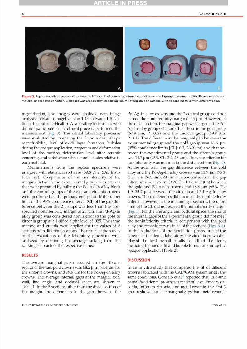
7252019 Varianta 2 Articol Epr
httpslidepdfcomreaderfullvarianta-2-articol-epr 48
magni1047297cation and images were analyzed with image
analysis software (ImageJ version 145 software US Na-tional Institutes of Health) A laboratory technician whodid not participate in the clinical process performed themeasurement ( Fig 3 ) The dental laboratory processes
were evaluated by comparing the 1047297t on a cast shapereproducibility level of oxide layer formation bubblesduring the opaque application properties and deformationlevel of the surface deformation level after ceramic
veneering and satisfaction with ceramic shades relative toeach material
Measurements from the replica specimen wereanalyzed with statistical software (SAS v92 SAS Insti-
tute Inc) Comparisons of the noninferiority of themargins between the experimental group with crownsthat were prepared by milling the Pd-Ag-In alloy blockand the control groups of the cast and zirconia crowns
were performed as the primary end point If the upperlimit of the 95 con1047297dence interval (CI) of the gap dif-ference between the 2 groups was less than the pre-speci1047297ed noninferiority margin of 25 mm the Pd-Ag-Inalloy group was considered noninferior to the gold orzirconia group at a 1-sided alpha level of 025 The samemethod and criteria were applied for the values of 6sections from different locations The results of the survey of the evaluations of the laboratory procedure wereanalyzed by obtaining the average ranking from therankings for each of the respective items
RESULTS
The average marginal gap measured on the siliconereplica of the cast gold crowns was 682 m m 754 mm forthe zirconia crowns and 769 mm for the Pd-Ag-In alloy crowns The average internal gaps at the margin axial
wall line angle and occlusal space are shown inTable 1 In the 5 sections other than the distal section of the margin the differences in the gaps between the
Pd-Ag-In alloy crowns and the 2 control groups did not
exceed the noninferiority margin of 25 mm However inthe distal section the marginal gap was larger in the Pd-
Ag-In alloy group (845 mm) than those in the gold group(679 mm P =002) and the zirconia group (698 mm P =01) The difference in the marginal gap between theexperimental group and the gold group was 166 mm(95 con1047297dence limits [CL] 63 269 mm) and that be-tween the experimental group and the zirconia group
was 147 mm (95 CL 34 26 mm) Thus the criterion fornoninferiority was not met in the distal sections ( Fig 4 )
At the axial wall the gap difference between the goldalloy and the Pd-Ag-In alloy crowns was 119 mm (95
CL minus
24 262 mm) At the mesiobuccal section the gapdifferences were 26 mm (95 CL 102 417 mm) betweenthe gold and Pd-Ag-In crowns and 188 mm (95 CL19 357 mm) between the zirconia and Pd-Ag-In alloy crowns These differences did not meet the noninferiority criteria However in the remaining 4 sections the upperlimit of the CL did not exceed the noninferiority margin( Fig 5 ) For the line angle and occlusal space the size of the internal gaps of the experimental group did not meetthe noninferiority criteria in comparison with the goldalloy and zirconia crowns in all of the sections ( Figs 6-8 )In the evaluations of the fabrication procedures of thecrowns in the dental laboratory the zirconia crown dis-played the best overall results for all of the itemsincluding the model 1047297t and bubble formation during theopaque application (Table 2)
DISCUSSION
In an in vitro study that compared the 1047297t of differentcrowns fabricated with the CA DCAM system under thesame conditions Gonzalo et al35 reported that in 3-unitpartial 1047297xed dental prostheses made of Lava Procera zir-conia InCeram zirconia and metal ceramic the 1047297rst 3groups showed smaller marginal gaps than metal ceramic
Figure 2 Replica technique procedure to measure internal 1047297t of crowns A Internal gaps of crowns in 3 groups were made with silicone registration
material under same condition B Replica was prepared by stabilizing volume of registration material with silicone material with different color
4 Volume - Issue -
THE JOURNAL OF PROSTHETIC DENTISTRY Park et al
7252019 Varianta 2 Articol Epr
httpslidepdfcomreaderfullvarianta-2-articol-epr 58
In addition Baig et al55 reported that the marginal gaps were 664 mm for the Cercon system and 366 mm for theIPS Empress II system both made with the CADCAMmethod and 371 mm for a cast complete metal crown Inan in vivo study that was conducted on 19 participants
Reich et al50 reported that the marginal gaps of the Lavaand Cerec systems were both approximately 65 mm which
was not signi1047297cantly different from the 54 mm for themetal ceramic crown Wettstein et al51 reported that whenthe Cercon zirconia framework and conventional metalceramic framework were selectively fabricated for 25 par-ticipants the internal 1047297ts of the zirconia framework werecervical 1896 mm axial 1405 mm occlusal cusp tip 1213
Figure 3 Cross-section view of each area to measure marginal and internal gaps in silicone replica after mesiodistal and buccolingual sectioning (times30
magni1047297cation) A Margin B Axial wall C Line angle D Occlusal space
300
T w o - S
i d e d 9 5
C I o f G a p D i ff e r e n c e
a t M a r g i n ( micro m )
200
M
e s i o b u c c a l
M
e s i o l i n g u a l
D i s t o b u c c a l
D
i s t o l i n g u a l
M e s i a l
D i s t a l
100
00
ndash100
ndash200
ndash300
Mp - Gold
Mp - Mz
Location
Figure 4 Two-sided 95 CI of gap differences at margin Blue dotted
line denotes noninferiority margin (25 mm) MP machined group of Pd-
Ag-In alloy MZ machined group of zirconia
M e s i o b u c c a l
M e s i o l i n g u a l
D i s t o b u c c a l
D i s t o l i n g u a l
M e s i a l
D i s t a l
Mp - Gold
Mp - Mz
450
350
250
150
50
ndash50
ndash150
ndash250
ndash350
T w o - S
i d e d 9 5
C I o f G a p D i ff e r e n c e
a t A x i a l W a l l ( micro m )
Location
Figure 5 Two-sided 95 CI of gap differences at axial wall
- 2015 5
Park et al THE JOURNAL OF PROSTHETIC DENTISTRY
7252019 Varianta 2 Articol Epr
httpslidepdfcomreaderfullvarianta-2-articol-epr 68
mm and centro-occlusal 1920 mm these values weresigni1047297cantly greater than those of the metal ceramicframework which were 1186 mm 957 mm 1225 mm and1531 mm Thus in most studies the crowns fabricated
with the CADCAM method were comparable with metalceramic or complete-metal crowns that were made by casting and had marginal and internal 1047297ts that were withinthe clinically acceptable range21384452 In addition Tanet al59 compared the vertical margin openings of titaniumcrowns that were designed by CAD software milled tita-nium crowns that were waxed followed by double scan-ning with a 3-dimensional scanner and cast high noblecrowns The marginal discrepancies were 79 mm 73 mmand 24 mm respectively In addition the CADCAM
method with and without double scanning did not show signi1047297cantly different marginal gaps
In this in vivo study the 1047297rst null hypothesis wasrejected because the crown that was fabricated by milling the Pd-Ag-In alloy block showed a marginal 1047297ton the distal cross-section of the crown that was infe-rior to that of the cast crowns and zirconia crownsHowever the marginal gap of the Pd-Ag-In alloy crown
was not statistically inferior to those of the controlgroups on all of the cross-sections except for the distalsection The average marginal gaps of the cast crowns(682 mm) zirconia crowns (754 mm) and Pd-Ag-In
alloy crowns (769 mm) were generally greatercompared with the results of previous in vivo studies55-58
In contrast other in vivo studies have reported largermarginal gaps than those found in our study5051 Inaddition the marginal gap (845 mm) of the distal section
was statistically inferior to the 120 mm that was pro-posed as the clinically acceptable gap by Hung et al38
indicating that the Pd-Ag-In alloy crown had a clinically applicable 1047297t The differences in the internal gaps at theaxial wall between the Pd-Ag-In group and the controlgroups exceeded the noninferiority margin at the mesialsection (1008 mm) and mesiobuccal section (1031 mm)
However similar to the marginal gap all of the internalgaps obtained in this study showed values less than 140 to150 mm
For the line angle and occlusal space the second nullhypothesis was rejected because the Pd-Ag-In alloy crown showed larger internal gaps in all of the crosssections compared with the control groups Both the Pd-
Ag-In alloy and zirconia crowns had signi1047297cantly greaterinternal gaps in the occlusal space compared with thecast crowns This was likely a result of the characteristicsof the CADCAM fabrication method such as the limitedsize of the milling tool and tool compensation64 Inaddition some crowns have their internal spacer pa-rameters set to be bigger in the CAD system to avoid
early contact on the sharp edge of a preparation and toreduce the marginal gap Therefore the gap mightbecome larger in the CAD process compared with a castrestoration5665
The larger gap in the line angle and the occlusal spaceof the Pd-Ag-In alloy crown compared with the zirconiacrown from the same CADCAM method was thought toresult from differences in the gaps that might haveoccurred as a result of the parameters set during milling because of the difference between zirconia which has
volume reduction during the sintering process and thePd-Ag-In alloy which has no volume change For the
general procedures for crowns in the dental laboratory evaluations the zirconia crowns appeared to be theeasiest to fabricate However this was thought to bebecause of skill differences and the fabrication andplacement of the gold or Pd-Ag-In alloy crowns was notexceptionally dif 1047297cult
The methods that are used to evaluate the 1047297t of acrown include direct visualization under a microscope
visualization by the attachment of the crown to a die thatis followed by sectioning evaluation after impressionmaking and evaluation with an explorer and the nakedeye6667 This in vivo study used the replica technique
M e s i o b u c c a l
M e s i o l i n g u a l
D i s t o b u c c a l
D i s t o l i n g u a l
M e s i a l
D i s t a l
Mp - Gold
Mp - Mz
750
650
550
450
350
250
150
50
ndash50
ndash150
T w o - S
i d e d 9 5
C I o f G a p D i ff e r e n c e
a t
L i n e A n g l e ( micro m )
Location
Figure 6 Two-sided 95 CI of gap differences at line angles
M e s i o b u c c a l
M e s i o l i n g u a l
D i s t o b u c c a l
D i s t o l i n g u a l
M e s i a l
D i s t a l
Mp - Gold
Mp - Mz
ndash250
ndash50
150
350
550
750
950
T w o - S
i d e d 9 5
C I o f G a p D i ff e r e n c e
a t O c c l u s a l S p a c e ( micro m )
Location
Figure 7 Two-sided 95 CI of gap differences at occlusal spaces
6 Volume - Issue -
THE JOURNAL OF PROSTHETIC DENTISTRY Park et al
7252019 Varianta 2 Articol Epr
httpslidepdfcomreaderfullvarianta-2-articol-epr 78
which duplicated the state of the abutment becausedirectly cutting the patientrsquos tooth was not possible45 Nosigni1047297cant differences have been reported between the diesectioning method and replica technique and these results
were not affected by the type of silicone registration ma-terial5354 Although the replica was prepared using themaximum clenching force of a patient the replica may nothave re1047298ected the precise positions possibly increasing the standard deviation of the occlusal space
A deep chamfer 1047297nish line was prepared for the labialor buccal side of the incisors canines and premolars
while a conventional chamfer margin was prepared forthe remaining margin Syu et al68 suggested that the typeof 1047297nish line does not affect the marginal gap In addi-tion the preparation design may not result in signi1047297cant
differences between the 1047297
t of the cast crowns and CADCAM crowns6970
The analysis in this clinical study indicated that themarginal and internal 1047297ts of the crowns fabricated by milling machining Pd-Ag-In semiprecious metal alloys
were comparable with those of the conventionally usedcast crowns and zirconia crowns Although the Pd-Ag-Incrowns did not meet the noninferiority criterioncompared with the existing materials for the line angleand occlusal space their values were within the clinically acceptable range Furthermore most areas of the mar-ginal and axial locations met the noninferiority criterionHowever because this study had a small sample size anda short period for crown placement long-term clinicalstudies that involve a larger sample are required Inaddition studies on possible events in the oral cavitysuch as antagonistic tooth wear or fracture chipping of the ceramic veneer and methods of fabricating moreprecise crowns through improvements of the CADCAMtechnique are necessary
CONCLUSIONS
Within the limitations of this in vivo study the following conclusions may be drawn
1 The average plusmnstandard deviation marginal gaps were 682 plusmn248 mm for the cast gold crowns 754plusmn288 mm for the zirconia crowns and 769 plusmn270mm for the Pd-Ag-In alloy crowns The Pd-Ag-Inalloy crowns met the noninferiority criterion in allthe areas except for the distal cross-sectioncompared with the control groups
2 For the line angle and occlusal space the Pd-Ag-Inalloy crowns were inferior to the control groups forthe internal gaps
REFERENCES
1 Fasbinder DJ Clinical performance of chairside CADCAM restorations J AmDent Assoc 200613722S-31S
2 Mormann WH Brandestini M Lutz F Barbakow F Chairside computer-aided direct ceramic inlays Quintessence Int 198920329-39
3 Bindl A Mormann WH Fit of all-ceramic posterior 1047297xed partial dentureframeworks in vitro Int J Periodontics Restorative Dent 200727567-75
4 Tinschert J Natt G Mautsch W Spiekermann H Anusavice KJ Marginal 1047297tof alumina-and zirconia-based 1047297xed partial dentures produced by a CADCAM system Oper Dent 200126367-74
5 Williams RJ Bibb R Eggbeer D CADCAM in the fabrication of removablepartial denture frameworks a virtual method of surveying 3D scanned dentalcasts Quint J Dent Technol 20042268-76
6 Sun Y Lu P Wang Y Study on CADampRP for removable complete dentureComput Methods Programs Biomed 200993266-72
7 Williams RJ Bibb R Eggbeer D Collis J Use of CADCAM technology tofabricate a removable partial denture framework J Prosthet Dent 20069696-9
Figure 8 Representative silicone replicas of internal gaps of crowns
evaluated A Gold alloy group B Zirconia group C Pd-Ag-In alloy group
(times30 magni1047297cation)
- 2015 7
Park et al THE JOURNAL OF PROSTHETIC DENTISTRY
7252019 Varianta 2 Articol Epr
httpslidepdfcomreaderfullvarianta-2-articol-epr 88
8 Andersson M Razzoog ME Oden A Hegenbarth EA Lang BR Procera anew way to achieve an all-ceramic crown Quintessence Int 199829285-96
9 Kapos T Evans C CADCAM technology for implant abutments crownsand superstructures Int J Oral Maxillofac Implants 201429 Suppl117-36
10 Luthardt RG Sandkuhl O Herold V Walter MH Accuracy of mechanicaldigitizing with a CADCAM system for 1047297xed restorations Int J Prosthodont200114146-51
11 McLaren EA Terry DA CADCAM systems materials and clinical guidelinesfor all-ceramic crowns and 1047297xed partial dentures Compend Contin EducDent 200223637-41 44 46 passim quiz 54
12 Piconi C Maccauro G Zirconia as a ceramic biomaterial Biomaterials1999201-25
13 Tinschert J Zwez D Marx R Anusavice KJ Structural reliability of alumina-feldspar- leucite- mica- and zirconia-based ceramics J Dent 200028529-35
14 Ardlin BI Transformation-toughened zirconia for dental inlays crowns andbridges chemical stability and effect of low-temperature aging on 1047298exuralstrength and surface structure Dent Mater 200218590-5
15 Guazzato M Albakry M Ringer SP Swain MV Strength fracture toughnessand microstructure of a selection of all-ceramic materials Part II Zirconia-based dental ceramics Dent Mater 200420449-56
16 Raigrodski AJ Contemporary materials and technologies for all-ceramic 1047297xedpartial dentures a review of the literature J Prosthet Dent 200492557-62
17 Tinschert J Natt G Hassenp1047298ug S Spiekermann H Status of current CADCAM technology in dental medicine Int J Comput Dent 2004725-45
18 Lee DH Lee BJ Kim SH Lee KB Shear bond strength of porcelain to a new millable alloy and a conventional castable alloy J Prosthet Dent 2015113329-35
19 Abduo Fit of CADCAM implant frameworks a comprehensive review
J Oral Implantol 2014 Dec40(6)758-6620 Kapos T Ashy LM Gallucci GO Weber HP Wismeijer D Computer-aided
design and computer-assisted manufacturing in prosthetic implant dentistryInt J Oral Maxillofac Implants 200924110-7
21 Boening KW Walter MH Reppel PD Non-cast titanium restorations in 1047297xedprosthodontics J Oral Rehabil 199219281-7
22 Gilbert JL Covey DA Lautenschlager EP Bond characteristics of porcelainfused to milled titanium Dent Mater 199410134-40
23 Walter M Boning K Reppel PD Clinical performance of machined titaniumrestorations J Dent 199422346-8
24 Goodacre CJ Palladium-silver alloys a review of the literature J ProsthetDent 19896234-7
25 Huget EF Civjan S Status report on palladium-silver-based crown andbridge alloys J Am Dent Assoc 197489383-5
26 Kansu G Aydin AK Evaluation of the biocompatibility of various dentalalloys Part 2e Allergenical potentials Eur J Prosthodont Restor Dent 19964155-61
27 Hong JT Shin SY A comparative study on the bond strength of porcelain to
the millingable Pd-Ag alloy J Adv Prosthodont 20146372-828 Sorensen JA A rationale for comparison of plaque-retaining properties of crown systems J Prosthet Dent 198962264-9
29 Sorensen SE Larsen IB Jorgensen KD Gingival and alveolar bone reactionto marginal 1047297t of subgingival crown margins Scand J Dent Res 198694109-14
30 Assif D Rimer Y Aviv I The 1047298ow of zinc phosphate cement under a full-coverage restoration and its effect on marginal adaptation according to thelocation of cement application Quintessence Int 198718765-74
31 Bader JD Rozier RG McFall WT Jr Ramsey DL Effect of crown margins onperiodontal conditions in regularly attending patients J Prosthet Dent19916575-9
32 Belser UC MacEntee MI Richter WA Fit of three porcelain-fused-to-metalmarginal designs in vivo a scanning electron microscope study J ProsthetDent 19855324-9
33 Felton DA Kanoy BE Bayne SC Wirthman GP Effect of in vivo crownmargin discrepancies on periodontal health J Prosthet Dent 199165357-64
34 Fusayama T Ide K Hosoda H Relief of resistance of cement of full castcrowns J Prosthet Dent 19641495-106
35 Gonzalo E Suarez MJ Serrano B Lozano JF Marginal 1047297t of Zirconia pos-terior 1047297xed partial dentures Int J Prosthodont 200821398-9
36 Grasso JE Nalbandian J Sanford C Bailit H Effect of restoration quality onperiodontal health J Prosthet Dent 19855314-9
37 Grey NJ Piddock V Wilson MA In vitro comparison of conventional crownsand a new all-ceramic system J Dent 19932147-51
38 Hung SH Hung KS Eick JD Chappell RP Marginal 1047297t of porcelain-fused-to-metal and two types of ceramic crown J Prosthet Dent 19906326-31
39 McLean JW von Fraunhofer JA The estimation of cement 1047297lm thickness by an in vivo technique Br Dent J 1971131107-11
40 Palomo F Peden J Periodontal considerations of restorative procedures J Prosthet Dent 197636387-94
41 Passon C Lambert RH Lambert RL Newman S The effect of multiple layersof die-spacer on crown retention Oper Dent 19921742-9
42 Schwartz NL Whitsett LD Berry TG Stewart JL Unserviceable crowns and1047297xed partial dentures life-span and causes for loss of serviceability J AmDent Assoc 1970811395-401
43 Walton JN Gardner FM Agar JR A survey of crown and 1047297xed partial denturefailures length of service and reasons for replacement J Prosthet Dent198656416-21
44 Christensen GJ Marginal 1047297t of gold inlay castings J Prosthet Dent 196616297-305
45 Molin M Karlsson S The 1047297t of gold inlays and three ceramic inlay systems A clinical and in vitro study Acta Odontol Scand 199351201-6
46 Boening KW Wolf BH Schmidt AE Kastner K Walter MH Clinical 1047297t of Procera AllCeram crowns J Prosthet Dent 200084419-24
47 Coli P Karlsson S Fit of a new pressure-sintered zirconium dioxide coping
Int J Prosthodont 20041759-6448 Kohorst P Brinkmann H Dittmer MP Borchers L Stiesch M In1047298uence of
the veneering process on the marginal 1047297t of zirconia 1047297xed dental prostheses J Oral Rehabil 201037283-91
49 Reich S Kappe K Teschner H Schmitt J Clinical 1047297t of four-unit zirconiaposterior 1047297xed dental prostheses Eur J Oral Sci 2008116579-84
50 Reich S Wichmann M Nkenke E Proeschel P Clinical 1047297t of all-ceramicthree-unit 1047297xed partial dentures generated with three different CADCAMsystems Eur J Oral Sci 2005113174-9
51 Wettstein F Sailer I Roos M Hammerle CH Clinical study of the internalgaps of zirconia and metal frameworks for 1047297xed partial dentures Eur J OralSci 2008116272-9
52 Fransson B Oilo G Gjeitanger R The 1047297t of metal-ceramic crowns a clinicalstudy Dent Mater 19851197-9
53 Rahme HY Tehini GE Adib SM Ardo AS Rifai KT In vitro evaluation of theldquoreplica techniquerdquo in the measurement of the 1047297t of Procera crowns
J Contemp Dent Pract 2008925-3254 Laurent M Scheer P Dejou J Laborde G Clinical evaluation of the marginal
1047297t of cast crownse validation of the silicone replica method J Oral Rehabil200835116-22
55 Baig MR Tan KB Nicholls JI Evaluation of the marginal 1047297t of a zirconiaceramic computer-aided machined (CAM) crown system J Prosthet Dent2010104216-27
56 Bindl A Mormann WH Marginal and internal 1047297t of all-ceramic CADCAMcrown-copings on chamfer preparations J Oral Rehabil 200532441-7
57 Hertlein GHS Frank S Suttor D Marginal 1047297t of CADCAM manufactured allceramic prosthesis J Dent Res 20018042-4
58 May KB Russell MM Razzoog ME Lang BR Precision of 1047297t the Procera AllCeram c rown J Prosthet Dent 199880394-404
59 Tan PL Gratton DG Diaz-Arnold AM Holmes DC An in vitro comparisonof vertical marginal gaps of CADCAM titanium and conventional cast res-torations J Prosthodont 200817378-83
60 Kokubo Y Tsumita M Kano T Sakurai S Fukushima S Clinical marginal andinternal gaps of zirconia all-ceramic crowns J Prosthodont Res 20115540-3
61 Chow SC Shao J Wang H Sample size calculations in clinical research 2nded Boca Raton FL Chapman and HallCRC 2007
62 Beuer F Stimmelmayr M Gueth JF Edelhoff D Naumann M In vitro per-formance of full-contour zirconia single crowns Dent Mater 201228449-5663 Kollar A Huber S Mericske E Mericske-Stern R Zirconia for teeth and
implants a case series Int J Periodontics Restorative Dent 200828479-8764 Sturdevant JR Bayne SC Heymann HO Margin gap size of ceramic inlays
using second-generation CADCAM equipment J Esthet Dent 199911206-14
65 Bornemann G Lemelson S Luthardt R Innovative method for the analysisof the internal 3D 1047297tting accuracy of Cerec-3 crowns Int J Comput Dent20025177-82
66 Sorensen JA A standardized method for determination of crown margin 1047297-delity J Prosthet Dent 19906418-24
67 Holmes JR Bayne SC Holland GA Sulik WD Considerations in measure-ment of marginal 1047297t J Prosthet Dent 198962405-8
68 Syu JZ Byrne G Laub LW Land MF In1047298uence of 1047297nish-line geometry onthe 1047297t of crowns Int J Prosthodont 1993625-30
69 Tsitrou EA Northeast SE van Noort R Evaluation of the marginal 1047297t of threemargin designs of resin composite crowns using CADCAM J Dent 20073568-73
70 Ayad MF Effect of the crown preparation margin and die type on the mar-ginal accuracy of 1047297ber-reinforced composite crowns J Contemp Dent Pract200899-16
Corresponding author
Dr Namsik OhInha University 7-2063 Shinhung-dong Jung-guIncheon 400-711REPUBLIC OF KOREA Email onsdoinhaackr
AcknowledgmentsThe authors thank Dr Kyoung-Ae Kong for performing statistical analyses in thisstudy
Copyright copy 2015 by the Editorial Council for The Journal of Prosthetic Dentistry
8 Volume - Issue -
THE JOURNAL OF PROSTHETIC DENTISTRY Park et al
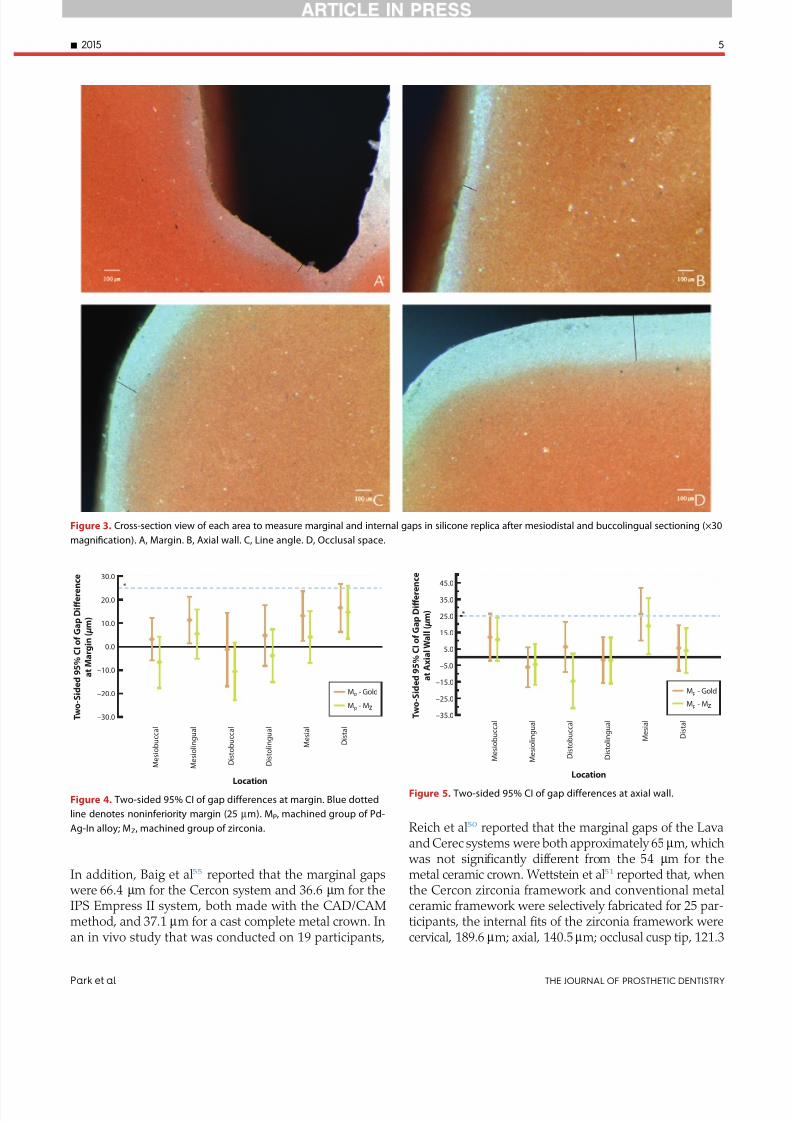
7252019 Varianta 2 Articol Epr
httpslidepdfcomreaderfullvarianta-2-articol-epr 58
In addition Baig et al55 reported that the marginal gaps were 664 mm for the Cercon system and 366 mm for theIPS Empress II system both made with the CADCAMmethod and 371 mm for a cast complete metal crown Inan in vivo study that was conducted on 19 participants
Reich et al50 reported that the marginal gaps of the Lavaand Cerec systems were both approximately 65 mm which
was not signi1047297cantly different from the 54 mm for themetal ceramic crown Wettstein et al51 reported that whenthe Cercon zirconia framework and conventional metalceramic framework were selectively fabricated for 25 par-ticipants the internal 1047297ts of the zirconia framework werecervical 1896 mm axial 1405 mm occlusal cusp tip 1213
Figure 3 Cross-section view of each area to measure marginal and internal gaps in silicone replica after mesiodistal and buccolingual sectioning (times30
magni1047297cation) A Margin B Axial wall C Line angle D Occlusal space
300
T w o - S
i d e d 9 5
C I o f G a p D i ff e r e n c e
a t M a r g i n ( micro m )
200
M
e s i o b u c c a l
M
e s i o l i n g u a l
D i s t o b u c c a l
D
i s t o l i n g u a l
M e s i a l
D i s t a l
100
00
ndash100
ndash200
ndash300
Mp - Gold
Mp - Mz
Location
Figure 4 Two-sided 95 CI of gap differences at margin Blue dotted
line denotes noninferiority margin (25 mm) MP machined group of Pd-
Ag-In alloy MZ machined group of zirconia
M e s i o b u c c a l
M e s i o l i n g u a l
D i s t o b u c c a l
D i s t o l i n g u a l
M e s i a l
D i s t a l
Mp - Gold
Mp - Mz
450
350
250
150
50
ndash50
ndash150
ndash250
ndash350
T w o - S
i d e d 9 5
C I o f G a p D i ff e r e n c e
a t A x i a l W a l l ( micro m )
Location
Figure 5 Two-sided 95 CI of gap differences at axial wall
- 2015 5
Park et al THE JOURNAL OF PROSTHETIC DENTISTRY
7252019 Varianta 2 Articol Epr
httpslidepdfcomreaderfullvarianta-2-articol-epr 68
mm and centro-occlusal 1920 mm these values weresigni1047297cantly greater than those of the metal ceramicframework which were 1186 mm 957 mm 1225 mm and1531 mm Thus in most studies the crowns fabricated
with the CADCAM method were comparable with metalceramic or complete-metal crowns that were made by casting and had marginal and internal 1047297ts that were withinthe clinically acceptable range21384452 In addition Tanet al59 compared the vertical margin openings of titaniumcrowns that were designed by CAD software milled tita-nium crowns that were waxed followed by double scan-ning with a 3-dimensional scanner and cast high noblecrowns The marginal discrepancies were 79 mm 73 mmand 24 mm respectively In addition the CADCAM
method with and without double scanning did not show signi1047297cantly different marginal gaps
In this in vivo study the 1047297rst null hypothesis wasrejected because the crown that was fabricated by milling the Pd-Ag-In alloy block showed a marginal 1047297ton the distal cross-section of the crown that was infe-rior to that of the cast crowns and zirconia crownsHowever the marginal gap of the Pd-Ag-In alloy crown
was not statistically inferior to those of the controlgroups on all of the cross-sections except for the distalsection The average marginal gaps of the cast crowns(682 mm) zirconia crowns (754 mm) and Pd-Ag-In
alloy crowns (769 mm) were generally greatercompared with the results of previous in vivo studies55-58
In contrast other in vivo studies have reported largermarginal gaps than those found in our study5051 Inaddition the marginal gap (845 mm) of the distal section
was statistically inferior to the 120 mm that was pro-posed as the clinically acceptable gap by Hung et al38
indicating that the Pd-Ag-In alloy crown had a clinically applicable 1047297t The differences in the internal gaps at theaxial wall between the Pd-Ag-In group and the controlgroups exceeded the noninferiority margin at the mesialsection (1008 mm) and mesiobuccal section (1031 mm)
However similar to the marginal gap all of the internalgaps obtained in this study showed values less than 140 to150 mm
For the line angle and occlusal space the second nullhypothesis was rejected because the Pd-Ag-In alloy crown showed larger internal gaps in all of the crosssections compared with the control groups Both the Pd-
Ag-In alloy and zirconia crowns had signi1047297cantly greaterinternal gaps in the occlusal space compared with thecast crowns This was likely a result of the characteristicsof the CADCAM fabrication method such as the limitedsize of the milling tool and tool compensation64 Inaddition some crowns have their internal spacer pa-rameters set to be bigger in the CAD system to avoid
early contact on the sharp edge of a preparation and toreduce the marginal gap Therefore the gap mightbecome larger in the CAD process compared with a castrestoration5665
The larger gap in the line angle and the occlusal spaceof the Pd-Ag-In alloy crown compared with the zirconiacrown from the same CADCAM method was thought toresult from differences in the gaps that might haveoccurred as a result of the parameters set during milling because of the difference between zirconia which has
volume reduction during the sintering process and thePd-Ag-In alloy which has no volume change For the
general procedures for crowns in the dental laboratory evaluations the zirconia crowns appeared to be theeasiest to fabricate However this was thought to bebecause of skill differences and the fabrication andplacement of the gold or Pd-Ag-In alloy crowns was notexceptionally dif 1047297cult
The methods that are used to evaluate the 1047297t of acrown include direct visualization under a microscope
visualization by the attachment of the crown to a die thatis followed by sectioning evaluation after impressionmaking and evaluation with an explorer and the nakedeye6667 This in vivo study used the replica technique
M e s i o b u c c a l
M e s i o l i n g u a l
D i s t o b u c c a l
D i s t o l i n g u a l
M e s i a l
D i s t a l
Mp - Gold
Mp - Mz
750
650
550
450
350
250
150
50
ndash50
ndash150
T w o - S
i d e d 9 5
C I o f G a p D i ff e r e n c e
a t
L i n e A n g l e ( micro m )
Location
Figure 6 Two-sided 95 CI of gap differences at line angles
M e s i o b u c c a l
M e s i o l i n g u a l
D i s t o b u c c a l
D i s t o l i n g u a l
M e s i a l
D i s t a l
Mp - Gold
Mp - Mz
ndash250
ndash50
150
350
550
750
950
T w o - S
i d e d 9 5
C I o f G a p D i ff e r e n c e
a t O c c l u s a l S p a c e ( micro m )
Location
Figure 7 Two-sided 95 CI of gap differences at occlusal spaces
6 Volume - Issue -
THE JOURNAL OF PROSTHETIC DENTISTRY Park et al
7252019 Varianta 2 Articol Epr
httpslidepdfcomreaderfullvarianta-2-articol-epr 78
which duplicated the state of the abutment becausedirectly cutting the patientrsquos tooth was not possible45 Nosigni1047297cant differences have been reported between the diesectioning method and replica technique and these results
were not affected by the type of silicone registration ma-terial5354 Although the replica was prepared using themaximum clenching force of a patient the replica may nothave re1047298ected the precise positions possibly increasing the standard deviation of the occlusal space
A deep chamfer 1047297nish line was prepared for the labialor buccal side of the incisors canines and premolars
while a conventional chamfer margin was prepared forthe remaining margin Syu et al68 suggested that the typeof 1047297nish line does not affect the marginal gap In addi-tion the preparation design may not result in signi1047297cant
differences between the 1047297
t of the cast crowns and CADCAM crowns6970
The analysis in this clinical study indicated that themarginal and internal 1047297ts of the crowns fabricated by milling machining Pd-Ag-In semiprecious metal alloys
were comparable with those of the conventionally usedcast crowns and zirconia crowns Although the Pd-Ag-Incrowns did not meet the noninferiority criterioncompared with the existing materials for the line angleand occlusal space their values were within the clinically acceptable range Furthermore most areas of the mar-ginal and axial locations met the noninferiority criterionHowever because this study had a small sample size anda short period for crown placement long-term clinicalstudies that involve a larger sample are required Inaddition studies on possible events in the oral cavitysuch as antagonistic tooth wear or fracture chipping of the ceramic veneer and methods of fabricating moreprecise crowns through improvements of the CADCAMtechnique are necessary
CONCLUSIONS
Within the limitations of this in vivo study the following conclusions may be drawn
1 The average plusmnstandard deviation marginal gaps were 682 plusmn248 mm for the cast gold crowns 754plusmn288 mm for the zirconia crowns and 769 plusmn270mm for the Pd-Ag-In alloy crowns The Pd-Ag-Inalloy crowns met the noninferiority criterion in allthe areas except for the distal cross-sectioncompared with the control groups
2 For the line angle and occlusal space the Pd-Ag-Inalloy crowns were inferior to the control groups forthe internal gaps
REFERENCES
1 Fasbinder DJ Clinical performance of chairside CADCAM restorations J AmDent Assoc 200613722S-31S
2 Mormann WH Brandestini M Lutz F Barbakow F Chairside computer-aided direct ceramic inlays Quintessence Int 198920329-39
3 Bindl A Mormann WH Fit of all-ceramic posterior 1047297xed partial dentureframeworks in vitro Int J Periodontics Restorative Dent 200727567-75
4 Tinschert J Natt G Mautsch W Spiekermann H Anusavice KJ Marginal 1047297tof alumina-and zirconia-based 1047297xed partial dentures produced by a CADCAM system Oper Dent 200126367-74
5 Williams RJ Bibb R Eggbeer D CADCAM in the fabrication of removablepartial denture frameworks a virtual method of surveying 3D scanned dentalcasts Quint J Dent Technol 20042268-76
6 Sun Y Lu P Wang Y Study on CADampRP for removable complete dentureComput Methods Programs Biomed 200993266-72
7 Williams RJ Bibb R Eggbeer D Collis J Use of CADCAM technology tofabricate a removable partial denture framework J Prosthet Dent 20069696-9
Figure 8 Representative silicone replicas of internal gaps of crowns
evaluated A Gold alloy group B Zirconia group C Pd-Ag-In alloy group
(times30 magni1047297cation)
- 2015 7
Park et al THE JOURNAL OF PROSTHETIC DENTISTRY
7252019 Varianta 2 Articol Epr
httpslidepdfcomreaderfullvarianta-2-articol-epr 88
8 Andersson M Razzoog ME Oden A Hegenbarth EA Lang BR Procera anew way to achieve an all-ceramic crown Quintessence Int 199829285-96
9 Kapos T Evans C CADCAM technology for implant abutments crownsand superstructures Int J Oral Maxillofac Implants 201429 Suppl117-36
10 Luthardt RG Sandkuhl O Herold V Walter MH Accuracy of mechanicaldigitizing with a CADCAM system for 1047297xed restorations Int J Prosthodont200114146-51
11 McLaren EA Terry DA CADCAM systems materials and clinical guidelinesfor all-ceramic crowns and 1047297xed partial dentures Compend Contin EducDent 200223637-41 44 46 passim quiz 54
12 Piconi C Maccauro G Zirconia as a ceramic biomaterial Biomaterials1999201-25
13 Tinschert J Zwez D Marx R Anusavice KJ Structural reliability of alumina-feldspar- leucite- mica- and zirconia-based ceramics J Dent 200028529-35
14 Ardlin BI Transformation-toughened zirconia for dental inlays crowns andbridges chemical stability and effect of low-temperature aging on 1047298exuralstrength and surface structure Dent Mater 200218590-5
15 Guazzato M Albakry M Ringer SP Swain MV Strength fracture toughnessand microstructure of a selection of all-ceramic materials Part II Zirconia-based dental ceramics Dent Mater 200420449-56
16 Raigrodski AJ Contemporary materials and technologies for all-ceramic 1047297xedpartial dentures a review of the literature J Prosthet Dent 200492557-62
17 Tinschert J Natt G Hassenp1047298ug S Spiekermann H Status of current CADCAM technology in dental medicine Int J Comput Dent 2004725-45
18 Lee DH Lee BJ Kim SH Lee KB Shear bond strength of porcelain to a new millable alloy and a conventional castable alloy J Prosthet Dent 2015113329-35
19 Abduo Fit of CADCAM implant frameworks a comprehensive review
J Oral Implantol 2014 Dec40(6)758-6620 Kapos T Ashy LM Gallucci GO Weber HP Wismeijer D Computer-aided
design and computer-assisted manufacturing in prosthetic implant dentistryInt J Oral Maxillofac Implants 200924110-7
21 Boening KW Walter MH Reppel PD Non-cast titanium restorations in 1047297xedprosthodontics J Oral Rehabil 199219281-7
22 Gilbert JL Covey DA Lautenschlager EP Bond characteristics of porcelainfused to milled titanium Dent Mater 199410134-40
23 Walter M Boning K Reppel PD Clinical performance of machined titaniumrestorations J Dent 199422346-8
24 Goodacre CJ Palladium-silver alloys a review of the literature J ProsthetDent 19896234-7
25 Huget EF Civjan S Status report on palladium-silver-based crown andbridge alloys J Am Dent Assoc 197489383-5
26 Kansu G Aydin AK Evaluation of the biocompatibility of various dentalalloys Part 2e Allergenical potentials Eur J Prosthodont Restor Dent 19964155-61
27 Hong JT Shin SY A comparative study on the bond strength of porcelain to
the millingable Pd-Ag alloy J Adv Prosthodont 20146372-828 Sorensen JA A rationale for comparison of plaque-retaining properties of crown systems J Prosthet Dent 198962264-9
29 Sorensen SE Larsen IB Jorgensen KD Gingival and alveolar bone reactionto marginal 1047297t of subgingival crown margins Scand J Dent Res 198694109-14
30 Assif D Rimer Y Aviv I The 1047298ow of zinc phosphate cement under a full-coverage restoration and its effect on marginal adaptation according to thelocation of cement application Quintessence Int 198718765-74
31 Bader JD Rozier RG McFall WT Jr Ramsey DL Effect of crown margins onperiodontal conditions in regularly attending patients J Prosthet Dent19916575-9
32 Belser UC MacEntee MI Richter WA Fit of three porcelain-fused-to-metalmarginal designs in vivo a scanning electron microscope study J ProsthetDent 19855324-9
33 Felton DA Kanoy BE Bayne SC Wirthman GP Effect of in vivo crownmargin discrepancies on periodontal health J Prosthet Dent 199165357-64
34 Fusayama T Ide K Hosoda H Relief of resistance of cement of full castcrowns J Prosthet Dent 19641495-106
35 Gonzalo E Suarez MJ Serrano B Lozano JF Marginal 1047297t of Zirconia pos-terior 1047297xed partial dentures Int J Prosthodont 200821398-9
36 Grasso JE Nalbandian J Sanford C Bailit H Effect of restoration quality onperiodontal health J Prosthet Dent 19855314-9
37 Grey NJ Piddock V Wilson MA In vitro comparison of conventional crownsand a new all-ceramic system J Dent 19932147-51
38 Hung SH Hung KS Eick JD Chappell RP Marginal 1047297t of porcelain-fused-to-metal and two types of ceramic crown J Prosthet Dent 19906326-31
39 McLean JW von Fraunhofer JA The estimation of cement 1047297lm thickness by an in vivo technique Br Dent J 1971131107-11
40 Palomo F Peden J Periodontal considerations of restorative procedures J Prosthet Dent 197636387-94
41 Passon C Lambert RH Lambert RL Newman S The effect of multiple layersof die-spacer on crown retention Oper Dent 19921742-9
42 Schwartz NL Whitsett LD Berry TG Stewart JL Unserviceable crowns and1047297xed partial dentures life-span and causes for loss of serviceability J AmDent Assoc 1970811395-401
43 Walton JN Gardner FM Agar JR A survey of crown and 1047297xed partial denturefailures length of service and reasons for replacement J Prosthet Dent198656416-21
44 Christensen GJ Marginal 1047297t of gold inlay castings J Prosthet Dent 196616297-305
45 Molin M Karlsson S The 1047297t of gold inlays and three ceramic inlay systems A clinical and in vitro study Acta Odontol Scand 199351201-6
46 Boening KW Wolf BH Schmidt AE Kastner K Walter MH Clinical 1047297t of Procera AllCeram crowns J Prosthet Dent 200084419-24
47 Coli P Karlsson S Fit of a new pressure-sintered zirconium dioxide coping
Int J Prosthodont 20041759-6448 Kohorst P Brinkmann H Dittmer MP Borchers L Stiesch M In1047298uence of
the veneering process on the marginal 1047297t of zirconia 1047297xed dental prostheses J Oral Rehabil 201037283-91
49 Reich S Kappe K Teschner H Schmitt J Clinical 1047297t of four-unit zirconiaposterior 1047297xed dental prostheses Eur J Oral Sci 2008116579-84
50 Reich S Wichmann M Nkenke E Proeschel P Clinical 1047297t of all-ceramicthree-unit 1047297xed partial dentures generated with three different CADCAMsystems Eur J Oral Sci 2005113174-9
51 Wettstein F Sailer I Roos M Hammerle CH Clinical study of the internalgaps of zirconia and metal frameworks for 1047297xed partial dentures Eur J OralSci 2008116272-9
52 Fransson B Oilo G Gjeitanger R The 1047297t of metal-ceramic crowns a clinicalstudy Dent Mater 19851197-9
53 Rahme HY Tehini GE Adib SM Ardo AS Rifai KT In vitro evaluation of theldquoreplica techniquerdquo in the measurement of the 1047297t of Procera crowns
J Contemp Dent Pract 2008925-3254 Laurent M Scheer P Dejou J Laborde G Clinical evaluation of the marginal
1047297t of cast crownse validation of the silicone replica method J Oral Rehabil200835116-22
55 Baig MR Tan KB Nicholls JI Evaluation of the marginal 1047297t of a zirconiaceramic computer-aided machined (CAM) crown system J Prosthet Dent2010104216-27
56 Bindl A Mormann WH Marginal and internal 1047297t of all-ceramic CADCAMcrown-copings on chamfer preparations J Oral Rehabil 200532441-7
57 Hertlein GHS Frank S Suttor D Marginal 1047297t of CADCAM manufactured allceramic prosthesis J Dent Res 20018042-4
58 May KB Russell MM Razzoog ME Lang BR Precision of 1047297t the Procera AllCeram c rown J Prosthet Dent 199880394-404
59 Tan PL Gratton DG Diaz-Arnold AM Holmes DC An in vitro comparisonof vertical marginal gaps of CADCAM titanium and conventional cast res-torations J Prosthodont 200817378-83
60 Kokubo Y Tsumita M Kano T Sakurai S Fukushima S Clinical marginal andinternal gaps of zirconia all-ceramic crowns J Prosthodont Res 20115540-3
61 Chow SC Shao J Wang H Sample size calculations in clinical research 2nded Boca Raton FL Chapman and HallCRC 2007
62 Beuer F Stimmelmayr M Gueth JF Edelhoff D Naumann M In vitro per-formance of full-contour zirconia single crowns Dent Mater 201228449-5663 Kollar A Huber S Mericske E Mericske-Stern R Zirconia for teeth and
implants a case series Int J Periodontics Restorative Dent 200828479-8764 Sturdevant JR Bayne SC Heymann HO Margin gap size of ceramic inlays
using second-generation CADCAM equipment J Esthet Dent 199911206-14
65 Bornemann G Lemelson S Luthardt R Innovative method for the analysisof the internal 3D 1047297tting accuracy of Cerec-3 crowns Int J Comput Dent20025177-82
66 Sorensen JA A standardized method for determination of crown margin 1047297-delity J Prosthet Dent 19906418-24
67 Holmes JR Bayne SC Holland GA Sulik WD Considerations in measure-ment of marginal 1047297t J Prosthet Dent 198962405-8
68 Syu JZ Byrne G Laub LW Land MF In1047298uence of 1047297nish-line geometry onthe 1047297t of crowns Int J Prosthodont 1993625-30
69 Tsitrou EA Northeast SE van Noort R Evaluation of the marginal 1047297t of threemargin designs of resin composite crowns using CADCAM J Dent 20073568-73
70 Ayad MF Effect of the crown preparation margin and die type on the mar-ginal accuracy of 1047297ber-reinforced composite crowns J Contemp Dent Pract200899-16
Corresponding author
Dr Namsik OhInha University 7-2063 Shinhung-dong Jung-guIncheon 400-711REPUBLIC OF KOREA Email onsdoinhaackr
AcknowledgmentsThe authors thank Dr Kyoung-Ae Kong for performing statistical analyses in thisstudy
Copyright copy 2015 by the Editorial Council for The Journal of Prosthetic Dentistry
8 Volume - Issue -
THE JOURNAL OF PROSTHETIC DENTISTRY Park et al
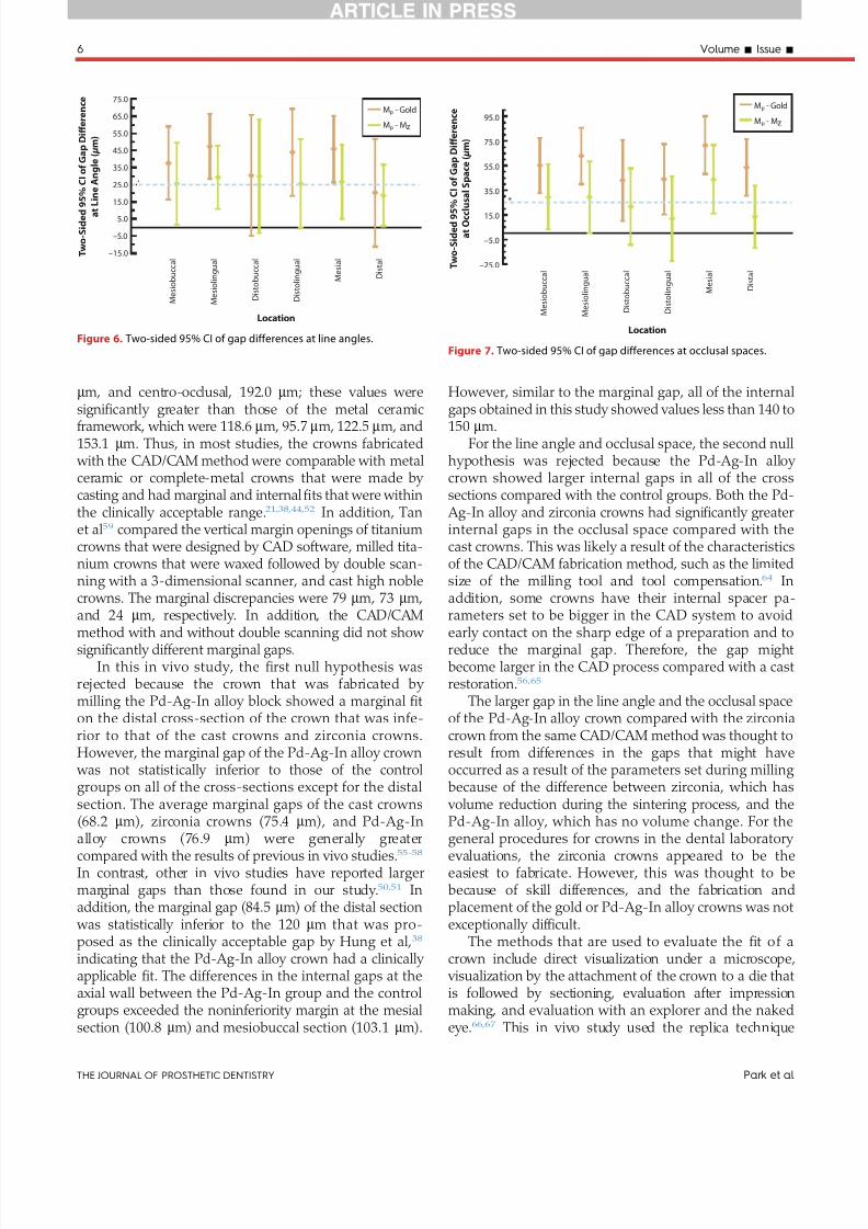
7252019 Varianta 2 Articol Epr
httpslidepdfcomreaderfullvarianta-2-articol-epr 68
mm and centro-occlusal 1920 mm these values weresigni1047297cantly greater than those of the metal ceramicframework which were 1186 mm 957 mm 1225 mm and1531 mm Thus in most studies the crowns fabricated
with the CADCAM method were comparable with metalceramic or complete-metal crowns that were made by casting and had marginal and internal 1047297ts that were withinthe clinically acceptable range21384452 In addition Tanet al59 compared the vertical margin openings of titaniumcrowns that were designed by CAD software milled tita-nium crowns that were waxed followed by double scan-ning with a 3-dimensional scanner and cast high noblecrowns The marginal discrepancies were 79 mm 73 mmand 24 mm respectively In addition the CADCAM
method with and without double scanning did not show signi1047297cantly different marginal gaps
In this in vivo study the 1047297rst null hypothesis wasrejected because the crown that was fabricated by milling the Pd-Ag-In alloy block showed a marginal 1047297ton the distal cross-section of the crown that was infe-rior to that of the cast crowns and zirconia crownsHowever the marginal gap of the Pd-Ag-In alloy crown
was not statistically inferior to those of the controlgroups on all of the cross-sections except for the distalsection The average marginal gaps of the cast crowns(682 mm) zirconia crowns (754 mm) and Pd-Ag-In
alloy crowns (769 mm) were generally greatercompared with the results of previous in vivo studies55-58
In contrast other in vivo studies have reported largermarginal gaps than those found in our study5051 Inaddition the marginal gap (845 mm) of the distal section
was statistically inferior to the 120 mm that was pro-posed as the clinically acceptable gap by Hung et al38
indicating that the Pd-Ag-In alloy crown had a clinically applicable 1047297t The differences in the internal gaps at theaxial wall between the Pd-Ag-In group and the controlgroups exceeded the noninferiority margin at the mesialsection (1008 mm) and mesiobuccal section (1031 mm)
However similar to the marginal gap all of the internalgaps obtained in this study showed values less than 140 to150 mm
For the line angle and occlusal space the second nullhypothesis was rejected because the Pd-Ag-In alloy crown showed larger internal gaps in all of the crosssections compared with the control groups Both the Pd-
Ag-In alloy and zirconia crowns had signi1047297cantly greaterinternal gaps in the occlusal space compared with thecast crowns This was likely a result of the characteristicsof the CADCAM fabrication method such as the limitedsize of the milling tool and tool compensation64 Inaddition some crowns have their internal spacer pa-rameters set to be bigger in the CAD system to avoid
early contact on the sharp edge of a preparation and toreduce the marginal gap Therefore the gap mightbecome larger in the CAD process compared with a castrestoration5665
The larger gap in the line angle and the occlusal spaceof the Pd-Ag-In alloy crown compared with the zirconiacrown from the same CADCAM method was thought toresult from differences in the gaps that might haveoccurred as a result of the parameters set during milling because of the difference between zirconia which has
volume reduction during the sintering process and thePd-Ag-In alloy which has no volume change For the
general procedures for crowns in the dental laboratory evaluations the zirconia crowns appeared to be theeasiest to fabricate However this was thought to bebecause of skill differences and the fabrication andplacement of the gold or Pd-Ag-In alloy crowns was notexceptionally dif 1047297cult
The methods that are used to evaluate the 1047297t of acrown include direct visualization under a microscope
visualization by the attachment of the crown to a die thatis followed by sectioning evaluation after impressionmaking and evaluation with an explorer and the nakedeye6667 This in vivo study used the replica technique
M e s i o b u c c a l
M e s i o l i n g u a l
D i s t o b u c c a l
D i s t o l i n g u a l
M e s i a l
D i s t a l
Mp - Gold
Mp - Mz
750
650
550
450
350
250
150
50
ndash50
ndash150
T w o - S
i d e d 9 5
C I o f G a p D i ff e r e n c e
a t
L i n e A n g l e ( micro m )
Location
Figure 6 Two-sided 95 CI of gap differences at line angles
M e s i o b u c c a l
M e s i o l i n g u a l
D i s t o b u c c a l
D i s t o l i n g u a l
M e s i a l
D i s t a l
Mp - Gold
Mp - Mz
ndash250
ndash50
150
350
550
750
950
T w o - S
i d e d 9 5
C I o f G a p D i ff e r e n c e
a t O c c l u s a l S p a c e ( micro m )
Location
Figure 7 Two-sided 95 CI of gap differences at occlusal spaces
6 Volume - Issue -
THE JOURNAL OF PROSTHETIC DENTISTRY Park et al
7252019 Varianta 2 Articol Epr
httpslidepdfcomreaderfullvarianta-2-articol-epr 78
which duplicated the state of the abutment becausedirectly cutting the patientrsquos tooth was not possible45 Nosigni1047297cant differences have been reported between the diesectioning method and replica technique and these results
were not affected by the type of silicone registration ma-terial5354 Although the replica was prepared using themaximum clenching force of a patient the replica may nothave re1047298ected the precise positions possibly increasing the standard deviation of the occlusal space
A deep chamfer 1047297nish line was prepared for the labialor buccal side of the incisors canines and premolars
while a conventional chamfer margin was prepared forthe remaining margin Syu et al68 suggested that the typeof 1047297nish line does not affect the marginal gap In addi-tion the preparation design may not result in signi1047297cant
differences between the 1047297
t of the cast crowns and CADCAM crowns6970
The analysis in this clinical study indicated that themarginal and internal 1047297ts of the crowns fabricated by milling machining Pd-Ag-In semiprecious metal alloys
were comparable with those of the conventionally usedcast crowns and zirconia crowns Although the Pd-Ag-Incrowns did not meet the noninferiority criterioncompared with the existing materials for the line angleand occlusal space their values were within the clinically acceptable range Furthermore most areas of the mar-ginal and axial locations met the noninferiority criterionHowever because this study had a small sample size anda short period for crown placement long-term clinicalstudies that involve a larger sample are required Inaddition studies on possible events in the oral cavitysuch as antagonistic tooth wear or fracture chipping of the ceramic veneer and methods of fabricating moreprecise crowns through improvements of the CADCAMtechnique are necessary
CONCLUSIONS
Within the limitations of this in vivo study the following conclusions may be drawn
1 The average plusmnstandard deviation marginal gaps were 682 plusmn248 mm for the cast gold crowns 754plusmn288 mm for the zirconia crowns and 769 plusmn270mm for the Pd-Ag-In alloy crowns The Pd-Ag-Inalloy crowns met the noninferiority criterion in allthe areas except for the distal cross-sectioncompared with the control groups
2 For the line angle and occlusal space the Pd-Ag-Inalloy crowns were inferior to the control groups forthe internal gaps
REFERENCES
1 Fasbinder DJ Clinical performance of chairside CADCAM restorations J AmDent Assoc 200613722S-31S
2 Mormann WH Brandestini M Lutz F Barbakow F Chairside computer-aided direct ceramic inlays Quintessence Int 198920329-39
3 Bindl A Mormann WH Fit of all-ceramic posterior 1047297xed partial dentureframeworks in vitro Int J Periodontics Restorative Dent 200727567-75
4 Tinschert J Natt G Mautsch W Spiekermann H Anusavice KJ Marginal 1047297tof alumina-and zirconia-based 1047297xed partial dentures produced by a CADCAM system Oper Dent 200126367-74
5 Williams RJ Bibb R Eggbeer D CADCAM in the fabrication of removablepartial denture frameworks a virtual method of surveying 3D scanned dentalcasts Quint J Dent Technol 20042268-76
6 Sun Y Lu P Wang Y Study on CADampRP for removable complete dentureComput Methods Programs Biomed 200993266-72
7 Williams RJ Bibb R Eggbeer D Collis J Use of CADCAM technology tofabricate a removable partial denture framework J Prosthet Dent 20069696-9
Figure 8 Representative silicone replicas of internal gaps of crowns
evaluated A Gold alloy group B Zirconia group C Pd-Ag-In alloy group
(times30 magni1047297cation)
- 2015 7
Park et al THE JOURNAL OF PROSTHETIC DENTISTRY
7252019 Varianta 2 Articol Epr
httpslidepdfcomreaderfullvarianta-2-articol-epr 88
8 Andersson M Razzoog ME Oden A Hegenbarth EA Lang BR Procera anew way to achieve an all-ceramic crown Quintessence Int 199829285-96
9 Kapos T Evans C CADCAM technology for implant abutments crownsand superstructures Int J Oral Maxillofac Implants 201429 Suppl117-36
10 Luthardt RG Sandkuhl O Herold V Walter MH Accuracy of mechanicaldigitizing with a CADCAM system for 1047297xed restorations Int J Prosthodont200114146-51
11 McLaren EA Terry DA CADCAM systems materials and clinical guidelinesfor all-ceramic crowns and 1047297xed partial dentures Compend Contin EducDent 200223637-41 44 46 passim quiz 54
12 Piconi C Maccauro G Zirconia as a ceramic biomaterial Biomaterials1999201-25
13 Tinschert J Zwez D Marx R Anusavice KJ Structural reliability of alumina-feldspar- leucite- mica- and zirconia-based ceramics J Dent 200028529-35
14 Ardlin BI Transformation-toughened zirconia for dental inlays crowns andbridges chemical stability and effect of low-temperature aging on 1047298exuralstrength and surface structure Dent Mater 200218590-5
15 Guazzato M Albakry M Ringer SP Swain MV Strength fracture toughnessand microstructure of a selection of all-ceramic materials Part II Zirconia-based dental ceramics Dent Mater 200420449-56
16 Raigrodski AJ Contemporary materials and technologies for all-ceramic 1047297xedpartial dentures a review of the literature J Prosthet Dent 200492557-62
17 Tinschert J Natt G Hassenp1047298ug S Spiekermann H Status of current CADCAM technology in dental medicine Int J Comput Dent 2004725-45
18 Lee DH Lee BJ Kim SH Lee KB Shear bond strength of porcelain to a new millable alloy and a conventional castable alloy J Prosthet Dent 2015113329-35
19 Abduo Fit of CADCAM implant frameworks a comprehensive review
J Oral Implantol 2014 Dec40(6)758-6620 Kapos T Ashy LM Gallucci GO Weber HP Wismeijer D Computer-aided
design and computer-assisted manufacturing in prosthetic implant dentistryInt J Oral Maxillofac Implants 200924110-7
21 Boening KW Walter MH Reppel PD Non-cast titanium restorations in 1047297xedprosthodontics J Oral Rehabil 199219281-7
22 Gilbert JL Covey DA Lautenschlager EP Bond characteristics of porcelainfused to milled titanium Dent Mater 199410134-40
23 Walter M Boning K Reppel PD Clinical performance of machined titaniumrestorations J Dent 199422346-8
24 Goodacre CJ Palladium-silver alloys a review of the literature J ProsthetDent 19896234-7
25 Huget EF Civjan S Status report on palladium-silver-based crown andbridge alloys J Am Dent Assoc 197489383-5
26 Kansu G Aydin AK Evaluation of the biocompatibility of various dentalalloys Part 2e Allergenical potentials Eur J Prosthodont Restor Dent 19964155-61
27 Hong JT Shin SY A comparative study on the bond strength of porcelain to
the millingable Pd-Ag alloy J Adv Prosthodont 20146372-828 Sorensen JA A rationale for comparison of plaque-retaining properties of crown systems J Prosthet Dent 198962264-9
29 Sorensen SE Larsen IB Jorgensen KD Gingival and alveolar bone reactionto marginal 1047297t of subgingival crown margins Scand J Dent Res 198694109-14
30 Assif D Rimer Y Aviv I The 1047298ow of zinc phosphate cement under a full-coverage restoration and its effect on marginal adaptation according to thelocation of cement application Quintessence Int 198718765-74
31 Bader JD Rozier RG McFall WT Jr Ramsey DL Effect of crown margins onperiodontal conditions in regularly attending patients J Prosthet Dent19916575-9
32 Belser UC MacEntee MI Richter WA Fit of three porcelain-fused-to-metalmarginal designs in vivo a scanning electron microscope study J ProsthetDent 19855324-9
33 Felton DA Kanoy BE Bayne SC Wirthman GP Effect of in vivo crownmargin discrepancies on periodontal health J Prosthet Dent 199165357-64
34 Fusayama T Ide K Hosoda H Relief of resistance of cement of full castcrowns J Prosthet Dent 19641495-106
35 Gonzalo E Suarez MJ Serrano B Lozano JF Marginal 1047297t of Zirconia pos-terior 1047297xed partial dentures Int J Prosthodont 200821398-9
36 Grasso JE Nalbandian J Sanford C Bailit H Effect of restoration quality onperiodontal health J Prosthet Dent 19855314-9
37 Grey NJ Piddock V Wilson MA In vitro comparison of conventional crownsand a new all-ceramic system J Dent 19932147-51
38 Hung SH Hung KS Eick JD Chappell RP Marginal 1047297t of porcelain-fused-to-metal and two types of ceramic crown J Prosthet Dent 19906326-31
39 McLean JW von Fraunhofer JA The estimation of cement 1047297lm thickness by an in vivo technique Br Dent J 1971131107-11
40 Palomo F Peden J Periodontal considerations of restorative procedures J Prosthet Dent 197636387-94
41 Passon C Lambert RH Lambert RL Newman S The effect of multiple layersof die-spacer on crown retention Oper Dent 19921742-9
42 Schwartz NL Whitsett LD Berry TG Stewart JL Unserviceable crowns and1047297xed partial dentures life-span and causes for loss of serviceability J AmDent Assoc 1970811395-401
43 Walton JN Gardner FM Agar JR A survey of crown and 1047297xed partial denturefailures length of service and reasons for replacement J Prosthet Dent198656416-21
44 Christensen GJ Marginal 1047297t of gold inlay castings J Prosthet Dent 196616297-305
45 Molin M Karlsson S The 1047297t of gold inlays and three ceramic inlay systems A clinical and in vitro study Acta Odontol Scand 199351201-6
46 Boening KW Wolf BH Schmidt AE Kastner K Walter MH Clinical 1047297t of Procera AllCeram crowns J Prosthet Dent 200084419-24
47 Coli P Karlsson S Fit of a new pressure-sintered zirconium dioxide coping
Int J Prosthodont 20041759-6448 Kohorst P Brinkmann H Dittmer MP Borchers L Stiesch M In1047298uence of
the veneering process on the marginal 1047297t of zirconia 1047297xed dental prostheses J Oral Rehabil 201037283-91
49 Reich S Kappe K Teschner H Schmitt J Clinical 1047297t of four-unit zirconiaposterior 1047297xed dental prostheses Eur J Oral Sci 2008116579-84
50 Reich S Wichmann M Nkenke E Proeschel P Clinical 1047297t of all-ceramicthree-unit 1047297xed partial dentures generated with three different CADCAMsystems Eur J Oral Sci 2005113174-9
51 Wettstein F Sailer I Roos M Hammerle CH Clinical study of the internalgaps of zirconia and metal frameworks for 1047297xed partial dentures Eur J OralSci 2008116272-9
52 Fransson B Oilo G Gjeitanger R The 1047297t of metal-ceramic crowns a clinicalstudy Dent Mater 19851197-9
53 Rahme HY Tehini GE Adib SM Ardo AS Rifai KT In vitro evaluation of theldquoreplica techniquerdquo in the measurement of the 1047297t of Procera crowns
J Contemp Dent Pract 2008925-3254 Laurent M Scheer P Dejou J Laborde G Clinical evaluation of the marginal
1047297t of cast crownse validation of the silicone replica method J Oral Rehabil200835116-22
55 Baig MR Tan KB Nicholls JI Evaluation of the marginal 1047297t of a zirconiaceramic computer-aided machined (CAM) crown system J Prosthet Dent2010104216-27
56 Bindl A Mormann WH Marginal and internal 1047297t of all-ceramic CADCAMcrown-copings on chamfer preparations J Oral Rehabil 200532441-7
57 Hertlein GHS Frank S Suttor D Marginal 1047297t of CADCAM manufactured allceramic prosthesis J Dent Res 20018042-4
58 May KB Russell MM Razzoog ME Lang BR Precision of 1047297t the Procera AllCeram c rown J Prosthet Dent 199880394-404
59 Tan PL Gratton DG Diaz-Arnold AM Holmes DC An in vitro comparisonof vertical marginal gaps of CADCAM titanium and conventional cast res-torations J Prosthodont 200817378-83
60 Kokubo Y Tsumita M Kano T Sakurai S Fukushima S Clinical marginal andinternal gaps of zirconia all-ceramic crowns J Prosthodont Res 20115540-3
61 Chow SC Shao J Wang H Sample size calculations in clinical research 2nded Boca Raton FL Chapman and HallCRC 2007
62 Beuer F Stimmelmayr M Gueth JF Edelhoff D Naumann M In vitro per-formance of full-contour zirconia single crowns Dent Mater 201228449-5663 Kollar A Huber S Mericske E Mericske-Stern R Zirconia for teeth and
implants a case series Int J Periodontics Restorative Dent 200828479-8764 Sturdevant JR Bayne SC Heymann HO Margin gap size of ceramic inlays
using second-generation CADCAM equipment J Esthet Dent 199911206-14
65 Bornemann G Lemelson S Luthardt R Innovative method for the analysisof the internal 3D 1047297tting accuracy of Cerec-3 crowns Int J Comput Dent20025177-82
66 Sorensen JA A standardized method for determination of crown margin 1047297-delity J Prosthet Dent 19906418-24
67 Holmes JR Bayne SC Holland GA Sulik WD Considerations in measure-ment of marginal 1047297t J Prosthet Dent 198962405-8
68 Syu JZ Byrne G Laub LW Land MF In1047298uence of 1047297nish-line geometry onthe 1047297t of crowns Int J Prosthodont 1993625-30
69 Tsitrou EA Northeast SE van Noort R Evaluation of the marginal 1047297t of threemargin designs of resin composite crowns using CADCAM J Dent 20073568-73
70 Ayad MF Effect of the crown preparation margin and die type on the mar-ginal accuracy of 1047297ber-reinforced composite crowns J Contemp Dent Pract200899-16
Corresponding author
Dr Namsik OhInha University 7-2063 Shinhung-dong Jung-guIncheon 400-711REPUBLIC OF KOREA Email onsdoinhaackr
AcknowledgmentsThe authors thank Dr Kyoung-Ae Kong for performing statistical analyses in thisstudy
Copyright copy 2015 by the Editorial Council for The Journal of Prosthetic Dentistry
8 Volume - Issue -
THE JOURNAL OF PROSTHETIC DENTISTRY Park et al
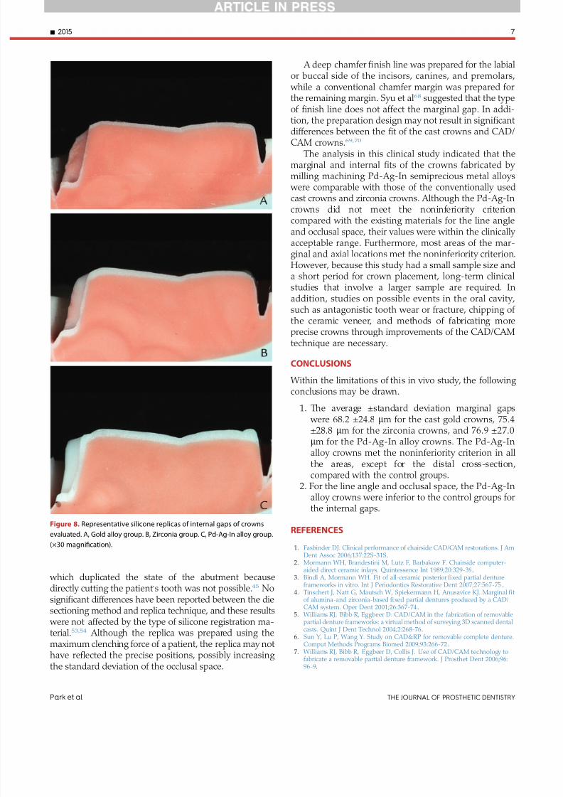
7252019 Varianta 2 Articol Epr
httpslidepdfcomreaderfullvarianta-2-articol-epr 78
which duplicated the state of the abutment becausedirectly cutting the patientrsquos tooth was not possible45 Nosigni1047297cant differences have been reported between the diesectioning method and replica technique and these results
were not affected by the type of silicone registration ma-terial5354 Although the replica was prepared using themaximum clenching force of a patient the replica may nothave re1047298ected the precise positions possibly increasing the standard deviation of the occlusal space
A deep chamfer 1047297nish line was prepared for the labialor buccal side of the incisors canines and premolars
while a conventional chamfer margin was prepared forthe remaining margin Syu et al68 suggested that the typeof 1047297nish line does not affect the marginal gap In addi-tion the preparation design may not result in signi1047297cant
differences between the 1047297
t of the cast crowns and CADCAM crowns6970
The analysis in this clinical study indicated that themarginal and internal 1047297ts of the crowns fabricated by milling machining Pd-Ag-In semiprecious metal alloys
were comparable with those of the conventionally usedcast crowns and zirconia crowns Although the Pd-Ag-Incrowns did not meet the noninferiority criterioncompared with the existing materials for the line angleand occlusal space their values were within the clinically acceptable range Furthermore most areas of the mar-ginal and axial locations met the noninferiority criterionHowever because this study had a small sample size anda short period for crown placement long-term clinicalstudies that involve a larger sample are required Inaddition studies on possible events in the oral cavitysuch as antagonistic tooth wear or fracture chipping of the ceramic veneer and methods of fabricating moreprecise crowns through improvements of the CADCAMtechnique are necessary
CONCLUSIONS
Within the limitations of this in vivo study the following conclusions may be drawn
1 The average plusmnstandard deviation marginal gaps were 682 plusmn248 mm for the cast gold crowns 754plusmn288 mm for the zirconia crowns and 769 plusmn270mm for the Pd-Ag-In alloy crowns The Pd-Ag-Inalloy crowns met the noninferiority criterion in allthe areas except for the distal cross-sectioncompared with the control groups
2 For the line angle and occlusal space the Pd-Ag-Inalloy crowns were inferior to the control groups forthe internal gaps
REFERENCES
1 Fasbinder DJ Clinical performance of chairside CADCAM restorations J AmDent Assoc 200613722S-31S
2 Mormann WH Brandestini M Lutz F Barbakow F Chairside computer-aided direct ceramic inlays Quintessence Int 198920329-39
3 Bindl A Mormann WH Fit of all-ceramic posterior 1047297xed partial dentureframeworks in vitro Int J Periodontics Restorative Dent 200727567-75
4 Tinschert J Natt G Mautsch W Spiekermann H Anusavice KJ Marginal 1047297tof alumina-and zirconia-based 1047297xed partial dentures produced by a CADCAM system Oper Dent 200126367-74
5 Williams RJ Bibb R Eggbeer D CADCAM in the fabrication of removablepartial denture frameworks a virtual method of surveying 3D scanned dentalcasts Quint J Dent Technol 20042268-76
6 Sun Y Lu P Wang Y Study on CADampRP for removable complete dentureComput Methods Programs Biomed 200993266-72
7 Williams RJ Bibb R Eggbeer D Collis J Use of CADCAM technology tofabricate a removable partial denture framework J Prosthet Dent 20069696-9
Figure 8 Representative silicone replicas of internal gaps of crowns
evaluated A Gold alloy group B Zirconia group C Pd-Ag-In alloy group
(times30 magni1047297cation)
- 2015 7
Park et al THE JOURNAL OF PROSTHETIC DENTISTRY
7252019 Varianta 2 Articol Epr
httpslidepdfcomreaderfullvarianta-2-articol-epr 88
8 Andersson M Razzoog ME Oden A Hegenbarth EA Lang BR Procera anew way to achieve an all-ceramic crown Quintessence Int 199829285-96
9 Kapos T Evans C CADCAM technology for implant abutments crownsand superstructures Int J Oral Maxillofac Implants 201429 Suppl117-36
10 Luthardt RG Sandkuhl O Herold V Walter MH Accuracy of mechanicaldigitizing with a CADCAM system for 1047297xed restorations Int J Prosthodont200114146-51
11 McLaren EA Terry DA CADCAM systems materials and clinical guidelinesfor all-ceramic crowns and 1047297xed partial dentures Compend Contin EducDent 200223637-41 44 46 passim quiz 54
12 Piconi C Maccauro G Zirconia as a ceramic biomaterial Biomaterials1999201-25
13 Tinschert J Zwez D Marx R Anusavice KJ Structural reliability of alumina-feldspar- leucite- mica- and zirconia-based ceramics J Dent 200028529-35
14 Ardlin BI Transformation-toughened zirconia for dental inlays crowns andbridges chemical stability and effect of low-temperature aging on 1047298exuralstrength and surface structure Dent Mater 200218590-5
15 Guazzato M Albakry M Ringer SP Swain MV Strength fracture toughnessand microstructure of a selection of all-ceramic materials Part II Zirconia-based dental ceramics Dent Mater 200420449-56
16 Raigrodski AJ Contemporary materials and technologies for all-ceramic 1047297xedpartial dentures a review of the literature J Prosthet Dent 200492557-62
17 Tinschert J Natt G Hassenp1047298ug S Spiekermann H Status of current CADCAM technology in dental medicine Int J Comput Dent 2004725-45
18 Lee DH Lee BJ Kim SH Lee KB Shear bond strength of porcelain to a new millable alloy and a conventional castable alloy J Prosthet Dent 2015113329-35
19 Abduo Fit of CADCAM implant frameworks a comprehensive review
J Oral Implantol 2014 Dec40(6)758-6620 Kapos T Ashy LM Gallucci GO Weber HP Wismeijer D Computer-aided
design and computer-assisted manufacturing in prosthetic implant dentistryInt J Oral Maxillofac Implants 200924110-7
21 Boening KW Walter MH Reppel PD Non-cast titanium restorations in 1047297xedprosthodontics J Oral Rehabil 199219281-7
22 Gilbert JL Covey DA Lautenschlager EP Bond characteristics of porcelainfused to milled titanium Dent Mater 199410134-40
23 Walter M Boning K Reppel PD Clinical performance of machined titaniumrestorations J Dent 199422346-8
24 Goodacre CJ Palladium-silver alloys a review of the literature J ProsthetDent 19896234-7
25 Huget EF Civjan S Status report on palladium-silver-based crown andbridge alloys J Am Dent Assoc 197489383-5
26 Kansu G Aydin AK Evaluation of the biocompatibility of various dentalalloys Part 2e Allergenical potentials Eur J Prosthodont Restor Dent 19964155-61
27 Hong JT Shin SY A comparative study on the bond strength of porcelain to
the millingable Pd-Ag alloy J Adv Prosthodont 20146372-828 Sorensen JA A rationale for comparison of plaque-retaining properties of crown systems J Prosthet Dent 198962264-9
29 Sorensen SE Larsen IB Jorgensen KD Gingival and alveolar bone reactionto marginal 1047297t of subgingival crown margins Scand J Dent Res 198694109-14
30 Assif D Rimer Y Aviv I The 1047298ow of zinc phosphate cement under a full-coverage restoration and its effect on marginal adaptation according to thelocation of cement application Quintessence Int 198718765-74
31 Bader JD Rozier RG McFall WT Jr Ramsey DL Effect of crown margins onperiodontal conditions in regularly attending patients J Prosthet Dent19916575-9
32 Belser UC MacEntee MI Richter WA Fit of three porcelain-fused-to-metalmarginal designs in vivo a scanning electron microscope study J ProsthetDent 19855324-9
33 Felton DA Kanoy BE Bayne SC Wirthman GP Effect of in vivo crownmargin discrepancies on periodontal health J Prosthet Dent 199165357-64
34 Fusayama T Ide K Hosoda H Relief of resistance of cement of full castcrowns J Prosthet Dent 19641495-106
35 Gonzalo E Suarez MJ Serrano B Lozano JF Marginal 1047297t of Zirconia pos-terior 1047297xed partial dentures Int J Prosthodont 200821398-9
36 Grasso JE Nalbandian J Sanford C Bailit H Effect of restoration quality onperiodontal health J Prosthet Dent 19855314-9
37 Grey NJ Piddock V Wilson MA In vitro comparison of conventional crownsand a new all-ceramic system J Dent 19932147-51
38 Hung SH Hung KS Eick JD Chappell RP Marginal 1047297t of porcelain-fused-to-metal and two types of ceramic crown J Prosthet Dent 19906326-31
39 McLean JW von Fraunhofer JA The estimation of cement 1047297lm thickness by an in vivo technique Br Dent J 1971131107-11
40 Palomo F Peden J Periodontal considerations of restorative procedures J Prosthet Dent 197636387-94
41 Passon C Lambert RH Lambert RL Newman S The effect of multiple layersof die-spacer on crown retention Oper Dent 19921742-9
42 Schwartz NL Whitsett LD Berry TG Stewart JL Unserviceable crowns and1047297xed partial dentures life-span and causes for loss of serviceability J AmDent Assoc 1970811395-401
43 Walton JN Gardner FM Agar JR A survey of crown and 1047297xed partial denturefailures length of service and reasons for replacement J Prosthet Dent198656416-21
44 Christensen GJ Marginal 1047297t of gold inlay castings J Prosthet Dent 196616297-305
45 Molin M Karlsson S The 1047297t of gold inlays and three ceramic inlay systems A clinical and in vitro study Acta Odontol Scand 199351201-6
46 Boening KW Wolf BH Schmidt AE Kastner K Walter MH Clinical 1047297t of Procera AllCeram crowns J Prosthet Dent 200084419-24
47 Coli P Karlsson S Fit of a new pressure-sintered zirconium dioxide coping
Int J Prosthodont 20041759-6448 Kohorst P Brinkmann H Dittmer MP Borchers L Stiesch M In1047298uence of
the veneering process on the marginal 1047297t of zirconia 1047297xed dental prostheses J Oral Rehabil 201037283-91
49 Reich S Kappe K Teschner H Schmitt J Clinical 1047297t of four-unit zirconiaposterior 1047297xed dental prostheses Eur J Oral Sci 2008116579-84
50 Reich S Wichmann M Nkenke E Proeschel P Clinical 1047297t of all-ceramicthree-unit 1047297xed partial dentures generated with three different CADCAMsystems Eur J Oral Sci 2005113174-9
51 Wettstein F Sailer I Roos M Hammerle CH Clinical study of the internalgaps of zirconia and metal frameworks for 1047297xed partial dentures Eur J OralSci 2008116272-9
52 Fransson B Oilo G Gjeitanger R The 1047297t of metal-ceramic crowns a clinicalstudy Dent Mater 19851197-9
53 Rahme HY Tehini GE Adib SM Ardo AS Rifai KT In vitro evaluation of theldquoreplica techniquerdquo in the measurement of the 1047297t of Procera crowns
J Contemp Dent Pract 2008925-3254 Laurent M Scheer P Dejou J Laborde G Clinical evaluation of the marginal
1047297t of cast crownse validation of the silicone replica method J Oral Rehabil200835116-22
55 Baig MR Tan KB Nicholls JI Evaluation of the marginal 1047297t of a zirconiaceramic computer-aided machined (CAM) crown system J Prosthet Dent2010104216-27
56 Bindl A Mormann WH Marginal and internal 1047297t of all-ceramic CADCAMcrown-copings on chamfer preparations J Oral Rehabil 200532441-7
57 Hertlein GHS Frank S Suttor D Marginal 1047297t of CADCAM manufactured allceramic prosthesis J Dent Res 20018042-4
58 May KB Russell MM Razzoog ME Lang BR Precision of 1047297t the Procera AllCeram c rown J Prosthet Dent 199880394-404
59 Tan PL Gratton DG Diaz-Arnold AM Holmes DC An in vitro comparisonof vertical marginal gaps of CADCAM titanium and conventional cast res-torations J Prosthodont 200817378-83
60 Kokubo Y Tsumita M Kano T Sakurai S Fukushima S Clinical marginal andinternal gaps of zirconia all-ceramic crowns J Prosthodont Res 20115540-3
61 Chow SC Shao J Wang H Sample size calculations in clinical research 2nded Boca Raton FL Chapman and HallCRC 2007
62 Beuer F Stimmelmayr M Gueth JF Edelhoff D Naumann M In vitro per-formance of full-contour zirconia single crowns Dent Mater 201228449-5663 Kollar A Huber S Mericske E Mericske-Stern R Zirconia for teeth and
implants a case series Int J Periodontics Restorative Dent 200828479-8764 Sturdevant JR Bayne SC Heymann HO Margin gap size of ceramic inlays
using second-generation CADCAM equipment J Esthet Dent 199911206-14
65 Bornemann G Lemelson S Luthardt R Innovative method for the analysisof the internal 3D 1047297tting accuracy of Cerec-3 crowns Int J Comput Dent20025177-82
66 Sorensen JA A standardized method for determination of crown margin 1047297-delity J Prosthet Dent 19906418-24
67 Holmes JR Bayne SC Holland GA Sulik WD Considerations in measure-ment of marginal 1047297t J Prosthet Dent 198962405-8
68 Syu JZ Byrne G Laub LW Land MF In1047298uence of 1047297nish-line geometry onthe 1047297t of crowns Int J Prosthodont 1993625-30
69 Tsitrou EA Northeast SE van Noort R Evaluation of the marginal 1047297t of threemargin designs of resin composite crowns using CADCAM J Dent 20073568-73
70 Ayad MF Effect of the crown preparation margin and die type on the mar-ginal accuracy of 1047297ber-reinforced composite crowns J Contemp Dent Pract200899-16
Corresponding author
Dr Namsik OhInha University 7-2063 Shinhung-dong Jung-guIncheon 400-711REPUBLIC OF KOREA Email onsdoinhaackr
AcknowledgmentsThe authors thank Dr Kyoung-Ae Kong for performing statistical analyses in thisstudy
Copyright copy 2015 by the Editorial Council for The Journal of Prosthetic Dentistry
8 Volume - Issue -
THE JOURNAL OF PROSTHETIC DENTISTRY Park et al