Embed Size (px)
Citation preview
Variation in Cell Surface Hydrophobicity among Cryptococcusneoformans Strains Influences Interactions with Amoebas
Raghav Vij,a* Carina Danchik,a Conor Crawford,a,b Quigly Dragotakes,a Arturo Casadevalla
aDepartment of Molecular Microbiology and Immunology, Johns Hopkins Bloomberg School of Public Health, Baltimore, Maryland, USAbCentre for Synthesis and Chemical Biology, University College Dublin, Belfield, Ireland
ABSTRACT Cryptococcus neoformans and Cryptococcus gattii are pathogenic fungithat cause significant morbidity and mortality. Cell surface hydrophobicity (CSH) is abiophysical parameter that influences the adhesion of fungal cells or spores to bioticand abiotic surfaces. C. neoformans is encased by polysaccharide capsule that ishighly hydrophilic and is a critical determinant of virulence. In this study, we reportlarge differences in the CSH of some C. neoformans and C. gattii strains. The capsularpolysaccharides of C. neoformans strains differ in repeating motifs and therefore varyin the number of hydroxyl groups, which, along with higher-order structure of thecapsule, may contribute to the variation in hydrophobicity that we observed. Wefound that cell wall composition, in the context of chitin-chitosan content, does notinfluence CSH. For C. neoformans, CSH correlated with phagocytosis by natural soilpredator Acanthamoeba castellanii. Furthermore, capsular binding of the protectiveantibody (18B7), but not the nonprotective antibody (13F1), altered the CSH of C.neoformans strains. Variability in CSH could be an important characteristic in com-paring the biological properties of cryptococcal strains.
IMPORTANCE The interaction of a microbial cell with its environment is influencedby the biophysical properties of a cell. The affinity of the cell surface for water, definedby the cell surface hydrophobicity (CSH), is a biophysical parameter that varies amongdifferent strains of Cryptococcus neoformans. The CSH influences the phagocytosis of theyeast by its natural predator in the soil, the amoeba. Studying variation in biophysicalproperties like CSH gives us insight into the dynamic host-predator interaction and host-pathogen interaction in a damage-response framework.
KEYWORDS cell surface hydrophobicity (CSH), Cryptococcus neoformans,Cryptococcus gattii, Acanthamoeba castellanii, capsular antibody, polysaccharidecapsule
The encapsulated Basidiomycetes that comprise the Cryptococcus species complexinclude several pathogenic species including Cryptococcus neoformans and Crypto-
coccus gattii. Cryptococcus spp. have a worldwide geographic distribution and areunusual among fungal pathogens in that they have polysaccharide capsules that areessential for mammalian virulence.
Human infection usually begins in the lung. Infectious propagules of C. neoformans,in the form of spore or yeast, may be inhaled to cause a pulmonary infection that isusually cleared in immunocompetent hosts or becomes latent. Conditions that impairimmunity, such as HIV infection, are associated with disseminated disease, whichusually manifests clinically as a meningoencephalitis. Recent evidence suggests thatthe nature of the infectious propagule has a significant effect on the outcome of theinfection, as spores from C. neoformans cause significantly higher fungal burden in thebrain of a murine model than small encapsulated yeast (1).
C. neoformans has been isolated from avian guano, soil, or arboreal sources. C. gattii
Citation Vij R, Danchik C, Crawford C,Dragotakes Q, Casadevall A. 2020. Variation incell surface hydrophobicity among Cryptococcusneoformans strains influences interactions withamoebas. mSphere 5:e00310-20. https://doi.org/10.1128/mSphere.00310-20.
Editor J. Andrew Alspaugh, Duke UniversityMedical Center
Copyright © 2020 Vij et al. This is an open-access article distributed under the terms ofthe Creative Commons Attribution 4.0International license.
Address correspondence to Arturo Casadevall,[email protected].
* Present address: Raghav Vij, Department ofMicrobial Pathogenicity Mechanisms, LeibnizInstitute for Natural Product Research andInfection Biology—Hans-Knöll-Institute, Jena,Germany.
Significant variation in cell surfacehydrophobicity of C. neoformans strains,correlates with phagocytosis by naturalpredator, amoeba. @ACasadevall1
Received 6 April 2020Accepted 14 April 2020Published
RESEARCH ARTICLEHost-Microbe Biology
crossm
March/April 2020 Volume 5 Issue 2 e00310-20 msphere.asm.org 1
29 April 2020
on August 1, 2020 by guest
http://msphere.asm
.org/D
ownloaded from

has been isolated from trees, soil, freshwater, and seawater. There are three serotypesof C. neoformans, now referred to as Cryptococcus neoformans var. neoformans (serotypeD), Cryptococcus neoformans var. grubii (serotype A), and hybrid (serotype AD). Phylo-genetic evidence suggests that they may be classified as separate species, C. neofor-mans, Cryptococcus deneoformans, and hybrid, respectively (2). Interestingly, C. neofor-mans var. grubii has been isolated from 63% of clinical samples collected worldwide,followed by C. neoformans hybrid (6%) and C. neoformans var. neoformans (5%) (3, 4).The genomic diversity in the cryptococcal species complex may contribute to differ-ences in the biophysical properties of cell surfaces within the Cryptococcus speciescomplex.
C. neoformans and C. gattii cells are surrounded by a polysaccharide capsule that candramatically vary in size during infection (5) and helps the pathogen evade themammalian immune system. Highly branched polysaccharides (6) radiate outward fromthe cell wall, to form a dense matrix whose porosity increases with the distance fromthe cell wall, with reducing ends localized at the cell wall (7, 8). The capsule is primarilycomposed of glucuronoxylomannan (GXM; 98%), along with minor components glucu-ronoxylomannogalactan (GXMGal) and mannoproteins. GXM contains a core repeatingstructure of a �-(1¡3)-mannose triad, with a �-(1¡2) glucuronic acid branch on everythird mannose (9). The capsules of different serotypes of C. neoformans and C. gattiihave distinguishable polysaccharide motifs characterized by a varied degree of �-(1¡2)or �-(1¡4) xylose substitutions, and 6-O-acetyl substitutions along the mannan back-bone (10). Polysaccharides are highly enriched in hydroxyl groups and form an exten-sive network of intramolecular and intermolecular hydrogen bonds, which includesbonding with water molecules. Therefore, polysaccharides are intrinsically hydrophilicmolecules, which could provide an explanation for approximately 95% of the capsule’sweight (11). Structure-function relationships in glycans are poorly understood, butbranching and substitution of polysaccharides likely affect the intra- and intermolecularhydrogen bonds, and therefore the rigidity of the polymer, thereby affecting thepolysaccharide’s ability to form hydrogen bonds with water, which would result invariation of the conformation and hydrophobicity (12–15).
Factors that affect the biophysical parameters of the microbial surface of theCryptococcus species complex have been previously described. For instance, melaniza-tion, capsule induction, and binding of capsular antibody alter the cell surface charge,which also varies by strain (16). Chronological aging of the yeast and antibody bindingalter the elasticity of the polysaccharide capsule that surrounds the C. neoformans cell(17, 18).
Cell surface hydrophobicity (CSH) is a property of a microbial surface that reflects theaffinity of components of the microbe’s cell surface for water and is calculated byestimating the affinity of cell surfaces for hydrophobic substances like hydrophobiccolumns, solvents, or polystyrene beads (Fig. 1). The biological role of CSH has beenstudied in bacteria such as Staphylococcus aureus and some fungi and has beensuccinctly reviewed (19). Previous studies of Candida albicans have established theimportance of CSH for the interaction of the pathogen with the host tissue (20).Furthermore, strain-specific variation in CSH of clinical isolates and variation betweenspecies of Candida species complex have been reported (21).
The biophysical properties of the infectious propagule of C. neoformans in the formof yeast or spore influence the interaction of the yeast with its environment and insidethe host during infection. For example, during infection, C. neoformans interacts withlung epithelial cells and macrophages and can pass through the blood-brain barrier. Inthe environment, Cryptococcus species complex is believed to interact with amoebas(22) and nematodes (23). Furthermore, hydrophobicity may influence the phagocytosisof microbial cells or particles by amoebas (24).
In this study, we report variation in CSH of C. neoformans and C. gattii strains usingtwo independent methods. Further, we observed that CSH correlated positively withphagocytosis by Acanthamoeba castellanii. Additionally, the higher-order structure ofthe capsule is affected by the different capsular polysaccharide motifs that vary
Vij et al.
March/April 2020 Volume 5 Issue 2 e00310-20 msphere.asm.org 2
on August 1, 2020 by guest
http://msphere.asm
.org/D
ownloaded from

between serotypes of C. neoformans and C. gattii, which may influence the CSH. We alsofound that binding of protective but not nonprotective antibodies altered the hydro-phobicity of C. neoformans grown in capsule induction medium.
RESULTSCryptococcal species manifest significant differences in CSH. Measuring CSH by
the MATH (microbial adhesion to hydrocarbons) and hydrophobic microsphere tech-niques (Fig. 1) revealed considerable variability among cells of C. neoformans and C.gattii strains cultured in Sabouraud dextrose broth (Fig. 2). By MATH assay, we foundthat serotype D strains B3501 and JEC21 were significantly more hydrophobic than thereference strain H99 (Fig. 2A). By the hydrophobic microsphere assay, we found that allstrains of serotype D for which CSH was estimated, including B3501, ATCC 24067, andJEC21, were significantly more hydrophobic than the reference strain H99 (Fig. 2B).However, there was considerable strain-to-strain variation, and no pattern emergedregarding differences between serotypes or species, except for the notable finding thatthe most strains manifesting highest CSH were C. neoformans serotype D.
Capsule composition may influence the CSH. The capsule that surrounds the C.neoformans cell is thought to be highly hydrophilic and is primarily composed of water(11). Hence, we sought to ascertain its contribution to CSH in C. neoformans strain H99(serotype A) by comparing encapsulated H99 and the nonencapsulated cap59 strain. Toour surprise, we observed no major difference in CSH between H99 and cap59 cellsgrown in Sabouraud dextrose broth, by the MATH assay (Fig. 3A). However, whengrown in capsule-inducing minimum medium (25), the nonencapsulated strain boundmore hydrophobic beads than the encapsulated strains (Fig. 3B). Next, we comparedthe CSH of C. neoformans strain B3501 (serotype D) to the acapsular cap67 mutant, onthe B3501 background (26, 27). We observed a significant decrease in the CSH by MATHassay (Fig. 3A).
Different strains and serotypes of C. neoformans and C. gattii have different domi-nant carbohydrate motifs in their capsule (10) that may influence the experimentallyobserved variation in CSH. To test this hypothesis, we used in silico method describedby Mannhold et al. (28), to calculate and compare the lipophilicity (log P) of the fourdominant GXM motifs. We observed the following trend in the predicted lipophilicityof GXM carbohydrate motifs: M4 (dominant in serotype C, log P 2.12) � M3 (dominant
FIG 1 Methods for estimation of C. neoformans CSH. (A) CSH estimated by MATH assay that quantifies theinteraction of C. neoformans cells in a suspension with the hydrocarbon solvent n-hexadecane. CSH% wascalculated as the percent change in OD of a C. neoformans cell suspension after vortexing the mixture of cells withn-hexadecane. (B) In addition, we estimated CSH by visualizing the interaction between C. neoformans cells andhydrophobic beads (0.8 �m) in a hemocytometer and counting cells that had �3 beads/100 cells to calculateCSH%. Image created with BioRender.
Cell Surface Hydrophobicity of C. neoformans Strains
March/April 2020 Volume 5 Issue 2 e00310-20 msphere.asm.org 3
on August 1, 2020 by guest
http://msphere.asm
.org/D
ownloaded from

in serotype B, log P 2.01) � M2 (dominant in serotype A, log P 1.9) � M1 (dominant inserotype D, log P 1.79) (Fig. 3C).
Based on the rationale that polysaccharides enriched in a greater number ofhydroxyl groups would have higher hydrophilicity, we counted the number of hydroxylgroups of each dominant GXM motif (Fig. 3C). The M4 motif (dominant in serotype C)contained the highest number of hydroxyl groups, 21, followed by 19 hydroxyl groupsin M3 (dominant in serotype B), 17 hydroxyl groups in M2 (dominant in serotype A), and15 hydroxyl groups in M1 (dominant in serotype D).
Next, we wanted to investigate if the cell wall composition influences the CSH of C.neoformans. The enzymes chitin deacetylases, encoded by the cda gene family,deacetylate chitin, a hydrophobic polymer that is an important cell wall component, tochitosan, a hydrophilic polymer (29). We found no significant differences between wildtype and strains knocked out for cda1 and for cda1, cda2, and cda3 (Fig. 3D).
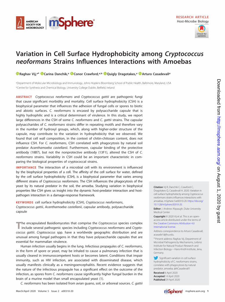
CSH of unopsonized C. neoformans correlates with phagocytosis by A. castel-lanii. To test whether CSH influences phagocytosis by soil predators like the amoeba,we incubated fungal and protozoal cells and estimated the phagocytosis index. Wefound a positive and linear correlation between CSH of C. neoformans strains andphagocytosis index of C. neoformans strains by A. castellanii (Fig. 4). In this series ofisolates, the phagocytosis index also correlated with strain serotype.
FIG 2 CSH of C. neoformans differs by strain. Graphical representation of CSH of C. neoformans and C.gattii strains. (A) Graphical representation of CSH estimated by MATH assay. (B) Graphical representationof CSH estimated by hydrophobic microsphere assay (left). Experiments have been performed 2 to 6times independently, as indicated by individual data points. Circles indicate data points of CSH of anencapsulated strain of C. neoformans and C. gattii. Error bars represent the standard deviation of themean. (C) Representative image of a mixture of hydrophobic beads with C. neoformans strain H99 (top)and relatively hydrophobic C. neoformans strain B3501 (bottom) used for the assay. Hydrophobic beads(small spheres, approximately 0.8 �m in diameter) adhere to the cell surface due to the high hydropho-bicity of the B3501 cell, covering it almost completely. The hydrophobic beads are all but absent fromthe surface of H99 cells. Ordinary one-way analysis of variance was used to compare the CSH of C.neoformans strain H99 with the CSH of C. neoformans and C. gattii strains. The following symbols wereused to annotate the statistical significance of the results: ***, P � 0.001; ****, P � 0.0001.
Vij et al.
March/April 2020 Volume 5 Issue 2 e00310-20 msphere.asm.org 4
on August 1, 2020 by guest
http://msphere.asm
.org/D
ownloaded from

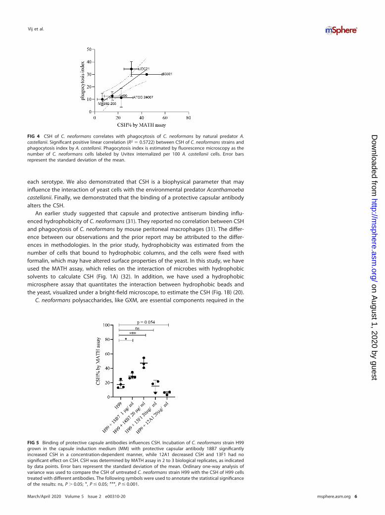
Effect of antibody binding on CSH. Previous studies have demonstrated thatcapsule antibody binding alters capsule structure and changes the surface charge of C.neoformans (16, 17). This led us to investigate the effect of binding of capsularantibodies to C. neoformans on the CSH. We demonstrated that binding of capsularantibody 18B7 (30) increases CSH in a concentration-dependent manner, while bindingof nonprotective antibody 13F1 has no significant effect on the CSH of C. neoformanscells grown in the capsule induction medium (Fig. 5).
DISCUSSION
In this study, we measured the CSH of C. neoformans and found considerableinterstrain variation. When CSH was estimated by hydrophobic microsphere assay, C.neoformans serotype D strains were likely to be more hydrophobic than C. neoformansserotype A strains, with the caveat that we analyzed a relatively small set of strains from
FIG 3 Capsular motifs, and not the cell wall composition, may contribute to the variation in hydrophobicity of C. neoformans.(A) Comparison of the CSH of C. neoformans H99 grown in Sabouraud broth with those of C. neoformans H99 grown in MMand acapsular cap59 strain and comparison of the CSH of strain B3501 with that of acapsular cap67 strain. One-way analysisof variance and t test were used, respectively, to compare the means. Data have been compiled from separate experimentsto draw comparisons and study the influence of presence or induction of capsule on the CSH of C. neoformans. Each data pointrepresents a biological replicate, and the error bar represents the SD of the mean. (B) CSH% measured by a hydrophobic beadassay of C. neoformans strain H99 and acapsular strain cap59 grown in capsule induction medium. The experiment wasperformed in two independent replicates, as represented by data points about the median. (C) Lipophilicity, log P, of dominantcarbohydrate motifs in the carbohydrate was predicted by an equation proposed by Mannhold et al. (28). M4 was found tobe the most hydrophobic motif and M1 the least. The number of hydroxyl groups on each polysaccharide motif was calculated(below). Glycan notification followed the Symbol Nomenclature for Glycans (SNFG) (71). (D) No significant differences werefound when the CSH% of C. neoformans strain K99 was compared to those of chitin deacetylase 1 mutant (cda1�) and chitindeacetylase triple-knockout mutant (cda123�) strains as measured by hydrophobic microsphere assay. The following symbolswere used to annotate the statistical significance of the results: *, P � 0.05.
Cell Surface Hydrophobicity of C. neoformans Strains
March/April 2020 Volume 5 Issue 2 e00310-20 msphere.asm.org 5
on August 1, 2020 by guest
http://msphere.asm
.org/D
ownloaded from
each serotype. We also demonstrated that CSH is a biophysical parameter that mayinfluence the interaction of yeast cells with the environmental predator Acanthamoebacastellanii. Finally, we demonstrated that the binding of a protective capsular antibodyalters the CSH.
An earlier study suggested that capsule and protective antiserum binding influ-enced hydrophobicity of C. neoformans (31). They reported no correlation between CSHand phagocytosis of C. neoformans by mouse peritoneal macrophages (31). The differ-ence between our observations and the prior report may be attributed to the differ-ences in methodologies. In the prior study, hydrophobicity was estimated from thenumber of cells that bound to hydrophobic columns, and the cells were fixed withformalin, which may have altered surface properties of the yeast. In this study, we haveused the MATH assay, which relies on the interaction of microbes with hydrophobicsolvents to calculate CSH (Fig. 1A) (32). In addition, we have used a hydrophobicmicrosphere assay that quantitates the interaction between hydrophobic beads andthe yeast, visualized under a bright-field microscope, to estimate the CSH (Fig. 1B) (20).
C. neoformans polysaccharides, like GXM, are essential components required in the
FIG 4 CSH of C. neoformans correlates with phagocytosis of C. neoformans by natural predator A.castellanii. Significant positive linear correlation (R2 � 0.5722) between CSH of C. neoformans strains andphagocytosis index by A. castellanii. Phagocytosis index is estimated by fluorescence microscopy as thenumber of C. neoformans cells labeled by Uvitex internalized per 100 A. castellanii cells. Error barsrepresent the standard deviation of the mean.
FIG 5 Binding of protective capsule antibodies influences CSH. Incubation of C. neoformans strain H99grown in the capsule induction medium (MM) with protective capsular antibody 18B7 significantlyincreased CSH in a concentration-dependent manner, while 12A1 decreased CSH and 13F1 had nosignificant effect on CSH. CSH was determined by MATH assay in 2 to 3 biological replicates, as indicatedby data points. Error bars represent the standard deviation of the mean. Ordinary one-way analysis ofvariance was used to compare the CSH of untreated C. neoformans strain H99 with the CSH of H99 cellstreated with different antibodies. The following symbols were used to annotate the statistical significanceof the results: ns, P � 0.05; *, P � 0.05; ***, P � 0.001.
Vij et al.
March/April 2020 Volume 5 Issue 2 e00310-20 msphere.asm.org 6
on August 1, 2020 by guest
http://msphere.asm
.org/D
ownloaded from

formation of microbial communities called biofilms that are protective for the fungi(33). C. neoformans biofilms have been reported on medical devices (34, 35). Biofilm-associated cells have been associated with increased tolerance against antifungal drugsand phagocytic cells, as they upregulate proteins associated with host defense (36–38).In vivo, C. neoformans forms biofilm-like structures called cryptococcomas that couldplay a role in its neurotropism (39). The surface property of cells may affect theaggregation of microbial communities in biofilms. Interestingly, ATCC 24067 and B3501strains, which are highly hydrophobic, also form biofilms more easily than the H99strain, which is relatively less hydrophobic (Fig. 1) (36, 37). A similar correlation betweenthe formation of biofilm and CSH was observed in Candida spp. (21, 40, 41) and inbacteria (42). Flocculation, another multicellular phenotype observed in yeasts, hasbeen observed in C. neoformans cells during growth in certain media (43) and could becaused by changes in CSH, as reported for brewer’s yeast (44).
Amoebas are natural predators of Cryptococcus species (22, 45) and have emergedas a powerful tool for studying mechanisms of intracellular pathogenesis and evolutionof virulence (46, 47). A growing body of evidence suggests that virulence traits haveemerged in environmental fungi, including Cryptococcus species, because of the selec-tion pressure that results from fungus-amoeba interaction (48). Our finding that themore hydrophobic Cryptococcus strains were more readily phagocytosed is congruentwith the observation that amoebas can phagocytose hydrophobic particles (24), al-though these mechanisms are not well understood. There is a remarkable correspon-dence between C. neoformans virulence traits that influence phagocytosis and enablesurvival of the fungi in A. castellanii and in human macrophages (46). For instance, thecapsule of C. neoformans masks cell wall components that are recognized by innateimmune receptors (49), and the absence of capsule leads to poor survival of C.neoformans incubated with A. castellanii (46). In vitro studies of macrophage and C.neoformans interaction usually require opsonins such as capsular antibodies and com-plement (50, 51) for phagocytosis by innate immune cells. Since opsonins change theCSH of C. neoformans and without opsonins phagocytosis of cryptococcal cells bymacrophages is essentially nil (52), comparable studies with mammalian phagocyticcells are difficult to do. Studying the effect of CSH on phagocytosis in amoebas maygive insights into factors independent of opsonin-receptor interaction that may influ-ence phagocytosis in macrophages.
Murine antibodies that recognize capsular epitopes of C. neoformans can conferpassive protection on the host and enhance macrophage activity (53, 54). In additionto facilitating phagocytosis of the yeast, the murine IgG antibody 18B7 (30) alterscapsule stiffness and impairs cellular replication of the yeast (17), significantly alters thecell surface charge (16), and has a catalytic activity that breaks down the capsule (55).In this study, we report that monoclonal antibody (MAb) 18B7 binding significantlyincreased the hydrophobicity of the cryptococcal cell surface in a concentration-dependent manner, while a nonprotective antibody, IgM 13F1, did not alter the CSH.We may attribute the differential effect of changes in CSH induced by MAbs 13F1 and18B7 to the pattern of MAb binding, since MAb 18B7 binds near the surface in anannular pattern (17, 30, 56), while MAb 13F1 binds throughout the capsule in apunctate pattern (57, 58). There is precedence for our observation in the encapsulatedbacterium Klebsiella aerogenes, where the pattern of diffusion of some MAbs throughthe polysaccharide capsule has been shown to influence the cell surface hydrophobic-ity (59, 60).
A surprising result in our study was that some C. neoformans strains manifest aconsiderably higher CSH than others, despite being surrounded by a hydrophiliccapsule. The origin and mechanism for variability in CSH in these strains are notunderstood. Glycans are intrinsically hydrophilic molecules. Lipophilicity for glycansmay be described by the partition coefficient (P), which is quantified as the distributionof a compound between two immiscible solvents, like water and octanol (61). Whileprior studies have compared the lipophilicities for monosaccharides, these efforts arenot standardized in the field (14). For small molecules, log P can be accurately predicted
Cell Surface Hydrophobicity of C. neoformans Strains
March/April 2020 Volume 5 Issue 2 e00310-20 msphere.asm.org 7
on August 1, 2020 by guest
http://msphere.asm
.org/D
ownloaded from
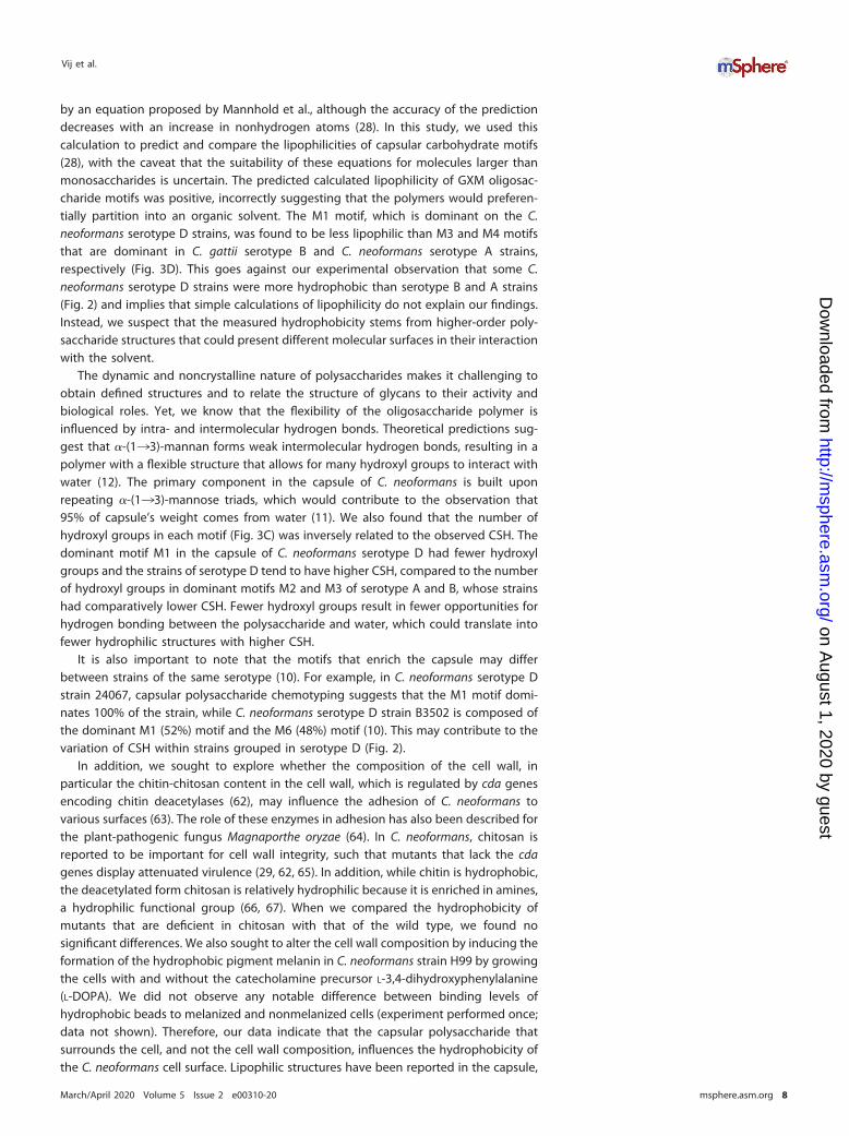
by an equation proposed by Mannhold et al., although the accuracy of the predictiondecreases with an increase in nonhydrogen atoms (28). In this study, we used thiscalculation to predict and compare the lipophilicities of capsular carbohydrate motifs(28), with the caveat that the suitability of these equations for molecules larger thanmonosaccharides is uncertain. The predicted calculated lipophilicity of GXM oligosac-charide motifs was positive, incorrectly suggesting that the polymers would preferen-tially partition into an organic solvent. The M1 motif, which is dominant on the C.neoformans serotype D strains, was found to be less lipophilic than M3 and M4 motifsthat are dominant in C. gattii serotype B and C. neoformans serotype A strains,respectively (Fig. 3D). This goes against our experimental observation that some C.neoformans serotype D strains were more hydrophobic than serotype B and A strains(Fig. 2) and implies that simple calculations of lipophilicity do not explain our findings.Instead, we suspect that the measured hydrophobicity stems from higher-order poly-saccharide structures that could present different molecular surfaces in their interactionwith the solvent.
The dynamic and noncrystalline nature of polysaccharides makes it challenging toobtain defined structures and to relate the structure of glycans to their activity andbiological roles. Yet, we know that the flexibility of the oligosaccharide polymer isinfluenced by intra- and intermolecular hydrogen bonds. Theoretical predictions sug-gest that �-(1¡3)-mannan forms weak intermolecular hydrogen bonds, resulting in apolymer with a flexible structure that allows for many hydroxyl groups to interact withwater (12). The primary component in the capsule of C. neoformans is built uponrepeating �-(1¡3)-mannose triads, which would contribute to the observation that95% of capsule’s weight comes from water (11). We also found that the number ofhydroxyl groups in each motif (Fig. 3C) was inversely related to the observed CSH. Thedominant motif M1 in the capsule of C. neoformans serotype D had fewer hydroxylgroups and the strains of serotype D tend to have higher CSH, compared to the numberof hydroxyl groups in dominant motifs M2 and M3 of serotype A and B, whose strainshad comparatively lower CSH. Fewer hydroxyl groups result in fewer opportunities forhydrogen bonding between the polysaccharide and water, which could translate intofewer hydrophilic structures with higher CSH.
It is also important to note that the motifs that enrich the capsule may differbetween strains of the same serotype (10). For example, in C. neoformans serotype Dstrain 24067, capsular polysaccharide chemotyping suggests that the M1 motif domi-nates 100% of the strain, while C. neoformans serotype D strain B3502 is composed ofthe dominant M1 (52%) motif and the M6 (48%) motif (10). This may contribute to thevariation of CSH within strains grouped in serotype D (Fig. 2).
In addition, we sought to explore whether the composition of the cell wall, inparticular the chitin-chitosan content in the cell wall, which is regulated by cda genesencoding chitin deacetylases (62), may influence the adhesion of C. neoformans tovarious surfaces (63). The role of these enzymes in adhesion has also been described forthe plant-pathogenic fungus Magnaporthe oryzae (64). In C. neoformans, chitosan isreported to be important for cell wall integrity, such that mutants that lack the cdagenes display attenuated virulence (29, 62, 65). In addition, while chitin is hydrophobic,the deacetylated form chitosan is relatively hydrophilic because it is enriched in amines,a hydrophilic functional group (66, 67). When we compared the hydrophobicity ofmutants that are deficient in chitosan with that of the wild type, we found nosignificant differences. We also sought to alter the cell wall composition by inducing theformation of the hydrophobic pigment melanin in C. neoformans strain H99 by growingthe cells with and without the catecholamine precursor L-3,4-dihydroxyphenylalanine(L-DOPA). We did not observe any notable difference between binding levels ofhydrophobic beads to melanized and nonmelanized cells (experiment performed once;data not shown). Therefore, our data indicate that the capsular polysaccharide thatsurrounds the cell, and not the cell wall composition, influences the hydrophobicity ofthe C. neoformans cell surface. Lipophilic structures have been reported in the capsule,
Vij et al.
March/April 2020 Volume 5 Issue 2 e00310-20 msphere.asm.org 8
on August 1, 2020 by guest
http://msphere.asm
.org/D
ownloaded from
which might extend to the surface and influence the hydrophobicity of the cell surface(12, 68).
In summary, we report that CSH of Cryptococcus species can differ significantlydepending on the strain. We have also demonstrated the correlation of the biophysicalparameter CSH with the phagocytosis by A. castellanii and shown that protectiveantibodies that bind to the capsule of C. neoformans can influence the hydrophobicityof C. neoformans. The findings that C. neoformans strains differ in CSH and that changesto this cell surface property correlates with biological properties suggest that theinvestigation of how this parameter is established and maintained could provide newinsights into capsular structure.
MATERIALS AND METHODSStrains and culture of C. neoformans and C. gattii. Cryptococcus neoformans and C. gattii strains
(Table 1) stored as frozen stocks at �80°C were streaked onto Sabouraud agar plates and incubated at30°C for 48 h. The plates were stored at 4°C for use up to 1 week. Multiple colonies were selected andinoculated into 5 ml of liquid medium and Sabouraud broth and incubated at 30°C with shaking for2 days. For capsule induction, 106 cells/ml were washed twice in phosphate-buffered saline (PBS) andinoculated into MM (10 mM MgSO4, 29.3 mM KH2PO4, 13 mM glycine, 3 �M thiamine-HCl, and 15 mMdextrose, with pH adjusted to 5.5).
Antibody incubation. C. neoformans (H99) grown in MM was washed twice in PBS. Protective andnonprotective capsular antibodies, 18B7, 12A1, and 13F1 (69), respectively, were incubated for 1 h at30°C with shaking. The CSH percentage (CSH%) was determined by MATH and microsphere assays asdetailed below.
Estimation of CSH by MATH. CSH was estimated by the MATH assay as described in reference 32.Yeast cultures were washed twice in PBS and resuspended in 3 ml of PBS at an estimated initial opticaldensity (OD) of 0.2 to 0.4 recorded as A0. An 0.4-ml amount of n-hexadecane was added, and the mixturewas vortexed for 30 s and incubated at 30°C for 2 min to allow the layers to separate. Final OD (A1) ofthe aqueous layer was recorded and estimated as an average from 3 technical replicates in a 96-wellplate read by an Emax Plus microplate reader (Molecular Devices). CSH% was estimated as [1 �(A0/A1)] � 100.
Estimation of CSH by hydrophobic microsphere assay. CSHs of C. neoformans and C. gattii wereestimated by the method detailed in reference 20 by adding 9.02 � 108 0.8-�m green hydrophobicbeads (Bang Laboratories) to 2 ml of 2 � 106 cells/ml in sodium phosphate buffer (0.05 M, pH 7.2) in cleanglass tubes. After equilibration at room temperature (RT) for 2 min, the mixture was vortexed vigorouslyfor 30 s. Cells were imaged at 40�, 100 cells were counted, and the percentage of cells having �3attached microspheres was considered the CSH% value.
Acanthamoeba castellanii culture. Acanthamoeba castellanii strain 30234 was obtained from theAmerican Type Culture Collection (ATCC). Cultures were maintained in PYG broth (ATCC medium 712) at25°C according to instructions from ATCC.
TABLE 1 Strains of C. neoformans and C. gattii used in the present study
Species and strain Mutant Serotype Source or referencea
Cryptococcus neoformansH99 A John Perfect (Durham, NC)
cap59K99 29
cda1Δ 29cda1,2,3Δ 29
J45 72J10 72J43 72J48 72SB6 73MAS92-203 AD 6ATCC 24067 D 6B3501 6
cap67 27JEC21 74J39 72
Cryptococcus gattiiR265 B ATCC (Manassas, VA) (75)NIH444 6WM179 ATCC (Manassas, VA) (75)
aThe references indicate the study in which the strains were serotyped or the study in which the strainsused had been characterized by serotype.
Cell Surface Hydrophobicity of C. neoformans Strains
March/April 2020 Volume 5 Issue 2 e00310-20 msphere.asm.org 9
on August 1, 2020 by guest
http://msphere.asm
.org/D
ownloaded from
Acanthamoeba castellanii phagocytosis index. The phagocytosis index was estimated as detailedin reference 70 with minor modifications. Briefly, 5 � 105 cells/ml of A. castellanii were incubated in35-mm no. 1.5 coverslip MatTek dishes with Dulbecco’s PBS (DPBS) (Ca2� and Mg2�) for 3 to 4 h. C.neoformans or C. gattii strains were incubated with 10 �g/ml Uvitex (fungal cell wall dye), inoculated ata multiplicity of infection (MOI) of 1 and incubated for 2 h at 25°C. The cells were imaged using a ZeissAxiovert 200M inverted microscope with a 20� phase objective. Phagocytosis index was estimated bycounting the number of C. neoformans or C. gattii cells engulfed per 100 amoeboid cells.
Estimation of lipophilicity and number of hydroxyl groups in carbohydrate motifs. The lipo-philicity of the carbohydrate motif dominant in the capsule of an C. neoformans serotype was estimatedby the method described by Mannhold et al. (28), as the log of the partition coefficient (P): log P � 1.46(�0.02) � 0.11 (�0.001)NC � 0.11 (�0.001)NHET, where NC is the number of carbon atoms in a moleculeand NHET is the number of hetero atoms.
The number of hydroxyl groups in each motif of C. neoformans capsule was counted manually, asproxy for the number of hydrogen bond donor and acceptor atoms.
ACKNOWLEDGMENTSA.C. is supported by grants HL059842, AI052733, and AI152078. C.C. was funded by
an Irish Research Council postgraduate award (GOIPG/2016/998).Special thanks to Jennifer K. Lodge at Department of Molecular Microbiology,
Washington University School of Medicine, St. Louis, MO, for generously gifting us withthe cda knockout strains. Special thanks to Radames J. B. Cordero for valuable discus-sions of experimental design and edits to the manuscript and to Daniel F. Q. Smith forthe valuable contribution of editing the figures.
R.V. designed and conducted the experiments, analyzed the data, and wrote themanuscript. C.C. performed computation and theoretical analysis and wrote the man-uscript. C.D. and Q.D. conducted experiments and analyzed data. A.C. contributed tothe experimental design, supervised the experiments, and edited and wrote parts ofthe manuscript.
REFERENCES1. Walsh NM, Botts MR, McDermott AJ, Ortiz SC, Wüthrich M, Klein B, Hull CM.
2019. Infectious particle identity determines dissemination and diseaseoutcome for the inhaled human fungal pathogen Cryptococcus. PLoSPathog 15:e1007777. https://doi.org/10.1371/journal.ppat.1007777.
2. Kwon-Chung KJ, Bennett JE, Wickes BL, Meyer W, Cuomo CA, Wollen-burg KR, Bicanic TA, Castañeda E, Chang YC, Chen J, Cogliati M, DromerF, Ellis D, Filler SG, Fisher MC, Harrison TS, Holland SM, Kohno S, KronstadJW, Lazera M, Levitz SM, Lionakis MS, May RC, Ngamskulrongroj P,Pappas PG, Perfect JR, Rickerts V, Sorrell TC, Walsh TJ, Williamson PR, XuJ, Zelazny AM, Casadevall A. 2017. The case for adopting the “speciescomplex” nomenclature for the etiologic agents of cryptococcosis.mSphere 2:e00357-16. https://doi.org/10.1128/mSphere.00357-16.
3. Kwon-Chung KJ, Fraser JA, Doering TL, Wang Z, Janbon G, Idnurm A,Bahn Y-S. 2014. Cryptococcus neoformans and Cryptococcus gattii, theetiologic agents of cryptococcosis. Cold Spring Harb Perspect Med4:a019760. https://doi.org/10.1101/cshperspect.a019760.
4. Meyer W, Gilgado F, Ngamskulrungroj P, Trilles L, Hagen F, Castañeda E,Boekhout T. 2011. Molecular typing of the Cryptococcus neoformans/Cryptococcus gattii species complex, p 327–357. In Heitman J, Kozel T,Kwon-Chung K, Perfect J, Casadevall A (ed), Cryptococcus: from humanpathogen to model yeast. ASM Press, Washington, DC.
5. Charlier C, Chrétien F, Baudrimont M, Mordelet E, Lortholary O, DromerF. 2005. Capsule structure changes associated with Cryptococcus neo-formans crossing of the blood-brain barrier. Am J Pathol 166:421– 432.https://doi.org/10.1016/S0002-9440(10)62265-1.
6. Cordero RJB, Frases S, Guimaräes AJ, Rivera J, Casadevall A. 2011.Evidence for branching in cryptococcal capsular polysaccharides andconsequences on its biological activity. Mol Microbiol 79:1101–1117.https://doi.org/10.1111/j.1365-2958.2010.07511.x.
7. Gates MA, Thorkildson P, Kozel TR. 2004. Molecular architecture of theCryptococcus neoformans capsule. Mol Microbiol 52:13–24. https://doi.org/10.1111/j.1365-2958.2003.03957.x.
8. Crawford CJ, Cordero RJB, Guazzelli L, Wear MP, Bowen A, Oscarson S,Casadevall A. 2020. Exploring Cryptococcus neoformans capsule struc-ture and assembly with a hydroxylamine-armed fluorescent probe. J BiolChem 295:4327– 4340. https://doi.org/10.1074/jbc.RA119.012251.
9. Cherniak R, Morris LC, Belay T, Spitzer ED, Casadevall A. 1995. Variation
in the structure of glucuronoxylomannan in isolates from patients withrecurrent cryptococcal meningitis. Infect Immun 63:1899 –1905. https://doi.org/10.1128/IAI.63.5.1899-1905.1995.
10. Cherniak R, Valafar H, Morris LC, Valafar F. 1998. Cryptococcus neofor-mans chemotyping by quantitative analysis of 1H nuclear magneticresonance spectra of glucuronoxylomannans with a computer-simulated artificial neural network. Clin Diagn Lab Immunol 5:146 –159.https://doi.org/10.1128/CDLI.5.2.146-159.1998.
11. Maxson ME, Cook E, Casadevall A, Zaragoza O. 2007. The volume andhydration of the Cryptococcus neoformans polysaccharide capsule. Fun-gal Genet Biol 44:180 –186. https://doi.org/10.1016/j.fgb.2006.07.010.
12. Almond A. 2005. Towards understanding the interaction between oli-gosaccharides and water molecules. Carbohydr Res 340:907–920.https://doi.org/10.1016/j.carres.2005.01.014.
13. Yu Y, Tyrikos�Ergas T, Zhu Y, Fittolani G, Bordoni V, Singhal A, Fair RJ,Grafmüller A, Seeberger PH, Delbianco M. 2019. Systematic hydrogen-bond manipulations to establish polysaccharide structure–property cor-relations. Angew Chem 131:13261–13266. https://doi.org/10.1002/ange.201906577.
14. Fu D, O’Neill RA. 1995. Monosaccharide composition analysis of oligo-saccharides and glycoproteins by high-performance liquid chromatog-raphy. Anal Biochem 227:377–384. https://doi.org/10.1006/abio.1995.1294.
15. Delbianco M, Kononov A, Poveda A, Yu Y, Diercks T, Jiménez-Barbero J,Seeberger PH. 2018. Well-defined oligo- and polysaccharides as idealprobes for structural studies. J Am Chem Soc 140:5421–5426. https://doi.org/10.1021/jacs.8b00254.
16. Nosanchuk JD, Casadevall A. 1997. Cellular charge of Cryptococcusneoformans: contributions from the capsular polysaccharide, melanin,and monoclonal antibody binding. Infect Immun 65:1836 –1841. https://doi.org/10.1128/IAI.65.5.1836-1841.1997.
17. Cordero RJB, Pontes B, Frases S, Nakouzi AS, Nimrichter L, Rodrigues ML,Viana NB, Casadevall A. 2013. Antibody binding to Cryptococcus neo-formans impairs budding by altering capsular mechanical properties. JImmunol 190:317–323. https://doi.org/10.4049/jimmunol.1202324.
18. Cordero RJB, Pontes B, Guimarães AJ, Martinez LR, Rivera J, Fries BC,Nimrichter L, Rodrigues ML, Viana NB, Casadevall A. 2011. Chronological
Vij et al.
March/April 2020 Volume 5 Issue 2 e00310-20 msphere.asm.org 10
on August 1, 2020 by guest
http://msphere.asm
.org/D
ownloaded from
aging is associated with biophysical and chemical changes in the cap-sule of Cryptococcus neoformans. Infect Immun 79:4990 –5000. https://doi.org/10.1128/IAI.05789-11.
19. Krasowska A, Sigler K. 2014. How microorganisms use hydrophobicityand what does this mean for human needs? Front Cell Infect Microbiol4:112. https://doi.org/10.3389/fcimb.2014.00112.
20. Hazen KC, Brawner DL, Riesselman MH, Jutila MA, Cutler JE. 1991.Differential adherence of hydrophobic and hydrophilic Candida albicansyeast cells to mouse tissues. Infect Immun 59:907–912. https://doi.org/10.1128/IAI.59.3.907-912.1991.
21. Borecká-Melkusová S, Bujdáková H. 2008. Variation of cell surface hy-drophobicity and biofilm formation among genotypes of Candida albi-cans and Candida dubliniensis under antifungal treatment. Can J Micro-biol 54:718 –724. https://doi.org/10.1139/W08-060.
22. Castellani A. 1930. An amoeba growing in cultures of a yeast. J Trop MedHyg 33:188 –191.
23. Mylonakis E, Ausubel FM, Perfect JR, Heitman J, Calderwood SB. 2002.Killing of Caenorhabditis elegans by Cryptococcus neoformans as amodel of yeast pathogenesis. Proc Natl Acad Sci U S A 99:15675–15680.https://doi.org/10.1073/pnas.232568599.
24. Vogel G, Thilo L, Schwarz H, Steinhart R. 1980. Mechanism of phagocy-tosis in Dictyostelium discoideum: phagocytosis is mediated by differentrecognition sites as disclosed by mutants with altered phagocytoticproperties. J Cell Biol 86:456 – 465. https://doi.org/10.1083/jcb.86.2.456.
25. Zaragoza O, Casadevall A. 2004. Experimental modulation of capsule sizein Cryptococcus neoformans. Biol Proced Online 6:10 –15. https://doi.org/10.1251/bpo68.
26. Vaishnav VV, Bacon BE, O’Neill M, Cherniak R. 1998. Structural charac-terization of the galactoxylomannan of Cryptococcus neoformansCap67. Carbohydr Res 306:315–330. https://doi.org/10.1016/S0008-6215(97)10058-1.
27. Jacobson ES, Ayers DJ, Harrell AC, Nicholas CC. 1982. Genetic andphenotypic characterization of capsule mutants of Cryptococcus neo-formans. J Bacteriol 150:1292–1296. https://doi.org/10.1128/JB.150.3.1292-1296.1982.
28. Mannhold R, Poda GI, Ostermann C, Tetko IV. 2009. Calculation ofmolecular lipophilicity: state-of-the-art and comparison of logP methodson more than 96,000 compounds. J Pharm Sci 98:861– 893. https://doi.org/10.1002/jps.21494.
29. Baker LG, Specht CA, Donlin MJ, Lodge JK. 2007. Chitosan, the deacetylatedform of chitin, is necessary for cell wall integrity in Cryptococcus neofor-mans. Eukaryot Cell 6:855–867. https://doi.org/10.1128/EC.00399-06.
30. Casadevall A, Cleare W, Feldmesser M, Glatman-Freedman A, Goldman DL,Kozel TR, Lendvai N, Mukherjee J, Pirofski LA, Rivera J, Rosas AL, Scharff MD,Valadon P, Westin K, Zhong Z. 1998. Characterization of a murine mono-clonal antibody to Cryptococcus neoformans polysaccharide that is a can-didate for human therapeutic studies. Antimicrob Agents Chemother 42:1437–1446. https://doi.org/10.1128/AAC.42.6.1437.
31. Kozel TR. 1983. Dissociation of a hydrophobic surface from phagocytosisof encapsulated and non-encapsulated cryptococcus neoformans. InfectImmun 39:1214 –1219. https://doi.org/10.1128/IAI.39.3.1214-1219.1983.
32. Rosenberg M. 1984. Bacterial adherence to hydrocarbons: a useful tech-nique for studying cell surface hydrophobicity. FEMS Microbiol Lett22:289 –295. https://doi.org/10.1111/j.1574-6968.1984.tb00743.x.
33. Martinez LR, Casadevall A. 2007. Cryptococcus neoformans biofilm for-mation depends on surface support and carbon source and reducesfungal cell susceptibility to heat, cold, and UV light. Appl EnvironMicrobiol 73:4592– 4601. https://doi.org/10.1128/AEM.02506-06.
34. Banerjee U, Gupta K, Venugopal P. 1997. A case of prosthetic valve endo-carditis caused by Cryptococcus neoformans var. neoformans. J Med VetMycol 35:139–141. https://doi.org/10.1080/02681219780001031.
35. Walsh TJ, Schlegel R, Moody MM, Costerton JW, Salcman M. 1986.Ventriculoatrial shunt infection due to Cryptococcus neoformans: anultrastructural and quantitative microbiological study. Neurosurgery 18:373–375. https://doi.org/10.1227/00006123-198603000-00025.
36. Martinez LR, Casadevall A. 2005. Specific antibody can prevent fungalbiofilm formation and this effect correlates with protective efficacy. InfectImmun 73:6350–6362. https://doi.org/10.1128/IAI.73.10.6350-6362.2005.
37. Martinez LR, Casadevall A. 2006. Susceptibility of Cryptococcus neoformansbiofilms to antifungal agents in vitro. Antimicrob Agents Chemother 50:1021–1033. https://doi.org/10.1128/AAC.50.3.1021-1033.2006.
38. Santi L, Beys-da-Silva WO, Berger M, Calzolari D, Guimarães JA, MorescoJJ, Yates JR. 2014. Proteomic profile of Cryptococcus neoformans biofilm
reveals changes in metabolic processes. J Proteome Res 13:1545–1559.https://doi.org/10.1021/pr401075f.
39. Aslanyan L, Sanchez DA, Valdebenito S, Eugenin EA, Ramos RL, MartinezLR. 2017. The crucial role of biofilms in Cryptococcus neoformans sur-vival within macrophages and colonization of the central nervous sys-tem. J Fungi 3:10. https://doi.org/10.3390/jof3010010.
40. Bujdáková H, Didiášová M, Drahovská H, Cernáková L. 2013. Role of cellsurface hydrophobicity in Candida albicans biofilm. Cent Eur J Biol8:259 –262.
41. Silva-Dias A, Miranda IM, Branco J, Monteiro-Soares M, Pina-Vaz C,Rodrigues AG. 2015. Adhesion, biofilm formation, cell surface hydropho-bicity, and antifungal planktonic susceptibility: relationship among Can-dida spp. Front Microbiol 6:205. https://doi.org/10.3389/fmicb.2015.00205.
42. Mirani ZA, Fatima A, Urooj S, Aziz M, Khan MN, Abbas T. 2018. Relation-ship of cell surface hydrophobicity with biofilm formation and growthrate: a study on Pseudomonas aeruginosa, Staphylococcus aureus, andEscherichia coli. Iran J Basic Med Sci 21:760 –769. https://doi.org/10.22038/IJBMS.2018.28525.6917.
43. Li L, Zaragoza O, Casadevall A, Fries BC. 2006. Characterization of aflocculation-like phenotype in Cryptococcus neoformans and its effectson pathogenesis. Cell Microbiol 8:1730 –1739. https://doi.org/10.1111/j.1462-5822.2006.00742.x.
44. Smit G, Straver MH, Lugtenberg BJ, Kijne JW. 1992. Flocculence ofSaccharomyces cerevisiae cells is induced by nutrient limitation, withcell surface hydrophobicity as a major determinant. Appl Environ Micro-biol 58:3709 –3714. https://doi.org/10.1128/AEM.58.11.3709-3714.1992.
45. Castellani A. 1955. Phagocytic and destructive action of Hartmanellacastellanii (Amoeba castellanii) on pathogenic encapsulated yeast-likefungi Torulopsis neoformans (Cryptococcus neoformans)]. Ann Inst Pas-teur (Paris) 89:1–7.
46. Steenbergen JN, Shuman HA, Casadevall A. 2001. Cryptococcus neofor-mans interactions with amoebae suggest an explanation for its virulenceand intracellular pathogenic strategy in macrophages. Proc Natl Acad SciU S A 98:15245–15250. https://doi.org/10.1073/pnas.261418798.
47. Rizzo J, Albuquerque PC, Wolf JM, Nascimento R, Pereira MD, NosanchukJD, Rodrigues ML. 2017. Analysis of multiple components involved in theinteraction between Cryptococcus neoformans and Acanthamoeba cas-tellanii. Fungal Biol 121:602– 614. https://doi.org/10.1016/j.funbio.2017.04.002.
48. Casadevall A, Fu MS, Guimaraes AJ, Albuquerque P. 2019. The ‘amoeboidpredator-fungal animal virulence’ hypothesis. J Fungi 5:10. https://doi.org/10.3390/jof5010010.
49. O’Meara TR, Alspaugh JA. 2012. The Cryptococcus neoformans capsule:a sword and a shield. Clin Microbiol Rev 25:387– 408. https://doi.org/10.1128/CMR.00001-12.
50. Kelly RM, Chen J, Yauch LE, Levitz SM. 2005. Opsonic requirements fordendritic cell-mediated responses to Cryptococcus neoformans. InfectImmun 73:592–598. https://doi.org/10.1128/IAI.73.1.592-598.2005.
51. Zhong Z, Pirofski LA. 1996. Opsonization of Cryptococcus neoformans byhuman anticryptococcal glucuronoxylomannan antibodies. Infect Im-mun 64:3446 –3450. https://doi.org/10.1128/IAI.64.9.3446-3450.1996.
52. Macura N, Zhang T, Casadevall A. 2007. Dependence of macrophagephagocytic efficacy on antibody concentration. Infect Immun 75:1904 –1915. https://doi.org/10.1128/IAI.01258-06.
53. Mukherjee J, Scharff MD, Casadevall A. 1992. Protective murine mono-clonal antibodies to Cryptococcus neoformans. Infect Immun 60:4534 – 4541. https://doi.org/10.1128/IAI.60.11.4534-4541.1992.
54. Mukherjee S, Lee SC, Casadevall A. 1995. Antibodies to Cryptococcusneoformans glucuronoxylomannan enhance antifungal activity of mu-rine macrophages. Infect Immun 63:573–579. https://doi.org/10.1128/IAI.63.2.573-579.1995.
55. Bowen A, Wear MP, Cordero RJB, Oscarson S, Casadevall A. 2017. Amonoclonal antibody to Cryptococcus neoformans glucuronoxyloman-nan manifests hydrolytic activity for both peptides and polysaccharides.J Biol Chem 292:417– 434. https://doi.org/10.1074/jbc.M116.767582.
56. Larsen RA, Pappas PG, Perfect J, Aberg JA, Casadevall A, Cloud GA, JamesR, Filler S, Dismukes WE. 2005. Phase I evaluation of the safety andpharmacokinetics of murine-derived anticryptococcal antibody 18B7 insubjects with treated cryptococcal meningitis. Antimicrob Agents Che-mother 49:952–958. https://doi.org/10.1128/AAC.49.3.952-958.2005.
57. Feldmesser M, Rivera J, Kress Y, Kozel TR, Casadevall A. 2000. Antibodyinteractions with the capsule of Cryptococcus neoformans. Infect Immun68:3642–3650. https://doi.org/10.1128/iai.68.6.3642-3650.2000.
Cell Surface Hydrophobicity of C. neoformans Strains
March/April 2020 Volume 5 Issue 2 e00310-20 msphere.asm.org 11
on August 1, 2020 by guest
http://msphere.asm
.org/D
ownloaded from

58. MacGill TC, MacGill RS, Casadevall A, Kozel TR. 2000. Biological correlatesof capsular (quellung) reactions of cryptococcus neoformans. J Immunol164:4835– 4842. https://doi.org/10.4049/jimmunol.164.9.4835.
59. Held TK, Jendrike NRM, Rukavina T, Podschun R, Trautmann M. 2000.Binding to and opsonophagocytic activity of O-antigen-specific mono-clonal antibodies against encapsulated and nonencapsulated Klebsiellapneumoniae serotype O1 strains. Infect Immun 68:2402–2409. https://doi.org/10.1128/iai.68.5.2402-2409.2000.
60. Williams P, Lambert PA, Brown M. 1988. Penetration of immunoglobulinsthrough the Klebsiella capsule and their effect on cell-surface hydro-phobicity. J Med Microbiol 26:29 –35. https://doi.org/10.1099/00222615-26-1-29.
61. Chiou CT, Freed VH, Schmedding DW, Kohnert RL. 1977. Partition coef-ficient and bioaccumulation of selected organic chemicals. Environ SciTechnol 11:475– 478. https://doi.org/10.1021/es60128a001.
62. Upadhya R, Baker LG, Lam WC, Specht CA, Donlin MJ, Lodge JK. 2018.Cryptococcus neoformans Cda1 and its chitin deacetylase activity arerequired for fungal pathogenesis. mBio 9:e02087-18. https://doi.org/10.1128/mBio.02087-18.
63. Teixeira PAC, Penha LL, Mendonça-Previato L, Previato JO. 2014. Man-noprotein MP84 mediates the adhesion of Cryptococcus neoformans toepithelial lung cells. Front Cell Infect Microbiol 4:106. https://doi.org/10.3389/fcimb.2014.00106.
64. Geoghegan IA, Gurr SJ. 2016. Chitosan mediates germling adhesion inMagnaporthe oryzae and is required for surface sensing and germlingmorphogenesis. PLoS Pathog 12:e1005703. https://doi.org/10.1371/journal.ppat.1005703.
65. Baker LG, Specht CA, Lodge JK. 2011. Cell wall chitosan is necessary forvirulence in the opportunistic pathogen Cryptococcus neoformans. Eu-karyot Cell 10:1264 –1268. https://doi.org/10.1128/EC.05138-11.
66. Jollès P, Muzzarelli R. 1999. Chitin and chitinases. Birkhäuser Verlag,Basel, Switzerland.
67. Ye J-R, Chen L, Zhang Y, Zhang Q-C, Shen Q. 2014. Turning the chitosansurface from hydrophilic to hydrophobic by layer-by-layer electro-assembly.RSC Adv 4:58200–58203. https://doi.org/10.1039/C4RA10327K.
68. Nicola AM, Frases S, Casadevall A. 2009. Lipophilic dye staining ofCryptococcus neoformans extracellular vesicles and capsule. EukaryotCell 8:1373–1380. https://doi.org/10.1128/EC.00044-09.
69. Mukherjee J, Casadevall A, Scharff MD. 1993. Molecular characterizationof the humoral responses to Cryptococcus neoformans infection andglucuronoxylomannan-tetanus toxoid conjugate immunization. J ExpMed 177:1105–1116. https://doi.org/10.1084/jem.177.4.1105.
70. Fu MS, Casadevall A. 2018. Divalent metal cations potentiate the pred-atory capacity of amoeba for Cryptococcus neoformans. Appl EnvironMicrobiol 84:e01717-17. https://doi.org/10.1128/AEM.01717-17.
71. Varki A, Cummings RD, Aebi M, Packer NH, Seeberger PH, Esko JD,Stanley P, Hart G, Darvill A, Kinoshita T, Prestegard JJ, Schnaar RL, FreezeHH, Marth JD, Bertozzi CR, Etzler ME, Frank M, Vliegenthart JF, Lütteke T,Perez S, Bolton E, Rudd P, Paulson J, Kanehisa M, Toukach P, Aoki-Kinoshita KF, Dell A, Narimatsu H, York W, Taniguchi N, Kornfeld S. 2015.Symbol nomenclature for graphical representations of glycans. Glycobi-ology 25:1323–1324. https://doi.org/10.1093/glycob/cwv091.
72. Steenbergen JN, Casadevall A. 2000. Prevalence of Cryptococcus neo-formans var. neoformans (serotype D) and Cryptococcus neoformansvar. grubii (serotype A) isolates in New York City. J Clin Microbiol38:1974 –1976. https://doi.org/10.1128/JCM.38.5.1974-1976.2000.
73. Cleare W, Casadevall A. 1998. The different binding patterns of twoimmunoglobulin M monoclonal antibodies to Cryptococcus neoformansserotype A and D strains correlate with serotype classification anddifferences in functional assays. Clin Diagn Lab Immunol 5:125–129.https://doi.org/10.1128/CDLI.5.2.125-129.1998.
74. Heitman J, Allen B, Alspaugh JA, Kwon-Chung KJ. 1999. On the origins ofcongenic MAT� and MATa strains of the pathogenic yeast Cryptococcusneoformans. Fungal Genet Biol 28:1–5. https://doi.org/10.1006/fgbi.1999.1155.
75. Freij JB, Fu MS, De Leon Rodriguez CM, Dziedzic A, Jedlicka AE,Dragotakes Q, Rossi DCP, Jung EH, Coelho C, Casadevall A. 2018. Con-servation of intracellular pathogenic strategy among distantly relatedcryptococcal species. Infect Immun 86:e00946-17. https://doi.org/10.1128/IAI.00946-17.
Vij et al.
March/April 2020 Volume 5 Issue 2 e00310-20 msphere.asm.org 12
on August 1, 2020 by guest
http://msphere.asm
.org/D
ownloaded from


![Differences in Nanostructure and Hydrophobicity of …downloads.hindawi.com/journals/abb/2018/5305847.pdfsurfaces among cicadas [18, 22, 35]. Some species show very weak hydrophobicity](https://img.pdfslide.net/doc/110x75/5f2b7ebc5322004a2502ee65/differences-in-nanostructure-and-hydrophobicity-of-surfaces-among-cicadas-18-22.jpg)