Embed Size (px)
Citation preview
219
nature of the lesion and serve as a complementary tool usedto clarify and confirm diagnosis. They also facilitate analysisof the extent of lesions and assessment of the nonvisiblecomponent. In certain situations, imaging techniques notonly determine the optimum therapeutic approach but alsoform an integral part of treatment when this involves theapplication of embolic or sclerosing agents.
Plain radiography has today clearly been surpassed byother imaging techniques and is of only limited value evenin demonstrating the degree of bone involvement and thepresence of calcifications. Computed tomography (CT) is
Diagnostic Imaging
The diagnosis of cutaneous vascular malformations is basedon the patient’s medical history and a physical examination.1-4
Imaging studies are used when there is doubt about the
REVIEW ARTICLE
Vascular malformations (II). Diagnosis, Pathology, and Treatment
P RedondoDepartamento de Dermatología, Clínica Universitaria de Navarra, Spain
Abstract. Diagnosis of vascular malformations is essentially clinical, based on the evolution and morphologyof lesions. A biopsy is rarely needed to evaluate the nature of the vessels. Imaging examinations are necessaryto assess the extension of malformations as well as the osteomuscular and visceral compromise. Newtechniques such as 3D angio-CT scan and angio-MRI improve the diagnosis of some vascularmalformations, especially the large combined ones such as Klippel-Trénaunay syndrome, thus limiting theneed for invasive procedures. On the other hand, the advances in laser technology, particularly pulsed dyelaser for port-wine stains and Nd:YAG laser for superficial venous malformations constitute new alternativesfor the management of these patients. Other emergent treatments include microfoam sclerotherapy forvenous and combined, slow-flow malformations, and new embolizing materials associated to surgery forarteriovenous malformations. The second part of this review is focused on the complementary diagnosis(imaging exams, pathology and accessory tests) and multidisciplinary and specific treatment based on thedifferent groups.
Key words: angio–CT scan, angio-MRI, pulsed dye laser, microfoam sclerotherapy, embolization, surgery.
MALFORMACIONES VASCULARES (II). DIAGNÓSTICO, HISTOPATOLOGÍAY TRATAMIENTOResumen. El diagnóstico de las malformaciones vasculares es fundamentalmente clínico, y está basado en laevolución y morfología de las lesiones, siendo necesaria en muy raras ocasiones la realización de una biopsia paravalorar histológicamente la naturaleza de los vasos. Para delimitar la extensión de las malformaciones, asícomo el compromiso músculo-esquelético y visceral, son necesarias las pruebas de imagen. La incorporación denuevas técnicas como la angio-tomografía computarizada (TC) o la angio-resonancia magnética (RM) en 3Dagilizan el diagnóstico de algunas malformaciones vasculares, especialmente las combinadas extensas tipo sín-drome de Klippel-Trenaunay, limitando la necesidad de procedimientos invasivos. Por otra parte, los avances entecnología láser, concretamente el láser de colorante pulsado para la mancha en vino de Oporto y el láser deNd:YAG para las malformaciones venosas superficiales, la escleroterapia con microespuma en las malforma-ciones venosas y combinadas de bajo flujo, y los nuevos materiales embolizantes asociados con la cirugía en mal-formaciones arteriovenosas, son terapias emergentes para el seguimiento de los pacientes. La segunda parte deesta revisión está enfocada al diagnóstico complementario (pruebas de imagen, histología y pruebas accesorias)y al tratamiento multidisciplinar y específico según los diferentes grupos.
Palabras clave: angio-TC, angio-RM, láser de colorante pulsado, esclerosante en microespuma, emboliza-ción, cirugía.
Actas Dermosifiliogr. 2007;98:219-35
Correspondence:Pedro RedondoDepartamento de Dermatología. Clínica Universitaria de NavarraAvda. Pío XII, s/n. Pamplona, [email protected]
Manuscript accepted for publication February 9, 2007.
Document downloaded from http://http::/www.revespcardiol.org, day 08/06/2017. This copy is for personal use. Any transmission of this document by any media or format is strictly prohibited.
a much more sensitive tool for these purposes and providesmuch more precise anatomic information.
Conventional radiography can be used to reveal thepresence of phleboliths in venous malformations and todetect the rare calcifications that occasionally occur in somelymphatic malformations.5,6 While venous and arteriovenousmalformations are not visible as such on a plain radiograph,their repercussions on adjacent bone structures are visible(for example, asymmetric hypertrophy or atrophy,osteoporosis, or lytic lesions).7-9
In addition to anatomic information, Dopplerultrasound also provides hemodynamic data, such as thevelocity and direction of flow, a contribution ofconsiderable value in both high flow (arteriovenous) andlow flow (venous) malformations.10-12 Ultrasound is aharmless noninvasive technique that does not involveexposure to ionizing radiation. Moreover it is veryaccessible, economical, and particularly effective inchildren because it does not require a great deal ofcooperation on the part of the patient.
The principal limitation of ultrasound is that it is anoperator-dependent technique, making good reproducibilitydifficult to achieve. Venous malformations are hypoechoicand their appearance is similar to that of cysts, although aDoppler system, unlike the techniques discussed above,will demonstrate venous flow, particularly when compressionmaneuvers have been carried out. Lymphatic macrocysticmalformations appear as hypoechoic or anechoicmultiloculated masses with septa of varying thickness. Inthe case of arteriovenous malformations, the role of Dopplerultrasound is restricted to confirming the vascular natureof the lesion, which will present both arterial and venouswaves.
The definition of soft tissues obtained with CT is betterthan that of conventional plain radiography but far inferiorto that obtained with magnetic resonance imaging (MRI).The great advantage of MRI is its excellent demonstrationof bone structures and calcifications, while one of thedisadvantages of this technique is that it is based on theuse of ionizing radiation and that a contrast media is nearlyalways necessary. These media are associated with risksbecause of their nephrotoxicity and the adverse reactionsthey may cause. In addition, sedation is necessary inpediatric patients because image quality is impaired bymovement.13-16
MRI provides excellent tissue differentiation and this,together with its capacity to acquire images in multiplespatial planes, makes it the best radiologic technique fordemonstrating anatomic relationships and studying thetissue adjacent to the vascular malformation. MRI providesboth anatomic and hemodynamic data. The presence of afast or turbulent flow decreases the intensity of the signal,and when the flow is slow or thrombosis is present, theintensity of the signal increases.17-19 Like ultrasound, MRI
Redondo P. Vascular malformations (II). Diagnosis, Pathology, and Treatment
Actas Dermosifiliogr. 2007;98:219-35220
does not use ionizing radiation, so that it is of great use notonly in diagnosis but also for monitoring disease progressionand for post-treatment follow-up. The gadolinium used asa contrast media is extremely safe and does not in generalprovoke any clinically significant side effects, although inpatients with nephropathy a possible pathogenic relationshiphas been shown with fibrosing dermopathy.
The chief limitation of MRI is that it requires cooperationon the part of the patient and sedation is necessary inclaustrophobic patients and children. The use of scannerswith open architecture can reduce this problem, althoughthese devices usually have a lower magnetic field strength,a characteristic that affects the quality of the image and thescan duration when multiple sequences are required.
MRI is the technique of choice for initial assessment ofvenous malformations because it is noninvasive and candefine the complete extension of the lesion on variousanatomic planes.20,21 These malformations produce a lowersignal intensity than adjacent fatty tissue in T1-weightedsequences and a higher signal intensity in T2-weightedimages.
In the case of lymphatic malformations, MRI can enhancethe contrast between the lesion and adjacent tissue anddelineate this border anatomically.20,21 In T1-weightedsequences, the intensity of the signal is similar or slightlylower than that produced by muscle, while in T2-weightedsequences there is a marked increase in the signal, whichis of even higher intensity than that of adjacent fat andmuscle.
Phlebography is indicated in the case of low-flow orvenous malformations.22 Phlebography by direct punctureof the malformation complemented by the use of tourniquetsto redirect blood flow facilitates anatomical delimitation ofthe extension and components of the malformation as wellas assessment of the volume of the venous compartments.Traditionally, phlebography has been the procedure of choicefor studying the deep venous system and its patency in thelimbs of patients with large venous malformations or thecombined malformations of the Klippel-Trenaunaysyndrome.
As mentioned earlier, in the study of arteriovenousmalformations MRI images do not define afferent andefferent vessels, the nidus, patterns, or flow velocities withsufficient precision. Consequently, despite advances in 3-dimensional MRI technology, angiography remains theprocedure of choice for evaluating the angioarchitecture ofthese lesions. It is, therefore, an indispensable prior requisitefor embolization treatment and, in this context, can play atherapeutic as well as a diagnostic role.
Today, new MRI and multi-ring CT scanners can acquireimages with great speed and produce high quality multiplanarreconstructions and noninvasive angiographic studies.13,23
We recently studied 16 patients with Klippel-Trenaunaysyndrome using multidetector CT venography and 3-
Document downloaded from http://http::/www.revespcardiol.org, day 08/06/2017. This copy is for personal use. Any transmission of this document by any media or format is strictly prohibited.
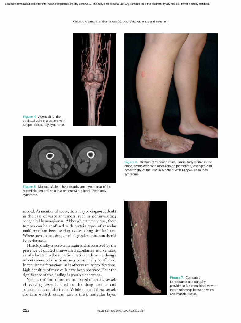
dimensional MRI venography by way of conventional axialimages, multiplanar reconstructions, maximum intensityprojections, and 3-dimensional images (Figures 1-3). Theconventional axial images were useful for assessing bone andsoft tissues and allowed us to locate both the deep venoussystem and anomalous superficial vessels and determinetheir relationship to adjacent structures. The axial images,multiplanar reconstructions, and maximum intensityprojections were used to define the origin, trajectory, andextension of the venous malformations. Skin surface imageswere useful for assessing the location and extension of theport-wine stain and for demonstrating limb hypertrophy.The 3-dimensional reconstructions demonstrated varicoseveins and the origin and trajectory of the anomalous vessels(Figures 4 and 5). No complementary invasive technique(conventional phlebography) was required to confirmdiagnosis or facilitate therapeutic planning in any of thesepatients.24
Thus, we can conclude that CT venography—or a 3-dimensional MRI venograph in children and pregnantwomen to limit radiation—can be the procedure of choicefor a comprehensive study of a large vascular malformationlocated on a limb. These techniques can pinpoint the exactlocation of the lesion on a 3-dimensional plane, detectmuscular skeletal infiltration and thoracic or abdominopelvicinvolvement, characterize bone density changes(osteoporosis) and asymmetries, and verify the existence ofa deep venous system while assessing its patency (Figures6-9). They are also useful for detecting abnormalities inthe superficial venous system, demonstrating the presenceof sciatic or abnormal veins, and determining the extensionand drainage of the malformation. Although theseprocedures are still not as good as conventional angiographyand venography in that they do not provide as muchinformation concerning the hemodynamics of themalformation, they nonetheless represent a very significantadvance. In our experience, invasive complementaryprocedures are only necessary in patients with vascularhypoplasia when the hemodynamic function of the vesselis unclear.24,25
Specifically, an MRI of the spine should be performedin the case of vascular malformations located along themidline of the back, particularly the lumbar sacral region,in order to detect defects in neural tube closure at variouslevels. Likewise, in the case of facial port-wine stainsand suspected Sturge-Weber syndrome, a brain MRIscan is required to assess possible neurologicalinvolvement.26
Pathological Study
The diagnosis of a vascular malformation is primarily clinical,and histopathologic examination of the lesion is rarely
Redondo P. Vascular malformations (II). Diagnosis, Pathology, and Treatment
Actas Dermosifiliogr. 2007;98:219-35 221
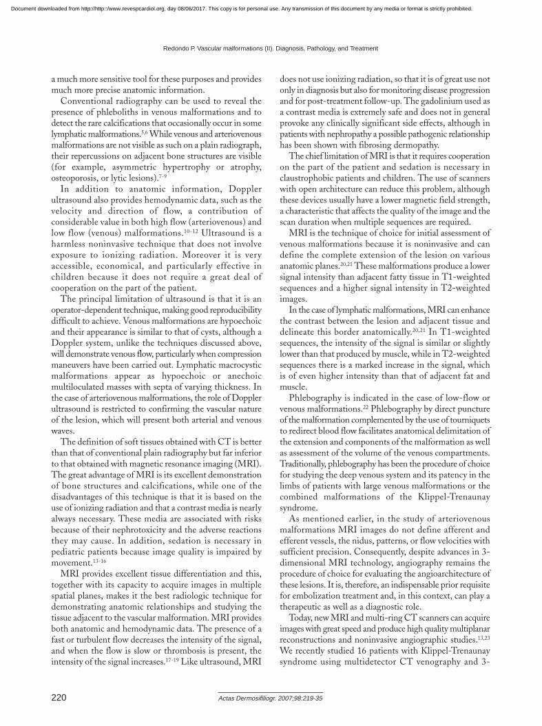
Figure 1. Sixteen-year-old girl with large, predominantly distal,venous malformation in the left arm.
Figure 2. Computedtomography angiography showsthe correlation between thesuperficial veins and the clinicalsituation.
Figure 3. A detailed view of thecomputed tomographyangiogram shows therelationship between the veinsand internal structures.
Document downloaded from http://http::/www.revespcardiol.org, day 08/06/2017. This copy is for personal use. Any transmission of this document by any media or format is strictly prohibited.
needed. As mentioned above, there may be diagnostic doubtin the case of vascular tumors, such as noninvolutingcongenital hemangiomas. Although extremely rare, thesetumors can be confused with certain types of vascularmalformations because they evolve along similar lines.Where such doubt exists, a pathological examination shouldbe performed.
Histologically, a port-wine stain is characterized by thepresence of dilated thin-walled capillaries and venules,usually located in the superficial reticular dermis althoughsubcutaneous cellular tissue may occasionally be affected.In venular malformations, as in other vascular proliferations,high densities of mast cells have been observed,27 but thesignificance of this finding is poorly understood.
Venous malformations are composed of ectatic vesselsof varying sizes located in the deep dermis andsubcutaneous cellular tissue. While some of these vesselsare thin walled, others have a thick muscular layer.
Redondo P. Vascular malformations (II). Diagnosis, Pathology, and Treatment
Actas Dermosifiliogr. 2007;98:219-35222
Figure 4. Agenesis of thepopliteal vein in a patient withKlippel-Trénaunay syndrome.
Figure 5. Musculoskeletal hypertrophy and hypoplasia of thesuperficial femoral vein in a patient with Klippel-Trénaunaysyndrome.
Figure 6. Dilation of varicose veins, particularly visible in theankle, associated with ulcer-related pigmentary changes andhypertrophy of the limb in a patient with Klippel-Trénaunaysyndrome.
Figure 7. Computedtomography angiographyprovides a 3-dimensional view ofthe relationship between veinsand muscle tissue.
Document downloaded from http://http::/www.revespcardiol.org, day 08/06/2017. This copy is for personal use. Any transmission of this document by any media or format is strictly prohibited.
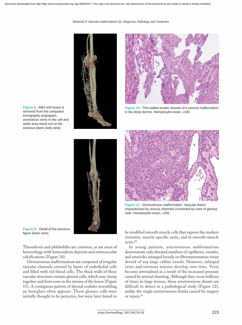
Thrombosis and phleboliths are common, as are areas ofhemorrhage with hemosiderin deposits and extravascularcalcifications (Figure 10).
Glomuvenous malformations are composed of irregularvascular channels covered by layers of endothelial cellsand filled with red blood cells. The thick walls of thesevascular structures contain glomus cells, which may clumptogether and form rows in the stroma of the lesion (Figure11). A contiguous pattern of dermal nodules resemblingan hourglass often appears. These glomus cells wereinitially thought to be pericytes, but were later found to
be modified smooth muscle cells that express the markersvimentin, muscle-specific actin, and α-smooth-muscleactin.28
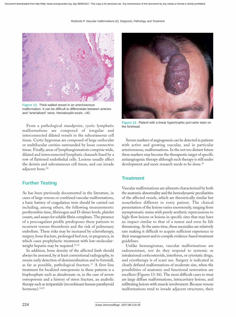
In young patients, arteriovenous malformationsdemonstrate only elevated numbers of capillaries, venules,and arterioles arranged loosely on fibromyxomatous tissuedevoid of any large caliber vessels. However, enlargedveins and tortuous arteries develop over time. Veinsbecome arterialized as a result of the increased pressurecaused by arterial shunting. Although they occur millionsof times in large lesions, these arteriovenous shunts aredifficult to detect in a pathological study (Figure 12),unlike the single arteriovenous fistula caused by surgeryor injury.29
Redondo P. Vascular malformations (II). Diagnosis, Pathology, and Treatment
Actas Dermosifiliogr. 2007;98:219-35 223
Figure 8. After soft tissue isremoved from the computedtomography angiogram,anomalous veins in the calf andankle area stand out on theosseous plane (side view).
Figure 9. Detail of the previousfigure (back view).
Figure 11. Glomuvenous malformation. Vascular lesioncharacterized by venous channels connected by rows of glomuscells. Hematoxylin-eosin, ×200.
Figure 10. Thin-walled ectatic vessels of a venous malformationin the deep dermis. Hematoxylin-eosin, ×100.
Document downloaded from http://http::/www.revespcardiol.org, day 08/06/2017. This copy is for personal use. Any transmission of this document by any media or format is strictly prohibited.
From a pathological standpoint, cystic lymphaticmalformations are composed of irregular andinterconnected dilated vessels in the subcutaneous celltissue. Cystic hygromas are composed of large unilocularor multilocular cavities surrounded by loose connectivetissue. Finally, areas of lymphangiomatosis comprise wide,dilated and interconnected lymphatic channels lined by arow of flattened endothelial cells. Lesions usually affectthe dermis and subcutaneous cell tissue, and can invadeadjacent bone.30
Further Testing
As has been previously documented in the literature, incases of large venous or combined vascular malformations,a basic battery of coagulation tests should be carried outincluding, among others, the following measurements:prothrombin time, fibrinogen and D-dimer levels, plateletcounts, and assays for soluble fibrin complexes. The presenceof a procoagulant profile predisposes these patients torecurrent venous thrombosis and the risk of pulmonaryembolism. These risks may be increased by sclerotherapy,surgery, bone fracture, prolonged bed rest, or pregnancy, inwhich cases prophylactic treatment with low-molecular-weight heparin may be required.31,32
In addition, bone density of the affected limb shouldalways be assessed, by at least conventional radiography, toensure early detection of demineralization and to forestall,as far as possible, pathological fracture.33 A first-linetreatment for localized osteoporosis in these patients is abisphosphate such as alendronate or, in the case of severeosteoporosis and a history of stress fracture, an anabolictherapy such as teriparatide (recombinant human parathyroidhormone).34,35
Serum markers of angiogenesis can be detected in patientswith active and growing vascular, and in particulararteriovenous, malformations. In the not too distant futurethese markers may become the therapeutic target of specificantiangiogenic therapy although such therapy is still underdevelopment and more research needs to be done.36
Treatment
Vascular malformations are ailments characterized by boththe anatomic abnormality and the hemodynamic peculiaritiesof the affected vessels, which are theoretically similar butnonetheless different in every patient. The clinicalpresentation of the lesions varies enormously, ranging fromasymptomatic stains with purely aesthetic repercussions tohigh-flow lesions or lesions in specific sites that may havean impact similar to that of a tumor and even be lifethreatening. At the same time, these anomalies are relativelyrare making it difficult to acquire sufficient experience intheir management and to compile evidence-based treatmentguidelines.
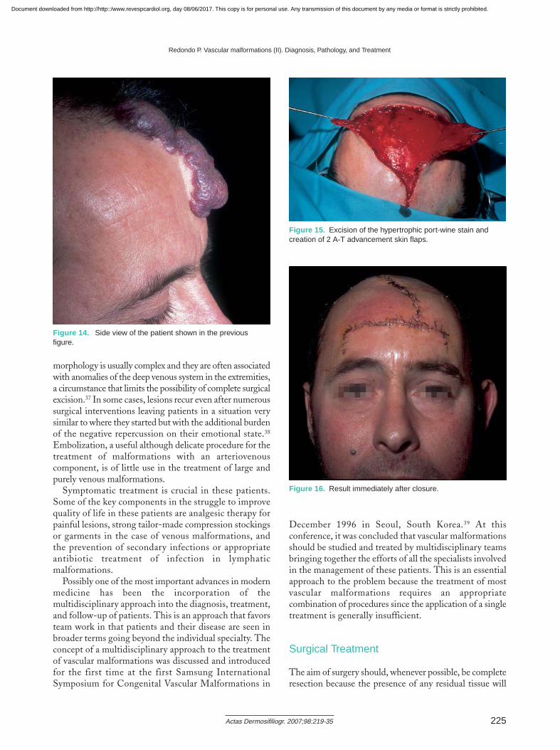
Unlike hemangiomas, vascular malformations areradioresistant, nor do they respond to systemic orintralesional corticosteroids, interferon, or cytostatic drugs,and cryotherapy is of scant use. Surgery is indicated inclearly defined malformations of moderate size, when thepossibilities of anatomic and functional restoration areexcellent (Figures 13-16). The most difficult cases to treatare large diffuse malformations, intracavitary lesions, andinfiltrating lesions with muscle involvement. Because venousmalformations tend to invade adjacent structures, their
Redondo P. Vascular malformations (II). Diagnosis, Pathology, and Treatment
Actas Dermosifiliogr. 2007;98:219-35224
Figure 12. Thick-walled vessel in an arteriovenousmalformation. It can be difficult to differentiate between arteriesand “arterialized” veins. Hematoxylin-eosin, ×40.
Figure 13. Patient with a linear hypertrophic port-wine stain onthe forehead.
Document downloaded from http://http::/www.revespcardiol.org, day 08/06/2017. This copy is for personal use. Any transmission of this document by any media or format is strictly prohibited.
morphology is usually complex and they are often associatedwith anomalies of the deep venous system in the extremities,a circumstance that limits the possibility of complete surgicalexcision.37 In some cases, lesions recur even after numeroussurgical interventions leaving patients in a situation verysimilar to where they started but with the additional burdenof the negative repercussion on their emotional state.38
Embolization, a useful although delicate procedure for thetreatment of malformations with an arteriovenouscomponent, is of little use in the treatment of large andpurely venous malformations.
Symptomatic treatment is crucial in these patients.Some of the key components in the struggle to improvequality of life in these patients are analgesic therapy forpainful lesions, strong tailor-made compression stockingsor garments in the case of venous malformations, andthe prevention of secondary infections or appropriateantibiotic treatment of infection in lymphaticmalformations.
Possibly one of the most important advances in modernmedicine has been the incorporation of themultidisciplinary approach into the diagnosis, treatment,and follow-up of patients. This is an approach that favorsteam work in that patients and their disease are seen inbroader terms going beyond the individual specialty. Theconcept of a multidisciplinary approach to the treatmentof vascular malformations was discussed and introducedfor the first time at the first Samsung InternationalSymposium for Congenital Vascular Malformations in
December 1996 in Seoul, South Korea.39 At thisconference, it was concluded that vascular malformationsshould be studied and treated by multidisciplinary teamsbringing together the efforts of all the specialists involvedin the management of these patients. This is an essentialapproach to the problem because the treatment of mostvascular malformations requires an appropriatecombination of procedures since the application of a singletreatment is generally insufficient.
Surgical Treatment
The aim of surgery should, whenever possible, be completeresection because the presence of any residual tissue will
Redondo P. Vascular malformations (II). Diagnosis, Pathology, and Treatment
Actas Dermosifiliogr. 2007;98:219-35 225
Figure 14. Side view of the patient shown in the previousfigure.
Figure 15. Excision of the hypertrophic port-wine stain andcreation of 2 A-T advancement skin flaps.
Figure 16. Result immediately after closure.
Document downloaded from http://http::/www.revespcardiol.org, day 08/06/2017. This copy is for personal use. Any transmission of this document by any media or format is strictly prohibited.
invariably give rise to recurrence of the lesion. Purely surgicaltreatment of vascular malformations is only possible in thecase of very localized and accessible lesions,40,41 such asvenous aneurysms, some small infiltrating lesions, and raremonopedicular arteriovenous lesions of limited size thatare easily accessible and can be treated by ligation and totalextirpation.
The resectability of the lesion should be rigorously assessedbefore the patient is proposed as a candidate for surgery.The malformation ideally suited for treatment exclusivelywith surgery is the isolated and superficial lesion that hasnot spread into deep planes or infiltrated adjacent structures.Subfascial extension with involvement of the muscles,tendons or bones or invasion of structures such as the pelvisor the gluteal region are contraindications for treatmentwith surgery alone since, in the best of cases notwithstandinggood therapeutic planning and meticulous surgery, thisoption represents a temporary palliative treatment42 andinvolves a high risk of thromboembolic events.43 The useof bipolar electrocautery is advised when surgery is carriedout in the vicinity of neural structures. Special attentionshould be paid to hemostasis of the surgical field given thatuncontrolled bleeding makes dissection much more difficultand increases the risk of damage to adjacent structures.
In the case of lymphatic malformations, treatment isusually conservative because these anomalies rarely affectthe function of the limb or organ involved and the presentingcomplaint is usually an aesthetic one. The exception,although quite a common one, corresponds to lesions thatcan be dangerous and obstruct the airway owing to theircervical location.44
In practice, it is difficult in the case of arteriovenous andlymphatic malformations, particularly diffuse ones, toidentify a dissection plane during excision because themargins of the lesion are almost impossible to delimit.Consequently, the resection margins should be as broad aspossible. Any residual lesion, for example a residual cutaneousmacular stain resembling a port-wine stain, will give riseto recurrence in the short term and this is the reason whyreoperations are common. Preoperative MRI and digitalangiography, particularly dynamic evaluations, are especiallyuseful for establishing these macroscopic margins.
The aim of surgery in the case of an arteriovenousmalformation is to eradicate the nidus of the malformation.In such cases, surgery is often very hemorrhagic andineffective due to the rapid recruitment of collateral vesselsto supply the nidus. Prior embolization facilitates surgeryand reduces bleeding. Resection should be performed assoon as and in the most complete way possible, with excisionof broad margins around the nidus.
Laser Therapy
Pulsed dye laser treatment targeting intravascularhemoglobin with selective photothermolysis is currentlythe first-line treatment for port-wine stains.45 Laser lightabsorbed by oxyhemoglobin generates heat and damagesthe vascular endothelium giving rise to thrombosis anddestruction of the vessel. The most commonly reportedadverse effects after pulsed dye laser treatment are theappearance of blisters, scabs, hyperpigmentation,hypopigmentation, hypertrophic or atrophic scarring,and infections. Blisters and scabs appear a few hours aftertreatment and rarely leave scars after healing. The mostcommon adverse effect is hyperpigmentation, whichusually resolves spontaneously within 6 to 12 months,46
although photoprotective measures should be taken tominimize this effect. The appearance of atrophic orhypertrophic scars varies between the different case seriesin the literature and has been reported in up to 5% ofpatients.46
Within the large range of pulsed dye laser systems onthe market, each more versatile than the last, we must limitourselves to the following parameters and oscillation values:laser beam diameter (5-10 mm), wavelength (585-600 nm),energy or fluence density (7-15 J/cm2), and pulse duration(450 µs-1500 ms). A recent study reported good results in63% of patients with a greater than 75% clearance of thelesion after 4 sessions using the following protocol: a dynamicskin cooling system, a wavelength of 595 nm, pulses of 1.5 ms,and energy fluences between 11-12 J/cm2.47 In the samestudy, 75% improvement was achieved in only 40% of asecond group of patients treated with 585 nm. Some of theparameters that appear to improve the final outcome arethe use of longer pulse durations (1.5 ms),48 longerwavelengths (600 nm),49 and higher energy fluences.47 Ininfants and children, treatment should be started with lowfluences, which can then be increased by 0.5-1 J/cm2 ateach session depending on the positive response and adverseeffects.
Traditionally, the standard interval between sessions hasbeen at least 6 weeks, although a recent study producedbetter results with sessions at 2-week intervals.50
While there is still debate about the most appropriatetiming for starting laser treatment, it is generally acceptedthat such treatment should be started as early as possible.51
Fewer sessions are required to treat children because theresponse is more rapid51; moreover, the rate of recurrenceis lower in the group treated for the first time under 10years of age.52 In most cases, between 4 and 8 sessions arerequired to obtain a satisfactory response, and completeclearance is achieved in only 10% of patients. Inapproximately half of the patients, at least 75% clearanceis achieved, and in one third the result is considered poor(improvement under 50%).
Redondo P. Vascular malformations (II). Diagnosis, Pathology, and Treatment
Actas Dermosifiliogr. 2007;98:219-35226
Document downloaded from http://http::/www.revespcardiol.org, day 08/06/2017. This copy is for personal use. Any transmission of this document by any media or format is strictly prohibited.
The chief drawback of early treatment is the need forsedation (general anesthesia) because the procedure ispainful and provokes uncontrollable distress and anxietyin children. The anesthetist must use low oxygenconcentrations because of the potential danger of ignitioncaused, for example, by the combustion of a hair shaftspreading through the air.53 This protocol has beenconfirmed by our experience because it facilitates longersessions with better long term results. It also favors a perfectbalance between the physician, the patient, and the family,a difficult outcome to achieve with invasive procedures inchildren in real life situations. The use of various systemsfor cooling the skin surface before irradiation with the laserbeam, such as dynamic cryogen spray cooling or continuouscold air cooling,54 can obviate the need for anesthesia insome prepubescent children and allow the use of higherenergy fluences while minimizing the risk of damage tothe epidermis.55
In venular malformations, 3 models of ectasia can bedistinguished in the papillary plexus using videomicroscopydepending on whether the lesion affects the vertical vessels,the deep horizontal vessels, or both.56 It is important todifferentiate between these 3 patterns when evaluatingprobable response to laser therapy because the first modelresponds better to such treatment than the other 2.57 Asmentioned earlier, venular malformations are classifiedaccording to the degree of ectasia present, with grade Icorresponding to those with the smallest vessels and gradeIV to those with the largest. The deepest vessels persistafter treatment, as they can dilate and even proliferatethrough angiogenesis and spread once again towards moresuperficial areas.58 The location and color of a lesion alsopredicts the level of response to laser treatment. Lesionssituated in V2 respond less well than those affecting V1 orV3; lesions on the face and neck respond much better thanthose located on the trunk and extremities.59
In one third of patients, lesions fail to improve evenafter numerous sessions partly because the limited depthof laser penetration into the tissue (1 to 1.5 mm) makes itimpossible to treat vessels situated at greater depth,60 andpartly because of the presence of other, nonvascular,elements of the hamartomatous type not targeted by thistechnique.61 Adult patients with deep purple port-winestains, tuberous lesions, or hypertrophy of the affected sideof the face require surgical treatment. The options arecurettage and electrocoagulation, simple excision, or surgicalreduction by wedge resection and vermilionectomy of liphypertrophy. In such cases, carbon dioxide (CO2) andneodymium:yttrium-aluminum-garnet (Nd:YAG) lasersystems are also effective.62 In resistant cases and areas, theadministration of 2 passes per session with the pulsed dyelaser improves response.63 Overlapping of pulses is anothertechnique shown to favor penetration without any adverseeffect on the epidermis.64
Patients whose condition does not respond to pulsed dyelaser can be treated with intense pulsed light, Nd:YAG laserlight (1064 nm),65 or potassium-titanyl-phosphate (KTP)laser light (532 nm). In a case series of patients withmalformations resistant to pulsed dye laser, satisfactoryresults (improvement of more than 50%) were obtained in17% of the patients treated with KTP laser light.66 The bestresults were obtained with fluences in the 18 to 24 J/cm2
range and pulse durations of between 9 and 14 ms. Theselaser systems deliver high energy fluences and afford thepossibility of adjusting the pulse duration to match thethermal relaxation time of the target structure. However,although tolerance is better, when no purpura is present,they may produce a higher incidence of scars or pigmentarychanges (Figures 17 and 18).66 Although there are fewerstudies in the literature on the use of intense pulsed lightthan on the laser systems described above, improvementsof between 70% and 100% have been achieved in 70% ofpatients with such treatment.67,68
Laser treatment of venous malformations is limited tovery superficial lesions, the superficial component of deeperlesions as an adjuvant to treatment with other procedures(for example sclerotherapy), or mucosal lesions. The lasersystem most often used is a continuous Nd:YAG laser,
Redondo P. Vascular malformations (II). Diagnosis, Pathology, and Treatment
Actas Dermosifiliogr. 2007;98:219-35 227
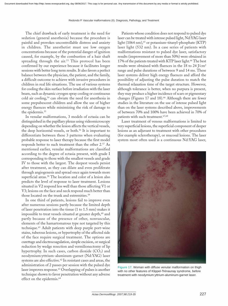
Figure 17. Woman with diffuse venular malformation on thighwith no other features of Klippel-Trénaunay syndrome, beforetreatment with neodymium:yttrium-aluminum-garnet laser.
Document downloaded from http://http::/www.revespcardiol.org, day 08/06/2017. This copy is for personal use. Any transmission of this document by any media or format is strictly prohibited.
which delivers infrared light at 1064 nm and has a skinpenetration depth of between 5 and 7 mm. This device candeliver intralesional therapy through a catheter insertedinto the lesion in a procedure controlled visually byultrasound.69-71
A Nd:YAG laser system with a constant power outputof 30 W, a 600 µm fiber diameter, and variable pulse durationis very useful in the treatment of mucosal lesions.72 Thisprocedure, which is usually performed under generalanesthesia, is safe, no simultaneous surface cooling systemis necessary, and the risk of fibrosis or residual scarring isnegligible. When the laser beam impinges on the mucosa,the targeted spot immediately retracts and whitish spotsappear in the area treated, providing a guide that helps theoperator to avoid overlapping impacts. In the case of largevenous malformations of the mucosa, laser treatment priorto surgery can eliminate the superficial component of themalformation and create a band of fibrosis that facilitatesbetter excision of the lesion and reduces bleeding duringthe intervention. If large areas of the oral cavity are treated,
especially at the back of the mouth, systemic corticosteroidsshould be administered to reduce inflammation and preventairway compromise.
Ablation with CO2 laser is particularly indicated insuperficial lymphatic malformations of mucosal membranes,characterized clinically by the presence of multiple vesicles.These are usually diffuse lesions that reach deep planes andcan permeate underlying muscle.72 Although this treatmentis not curative, it helps to control the disease and has aclearly beneficial effect on the patients’ quality of life.Nonetheless, recurrence is the norm and periodic treatmentis necessary. As infection can stimulate the growth of alymphatic malformation, patients undergoing this procedureshould receive appropriate prophylactic antibiotic treatmentand an anti-inflammatory regimen of oral corticosteroids.
Embolization
Embolization is a treatment particularly useful inarteriovenous malformations, and it can be used in themanagement of such lesions as a complete or adjuvanttherapy. Despite advances in recent years, particularly inthe new materials used (coils, different particles, polymers,and glues),73-75 this technique is still a temporary solutionsince the blood supply in any specific anatomic area is notstatic and the development of collateral circulation isinevitable.76
As an adjuvant therapy prior to surgery, embolization isusually the most appropriate procedure for use in the caseof arteriovenous malformations. Once the lesion has beenmapped by angiography and the nidus and its afferents havebeen delineated, the lesion is embolized from distal toproximal. This reduces the vascularization of the lesionthereby minimizing the potential risk of bleeding duringsurgery. Because a new collateral circulation will beestablished very quickly, surgery should not be delayed morethan 48 hours after embolization has been performed.77
Embolization can be use curatively or as a palliativetreatment to prevent bleeding from large nonresectablelesions. The use of embolization to achieve a cure is not,however, a very realistic aim and it is preferable to envisagemanagement of the lesion rather than a cure. In such cases,a gradual staged procedure is required, proceeding throughthe nidus from proximal to distal including both active andany potential arterial supply. The treatment must be repeatedregularly because of revascularization of the lesion. Thisprocedure is not risk free and should be carried out by aninterventional radiologist familiar with the technique becauseone of the possible adverse events is the inadvertent releaseof embolic material leading to distant occlusion, distalnecrosis, and neurological damage. Moreover, all of theembolic material used must be applied under fluoroscopicguidance.
Redondo P. Vascular malformations (II). Diagnosis, Pathology, and Treatment
Actas Dermosifiliogr. 2007;98:219-35228
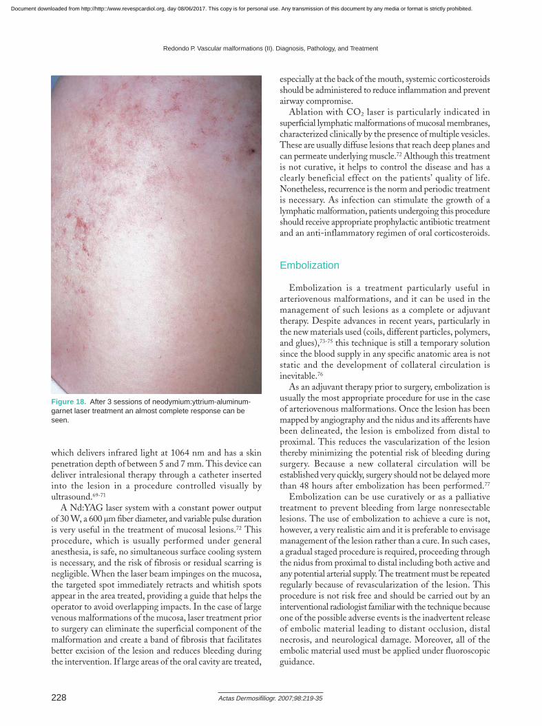
Figure 18. After 3 sessions of neodymium:yttrium-aluminum-garnet laser treatment an almost complete response can beseen.
Document downloaded from http://http::/www.revespcardiol.org, day 08/06/2017. This copy is for personal use. Any transmission of this document by any media or format is strictly prohibited.
Sclerotherapy
Sclerotherapy is a procedure used to eliminate varicoseveins through the injection of a sclerosing substance intothe vein. The inflammation resulting from contact betweenthe sclerosing agents and the endothelial cells on the innersurface of the vein leads to chemical irritation.78 This processgives rise to the formation of a thrombus that blocks bloodflow through the vein. Targeted veins gradually becomefibrous sclerosed cords and are eventually reabsorbed bythe body. Sclerosing agents can be classified as high potency(alcohol, iodine, and sodium tetradecyl sulfate), intermediatepotency (sodium salicylate and polidocanol), or low potency(chromated glycerin). Polidocanol (aethoxysclerol), likesodium tetradecyl sulfate, is a detergent with a hydrophilicand a hydrophobic pole that acts by changing the surfacetension of endothelial cells. The hydrophobic pole attachesto the cell surface while the hydrophilic portion attracts thewater contained in the cell, thereby causing rapid and intenseendothelial hydratation.79 Local and general tolerance ofpolidocanol is excellent,80 and this agent has been marketedin Europe for over 20 years for sclerosing varicose veinsalthough it has not yet been approved by the AmericanFood and Drug Administration. Its advantages contrastwith the drawbacks associated with ethanol sclerotherapy,which has an aggressive impact on both the malformationand adjacent tissue. Injection of ethanol is painful and canonly be carried out in hospitalized patients under generalanesthesia. The use of ethanol as a sclerosing agent is notindicated in children and it can produce necrosis of the skinand mucous membranes, thrombosis of the deep venoussystem in the extremities, pulmonary embolism, andcardiorespiratory collapse due to pulmonary spasm.Moreover, repeated administration of ethanol is not easy,and repetition is essential in sclerotherapy because partialrecanalization after initial intravascular thrombosis iscommon.81
Conventional sclerotherapy using liquid sclerosing agents,which is a palliative treatment in most types of vascularanomalies, produces good outcomes in smaller lesions.82 Inthe context of surgery it is indicated as a preoperativeintervention undertaken to reduce the size of the lesion, oras a postoperative complement.83 However, conventionalsclerotherapy is ineffective in the treatment of large venousmalformations. This is due to a number of factors: theintrinsic limitations of the injected liquids, which are subjectto dilution and progressive inactivation in large volumes ofblood; the irregular distribution of the sclerosing agent onthe endothelial cells of the target area; the difficulty ofmanipulating and controlling the sclerosing agent afterinjection; and the fact that the liquid agent cannot bevisualized inside the blood vessels on Doppler ultrasound.
These problems have been addressed by thedevelopment of a pharmaceutical microfoam that serves
as a vehicle for administering the sclerosing agent.84 Themodern use of foams in sclerotherapy started in 1944with the work of Orbach, who used an air block techniqueto introduce air into the vein before injection of thesclerosing liquid. His aim was to potentiate the action ofthe agent by emptying the target vessel of blood.85 Orbachlater used large bubbles of sclerosant (obtained by shakingthe ampule containing the liquid sclerosant) anddemonstrated that the action of this foam was greaterthan that of the liquid form. This procedure was onlyshown to be of use in small veins, and the initial interestfaded over the decades. In 1993, Juan Cabrera, a Spanishvascular surgeon, created a pharmaceutical form ofinjectable microfoam by creating microbubbles ofsurfactant sclerosing solutions using physiological gasesthat are highly soluble in body liquids.86,87 Other authorshave produced foams with such solutions using specifictechniques commonly designated by the names of theresearchers who have described them: Monfreux, Tessari,Frullini, etc.88 Currently, one of the most populartechniques is the method described by Tessari in whichthe foam is obtained using 2 connected syringes and a 3-way stopcock. These “homemade” foams made withambient air rich in nitrogen—a gas with low solubilityin body fluids—have irregular bubble size and highlyvariable internal cohesion. The techniques used to applythe foams also vary,89 although the statement issued aftera consensus meeting in Tegernsee, Germany, recommended—for reasons of safety—the injection of no more than 5 mL of foam in any one treatment session.90
The pharmaceutical sclerosing microfoam, on the otherhand, is made with physiologic gases, such as oxygen andCO2, and is composed of tiny microbubbles with sufficientstability and internal cohesion to withstand injection intothe vessels. The contact surface between the sclerosingagent and the vessel wall increases enormously in inverseproportion to the diameter of the bubble. Polidocanolmicrofoam displaces blood, thereby promoting homogeneouscontact between the sclerosant and the endothelium,facilitating endothelial destruction.86,87 Moreover, theprocedure is visible in real time on ultrasound. Owing tothe high solubility and lack of toxicity of CO2, large amountsof the microfoam can be injected in a single session, whichis not the case with foams made from ambient air.90-94 It issometimes necessary to inject between 20 and 100 mL ofmicrofoam to treat large venous malformations. By contrast,the volume of “homemade” foam that can be injected in asingle session cannot exceed the recommended 5 mL becauseof the low solubility of nitrogen, and only the concentrationcan be modified.
To date, we have treated congenital vascularmalformations (mainly venous) in over 120 patients withsclerotherapy using polidocanol microfoam. In an initialpublished series, 50 patients (18 males and 32 females
Redondo P. Vascular malformations (II). Diagnosis, Pathology, and Treatment
Actas Dermosifiliogr. 2007;98:219-35 229
Document downloaded from http://http::/www.revespcardiol.org, day 08/06/2017. This copy is for personal use. Any transmission of this document by any media or format is strictly prohibited.
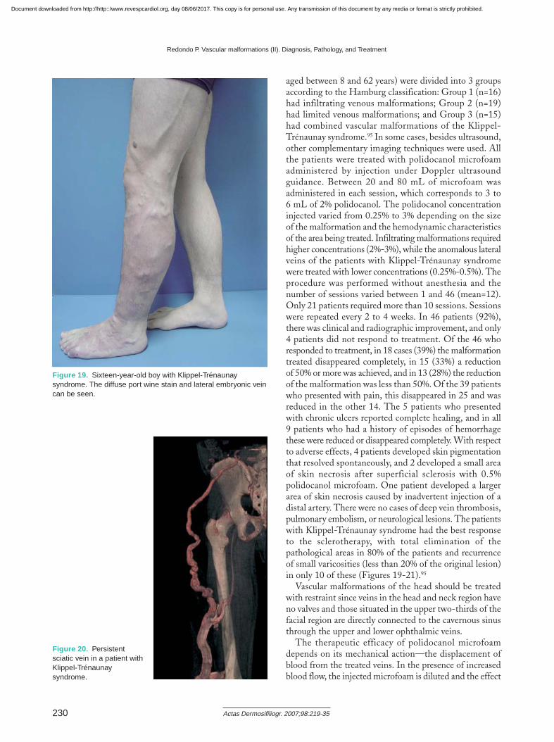
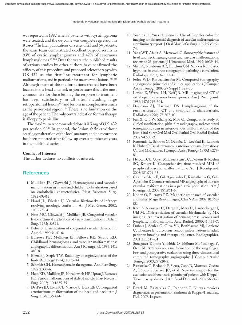
aged between 8 and 62 years) were divided into 3 groupsaccording to the Hamburg classification: Group 1 (n=16)had infiltrating venous malformations; Group 2 (n=19)had limited venous malformations; and Group 3 (n=15)had combined vascular malformations of the Klippel-Trénaunay syndrome.95 In some cases, besides ultrasound,other complementary imaging techniques were used. Allthe patients were treated with polidocanol microfoamadministered by injection under Doppler ultrasoundguidance. Between 20 and 80 mL of microfoam wasadministered in each session, which corresponds to 3 to 6 mL of 2% polidocanol. The polidocanol concentrationinjected varied from 0.25% to 3% depending on the sizeof the malformation and the hemodynamic characteristicsof the area being treated. Infiltrating malformations requiredhigher concentrations (2%-3%), while the anomalous lateralveins of the patients with Klippel-Trénaunay syndromewere treated with lower concentrations (0.25%-0.5%). Theprocedure was performed without anesthesia and thenumber of sessions varied between 1 and 46 (mean=12).Only 21 patients required more than 10 sessions. Sessionswere repeated every 2 to 4 weeks. In 46 patients (92%),there was clinical and radiographic improvement, and only4 patients did not respond to treatment. Of the 46 whoresponded to treatment, in 18 cases (39%) the malformationtreated disappeared completely, in 15 (33%) a reductionof 50% or more was achieved, and in 13 (28%) the reductionof the malformation was less than 50%. Of the 39 patientswho presented with pain, this disappeared in 25 and wasreduced in the other 14. The 5 patients who presentedwith chronic ulcers reported complete healing, and in all9 patients who had a history of episodes of hemorrhagethese were reduced or disappeared completely. With respectto adverse effects, 4 patients developed skin pigmentationthat resolved spontaneously, and 2 developed a small areaof skin necrosis after superficial sclerosis with 0.5%polidocanol microfoam. One patient developed a largerarea of skin necrosis caused by inadvertent injection of adistal artery. There were no cases of deep vein thrombosis,pulmonary embolism, or neurological lesions. The patientswith Klippel-Trénaunay syndrome had the best responseto the sclerotherapy, with total elimination of thepathological areas in 80% of the patients and recurrenceof small varicosities (less than 20% of the original lesion)in only 10 of these (Figures 19-21).95
Vascular malformations of the head should be treatedwith restraint since veins in the head and neck region haveno valves and those situated in the upper two-thirds of thefacial region are directly connected to the cavernous sinusthrough the upper and lower ophthalmic veins.
The therapeutic efficacy of polidocanol microfoamdepends on its mechanical action—the displacement ofblood from the treated veins. In the presence of increasedblood flow, the injected microfoam is diluted and the effect
Redondo P. Vascular malformations (II). Diagnosis, Pathology, and Treatment
Actas Dermosifiliogr. 2007;98:219-35230
Figure 19. Sixteen-year-old boy with Klippel-Trénaunaysyndrome. The diffuse port wine stain and lateral embryonic veincan be seen.
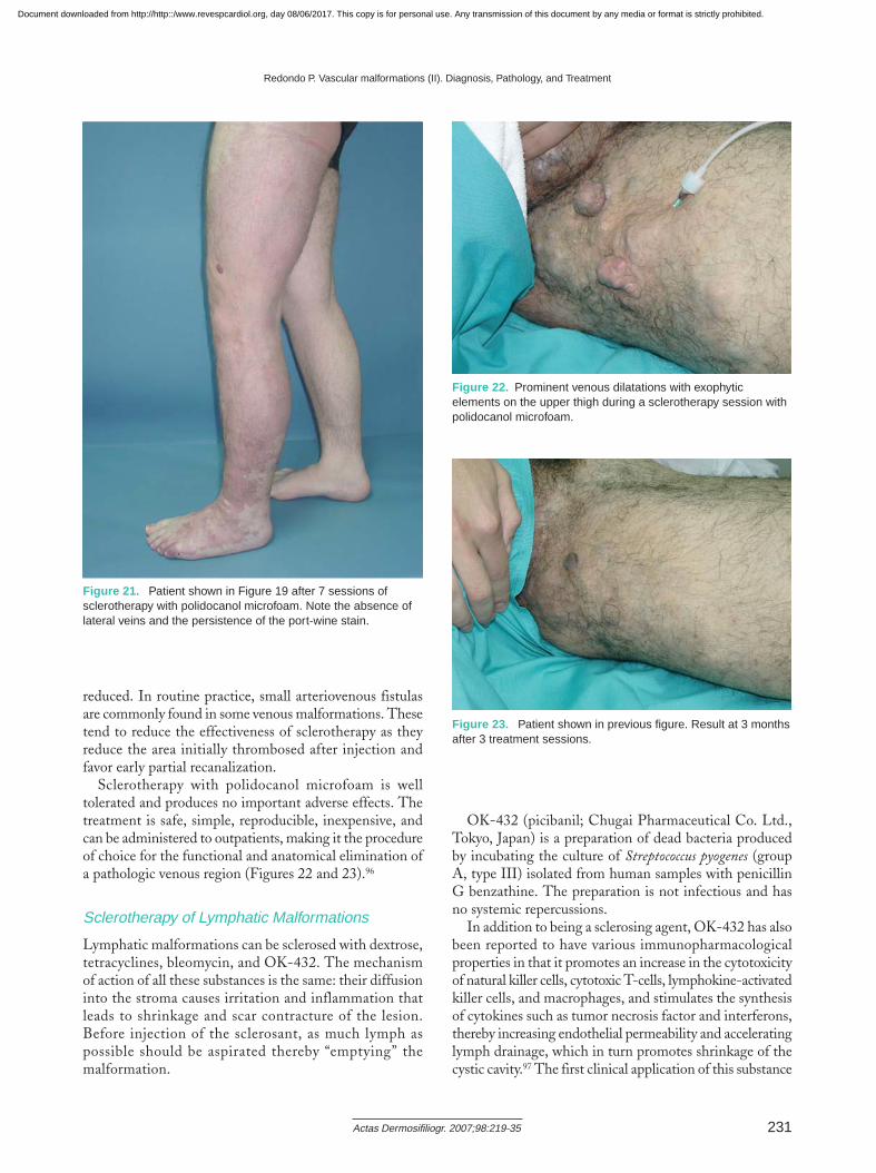
Figure 20. Persistentsciatic vein in a patient withKlippel-Trénaunaysyndrome.
Document downloaded from http://http::/www.revespcardiol.org, day 08/06/2017. This copy is for personal use. Any transmission of this document by any media or format is strictly prohibited.
reduced. In routine practice, small arteriovenous fistulasare commonly found in some venous malformations. Thesetend to reduce the effectiveness of sclerotherapy as theyreduce the area initially thrombosed after injection andfavor early partial recanalization.
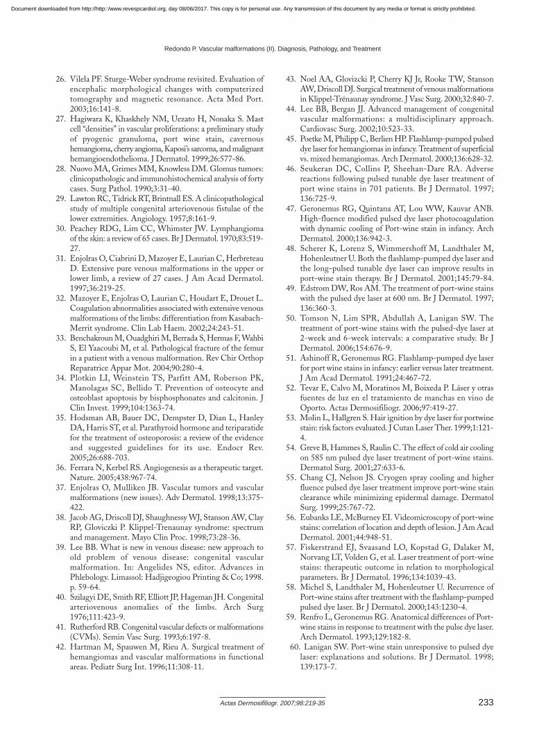
Sclerotherapy with polidocanol microfoam is welltolerated and produces no important adverse effects. Thetreatment is safe, simple, reproducible, inexpensive, andcan be administered to outpatients, making it the procedureof choice for the functional and anatomical elimination ofa pathologic venous region (Figures 22 and 23).96
Sclerotherapy of Lymphatic Malformations
Lymphatic malformations can be sclerosed with dextrose,tetracyclines, bleomycin, and OK-432. The mechanismof action of all these substances is the same: their diffusioninto the stroma causes irritation and inflammation thatleads to shrinkage and scar contracture of the lesion.Before injection of the sclerosant, as much lymph aspossible should be aspirated thereby “emptying” themalformation.
OK-432 (picibanil; Chugai Pharmaceutical Co. Ltd.,Tokyo, Japan) is a preparation of dead bacteria producedby incubating the culture of Streptococcus pyogenes (groupA, type III) isolated from human samples with penicillinG benzathine. The preparation is not infectious and hasno systemic repercussions.
In addition to being a sclerosing agent, OK-432 has alsobeen reported to have various immunopharmacologicalproperties in that it promotes an increase in the cytotoxicityof natural killer cells, cytotoxic T-cells, lymphokine-activatedkiller cells, and macrophages, and stimulates the synthesisof cytokines such as tumor necrosis factor and interferons,thereby increasing endothelial permeability and acceleratinglymph drainage, which in turn promotes shrinkage of thecystic cavity.97 The first clinical application of this substance
Redondo P. Vascular malformations (II). Diagnosis, Pathology, and Treatment
Actas Dermosifiliogr. 2007;98:219-35 231
Figure 22. Prominent venous dilatations with exophyticelements on the upper thigh during a sclerotherapy session withpolidocanol microfoam.
Figure 23. Patient shown in previous figure. Result at 3 monthsafter 3 treatment sessions.
Figure 21. Patient shown in Figure 19 after 7 sessions ofsclerotherapy with polidocanol microfoam. Note the absence oflateral veins and the persistence of the port-wine stain.
Document downloaded from http://http::/www.revespcardiol.org, day 08/06/2017. This copy is for personal use. Any transmission of this document by any media or format is strictly prohibited.
was reported in 1987 when 9 patients with cystic hygromawere treated, and the outcome was complete regression in8 cases.98 In later publications on series of 23 and 64 patients,the same team demonstrated excellent or good results in93% of cystic lymphangiomas and 47% of cavernouslymphangiomas.99,100 Over the years, the published resultsof various studies by other authors have confirmed theefficacy of this procedure and proposed sclerotherapy withOK-432 as the first-line treatment for lymphaticmalformations, and in particular for macrocystic lesions.101,102
Although most of the malformations treated have beenlocated in the head and neck region because this is the mostcommon site for these lesions, the response to treatmenthas been satisfactory in all sites, including largeretroperitoneal lesions103 and lesions in complex sites, suchas the periorbital region.104 There is no limitation on theage of the patient. The only contraindication for this therapyis allergy to penicillin.
The maximum recommended dose is 0.3 mg of OK-432per session.99,105 In general, the lesion shrinks withoutscarring or alteration of the local anatomy and no recurrencehas been reported after follow-up over a number of yearsin the published series.
Conflict of InterestsThe author declares no conflicts of interest.
References
1. Mulliken JB, Glowacki J. Hemangiomas and vascularmalformations in infants and children: a classification basedon endothelial characteristics. Plast Reconstr Surg.1982;69:412.
2. Hand JL, Frieden IJ. Vascular Birthmarks of infancy:resolving nosologic confusion. Am J Med Genet. 2002;108:257-64.
3. Finn MC, Glowacki J, Mulliken JB. Congenital vascularlesions: clinical application of a new classification. J PediatrSurg. 1983;18:894.
4. Belov S. Classification of congenital vascular defects. IntAngiol. 1990;9:141-6.
5. Burrows PE, Mulliken JB, Fellows KE, Strand RD.Childhood hemangiomas and vascular malformations:angiographic differentiation. Am J Roentgenol. 1983;141:483-8.
6. Bliznak J, Staple TW. Radiology of angiodysplasias of thelimb. Radiology 1974;110:35-44.
7. Schmidt GH. Hemangioma in the zygoma. Ann Plast Surg.1982;3:330-6.
8. Hein KD, Mulliken JB, Kozakewich HP, Upton J, BurrowsPE. Venous malformations of skeletal muscle. Plast ReconstrSurg. 2002;110:1625-35.
9. DesPrez JD, Kiehn CL, Vlastou C, Bonstelle C. Congenitalarteriovenous malformation of the head and neck. Am JSurg. 1978;136:424-9.
10. Yoshida H, Yusa H, Ueno E. Use of Doppler color forimaging for differential diagnosis of vascular malformations:a preliminary report. J Oral Maxillofac Surg. 1995;53:369-74.
11. Yang WT, Ahuja A, Metreweli C. Sonographic features ofhead and neck hemangiomas and vascular malformations:review of 23 patients. J Ultrasound Med. 1997;16:39-44.
12. Sheth S, Nussbaum AR, Hutchins GM, Sanders RC. Cystichygromas in children: sonographic-pathologic correlation.Radiology. 1987;162:821-4.
13. Foley WD, Karcaaltincaba M. Computed tomographyangiography: principles and clinical applications. J ComputAssist Tomogr. 2003;27 Suppl 1:S23-30.
14. Levine E, Wetzel LH, Neff JR. MR imaging and CT ofextrahepatic cavernous hemangiomas. Am J Roentgenol.1986;147:1299-304.
15. Davidson AJ, Hartman DS. Lymphangioma of theretroperitoneum: CT and sonographic characteristic.Radiology. 1990;175:507-10.
16. Fan X, Qiu W, Zhang Z, Mao Q. Comparative study ofclinical manifestation, plain-film radiography, and computedtomographic scan in arteriovenous malformations of thejaws. Oral Surg Oral Med Oral Pathol Oral Radiol Endod.2002;94:503-9.
17. Remonda L, Schroth G, Ozdoba C, Lovblad K, LadrachK, Huber P. Facial intraosseous arteriovenous malformations:CT and MR features. J Comput Assist Tomogr. 1995;19:277-81.
18. Herborn CU, Goyen M, Lauenstein TC, Debatin JF, RuehmSG, Kroger K. Comprehensive time-resolved MRI ofperipheral vascular malformations. Am J Roentgenol.2003;181:729-35.
19. Caseiro-Alves F, Gil-Agostinho P, Ramalheiro G, Gil-Agostinho P. Contrast-enhanced MR angiography of thoracicvascular malformations in a pediatric population. Am JRoentgenol. 2003;181:861-6.
20. Konez O, Burrows PE. Magnetic resonance of vascularanomalies. Magn Reson Imaging Clin N Am. 2002;10:363-88.
21. Kern S, Niemeyer C, Darge K, Merz C, Laubenberger J,Uhl M. Differentiation of vascular birthmarks by MRimaging. An investigation of hemangiomas, venous andlymphatic malformations. Acta Radiol. 2000;41:453-7.
22. Dubois J, Soulez G, Oliva VL, Berthiaume MJ, LapierreC, Therasse E. Soft-tissue venous malformations in adultpatients: imaging and therapeutic issues. Radiographics.2001;21:1519-31.
23. Sunagawa T, Ikuta Y, Ishida O, Ishiburo M, Yasunaga Y,Ochi M. Arteriovenous malformation of the ring finger.Pre- and postoperative evaluation using three-dimensionalcomputed tomography angiography. J Comput AssistTomogr. 2003;27:820-3.
24. Bastarrika G, Redondo P, Sierra, Cano D, Martínez-CuestaA, López-Gutierrez JC, et al. New techniques for theevaluation and therapeutic planning of patients with Klippel-Trenaunay syndrome. J Am Acad Dermatol. 2007;56:242-9.
25. Pretel M, Bastarrika G, Redondo P. Nuevas técnicasdiagnósticas en pacientes con síndrome de Klippel-Trenaunay.Piel. 2007. In press.
Redondo P. Vascular malformations (II). Diagnosis, Pathology, and Treatment
Actas Dermosifiliogr. 2007;98:219-35232
Document downloaded from http://http::/www.revespcardiol.org, day 08/06/2017. This copy is for personal use. Any transmission of this document by any media or format is strictly prohibited.
26. Vilela PF. Sturge-Weber syndrome revisited. Evaluation ofencephalic morphological changes with computerizedtomography and magnetic resonance. Acta Med Port.2003;16:141-8.
27. Hagiwara K, Khaskhely NM, Uezato H, Nonaka S. Mastcell “densities” in vascular proliferations: a preliminary studyof pyogenic granuloma, port wine stain, cavernoushemangioma, cherry angioma, Kaposi’s sarcoma, and malignanthemangioendothelioma. J Dermatol. 1999;26:577-86.
28. Nuovo MA, Grimes MM, Knowless DM. Glomus tumors:clinicopathologic and immunohistochemical analysis of fortycases. Surg Pathol. 1990;3:31-40.
29. Lawton RC, Tidrick RT, Brintnall ES. A clinicopathologicalstudy of multiple congenital arteriovenous fistulae of thelower extremities. Angiology. 1957;8:161-9.
30. Peachey RDG, Lim CC, Whimster JW. Lymphangiomaof the skin: a review of 65 cases. Br J Dermatol. 1970;83:519-27.
31. Enjolras O, Ciabrini D, Mazoyer E, Laurian C, HerbreteauD. Extensive pure venous malformations in the upper orlower limb, a review of 27 cases. J Am Acad Dermatol.1997;36:219-25.
32. Mazoyer E, Enjolras O, Laurian C, Houdart E, Drouet L.Coagulation abnormalities associated with extensive venousmalformations of the limbs: differentiation from Kasabach-Merrit syndrome. Clin Lab Haem. 2002;24:243-51.
33. Benchakroun M, Ouadghiri M, Berrada S, Hermas F, WahbiS, El Yaacoubi M, et al. Pathological fracture of the femurin a patient with a venous malformation. Rev Chir OrthopReparatrice Appar Mot. 2004;90:280-4.
34. Plotkin LI, Weinstein TS, Parfitt AM, Roberson PK,Manolagas SC, Bellido T. Prevention of osteocyte andosteoblast apoptosis by bisphosphonates and calcitonin. JClin Invest. 1999;104:1363-74.
35. Hodsman AB, Bauer DC, Dempster D, Dian L, HanleyDA, Harris ST, et al. Parathyroid hormone and teriparatidefor the treatment of osteoporosis: a review of the evidenceand suggested guidelines for its use. Endocr Rev.2005;26:688-703.
36. Ferrara N, Kerbel RS. Angiogenesis as a therapeutic target.Nature. 2005;438:967-74.
37. Enjolras O, Mulliken JB. Vascular tumors and vascularmalformations (new issues). Adv Dermatol. 1998;13:375-422.
38. Jacob AG, Driscoll DJ, Shaughnessy WJ, Stanson AW, ClayRP, Gloviczki P. Klippel-Trenaunay syndrome: spectrumand management. Mayo Clin Proc. 1998;73:28-36.
39. Lee BB. What is new in venous disease: new approach toold problem of venous disease: congenital vascularmalformation. In: Angelides NS, editor. Advances inPhlebology. Limassol: Hadjigeogiou Printing & Co; 1998.p. 59-64.
40. Szilagyi DE, Smith RF, Elliott JP, Hageman JH. Congenitalarteriovenous anomalies of the limbs. Arch Surg1976;111:423-9.
41. Rutherford RB. Congenital vascular defects or malformations(CVMs). Semin Vasc Surg. 1993;6:197-8.
42. Hartman M, Spauwen M, Rieu A. Surgical treatment ofhemangiomas and vascular malformations in functionalareas. Pediatr Surg Int. 1996;11:308-11.
43. Noel AA, Glovizcki P, Cherry KJ Jr, Rooke TW, StansonAW, Driscoll DJ. Surgical treatment of venous malformationsin Klippel-Trénaunay syndrome. J Vasc Surg. 2000;32:840-7.
44. Lee BB, Bergan JJ. Advanced management of congenitalvascular malformations: a multidisciplinary approach.Cardiovasc Surg. 2002;10:523-33.
45. Poetke M, Philipp C, Berlien HP. Flashlamp-pumped pulseddye laser for hemangiomas in infancy. Treatment of superficialvs. mixed hemangiomas. Arch Dermatol. 2000;136:628-32.
46. Seukeran DC, Collins P, Sheehan-Dare RA. Adversereactions following pulsed tunable dye laser treatment ofport wine stains in 701 patients. Br J Dermatol. 1997;136:725-9.
47. Geronemus RG, Quintana AT, Lou WW, Kauvar ANB.High-fluence modified pulsed dye laser photocoagulationwith dynamic cooling of Port-wine stain in infancy. ArchDermatol. 2000;136:942-3.
48. Scherer K, Lorenz S, Wimmershoff M, Landthaler M,Hohenleutner U. Both the flashlamp-pumped dye laser andthe long-pulsed tunable dye laser can improve results inport-wine stain therapy. Br J Dermatol. 2001;145:79-84.
49. Edstrom DW, Ros AM. The treatment of port-wine stainswith the pulsed dye laser at 600 nm. Br J Dermatol. 1997;136:360-3.
50. Tomson N, Lim SPR, Abdullah A, Lanigan SW. Thetreatment of port-wine stains with the pulsed-dye laser at2-week and 6-week intervals: a comparative study. Br JDermatol. 2006;154:676-9.
51. Ashinoff R, Geronemus RG. Flashlamp-pumped dye laserfor port wine stains in infancy: earlier versus later treatment.J Am Acad Dermatol. 1991;24:467-72.
52. Tevar E, Calvo M, Moratinos M, Boixeda P. Láser y otrasfuentes de luz en el tratamiento de manchas en vino deOporto. Actas Dermosifiliogr. 2006;97:419-27.
53. Molin L, Hallgren S. Hair ignition by dye laser for portwinestain: risk factors evaluated. J Cutan Laser Ther. 1999;1:121-4.
54. Greve B, Hammes S, Raulin C. The effect of cold air coolingon 585 nm pulsed dye laser treatment of port-wine stains.Dermatol Surg. 2001;27:633-6.
55. Chang CJ, Nelson JS. Cryogen spray cooling and higherfluence pulsed dye laser treatment improve port-wine stainclearance while minimizing epidermal damage. DermatolSurg. 1999;25:767-72.
56. Eubanks LE, McBurney EI. Videomicroscopy of port-winestains: correlation of location and depth of lesion. J Am AcadDermatol. 2001;44:948-51.
57. Fiskerstrand EJ, Svaasand LO, Kopstad G, Dalaker M,Norvang LT, Volden G, et al. Laser treatment of port-winestains: therapeutic outcome in relation to morphologicalparameters. Br J Dermatol. 1996;134:1039-43.
58. Michel S, Landthaler M, Hohenleutner U. Recurrence ofPort-wine stains after treatment with the flashlamp-pumpedpulsed dye laser. Br J Dermatol. 2000;143:1230-4.
59. Renfro L, Geronemus RG. Anatomical differences of Port-wine stains in response to treatment with the pulse dye laser.Arch Dermatol. 1993;129:182-8.
60. Lanigan SW. Port-wine stain unresponsive to pulsed dyelaser: explanations and solutions. Br J Dermatol. 1998;139:173-7.
Redondo P. Vascular malformations (II). Diagnosis, Pathology, and Treatment
Actas Dermosifiliogr. 2007;98:219-35 233
Document downloaded from http://http::/www.revespcardiol.org, day 08/06/2017. This copy is for personal use. Any transmission of this document by any media or format is strictly prohibited.
61. Sánchez-Carpintero I, Mihm MC, Mizeracki A, WanerM, North PE. Epithelial and mesenchymal hamartomatouschanges in a mature port-wine stain: morphologic evidencefor a multiple germ layer field defect. J Am Acad Dermatol.2004;50:608-12.
62. del Pozo J, Fonseca E. Port-wine stain nodules in the adult:report of 20 cases treated by CO2 laser vaporization. DermatolSurg. 2001;27:699-702.
63. Bencini PL. The multilayer technique: a new and fastapproach for flashlamp-pumped pulsed (FLPP) dye lasertreatment of port-wine stains (preliminary reports).Dermatology Surg. 1999;25:786-9.
64. Koster PH, van der Horst CM, van Gemert MJ, van derWal AC. Histologic evaluation of skin damage afteroverlapping and nonoverlapping flashlamp pumped pulseddye laser pulses: A study on normal human skin as a modelfor Port wine stains. Lasers Surg Med. 2001;28:176-81.
65. Groot D, Rao J, Johnston P, Nakatsui T. Algorithm for usinga long-pulsed Nd:YAG laser in the treatment of deepcutaneous vascular lesions. Dermatol Surg. 2003;29:35-42.
66. Chowdhury MM, Harris S, LAnigan SW. Potassium titanylphosphate laser treatment of resistant port-wine stains. BrJ Dermatol. 2001;144:814-7.
67. Raulin C, Schroeter CA, Weiss RA, Keiner M, Werner S.Treatment of port-wine stain with a noncoherent pulsedlight source. Arch Dermatol. 1999;135:679-83.
68. Cliff S, Misch K. Treatment of mature port wine stains withthe PhotoDerm VL. J Cutan Laser Ther. 1999;1:101-4.
69. Werner JA, Lippert BM, Hoffmann P, Rudert H. Nd:Yaglaser therapy of voluminous hemangiomas and vascularmalformations. Adv Otorhinolaryngol. 1995;49:75-80.
70. Glaessl A, Schreyer AG, Wimmershoff MB, LandthalerM, Feuerbach S, Hohenleutner U. Laser surgical planningwith magnetic resonance imaging-based 3-dimensionalreconstructions of intralesional Nd:Yag laser therapy of avenous malformation of the Neck. Arch Dermatol. 2001;137:1331-5.
71. Derby LD, Low DW. Laser treatment of facial venousvascular malformations. Ann Plast Surg. 1997;38:371-8.
72. Waner M, Suen JY. Treatment options for the managementof vascular malformations. In: Waner M, Suen JY, editors.Hemangiomas and vascular malformations of the head andneck. Chapter 10. New York: Wiley-Liss; 1999. p. 315-50.
73. Fujii Y, Keene BW, Mathews KG, Atkins CE, DefrancescoTC, Hardie EM, et al. Coil occlusion of residual shuntsafter surgical closure of patent ductus arteriosus. Vet Surg.2006;35:781-5.
74. Cekirge HS, Saatci I, Geyik S, Yayuz K, Ozturk H, PamukG. Intrasaccular combination of metallic coils and onyxliquid embolic agent for the endovascular treatment of cerebralaneurysms. J Neurosurg. 2006;105:706-12.
75. Sánchez MJ, Ananian CL, Berkmen T. Embolization of anaortic arch pseudoaneurysm with coils and N-butyl-cyanoacrylate. J Vasc Interv Radiol. 2006;17:1677-9.
76. Gomes AS. Embolization therapy of congenital arteriovenousmalformations: use of alternate approaches. Radiology.1994;190:191-5.
77. Latchaw RE, Gold LHA. Polyvinyl foam embolization ofvascular and neoplastic lesions of the head, neck, and spine.Radiology. 1979;131:669-74.
78. Weiss RA, Goldman MP. Advances in sclerotherapy.Dermatologic Clin. 1995;13:431-45.
79. Van der Stricht J. The sclerosing therapy in congenitalvascular defects. Int Angiol. 1990;9:224-7.
80. Cadere T. Treatment of varices with ethoxysclerol.Phebologie. 1980;33:377-8.
81. Villavicencio JL. Primum non nocere: Is it always true? Theuse of absolute ethanol in the management of congenitalvascular malformations. J Vasc Surg. 2001;33:904-6.
82. Goldman MP, Bennet RG. Treatment of telangiectasia: areview. J Am Acad Dermatol. 1987;17:167-82.
83. de Lorimier AA. Sclerotherapy for venous malformations.J Pediatr Surg. 1995;30:188-94.
84. Bikerman JJ. Foams. New York: Springer-Verlag; 1973. 85. Orbach EJ. Sclerotherapy of varicose veins: utilization of
intravenous air block. Am J Surg. 1994;66:362-6.86. Cabrera J, Cabrera J Jr, García-Olmedo MA. Elargissement
des limites de la sclerotherapie: Nouveaux produits sclerosants.Phlebologie. 1997;2:181-8.
87. Cabrera J, Cabrera J Jr, García-Olmedo MA. Treatment ofvaricose long saphenous veins with sclerosant in microfoamform: long-term outcomes. Phebology. 2000;15:19-23.
88. Wollmann J-CGR. The history of sclerosing foams.Dermatol Surg. 2004;30:694-703.
89. Redondo P, Cabrera J. Microfoam sclerotherapy. SemenCutan Med Surg. 2005;24:175-83.
90. European consensus meeting on foam sclerotherapy. April4-6, 2003, Tegernsee, Germany. Dermatol Surg. 2004;30:709-17.
91. Paul RE, Durant TM, Oppenheimer MJ, Stauffer HM.Intravenous carbon dioxide for intracardiac gas contrast inthe roentgen diagnosis of pericardial effusion and thickening.Am J Roentgenol Radium Ther Nucl Med. 1957;78:224-5.
92. Meltzer RS, Serruys PW, Hugenholtz PG, Roelandt J.Intravenous carbon dioxide as an echocardiographic contrastagent. J Clin Ultrasound. 1981;9:127-31.
93. Martínez-Cuesta A, Elduayen B, Vivas I, Delgado C,González-Crespo I, Bilbao JI. CO2 wedged hepaticvenography technical considerations and comparison withdirect and indirect portography with iodinated contrast.Abdom Imaging. 2000;25:576-82.
94. Bendib M, Toumi M, Boudjellab A. CO2 angiography andenlarged CO2 angiography in cardiology. Ann Radiol (Paris).1977;20:673-86.
95. Cabrera J, Cabrera J Jr, García-Olmedo MA, Redondo P.Treatment of venous malformations with sclerosant inmicrofoam form. Arch Dermatol. 2003;139:1409-16.
96. Hsu TS, Weiss RA. Foam sclerotherapy: a new era. ArchDermatol. 2003;139:1494-6.
97. Ogita S, Tsuto T, Nakamura K, Deguchi E, Tokiwa K, IwaiN. OK-432 therapy for lymphangiomas in children: whyand how does it work? J Pediatr Surg. 1996;31:477-80.
98. Ogita S, Tsuto T, Tokiwa K, Takahashi T. Intracystic injectionof OK-432: a new sclerosing therapy for cystic hygroma inchildren. Br J Surg. 1987;74:690-1.
99. Ogita S, Tsuto T, Deguchi E, Tokiwa K, Nagashima M,Iwai N. OK-432 therapy for unresectable lymphangiomasin children. J Pediatr Surg. 1991;26:263-70. 100. Ogita S,Tsuto T, Nakamura K, Deguchi E, Iwai N. OK-432 therapy
Redondo P. Vascular malformations (II). Diagnosis, Pathology, and Treatment
Actas Dermosifiliogr. 2007;98:219-35234
Document downloaded from http://http::/www.revespcardiol.org, day 08/06/2017. This copy is for personal use. Any transmission of this document by any media or format is strictly prohibited.
in 64 patients with lymphangioma. J Pediatr Surg.1994;29:784-5.
100. Ogita S, Tsuto T, Nakamura K, Deguchi E, Iwai N. OK-432 therapy in 64 patients with lymphangioma. J PediatrSurg. 1994;29:784-5.
101. Luzzatto C, Midrio P, Tchaprassian Z, Guglielmi M.Sclerosing treatment of lymphangiomas with OK-432. ArchDis Child. 2000;82:316-8.
102. Claesson G, Kuyelenstierna R. OK-432 therapy for lymphaticmalformation in 32 patients (28 children). Int J PediatrOtorhinolaryngol. 2002;65:1-6.
103. Uchida K, Inoue M, Araki T, Miki C, Kusunoki M. Hugescrotal, flank and retroperitoneal lymphangioma successfullytreated by OK-432 sclerotherapy. Urology. 2002;60:1112xxiv-1112xxvi.
104. Suzuki Y, Obana A, Gohto Y, Miki T, Otuka H, InoueY. Management of orbital lymphamgioma usingintralesional injection of OK-432. Br J Ophtalmol.2000;84:614-7.
105. Banieghlbal B, Davies MR. Guidelines for the successfultreatment of lymphangioma with OK-432. Eur J PediatrSurg. 2003;13:103-7.
Redondo P. Vascular malformations (II). Diagnosis, Pathology, and Treatment
Actas Dermosifiliogr. 2007;98:219-35 235
Document downloaded from http://http::/www.revespcardiol.org, day 08/06/2017. This copy is for personal use. Any transmission of this document by any media or format is strictly prohibited.