Embed Size (px)
Citation preview

Vascular Occlusion Associated With Incontinentia Pigmenti Richard J. PeHegrino, MD* and Ami J. Shah, MD*
A 4V2-year-old girl with incontinentia pigmenti devel- oped acute-onset hemiparesis. Magnetic resonance an- giography revealed occlusion of the left middle cere- bral artery. Many neurocutaneous syndromes have been linked to ischemic stroke syndromes. Incontinen- tia pigmenti has not been associated with stroke syn- dromes in the past; however, this report illustrates that the patient and her mother, who also has incontinentia pigmenti, may have had similar ischemic stroke events.
Pellegrino RJ, Shah AJ. Vascular occlusion associated with incontinentia pigmenti. Pediatr Neurol 1994;10: 73-74.
Introduction
Incontinentia pigmenti (Bloch-Sulzberger syndrome) is a rare neurocutaneous syndrome characterized by congen- ital skin lesions, dental anomalies, and scalp and ocular abnormalities [1]. About 30-50% of patients have evi- dence of central nervous system (CNS) involvement, in- cluding mental retardation, seizures, microcephaly, and cerebellar ataxia [1,2].
We report 1 patient who presented with an ischemic stroke syndrome related to incontinentia pigmenti.
Case Report
This 4I/2-year-old white female presented with acute onset of hemipa- resis. She had been in her usual state of good health until 2 days prior to admission when she began to complain of nausea. A flu-like syndrome was manifest in the family at the same time. One day prior to admission the patient experienced difficulty with speech. She was unable to respond to questions, but was able to follow commands. On the morning of admission, she was unable to bear weight, falling to the fight side. She had no history of fever, abdominal pain, nausea, vomiting, visual dis- turbance, or trauma.
The patient had been born to a 35-year-old female (1 spontaneous abortion at 11 wks estimated gestational age). No prenatal complications occurred, but the medical history was notable for incontinentia pigmenti
diagnosed at birth. The patient's development was normal. Recent find- ings included mild alopecia, anodontia, and streaking skin lesions. She also had hip dysplasias which were treated.
Family History. The patient's mother had a paralytic episode as a child which resolved. The episode was attributed to polio, but the mother had not been diagnosed at that time with incontinentia pigmenti which was later determined in adulthood.
Physical Examination. The general physical examination was normal except for alopecia, cone-shaped teeth, and marbled pigmentation of the skin of the right leg. Neurologic examination disclosed weakness of the right limbs, right central facial paresis, and right homonymous hemi- anopsia. Deep tendon reflexes were absent on the right and 2 + on the left and abdominal reflexes were absent on the right and intact on the left. Hemogram and electrolytes were within normal limits. Lumbar puncture revealed 10 leukocytes/mm 3 which were 100% monocytes; no erythro- cytes were observed.
CT demonstrated an infarct of the left frontoparietal area. MRI re- vealed increased intensity in the subcortical area, possibly due to de- creased flow (Fig 1). MRA disclosed occlusion of the proximal aspect of the left middle cerebral artery. There appeared to be some flow within the left middle cerebral branches, possibly representing retrograde collateral flow (Fig 2).
Hospital Course. Because of CSF pleocytosis, the patient was initially begun on cefotaxime which was discontinued after 3 days of negative cultures. The patient improved slowly throughout her hospital course. She remained in the hospital for 5 days, during which time she regained some function of her fight arm. Her fight central facial paresis improved somewhat; however, right homonymous hemianopsia and aphasia per- sisted.
After being discharged, she experienced 2 right-sided focal seizures which consisted of eyes rolling up and stiffening of the right arm. She also had some staring episodes. She now remains on antiepileptic med- ication and her seizures are well controlled.
Discussion
Incontinentia pigmenti is an inherited neurocutaneous disorder of uncertain transmission. It has been suggested that it is a sex-linked disorder which is lethal in males [3]. The female-to-male ratio is 35:1 [4]. Of 465 patients stud- ied, 30.5% had CNS disease. Among these, 16.6% had motor abnormalities including slow motor development, psychomotor retardation, spastic tetraplegia, hemiplegia, and diplegia. A few patients also had abnormal EEGs but no specific abnormality was observed [1].Our patient ex- hibited many other signs of incontinentia pigmenti, in- cluding alopecia (38% of patients), anodontia (43%), and streaking skin lesions (about 93%) [1]. MRA demon- strated partial occlusion of the left middle cerebral artery. Although other neurocutaneous syndromes, such as neu- rofibromatosis, tuberous sclerosis, Sturge-Weber syn- drome, and Klippel-Trenaunay-Weber syndrome, have been associated with ischemic strokes, incontinentia pig- menti has not [5].
This patient, like her mother, exhibited an acute onset of hemiparesis, which through noninvasive studies was attributed to partial occlusion of the left middle cerebral artery. Because the patient had no known cause of isch-
From the *Department of Pediatrics; UCLA School of Medicine; tDepartment of Pediatrics; Children's Hospital of Los Angeles; Los Angeles, California.
Communications should be addressed to: Dr. Pellegrino; Pediatric Neurology; 18411 Clark Street; Suite 201; Tarzana, CA 91356. Received November 11, 1992; accepted August 17, 1993.
© 1994 by Elsevier Science Inc. • 0887-8994/94/$7.00 Pellegrino and Shah: Incontinentia Pigmenti 73
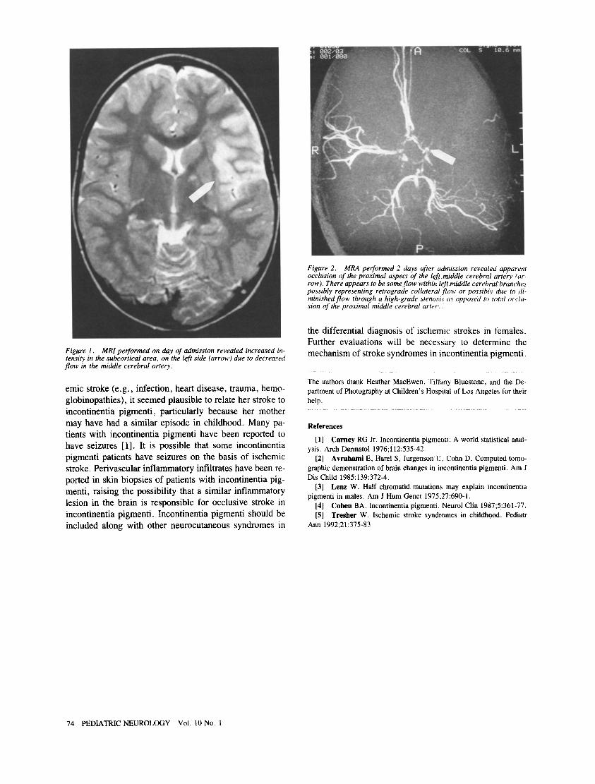
Figure 1. MRI performed on day of admission revealed increased in- tensity in the subcortical area, on the left side (arrow) due to decreased flow in the middle cerebral artery.
emic stroke (e.g., infection, heart disease, trauma, hemo- globinopathies), it seemed plausible to relate her stroke to incontinentia pigmenti, particularly because her mother may have had a similar episode in childhood. Many pa- tients with incontinentia pigmenti have been reported to have seizures [1]. It is possible that some incontinentia pigmenti patients have seizures on the basis of ischemic stroke. Perivascular inflammatory infiltrates have been re- ported in skin biopsies of patients with incontinentia pig- menti, raising the possibility that a similar inflammatory lesion in the brain is responsible for occlusive stroke in incontinentia pigmenti. Incontinentia pigmenti should be included along with other neurocutaneous syndromes in
Figure 2. MRA performed 2 days after admission revealed apparent occlusion of the proximal aspect of the left middte cerebral artery (ar- row). There appears to be some flow within left middle cerebral branche,~ possibly representing retrograde collateral ,flow or possibly due to di- minished flow through a high*grade stenosis as opposed to total acclt~- sion of the proximal middle cerebral artery._
the differential diagnosis of ischemic strokes in females. Further evaluations will be necessary to determine the mechanism of stroke syndromes in incontinentia pigmenti.
The authors thank Heather MacEwen, Tiftany Bluestone, and the De- partment of Photography at Children's Hospital of Los Angeles for their help.
References
[1] Carney RG Jr. Incontinentia pigmenti: A world statistical anal- ysis. Arch Dermatol 1976;112:535-42.
[2] Avrahami E, Harel S, Jurgenson U, Cohn D. Computed tomo- graphic demonstration of brain changes in incontinentia pigmenti. Am J Dis Child 1985;139:372-4.
[3] Lenz W. Half chromatid mutations may explain incontinentia pigmenti in males. Am J Hum Genet 1975;27:690-1.
[4] Cohen BA. Incontinentia pigmenti. Neurol Clin 1987;5:361-77. [5] Tresher W. Ischemic stroke syndromes in childhood. Pediatr
Ann 1992;21:375-83.
74 PEDIATRIC NEUROLOGY Vol. 10 No. 1


![Tnfa Signaling Through Tnfr2 Protects Skin Against ...eprints.whiterose.ac.uk/81541/1/Tnfa signaling through tnfr2 protects... · genodermatosis incontinentia pigmenti (IP) [17]](https://img.pdfslide.net/doc/110x75/5f3bedf6651a4c137761035c/tnfa-signaling-through-tnfr2-protects-skin-against-signaling-through-tnfr2-protects.jpg)


