Embed Size (px)
Citation preview
Vertebrate Hh Signaling
1
Hedgehog Secretion and Signal Transduction in Vertebrates
Kaitlyn E. Ryan and Chin Chiang*
From the Department of Cell and Developmental Biology Vanderbilt University, Nashville, TN 37232
*To whom correspondence should be addressed: Chin Chiang, Department of Cell and Developmental Biology, Vanderbilt University, Nashville, TN 37232. Email: [email protected]
Keywords: Hedgehog, Sonic Hedgehog, Indian Hedgehog, Desert Hedgehog, morphogen, long-range, mitogen, Sufu, Smoothened, Gli, Patched, Kif7
Summary Signaling by the Hedgehog (Hh) family of secreted proteins is essential for proper embryonic patterning and development. Dysregulation of Hh signaling is associated with a variety of human diseases ranging from developmental disorders such as holoprosencephaly to certain forms of cancer, including medulloblastoma and basal cell carcinoma. Genetic studies in flies and mice have shaped our understanding of Hh signaling and revealed that nearly all core components of the pathway are highly conserved. While many aspects of the Drosophila Hh pathway are conserved in vertebrates, mechanistic differences between the two species have begun to emerge. Perhaps the most striking divergence in vertebrate Hh signaling is its dependence on the primary cilium, a vestigial organelle that is largely absent in flies. This minireview will provide an overview of Hedgehog signaling and present recent insights into vertebrate Hh secretion, receptor binding, and signal transduction.
Originally discovered for its role in Drosophila embryonic patterning, the Hedgehog (Hh) pathway is among a handful of signaling pathways governing the development of multicellular organisms. Hh signaling is essential for the development of nearly every organ system in vertebrates, from patterning the neural tube and limbs to regulating lung morphogenesis and hair follicle formation (1). While the Drosophila genome encodes a single hh gene, vertebrates harbor between three (Sonic hedgehog [Shh], Desert hedgehog [Dhh] and Indian hedgehog [Ihh] in birds and mammals) and six (Shh, Dhh, and Ihh plus Tiggywinkle hedgehog [Twhh], Echidna
hedgehog [Ehh] and Qiqihar hedgehog [Qhh] in fish) homologs, differing primarily in tissue distribution (2). In vertebrates, Shh is expressed throughout the developing nervous system and in many epithelial tissues, Ihh functions primarily in bone development, and Dhh expression is limited to the peripheral nervous system and reproductive organs (1). As a result of its widespread expression, much of what is known about vertebrate Hh signaling stems from work on Shh. All Hh ligands undergo a similar series of processing events that result in the covalent attachment of two lipid moieties and are essential for proper signaling activity and tissue distribution (Figure 1). Secreted Hh ligands interact with Patched (Ptc)/co-receptor complexes on the surface of responding cells, relieving Ptc-mediated inhibition of the signal transducer Smoothened (Smo) (Figure 4). Activated Smo prevents the processing of full-length Gli transcription factors (Gli-FL) into transcriptional repressors (Gli-R) so as to allow full-length Gli to activate the transcription of Hh target genes. Thus, the relative abundance of Gli transcriptional activators and inhibitors ultimately regulates the transcription of Hh target genes. Although many aspects of Drosophila Hh signaling are conserved in vertebrates, vertebrate Hh signal transduction differs in its requirement for the primary cilium. Primary cilia are slim, microtubule-based non-motile structures that project from the surface of nearly all vertebrate cells but are conspicuously absent from most Drosophila cell types (3). The assembly and maintenance of primary cilia requires intraflagellar transport (IFT) proteins, and several members of the IFT family are essential for proper vertebrate
http://www.jbc.org/cgi/doi/10.1074/jbc.R112.356006The latest version is at JBC Papers in Press. Published on April 4, 2012 as Manuscript R112.356006
Copyright 2012 by The American Society for Biochemistry and Molecular Biology, Inc.
by guest on January 23, 2019http://w
ww
.jbc.org/D
ownloaded from
Vertebrate Hh Signaling
2
Hh signaling (3,4). Mutations in components of the kinesin-driven IFT-B complex, which mediates the anterograde transport of molecules from the base of the cilium to the tip, lead to a complete loss of Hh signaling (3). By contrast, mutations in members of the dynein-driven IFT-A complex, which controls retrograde transport, lead to aberrant Hh pathway activation (3). Nonetheless, it is not currently known whether IFT-A and -B complexes interact directly with Hh pathway components to control their localization and activity or if, instead, these complexes facilitate Hh signaling simply by maintaining proper cilia architecture. Indeed, recent genetic studies suggest that the primary cilium may function primarily as a scaffold for Hh signaling, arguing against a direct role for IFT proteins in regulating the movement of Hh pathway components (5). In this minireview, we provide an overview of Hh production and cytosolic signaling in vertebrates (for excellent reviews of Drosophila Hh signaling, see references (2,6)). We discuss recent insights into ligand release, receptor binding, and signal transduction and attempt to incorporate these findings into existing models of Hh signaling. Additionally, we present remaining questions regarding Hh secretion and signal transduction that warrant further investigation. Hedgehog processing and release The signaling activity of Hedgehog ligands is intimately linked to a complex sequence of post-translational modifications ultimately resulting in the covalent attachment of two lipid moieties, one at each terminus (Figure 1). Following translation, Hh precursor peptide approximately 45 kDa in size translocates into the ER lumen where it undergoes a cholesterol-dependent autocatalytic cleavage to generate a 19 kDa cholesterol modified N-terminal peptide fragment and a 25 kD C-terminal fragment (Figure 1). This cleavage reaction occurs in two steps. In the first step, the free thiol of Cys198 (human Shh) acts as a nucleophile, attacking the carbonyl carbon of the preceding glycine residue and generating a thioester intermediate (7-10). In the second step, this thioester intermediate is subject to nucleophilic attack by the 3β hydroxyl group of cholesterol, generating a cholesterol-modified N-terminal fragment (Hh-N) and displacing the C-terminal fragment (Hh-C). While Cys198 has long
been recognized for its role in autocatalytic cleavage, a second conserved cysteine, Cys363, is also required for cleavage, forming a disulfide bond with Cys198 that likely facilitates protein folding and reduction of which generates the reactive thiol required for cleavage (11). As such, mutating either cysteine residue prevents autoproteolysis of Hh precursors (11). Although processing-deficient mutants of Shh are able to illicit juxtacrine signaling in cell-based assays (12), the significance of this finding remains enigmatic, as Shh is found exclusively in its cleaved form during embryogenesis (13). Indeed, mutations disrupting the cleavage of full-length Hh peptides have been linked to developmental disorders such as holoprosencephaly (14,15). All of the signaling properties of Hh proteins reside within the N-terminal fragment. The C-terminal fragment undergoes ER-associated degradation (ERAD), a process that requires the lectins OS9 and XTP3, the ubiquitin ligase Hrd1 and its partner Sel1, and the p97 ATPase (Figure 1).The N-terminal fragment (Hh-N) is subject to a second covalent modification by Hh acyltransferase (Hhat)/Skinny Hh (Ski), which catalyzes the attachment of palmitate to the free amino group of the N-terminal cysteine (16-18). Thus, Hh-N has two covalently attached lipid moieties: cholesterol at its C-terminal end, and palmitate at its N-terminal end.
One unique feature of Hedgehog proteins is their capacity to travel very long distances, up to 300 µm in vertebrate limb, to reach their targets. The release and long-range signaling of the cholesterol- and palmitate-modified Hh-N (hereafter referred to as Hh) requires the activity of Dispatched (Disp), a twelve-pass transmembrane protein belonging to the RND family of bacterial transporters (13,19-21). While mice and flies deficient in Disp synthesize Hh properly, Hh accumulates in producing cells, able to activate the pathway in neighboring cells but not competent for long-range signaling (19,20,22-24). While the Hh-distributing function of murine Disp requires two presumptive proton-binding domains in TM4 and TM10, little else is known about how Disp facilitates Hh secretion and long-range signaling (20). Recent studies of Drosophila imaginal discs indicate that Hh and Disp co-localize within endocytic vesicles and suggest that Disp may traffic Hh to the basolateral membrane
by guest on January 23, 2019http://w
ww
.jbc.org/D
ownloaded from
Vertebrate Hh Signaling
3
where it is released (24). Whether or not the trafficking function of Disp is coupled to its Hh-releasing function, or if these two activities are distinct, remains to be shown, and additional studies are needed to determine if the trafficking function of Disp is conserved in vertebrates. Lipid modifications regulate the activity and distribution of Hedgehog Genetic studies in flies and mice indicate that cholesterol and palmitate are essential for the proper activity and distribution of Hh ligands. The C-terminal cholesterol moiety is required for the formation of multimeric Hh complexes, which are thought to be the biologically relevant form of the morphogen (25-27). In cells expressing a truncated form of Hh that cannot be cholesterol modified, Hh proteins are secreted as monomers in a Disp-independent manner (19,23,28). While the process by which cholesterol mediates multimerization remains uncertain, one possibility is that by tethering Hh proteins to the membrane, the cholesterol moiety concentrates Hh within specific microdomains, such as lipid rafts, and promotes electrostatic interactions between Hh monomers (29-31). Cholesterol-mediated clustering may also promote interactions between Hh and other membrane-associated molecules such as heparin sulfate proteoglycans (HSPGs), whose heparin sulfate moieties are known to interact with positively charged residues within a conserved Cardin Weintraub (CW) motif present in all Hh proteins (Figure 2) (26,27,30,31). In Drosophila, the HS-containing glypicans Dally and Dally-like interact with both Hh and the hemolymph-derived lipoprotein lipophorin, leading to the formation of soluble lipoprotein complexes that mediate patterning in the wing imaginal disc (27,32). Although the addition of HS is sufficient to induce dimerization of non-cholesterol modified Shh in vitro, the composition of vertebrate Hh multimers remains uncharacterized (30). In addition to its role in multimerization, cholesterol also regulates the distribution of Hh ligands (23,33,34). Although there have been conflicting reports regarding how cholesterol affects Hh distribution, the majority of data are in agreement with a role for cholesterol in restricting the spread of Hh ligands (23,33,35,36). Nonetheless, the mechanism by which cholesterol limits the distribution of Hh remains unclear, and
the increased range of non-cholesterol modified Hh ligands may be secondary to loss of multimerization or Disp-mediated release. Such an indirect role for cholesterol in regulating Hh distribution is supported by the finding that in Drosophila, a cholesterol-modified-form of Hh that cannot multimerize (due to a Lys132Asp mutation) has a restricted distribution and signaling range (Figure 2) (26). Additionally, recent work in vertebrate cell lines suggests that the cholesterol moiety of Shh may be removed by membrane proximal proteases prior to its release (30). Taken together, these data indicate that the role of cholesterol in determining the range of Hh signaling may not be straightforward and warrants further investigation.
Whereas non-cholesterol-modified Hh ligands maintain some of their signaling capacity, loss of palmitoylation abolishes the signaling activity of Hh almost entirely (17,18,29,37), indicating that palmitate is absolutely required for Hh signaling. Although the importance of palmitate has long been recognized, only recently have inroads been made in understanding why. Recent work in vitro suggests that palmitate facilitates the cleavage of N-terminal amino acids by membrane-proximal proteases such as ADAM (A disintegrin and metalloprotease) family members (38). Such cleavage is required for the formation of active Shh multimers, as these residues otherwise obstruct the Zn2+ coordination site on adjacent molecules, a region that likely interacts with Ptc and is known to regulate Shh stability and activity (Figure 3) (39-42). Thus, in the absence of palmitoylation (due to mutation of the N-terminal Cys), Shh maintains the capacity to multimerize, but these multimers have significantly reduced signaling activity due to their inability to properly interact with Ptc (38). While these data provide insight into the role of palmitoylation in Hh signaling, they also raise a number of questions regarding the production and secretion of Hh. For instance, how is the cleavage of lipid moieties coupled to Disp-mediated release? Are the lipid moieties of Drosophila Hh also cleaved? Future studies are needed to address these questions and to determine if lipid moieties are also cleaved in vivo. Dual roles of Patched in Hedgehog reception and pathway inhibition
by guest on January 23, 2019http://w
ww
.jbc.org/D
ownloaded from
Vertebrate Hh Signaling
4
The Hh receptor Patched (Ptc) is a twelve-pass transmembrane protein homology to the RND family of bacterial transporter proteins. Reception of Hh by Ptc is enhanced by the presence of additional Hh-binding proteins on the cell surface. These presumptive co-receptors include a family of immunoglobulin- and Fibronectin type III (FnIII)-containing integral membrane proteins (Ihog in Boi in Drosophila; Cdo and Boc in vertebrates) and the vertebrate-specific cell surface protein Gas1 (43-45). While removal of a single co-receptor leads to a modest, tissue-specific reduction in Hh pathway activity, removal of two or three co-receptors from Drosophila or mice, respectively, leads to a complete loss of signaling, indicating that these co-receptors play an essential role in Hh signaling (43,45,46).
In addition to Boc, Cdo, and Gas1, vertebrates harbor a fourth Hh binding protein, Hip, that has no downstream signaling function and likely acts as a decoy receptor by competing with Ptc for Hh binding (39,47). Analysis of the crystal structure of Hip in complex with Shh reveals that Asp383 of Hip displaces water and completes the tetrahedral coordination of Zn2+ in the Shh pseudoactive site (Figure 3) (39,40). Sequence comparisons of Hip and Ptc reveals that Ptc contains a similar sequence of amino acids capable of binding Shh and competing with Hip for Shh binding, providing novel insight into Hh-receptor interactions (39). Given that Drosophila Hh lacks a Zn2+ coordination site and is unable to directly bind Ptc, these data also suggest that Hh-Ptc interactions differs between flies and vertebrates (44). This possible divergence is further supported by the finding that while Drosophila Hh binds the second fibronectin III (FnIII) repeat in Ihog, vertebrate Hhs bind a third, non-orthologous FnIII repeat in Cdo (48). Thus, despite the conserved function of Ptc and co-receptors in Hh signaling, the mode of binding between Hh and these receptor complexes does not appear to be conserved. In addition to serving as the Hh receptor, Ptc functions as a potent negative regulator of the Hh pathway by inhibiting the seven-pass transmembrane protein Smoothened (Smo). In the absence of Hh, Ptc localizes to the primary cilium and maintains Smo in an inactive conformation, preventing Smo from entering the cilium (49). While early studies suggested that Ptc could
directly bind to and inhibit Smo (50), subsequent work revealed that Ptc-mediated inhibition is non-stoichiometric, making direct inhibition unlikely (51). The mechanism by which Ptc inhibits Smo remains enigmatic. Sequence similarities between Ptc and the RND family of bacterial transporter proteins have led many to hypothesize that Ptc may regulate the flux molecules that activate or inhibit Smo, a theory that is supported by the susceptibility of Smo to modulation by small molecules such as the steroidal alkaloid cyclopamine (52-54). Given that Ptc is enriched around the base of the primary cilium, where vertebrate Hh signaling likely occurs, Ptc might locally control the abundance of Smo inhibitors or activators (49). Although a number of Smo agonists and antagonists have been identified, to date none have been shown to be regulated by Ptc. Recent work in Drosophila suggests that Ptc may inhibit Hh signaling by regulating the synthesis of phosphotidylinositol 4-phosphate (PI4P), revealing that increased and decreased levels PI4P lead to Hh pathway activation and repression, respectively (55). Importantly, by showing that cells deficient in Ptc have increased PI4P levels, this work provides the first evidence of an endogenous Hh activator that is regulated by Ptc. Nonetheless, future studies are needed to determine how Ptc regulates PI4P synthesis and verify that PI4P activates the pathway at the level of Smo rather than acting further downstream. Transcriptional repression in the absence of Hedgehog The zinc finger-containing Gli transcription factors are the principle effectors of canonical Hh signaling. Depending on the availability of Hh ligands, Gli proteins function either as transcriptional activators or repressors. In the absence of Hh, full-length Gli (Gli-FL) is proteolytically processed to yield a truncated N-terminal transcriptional repressor (Gli-R) (Figure 4a). Whereas Drosophila harbor a single Gli family member, Cubitus Interruptus (Ci), vertebrates have three, Gli1-Gli3. Of these, Gli2 and Gli3 function as both transcriptional activators and repressors while Gli1 is a target of Hh signaling and exists only as an activator.
Although many aspects of vertebrate Gli-R formation remain enigmatic, Suppressor of Fused (Sufu), the kinesin Kif7 and the primary
by guest on January 23, 2019http://w
ww
.jbc.org/D
ownloaded from
Vertebrate Hh Signaling
5
cilium are required for efficient processing of Gli-FL into Gli-R (Figure 4a) (3,56-59). Sufu stabilizes full-length Gli2 and Gli3 and sequesters both proteins in the cytosol, thus preventing their nuclear translocation and activation (6,60-62). Sufu also promotes the phosphorylation of C-terminal residues in Gli-FL by protein kinase A (PKA), which primes full-length Gli for further phosphorylation by glycogen synthase kinase 3β (GSK3β) and casein kinase 1α (CK1α) (63,64). Phosphorylated Gli-FL is recognized by the E3 ubiquitin ligase β TrCP, leading to the ubiquitylation and degradation of C-terminal peptides to generate Gli-R (63-66). In contrast to its relatively minor role in Drosophila, Sufu is absolutely required for proper development and essential for Gli-R formation in vertebrates (56,67). Mice deficient in Sufu die around embryonic day 9.5 with significantly reduced levels of both full-length and repressor forms of Gli and features of aberrant Hedgehog activation that resemble loss of Ptc (56,67). In the absence of Sufu, Gli-FL enters the nucleus and is converted into a labile transcriptional activator (Gli-A) that is quickly degraded within the nucleus in a manner that depends upon the cullin3-based ubiquitin ligase adaptor Spop (62,68-70). Indeed, Sufu and Spop have been shown to compete for Gli-FL binding, and loss of Spop from Sufu-/- cells leads to a significant recovery in full-length Gli levels (62). Together, these data indicate that Sufu regulates Gli-R formation by stabilizing full-length Gli in the cytosol and preventing Spop-dependent degradation in the nucleus. In addition to its role in Gli processing, Sufu may also inhibit the transcription of Hh target genes through its interaction with SAP18, a component of the mSin3-histone deacetylase repressor complex (71). However, this processing-independent role for Sufu was recently challenged (69), and additional data are needed to clarify the function of nuclear Sufu in Hh pathway inhibition.
In addition to Sufu, the kinesin 4 family member Kif7 also appears to be required for optimal Gli processing (57-59,72). Mice deficient in Kif7 have increased levels of Gli-FL, decreased levels of Gli-R and exhibit features of pathway de-repression such as polydactyly (57-59). Although the mechanism by which Kif7 promotes Gli processing remains unclear, one possibility is that, like its Drosophila homolog Costal2 (Cos2), Kif7
recruits PKA, GSK3β and CK1α to phosphorylate Gli-FL (Figure 4a). Although Kif7 has been shown to interact with Gli, additional data are needed to determine if the scaffolding function of Kif7 is conserved in vertebrates.
Studies both in vivo and in vitro indicate that the primary cilium is required for efficient processing of Gli-FL into Gli-R (3). Interestingly, the role of Sufu in Gli-R production appears to be independent of cilia, as cells lacking both primary cilia and Sufu exhibit unkempt Hh pathway activity akin to Sufu-/- cells (69,73). By contrast, the role of Kif7 in Gli processing is cilia-dependent, as mice lacking both cilia and Kif7 resemble cilia mutants (57). Although the exact function of the cilium in Gli processing remains enigmatic, the cilium may serve as a platform for Gli processing machinery. Indeed, Kif7, PKA, GSK3β and CK1α are present in the primary cilia and/or basal body in the absence of Hh signaling (57-59,74-76). Although Sufu cannot localize to the cilium on its own, it is likely recruited there by Gli, as low levels of both Sufu and Gli can be observed in the cilium even in the absence of Hh signaling (60,61). Thus, although Gli-Sufu complexes form throughout the cytosol, they may be directed to the cilium by Gli for efficient processing in a Kif7- and kinase-dependent manner.
Although Gli2 and Gli3 both undergo partial proteolytic degradation in the absence of Hh, the processing of Gli3 is significantly more efficient than that of Gli2 (77). Consequently, Gli3-R serves as the principle transcriptional repressor of Hh signaling in the absence of ligand, while Gli2-A functions as the predominant transcriptional activator (78). The increased efficiency of Gli3 processing is due in large part to the sequence of a 200 residue processing determinant domain (PDD) in its C-terminus (79). Together with an appropriate degron and the zinc finger domain, the PDD forms a three part signal that is essential for efficient Gli3 processing (80). But what happens to full-length Gli2 in the absence of Hh? Like Gli3, the C-terminus of Gli2 is phosphorylated by PKA in the absence of Hh. Although this phosphorylation leads to a limited amount of processing, it may also destabilize Gli2-FL, leading to complete degradation by the proteosome (77,81) . Such a processing-independent role of PKA in Hh pathway inhibition
by guest on January 23, 2019http://w
ww
.jbc.org/D
ownloaded from
Vertebrate Hh Signaling
6
is supported by recent genetic data showing that mice lacking both catalytic subunits of PKA (Prkaca-/-; Prkacb-/-) die mid-gestation with a completely ventralized neural tube, a defect that cannot be explained by loss of Gli processing alone and suggests a increase in Gli activation (75,82). Given that PKA may also regulate the entry of Sufu-Gli complexes into the cilium, additional studies are required to clarify the mechanism(s) by which PKA inhibits Gli activation and determine to what extent Gli2 phosphorylation inhibits pathway activation (61,75,83). Smoothened and Gli activation in the presence of Hedgehog In the presence of Hh, Ptc relieves its inhibition of Smo and allows Smo to become activated. Despite significant sequence differences, many aspects of Drosophila Smo activation are conserved in vertebrates. In Drosophila, phosphorylation of C-terminal residues by PKA, CK1, and G-coupled protein receptor kinase 2 (GRK2) cause Smo to adopt an open conformation and promote its accumulation on the membrane (84-89). Although the C-terminus of vertebrate Smo differs significantly from Drosophila and lacks PKA phosphorylation sites, recent data indicate that vertebrate Smo is also phosphorylated in response to Hh signaling (76,90,91). CK1α and GRK2 phosphorylate the C-terminal tail of vertebrate Smo, inducing conformational changes and facilitating its lateral translocation into the primary cilium (Figure 4b) (76). The movement of Smo into the cilium is dependent upon β-Arrestins and the kinesin 2 motor subunit Kif3a, both of which are recruited to Smo following its phosphorylation by CK1α and GRK2 (76,90,92,93).
Activated Smo both inhibits Gli processing as well as promotes additional ill-defined modifications that convert full-length Gli proteins into transcriptional activators. Although the details of this process remain somewhat enigmatic, activated Smo likely promotes the disassembly of Sufu-Gli complexes that accumulate in the cilium following pathway activation (Figure 4b) (60-62,94). Kif7 may also promote Sufu-Gli disassembly, as it localizes to the cilium in response to Hh and interacts with overexpressed Smo in tissue culture cells (58). Indeed, such a positive role of Kif7 in Hh
signaling is consistent with the finding that mice deficient in Kif7 exhibit features of decreased Hh pathway activity, such as reduced Ptc expression in the notochord and floor plate (57,58). Nonetheless, additional studies are needed to determine if Kif7-Smo interactions are dependent on Smo phosphorylation, as they are for Drosophila Cos2 (95,96). The disassembly of Sufu-Gli complexes allows full-length Gli to enter the nucleus where it is converted to its activator form (Gli-A) (61). The translocation of Gli requires cytoplasmic microtubules, as microtubule de-stabilizing agents such as nocodazole have been shown to inhibit its nuclear accumulation and activity (60,97). While the details of Gli activation remain nebulous, they may involve phosphorylation, as Gli2 and Gli3 appear to be phosphorylated within the nucleus in response to Hh (60). Given that the nucleus is also the site of Spop-mediated degradation, however, it is difficult to ascertain whether this phosphorylation is coupled to Gli activation or degradation (62,69). Gli proteins might also be deacetylated in response to Hh stimulation, as HDAC1 overexpression in tissue culture cells leads to Gli1 deacetylation (98). Activated Gli promotes the transcription of genes involved in differentiation, proliferation, and cell survival as well as several negative regulators of the pathway, such as Ptc and Hip to downregulate pathway activity. Conclusions and Perspectives
Over the past two decades, mouse and fly genetics have been instrumental in identifying components of the Hh pathway and elucidating their functions, revealing a high degree of conservation between the two species. The discovery that vertebrate Hh signaling requires the primary cilium, however, has significantly changed how the pathway is studied and made it somewhat more difficult to draw comparisons between vertebrates and flies. Despite these challenges, significant progress has been made in defining vertebrate Hh signal transduction. Nonetheless, several questions regarding vertebrate Hh secretion and signal transduction remain unanswered. The mechanistic details of Disp-mediated secretion remain elusive, as does the composition of secreted Hh multimers. The mechanism by which Ptc inhibits Smo continues to be a mystery, and a detailed understanding of
by guest on January 23, 2019http://w
ww
.jbc.org/D
ownloaded from
Vertebrate Hh Signaling
7
how activated Smo promotes Gli activation is lacking. Additional studies are needed to examine Kif7’s role in Gli processing and activation as well as determine to what extent the motor function of Kif7 is important for Hh signaling. But perhaps most intriguing is the question of how, and why, the primary cilium plays such an essential role in
vertebrate Hh signal transduction. As cell and developmental biologists continue to adapt to the challenges inherent in the study of vertebrate Hh signaling, the answers to these and other questions will undoubtedly be revealed.
References 1. Ingham, P. W., and McMahon, A. P. (2001) Hedgehog signaling in animal development: paradigms
and principles. Genes Dev 15, 3059-3087 2. Ingham, P. W., Nakano, Y., and Seger, C. (2011) Mechanisms and functions of Hedgehog signalling
across the metazoa. Nat Rev Genet 12, 393-406 3. Goetz, S. C., and Anderson, K. V. (2010) The primary cilium: a signalling centre during vertebrate
development. Nat Rev Genet 11, 331-344 4. Pedersen, L. B., and Rosenbaum, J. L. (2008) Intraflagellar transport (IFT) role in ciliary assembly,
resorption and signalling. Curr Top Dev Biol 85, 23-61 5. Ocbina, P. J., Eggenschwiler, J. T., Moskowitz, I., and Anderson, K. V. (2011) Complex interactions
between genes controlling trafficking in primary cilia. Nat Genet 43, 547-553 6. Wilson, C. W., and Chuang, P. T. (2010) Mechanism and evolution of cytosolic Hedgehog signal
transduction. Development 137, 2079-2094 7. Porter, J. A., von Kessler, D. P., Ekker, S. C., Young, K. E., Lee, J. J., Moses, K., and Beachy, P. A.
(1995) The product of hedgehog autoproteolytic cleavage active in local and long-range signalling. Nature 374, 363-366
8. Porter, J. A., Ekker, S. C., Park, W. J., von Kessler, D. P., Young, K. E., Chen, C. H., Ma, Y., Woods, A. S., Cotter, R. J., Koonin, E. V., and Beachy, P. A. (1996) Hedgehog patterning activity: role of a lipophilic modification mediated by the carboxy-terminal autoprocessing domain. Cell 86, 21-34
9. Lee, J. J., Ekker, S. C., von Kessler, D. P., Porter, J. A., Sun, B. I., and Beachy, P. A. (1994) Autoproteolysis in hedgehog protein biogenesis. Science 266, 1528-1537
10. Porter, J. A., Young, K. E., and Beachy, P. A. (1996) Cholesterol modification of hedgehog signaling proteins in animal development. Science 274, 255-259
11. Chen, X., Tukachinsky, H., Huang, C. H., Jao, C., Chu, Y. R., Tang, H. Y., Mueller, B., Schulman, S., Rapoport, T. A., and Salic, A. (2011) Processing and turnover of the Hedgehog protein in the endoplasmic reticulum. J Cell Biol 192, 825-838
12. Tokhunts, R., Singh, S., Chu, T., D'Angelo, G., Baubet, V., Goetz, J. A., Huang, Z., Yuan, Z., Ascano, M., Zavros, Y., Therond, P. P., Kunes, S., Dahmane, N., and Robbins, D. J. (2010) The full-length unprocessed hedgehog protein is an active signaling molecule. J Biol Chem 285, 2562-2568
13. Kawakami, T., Kawcak, T., Li, Y. J., Zhang, W., Hu, Y., and Chuang, P. T. (2002) Mouse dispatched mutants fail to distribute hedgehog proteins and are defective in hedgehog signaling. Development 129, 5753-5765
14. Traiffort, E., Dubourg, C., Faure, H., Rognan, D., Odent, S., Durou, M. R., David, V., and Ruat, M. (2004) Functional characterization of sonic hedgehog mutations associated with holoprosencephaly. J Biol Chem 279, 42889-42897
15. Maity, T., Fuse, N., and Beachy, P. A. (2005) Molecular mechanisms of Sonic hedgehog mutant effects in holoprosencephaly. Proc Natl Acad Sci U S A 102, 17026-17031
16. Buglino, J. A., and Resh, M. D. (2008) Hhat is a palmitoylacyltransferase with specificity for N-palmitoylation of Sonic Hedgehog. J Biol Chem 283, 22076-22088
by guest on January 23, 2019http://w
ww
.jbc.org/D
ownloaded from
Vertebrate Hh Signaling
8
17. Pepinsky, R. B., Zeng, C., Wen, D., Rayhorn, P., Baker, D. P., Williams, K. P., Bixler, S. A., Ambrose, C. M., Garber, E. A., Miatkowski, K., Taylor, F. R., Wang, E. A., and Galdes, A. (1998) Identification of a palmitic acid-modified form of human Sonic hedgehog. J Biol Chem 273, 14037-14045
18. Chamoun, Z., Mann, R. K., Nellen, D., von Kessler, D. P., Bellotto, M., Beachy, P. A., and Basler, K. (2001) Skinny hedgehog, an acyltransferase required for palmitoylation and activity of the hedgehog signal. Science 293, 2080-2084
19. Burke, R., Nellen, D., Bellotto, M., Hafen, E., Senti, K. A., Dickson, B. J., and Basler, K. (1999) Dispatched, a novel sterol-sensing domain protein dedicated to the release of cholesterol-modified hedgehog from signaling cells. Cell 99, 803-815
20. Ma, Y., Erkner, A., Gong, R., Yao, S., Taipale, J., Basler, K., and Beachy, P. A. (2002) Hedgehog-mediated patterning of the mammalian embryo requires transporter-like function of dispatched. Cell 111, 63-75
21. Caspary, T., Garcia-Garcia, M. J., Huangfu, D., Eggenschwiler, J. T., Wyler, M. R., Rakeman, A. S., Alcorn, H. L., and Anderson, K. V. (2002) Mouse Dispatched homolog1 is required for long-range, but not juxtacrine, Hh signaling. Curr Biol 12, 1628-1632
22. Gallet, A., Rodriguez, R., Ruel, L., and Therond, P. P. (2003) Cholesterol modification of hedgehog is required for trafficking and movement, revealing an asymmetric cellular response to hedgehog. Dev Cell 4, 191-204
23. Li, Y., Zhang, H., Litingtung, Y., and Chiang, C. (2006) Cholesterol modification restricts the spread of Shh gradient in the limb bud. Proc Natl Acad Sci U S A 103, 6548-6553
24. Callejo, A., Bilioni, A., Mollica, E., Gorfinkiel, N., Andres, G., Ibanez, C., Torroja, C., Doglio, L., Sierra, J., and Guerrero, I. (2011) Dispatched mediates Hedgehog basolateral release to form the long-range morphogenetic gradient in the Drosophila wing disk epithelium. Proc Natl Acad Sci U S A 108, 12591-12598
25. Zeng, X., Goetz, J. A., Suber, L. M., Scott, W. J., Jr., Schreiner, C. M., and Robbins, D. J. (2001) A freely diffusible form of Sonic hedgehog mediates long-range signalling. Nature 411, 716-720
26. Vyas, N., Goswami, D., Manonmani, A., Sharma, P., Ranganath, H. A., VijayRaghavan, K., Shashidhara, L. S., Sowdhamini, R., and Mayor, S. (2008) Nanoscale organization of hedgehog is essential for long-range signaling. Cell 133, 1214-1227
27. Eugster, C., Panakova, D., Mahmoud, A., and Eaton, S. (2007) Lipoprotein-heparan sulfate interactions in the Hh pathway. Dev Cell 13, 57-71
28. Huang, X., Litingtung, Y., and Chiang, C. (2007) Region-specific requirement for cholesterol modification of sonic hedgehog in patterning the telencephalon and spinal cord. Development 134, 2095-2105
29. Chen, M. H., Li, Y. J., Kawakami, T., Xu, S. M., and Chuang, P. T. (2004) Palmitoylation is required for the production of a soluble multimeric Hedgehog protein complex and long-range signaling in vertebrates. Genes Dev 18, 641-659
30. Dierker, T., Dreier, R., Petersen, A., Bordych, C., and Grobe, K. (2009) Heparan sulfate-modulated, metalloprotease-mediated sonic hedgehog release from producing cells. J Biol Chem 284, 8013-8022
31. Dierker, T., Dreier, R., Migone, M., Hamer, S., and Grobe, K. (2009) Heparan sulfate and transglutaminase activity are required for the formation of covalently cross-linked hedgehog oligomers. J Biol Chem 284, 32562-32571
32. Panakova, D., Sprong, H., Marois, E., Thiele, C., and Eaton, S. (2005) Lipoprotein particles are required for Hedgehog and Wingless signalling. Nature 435, 58-65
33. Guerrero, I., and Chiang, C. (2007) A conserved mechanism of Hedgehog gradient formation by lipid modifications. Trends Cell Biol 17, 1-5
34. Lewis, P. M., Dunn, M. P., McMahon, J. A., Logan, M., Martin, J. F., St-Jacques, B., and McMahon, A. P. (2001) Cholesterol modification of sonic hedgehog is required for long-range signaling activity and effective modulation of signaling by Ptc1. Cell 105, 599-612
by guest on January 23, 2019http://w
ww
.jbc.org/D
ownloaded from

Vertebrate Hh Signaling
9
35. Dawber, R. J., Hebbes, S., Herpers, B., Docquier, F., and van den Heuvel, M. (2005) Differential range and activity of various forms of the Hedgehog protein. BMC Dev Biol 5, 21
36. Callejo, A., Torroja, C., Quijada, L., and Guerrero, I. (2006) Hedgehog lipid modifications are required for Hedgehog stabilization in the extracellular matrix. Development 133, 471-483
37. Lee, J. D., Kraus, P., Gaiano, N., Nery, S., Kohtz, J., Fishell, G., Loomis, C. A., and Treisman, J. E. (2001) An acylatable residue of Hedgehog is differentially required in Drosophila and mouse limb development. Dev Biol 233, 122-136
38. Ohlig, S., Farshi, P., Pickhinke, U., van den Boom, J., Hoing, S., Jakuschev, S., Hoffmann, D., Dreier, R., Scholer, H. R., Dierker, T., Bordych, C., and Grobe, K. (2011) Sonic hedgehog shedding results in functional activation of the solubilized protein. Dev Cell 20, 764-774
39. Bosanac, I., Maun, H. R., Scales, S. J., Wen, X., Lingel, A., Bazan, J. F., de Sauvage, F. J., Hymowitz, S. G., and Lazarus, R. A. (2009) The structure of SHH in complex with HHIP reveals a recognition role for the Shh pseudo active site in signaling. Nat Struct Mol Biol 16, 691-697
40. Bishop, B., Aricescu, A. R., Harlos, K., O'Callaghan, C. A., Jones, E. Y., and Siebold, C. (2009) Structural insights into hedgehog ligand sequestration by the human hedgehog-interacting protein HHIP. Nat Struct Mol Biol 16, 698-703
41. Day, E. S., Wen, D., Garber, E. A., Hong, J., Avedissian, L. S., Rayhorn, P., Shen, W., Zeng, C., Bailey, V. R., Reilly, J. O., Roden, J. A., Moore, C. B., Williams, K. P., Galdes, A., Whitty, A., and Baker, D. P. (1999) Zinc-dependent structural stability of human Sonic hedgehog. Biochemistry 38, 14868-14880
42. Fuse, N., Maiti, T., Wang, B., Porter, J. A., Hall, T. M., Leahy, D. J., and Beachy, P. A. (1999) Sonic hedgehog protein signals not as a hydrolytic enzyme but as an apparent ligand for patched. Proc Natl Acad Sci U S A 96, 10992-10999
43. Allen, B. L., Song, J. Y., Izzi, L., Althaus, I. W., Kang, J. S., Charron, F., Krauss, R. S., and McMahon, A. P. (2011) Overlapping roles and collective requirement for the coreceptors GAS1, CDO, and BOC in SHH pathway function. Dev Cell 20, 775-787
44. Beachy, P. A., Hymowitz, S. G., Lazarus, R. A., Leahy, D. J., and Siebold, C. (2010) Interactions between Hedgehog proteins and their binding partners come into view. Genes Dev 24, 2001-2012
45. Izzi, L., Levesque, M., Morin, S., Laniel, D., Wilkes, B. C., Mille, F., Krauss, R. S., McMahon, A. P., Allen, B. L., and Charron, F. (2011) Boc and Gas1 each form distinct Shh receptor complexes with Ptch1 and are required for Shh-mediated cell proliferation. Dev Cell 20, 788-801
46. Zheng, X., Mann, R. K., Sever, N., and Beachy, P. A. (2010) Genetic and biochemical definition of the Hedgehog receptor. Genes Dev 24, 57-71
47. Chuang, P. T., and McMahon, A. P. (1999) Vertebrate Hedgehog signalling modulated by induction of a Hedgehog-binding protein. Nature 397, 617-621
48. McLellan, J. S., Zheng, X., Hauk, G., Ghirlando, R., Beachy, P. A., and Leahy, D. J. (2008) The mode of Hedgehog binding to Ihog homologues is not conserved across different phyla. Nature 455, 979-983
49. Rohatgi, R., Milenkovic, L., and Scott, M. P. (2007) Patched1 regulates hedgehog signaling at the primary cilium. Science 317, 372-376
50. Murone, M., Rosenthal, A., and de Sauvage, F. J. (1999) Sonic hedgehog signaling by the patched-smoothened receptor complex. Curr Biol 9, 76-84
51. Taipale, J., Cooper, M. K., Maiti, T., and Beachy, P. A. (2002) Patched acts catalytically to suppress the activity of Smoothened. Nature 418, 892-897
52. Cooper, M. K., Porter, J. A., Young, K. E., and Beachy, P. A. (1998) Teratogen-mediated inhibition of target tissue response to Shh signaling. Science 280, 1603-1607
53. Taipale, J., Chen, J. K., Cooper, M. K., Wang, B., Mann, R. K., Milenkovic, L., Scott, M. P., and Beachy, P. A. (2000) Effects of oncogenic mutations in Smoothened and Patched can be reversed by cyclopamine. Nature 406, 1005-1009
54. Chen, J. K., Taipale, J., Cooper, M. K., and Beachy, P. A. (2002) Inhibition of Hedgehog signaling by direct binding of cyclopamine to Smoothened. Genes Dev 16, 2743-2748
by guest on January 23, 2019http://w
ww
.jbc.org/D
ownloaded from
Vertebrate Hh Signaling
10
55. Yavari, A., Nagaraj, R., Owusu-Ansah, E., Folick, A., Ngo, K., Hillman, T., Call, G., Rohatgi, R., Scott, M. P., and Banerjee, U. (2010) Role of lipid metabolism in smoothened derepression in hedgehog signaling. Dev Cell 19, 54-65
56. Svard, J., Heby-Henricson, K., Persson-Lek, M., Rozell, B., Lauth, M., Bergstrom, A., Ericson, J., Toftgard, R., and Teglund, S. (2006) Genetic elimination of Suppressor of fused reveals an essential repressor function in the mammalian Hedgehog signaling pathway. Dev Cell 10, 187-197
57. Liem, K. F., Jr., He, M., Ocbina, P. J., and Anderson, K. V. (2009) Mouse Kif7/Costal2 is a cilia-associated protein that regulates Sonic hedgehog signaling. Proc Natl Acad Sci U S A 106, 13377-13382
58. Endoh-Yamagami, S., Evangelista, M., Wilson, D., Wen, X., Theunissen, J. W., Phamluong, K., Davis, M., Scales, S. J., Solloway, M. J., de Sauvage, F. J., and Peterson, A. S. (2009) The mammalian Cos2 homolog Kif7 plays an essential role in modulating Hh signal transduction during development. Curr Biol 19, 1320-1326
59. Cheung, H. O., Zhang, X., Ribeiro, A., Mo, R., Makino, S., Puviindran, V., Law, K. K., Briscoe, J., and Hui, C. C. (2009) The kinesin protein Kif7 is a critical regulator of Gli transcription factors in mammalian hedgehog signaling. Sci Signal 2, ra29
60. Humke, E. W., Dorn, K. V., Milenkovic, L., Scott, M. P., and Rohatgi, R. (2010) The output of Hedgehog signaling is controlled by the dynamic association between Suppressor of Fused and the Gli proteins. Genes Dev 24, 670-682
61. Tukachinsky, H., Lopez, L. V., and Salic, A. (2010) A mechanism for vertebrate Hedgehog signaling: recruitment to cilia and dissociation of SuFu-Gli protein complexes. J Cell Biol 191, 415-428
62. Wang, C., Pan, Y., and Wang, B. (2010) Suppressor of fused and Spop regulate the stability, processing and function of Gli2 and Gli3 full-length activators but not their repressors. Development 137, 2001-2009
63. Tempe, D., Casas, M., Karaz, S., Blanchet-Tournier, M. F., and Concordet, J. P. (2006) Multisite protein kinase A and glycogen synthase kinase 3beta phosphorylation leads to Gli3 ubiquitination by SCFbetaTrCP. Mol Cell Biol 26, 4316-4326
64. Kise, Y., Morinaka, A., Teglund, S., and Miki, H. (2009) Sufu recruits GSK3beta for efficient processing of Gli3. Biochem Biophys Res Commun 387, 569-574
65. Bhatia, N., Thiyagarajan, S., Elcheva, I., Saleem, M., Dlugosz, A., Mukhtar, H., and Spiegelman, V. S. (2006) Gli2 is targeted for ubiquitination and degradation by beta-TrCP ubiquitin ligase. J Biol Chem 281, 19320-19326
66. Wang, B., and Li, Y. (2006) Evidence for the direct involvement of {beta}TrCP in Gli3 protein processing. Proc Natl Acad Sci U S A 103, 33-38
67. Cooper, A. F., Yu, K. P., Brueckner, M., Brailey, L. L., Johnson, L., McGrath, J. M., and Bale, A. E. (2005) Cardiac and CNS defects in a mouse with targeted disruption of suppressor of fused. Development 132, 4407-4417
68. Zhang, Q., Zhang, L., Wang, B., Ou, C. Y., Chien, C. T., and Jiang, J. (2006) A hedgehog-induced BTB protein modulates hedgehog signaling by degrading Ci/Gli transcription factor. Dev Cell 10, 719-729
69. Chen, M. H., Wilson, C. W., Li, Y. J., Law, K. K., Lu, C. S., Gacayan, R., Zhang, X., Hui, C. C., and Chuang, P. T. (2009) Cilium-independent regulation of Gli protein function by Sufu in Hedgehog signaling is evolutionarily conserved. Genes Dev 23, 1910-1928
70. Zhang, Q., Shi, Q., Chen, Y., Yue, T., Li, S., Wang, B., and Jiang, J. (2009) Multiple Ser/Thr-rich degrons mediate the degradation of Ci/Gli by the Cul3-HIB/SPOP E3 ubiquitin ligase. Proc Natl Acad Sci U S A 106, 21191-21196
71. Cheng, S. Y., and Bishop, J. M. (2002) Suppressor of Fused represses Gli-mediated transcription by recruiting the SAP18-mSin3 corepressor complex. Proc Natl Acad Sci U S A 99, 5442-5447
by guest on January 23, 2019http://w
ww
.jbc.org/D
ownloaded from
Vertebrate Hh Signaling
11
72. Tay, S. Y., Ingham, P. W., and Roy, S. (2005) A homologue of the Drosophila kinesin-like protein Costal2 regulates Hedgehog signal transduction in the vertebrate embryo. Development 132, 625-634
73. Jia, J., Kolterud, A., Zeng, H., Hoover, A., Teglund, S., Toftgard, R., and Liu, A. (2009) Suppressor of Fused inhibits mammalian Hedgehog signaling in the absence of cilia. Dev Biol 330, 452-460
74. Fumoto, K., Hoogenraad, C. C., and Kikuchi, A. (2006) GSK-3beta-regulated interaction of BICD with dynein is involved in microtubule anchorage at centrosome. EMBO J 25, 5670-5682
75. Tuson, M., He, M., and Anderson, K. V. (2011) Protein kinase A acts at the basal body of the primary cilium to prevent Gli2 activation and ventralization of the mouse neural tube. Development 138, 4921-4930
76. Chen, Y., Sasai, N., Ma, G., Yue, T., Jia, J., Briscoe, J., and Jiang, J. (2011) Sonic Hedgehog dependent phosphorylation by CK1alpha and GRK2 is required for ciliary accumulation and activation of smoothened. PLoS Biol 9, e1001083
77. Pan, Y., Bai, C. B., Joyner, A. L., and Wang, B. (2006) Sonic hedgehog signaling regulates Gli2 transcriptional activity by suppressing its processing and degradation. Mol Cell Biol 26, 3365-3377
78. Hui, C. C., and Angers, S. (2011) Gli proteins in development and disease. Annu Rev Cell Dev Biol 27, 513-537
79. Pan, Y., and Wang, B. (2007) A novel protein-processing domain in Gli2 and Gli3 differentially blocks complete protein degradation by the proteasome. J Biol Chem 282, 10846-10852
80. Schrader, E. K., Harstad, K. G., Holmgren, R. A., and Matouschek, A. (2011) A three-part signal governs differential processing of Gli1 and Gli3 proteins by the proteasome. J Biol Chem 286, 39051-39058
81. Pan, Y., Wang, C., and Wang, B. (2009) Phosphorylation of Gli2 by protein kinase A is required for Gli2 processing and degradation and the Sonic Hedgehog-regulated mouse development. Dev Biol 326, 177-189
82. Huang, Y., Roelink, H., and McKnight, G. S. (2002) Protein kinase A deficiency causes axially localized neural tube defects in mice. J Biol Chem 277, 19889-19896
83. Chen, Y., Yue, S., Xie, L., Pu, X. H., Jin, T., and Cheng, S. Y. (2011) Dual Phosphorylation of suppressor of fused (Sufu) by PKA and GSK3beta regulates its stability and localization in the primary cilium. J Biol Chem 286, 13502-13511
84. Lum, L., Yao, S., Mozer, B., Rovescalli, A., Von Kessler, D., Nirenberg, M., and Beachy, P. A. (2003) Identification of Hedgehog pathway components by RNAi in Drosophila cultured cells. Science 299, 2039-2045
85. Jia, J., Tong, C., Wang, B., Luo, L., and Jiang, J. (2004) Hedgehog signalling activity of Smoothened requires phosphorylation by protein kinase A and casein kinase I. Nature 432, 1045-1050
86. Apionishev, S., Katanayeva, N. M., Marks, S. A., Kalderon, D., and Tomlinson, A. (2005) Drosophila Smoothened phosphorylation sites essential for Hedgehog signal transduction. Nat Cell Biol 7, 86-92
87. Molnar, C., Holguin, H., Mayor, F., Jr., Ruiz-Gomez, A., and de Celis, J. F. (2007) The G protein-coupled receptor regulatory kinase GPRK2 participates in Hedgehog signaling in Drosophila. Proc Natl Acad Sci U S A 104, 7963-7968
88. Chen, Y., Li, S., Tong, C., Zhao, Y., Wang, B., Liu, Y., Jia, J., and Jiang, J. (2010) G protein-coupled receptor kinase 2 promotes high-level Hedgehog signaling by regulating the active state of Smo through kinase-dependent and kinase-independent mechanisms in Drosophila. Genes Dev 24, 2054-2067
89. Su, Y., Ospina, J. K., Zhang, J., Michelson, A. P., Schoen, A. M., and Zhu, A. J. (2011) Sequential phosphorylation of smoothened transduces graded hedgehog signaling. Sci Signal 4, ra43
90. Chen, W., Ren, X. R., Nelson, C. D., Barak, L. S., Chen, J. K., Beachy, P. A., de Sauvage, F., and Lefkowitz, R. J. (2004) Activity-dependent internalization of smoothened mediated by beta-arrestin 2 and GRK2. Science 306, 2257-2260
by guest on January 23, 2019http://w
ww
.jbc.org/D
ownloaded from
Vertebrate Hh Signaling
12
91. Meloni, A. R., Fralish, G. B., Kelly, P., Salahpour, A., Chen, J. K., Wechsler-Reya, R. J., Lefkowitz, R. J., and Caron, M. G. (2006) Smoothened signal transduction is promoted by G protein-coupled receptor kinase 2. Mol Cell Biol 26, 7550-7560
92. Kovacs, J. J., Whalen, E. J., Liu, R., Xiao, K., Kim, J., Chen, M., Wang, J., Chen, W., and Lefkowitz, R. J. (2008) Beta-arrestin-mediated localization of smoothened to the primary cilium. Science 320, 1777-1781
93. Milenkovic, L., Scott, M. P., and Rohatgi, R. (2009) Lateral transport of Smoothened from the plasma membrane to the membrane of the cilium. J Cell Biol 187, 365-374
94. Zeng, H., Jia, J., and Liu, A. (2010) Coordinated translocation of mammalian Gli proteins and suppressor of fused to the primary cilium. PLoS One 5, e15900
95. Shi, Q., Li, S., Jia, J., and Jiang, J. (2011) The Hedgehog-induced Smoothened conformational switch assembles a signaling complex that activates Fused by promoting its dimerization and phosphorylation. Development 138, 4219-4231
96. Jia, J., Tong, C., and Jiang, J. (2003) Smoothened transduces Hedgehog signal by physically interacting with Costal2/Fused complex through its C-terminal tail. Genes Dev 17, 2709-2720
97. Kim, J., Kato, M., and Beachy, P. A. (2009) Gli2 trafficking links Hedgehog-dependent activation of Smoothened in the primary cilium to transcriptional activation in the nucleus. Proc Natl Acad Sci U S A 106, 21666-21671
98. Canettieri, G., Di Marcotullio, L., Greco, A., Coni, S., Antonucci, L., Infante, P., Pietrosanti, L., De Smaele, E., Ferretti, E., Miele, E., Pelloni, M., De Simone, G., Pedone, E. M., Gallinari, P., Giorgi, A., Steinkuhler, C., Vitagliano, L., Pedone, C., Schinin, M. E., Screpanti, I., and Gulino, A. (2010) Histone deacetylase and Cullin3-REN(KCTD11) ubiquitin ligase interplay regulates Hedgehog signalling through Gli acetylation. Nat Cell Biol 12, 132-142
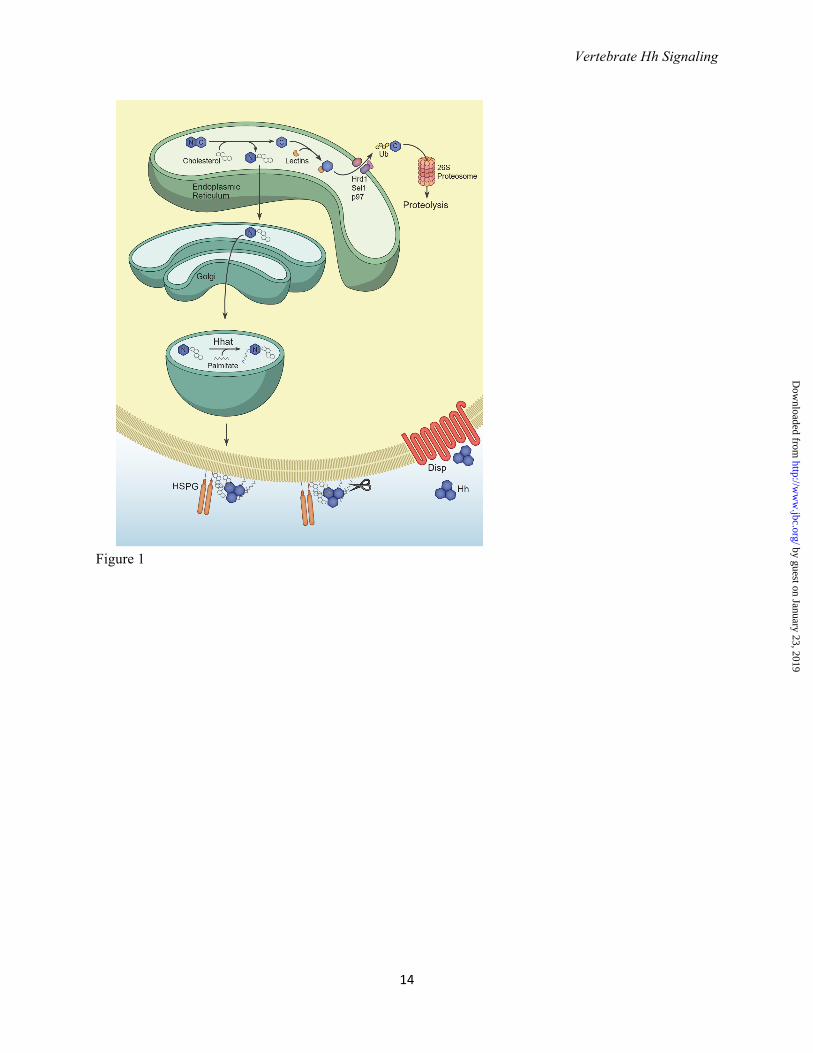
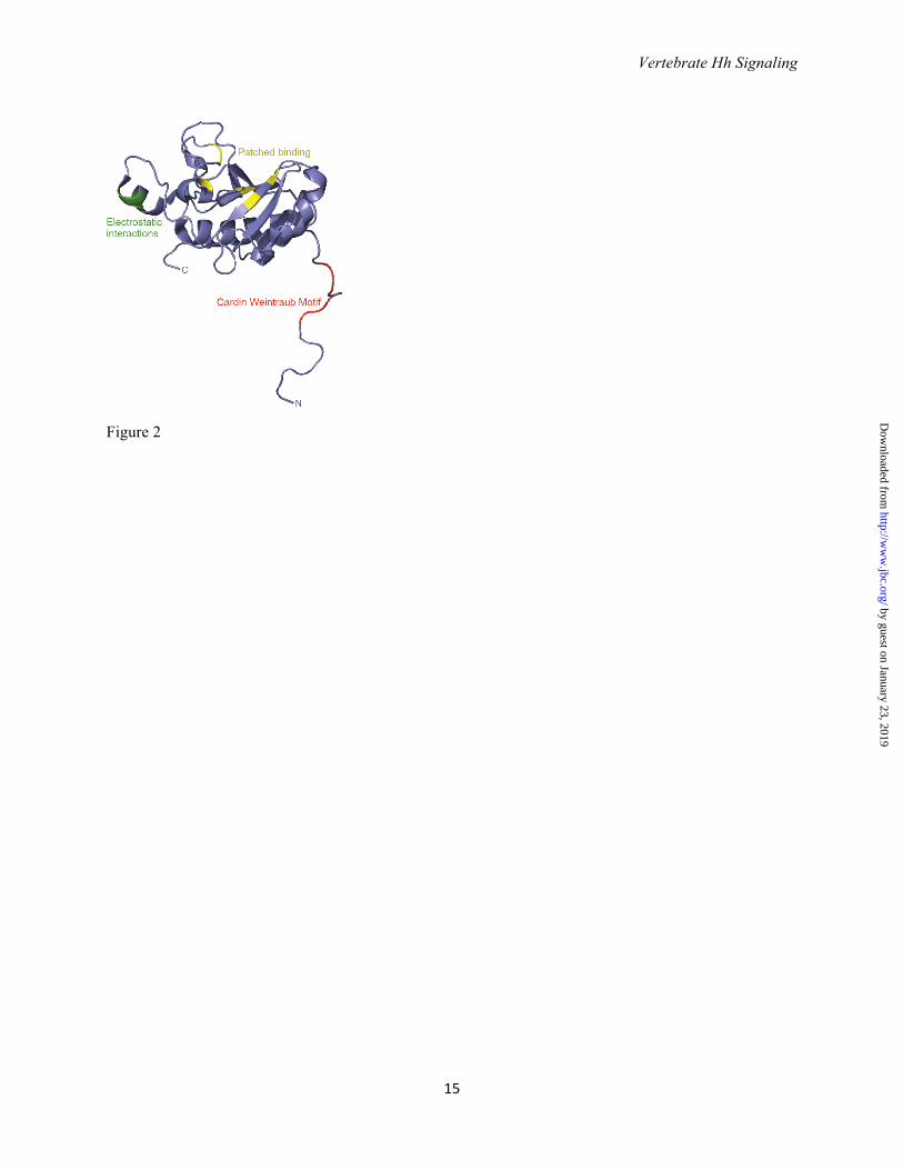
Acknowledgements We thank members of the Chiang lab for their support and suggestions, Elizabeth Dong for her help generating Figures 2 and 3, and Bruce Carter and Laura Lee for their assistance revising and organizing the manuscript. FIGURES Figure 1. Hedgehog processing and release. Hedgehog precursor peptides 45 kDa in size undergo a cholesterol-dependent autocatalytic cleavage in the endoplasmic reticulum to generate a cholesterol-modified N-terminal fragment (Hh-N; denoted by N) and a 25 kDa C-terminal fragment (Hh-C, denoted by C). Hh-C is recognized by the lectins OS-9 and XTP3 and ubiquitylated by the ubiquitin ligase Hrd1 and its partner, Sel1. Ubiquitylated Hh-C is moved into the cytosol by the p97 ATPase and subsequently degraded by the proteasome. Cholesterol-modified Hh-N enters the secretory pathway where the acyltransferase Hhat catalyzes the covalent attachment of palmitate to the N-terminal cysteine. Dually lipidated Hh is targeted to the cell membrane, where cholesterol facilitates the assembly of multimeric Hh complexes possibly by tethering Hh to the membrane and promoting interactions with heparin sulfate proteoglycans (HSPGs). Prior to its release, N- and C-terminal peptides may be cleaved by membrane-proximal proteases such as those belonging to the ADAM (A disintegrin and matrix metalloprotease) family, resulting in the removal of both lipid moieties. The twelve-pass transmembrane protein Dispatched (Disp) facilitates the release of Hh multimers into the extracellular environment although the mechanistic details of this process are not well understood. Figure 2. Regions of Shh important for receptor binding and multimerization. Structure of human SHH-N (non-cholesterol-modified N-terminal fragment, PDB: 3M1N (99)). Residues in green (E72, R73 and K75) mediate electrostatic interactions between Hh monomers and are required for multimerization (38). Arg73 is the vertebrate equivalent of Drosophila Lys132, the mutation of which results in decreased
by guest on January 23, 2019http://w
ww
.jbc.org/D
ownloaded from
Vertebrate Hh Signaling
13
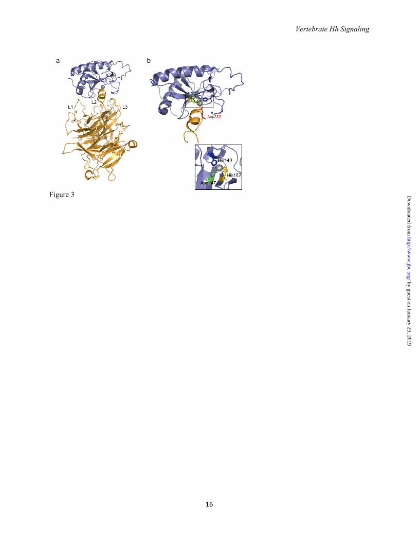
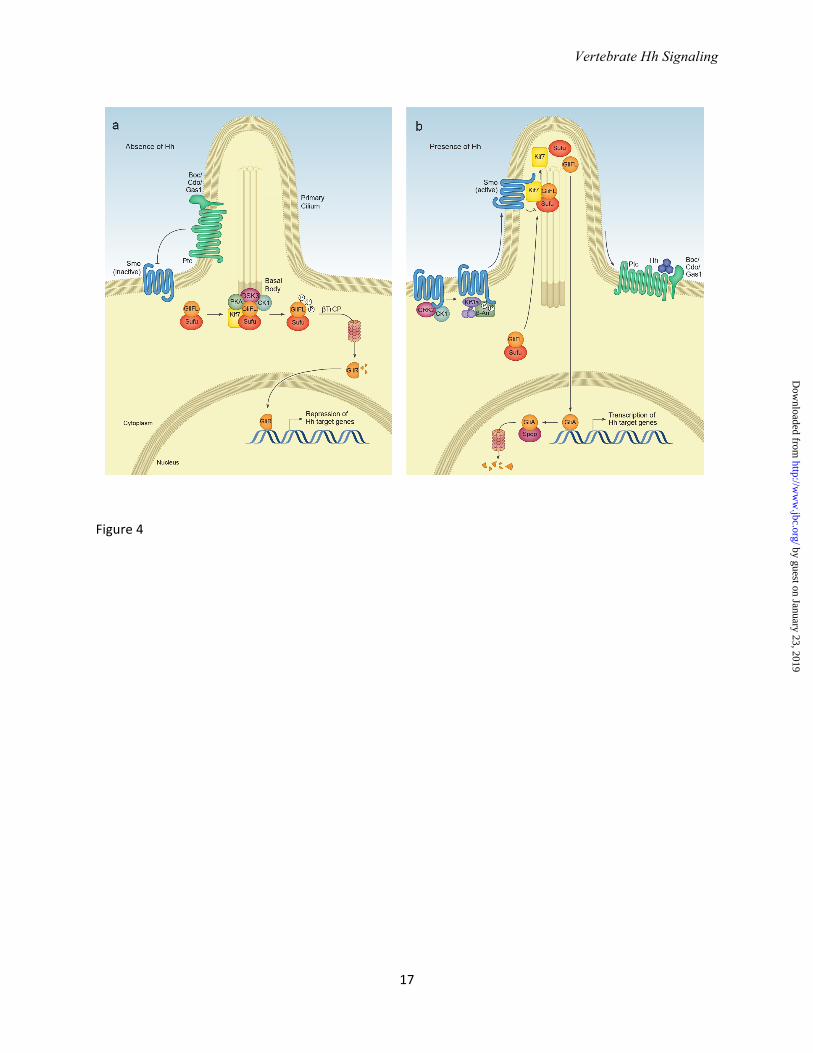
long range signaling in the imaginal disc (26). Residues in yellow (H133, H134, H140, H180 and H182) are important for Ptc binding (note that H140 and H182 coordinate with Zinc). Residues in red (K32, R33, R34, K37, K38) form the Cardin Weintraub motif and interact with heparin sulfate. Note how the N-terminus extends away from the globular domain of SHH-N; some of these residues may be cleaved in the formation of active Shh multimers (see text). Figure 3. SHH-N receptor binding involves the Zn2+ coordination site. a. Structure of human SHH-N in complex with HIP (Hh interacting protein) (PDB: 3HO5 (39)). The L2 loop in the beta-propeller domain of HIP interacts with SHH-N. b. HIP binds the pseudoactive site in SHH-N and Asp383 completes the tetrahedral coordination of Zn2+ in SHH-N. Inset: His140, His142, and Arg147 of SHH-N coordinate Zn2+. Note that the Zn2+ coordination site is also requires for binding to PTC, and PTC likely binds SHH in a manner similar to HIP (see text). Figure 4. Vertebrate Hedgehog signal transduction.a. In the absence of ligand, the twelve-pass transmembrane protein Patched (Ptc) localizes to the primary cilium base and maintains Smo in an inactive conformation. Full length Gli transcription factors (Gli-FL) complex with Suppressor of Fused (Sufu). Sufu sequesters Gli-FL in the cytosol and stabilizes the protein. Sufu and the kinesin 4 family member Kif7 promote the phosphorylation of C-terminal residues in full length Gli by protein kinase A (PKA), glycogen synthase kinase 3β (GSK3) and casein kinase 1α (CK1), which may occur at the basal body of the primary cilium. Phosphorylated Gli-FL is recognized by the E3 ubiquitin ligase βTrCP, resulting in ubiquitylation and proteosomal degradation of C-terminal residues to generate a truncated N-terminal transcriptional repressor (Gli-R) that inhibits Hh target gene transcription. b. In the presence of ligand, Hh binding to Ptc causes Ptc to exit the cilium and relieves its inhibition of Smo. Smo is phosphorylated by CK1α and G-coupled protein receptor kinase 2 (GRK2), inducing a conformational change and enabling β -arrestin- and Kif3a-dependent transport into the cilium. Within the cilium, activated Smo promotes the disassembly of Sufu-Gli complexes. Kif7 also localizes to the cilium in the presence of Hh likely assists Smo in this disassembly. Full-length Gli accumulates in the tip of the cilium and is shuttled into the nucleus, perhaps on cytoplasmic microtubules. Within the nucleus, Gli-FL receives additional modifications that convert it to a labile transcriptional activator (Gli-A) that activates Hh target genes. Gli-A is subsequently degraded in a manner that requires the Cullin3-adaptor Spop.
by guest on January 23, 2019http://w
ww
.jbc.org/D
ownloaded from
Vertebrate Hh Signaling
14
Figure 1
by guest on January 23, 2019http://w
ww
.jbc.org/D
ownloaded from
Vertebrate Hh Signaling
15
Figure 2
by guest on January 23, 2019http://w
ww
.jbc.org/D
ownloaded from
Vertebrate Hh Signaling
16
Figure 3
by guest on January 23, 2019http://w
ww
.jbc.org/D
ownloaded from
Vertebrate Hh Signaling
17
Figure 4
by guest on January 23, 2019http://w
ww
.jbc.org/D
ownloaded from
Kaitlyn Ryan and Chin ChiangHedgehog secretion and signal transduction in vertebrates
published online April 2, 2012 originally published online April 2, 2012J. Biol. Chem.
10.1074/jbc.R112.356006Access the most updated version of this article at doi:
Alerts:
When a correction for this article is posted•
When this article is cited•
to choose from all of JBC's e-mail alertsClick here
by guest on January 23, 2019http://w
ww
.jbc.org/D
ownloaded from