Embed Size (px)
Citation preview
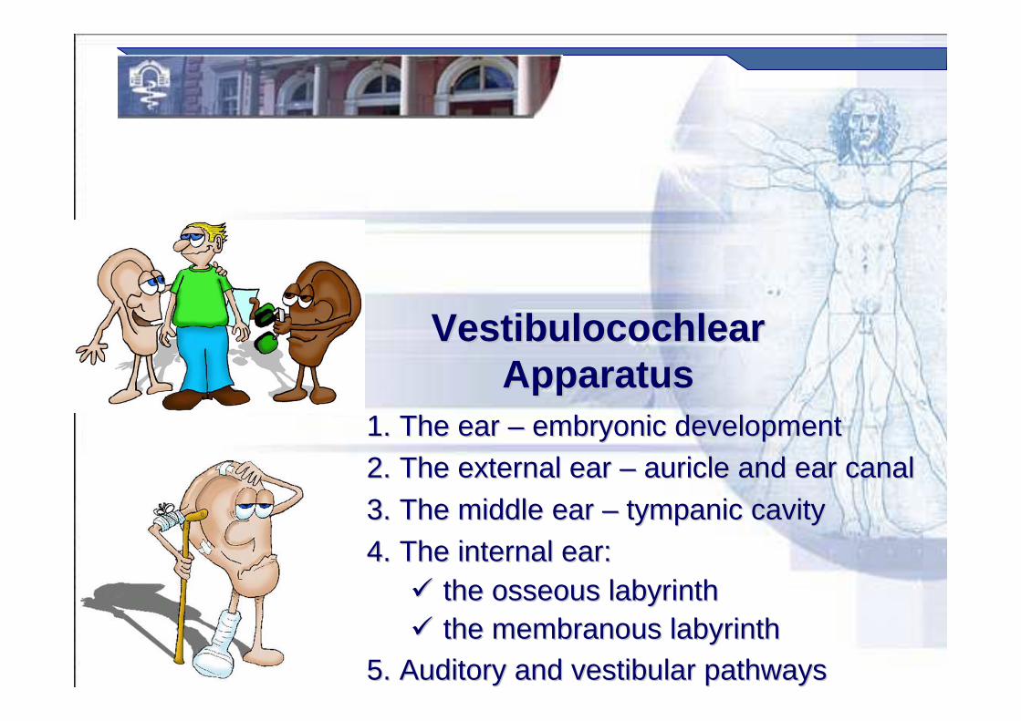
VestibulocochlearVestibulocochlearApparatusApparatus
1.1. The ear The ear –– embryonic development embryonic development
2.2. The external ear The external ear –– auricle and ear canalauricle and ear canal
3.3. The middle ear The middle ear –– tympanic cavitytympanic cavity
4.4. The internal ear:The internal ear:�� the osseous labyrinththe osseous labyrinth�� the membranous labyrinththe membranous labyrinth
5.5. Auditory and vestibular pathwaysAuditory and vestibular pathways

Prof. Dr. Nikolai LazarovProf. Dr. Nikolai Lazarov 2
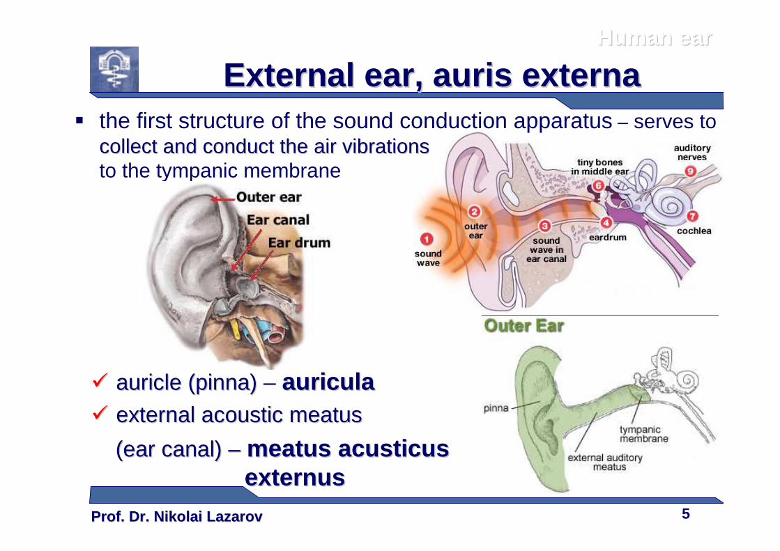
� The peripheral auditory apparatus, the earthe ear , auris, Gr. us, Gr. us, ωτοωτο == genitive genitive for earfor ear::� external (outer) earexternal (outer) ear, aurisauris externaexterna
�� auricle (pinna)auricle (pinna)
�� external acoustic external acoustic meatusmeatus (ear canal)(ear canal)
�� middle earmiddle ear, aurisauris mediamedia�� tympanic membrane (ear drum)tympanic membrane (ear drum)
�� tympanic cavitytympanic cavity
�� auditory (Eustachian) tubeauditory (Eustachian) tube
�� auditory auditory ossiclesossicles
�� internal (inner) earinternal (inner) ear, aurisauris internainternaauditory and vestibular portionsauditory and vestibular portions:�� osseous labyrinthosseous labyrinth
�� membranous labyrinthmembranous labyrinth
Anatomy of the earAnatomy of the earHuman earHuman ear
NB: NB: HumanHumanHumanHumanHumanHumanHumanHuman ear:ear:ear:ear:ear:ear:ear:ear: the organ of hearing and balancethe organ of hearing and balancethe organ of hearing and balancethe organ of hearing and balancethe organ of hearing and balancethe organ of hearing and balancethe organ of hearing and balancethe organ of hearing and balance
Prof. Dr. Nikolai LazarovProf. Dr. Nikolai Lazarov 3
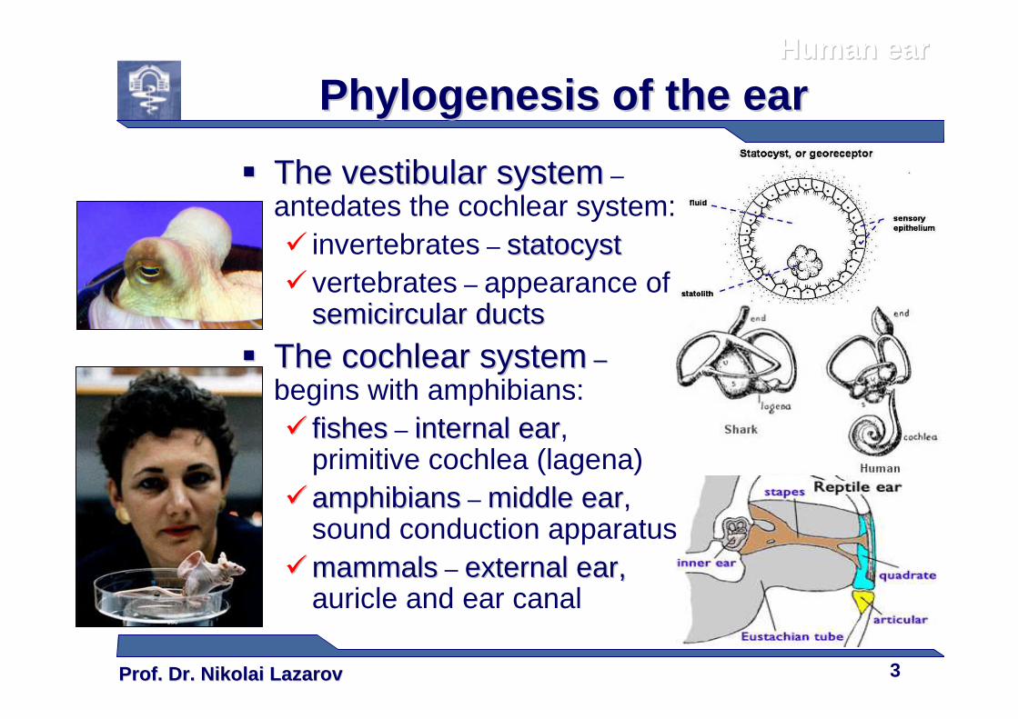
�� The vestibular systemThe vestibular system –antedates the cochlear system:� invertebrates – statocyststatocyst� vertebrates – appearance of
semicircular ductssemicircular ducts
�� The cochlear systemThe cochlear system ––begins with amphibians:�� fishesfishes – internal earinternal ear,
primitive cochlea (lagena)�� amphibiansamphibians – middle earmiddle ear,
sound conduction apparatus ��mammalsmammals – external earexternal ear, ,
auricle and ear canal
PhylogenesisPhylogenesis of the earof the earHuman earHuman ear
Prof. Dr. Nikolai LazarovProf. Dr. Nikolai Lazarov 4
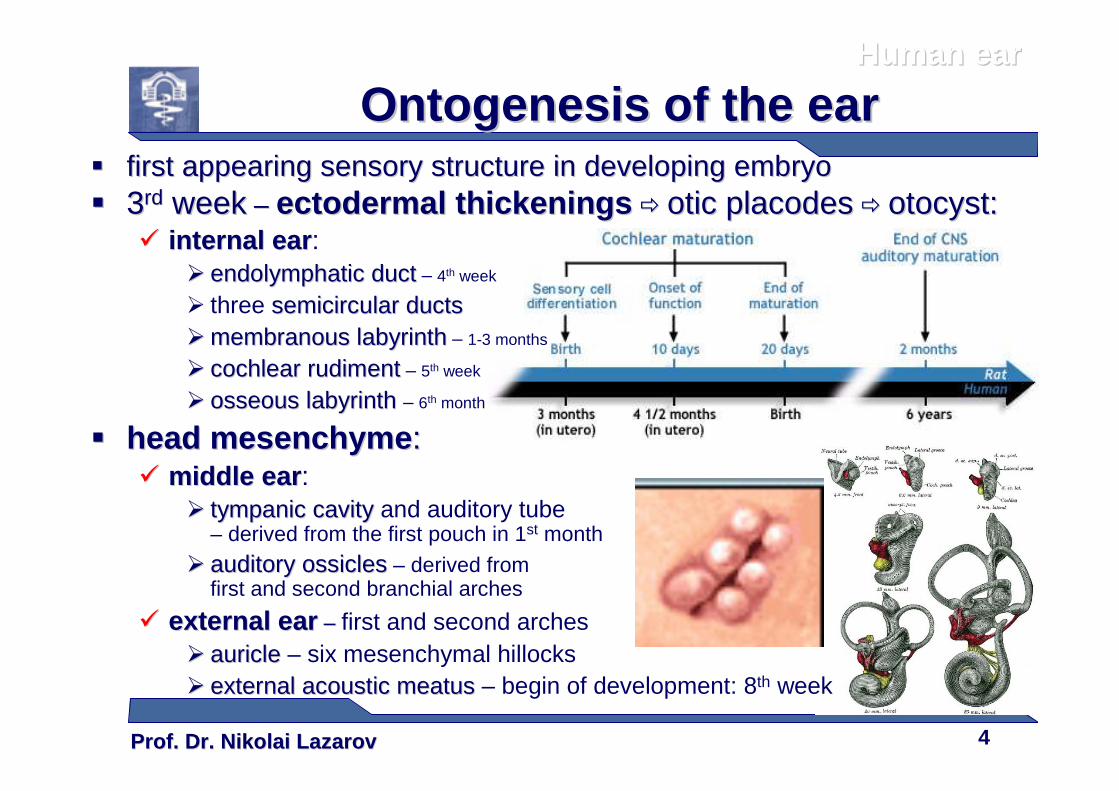
�� first appearing sensory structure in developing embryofirst appearing sensory structure in developing embryo�� 33rdrd weekweek –– ectodermal thickeningsectodermal thickenings �� oticotic placodesplacodes �� otocystotocyst::
�� internal earinternal ear :�� endolymphaticendolymphatic ductduct – 4th week
� three semicircular ductssemicircular ducts�� membranous labyrinthmembranous labyrinth – 1-3 months
�� cochlear rudimentcochlear rudiment – 5th week
�� osseous labyrinthosseous labyrinth – 6th month
�� head head mesenchymemesenchyme ::�� middle earmiddle ear :
�� tympanic cavitytympanic cavity and auditory tube– derived from the first pouch in 1st month
�� auditory auditory ossiclesossicles – derived from first and second branchial arches
�� external earexternal ear –– first and second arches�� auricleauricle – six mesenchymal hillocks�� external acoustic external acoustic meatusmeatus – begin of development: 8th week
Human earHuman ear
Ontogenesis of the earOntogenesis of the ear
Prof. Dr. Nikolai LazarovProf. Dr. Nikolai Lazarov 5
External earExternal ear , , aurisauris externaexterna� the first structure of the sound conduction apparatus – serves to
collectcollect and conductand conduct the air vibrationsthe air vibrationsto the tympanic membrane
�� auricle (pinna)auricle (pinna) – auriculaauricula�� external acoustic external acoustic meatusmeatus
((ear canalear canal) ) –– meatusmeatus acusticusacusticusexternusexternus
Human earHuman ear
Prof. Dr. Nikolai LazarovProf. Dr. Nikolai Lazarov 6
Auricle,Auricle, auriculaauricula� pinna – Lat. Lat. pinnapinna, a feather, a feather:
� thin skin with fine hairs
� elastic fibrocartilagefibrocartilage� lobule of auricle� auricular tubercle
(of Darwin)�� ligaments of auricleligaments of auricle, ,
extrinsic and intrinsic,
�� auricularauricular muscles muscles –extrinsic and intrinsic,
n. facialis
Human earHuman ear
� collects and funnels the sound wavescollects and funnels the sound waves into the meatus
Prof. Dr. Nikolai LazarovProf. Dr. Nikolai Lazarov 7
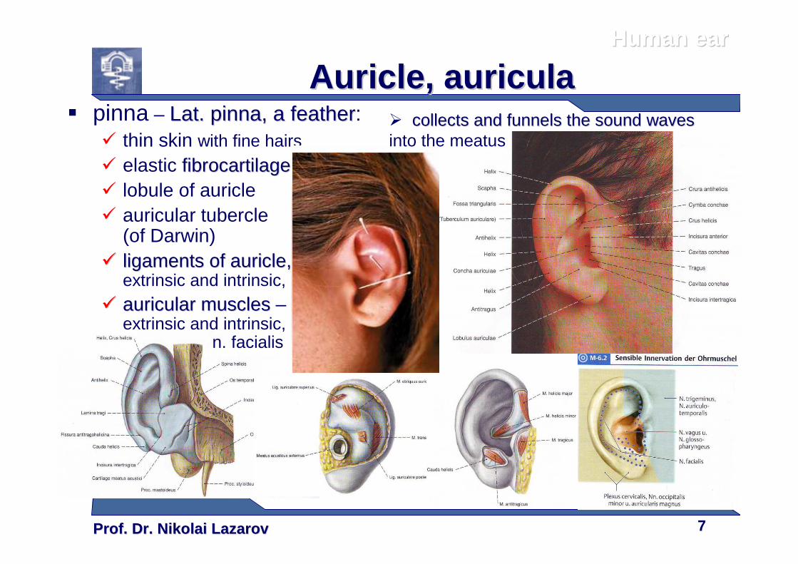
Auricle,Auricle, auriculaauricula� pinna – Lat. Lat. pinnapinna, a feather, a feather:
� thin skin with fine hairs
� elastic fibrocartilagefibrocartilage� lobule of auricle� auricular tubercle
(of Darwin)�� ligaments of auricleligaments of auricle, ,
extrinsic and intrinsic,
�� auricularauricular muscles muscles –extrinsic and intrinsic,
n. facialis
Human earHuman ear
� collects and funnels the sound wavescollects and funnels the sound wavesinto the meatus
Prof. Dr. Nikolai LazarovProf. Dr. Nikolai Lazarov 8
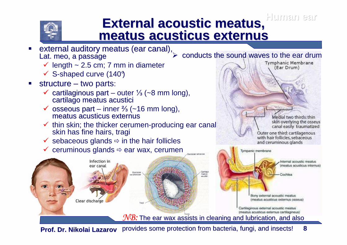
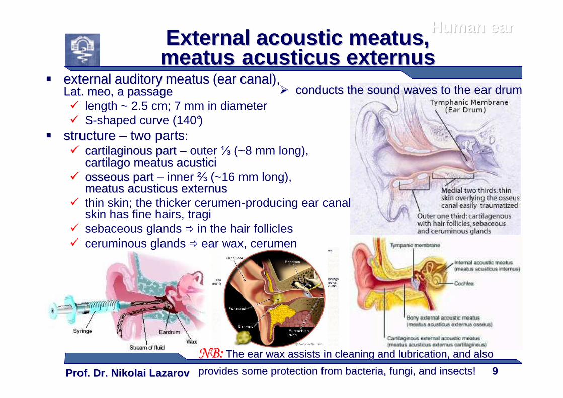
�� externalexternal auditoryauditory meatusmeatus (earear canalcanal), Lat. Lat. meomeo, a passage, a passage� length ~ 2.5 cm; 7 mm in diameter� S-shaped curve (140°)
�� structurestructure – two parts:�� cartilaginous partcartilaginous part – outer ⅓ (~8 mm long),
cartilagocartilago meatusmeatus acusticiacustici�� osseous partosseous part – inner ⅔ (~16 mm long),
meatusmeatus acusticusacusticus externusexternus� thin skin; the thicker cerumen-producing ear canal
skin has fine hairs, tragi� sebaceous glands � in the hair follicles� ceruminous glands � ear wax, cerumen
Human earHuman earExternal acoustic External acoustic meatusmeatus , , meatusmeatus acusticusacusticus externusexternus
�� conducts the sound wavesconducts the sound waves to the ear drum
NB:NB: The The ear waxear wax assistassists s in cleaning and lubrication, and also in cleaning and lubrication, and also
provides some protection from provides some protection from bacteria, fungibacteria, fungi, and , and insects!insects!
Prof. Dr. Nikolai LazarovProf. Dr. Nikolai Lazarov 9
�� externalexternal auditoryauditory meatusmeatus (earear canalcanal), Lat. Lat. meomeo, a passage, a passage� length ~ 2.5 cm; 7 mm in diameter� S-shaped curve (140°)
�� structurestructure – two parts:�� cartilaginous partcartilaginous part – outer ⅓ (~8 mm long),
cartilagocartilago meatusmeatus acusticiacustici�� osseous partosseous part – inner ⅔ (~16 mm long),
meatusmeatus acusticusacusticus externusexternus� thin skin; the thicker cerumen-producing ear canal
skin has fine hairs, tragi� sebaceous glands � in the hair follicles� ceruminous glands � ear wax, cerumen
Human earHuman earExternal acoustic External acoustic meatusmeatus , , meatusmeatus acusticusacusticus externusexternus
�� conducts the sound wavesconducts the sound waves to the ear drum
NB:NB: The The ear waxear wax assistassists s in cleaning and lubrication, and also in cleaning and lubrication, and also
provides some protection from provides some protection from bacteria, fungibacteria, fungi, and , and insects!insects!
Prof. Dr. Nikolai LazarovProf. Dr. Nikolai Lazarov 10
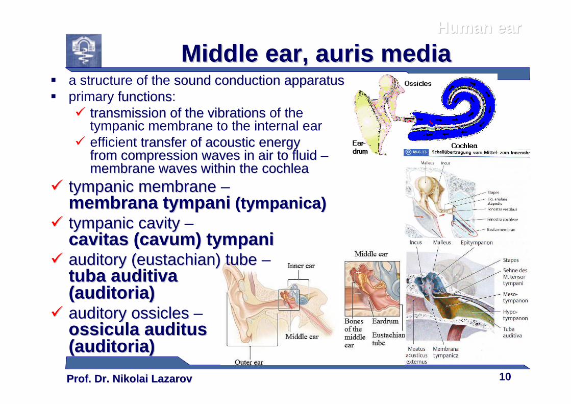
� a structure of the sound conduction apparatussound conduction apparatus� primary functionsfunctions:
�� transmission of the vibrationstransmission of the vibrations of the tympanic membrane to the internal ear
� efficient transfertransfer of of acoustic energy acoustic energy from compression waves in air to fluid from compression waves in air to fluid ––membrane waves within the membrane waves within the cochleacochlea
�� tympanic membranetympanic membrane –membranamembrana tympanitympani ((tympanicatympanica))
�� tympanic cavity tympanic cavity –cavitascavitas ((cavumcavum) tympani) tympani
�� auditory (auditory (eustachianeustachian) tube) tube –tuba tuba auditivaauditiva(auditoria)(auditoria)
�� auditory auditory ossiclesossicles –ossiculaossicula auditusauditus(auditoria)(auditoria)
Human earHuman ear
Middle earMiddle ear , , aurisauris mediamedia
Prof. Dr. Nikolai LazarovProf. Dr. Nikolai Lazarov 11
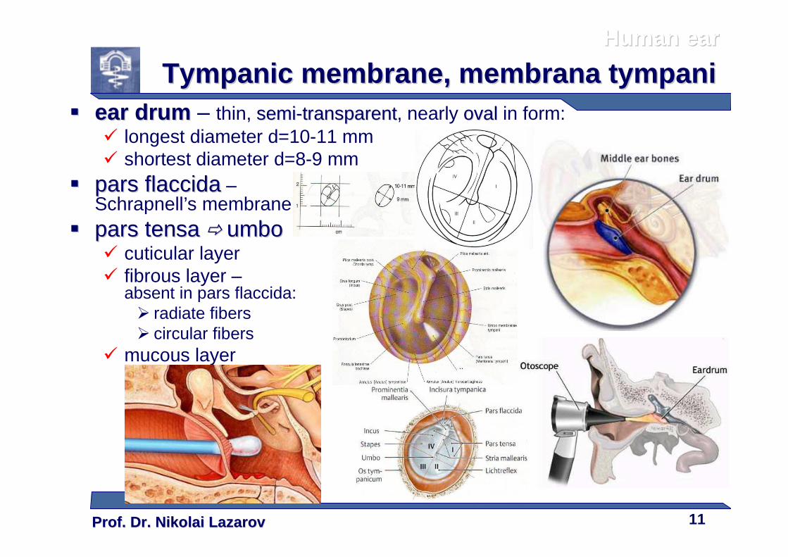
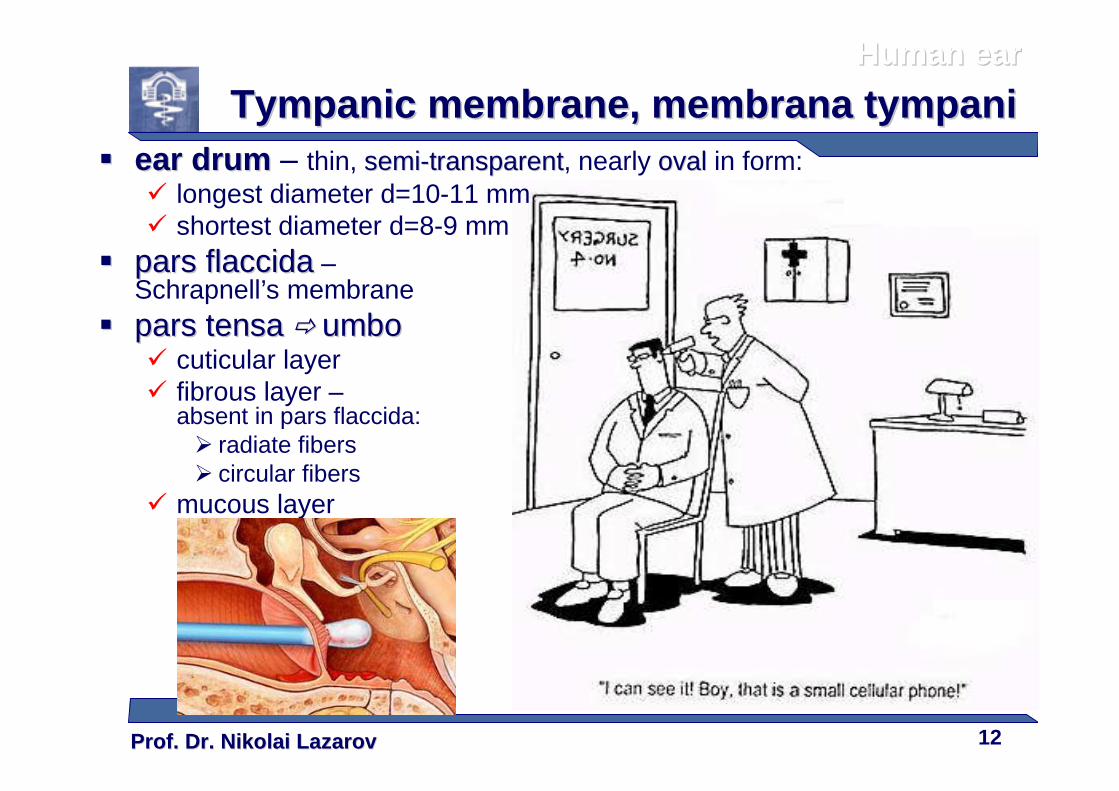
�� ear drumear drum – thin, semisemi--transparenttransparent, nearly ovaloval in form:� longest diameter d=10-11 mm� shortest diameter d=8-9 mm
�� pars pars flaccidaflaccida –Schrapnell’s membrane
�� pars pars tensatensa �� umboumbo� cuticular layer� fibrous layer –
absent in pars flaccida:� radiate fibers� circular fibers
� mucous layer
Human earHuman ear
Tympanic membraneTympanic membrane , , membranamembrana tympanitympani
Prof. Dr. Nikolai LazarovProf. Dr. Nikolai Lazarov 12
�� ear drumear drum – thin, semisemi--transparenttransparent, nearly ovaloval in form:� longest diameter d=10-11 mm� shortest diameter d=8-9 mm
�� pars pars flaccidaflaccida –Schrapnell’s membrane
�� pars pars tensatensa �� umboumbo� cuticular layer� fibrous layer –
absent in pars flaccida:� radiate fibers� circular fibers
� mucous layer
Human earHuman ear
Tympanic membraneTympanic membrane , , membranamembrana tympanitympani
Prof. Dr. Nikolai LazarovProf. Dr. Nikolai Lazarov 13
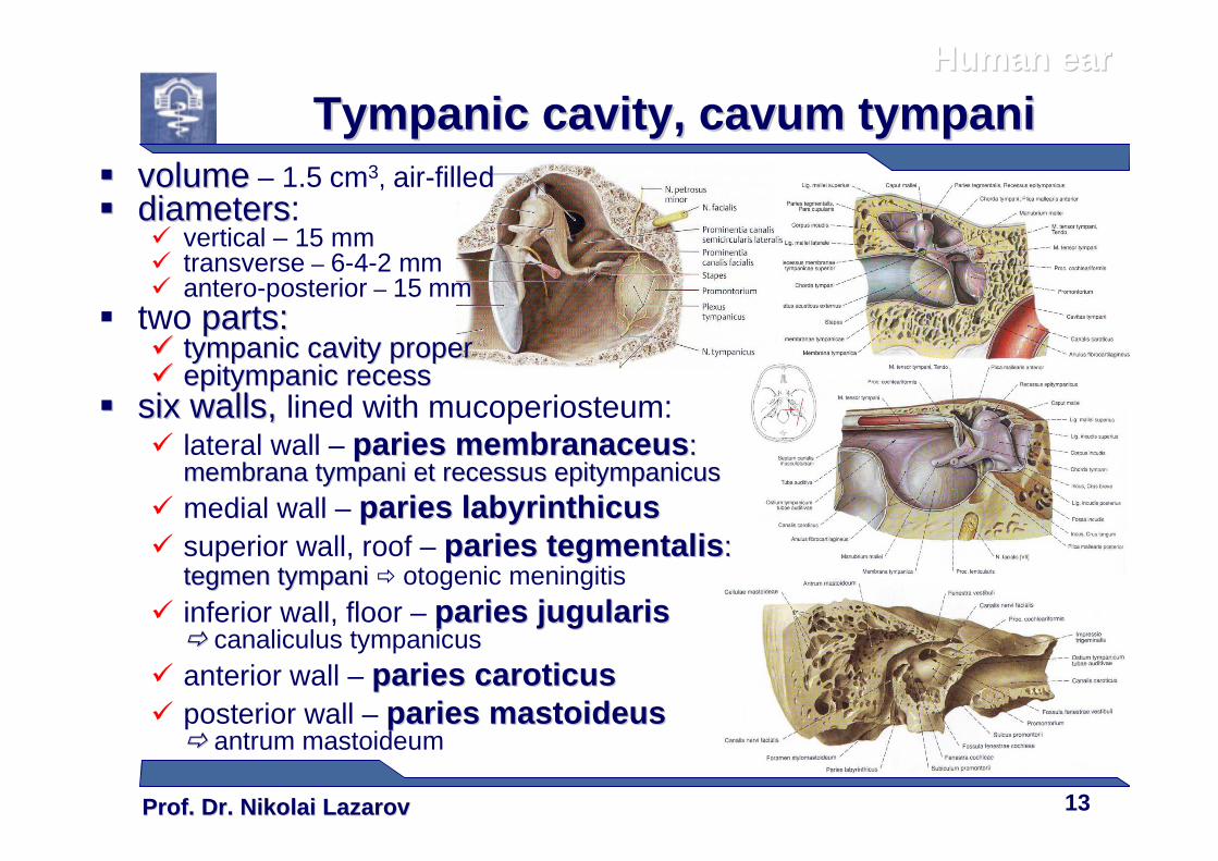
�� volumevolume – 1.5 cm3, air-filled�� diametersdiameters:
� vertical – 15 mm� transverse – 6-4-2 mm� antero-posterior – 15 mm
� two parts:parts:�� tympanic cavity propertympanic cavity proper�� epitympanicepitympanic recessrecess
�� six walls,six walls, lined with mucoperiosteum:� lateral wall – pariesparies membranaceusmembranaceus: :
membranamembrana tympani et tympani et recessusrecessus epitympanicusepitympanicus� medial wall – pariesparies labyrinthicuslabyrinthicus� superior wall, roof – pariesparies tegmentalistegmentalis: :
tegmentegmen tympani tympani � otogenic meningitis� inferior wall, floor – pariesparies jugularisjugularis
�� canaliculus tympanicus� anterior wall – pariesparies caroticuscaroticus� posterior wall – pariesparies mastoideusmastoideus
�� antrum mastoideum
Human earHuman ear
Tympanic cavityTympanic cavity , , cavumcavum tympanitympani
Prof. Dr. Nikolai LazarovProf. Dr. Nikolai Lazarov 14
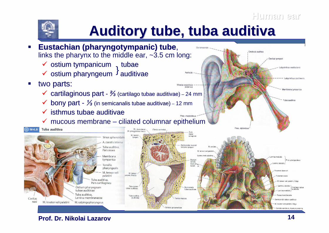
�� Eustachian (Eustachian ( pharyngotympanicpharyngotympanic ) tube) tube , links the pharynx to the middle ear, ~3.5 cm long:�� ostiumostium tympanicumtympanicum tubaetubae�� ostiumostium pharyngeumpharyngeum auditivaeauditivae
� two parts:parts:�� cartilaginouscartilaginous partpart -- ⅔⅔ ((cartilagocartilago tubaetubae auditivaeauditivae)) – 24 mm
�� bony partbony part -- ⅓⅓ ((in semicanalissemicanalis tubaetubae auditivaeauditivae)) – 12 mm
�� isthmus isthmus tubaetubae auditivaeauditivae� mucous membrane – ciliated columnar epithelium
Human earHuman ear
Auditory tubeAuditory tube , , tuba tuba auditivaauditiva
}
Prof. Dr. Nikolai LazarovProf. Dr. Nikolai Lazarov 15
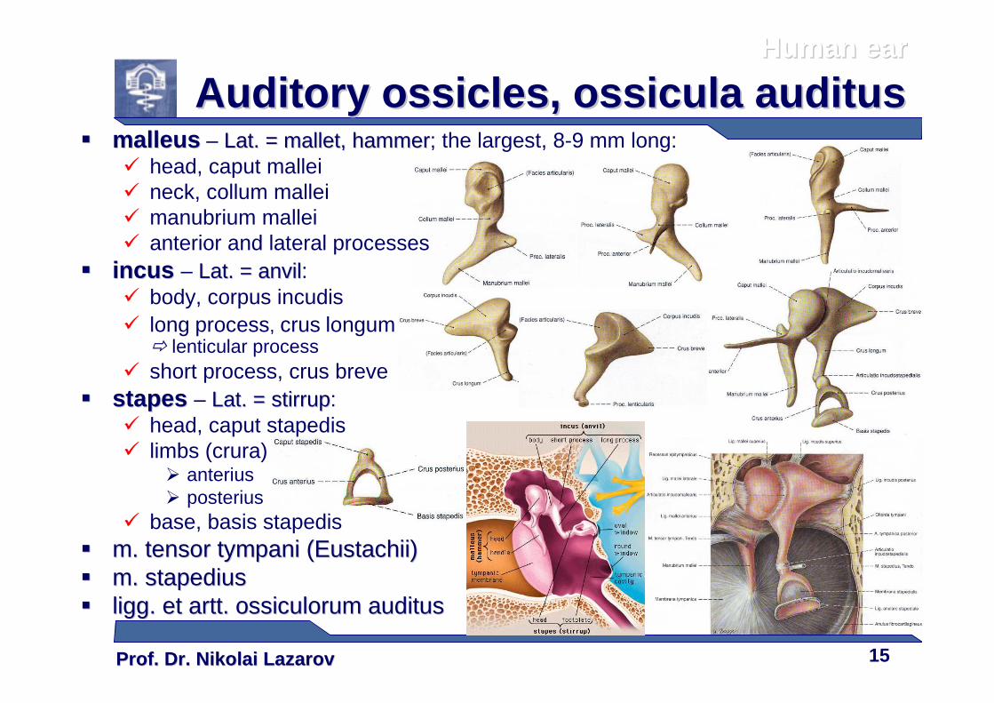
�� malleusmalleus – Lat. = mallet, hammerLat. = mallet, hammer; the largest, 8-9 mm long:� head, caput mallei� neck, collum mallei� manubrium mallei� anterior and lateral processes
�� incusincus – Lat. = anvilLat. = anvil:� body, corpus incudis� long process, crus longum
� lenticular process� short process, crus breve
�� stapes stapes – Lat. = stirrupLat. = stirrup:� head, caput stapedis� limbs (crura)
� anterius� posterius
� base, basis stapedis�� m. tensor tympani (m. tensor tympani (EustachiiEustachii))�� m. m. stapediusstapedius�� liggligg. et . et arttartt. . ossiculorumossiculorum auditusauditus
Human earHuman ear
Auditory Auditory ossiclesossicles , , ossiculaossicula auditusauditus
Prof. Dr. Nikolai LazarovProf. Dr. Nikolai Lazarov 16
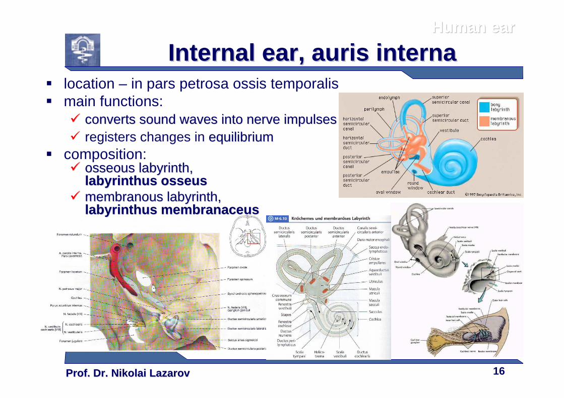
� location – in pars petrosa ossis temporalis� main functions:
�� converts sound waves into nerve impulsesconverts sound waves into nerve impulses� registers changes in equilibriumequilibrium
� composition:�� osseous labyrinth,osseous labyrinth,
labyrinthuslabyrinthus osseusosseus�� membranous labyrinthmembranous labyrinth,,
labyrinthuslabyrinthus membranaceusmembranaceus
Human earHuman ear
Internal earInternal ear , , aurisauris internainterna

Prof. Dr. Nikolai LazarovProf. Dr. Nikolai Lazarov 17
�� vestibulevestibule, vestibulum�� three semicircular canalsthree semicircular canals,,
canales semicirculares::� canalis semicircularis lateralis� canalis semicircularis anterior� canalis semicircularis posterior
� cochlea� filled with perilymphperilymph
Human earHuman ear
Osseous labyrinth, Osseous labyrinth, labyrinthuslabyrinthus osseusosseus
Prof. Dr. Nikolai LazarovProf. Dr. Nikolai Lazarov 18
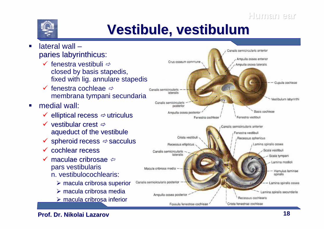
VestibuleVestibule , , vestibulumvestibulum� lateral wall –
pariesparies labyrinthicuslabyrinthicus:� fenestra vestibuli �
closed by basis stapedis, fixed with lig. annulare stapedis
� fenestra cochleae �membrana tympani secundaria
� medial wall:�� ellipticalelliptical recessrecess �� utriculusutriculus�� vestibular crestvestibular crest ��
aqueduct of the vestibuleaqueduct of the vestibule�� spheroidspheroid recessrecess �� sacculussacculus�� cochlear recesscochlear recess�� maculae maculae cribrosaecribrosae
pars vestibularisn. vestibulocochlearis::�� macula macula cribrosacribrosa superiorsuperior�� macula macula cribrosacribrosa mediamedia�� macula macula cribrosacribrosa inferiorinferior
Human earHuman ear
Prof. Dr. Nikolai LazarovProf. Dr. Nikolai Lazarov 19
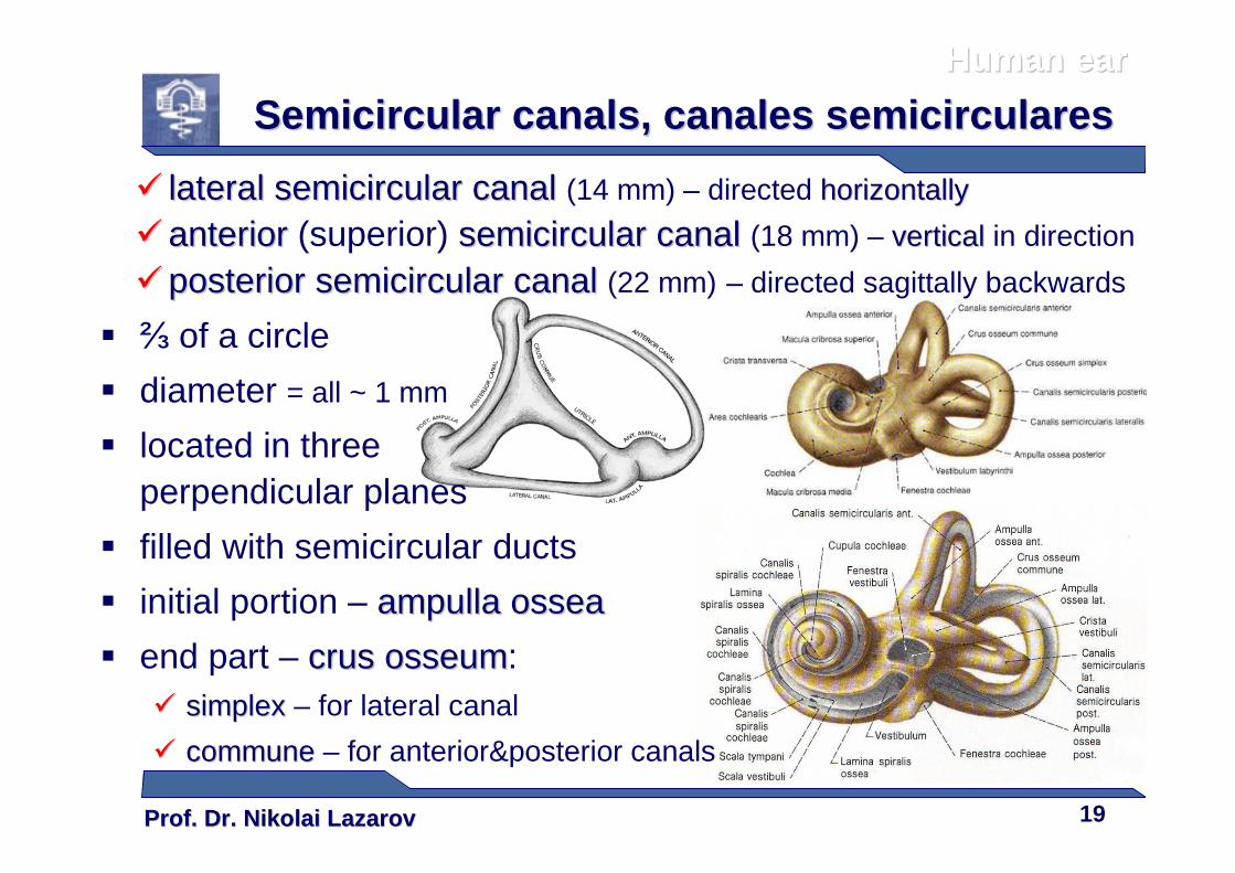
�� lateral semicircular canallateral semicircular canal (14 mm) – directed horizontallyhorizontally
�� anterioranterior (superior) semicircular canalsemicircular canal (18 mm) – verticalvertical in direction
�� posteriorposterior semicircular canal semicircular canal (22 mm) – directed sagittally backwards
� ⅔ of a circle
� diameter = all ~ 1 mm
� located in three perpendicular planes
� filled with semicircular ducts
� initial portion – ampullaampulla osseaossea
� end part – cruscrus osseumosseum:�� simplex simplex – for lateral canal
�� communecommune – for anterior&posterior canals
Human earHuman ear
Semicircular canals, Semicircular canals, canales canales semicircularessemicirculares
Prof. Dr. Nikolai LazarovProf. Dr. Nikolai Lazarov 20
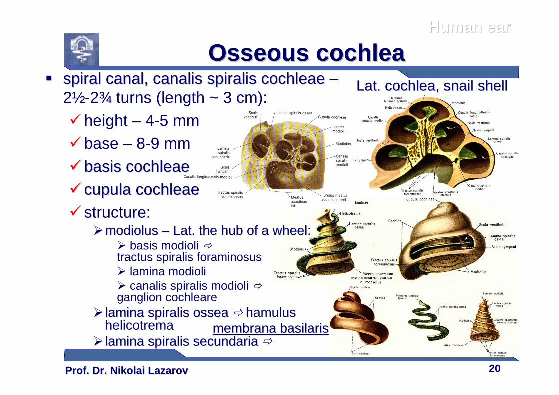
Osseous cochleaOsseous cochlea�� spiral canalspiral canal,, canaliscanalis spiralisspiralis cochleaecochleae –
2½-2¾ turns (length ~ 3 cm):�height – 4-5 mm�base – 8-9 mm
�� basis cochleaebasis cochleae
�� cupulacupula cochleaecochleae� structure:
��modiolusmodiolus –– Lat. Lat. the hub of a wheelthe hub of a wheel:� basis modioli �tractus spiralis foraminosus� lamina modioli� canalis spiralis modioli �ganglion cochleare
�� lamina lamina spiralisspiralis osseaossea � hamulushelicotrema
�� lamina lamina spiralisspiralis secundariasecundaria �
membranamembrana basilarisbasilaris
Human earHuman ear
Lat. Lat. cochleacochlea, snail shell, snail shell
Prof. Dr. Nikolai LazarovProf. Dr. Nikolai Lazarov 21
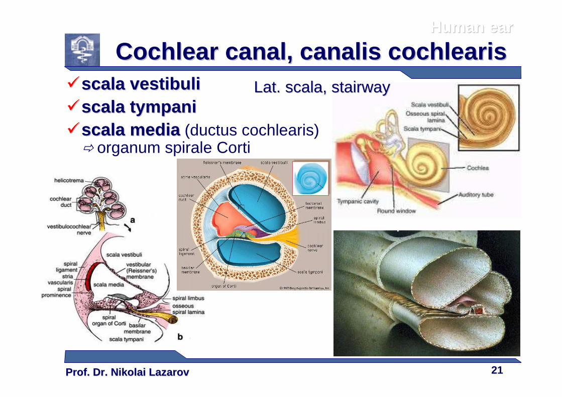
Cochlear canalCochlear canal ,, canaliscanalis cochleariscochlearis��scalascala vestibulivestibuli��scalascala tympanitympani��scalascala mediamedia (ductus cochlearis)
� organum spirale Corti
Human earHuman ear
Lat.Lat. scalascala, stairway, stairway
Prof. Dr. Nikolai LazarovProf. Dr. Nikolai Lazarov 22
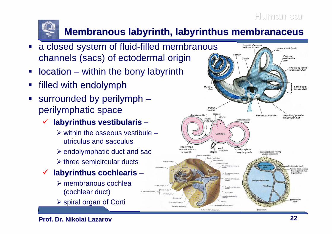
� a closed system of fluid-filled membranous channels (sacs) of ectodermal origin
�� locationlocation – within the bony labyrinth� filled with endolymphendolymph� surrounded by perilymphperilymph –
perilymphatic space� labyrinthuslabyrinthus vestibularisvestibularis ––
� within the osseous vestibule –utriculus and sacculus
� endolymphatic duct and sac� three semicircular ducts
�� labyrinthuslabyrinthus cochleariscochlearis ––� membranous cochlea
(cochlear duct)� spiral organ of Corti
Human earHuman ear
Membranous labyrinth, Membranous labyrinth, labyrinthuslabyrinthus membranaceusmembranaceus
Prof. Dr. Nikolai LazarovProf. Dr. Nikolai Lazarov 23
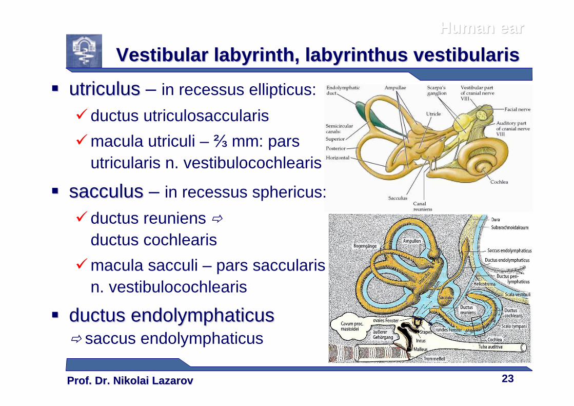
Vestibular labyrinthVestibular labyrinth , , labyrinthuslabyrinthus vestibularisvestibularis
�� utriculusutriculus – in recessus ellipticus:
�ductus utriculosaccularis
�macula utriculi – ⅔ mm: pars utricularis n. vestibulocochlearis
�� sacculussacculus – in recessus sphericus:
�ductus reuniens �
ductus cochlearis
�macula sacculi – pars saccularisn. vestibulocochlearis
�� ductusductus endolymphaticusendolymphaticus� saccus endolymphaticus
Human earHuman ear
Prof. Dr. Nikolai LazarovProf. Dr. Nikolai Lazarov 24
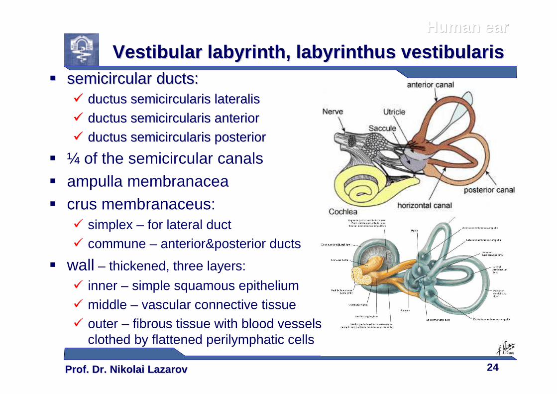
�� semicircular ductssemicircular ducts::�� ductusductus semicircularissemicircularis lateralislateralis
�� ductusductus semicircularissemicircularis anterioranterior
�� ductusductus semicircularissemicircularis posteriorposterior
� ¼ of the semicircular canals� ampulla membranacea� crus membranaceus:
� simplex – for lateral duct� commune – anterior&posterior ducts
� wall – thickened, three layers:
� inner – simple squamous epithelium� middle – vascular connective tissue� outer – fibrous tissue with blood vessels
clothed by flattened perilymphatic cells
Human earHuman ear
Vestibular labyrinthVestibular labyrinth , , labyrinthuslabyrinthus vestibularisvestibularis
Prof. Dr. Nikolai LazarovProf. Dr. Nikolai Lazarov 25
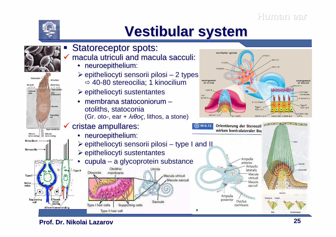
�� StatoreceptorStatoreceptor spotsspots::�� macula macula utriculiutriculi andand macula macula sacculisacculi::
•• neuroepitheliumneuroepithelium:� epitheliocyti sensorii pilosi – 2 types
� 40-80 stereocilia; 1 kinocilium� epitheliocyti sustentantes•• membranamembrana statoconiorumstatoconiorum –
otoliths, statoconia(Gr. oto-, ear + λιθος, lithos, a stone)
�� cristaecristae ampullaresampullares::•• neuroepitheliumneuroepithelium:� epitheliocyti sensorii pilosi – type I and II� epitheliocyti sustentantes•• cupulacupula – a glycoprotein substance
Human earHuman ear
Vestibular systemVestibular system
Prof. Dr. Nikolai LazarovProf. Dr. Nikolai Lazarov 26
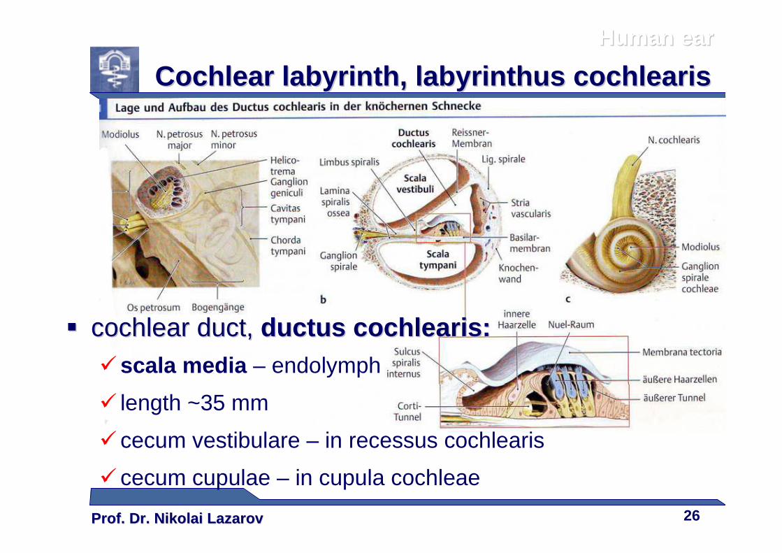
�� cochlear duct, cochlear duct, ductusductus cochleariscochlearis::�scala media – endolymph
� length ~35 mm
� cecum vestibulare – in recessus cochlearis
� cecum cupulae – in cupula cochleae
Human earHuman ear
Cochlear labyrinthCochlear labyrinth , , labyrinthuslabyrinthus cochleariscochlearis
Prof. Dr. Nikolai LazarovProf. Dr. Nikolai Lazarov 27
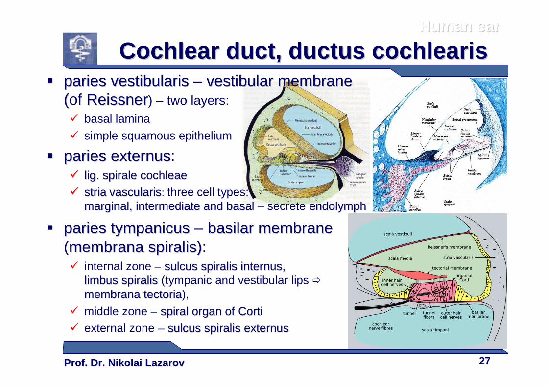
Cochlear duct,Cochlear duct, ductusductus cochleariscochlearis�� pariesparies vestibularisvestibularis – vestibular membranevestibular membrane
((of ReissnerReissner) – two layers:� basal lamina
� simple squamous epithelium
�� pariesparies externusexternus:�� liglig. . spiralespirale cochleaecochleae
�� striastria vascularisvascularis: three cell types:marginalmarginal, , intermediateintermediate and basaland basal – secrete endolymphendolymph
�� pariesparies tympanicustympanicus – basilarbasilar membranemembrane(membranamembrana spiralisspiralis)):� internal zone – sulcussulcus spiralisspiralis internusinternus, ,
limbuslimbus spiralisspiralis (tympanic and vestibular lips �
membranamembrana tectoriatectoria),
� middle zone – spiral organ of spiral organ of CortiCorti� external zone – sulcussulcus spiralisspiralis externusexternus
Human earHuman ear
Prof. Dr. Nikolai LazarovProf. Dr. Nikolai Lazarov 28
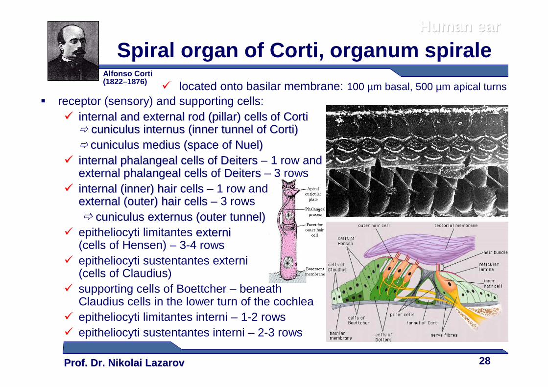
Spiral organ of Corti, organum spirale
� receptor (sensory) and supporting cells:�� internal and external rod (pillar) cellsinternal and external rod (pillar) cells of of CortiCorti
� cuniculuscuniculus internusinternus ((innerinner tunneltunnel of of CortiCorti))� cuniculuscuniculus mediusmedius ((space ofspace of NuelNuel))
�� internal internal phalangealphalangeal cellscells ofof DeitersDeiters – 1 row andexternal external phalangealphalangeal cellscells ofof DeitersDeiters – 3 rows
�� internal (inner) hair cellsinternal (inner) hair cells – 1 row andexternal (outer) hair cellsexternal (outer) hair cells – 3 rows� cuniculuscuniculus externusexternus ((outerouter tunnel)tunnel)
� epitheliocyti limitantes externiexterni(cells of Hensen) – 3-4 rows
� epitheliocyti sustentantes externi(cells of Claudius)
� supporting cells of Boettcher – beneathClaudius cells in the lower turn of the cochlea
� epitheliocyti limitantes interni – 1-2 rows� epitheliocyti sustentantes interni – 2-3 rows
Alfonso Corti(1822–1876)
� located onto basilar membrane: 100 µm basal, 500 µm apical turns
Human earHuman ear
Prof. Dr. Nikolai LazarovProf. Dr. Nikolai Lazarov 29
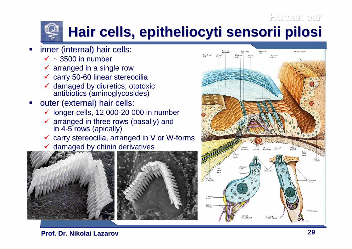
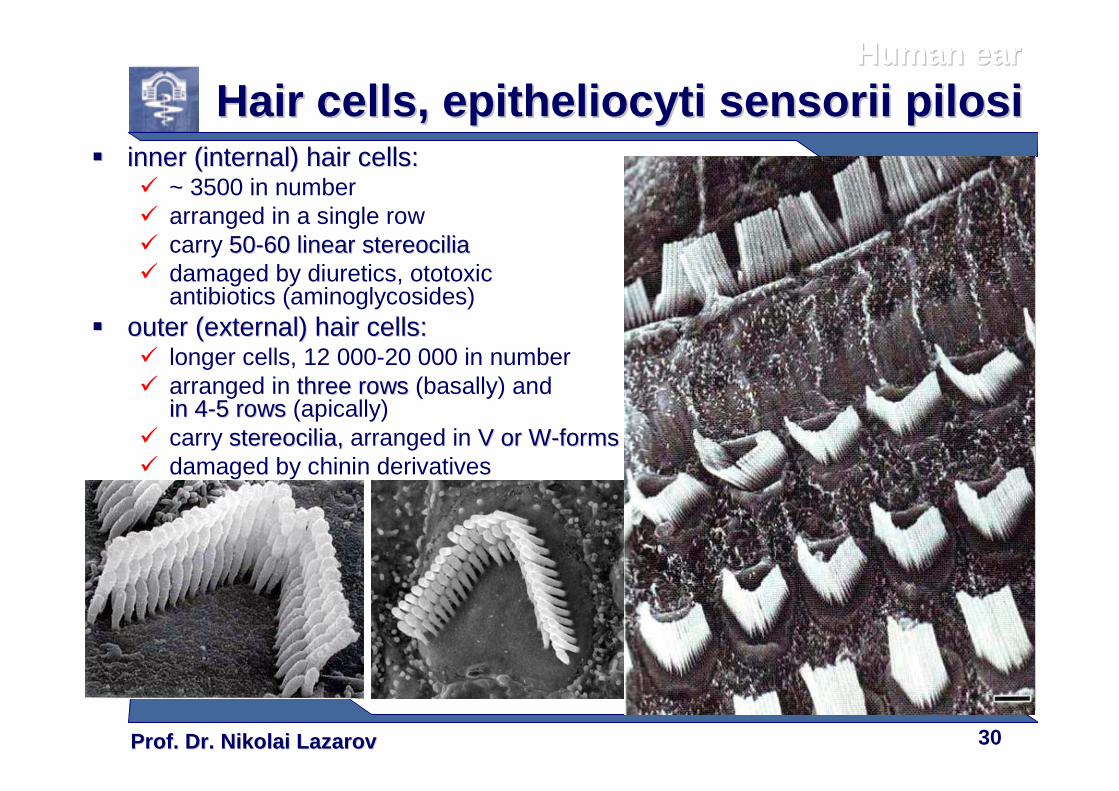
Hair cellsHair cells , , epitheliocytiepitheliocyti sensoriisensorii pilosipilosi�� inner (internal) hair cells:inner (internal) hair cells:
� ~ 3500 in number� arranged in a single row� carry 5050--60 linear 60 linear stereociliastereocilia� damaged by diuretics, ototoxic
antibiotics (aminoglycosides)�� outer (external) hair cells:outer (external) hair cells:
� longer cells, 12 000-20 000 in number� arranged in three rows three rows (basally) and
inin 44--5 5 rowsrows (apically)� carry stereociliastereocilia, , arranged in V or WV or W--formsforms� damaged by chinin derivatives
Human earHuman ear
Prof. Dr. Nikolai LazarovProf. Dr. Nikolai Lazarov 30
Hair cellsHair cells , , epitheliocytiepitheliocyti sensoriisensorii pilosipilosi�� inner (internal) hair cells:inner (internal) hair cells:
� ~ 3500 in number� arranged in a single row� carry 5050--60 linear 60 linear stereociliastereocilia� damaged by diuretics, ototoxic
antibiotics (aminoglycosides)�� outer (external) hair cells:outer (external) hair cells:
� longer cells, 12 000-20 000 in number� arranged in three rows three rows (basally) and
inin 44--5 5 rowsrows (apically)� carry stereociliastereocilia, , arranged in V or WV or W--formsforms� damaged by chinin derivatives
Human earHuman ear

Prof. Dr. Nikolai LazarovProf. Dr. Nikolai Lazarov 31
Cochlear innervationCochlear innervation� Afferent innervation –– from spiral ganglion (in modiolus):
� large bipolar type I cells – ~ 95% of all afferent neurons� inner hair cells� about ten fibers to each cell
� small pseudounipolar type II cells � outer hair cells� one fiber to about ten cells
�� Efferent innervationEfferent innervation –– tractustractusolivocochlearisolivocochlearis (Rasmussen’s tract)� cholinergic inhibitory synapses
Human earHuman ear
Prof. Dr. Nikolai LazarovProf. Dr. Nikolai Lazarov 32
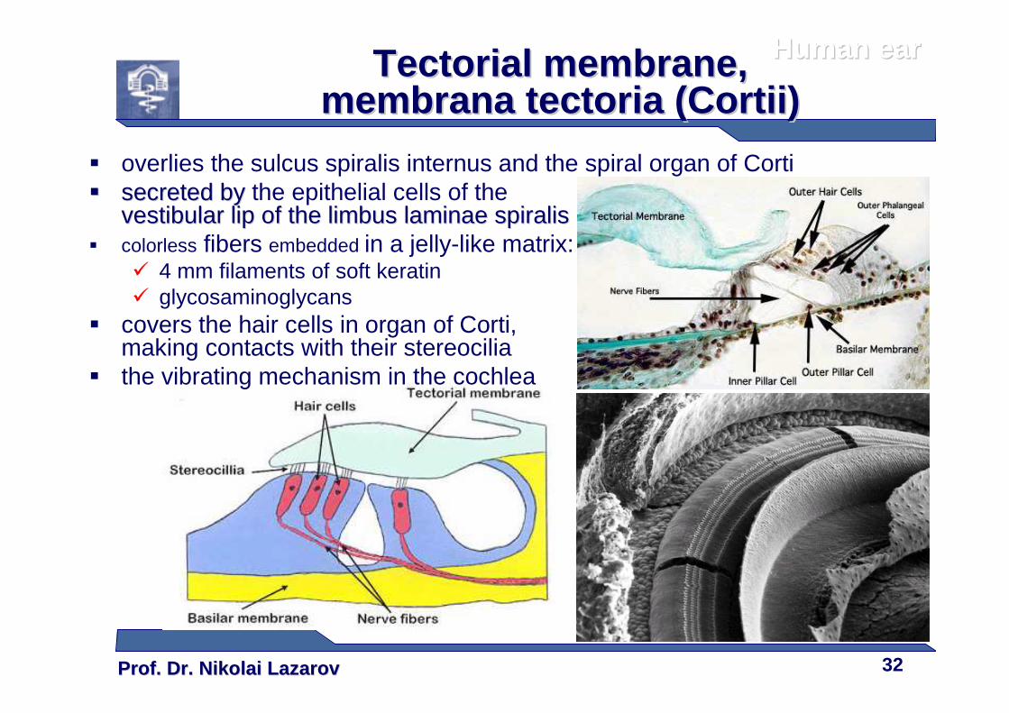
TectorialTectorial membranemembrane , , membranamembrana tectoriatectoria ((CortiiCortii))
� overlies the sulcus spiralis internus and the spiral organ of Corti�� secretedsecreted byby the epithelial cells of the
vestibular lipvestibular lip of theof the limbuslimbus laminaelaminae spiralisspiralis� colorless fibers embedded in a jelly-like matrix:
� 4 mm filaments of soft keratin� glycosaminoglycans
� covers the hair cells in organ of Corti, making contacts with their stereocilia
� the vibrating mechanism in the cochlea
Human earHuman ear
Prof. Dr. Nikolai LazarovProf. Dr. Nikolai Lazarov 33
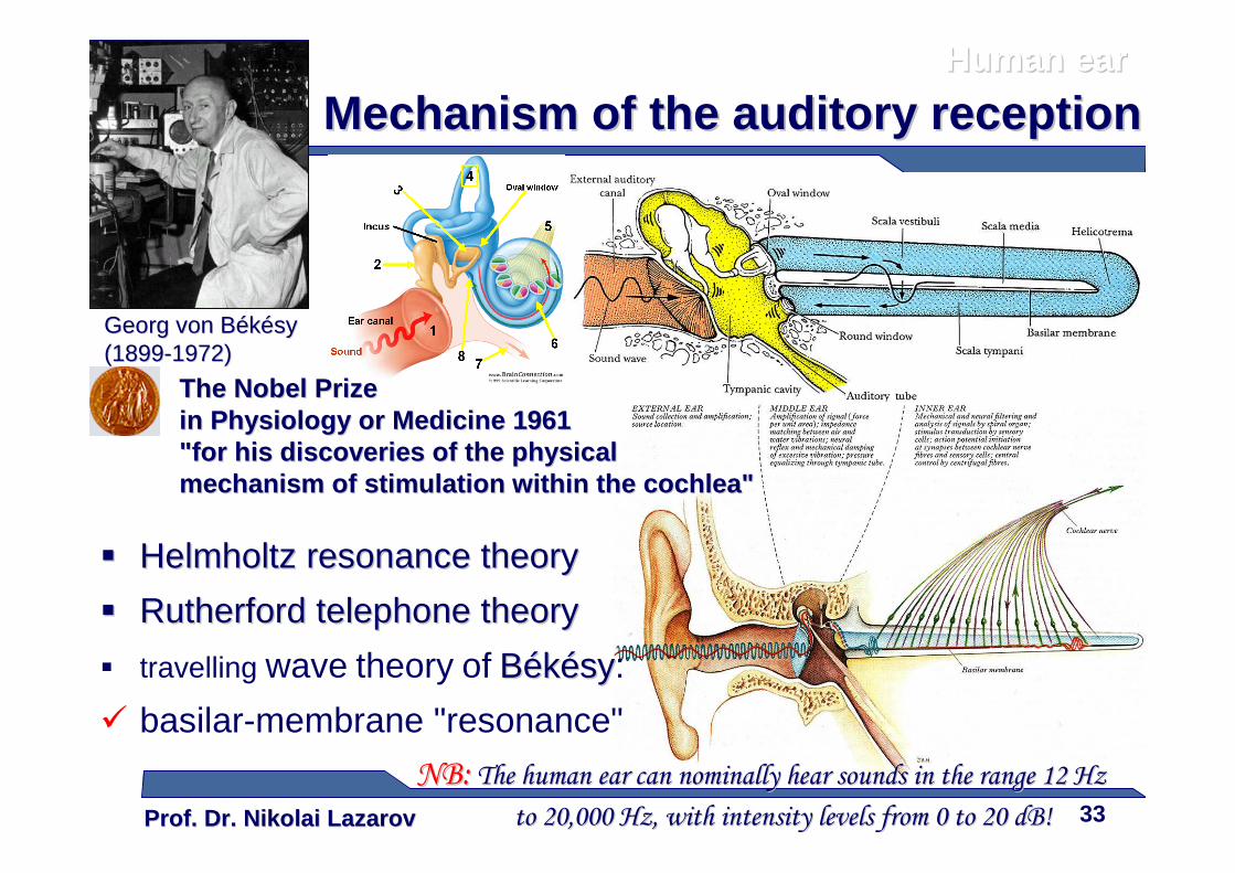
Mechanism of the auditory receptionMechanism of the auditory reception
GeorgGeorg von von BBéékkéésysy((18991899--19721972))
�� HelmholtzHelmholtz resonance theoryresonance theory
�� RutherfordRutherford telephone theorytelephone theory
� travelling wave theory of BBéékkéésysy:
� basilar-membrane "resonance"
NB:NB: The The humanhuman ear can nominally hear ear can nominally hear soundssounds in the rangein the range 12 12 Hz Hz
toto 20,000 Hz20,000 Hz, with intensity levels from , with intensity levels from 0 0 toto 20 20 dB!dB!
Human earHuman ear
The Nobel Prize The Nobel Prize in Physiology or Medicine 1961in Physiology or Medicine 1961"for his discoveries of the physical "for his discoveries of the physical mechanism of stimulation within the cochlea"mechanism of stimulation within the cochlea"
Prof. Dr. Nikolai LazarovProf. Dr. Nikolai Lazarov 34
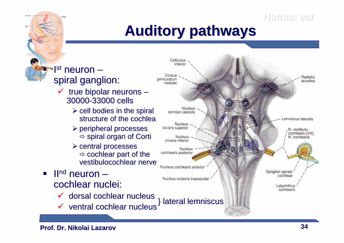
�� IIstst neuronneuron –spiral ganglion:spiral ganglion:�� true bipolar neuronstrue bipolar neurons ––
3000030000--33000 33000 cellscells�� cell bodies in cell bodies in the spiral the spiral
structure of the cochleastructure of the cochlea�� peripheral processesperipheral processes
�� spiral organ of spiral organ of CortiCorti�� central processescentral processes
�� cochlear part of the cochlear part of the vestibulocochlearvestibulocochlear nervenerve
�� IIIIndnd neuronneuron ––cochlear nuclei:cochlear nuclei:�� dorsaldorsal cochlear nucleuscochlear nucleus�� ventralventral cochlear nucleuscochlear nucleus
}} laterallateral lemniscuslemniscus
Human earHuman ear
Auditory pathwaysAuditory pathways
Prof. Dr. Nikolai LazarovProf. Dr. Nikolai Lazarov 35
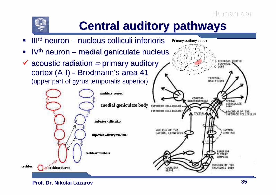
�� IIIIIIrdrd neuronneuron – nucleus nucleus colliculicolliculi inferiorisinferioris
�� IVIVthth neuronneuron – medial geniculate nucleusmedial geniculate nucleus
�� acoustic radiationacoustic radiation �� primary auditory primary auditory cortexcortex (A-I) = Brodmann’s area area 441 1 ((upper part of gyrus temporalis superior)
Human earHuman ear
Central auditory pathwaysCentral auditory pathways
Prof. Dr. Nikolai LazarovProf. Dr. Nikolai Lazarov 36
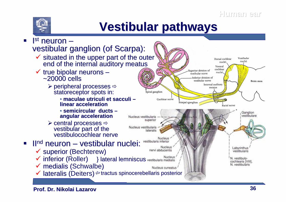
�� IIstst neuronneuron –vestibularvestibular ganglionganglion ((of of ScarpaScarpa):):�� situated in the upper part of the outer situated in the upper part of the outer
end of the end of the internal auditory internal auditory meatusmeatus�� true bipolar neuronstrue bipolar neurons ––
~~220000 0000 cellscells�� peripheral processesperipheral processes ��
statoreceptorstatoreceptor spots inspots in::•• maculae maculae utriculiutriculi et et sacculisacculi ––linear accelerationlinear acceleration•• semicircular semicircular ducts ducts ––angular accelerationangular acceleration
�� central processescentral processes ��
vestibular part of the vestibular part of the vestibulocochlearvestibulocochlear nervenerve
�� IIIIndnd neuronneuron –– vestibular nuclei:vestibular nuclei:�� superior (superior (Bechterew) ) �� inferior (inferior (Roller))�� medialismedialis ((Schwalbe))�� lateralislateralis ((Deiters)) tractustractus spinocerebellarisspinocerebellaris posteriorposterior
Human earHuman ear
Vestibular pathwaysVestibular pathways
}} laterallateral lemniscuslemniscus
Prof. Dr. Nikolai LazarovProf. Dr. Nikolai Lazarov 37
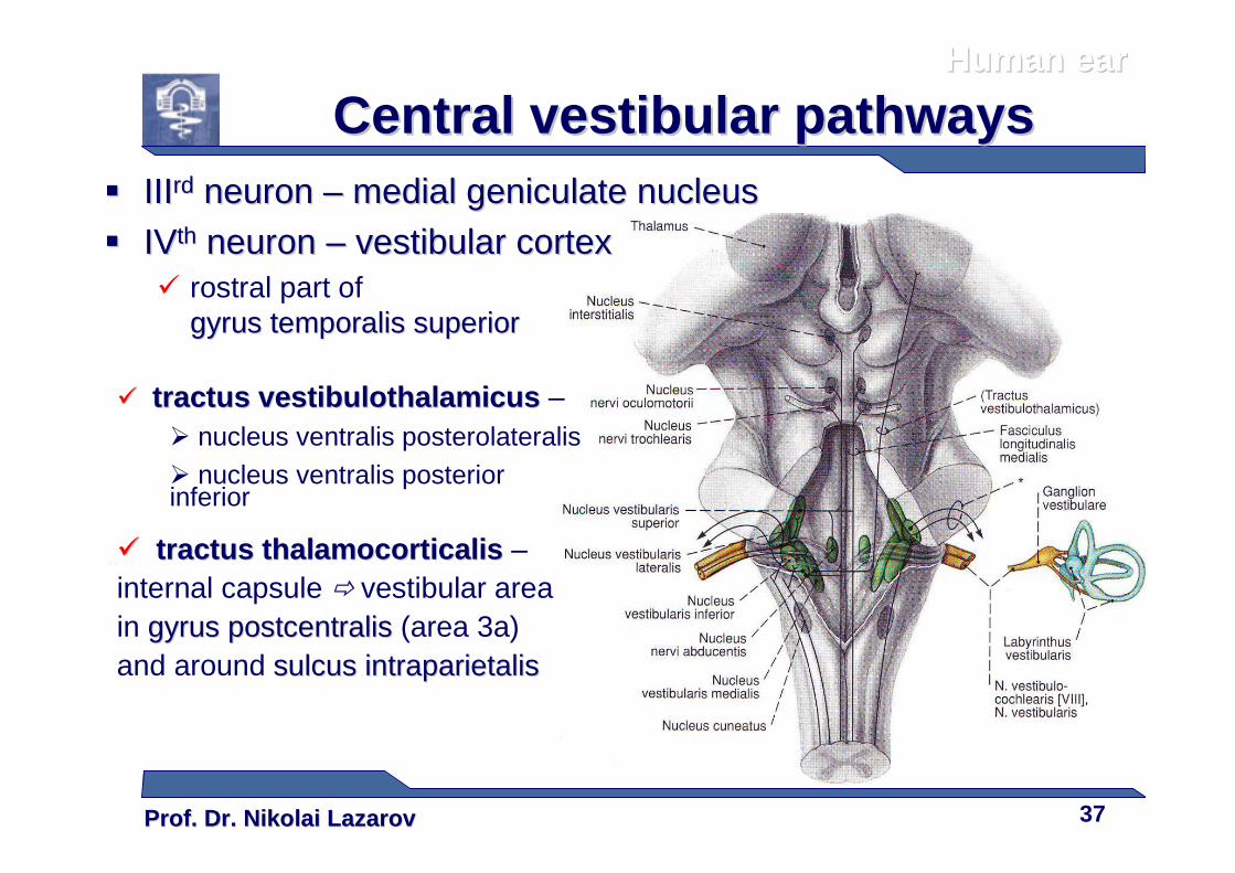
�� IIIIIIrdrd neuronneuron –– medial geniculate nucleusmedial geniculate nucleus
�� IVIVthth neuronneuron –– vestibular cortexvestibular cortex� rostral part of
gyrus gyrus temporalistemporalis superiorsuperior
�� tractustractus vestibulothalamicusvestibulothalamicus –� nucleus ventralis posterolateralis
� nucleus ventralis posterior inferior
�� tractustractus thalamocorticalisthalamocorticalis –internal capsule � vestibular area in gyrus gyrus postcentralispostcentralis (area 3a)and around sulcussulcus intraparietalisintraparietalis
Human earHuman ear
Central vestibular pathwaysCentral vestibular pathways

Prof. Dr. Nikolai LazarovProf. Dr. Nikolai Lazarov 38
Thank youThank you……