Embed Size (px)
Citation preview
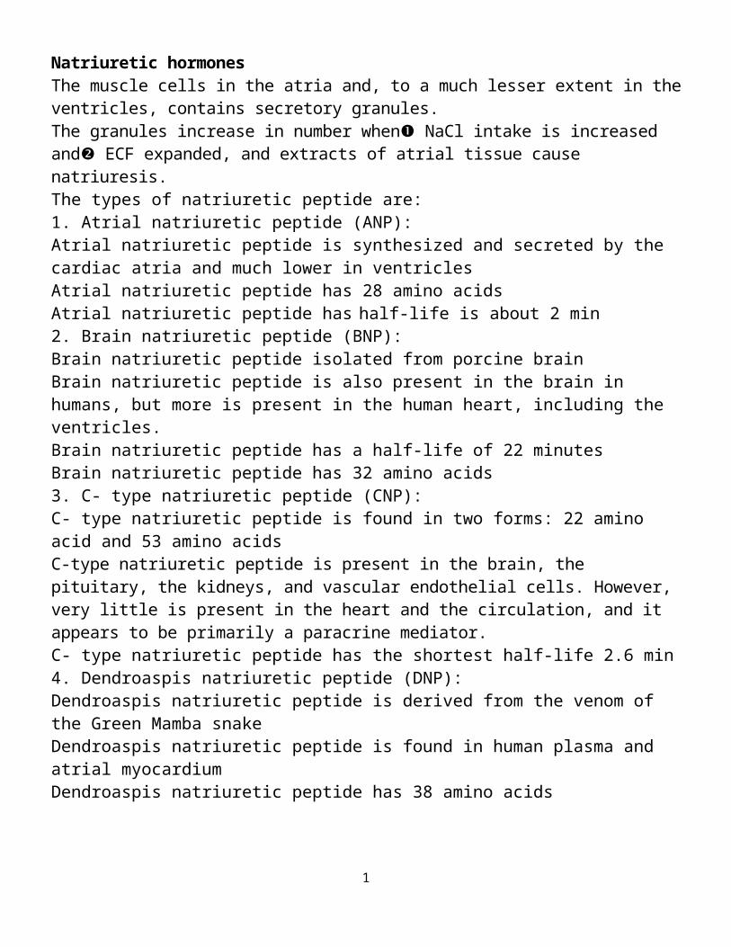
Natriuretic hormones The muscle cells in the atria and, to a much lesser extent in the ventricles, contains secretory granules. The granules increase in number when NaCl intake is increased and ECF expanded, and extracts of atrial tissue cause natriuresis.The types of natriuretic peptide are:1. Atrial natriuretic peptide (ANP): Atrial natriuretic peptide is synthesized and secreted by the cardiac atria and much lower in ventriclesAtrial natriuretic peptide has 28 amino acidsAtrial natriuretic peptide has half-life is about 2 min 2. Brain natriuretic peptide (BNP): Brain natriuretic peptide isolated from porcine brain Brain natriuretic peptide is also present in the brain in humans, but more is present in the human heart, including the ventricles. Brain natriuretic peptide has a half-life of 22 minutesBrain natriuretic peptide has 32 amino acids3. C- type natriuretic peptide (CNP): C- type natriuretic peptide is found in two forms: 22 amino acid and 53 amino acidsC-type natriuretic peptide is present in the brain, the pituitary, the kidneys, and vascular endothelial cells. However, very little is present in the heart and the circulation, and it appears to be primarily a paracrine mediator.C- type natriuretic peptide has the shortest half-life 2.6 min 4. Dendroaspis natriuretic peptide (DNP): Dendroaspis natriuretic peptide is derived from the venom of the Green Mamba snakeDendroaspis natriuretic peptide is found in human plasma and atrial myocardium Dendroaspis natriuretic peptide has 38 amino acidsThe existence of various natriuretic hormones has characteristic 17-amino-acid ring formed by a disulfide bond between two cysteines.
Natriuretic peptide receptors:Three different natriuretic peptide receptors (NPR: NPR-A, NPR-B, and NPR-C) have been isolated and characterized. The natriuretic peptides can stimulate the production of cGMP through the activation of specific receptors: the type A natriuretic peptide receptor (NPRA) and the type B natriuretic peptide receptor (NPRB). The NPRA receptor selectively responds to ANP and, to a lesser extent, to BNP.
1
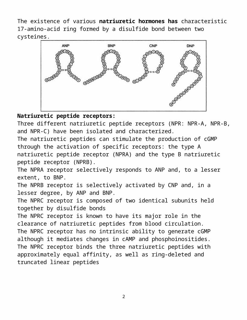
The NPRB receptor is selectively activated by CNP and, in a lesser degree, by ANP and BNP. The NPRC receptor is composed of two identical subunits held together by disulfide bondsThe NPRC receptor is known to have its major role in the clearance of natriuretic peptides from blood circulation. The NPRC receptor has no intrinsic ability to generate cGMP although it mediates changes in cAMP and phosphoinositides. The NPRC receptor binds the three natriuretic peptides with approximately equal affinity, as well as ring-deleted and truncated linear peptides
Secretion and metabolism Atrial natriuretic peptide (ANP) is released in response to
2
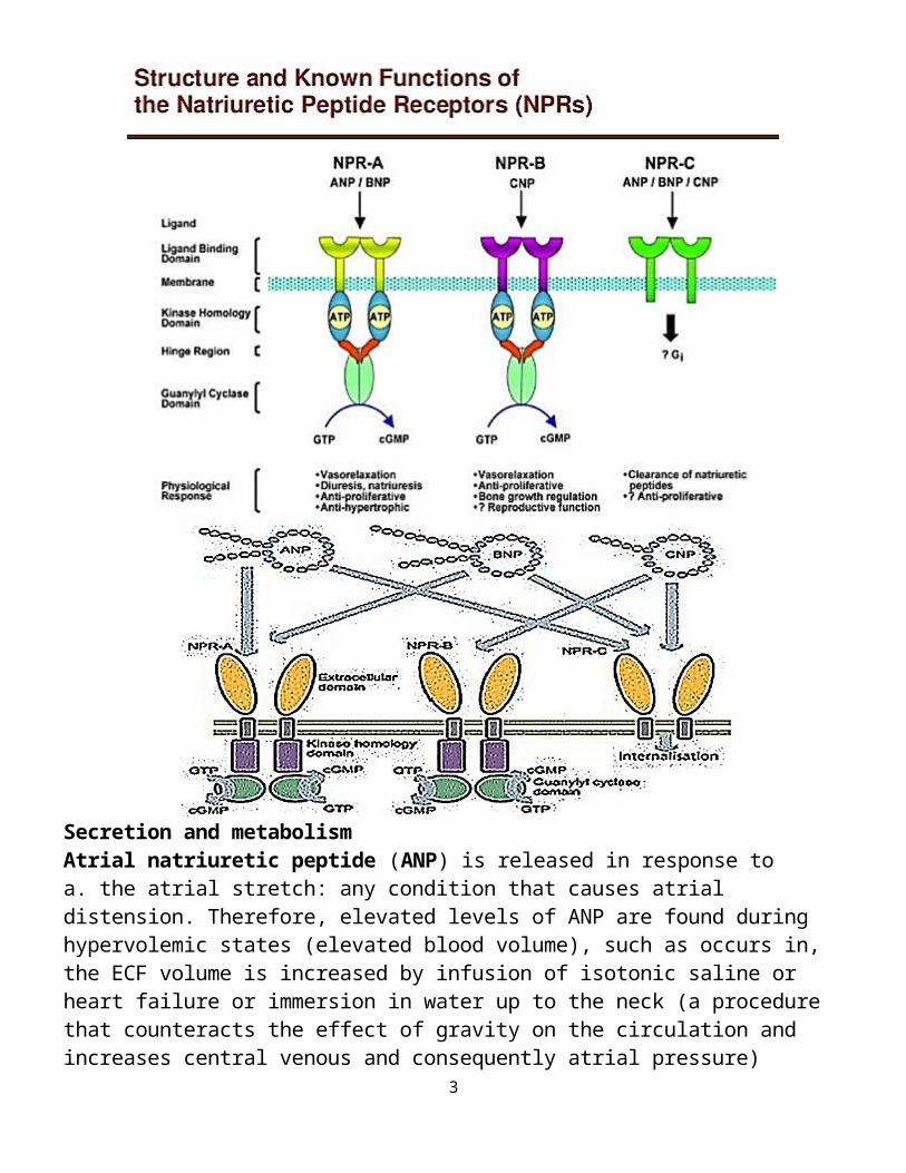
a. the atrial stretch: any condition that causes atrial distension. Therefore, elevated levels of ANP are found during hypervolemic states (elevated blood volume), such as occurs in, the ECF volume is increased by infusion of isotonic saline or heart failure or immersion in water up to the neck (a procedure that counteracts the effect of gravity on the circulation and increases central venous and consequently atrial pressure)(Note: immersion causes decrease in renin and aldosterone and increase ANP)
The reverse is also true such as dehydration. A small but measurable decrease in plasma ANP occurs in association with a decrease in central venous pressure on rising from the supine to the standing position.b. Blood pressure c. High NaCl intake d. Humoral effect:i. Rennin-angiotensin-aldosterone system ii. endotheline. Sympathetic stimulation (beta-adrenoreceptors mediated)
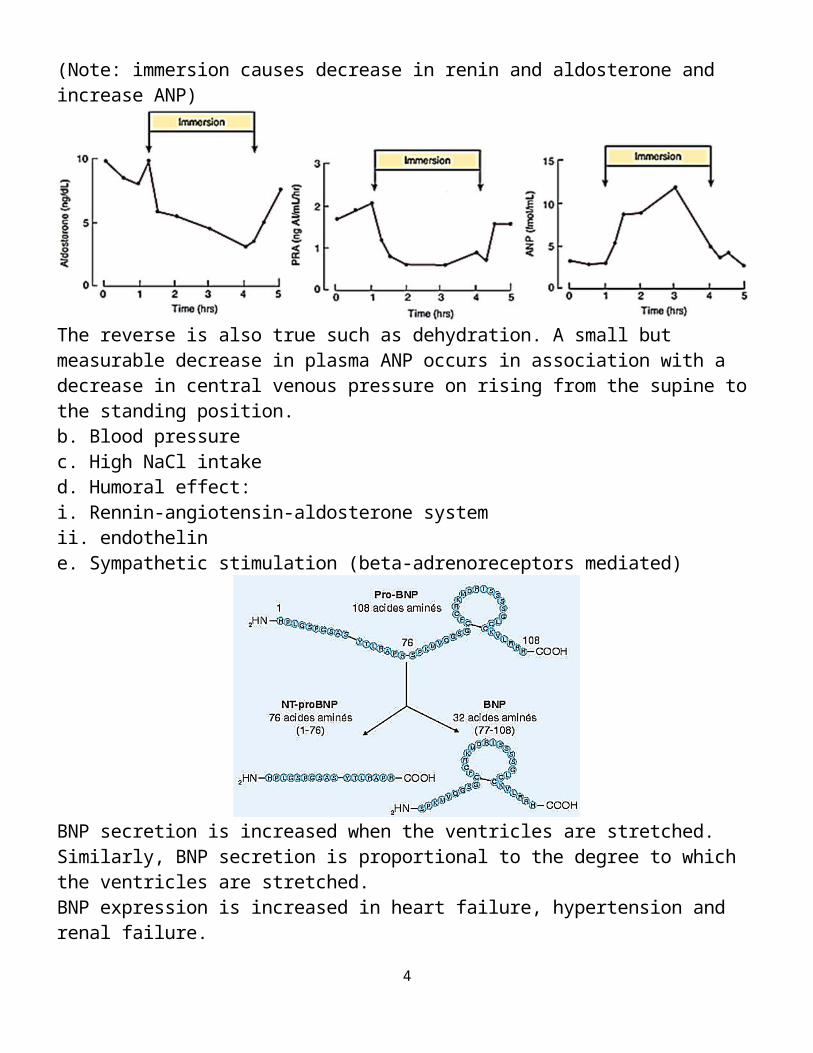
BNP secretion is increased when the ventricles are stretched.Similarly, BNP secretion is proportional to the degree to which the ventricles are stretched. BNP expression is increased in heart failure, hypertension and renal failure.Proteolysis of pro-BNP (108 amino acids) results in BNP (32 amino acids) and the N-terminal piece of pro-BNP (NT-pro-BNP; 76 amino acids). Both BNP and NT-pro-BNP are sensitive, diagnostic markers for heart failure in patients.Neutral endopeptidase (NEP; also called neprilysin) is a circulating enzyme that degrades natriuretic peptides. Physiological effects:
3
A. Renal:1. Increase of glomerular filtration and aqueous diuresisDilating afferent arterioles ► Increases Renal Blood Flow► increases glomerular filtration► Increases Urine Volume
2. Enhanced urinary excretion of sodium (= natriuresis) and of other electrolytes, phosphate, magnesium, calcium, potassiuma. act on the renal tubules to inhibit Na+ reabsorption► Increases Sodium Excretionb. decreases blood flow through the vasa recta, which will wash the solutes (NaCl and urea) out of the medullary interstitium ►This will lower osmolarity of the medullary interstitium leads to less reabsorption of tubular fluid and increased excretion.B. Cardiovascular:1. Vasodilation (relax vascular smooth muscle in arterioles and venules) 2. CNP has a greater dilator effect on veins than ANP and BNP.3. Decrease of vascular reactivity to vasoconstrictive agents (pressor effects of catecholamines and angiotensin II).4. In the brain, ANP is present in neurons, and an ANP-containing neural pathway projects from the anteromedial part of the hypothalamus to the areas in the lower brain stem that are concerned with neural regulation of the cardiovascular system.In general, the effects of ANP in the brain are opposite to those of angiotensin II, and ANP-containing neural circuits appear to be involved in lowering blood pressure and promoting natriuresis5. ANP reduces sympathetic tone by dampening of baroreceptors, suppressing the release of catecholamines from autonomic nerve endings and suppressing sympathetic outflow from the CNS. 6. An increase in capillary permeability, leading to extravasation of fluid The over all of these effects is decline in blood pressureC. Cellular effects: 1. antimitogenic effectsD. Hormonal:1. Inhibition of aldosterone and renin secretion and perhaps of ACTH and arginine vasopressin secretion2. Overall opposite effects to angiotensin II.3. Decrease the feeling of thirst and appetite for salt.E. Stimulation of lipolysis.Natriuretic peptides (NPs) are involved in the long-term regulation of sodium and water balance, blood volume and arterial pressure. There are two major pathways of natriuretic peptide actions: 1) Vasodilator effects2) Renal effects that leads to natriuresis and diuresis.
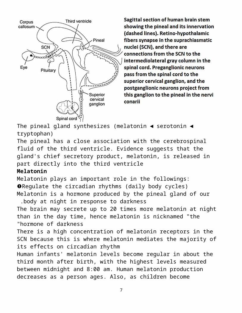
Pineal Gland:The pineal gland is a coned-shaped, pea-sized gland located just beneath the center of brain; behind the hypothalamus
4
After the age 7 pineal gland undergo involution (shrinkage), down 75% by end of puberty and to tiny mass of shrunk tissue in adult. Involution is accompanied by the appearance of granules of calcium phosphate and calcium carbonate called pineal sand The pineal gland is innervated by sympathetic neurons.
The pineal gland synthesizes (melatonin ◄ serotonin ◄ tryptophan) The pineal has a close association with the cerebrospinal fluid of the third ventricle. Evidence suggests that the gland's chief secretory product, melatonin, is released in part directly into the third ventricleMelatoninMelatonin plays an important role in the followings:Regulate the circadian rhythms (daily body cycles)
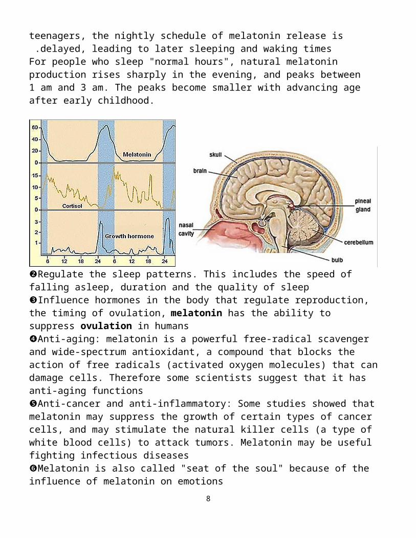
Melatonin is a hormone produced by the pineal gland of our body at night in response to darkness .The brain may secrete up to 20 times more melatonin at night than in the day time, hence melatonin is nicknamed "the hormone of darkness"
There is a high concentration of melatonin receptors in the SCN because this is where melatonin mediates the majority of its effects on circadian rhythmHuman infants' melatonin levels become regular in about the third month after birth, with the highest levels measured between midnight and 8:00 am. Human melatonin production decreases as a person ages. Also, as children become teenagers, the nightly schedule of melatonin release is delayed, leading
to later sleeping and waking times .For people who sleep "normal hours", natural melatonin production rises sharply in the evening, and peaks between 1 am and 3 am. The peaks become smaller with advancing age after early childhood.
5
Regulate the sleep patterns. This includes the speed of falling asleep, duration and the quality of sleep Influence hormones in the body that regulate reproduction, the timing of ovulation, melatonin has the ability to suppress ovulation in humans Anti-aging: melatonin is a powerful free-radical scavenger and wide-spectrum antioxidant, a compound that blocks the action of free radicals (activated oxygen molecules) that can damage cells. Therefore some scientists suggest that it has anti-aging functionsAnti-cancer and anti-inflammatory: Some studies showed that melatonin may suppress the growth of certain types of cancer cells, and may stimulate the natural killer cells (a type of white blood cells) to attack tumors. Melatonin may be useful fighting infectious diseasesMelatonin is also called "seat of the soul" because of the influence of melatonin on emotionsAdipose tissue as endocrinal gland:The classic function of the adipocyte is to store and release lipid fuelMost recent data emphasize the role of adipose tissue as a hormonally active system, influencing inflammation, metabolism, and body weight, and lipid and glucose metabolismThese secreted proteins, which include tumor necrosis factor (TNF)-alpha, resistin, IL-6, Acylation Stimulating Protein, angiotensinogen, plasminogen activator inhibitor-1 (PAI-1) ("bad" adipokines) and leptin, adiponectin ("good" adipokines) seem to play important regulatory roles in a variety of complex processes, including fat metabolism, feeding behavior, hemostasis, vascular tone, energy balance, and insulin sensitivity, but none is without controversy regarding its respective mechanism and scope of action.Endocrine function of the adipocyte can be divided into proteins and enzymes involved in steroid metabolismA. adipocytokines (or adipokines or Protein): Leptin Leptin is secreted not only from white adipocytes, but also from placenta and stomach Leptin is found more in subcutaneous than visceral fat. Leptin identification in 1994 Leptin endocrine effects include regulation of immune function, hematopoiesis, angiogenesis, and bone development.
6
Leptin has been known for its role in controlling body weight, food intake through hypothalamic pathways, glucose homeostasis Leptin primary function is to increase satiety and energy expenditure through action on the hypothalamus.Leptin action in the muscle, increases glucose uptake and glucose metabolism. Leptin action in the liver increases glucose productionLeptin action in the pancreases, increases insulin Leptin activates the hypothalamic-pituitary-adrenal axis Leptin suppresses the hypothalamic-pituitary - thyroid axis and the hypothalamic -pituitary-gonadal axisLeptin is involved in the regulation of reproductive development and function by indirectly influencing GnRH neuron activity.Adiponectin:Adiponectin is exclusively secreted by adipocytesAdiponectin with higher serum concentration in femalesAdiponectin is found more in the subcutaneous adipose deposits than in the visceral deposits.Adiponectin In the liver decreases glucose production and free fatty acid synthesis. Adiponectin In the muscle increases free fatty acid oxidation and decreases triglyceride productionAdiponectin has anti-atherogenic activity because it acts through:A. the insulin receptor of the vascular endothelium to increase nitric oxide (NO) B. positive correlation to high-density lipoprotein cholesterol and inverse correlations to low-density lipoprotein cholesterol, triglycerides, insulin resistance, and diastolic blood pressureA strong and consistent inverse association between adiponectin and both insulin resistance and an inflammatory state has been established:A. Adiponectin activity increases during insulin sensitivity and decreases insulin resistance B. Adiponectin seems to have anti-inflammatoryTaken together, these studies suggest that adiponectin is a unique adipocyte-derived hormone with anti-diabetic, anti -inflammatory and anti-atherogenic effects.
7
Adipsin & Acylation Stimulating ProteinAdipsin is found more in the subcutaneous adipose deposits than in the visceral deposits. Adipsin is the enzyme required for the production of Acylation Stimulating Protein (ASP).Both adipsin and Acylation Stimulating Protein positively correlate with adiposity, insulin resistance, dyslipidemia, and cardiovascular diseaseAcylation Stimulating Protein increases insulin secretion. Acylation Stimulating Protein in the adipose tissue:A. increases glucose transport and Triglycerides synthesis by increasing the activity of diacylglycerol acyltransferase, and decreases lipolysis and release of Non-esterified ("free" or unsaturated) fatty acid from adipocytesB. decreases lipolysis and free fatty acid releaseResistinResistin is 15 times greater in the visceral adipose deposits than in the subcutaneous deposits. Resistin increase in insulin resistance and type 2 diabetes mellitusResistin potentially linking obesity with insulin resistanceTumor necrosis factor-alpha (TNP-ά) Tumor necrosis factor-alpha is secreted more from the subcutaneous adipose deposits than from the visceral deposits and may be dependent on regional fat mass. The primary action of Tumor necrosis factor-alpha is to increase insulin resistance in liver, muscle and adipose tissue. Additional action on the liver include increased free fatty acid (FFA) production and cholesterol synthesis and decreased glucose uptake and free fatty acid storageProtein from the renin angiotensin systemRenin angiotensin system proteins are found more in visceral adipose deposits than subcutaneous deposits. RAS proteins include: rennin, angiotensinogen, angiotensin I, angiotensin II, angiotensin converting enzymeSimilar to the renin-angiotensin system in the kidney, these proteins affect the same target organs. In the adrenal gland, aldosterone is increased, increasing the reabsorption of sodium and water in the kidneys. In the vasculature, vasoconstriction is primary activity from adipocyte renin angiotensin system. In the liver, renin angiotensin system results in decreased lipolysis with increased lipogenesis, gluconeogenesis, glycogenolysis, and insulin resistanceInterleukin-6Interleukin-6 is found more in visceral adipose deposits than in subcutaneous deposits. Approximately 1/3rd of the circulating Interleukin-6 originates from the adipose tissue.Interleukin-6 primary function is to increase insulin resistance at the insulin receptor or insulin signaling pathway in hepatic, muscle and adipose tissue.Interleukin-6 increases hepatic hyperlipidemia and glucose production. In the central nervous system, Interleukin-6 deficiency decreases energy expenditure Plasminogen activator inhibitor-1Plasminogen activator inhibitor-1 is found more in visceral adipose deposits than in subcutaneous deposits.Fibrinolysis the breaking down of blood clots. Plasminogen activator inhibitor-1inhibits tissue plasminogen activator which initiates the fibrinolysis cascade: thus, Plasminogen activator inhibitor-1 inhibits fibrinolysis.
8
Tumor necrosis factor-alpha increases Plasminogen activator inhibitor-1 in adipose tissueB. Steroidgenic enzymes:Several enzymes from the adipose tissue are involved in steroid metabolism; activation, inter-conversion and inactivation.The steroid activity can be divided into sex steroids and glucocorticoid activityFor the most part, the enzymes involved in sex steroids convert androgens to estrogens. More specifically, they also convert specific sex steroids to their more active form; that is, androstenedione to testosterone and estrone to estrodiol.In glucocorticoid activity, steroidgenic enzymes increase insulin sensitivity and control visceral adipose tissue depositionHunger & satiety:There are several levels of energy balance in metabolism. The obvious balance is that of food intake and energy expenditure. In the balance of caloric intake and energy expenditure, the factors affecting caloric intake include: Hunger is a physiological urge to consume food. Satiety is the signal for cessation of food intake. Appetite is a physiological preference for specific foods.The feelings of hunger and satiety are stimulated by the “gut-brain axis”, where a crucial role is played by gastrointestinal hormones: glucagon-like peptide 1, glucose-dependent insulinotropic polypeptide, pancreatic polypeptide, peptide YY, oxyntomodulin, cholecystokinin and ghrelin. These hormones affect not only the functioning of the digestive tract, but also might have effects on insulin secretion and are mediators which affect brain areas involved in the regulation of food intake. How much of these factors can be modified by psychological factors in the control of food intake for humans?GhrelinGhrelin is an endogenous peptide with 28 amino acid peptideGhrelin is the substance found to be associated with meal initiation in humans. Ghrelin is secreted by the stomach and intestine. Ghrelin has been found to increase before mealsGhrelin believed that glucose concentrations and energy balance have a role in ghrelin production and release. Ghrelin fall to its lowest point within one hour following the meal;Functions of GhrelinGhrelin increases growth hormone. Ghrelin acts as an antagonist of somatostatin that inhibits the secretion but not the synthesis of GH. GHRH+ ghrelin acts synergistically to stimulate the release of GH from the somatotrophic cells of the hypophysis.Ghrelin actions of feeding are independent from the growth hormone actions. As ghrelin is adipogenic,Ghrelin by lowering the catabolism of fat participates in a. the regulation of energy homeostasis, b. increases food intake, and c. decreases energy expenditure. orexigenic (appetite-stimulating) Ghrelin increases food intake + weight gain in experimental animals
9
Ghrelin induces hunger in humans. Ghrelin is part of a complex neuroendocrine network involved in the regulation of appetite and energy homeostasis. Several studies suggested that the orexigenic signal of ghrelin secreted from the stomach is transmitted to the brain via the vagal afferent nerve. Peripheral ghrelin may exert its effects on the CNS by crossing the blood brain barrier (BBB) although the rate at which it passes the BBB is very low. Areas in the brain that are implicated in the regulation of feeding behavior express receptors for ghrelinObestatinObestatin is 23 amino acid peptide Obestatin was initially considered to oppose the orexigenic effects of ghrelin. Later studies, however, cast doubt on the initial findings as subsequent studies failed to confirm the anorexigenic effects (appetite suppressant) of obestatin. Obestatin has been reported to have additional roles such as the inhibition of thirst the regulation of memory, anxiety, and sleepstimulate the proliferation of human retinal cells promote the survival of pancreatic β-cells and human islets the regulation of adipocyte metabolism and adipogenesis.Leptin, Leptin a satiety hormone produced by white adipose tissue, and represents another appetite regulator. Leptin and ghrelin are supposed to share hypothalamic pathways regulating food intake and energy homeostasis.
10