Embed Size (px)
Citation preview
INTRODUCTION
Testes, as a critical component of the male reproduc-tive and endocrine tract, the primary functions is to produce sperm and androgens. Mammalian testes con-sists of several well-defined cell types, including germ cells of different stages, Sertoli cells and Leydig cells.
Each of them is configured with a different location and has specific functions: Germ cells produce sperm and are located in the seminiferous tubules; Leydig cells produce sex hormones and are distributed in the connective tissue of the convoluted seminiferous tu-bules; Sertoli cells form the basement membrane of the seminiferous tubules and offer the environment neces-
Received: May 21, 2018 Revised: Sep 10, 2018 Accepted: Sep 10, 2018 Published online Oct 23, 2018Correspondence to: Lei Yang https://orcid.org/0000-0001-8859-9692 College of Basic Medical Science, Jiujiang University, 17 Lufeng Rd, Xunyang Qu, Jiujiang Shi, Jiangxi Sheng 332000, China.Tel: +86-0792-8570043, Fax: +86-0792-8577050, E-mail: [email protected] to: Shaoxin Huang https://orcid.org/0000-0001-9801-4133 College of Basic Medical Science, Jiujiang University, 17 Lufeng Rd, Xunyang Qu, Jiujiang Shi, Jiangxi Sheng 332000, China.Tel: +86-0792-8570043, Fax: +86-0792-8577050, E-mail: [email protected]
Copyright © 2019 Korean Society for Sexual Medicine and Andrology
Original ArticlepISSN: 2287-4208 / eISSN: 2287-4690World J Mens Health 2019 May 37(2): 186-198https://doi.org/10.5534/wjmh.180041
C-Type Natriuretic Peptide/Natriuretic Peptide Receptor 2 Is Involved in Cell Proliferation and Testosterone Production in Mouse Leydig Cells
Lei Yang1,3 , Lanjie Lei2 , Qihan Zhao1 , Ying Gong1,3 , Gaopeng Guan2,3 , Shaoxin Huang1
1College of Basic Medical Science, 2Clinical Skills Center, Affiliated Hospital of Jiujiang University, 3Key Laboratory of System Bio-medicine of Jiangxi Province, Jiujiang University, Jiujiang, China
Purpose: This study investigated the role of natriuretic peptide receptor 2 (NPR2) on cell proliferation and testosterone secre-tion in mouse Leydig cells.Materials and Methods: Mouse testis of different postnatal stages was isolated to detect the expression C-type natriuretic peptide (CNP) and its receptor NPR2 by quantitative reverse transcription polymerase chain reaction (RT-qPCR). Leydig cells isolated from mouse testis were cultured and treated with shNPR2 lentiviruses or CNP. And then the cyclic guanosine mono-phosphate production, testosterone secretion, cell proliferation, cell cycle and cell apoptosis in mouse Leydig cells were ana-lyzed by ELISA, RT-qPCR, Cell Counting Kit-8, and flow cytometry. Moreover, the expression of NPR2, cell cycle, apoptosis proliferation and cell cycle related gene were detected by RT-qPCR and Western blot. Results: Knockdown of NPR2 by RNAi resulted in S phase cell cycle arrest, cell apoptosis, and decreased testosterone secre-tion in mouse Leydig cells.Conclusions: Our study provides more evidences to better understand the function of CNP/NPR2 pathway in male reproduc-tion, which may help us to treat male infertility.
Keywords: Germ cells; Leydig cells; Testicular diseases; Testosterone
This is an Open Access article distributed under the terms of the Creative Commons Attribution Non-Commercial License (http://creativecommons.org/licenses/by-nc/4.0) which permits unrestricted non-commercial use, distribution, and reproduction in any medium, provided the original work is properly cited.
Lei Yang, et al: CNP/NPR2 Promotes Testosterone Production
187www.wjmh.org
sary for the differentiation and maturation of germ cells [1]. The importance of Leydig cells in male repro-duction is exemplified by the fact that loss of func-tion of gene such as the luteinizing hormone receptor (LHR) [2], insulin-like factor 3 (Insl3) [3], and enzymes involved in testosterone biosynthesis [4].
C-type natriuretic peptide (CNP) and its receptor (natriuretic peptide receptor 2, NPR2) play a role as an oocyte maturation inhibitor in mammalian [5]. Re-cent studies suggested that CNP can improve oocyte maturation and developmental competence in vitro [6]. Compared with numerous studies about CNP/NPR2 on female reproduction, there is little research about CNP/NPR2 in male reproduction. Previous studies reported that CNP regulates blood-testis barrier dynamics and is related with spermatogenesis [7]. Sogawa et al [8] inves-tigated phenotype of NPR2-defcient short-limbed-dwarfs mice, and found that the developmental onset and ac-quisition of spermatogenic function is delayed in NPR2 mutant mice. Subsequently, two studies reported that CNP/NPR2 is related with sperm motility, acrosome reaction and induces sperm attraction for fertilization, thus regulating the reproductive function of males [9,10].
However, the function of CNP/NPR2 in male repro-duction remains largely unknown. In this study, we aimed to study the role of CNP/NPR2 on cell prolif-eration, testosterone secretion and related regulatory mechanisms in mouse Leydig cells.
MATERIALS AND METHODS
1. Chemicals and miceAll chemicals were purchased from Sigma-Aldrich
(St. Louis, MO, USA) unless otherwise stated. Male Kunming mice were purchased from the Laboratory Animal Central of Jiujiang University. All mice were fed a typical diet of lab chow and housed in a single room under conditions of constant temperature (25°C–28°C), humidity (55%±5%) and lighting (12 hours light, 12 hours dark cycle) [11]. All procedures were approved by the Committee for the Ethics on Animal Care and Experiments of Jiujiang University (approval No. SYXK(GAN)2017-0001).
2. Isolation and cultures of Leydig cellsLeydig cells were isolated from the testes of 42- to
49-day-old Kunming mice and cultured as previous report [12]. The purity of Leydig cells was assessed by
3β-hydroxysteroid dehydrogenase (3β-HSD) staining using the modified Wiebe method. Simply, Leydig cells were fixed in 1% paraformaldehyde for 20 minutes and then washed with phosphate buffer saline (PBS) for three times. After washed the cells were incubated with 1 mL PBS containing 1 mg bovine serum albumin, 1.5 mg nicotinamide adenine dinucleotide, 0.2 mg nitro-blue tetrazolium and 0.25 mg dehydroepiandrosterone for 2 hours at 37°C in the dark. Then the cells were gently rinsed with PBS and observed microscopically. During all the culture, the cells were cultured with Dulbecco’s modified Eagle’s medium/F-12 containing 10% fetal bovine serum with or without CNP (CNP were used to active CNP/NPR2 signaling pathway), unless otherwise stated.
3. Transfection of cells with shRNA-natriuretic peptide receptor 2 lentiviral
shRNA-NPR2 and shRNA-negative lentiviral vec-tor were purchased from Genechem (Shanghai, China). The mouse Leydig cells were seeded into 6-well plates, which were cultured to 30% to 40% confluence and in-fected by addition of 1×10⁸ TU/mL lentivirus, 5 µg/mL polybrene and complete medium. After 12 hours, the lentivirus solution was replaced by complete culture medium and cultured for 36 hours. And the cells were observed under a fluorescence microscope to evaluate the transfection efficiency. The efficiency and specific-ity of siRNA mediated knockdown were examined by Western blot and quantitative reverse transcription polymerase chain reaction (RT-qPCR). After determine the NPR2 knockdown efficiency, the cells were cul-tured for subsequent experiments.
4. Cell proliferation assayAfter culturing with CNP for 24 hours, Cell Count-
ing Kit-8 (CCK8) was added to the cells (10 µL/well). Then the cells were incubated for 1 hour at 37°C and measured at 450 nm by a Microplate Reader (Bio-Rad 680, Hercules, CA, USA). The experiments were per-formed in triplicate.
5. Cyclic guanosine monophosphate measurement
After 30 minutes of treatment, the cells were col-lected to measure the cyclic guanosine monophosphate (cGMP). The cGMP measurement was according to the procedure described as our previous report [6]. The lev-
https://doi.org/10.5534/wjmh.180041
188 www.wjmh.org
els of cGMP were determined using cGMP-EIA kits ob-tained from Cayman Chemicals (Ann Arbor, MI, USA). Each sample was measured in triplicate.
6. Testosterone measurementAfter 24 hours of treatment, mouse Leydig cells
were counted. The concentration of testosterone in the culture supernatants (100,000 cell/mL culture superna-tant) was measured with ELISA kits (Ji Yin Mei; Co. Ltd., Wuhan, China) according to the manufacturer’s instructions. Each sample was measured in triplicate.
7. Cell cycle analysisMouse Leydig cells of the respective experimental
groups were collected and fixed in ice-cold 70% etha-nol overnight at 4°C after the cells were treatment and count. Then, the cells were washed with PBS and stained with propidium iodide (PI) solution for 20 minutes at 25°C in the dark. Finally, the cells were analyzed by flow cytometry using a BD FACS Calibur system and Mod Fit LT for MacV3.0 software. For each analysis, a minimum of 10,000 cells were analyzed. Each sample was measured in triplicate.
8. Cell apoptosis assayThe apoptotic rate was determined using the phyco-
erythrin (PE) and PI double-staining apoptosis analysis kit (Nanjing Key Gen Biotech, Nanjing, China). After washes by PBS for two times, followed by centrifuga-tion, the cells were suspended in 500 µL binding buffer. Then, 5 µL Annexin PE and PI staining solution were added and incubated at 25°C for 10 minutes. The mix-ture was then analyzed by flow cytometry within 60 minutes. Each sample was measured in triplicate.
9. RNA isolation and quantitative reverse transcription polymerase chain reaction
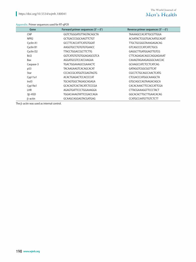
Total RNA was extracted using Trizol reagent (In-vitrogen, Carlsbad, CA, USA). First-strand cDNA was synthesized according to the manufacturer’s instruc-tions (PrimeScript® RT reagent Kit). RT-qPCR was per-formed using an ABI StepOnePlus Real-time Detection System (ABI, Foster, CA, USA) and SYBR Green qPCR SuperMix (Invitrogen). Each experiment was repeated independently at least three times, and the fold change in the expression of each gene was analyzed using the 2-ΔΔCT method. All the primers were used as Appendix and the β-actin was used as internal control.
10. ImmunohistochemistryTestes of 42- to 49-day-old Kunming mice were fixed
in 4% paraformaldehyde in PBS for one week, dehy-drated through a graded ethanol series, and embed-ded in paraffin. Sections 7 mm thick were mounted onto glass slides precoated with Poly-L-Lysine Solution and incubated overnight at 37°C. After dehydrating, samples were placed in citrate buffer (pH=6.0). Antigen retrieval was performed by treating samples in a mi-crowave oven at 92°C for 15 minutes; slides were cooled and then washed in PBS. The sections were pretreated with 3% (vol/vol) H2O2 in methanol to quench endog-enous peroxidase activity. After being washed with PBS, sections were incubated with 10% goat serum for 30 minutes at 37°C. After blocking, sections were incu-bated overnight at 4°C with rabbit polyclonal antibody against NPR2 (Sigma-Aldrich; 1:50 dilutions) in a hu-midified chamber. After washing, followed by incuba-tion with biotinylated anti-rabbit immunoglobulin G antibody (Beijing 4A Biotech Co., Ltd, Beijing, China) at 37°C for 1 hour, and then sections were incubated with horseradish peroxidase-labeled streptavidin at 37°C for 30 minutes. Thereafter, positive reactions were visual-ized with a diaminobenzidine-peroxidase substrate and 30 seconds counterstaining with hematoxylin. Finally, the sections were counterstained with dehydrated and mounted. Slides were imaged using a digital microscope (BA400; Motic, Wetzlar, Germany).
11. Western blot analysisThe extraction of cell proteins was used radioim-
munoprecipitation assay buffer. Protein determination was performed by the bicinchoninic acid (BCA) assay. 200 µg total protein per sample were separated by 12% sodium dodecyl sulfate polyacrylamide gel electropho-resis and electro transferred to a to polyvinylidene difluoride membrane. After incubation in blocking buffer for 1 hour at room temperature, the membrane was incubated overnight at 4°C with the primary an-tibodies (p-AKT, AKT, β-actin, 1:1,000, Cell Signaling Technology, Beverly, MA, USA; NPR2, 1:500, Sigma-Aldrich; Cyp11a1, Star, Bax, Bcl-2, 1:500, Santa Cruz Biotechnology, Santa Cruz, CA, USA). After washing, the membranes were incubated with a secondary anti-body conjugated to horseradish peroxidase at 25°C for 1 hour. Finally, immunoreactive bands were visual-ized using a Super Signal West Pico kit according to the manufacturer’s (Pierce Biotechnology, Rockford,
Lei Yang, et al: CNP/NPR2 Promotes Testosterone Production
189www.wjmh.org
IL, USA) instructions. The β-actin was used as inter-nal control and the protein band densities were semi-quantified by densitometric analysis using ImageJ (ver. 1.49).
12. Caspase-3 activity measurement Caspase-3 activity was measured using a caspase-3
Activity Colorimetric Assay Kit. Simply, after treat-ment, the cells were harvested by centrifugation and incubated in lysis buffer on ice for 15 minutes. The ly-sate was then centrifuged at 15,000 rpm and 4°C for 15 minutes, and the protein content was determined using the BCA Protein Assay Kit according to the manufac-turer’s (Nanjing Biobox Biotech, Naniing, China) in-structions. Then, 100 mg of protein in each sample was incubated with the caspase-3 substrate (200 mM final concentration) at 37°C in a microplate for 4 hours. The samples were measured at 405 nm using a microplate reader (Bio-Rad 680).
13. Statistical analysesAll experiments were replicated at least three times
for each group, and the data are presented as the mean±standard error of mean. The data were analyzed by ANOVA, followed by Fisher’s least significant dif-ferent test and independent samples Student t-test, with the SPSS software ver. 13.0 (SPSS Inc., Chicago, IL, USA). Differences were considered significant at p<0.05.
RESULTS
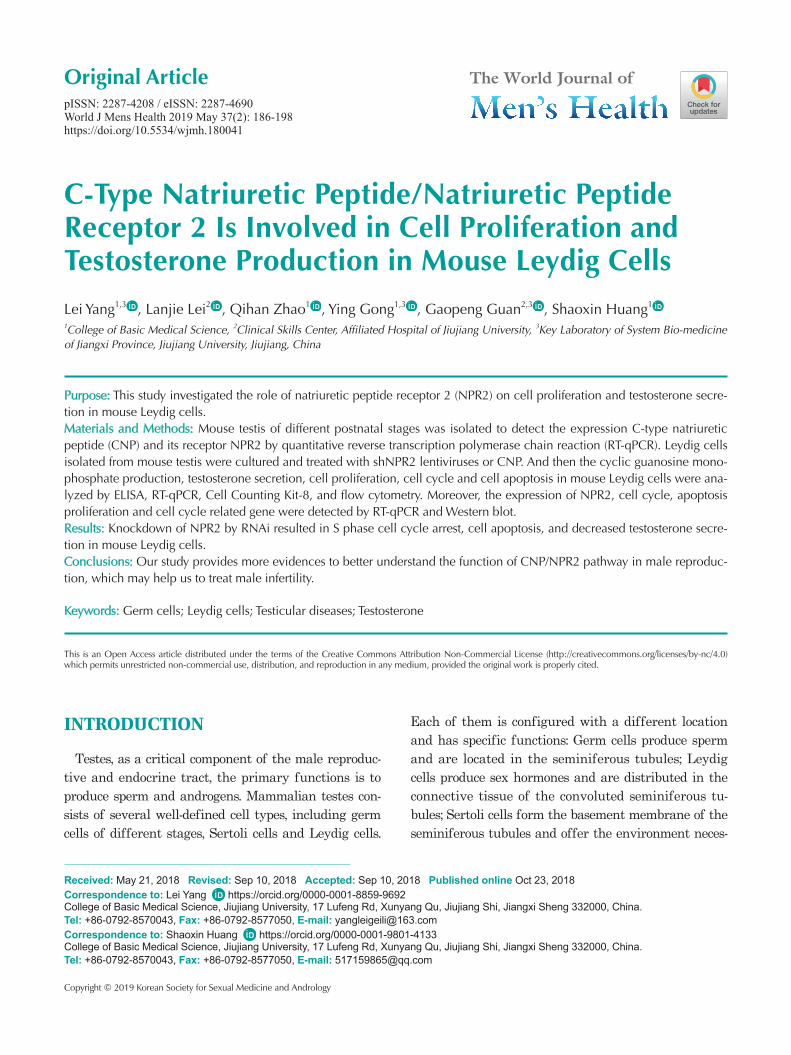
1. The expression pattern of C-type natriuretic peptide/natriuretic peptide receptor 2 at different postnatal stages of testes
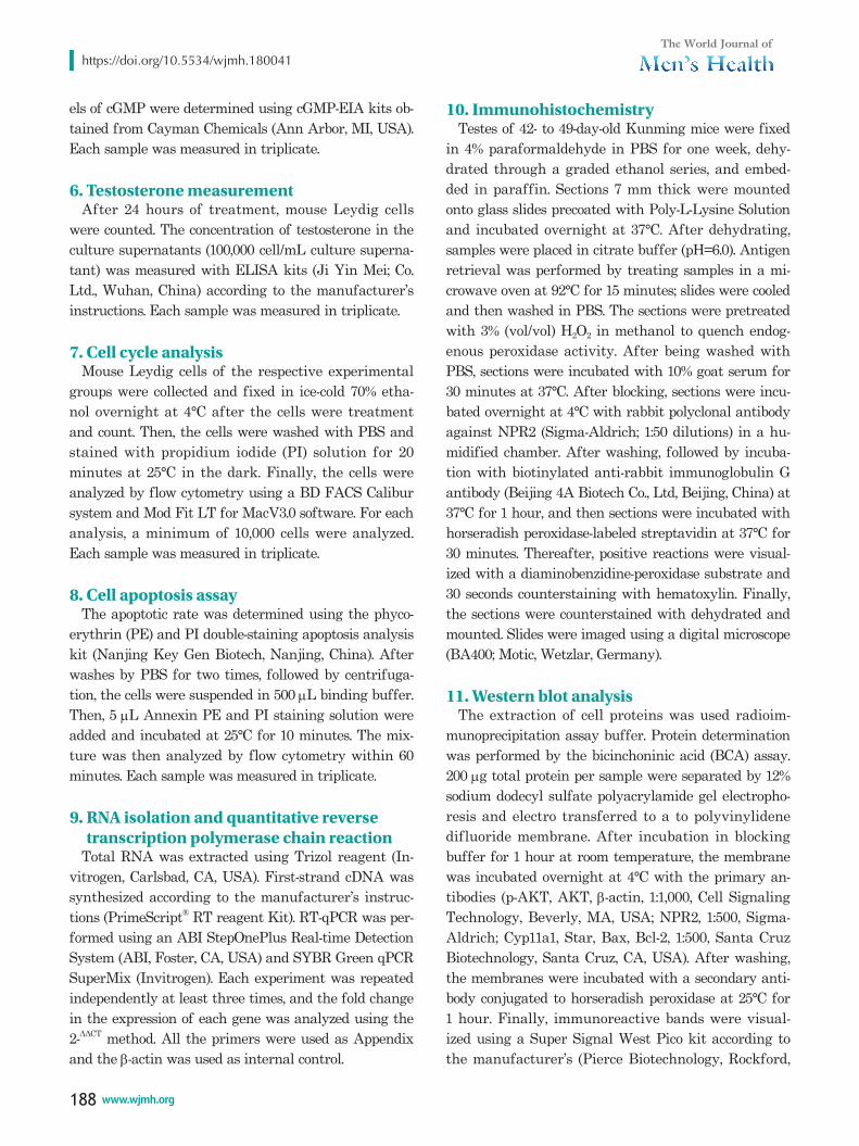
Previous studies reported that CNP is mainly located and expressed in rat Leydig cells [13,14]. But the expres-sion and location of NPR2 in rodent are still unknown. Here, we first detected the location and expression of NPR2 in adult mouse testes by semiquantitative RT-PCR, Western blot and immunohistochemistry. Results showed that NPR2 is mainly expressed in Leydig cells
CNP
NPR2
�-actin
Testes Leydig cellsA B
0
2.0
1.5
1.0
0.5
CN
Pexpre
ssio
nle
vels
Time (d)
0.07 14 21 28 35 42 49 56
b
gf
e
d
c
b
a
b
D
0
2.0
1.5
1.0
0.5
NP
R2
expre
ssio
nle
vels
Time (d)
0.07 14 21 28 35 42 49 56
c
e e
d
c
b
aa
ab
E
NPR2
�-actin
CTestes Leydig cells
Fig. 1. The expression of C-type natriuretic peptide (CNP)/natriuretic peptide receptor 2 (NPR2) at different postnatal stages of testes. (A) The expression of CNP/NPR2 mRNA in testes and Leydig cells. (B, C) The location by immunohistochemistry (×200) and expression by Western blot of NPR2 in testes and Leydig cells. (D) The expression of CNP at mouse different postnatal stages of testes. (E) The expression of NPR2 at mouse different postnatal stages of testes. The β-actin was used as internal control. The data are presented as the mean±standard error of mean of three independent experiments. Bars with different letters are significantly different (p<0.05).
https://doi.org/10.5534/wjmh.180041
190 www.wjmh.org
(Fig. 1A–1C). Moreover, CNP is also mainly expressed in Leydig cells (Fig. 1A). In addition, we detected the ex-pression pattern of CNP and NPR2 at different time of testes after born (0, 7, 14, 21, 28, 35, 42, 49, and 56 days) by RT-qPCR. Result showed that CNP and NPR2 have a similar expression patterns in the mouse testis at dif-ferent postnatal stages (Fig. 1D, 1E). The mRNA expres-sion of CNP and NPR2 has a higher expression levels on 0 day, reach its lowest level on postnatal 7 days, and then increases again from 14 to 56 days (Fig. 1D, 1E).
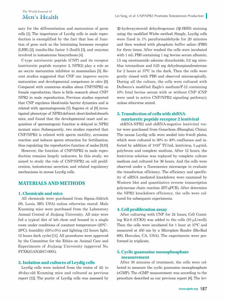
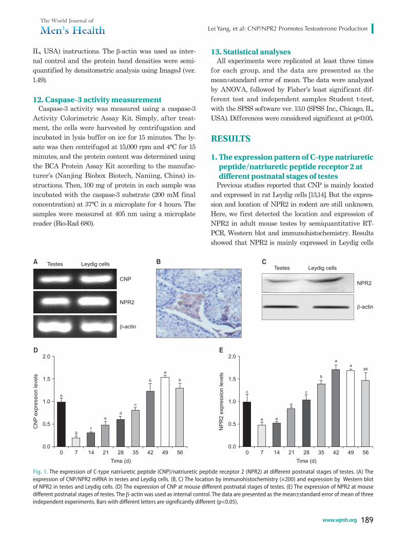
2. Effects of C-type natriuretic peptide on cyclic guanosine monophosphate production, natriuretic peptide receptor 2 expression, cell proliferation, testosterone secretion in mouse Leydig cells
To measure the effects of CNP on cGMP production,
NPR2 expression, cell proliferation, testosterone secre-tion in mouse Leydig cells, the cells were treated with different concentrations of CNP (0, 15, 30, 60, 120 nM) to measure cGMP production, detect NPR2 expres-sion, cell proliferation and testosterone secretion. The results showed that CNP enhances cGMP production in a dose-dependent manner, and the lowest effective concentration is 15 nM (Fig. 2A). Moreover, RT-qPCR results showed that CNP also increases NPR2 expres-sion in dose-dependent manner and the lowest effective concentration is 30 nM (Fig. 2B). Meanwhile, CCK8 results showed that CNP enhances cell proliferation in a dose-dependent manner, and the lowest effective concentration is 30 nM (Fig. 2C). In addition, ELISA results showed that CNP promotes the testosterone se-cretion and the lowest effective concentration is 30 nM (Fig. 2D).
0
140
120
100
80
60
40
20
cG
MP
(pm
ol/m
L)
CNP (nM)
015 30 60 120
0
180
160
140
120
100
80
Cell
via
bili
ty(%
)
CNP (nM)
015 30 60 120
e
d
c
b
a
c
c
b
aa
0
2.0
1.5
1.0
0.5
NP
R2
expre
ssio
nle
vels
CNP (nM)
0.015 30 60 120
0
80
70
60
50
40
30
20
10
Testo
ste
rone
(ng/m
L)
CNP (nM)
015 30 60 120
c c
b
aa
dd
c
b
a
A B
C D
Fig. 2. Effects of C-type natriuretic peptide (CNP) on cyclic guanosine monophosphate (cGMP) production, natriuretic peptide receptor 2 (NPR2) expression, cell proliferation, testosterone secretion in mouse Leydig cells. (A) The cGMP production in mouse Leydig cells. (B) The NPR2 expres-sion in mouse Leydig cells. (C) The cell activity in mouse Leydig cells. (D) The testosterone secretion in mouse Leydig cells. The β-actin was used as internal control. The data are presented as the mean±standard error of mean of three independent experiments. Bars with different letters are significantly different (p<0.05).
Lei Yang, et al: CNP/NPR2 Promotes Testosterone Production
191www.wjmh.org
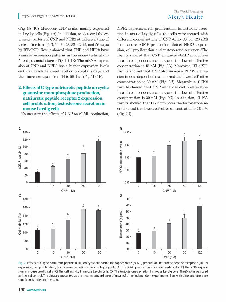
3. Natriuretic peptide receptor 2 knockdown inhibits cyclic guanosine monophosphate production and cell proliferation in mouse Leydig cells
To investigate role NPR2 on cell proliferation, the mouse Leydig cells were transfected with shRNA-NPR2 and shRNA-negative lentivirus. And the NPR2 knockdown or shRNA-negative mouse Leydig cells were treated with 30 nM CNP for 24 hours. RT-qPCR and western blot showed that shRNA-NPR2 lentiviral effectively suppresses NPR2 expression in both mRNA and protein levels (Fig. 3A, 3B). Moreover, ELISA showed that NPR2 depleted Leydig cells have a lower expression levels of cGMP (Fig. 3C). CCK8 results in-dicated that a decreased cell viability is observed in NPR2 knockdown group compared with the control group (Fig. 3D). Flow cytometry analysis showed that
the S phase of cells in the NPR2 knockdown group is higher compared the control group (Table 1). In addi-tion, the mRNA expression of cell cycle factors (cyclin A1, cyclin B1, and cyclin D2) is downregulation in NPR2 knockdown mouse Leydig cells compared the control group (Fig. 3E).
shRNA-negative
1.2
1.0
0.8
0.6
0.4
0.2
NP
R2
mR
NA
expre
ssio
nle
vels
0.0
A
shRNA-NPR2 shRNA-negative
1.5
1.0
0.5
NP
R2
pro
tein
expre
ssio
nle
vels
0.0
B
shRNA-NPR2 shRNA-negative
50
40
30
20
10
cG
MP
(pm
ol/m
L)
0
C
shRNA-NPR2
shRNA-negative
120
100
80
60
40
20
Cell
via
bili
ty(%
)
0
D
shRNA-NPR2 CyclinA1
1.2
1.0
0.8
0.6
0.4mR
NA
expre
ssio
nle
vels
0.2
E
**
**
*
CyclinB1
CyclinD1
*
*
**
**
shRNA-negativeshRNA-NPR2
NPR2
�-actin
Fig. 3. Effects of natriuretic peptide receptor 2 (NPR2) knockdown on cyclic guanosine monophosphate (cGMP) production and cell proliferation in mouse Leydig cells. (A and B) The NPR2 expression in mRNA and Western blot levels in mouse Leydig transducted with shRNA-NPR2 lentivirus. (C) The cGMP production in mouse Leydig cells transducted with shRNA-NPR2 lentivirus. (D) The cell activity in mouse Leydig cells transducted with shRNA-NPR2 lentivirus. (E) The mRNA expression of cyclin A1, cyclin B1, and cyclin D2 in mouse Leydig cells transducted with shRNA-NPR2 lentivirus. The β-actin was used as internal control. The protein of NPR2 were normalized to that of β-actin. The results are presented as the mean±standard error of mean and are representative of three independent experiments. *p<0.05 and **p<0.01 compared with each correspond-ing group in the control.
Table 1. Cell cycle value comparison between shRNA-negative and shRNA-natriuretic peptide receptor 2
Group G1-phase (%) S-phase (%) G2-phase (%)
shRNA-negative 63.37±2.35 15.43±1.83 21.23±2.49 shRNA-NPR2 57.33±1.49* 23.18±2.28* 19.67±1.34
Values are presented as mean±standard error of mean of three inde-pendent experiments. *An asterisk indicates the level of significance within the columns (p<0.05).
https://doi.org/10.5534/wjmh.180041
192 www.wjmh.org
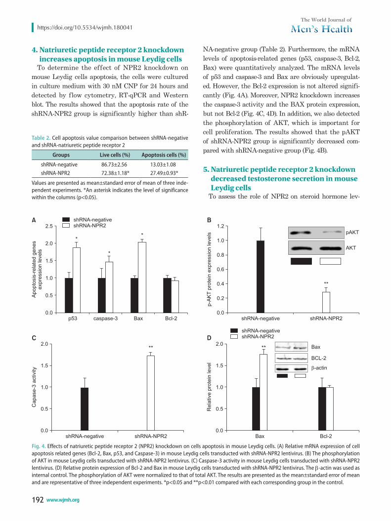
4. Natriuretic peptide receptor 2 knockdown increases apoptosis in mouse Leydig cells
To determine the effect of NPR2 knockdown on mouse Leydig cells apoptosis, the cells were cultured in culture medium with 30 nM CNP for 24 hours and detected by flow cytometry, RT-qPCR and Western blot. The results showed that the apoptosis rate of the shRNA-NPR2 group is significantly higher than shR-
NA-negative group (Table 2). Furthermore, the mRNA levels of apoptosis-related genes (p53, caspase-3, Bcl-2, Bax) were quantitatively analyzed. The mRNA levels of p53 and caspase-3 and Bax are obviously upregulat-ed. However, the Bcl-2 expression is not altered signifi-cantly (Fig. 4A). Moreover, NPR2 knockdown increases the caspase-3 activity and the BAX protein expression, but not Bcl-2 (Fig. 4C, 4D). In addition, we also detected the phosphorylation of AKT, which is important for cell proliferation. The results showed that the pAKT of shRNA-NPR2 group is significantly decreased com-pared with shRNA-negative group (Fig. 4B).
5. Natriuretic peptide receptor 2 knockdown decreased testosterone secretion in mouse Leydig cells
To assess the role of NPR2 on steroid hormone lev-
shRNA-negative
2.0
1.5
1.0
0.5
Capase-3
activity
0.0Bax
1.5
1.0
0.5
2.0
Rela
tive
pro
tein
level
0.0
A B
C D
p53
2.5
2.0
1.5
1.0
0.5Apopto
sis
-rela
ted
genes
expre
ssio
nle
vels
0.0caspase-3 Bax Bcl-2
1.2
1.0
0.8
0.6
0.4
0.2
p-A
KT
pro
tein
expre
ssio
nle
vels
0.0shRNA-negative shRNA-NPR2
shRNA-NPR2 Bcl-2
*
*
*
shRNA-negativeshRNA-NPR2
**
pAKT
AKT
** Bax
BCL-2
�-actin
shRNA-negativeshRNA-NPR2
**
Fig. 4. Effects of natriuretic peptide receptor 2 (NPR2) knockdown on cells apoptosis in mouse Leydig cells. (A) Relative mRNA expression of cell apoptosis related genes (Bcl-2, Bax, p53, and Caspase-3) in mouse Leydig cells transducted with shRNA-NPR2 lentivirus. (B) The phosphorylation of AKT in mouse Leydig cells transducted with shRNA-NPR2 lentivirus. (C) Caspase-3 activity in mouse Leydig cells transducted with shRNA-NPR2 lentivirus. (D) Relative protein expression of Bcl-2 and Bax in mouse Leydig cells transducted with shRNA-NPR2 lentivirus. The β-actin was used as internal control. The phosphorylation of AKT were normalized to that of total AKT. The results are presented as the mean±standard error of mean and are representative of three independent experiments. *p<0.05 and **p<0.01 compared with each corresponding group in the control.
Table 2. Cell apoptosis value comparison between shRNA-negative and shRNA-natriuretic peptide receptor 2
Groups Live cells (%) Apoptosis cells (%)
shRNA-negative 86.73±2.56 13.03±1.08 shRNA-NPR2 72.38±1.18* 27.49±0.93*
Values are presented as mean±standard error of mean of three inde-pendent experiments. *An asterisk indicates the level of significance within the columns (p<0.05).
Lei Yang, et al: CNP/NPR2 Promotes Testosterone Production
193www.wjmh.org
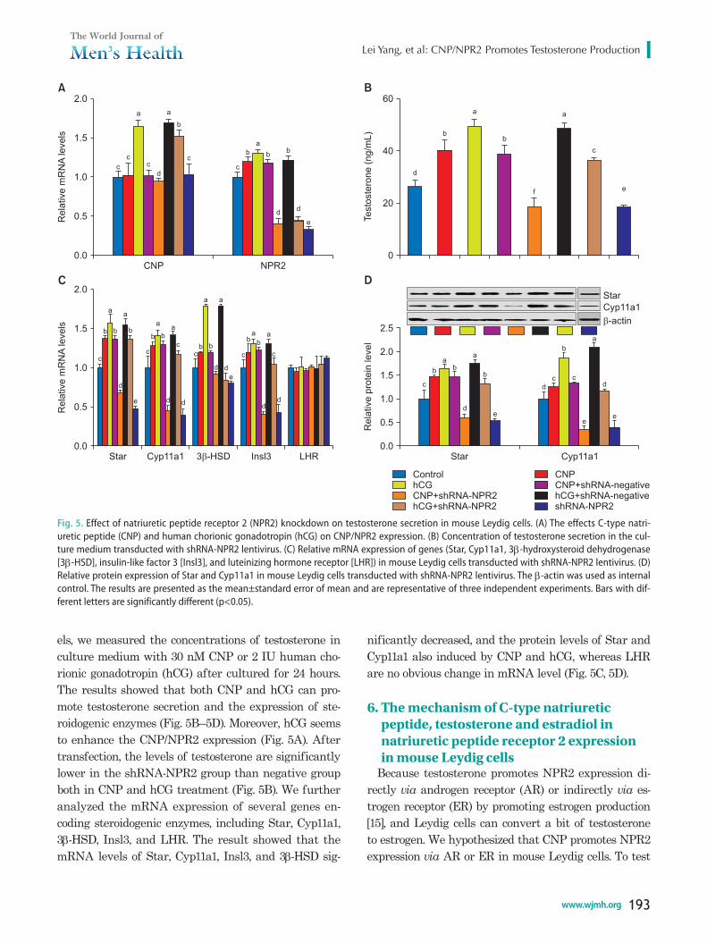
els, we measured the concentrations of testosterone in culture medium with 30 nM CNP or 2 IU human cho-rionic gonadotropin (hCG) after cultured for 24 hours. The results showed that both CNP and hCG can pro-mote testosterone secretion and the expression of ste-roidogenic enzymes (Fig. 5B–5D). Moreover, hCG seems to enhance the CNP/NPR2 expression (Fig. 5A). After transfection, the levels of testosterone are significantly lower in the shRNA-NPR2 group than negative group both in CNP and hCG treatment (Fig. 5B). We further analyzed the mRNA expression of several genes en-coding steroidogenic enzymes, including Star, Cyp11a1, 3β-HSD, Insl3, and LHR. The result showed that the mRNA levels of Star, Cyp11a1, Insl3, and 3β-HSD sig-
nificantly decreased, and the protein levels of Star and Cyp11a1 also induced by CNP and hCG, whereas LHR are no obvious change in mRNA level (Fig. 5C, 5D).
6. The mechanism of C-type natriuretic peptide, testosterone and estradiol in natriuretic peptide receptor 2 expression in mouse Leydig cells
Because testosterone promotes NPR2 expression di-rectly via androgen receptor (AR) or indirectly via es-trogen receptor (ER) by promoting estrogen production [15], and Leydig cells can convert a bit of testosterone to estrogen. We hypothesized that CNP promotes NPR2 expression via AR or ER in mouse Leydig cells. To test
Fig. 5. Effect of natriuretic peptide receptor 2 (NPR2) knockdown on testosterone secretion in mouse Leydig cells. (A) The effects C-type natri-uretic peptide (CNP) and human chorionic gonadotropin (hCG) on CNP/NPR2 expression. (B) Concentration of testosterone secretion in the cul-ture medium transducted with shRNA-NPR2 lentivirus. (C) Relative mRNA expression of genes (Star, Cyp11a1, 3β-hydroxysteroid dehydrogenase [3β-HSD], insulin-like factor 3 [Insl3], and luteinizing hormone receptor [LHR]) in mouse Leydig cells transducted with shRNA-NPR2 lentivirus. (D) Relative protein expression of Star and Cyp11a1 in mouse Leydig cells transducted with shRNA-NPR2 lentivirus. The β-actin was used as internal control. The results are presented as the mean±standard error of mean and are representative of three independent experiments. Bars with dif-ferent letters are significantly different (p<0.05).
CNP
2.0
1.5
1.0
0.5Rela
tive
mR
NA
levels
0.0NPR2
c
c
a
c
d
a
b
c
c
b
a
bb
dd
e
Star
2.0
1.5
1.0
0.5Rela
tive
mR
NA
levels
0.0Cyp11a1 3 -HSD� Insl3 LHR
c
b
a
b
a
b
d
e
c
b b
aa
c
d d
cb b
d d
e
a a
c
ba
ba
c
dd
A
C
60
40
20
Testo
ste
rone
(ng/m
L)
0
d
b
a
b
a
c
ef
2.5
2.0
1.5
1.0
0.5
Rela
tive
pro
tein
level
0.0Star Cyp11a1
Star
Cyp11a1
�-actin
a
b b
c
d
a
b
e
d
c
b
c
a
d
ee
ControlhCGCNP+shRNA-NPR2hCG+shRNA-NPR2
CNPCNP+shRNA-negativehCG+shRNA-shRNA-NPR2
negative
B
D
https://doi.org/10.5534/wjmh.180041
194 www.wjmh.org
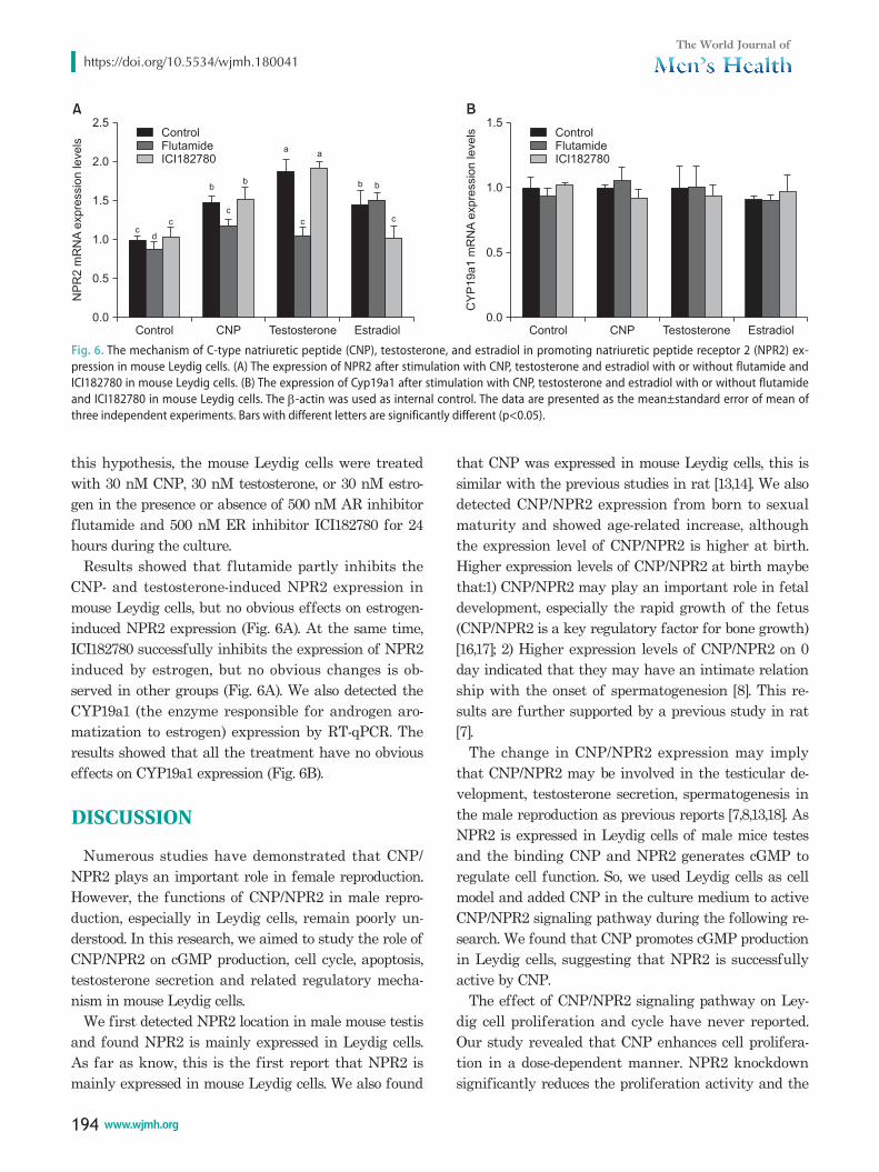
this hypothesis, the mouse Leydig cells were treated with 30 nM CNP, 30 nM testosterone, or 30 nM estro-gen in the presence or absence of 500 nM AR inhibitor flutamide and 500 nM ER inhibitor ICI182780 for 24 hours during the culture.
Results showed that flutamide partly inhibits the CNP- and testosterone-induced NPR2 expression in mouse Leydig cells, but no obvious effects on estrogen-induced NPR2 expression (Fig. 6A). At the same time, ICI182780 successfully inhibits the expression of NPR2 induced by estrogen, but no obvious changes is ob-served in other groups (Fig. 6A). We also detected the CYP19a1 (the enzyme responsible for androgen aro-matization to estrogen) expression by RT-qPCR. The results showed that all the treatment have no obvious effects on CYP19a1 expression (Fig. 6B).
DISCUSSION
Numerous studies have demonstrated that CNP/NPR2 plays an important role in female reproduction. However, the functions of CNP/NPR2 in male repro-duction, especially in Leydig cells, remain poorly un-derstood. In this research, we aimed to study the role of CNP/NPR2 on cGMP production, cell cycle, apoptosis, testosterone secretion and related regulatory mecha-nism in mouse Leydig cells.
We first detected NPR2 location in male mouse testis and found NPR2 is mainly expressed in Leydig cells. As far as know, this is the first report that NPR2 is mainly expressed in mouse Leydig cells. We also found
that CNP was expressed in mouse Leydig cells, this is similar with the previous studies in rat [13,14]. We also detected CNP/NPR2 expression from born to sexual maturity and showed age-related increase, although the expression level of CNP/NPR2 is higher at birth. Higher expression levels of CNP/NPR2 at birth maybe that:1) CNP/NPR2 may play an important role in fetal development, especially the rapid growth of the fetus (CNP/NPR2 is a key regulatory factor for bone growth) [16,17]; 2) Higher expression levels of CNP/NPR2 on 0 day indicated that they may have an intimate relation ship with the onset of spermatogenesion [8]. This re-sults are further supported by a previous study in rat [7].
The change in CNP/NPR2 expression may imply that CNP/NPR2 may be involved in the testicular de-velopment, testosterone secretion, spermatogenesis in the male reproduction as previous reports [7,8,13,18]. As NPR2 is expressed in Leydig cells of male mice testes and the binding CNP and NPR2 generates cGMP to regulate cell function. So, we used Leydig cells as cell model and added CNP in the culture medium to active CNP/NPR2 signaling pathway during the following re-search. We found that CNP promotes cGMP production in Leydig cells, suggesting that NPR2 is successfully active by CNP.
The effect of CNP/NPR2 signaling pathway on Ley-dig cell proliferation and cycle have never reported. Our study revealed that CNP enhances cell prolifera-tion in a dose-dependent manner. NPR2 knockdown significantly reduces the proliferation activity and the
Control
2.5
2.0
1.5
1.0
0.5
NP
R2
mR
NA
expre
ssio
nle
vels
0.0CNP Testosterone Estradiol
cd
c
b
c
b
a
c
a
b b
c
ControlFlutamideICI182780
A
Control
1.5
1.0
0.5
CY
P19a1
mR
NA
expre
ssio
nle
vels
0.0CNP Testosterone Estradiol
ControlFlutamideICI182780
B
Fig. 6. The mechanism of C-type natriuretic peptide (CNP), testosterone, and estradiol in promoting natriuretic peptide receptor 2 (NPR2) ex-pression in mouse Leydig cells. (A) The expression of NPR2 after stimulation with CNP, testosterone and estradiol with or without flutamide and ICI182780 in mouse Leydig cells. (B) The expression of Cyp19a1 after stimulation with CNP, testosterone and estradiol with or without flutamide and ICI182780 in mouse Leydig cells. The β-actin was used as internal control. The data are presented as the mean±standard error of mean of three independent experiments. Bars with different letters are significantly different (p<0.05).
Lei Yang, et al: CNP/NPR2 Promotes Testosterone Production
195www.wjmh.org
cells are arrested at the S phase. This may be because CNP/NPR2 is related with expression of cell cycle fac-tors (cyclin A1, cyclin B1, and cyclin D2). Cyclin A1 and cyclin D2 are key regulators associated with the S-to-G2/M transition. Moreover, cyclin B1 is an important gene related to regulating the normal cell-cycle pro-gression and plays a vital role in mitotic exit and the beginning of the subsequent cell division. Thus, the cell-cycle results imply that CNP/NPR2 is involved in the growth and proliferation in Leydig cells. In addi-tion, some studies report that CNP is a growth inhibi-tor in smooth muscle cells [19,20], and some studies reported that CNP promotes the growth of the bone [17]. This difference may be related to cell types or the indirect effects of CNP, such as testosterone synthesis [21] and insulin-like growth factor I production [16].
Previous study showed that NPR2 gene knockout mice have smaller size [16] and smaller testis [8] com-pared with the normal mice. However, whether CNP/NPR2 regulates mouse Leydig cells viability and apoptosis remains unclear. In the current study, we found that CNP effectively promotes the viability of mouse Leydig cells. At the same time, NPR2 depletion remarkably induces apoptosis in mouse Leydig cells. To clarify this result further, we measured the expres-sion of key molecules associated with apoptosis via RT-qPCR and Western blot. As an important transcription factor, p53 can promote cell apoptosis, and elevated p53 mRNA levels result in higher apoptosis rates in Leydig cells [22]. Caspase-3, acts as an executioner in caspase-mediated apoptosis, is extensively involved various cell apoptosis. Moreover, in the Bcl-2 family, Bcl-2 plays an anti-apoptotic role, whereas Bax has a proapoptotic effect [23,24]. In the present study, the expression of Bax, p53 and caspase-3 was significantly upregulated after NPR2 disruption, whereas the expression Bax levels appeared unchanged. Based on these results, we concluded that the apoptosis cells in mouse Leydig cells caused by disrupting NPR2 is closely correlated with these molecules. In addition, PI3K-AKT pathway plays an key role in cell proliferation and survival [25] and NPR2 knockdown inhibits the activation of AKT in in mouse Leydig cells. This also showed that NPR2 regu-lates cells viability and apoptosis in another perspec-tive.
Given the importance of Leydig cells in secretion of testosterone, we evaluated the CNP/NPR2 signaling pathway on testosterone synthesis in mouse Leydig
cells. Our results indicated that CNP promotes testos-terone secretion in mouse Leydig cells as a previous report [21] and NPR2 silencing inhibits testosterone production. The possible reason for the reductions of testosterone could be the decreases in the Star, Cyp11a1 and 3β-HSD, which is essential for testosterone biosyn-thesis [4,26]. Moreover, the downregulation of cGMP may also inhibits testosterone production [27]. How-ever, the expression levels of LHR was no obvious after NPR2 knockdown in Leydig cells. In addition, we also found that activated LH signal by hCG induces the CNP/NPR2 expression and NPR2 disruption partly reduces the hCG-induced testosterone secretion. This result suggested that CNP/NPR2 signal maybe related with LHR signal during the testosterone secretion. Thus, we inferred that CNP/NPR2 may play an im-portant role in the development of male reproductive tissues as well as promoting secondary sexual charac-teristics, such as increased muscle and bone mass by promoting testosterone production.
We also found that CNP promotes NPR2 expression in mouse Leydig cells. And previous studies report that testosterone enhances NPR2 expression in cumulus cell [15,28]. So, we hypothesized that CNP-induced NPR2 expression is by promoting testosterone production, which further enhances NPR2 expression via AR in mouse Leydig cells. Consequently, we inhibited AR by flutamide during the culture with CNP and tes-tosterone. The result that inhibited AR by flutamide partially improves NPR2 expression in Leydig cells. We therefore speculated that testosterone is an impor-tant factor in regulating CNP/NPR2 in Leydig cells as previous report in ovary [15]. As Leydig cells can con-vert a bit of testosterone to estrogen [29] and estrogen promotes NPR2 expression via ER [28,30], we detected the role of ER on CNP- or testosterone-induced NPR2 expression. We found inhibited ER by ICI182780 has no obvious effects on NPR2 expression in mouse Leydig cells and this result is further supported by the result of CYP19a1 mRNA expression. These results indicated that CNP promotes testosterone secretion, which fur-ther induces NPR2 expression via AR in mouse Leydig cells.
CONCLUSIONS
Our study highlights the important role of CNP/NPR2 in mouse testes. We found that CNP/NPR2 is re-
https://doi.org/10.5534/wjmh.180041
196 www.wjmh.org
lated with cell proliferation and testosterone secretion in mouse Leydig cells. In addition, CNP can promote NPR2 expression via AR in mouse Leydig cells. Our findings not only provided new insights into the func-tion of CNP/NPR2 in female reproduction but also pro-vide potential therapeutic targets for treating reduced testosterone.
ACKNOWLEDGEMENTS
This research study was funded by the National Natural Science Foundation of China (No. 81660535), Project of Science and Technology of Jiangxi Provin-cial Education Department (GJJ170966), and Doctoral Research Start-Up Foundation of Jiujiang University (No. 8879522).
Disclosure
The authors have no potential conflicts of interest to disclose.
Author Contribution
Conceptualization: Yang L. Data curation: Huang S. Formal analysis: Yang L. Funding acquisition: Yang L, Huang S. Inves-tigation: Lei L, Zhao Q. Methodology: Yang L, Gong Y, Guan G. Project administration: Huang S. Resources: Huang S. Software: Lei L, Guan G. Supervision: Gong Y. Validation: Yang L. Visual-ization: Yang L, Zhao Q. Writing–original draft: Lei L. Writing–review & editing: Yang L.
REFERENCES
1. Ito A, Shirakawa H, Takumi N, Minegishi Y, Ohashi A, How-lader ZH, et al. Menaquinone-4 enhances testosterone pro-duction in rats and testis-derived tumor cells. Lipids Health Dis 2011;10:158.
2. Huhtaniemi IT. LH and FSH receptor mutations and their ef-fects on puberty. Horm Res 2002;57 Suppl 2:35-8.
3. Ferlin A, Bogatcheva NV, Gianesello L, Pepe A, Vinanzi C, Agoulnik AI, et al. Insulin-like factor 3 gene mutations in testicular dysgenesis syndrome: clinical and functional char-acterization. Mol Hum Reprod 2006;12:401-6.
4. Tremblay JJ. Molecular regulation of steroidogenesis in endo-crine Leydig cells. Steroids 2015;103:3-10.
5. Zhang M, Su YQ, Sugiura K, Xia G, Eppig JJ. Granulosa cell ligand NPPC and its receptor NPR2 maintain meiotic arrest in mouse oocytes. Science 2010;330:366-9.
6. Yang L, Wei Q, Li W, Ge J, Zhao X, Ma B. C-type natriuretic peptide improved vitrified-warmed mouse cumulus oocyte complexes developmental competence. Cryobiology 2016;72: 161-4.
7. Huang DH, Zhang SW, Zhao H, Zhang L. The role of C-type natriuretic peptide in rat testes during spermatogenesis. Asian J Androl 2011;13:275-80.
8. Sogawa C, Fujiwara Y, Tsukamoto S, Ishida Y, Yoshii Y, Furu-kawa T, et al. Mutant phenotype analysis suggests potential roles for C-type natriuretic peptide receptor (NPR-B) in male mouse fertility. Reprod Biol Endocrin 2014;12:64.
9. Kong N, Xu X, Zhang Y, Wang Y, Hao X, Zhao Y, et al. Natri-uretic peptide type C induces sperm attraction for fertiliza-tion in mouse. Sci Rep 2017;7:39711.
10. Xia H, Chen Y, Wu KJ, Zhao H, Xiong CL, Huang DH. Role of C-type natriuretic peptide in the function of normal hu-man sperm. Asian J Androl 2016;18:80-4.
11. Yang L, Wei Q, Li W, Xi Q, Zhao X, Ma B. NPR2 is involved in FSH-mediated mouse oocyte meiotic resumption. J Ovarian Res 2016;9:6.
12. Chen H, Gao L, Xiong Y, Yang D, Li C, Wang A, et al. Circa-dian clock and steroidogenic-related gene expression profiles in mouse Leydig cells following dexamethasone stimulation. Biochem Bioph Res Co 2017;483:294-300.
13. Xia W, Mruk DD, Cheng CY. C-type natriuretic peptide regu-lates blood-testis barrier dynamics in adult rat testes. Proc Natl Acad Sci USA 2007;104:3841-6.
14. Collin O, Lissbrant E, Bergh A. Atrial natriuretic peptide, brain natriuretic peptide and c-type natriuretic peptide: ef-fects on testicular microcirculation and immunohistochemi-cal localization. Int J Androl 1997;20:55-60.
15. Wang X, Wang H, Liu W, Zhang Z, Zhang Y, Zhang W, et al. High level of C-type natriuretic peptide induced by hyperan-drogen-mediated anovulation in polycystic ovary syndrome mice. Clin Sci (Lond) 2018;132:759-76.
16. Tsuji T, Kunieda T. A loss-of-function mutation in natriuretic peptide receptor 2 (Npr2) gene is responsible for dispropor-tionate dwarfism in cn/cn mouse. J Biol Chem 2005;280: 14288-92.
17. Mericq V, Uyeda JA, Barnes KM, De LF, Baron J. Regulation of fetal rat bone growth by C-type natriuretic peptide and cGMP. Pediatr Res 2000;47:189-93.
18. Karan S, Frederick JW, Baehr W. Novel functions of photore-ceptor guanylate cyclases revealed by targeted deletion. Mol Cell Biochem 2010;334:141-55.
19. Porter JG, Catalano R, Mcenroe G, Lewicki JA, Protter AA. C-type natriuretic peptide inhibits growth factor-dependent DNA synthesis in smooth muscle cells. Am J Physiol 1992;
Lei Yang, et al: CNP/NPR2 Promotes Testosterone Production
197www.wjmh.org
263:C1001-6.20. Furuya M, Yoshida M, Hayashi Y, Ohnuma N, Minamino N,
Kangawa K, et al. C-type natriuretic peptide is a growth in-hibitor of rat vascular smooth muscle cells. Biochem Biophys Res Commun 1991;177:927-31.
21. Khurana ML, Pandey KN. Receptor-mediated stimulatory ef-fect of atrial natriuretic factor, brain natriuretic peptide, and C-type natriuretic peptide on testosterone production in pu-rified mouse Leydig cells: activation of cholesterol side-chain cleavage enzyme. Endocrinology 1993;133:2141-9.
22. Fouchécourt S, Livera G, Messiaen S, Fumel B, Parent AS, Marine JC, et al. Apoptosis of sertoli cells after conditional ablation of murine double minute 2 (Mdm2) gene is p53-de-pendent and results in male sterility. Cell Death Differ 2016; 23:521-30.
23. Zhao F, Wang N, Yi Y, Lin P, Tang K, Wang A, et al. Knock-down of CREB3/Luman by shRNA in mouse granulosa cells results in decreased estradiol and progesterone synthesis and promotes cell proliferation. Plos One 2016;11:e0168246.
24. Sifer C, Bénifla JL, Bringuier AF, Porcher R, Blanc-Layrac G, Madélénat P, et al. Could induced apoptosis of human granu-losa cells predict in vitro fertilization-embryo transfer out-come? A preliminary study of 25 women. Eur J Obstet Gyn R
B 2002;103:150-3.25. Datta SR, Dudek H, Tao X, Masters S, Fu H, Gotoh Y, et al.
Akt phosphorylation of bad couples survival signals to the cell-intrinsic death machinery. Cell 1997;91:231-41.
26. Hatano M, Migita T, Ohishi T, Shima Y, Ogawa Y, Morohashi KI, et al. SF-1 deficiency causes lipid accumulation in Leydig cells via suppression of STAR and CYP11A1. Endocrine 2016; 54:484-96.
27. Moon HW, Park JW, Lee KW, Jeong HC, Choi JB, Choi SW, et al. Administration of Goji (lycium Chinense mill.) Extracts improves erectile function in old aged rat model. World J Mens Health 2016;35:43-50.
28. Zhang M, Su YQ, Sugiura K, Wigglesworth K, Xia G, Eppig JJ. Estradiol promotes and maintains cumulus cell expression of natriuretic peptide receptor 2 (npr2) and meiotic arrest in mouse oocytes in vitro. Endocrinology 2011;152:4377-85.
29. Abney TO. The potential roles of estrogens in regulating Ley-dig cell development and function: a review. Steroids 1999; 64:610-7.
30. Zhang J, Wei Q, Cai J, Zhao X, Ma B. Effect of C-type natri-uretic peptide on maturation and developmental competence of goat oocytes matured in vitro. Plos One 2015;10:e0132318.
https://doi.org/10.5534/wjmh.180041
198 www.wjmh.org
Appendix. Primer sequences used for RT-qPCR
Gene Forward primer sequences (5'→3’) Reverse primer sequences (5'→3’)
CNP GGTCTGGGATGTTAGTACAGCTA TAAAAGCCACATTGCGTTGGANPR2 GCTGACCCGGCAAGTTCTGT ACAATACTCGGTGACAATGCAGATCyclin A1 GCCTTCACCATTCATGTGGAT TTGCTGCGGGTAAAGAGACAGCyclin B1 AAGGTGCCTGTGTGTGAACC GTCAGCCCCATCATCTGCGCyclin D2 TTACCTGGACCGCTTCTTG GAGGCTTGATGGAGTTGTCGBcl2 GGTCATGTGTGTGGAGAGCGTCA CTTCAGAGACAGCCAGGAGAAATBax AGGATGCGTCCACCAAGAA CAAAGTAGAAGAGGGCAACCACCaspase-3 TGACTGGAAAGCCGAAACTC GCAAGCCATCTCCTCATCAGp53 TACAAGAAGTCACAGCACAT GATAGGTCGGCGGTTCATStar CCCACCGCATGGTCGAGTAGTG CGCCTCTGCAGCCAACTCATGCyp11a1 ACACTGAGACTCCACCCCAT CTCGACCCATGGCAAAGCTAInsl3 TGCAGTGGCTAGAGCAGAGA GTGCAGCCAGTAAGACAGCACyp19a1 GCACAGTCACTACATCTCCCGA CACACAAACTTCCACCATTCGALHR AGAGTGATTCCCTGGAAAGGA CTTACGAAAGGTTCCCTACT3β-HSD TGGACAAAGTATTCCGACCAGA GGCACACTTGCTTGAACACAGβ-actin GCAAGCAGGAGTACGATGAG CCATGCCAATGTTGTCTCTT
The β-actin was used as internal control.