Embed Size (px)
Citation preview
1
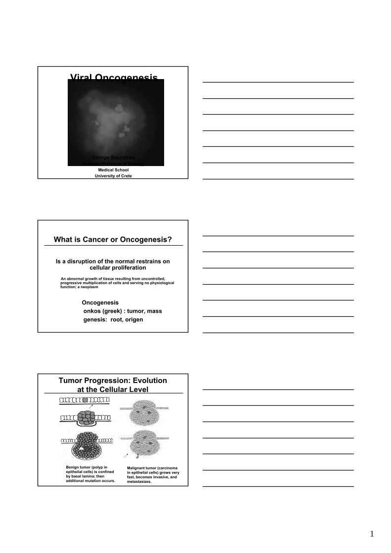
Viral Oncogenesis
George SourvinosAssistant Professor of Virology
Medical SchoolUniversity of Crete
What is Cancer or Oncogenesis?
Is a disruption of the normal restrains on cellular proliferation
An abnormal growth of tissue resulting from uncontrolled, progressive multiplication of cells and serving no physiologicalfunction; a neoplasm
Oncogenesisonkos (greek) : tumor, massgenesis: root, origen
Tumor Progression: Evolutionat the Cellular Level
Benign tumor (polyp in epithelial cells) is confined by basal lamina; then additional mutation occurs.
Malignant tumor (carcinoma in epithelial cells) grows very fast, becomes invasive, and metastasizes.
2
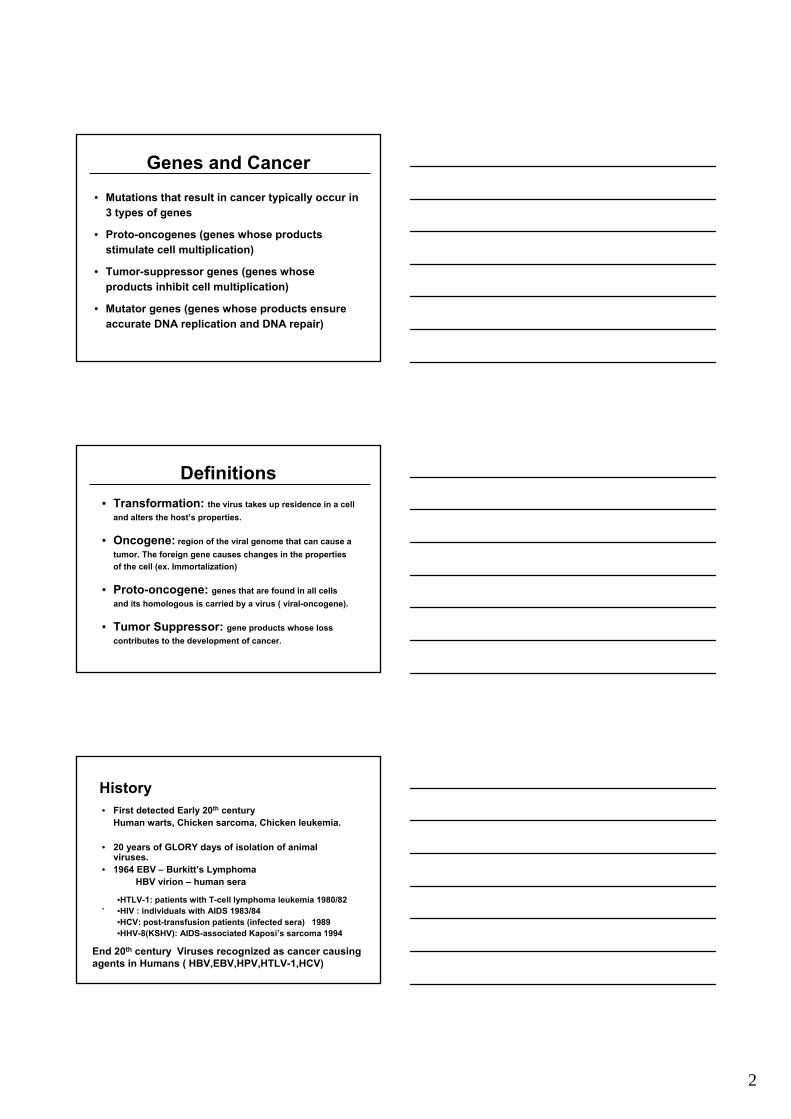
Genes and Cancer• Mutations that result in cancer typically occur in
3 types of genes
• Proto-oncogenes (genes whose products stimulate cell multiplication)
• Tumor-suppressor genes (genes whose products inhibit cell multiplication)
• Mutator genes (genes whose products ensure accurate DNA replication and DNA repair)
Definitions• Transformation: the virus takes up residence in a cell
and alters the host’s properties.
• Oncogene: region of the viral genome that can cause a tumor. The foreign gene causes changes in the properties of the cell (ex. Immortalization)
• Proto-oncogene: genes that are found in all cells and its homologous is carried by a virus ( viral-oncogene).
• Tumor Suppressor: gene products whose loss contributes to the development of cancer.
History• First detected Early 20th century
Human warts, Chicken sarcoma, Chicken leukemia.
• 20 years of GLORY days of isolation of animal viruses.
• 1964 EBV – Burkitt’s Lymphoma HBV virion – human sera
.
End 20th century Viruses recognized as cancer causingagents in Humans ( HBV,EBV,HPV,HTLV-1,HCV)
•HTLV-1: patients with T-cell lymphoma leukemia 1980/82•HIV : individuals with AIDS 1983/84•HCV: post-transfusion patients (infected sera) 1989•HHV-8(KSHV): AIDS-associated Kaposi’s sarcoma 1994
3
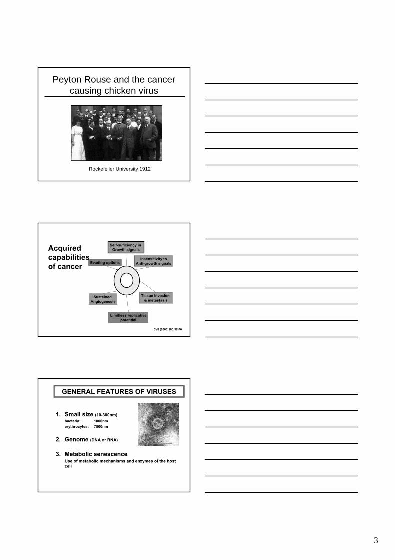
Peyton Rouse and the cancer causing chicken virus
Rockefeller University 1912
Self-suficiency in Growth signals
Insensitivity toAnti-growth signalsEvading options
Tissue invasion & metastasis
Sustained Angiogenesis
Limitless replicativepotential
Acquiredcapabilitiesof cancer
Cell (2000)100:57-70
GENERAL FEATURES OF VIRUSES
1. Small size (10-300nm)bacteria: 1000nmerythrocytes: 7500nm
2. Genome (DNA or RNA)
3. Metabolic senescenceUse of metabolic mechanisms and enzymes of the host cell
4
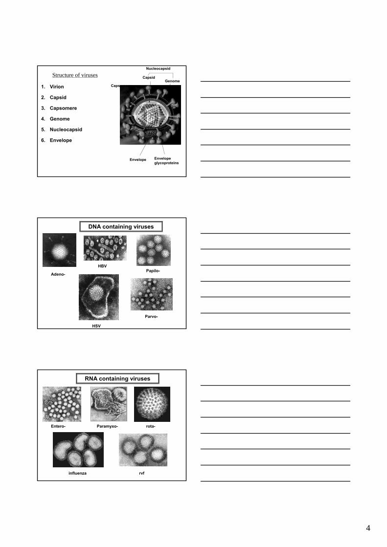
Structure of virusesStructure of viruses
1. Virion
2. Capsid
3. Capsomere
4. Genome
5. Nucleocapsid
6. Envelope
GenomeCapsid
Nucleocapsid
Envelope
Capsomere
Envelopeglycoproteins
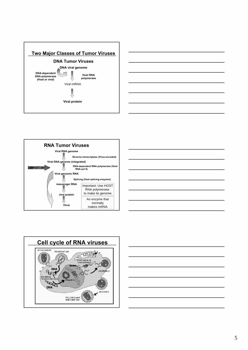
DNA containing viruses
Αdeno-Papilo-
Parvo-
HSV
HBV
RNA containing viruses
Entero- rota-
rvfinfluenza
Paramyxo-
5
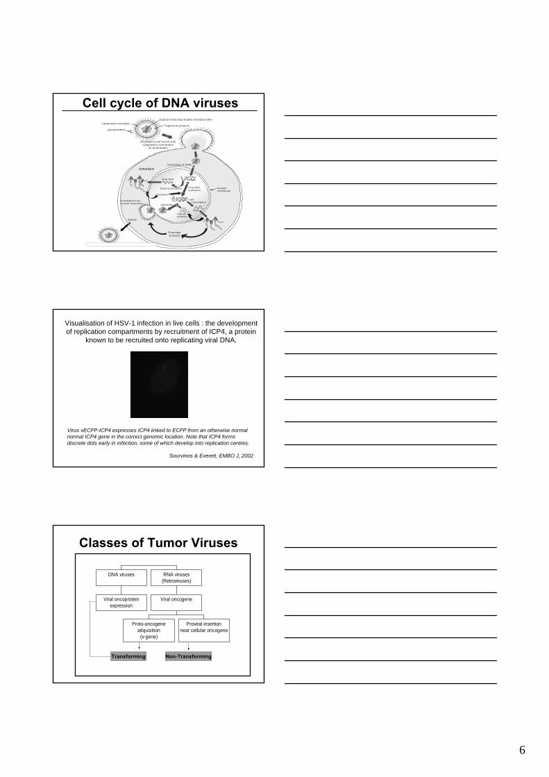
Two Major Classes of Tumor VirusesDNA Tumor Viruses
DNA viral genome
Host RNA polymerase
Viral mRNA
Viral protein
DNA-dependentDNA polymerase
(Host or viral)
RNA Tumor VirusesViral RNA genome
Reverse transcriptase (Virus-encoded)
Viral DNA genome (integrated)DNA-dependent RNA polymerase (Host Host
RNA pol II)
Viral genomic RNA
Splicing (Host splicing enzymes)
messenger RNA
viral protein
Virus
Important: Use HOSTRNA polymerase
to make its genome
An enzyme that normally
makes mRNA
IMPORTANT
Cell cycle of RNA viruses
6
1
Lipoprote in enve lope
glycop rote ins
C apsid enc losing doub le-stranded D N A
Tegum ent p ro te ins
2
Adsorption and fus ion w ith cytop lasm ic m em brane
for penetration
U ncoating of D N ACyto plasm
C apsid proteins
vira l DN A
Vira l DN A rep lica tio n
Early tra nscr ip tio n
vira l RN A
Late transcr iption
enzym es
Enve lopeprote ins
N uc lear m em brane
Assem bly
3 4
5Enve lopm ent b ynuc lear m em brane
egress6
R N A
Fi 1 2 R li ti f H SV 1 d i l ti i f ti
Cell cycle of DNA viruses
Visualisation of HSV-1 infection in live cells : the development of replication compartments by recruitment of ICP4, a protein
known to be recruited onto replicating viral DNA.
Virus vECFP-ICP4 expresses ICP4 linked to ECFP from an otherwise normal normal ICP4 gene in the correct genomic location. Note that ICP4 forms discrete dots early in infection, some of which develop into replication centres.
Sourvinos & Everett, EMBO J, 2002
Classes of Tumor Viruses
Viral oncoproteinexpression
DNA viruses
Proto-oncogeneadquisition
(v-gene)
Proviral insertionnear cellular oncogene
Viral oncogene
RNA viruses(Retroviruses)
Transforming Non-Transforming
7
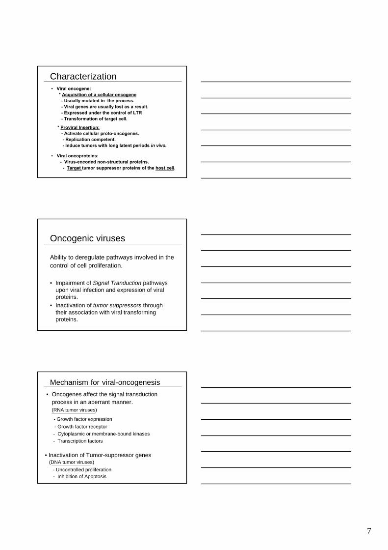
Characterization
• Viral oncoproteins: - Virus-encoded non-structural proteins.- Target tumor suppressor proteins of the host cell.
• Viral oncogene:* Acquisition of a cellular oncogene- Usually mutated in the process.- Viral genes are usually lost as a result.- Expressed under the control of LTR - Transformation of target cell.
* Proviral Insertion:- Activate cellular proto-oncogenes.- Replication competent.- Induce tumors with long latent periods in vivo.
Oncogenic viruses
• Impairment of Signal Tranduction pathways upon viral infection and expression of viral proteins.
• Inactivation of tumor suppressors through their association with viral transforming proteins.
Ability to deregulate pathways involved in thecontrol of cell proliferation.
Mechanism for viral-oncogenesis• Oncogenes affect the signal transduction
process in an aberrant manner.(RNA tumor viruses)
- Growth factor expression- Growth factor receptor- Cytoplasmic or membrane-bound kinases- Transcription factors
• Inactivation of Tumor-suppressor genes(DNA tumor viruses)
- Uncontrolled proliferation- Inhibition of Apoptosis
8
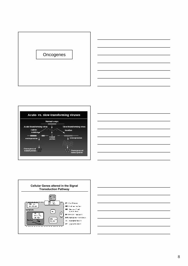
Oncogenes
Cellular Genes altered in the Signal Transduction Pathway
9
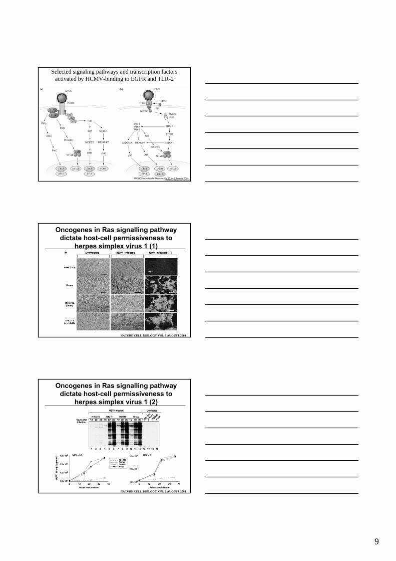
Selected signaling pathways and transcription factorsactivated by HCMV-binding to EGFR and TLR-2
TRENDS in Molecular Medicine Vol.10 No.1 January 2004
Oncogenes in Ras signalling pathwaydictate host-cell permissiveness to
herpes simplex virus 1 (1)
NATURE CELL BIOLOGY VOL 3 AUGUST 2001
Oncogenes in Ras signalling pathwaydictate host-cell permissiveness to
herpes simplex virus 1 (2)
NATURE CELL BIOLOGY VOL 3 AUGUST 2001
10
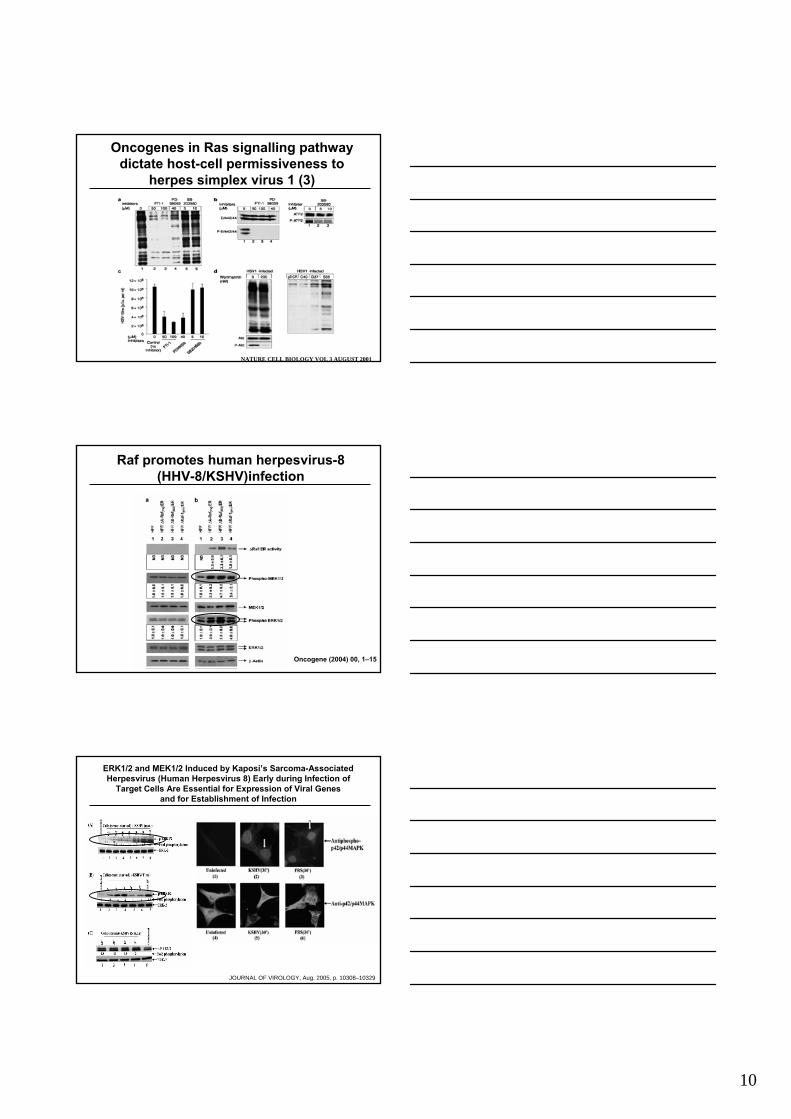
Oncogenes in Ras signalling pathwaydictate host-cell permissiveness to
herpes simplex virus 1 (3)
NATURE CELL BIOLOGY VOL 3 AUGUST 2001
Raf promotes human herpesvirus-8 (HHV-8/KSHV)infection
Oncogene (2004) 00, 1–15
ERK1/2 and MEK1/2 Induced by Kaposi’s Sarcoma-AssociatedHerpesvirus (Human Herpesvirus 8) Early during Infection of
Target Cells Are Essential for Expression of Viral Genesand for Establishment of Infection
JOURNAL OF VIROLOGY, Aug. 2005, p. 10308–10329
11
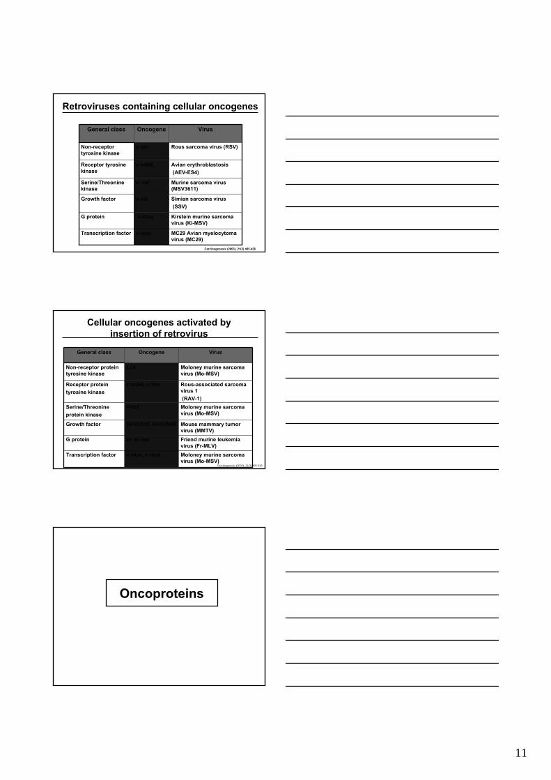
Retroviruses containing cellular oncogenes
MC29 Avian myelocytomavirus (MC29)
v-mycTranscription factor
Kirstein murine sarcoma virus (Ki-MSV)
v-KrasG protein
Simian sarcoma virus(SSV)
v-sisGrowth factor
Murine sarcoma virus (MSV3611)
v- rafSerine/Threoninekinase
Avian erythroblastosis(AEV-ES4)
v-erbB, Receptor tyrosine kinase
Rous sarcoma virus (RSV)v-srcNon-receptor tyrosine kinase
VirusOncogeneGeneral class
Carcinogenesis (2003), 21(3) 405-426
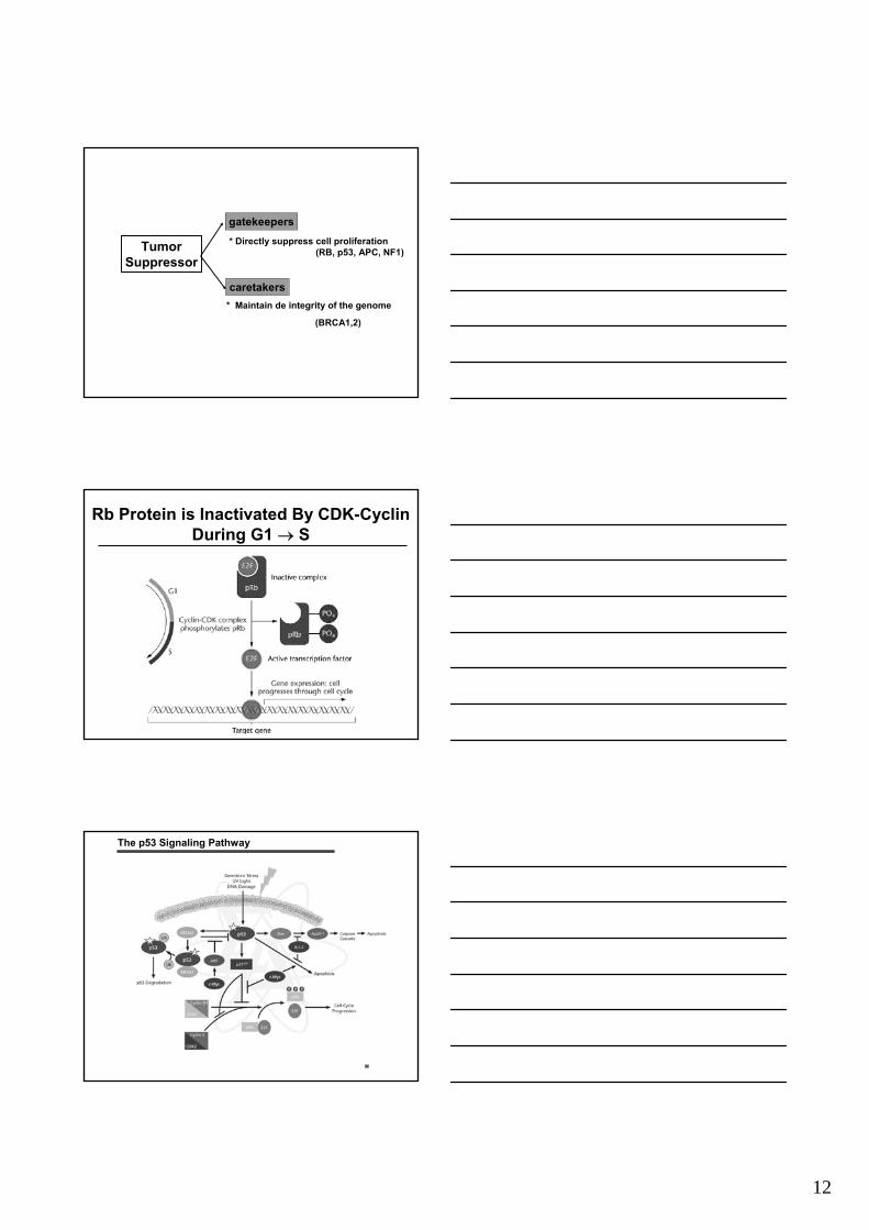
Cellular oncogenes activated by insertion of retrovirus
Moloney murine sarcoma virus (Mo-MSV)
c-myc, c-mybTranscription factor
Friend murine leukemia virus (Fr-MLV)
ci- Ki-rasG protein
Mouse mammary tumor virus (MMTV)
Wnt1/IntI, Wnt3/Int4Growth factor
Moloney murine sarcoma virus (Mo-MSV)
Pim1Serine/Threonineprotein kinase
Rous-associated sarcoma virus 1 (RAV-1)
c-erbB, c-fmsReceptor proteintyrosine kinase
Moloney murine sarcoma virus (Mo-MSV)
LckNon-receptor protein tyrosine kinase
VirusOncogeneGeneral class
Carcinogenesis (2003), 21(3) 405-426
Oncoproteins
12
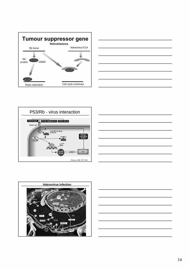
TumorSuppressor
gatekeepers
caretakers
* Directly suppress cell proliferation
* Maintain de integrity of the genome
(RB, p53, APC, NF1)
(BRCA1,2)
Rb Protein is Inactivated By CDK-Cyclin During G1 → S
The p53 Signaling Pathway
13
DNA virus oncoproteins andcellular protein interaction
TRAFsLMP1EBV
pRbP53
E1AE1B-55K
Adenovirus
p53pRb
E6E7
HPV
p53,pRbPP2A
Large T agSmall T ag
SV40
cellular proteinoncoproteinVirus
Functional Domains of p53
MDM2
EMBO J (99)18:1661
http://www.novocastra.co.uk/oapdgs.htm
HPV E6Ad E1b p55
Tumor suppressor genesp53
P53 gene P53 gene P53 gene
P53
P53 DNA
Stops replication
Hepatitis C
P53
replication replication
Papillomaproteolysis
P53
HPV
14
Tumour suppressor geneRb Gene
RbRbprotein
Rb
Stops replication
Rb
Adenovirus E1A
Cell cycle continues
Retinoblastoma
105kD
P53/Rb - virus interaction
(Nature, 408, 307-310)
Adenovirus infection
15
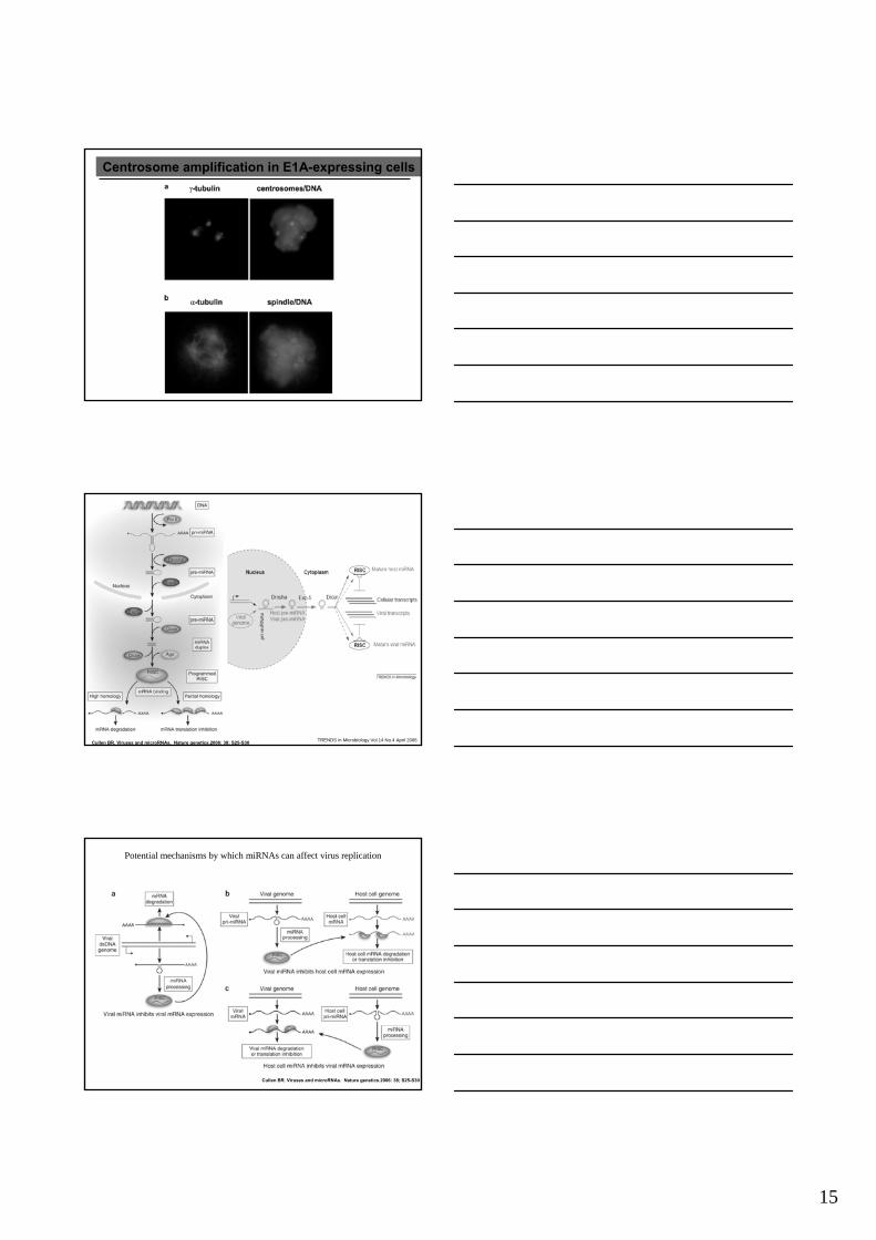
Centrosome amplification in E1A-expressing cells
Cullen BR. Viruses and microRNAs. Nature genetics.2006: 38; S25-S30 TRENDS in Microbiology Vol.14 No.4 April 2006
Potential mechanisms by which miRNAs can affect virus replication
Cullen BR. Viruses and microRNAs. Nature genetics.2006: 38; S25-S30
16
Cullen BR. Viruses and microRNAs. Nature genetics.2006: 38; S25-S30TRENDS in Microbiology Vol.14 No.4 April 2006
HCMV miRNAs
MicroRNAs can function as tumour suppressors and oncogenes
Esquela-Kerscher A and Slack FJ Nature 2006: 6; 259-269
The future: Silencing MicroRNAs with Antagomirs
NEJM 2006:354;1194-1195. Nature 2006: 38; S14- S19
17
HPV• DNA Tumor virus
– Over 100 HPV types• infect different areas of
skin• 38 genital HPV types• do not circulate in blood
• Not easy to grow– Test for HPV DNA in patient
samples
HPV Genome•E6 & E7 genes
–code for proteins that inactivate human tumor suppressor proteins
•L1 gene–codes for a protein that self-assembles into the shell (capsid) of the virus–empty shells (capsids) are called virus–like particles(VLPs)
HPV life cycle
18
HPV L1 Virus-Like Particle(protein from the L1 gene of HPV)
HPV
Papilloma Viruses
Squamous cell carcinoma:LarynxEsophagus All histologically similar Lung
urogenital cancer
wart Malignant Squamous cell carcinoma
10% of human cancers may be HPV-linked
Model of the roles of UV irradiation and HPV infectionin skin cancer development
J Pathol 2006; 208: 165–175
19
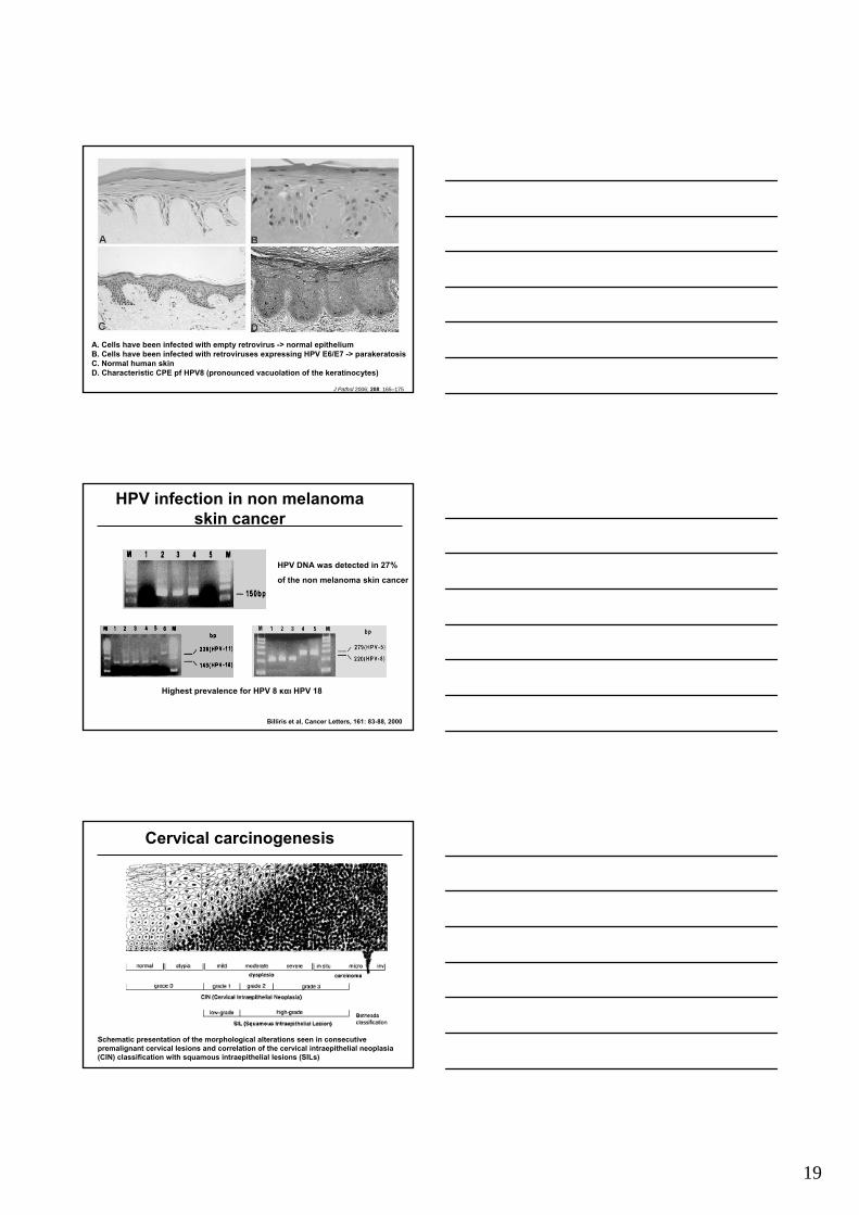
A. Cells have been infected with empty retrovirus -> normal epitheliumB. Cells have been infected with retroviruses expressing HPV E6/E7 -> parakeratosisC. Normal human skinD. Characteristic CPE pf HPV8 (pronounced vacuolation of the keratinocytes)
J Pathol 2006; 208: 165–175
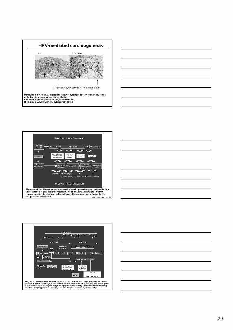
HPV infection in non melanomaskin cancer
Billiris et al, Cancer Letters, 161: 83-88, 2000
HPV DNA was detected in 27%
of the non melanoma skin cancer
Highest prevalence for HPV 8 και HPV 18
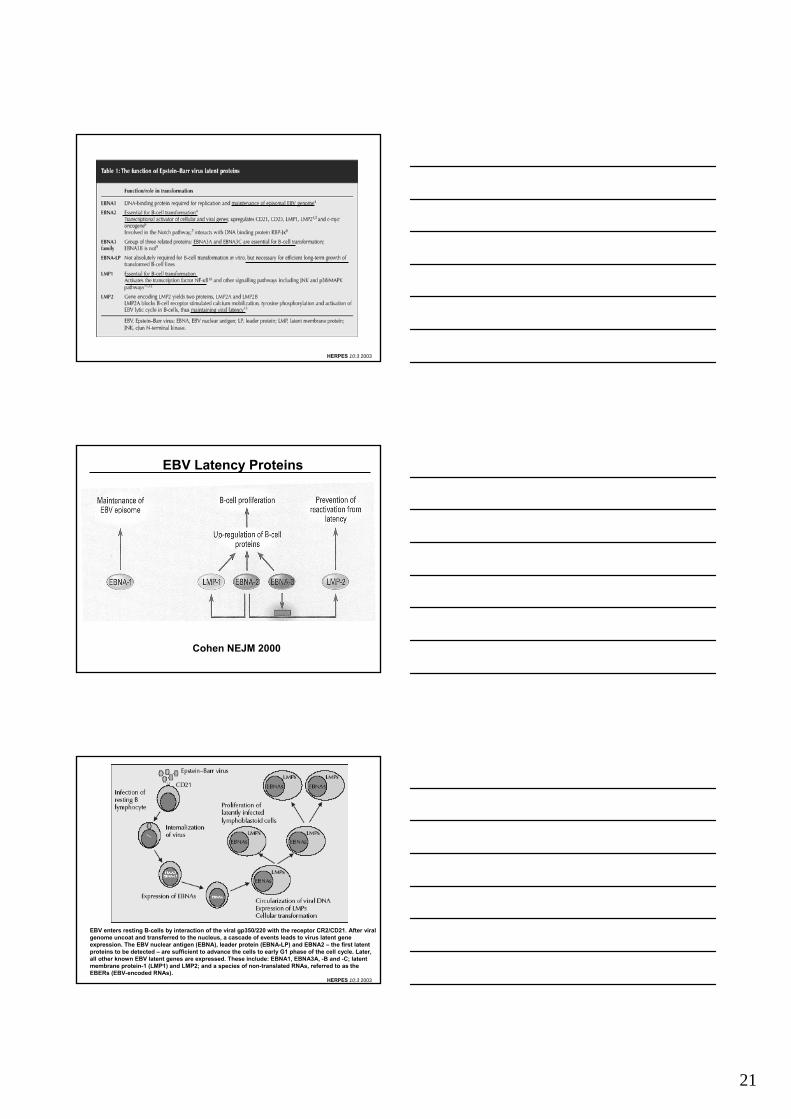
Cervical carcinogenesis
Schematic presentation of the morphological alterations seen in consecutivepremalignant cervical lesions and correlation of the cervical intraepithelial neoplasia(CIN) classification with squamous intraepithelial lesions (SILs)
20
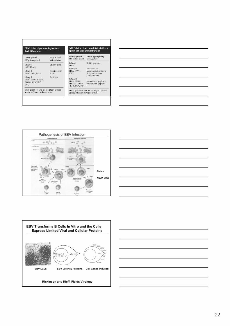
Deregulated HPV 16 E6/E7 expression in lower, dysplastic cell layers of a CIN 2 lesionat the transition to normal cervical epitheliumLeft panel: Haematoxylin–eosin (HE)-stained section.Right panel: E6/E7 RNA in situ hybridization (RISH)
HPV-mediated carcinogenesis
Alignment of the different steps during cervical carcinogenesis (upper part) and in vitrotransformation of epithelial cells mediated by high risk HPV (lower part). Potential relevant genetic alterations are indicated in red. Chromosomes are indicated by ‘#’. Compl. = complementation J Pathol 2006; 208: 152–164
Progression model of cervical cancer based on in vitro transformation steps and data from clinical samples. Potential relevant genetic alterations are indicated in red. TSGs = tumour suppressor genes.↑ indicates increased activity resulting from (epi)genetic alteration(s). ↓ indicates decreased activity resulting from (epi)genetic alteration(s), such as deletion or promoter hypermethylation
21
HERPES 10:3 2003
EBV Latency Proteins
Cohen NEJM 2000
EBV enters resting B-cells by interaction of the viral gp350/220 with the receptor CR2/CD21. After viral genome uncoat and transferred to the nucleus, a cascade of events leads to virus latent gene expression. The EBV nuclear antigen (EBNA), leader protein (EBNA-LP) and EBNA2 – the first latent proteins to be detected – are sufficient to advance the cells to early G1 phase of the cell cycle. Later, all other known EBV latent genes are expressed. These include: EBNA1, EBNA3A, -B and -C; latent membrane protein-1 (LMP1) and LMP2; and a species of non-translated RNAs, referred to as theEBERs (EBV-encoded RNAs).
HERPES 10:3 2003
22
Pathogenesis of EBV Infection
Cohen
NEJM 2000
EBV Transforms B Cells In Vitro and the Cells Express Limited Viral and Cellular Proteins
Rickinson and Kieff, Fields Virology
EBV LCLs EBV Latency Proteins Cell Genes Induced
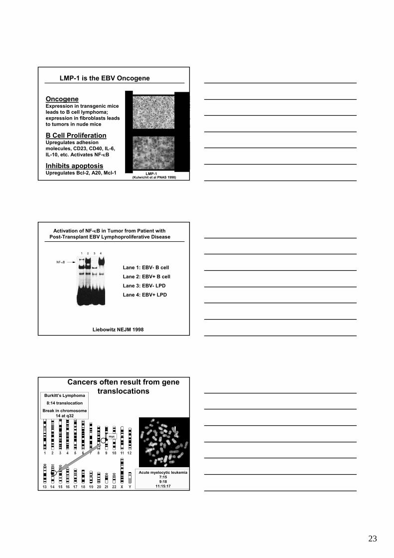
23
OncogeneExpression in transgenic mice leads to B cell lymphoma; expression in fibroblasts leads to tumors in nude mice
B Cell ProliferationUpregulates adhesion molecules, CD23, CD40, IL-6, IL-10, etc. Activates NF-κB
Inhibits apoptosisUpregulates Bcl-2, A20, Mcl-1
LMP-1 is the EBV Oncogene
LMP-1
H & E
(Kulwichit et al PNAS 1998)
Liebowitz NEJM 1998
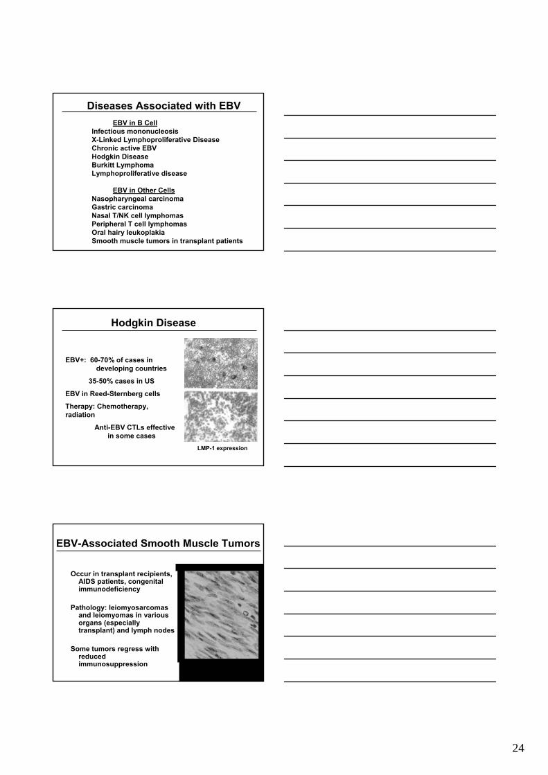
Activation of NF-κB in Tumor from Patient with Post-Transplant EBV Lymphoproliferative Disease
Lane 1: EBV- B cell
Lane 2: EBV+ B cell
Lane 3: EBV- LPD
Lane 4: EBV+ LPD
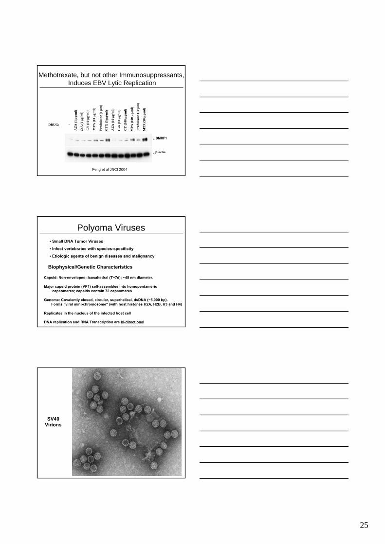
Cancers often result from gene translocations
Burkitt’s Lymphoma
8:14 translocation
Break in chromosome 14 at q32
Acute myelocytic leukemia7:159:18
11:15:17
myc
24
EBV in B CellInfectious mononucleosisX-Linked Lymphoproliferative DiseaseChronic active EBVHodgkin Disease Burkitt LymphomaLymphoproliferative disease
EBV in Other CellsNasopharyngeal carcinomaGastric carcinomaNasal T/NK cell lymphomasPeripheral T cell lymphomasOral hairy leukoplakiaSmooth muscle tumors in transplant patients
Diseases Associated with EBV
EBV+: 60-70% of cases in developing countries
35-50% cases in US
EBV in Reed-Sternberg cells
Therapy: Chemotherapy, radiation
Anti-EBV CTLs effective in some cases
Hodgkin Disease
LMP-1 expression
EBV-Associated Smooth Muscle Tumors
Occur in transplant recipients, AIDS patients, congenital immunodeficiency
Pathology: leiomyosarcomasand leiomyomas in various organs (especially transplant) and lymph nodes
Some tumors regress with reduced immunosuppression
25
Methotrexate, but not other Immunosuppressants, Induces EBV Lytic Replication
BMRF1
CY
(100
µg/
ml)
Pred
niso
ne (1
0 µm
)
_
AZA
(1 µ
g/m
l)
CsA
(1 µ
g/m
l)
CY
(10 µg
/ml)
MPA
(10 µg
/ml)
Pred
niso
ne (1
µm
)
MT
X (5
µg/
ml)
AZA
(10 µg
/ml)
CsA
(10 µg
/ml)
MPA
(100
µg/
ml)
MT
X (5
0 µg
/ml)
DRUG:
β-actin
Feng et al JNCI 2004
Polyoma Viruses• Small DNA Tumor Viruses
• Infect vertebrates with species-specificity
• Etiologic agents of benign diseases and malignancy
Biophysical/Genetic Characteristics
Capsid: Non-enveloped; icosahedral (T=7d); ~45 nm diameter.
Major capsid protein (VP1) self-assembles into homopentamericcapsomeres; capsids contain 72 capsomeres
Genome: Covalently closed, circular, superhelical, dsDNA (~5,000 bp). Forms "viral mini-chromosome" (with host histones H2A, H2B, H3 and H4)
Replicates in the nucleus of the infected host cell
DNA replication and RNA Transcription are bi-directional
SV40 Virions
26
Py Host Range:
Virus Host
Simian virus 40 (SV40) Human, Rhesus monkey
Simian Agent 12 (SA12) Baboon
Lymphotropic Papovavirus (LPV) African green monkey
JC virus (JCV) Human
BK virus (BKV) Human
Bovine polyomavirus (BPy) Cattle
Rabbit polyomavirus (RKV) Rabbit
Murine Polyoma virus (Py) Mouse
Kirsten virus (KV) Mouse
Hamster polyomavirus (HaPy) Hamster
Rat polyomavirus (RPV) Rat
Budgerigar fledgling disease virus Parakeet
Pathogenesis
This family causes sub-clinical persistent infections
Some transmitted transplacentally, Polyoma and SV40 viruses
Pathology only after immunosuppression
eg. Progressive multifocal leucoencephalopathycaused by JCV
tumors caused by Polyomavirus in mice
SV405243 bp
ELP
LARGE T
AGNOPROTEI
VP2
VP3
VP1
SMALL T
L 19S
L 16S
E 19S
E 18S
E P
E POLY A
E POLY A
L POLY A
SV40 ORI
AUX ORI
RPT I
RPT II
RPT III
RPT ENH A
RPT ENH B
SAS RNA
Eco R I (1783)
SV405243 bp
Small T Antigen
VP1
VP3
VP2
Large T Antigen
ori(5208-28) Agnoprotein
Polyomavirus (SV40) Genomic Organization
Replication
Transcription
27
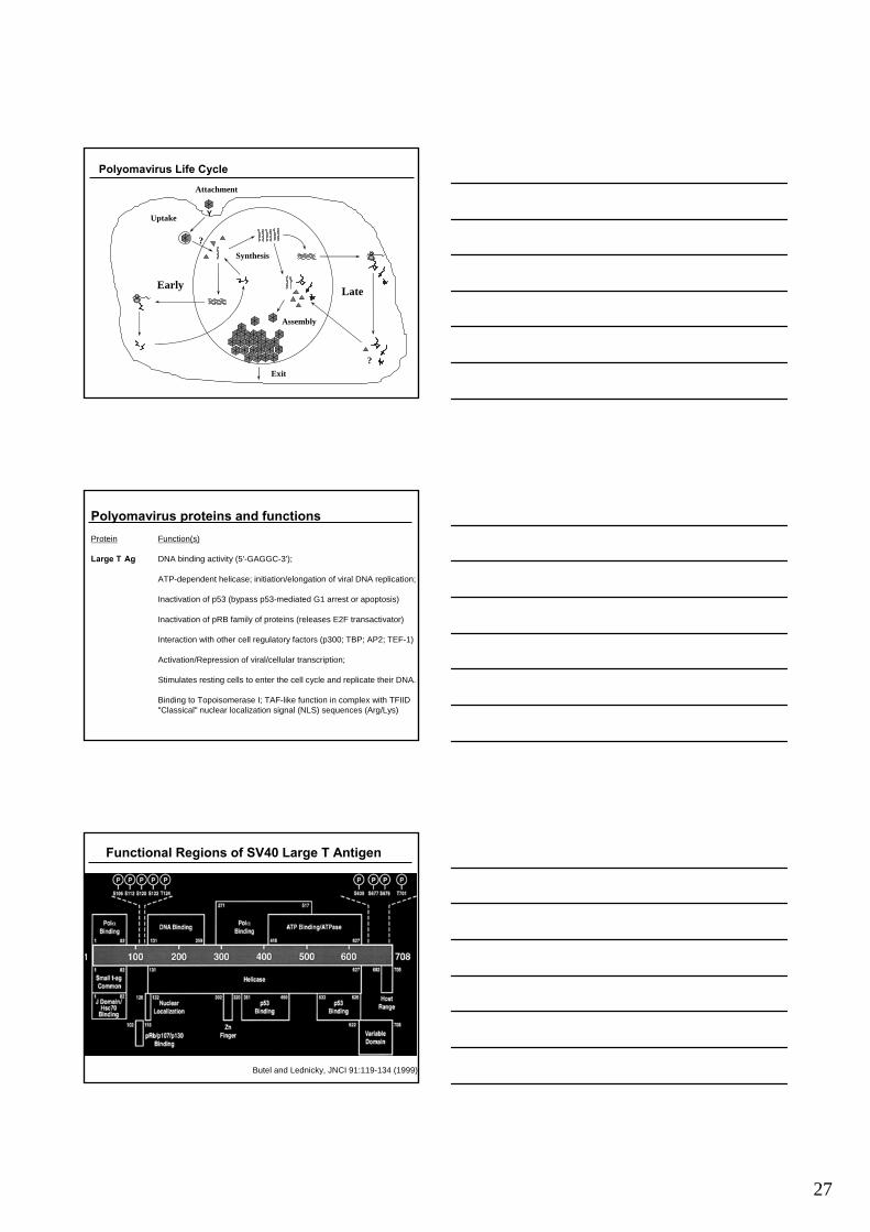
Polyomavirus Life Cycle
?
Early Late
Uptake
Assembly
Synthesis
Y
?Exit
Attachment
Polyomavirus proteins and functionsProtein Function(s)
Large T Ag DNA binding activity (5'-GAGGC-3');
ATP-dependent helicase; initiation/elongation of viral DNA replication;
Inactivation of p53 (bypass p53-mediated G1 arrest or apoptosis)
Inactivation of pRB family of proteins (releases E2F transactivator)
Interaction with other cell regulatory factors (p300; TBP; AP2; TEF-1)
Activation/Repression of viral/cellular transcription;
Stimulates resting cells to enter the cell cycle and replicate their DNA.
Binding to Topoisomerase I; TAF-like function in complex with TFIID"Classical" nuclear localization signal (NLS) sequences (Arg/Lys)
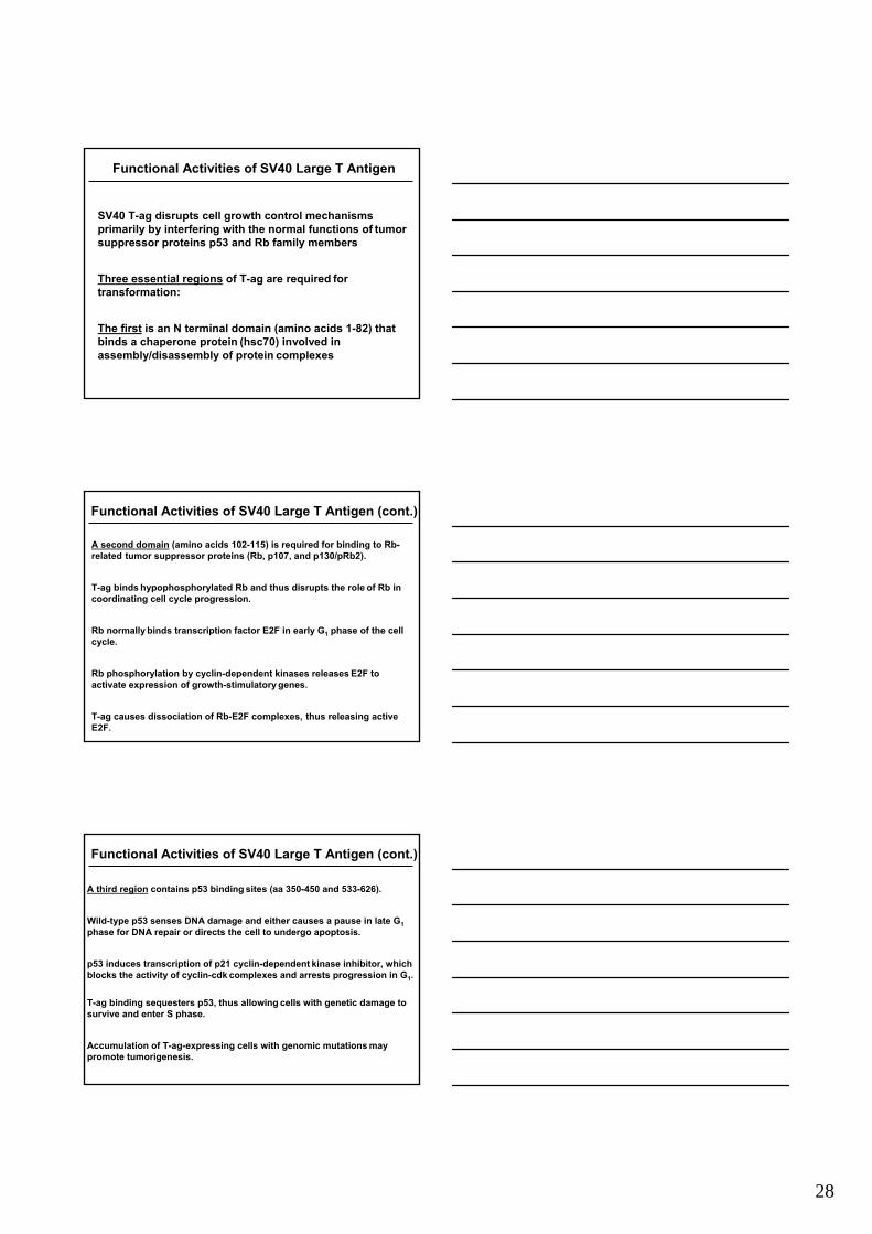
Functional Regions of SV40 Large T Antigen
Butel and Lednicky, JNCI 91:119-134 (1999)
28
SV40 T-ag disrupts cell growth control mechanismsprimarily by interfering with the normal functions of tumor suppressor proteins p53 and Rb family members
Three essential regions of T-ag are required for transformation:
The first is an N terminal domain (amino acids 1-82) that binds a chaperone protein (hsc70) involved in assembly/disassembly of protein complexes
Functional Activities of SV40 Large T Antigen
A second domain (amino acids 102-115) is required for binding to Rb-related tumor suppressor proteins (Rb, p107, and p130/pRb2).
T-ag binds hypophosphorylated Rb and thus disrupts the role of Rb in coordinating cell cycle progression.
Rb normally binds transcription factor E2F in early G1 phase of the cellcycle.
Rb phosphorylation by cyclin-dependent kinases releases E2F to activate expression of growth-stimulatory genes.
T-ag causes dissociation of Rb-E2F complexes, thus releasing active E2F.
Functional Activities of SV40 Large T Antigen (cont.)
A third region contains p53 binding sites (aa 350-450 and 533-626).
Wild-type p53 senses DNA damage and either causes a pause in late G1phase for DNA repair or directs the cell to undergo apoptosis.
p53 induces transcription of p21 cyclin-dependent kinase inhibitor, which blocks the activity of cyclin-cdk complexes and arrests progression in G1.
T-ag binding sequesters p53, thus allowing cells with genetic damage to survive and enter S phase.
Accumulation of T-ag-expressing cells with genomic mutations may promote tumorigenesis.
Functional Activities of SV40 Large T Antigen (cont.)
29
• Binds protein phosphatase-2A (PP2A), which activates the mitogen-activated protein (MAP) kinase pathway and growth stimulation of quiescent cells.
• Activates AKT and telomerase and induces anchorage-independent growth of human epithelial cells.
Functional Activities of SV40 Small T Antigen
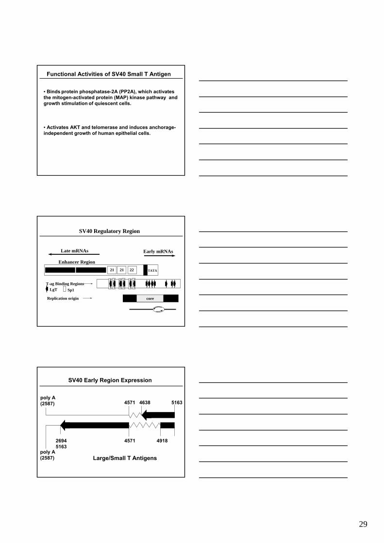
SV40 Regulatory Region
aux-2 aux-1core
7272 2221 21 TATA
Enhancer Region
Late mRNAs Early mRNAs
LgT Sp1T-ag Binding Regions
Replication origin
4571 4638 5163
poly A (2587)
2694 4571 4918 5163
poly A (2587)
SV40 Early Region Expression
Sm T
Lg T Lg T
Large/Small T Antigens
30
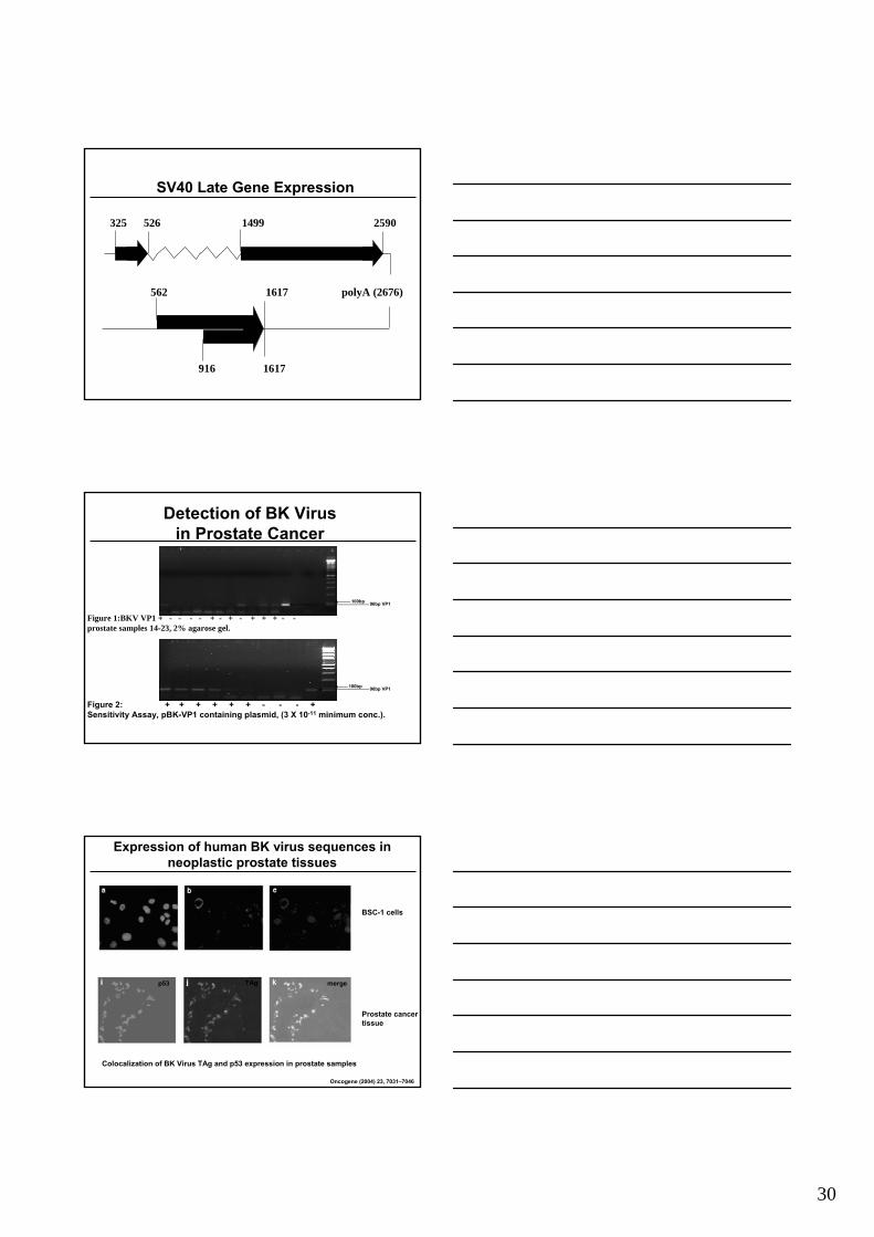
325 526 1499 2590
562 1617 polyA (2676)
916 1617
SV40 Late Gene Expression
Agno VP1
VP2VP3
Detection of ΒΚ Virusin Prostate Cancer
Figure 1:BKV VP1 + - - - - + - + - + + + - -prostate samples 14-23, 2% agarose gel.
100bp 96bp VP1
100bp 96bp VP1
Figure 2: + + + + + + Figure 2: + + + + + + -- -- -- + + Sensitivity Assay, pBKSensitivity Assay, pBK--VP1 containing plasmid, (3 X 10VP1 containing plasmid, (3 X 10--1111 minimum conc.)minimum conc.)..
Expression of human BK virus sequences in neoplastic prostate tissues
Oncogene (2004) 23, 7031–7046
Colocalization of BK Virus TAg and p53 expression in prostate samples
BSC-1 cells
Prostate cancertissue
DAPI p53
TAgp53 merge
31
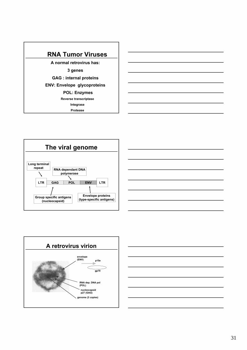
RNA Tumor Viruses
POL: EnzymesReverse transcriptase
Integrase
Protease
A normal retrovirus has:
3 genes
GAG : internal proteinsENV: Envelope glycoproteins
The viral genome
LTR LTRGAG POL ENV
Long terminalrepeat
Group specific antigens(nucleocapsid)
RNA dependant DNApolymerase
Envelope proteins(type-specific antigens)
A retrovirus virionenvelope (ENV)
nucleocapsid p27 (GAG)
genome (2 copies)
RNA dep. DNA pol (POL)
p15e
gp70
32
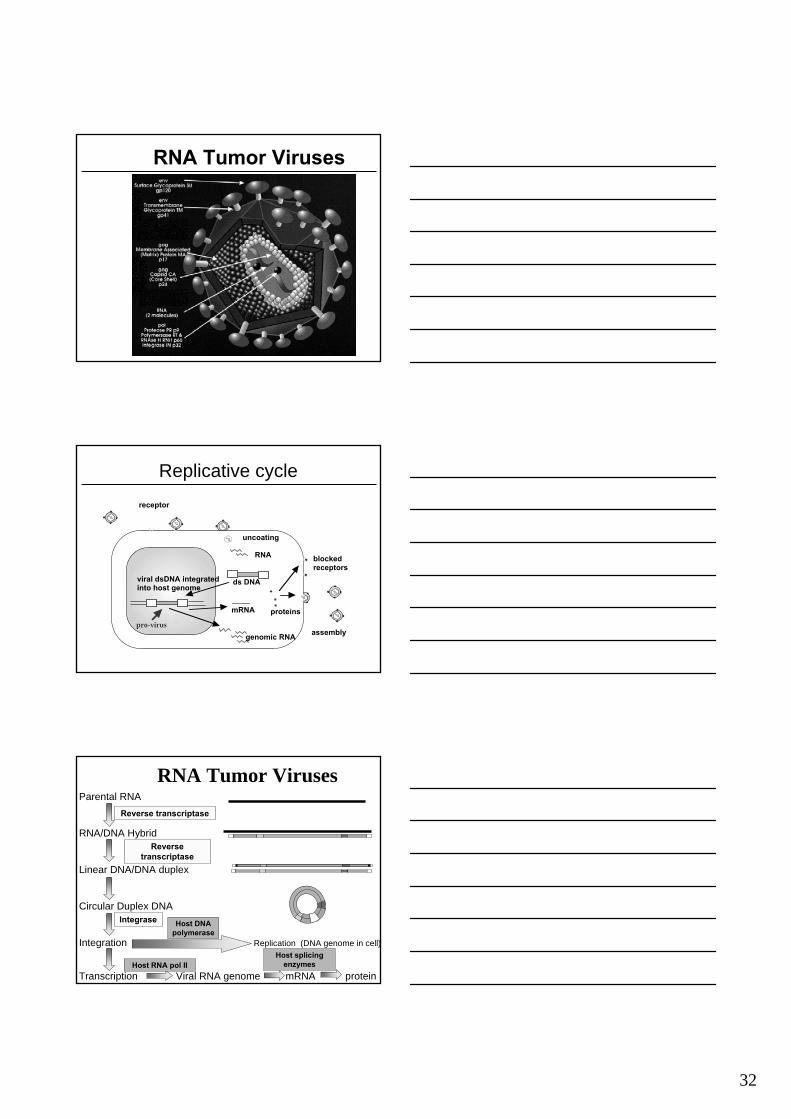
RNA Tumor Viruses
Replicative cyclereceptor
uncoating
RNA
ds DNA
proteinsmRNA
viral dsDNA integrated into host genome
genomic RNA
blocked receptors
assemblypro-virus
Parental RNA
RNA/DNA Hybrid
Linear DNA/DNA duplex
Circular Duplex DNA
Integration Replication (DNA genome in cell)
Transcription Viral RNA genome mRNA protein
RNA Tumor Viruses
Reverse transcriptase
Reverse transcriptase
Integrase
Host RNA pol II
Host DNA polymerase
Host splicing enzymes
33
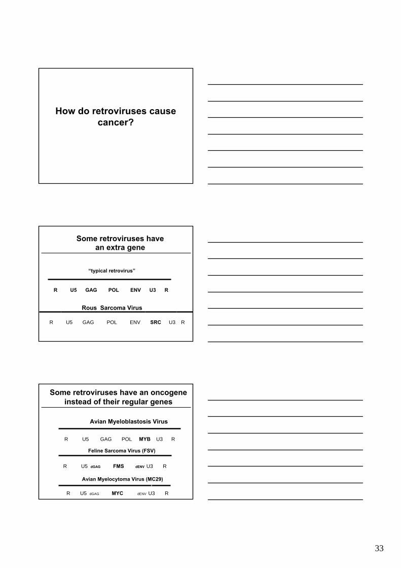
How do retroviruses cause cancer?
R U5 GAG POL ENV U3 R
Rous Sarcoma Virus
R U5 GAG POL ENV U3 R
Some retroviruses have an extra gene
“typical retrovirus”
SRC
Feline Sarcoma Virus (FSV)
R U5 dGAG FMS dENV U3 R
Avian Myelocytoma Virus (MC29)
R U5 dGAG MYC dENV U3 R
Avian Myeloblastosis Virus
R U5 GAG POL MYB U3 R
Some retroviruses have an oncogeneinstead of their regular genes
34
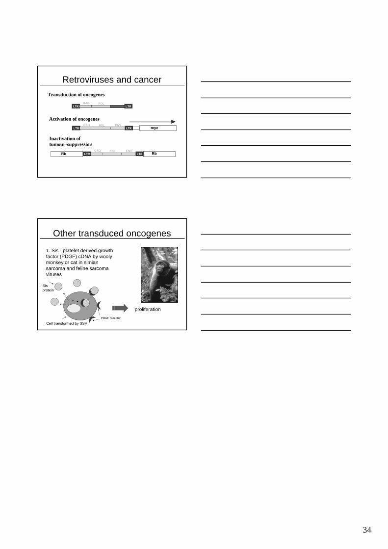
Retroviruses and cancerTransduction of oncogenes
Activation of oncogenes
Inactivation of tumour-suppressors
LTRGAG POL
LTRsrc
LTRGAG POL ENV
LTR myc
LTRGAG POL ENV
LTRRb Rb
Other transduced oncogenes
1. Sis - platelet derived growth factor (PDGF) cDNA by wooly monkey or cat in simian sarcoma and feline sarcoma viruses
Cell transformed by SSVPDGF receptor
Sis protein
proliferation


![Viral integration transforms chromatin to drive oncogenesis...2020/02/12 · [36] JüriReimand,TambetArak,PriitAdler,LiisKolberg,SulevReisberg,HediPeterson,andJaakVilo.g:Profiler—awebserverforfunctionalinterpretation](https://img.pdfslide.net/doc/110x75/603c19f7499e610de0739366/viral-integration-transforms-chromatin-to-drive-oncogenesis-20200212-36.jpg)