Embed Size (px)
Citation preview
Structure
Theory
Two Pathways Mediate InterdomainAllosteric Regulation in Pin1Jingjing Guo,1,2 Xiaodong Pang,1 and Huan-Xiang Zhou1,*1Department of Physics and Institute of Molecular Biophysics, Florida State University, Tallahassee, FL 32306, USA2Department of Chemistry, School of Pharmacy, State Key Laboratory of Applied Organic Chemistry, Lanzhou University, Lanzhou 730000,
China*Correspondence: [email protected]
http://dx.doi.org/10.1016/j.str.2014.11.009
SUMMARY
Allostery is an essential means for regulating biomol-ecular functions and provides unique opportunitiesfor drug design, yet our ability to elucidate allostericmechanisms remains limited. Here, based on exten-sive molecular dynamics simulations, we present anatomistic picture of the pathways mediating the allo-steric regulation of the PPIase domain of Pin1 by itsWWdomain. Two pathways jointly propagate the ac-tion of substrate-WWbinding to produce closure andrigidification of three PPIase catalytic-site loops. Onepathway preexists in the apo protein, but remainsdormant until substrate-WW binding completes thesecond. The reduction in conformational entropyand preorganization of the catalytic-site loopsobserved here may explain why substrate-WW bind-ing enhances ligand affinity and catalytic activity ofthe PPIase domain and suggest a combinationdrug therapy for Pin1-related diseases. Whereasthe traditional view of allostery has emphasizedconformational transition, our study identifies adistinct role of conformational dynamics in elicitingallostery.
INTRODUCTION
Pin1, a peptidyl-prolyl cis/trans isomerase (PPIase), acts on
phosphoSer/Thr-Pro (pSer/Thr-Pro) motifs present in mitotic
phosphoproteins (Lu et al., 1996), thereby controlling their fates
(Liou et al., 2011). Pin1 dysregulation is implicated in various dis-
eases, including cancer and Alzheimer’s disease (Lu, 2004; Lu
and Zhou, 2007; Lu et al., 1999b; Wulf et al., 2001). Therefore,
Pin1 is an attractive therapeutic target, and a number of inhibi-
tors have been designed (Moore and Potter, 2013; Wang and
Etzkorn, 2006; Wang et al., 2004). The full-length Pin1 can be
divided into an N-terminal WW domain (residues 1–39) and the
C-terminal PPIase domain (residues 50–163) (Figures 1A and
1B). Both domains can selectively bind pSer/Thr-Pro containing
substrates motifs, but only the PPIase domain can isomerize the
peptidyl-prolyl bonds (Lu et al., 1999a; Zhou et al., 2000). The
roles of theWWdomain and,more specifically, substrate binding
to it, have long been studied (Lu and Zhou, 2007; Lu et al., 1999a,
Structure 23, 23
2002; Ranganathan et al., 1997; Verdecia et al., 2000). These
roles may provide both better understanding of the functional
mechanism of Pin1 and unique opportunities for designing
Pin1-targeting drugs. Here, we report a computational study
on the conformational and dynamical effects of substrate-WW
binding.
Earlier studies have emphasized the potential of the WW
domain as a noncatalytic binder in increasing local substrate
concentration and in subcellular localization (Lu et al., 1999a,
2002). However, the substrate affinity and catalytic activity of
the isolated PPIase domain are different from those of the full-
length protein (Lu et al., 1999a; Namanja et al., 2011; Zhou
et al., 2000), therefore suggesting that the WW domain can
modulate substrate binding and catalysis. Indeed, numerous
crystal structures of Pin1 have shown that the two domains are
tightly packed against each other, although the linker between
them is disordered (Ranganathan et al., 1997; Verdecia et al.,
2000; Zhang et al., 2012). Nuclear magnetic resonance (NMR)
studies have shown that binding of both substrates and a non-
peptidic ligand, polyethylene glycol (PEG), to theWWdomain re-
sults in tighter coupling between the two domains (Jacobs et al.,
2003; Vanwart et al., 2012). Side-chain methyl dynamics studies
(Namanja et al., 2007, 2011) have further shown that substrate
binding to the WW domain leads to a loss of side-chain flexibility
along a ‘‘conduit’’ of conserved hydrophobic residues linking the
interdomain interface and the catalytic site. Moreover, an I28A
mutation in the interdomain interface has been found to weaken
interdomain communication (Wilson et al., 2013).
Together, the foregoing studies suggest that the WW domain
may modulate the activity of the PPIase domain through allo-
steric regulation. However, the underlying mechanism remains
poorly defined. Potentially, the large number of crystal structures
of Pin1 in the Protein Data Bank (PDB) could provide clues to the
various conformations accessible to the proteins. Unfortunately,
in all these structures, both the WW site and the PPIase catalytic
site are occupied, often by PEG, an additive for protein crystalli-
zation. The side-chain methyl dynamics studies (Namanja et al.,
2007, 2011) have given rise to the most detailed picture of the
pathway for interdomain communication, yet these studies are
limited to methyl-containing side chains and the possibility of
other participating residues cannot be excluded. It is also un-
clear how ligand binding to the WW domain induces effects on
substrate binding to, and catalytic activity of, the PPIase domain.
In recent years, computational studies have been found to be
very useful in complementing experiments in elucidating allo-
steric mechanisms (Elber, 2011; Feher et al., 2014; Rousseau
7–247, January 6, 2015 ª2015 Elsevier Ltd All rights reserved 237

Figure 1. The Protein and Ligands in This
Study
(A) Structure of Pin1 with FFpSPR bound to the
WW domain; protein from 3TDB and substrate
modeled after 1F8A. The b1–b2 loop and three
loops around the catalytic site are highlighted in
darker cyan.
(B) Secondary structures of Pin1.
(C) The three Pin1 ligands, from top to bottom:
FFpSPR, cis ligand, and trans ligand.
Structure
Allosteric Pathways in Pin1
and Schymkowitz, 2005), including algorithms for identifying
allosteric networks (Gerek and Ozkan, 2011; Ghosh and Vish-
veshwara, 2007; Kannan and Vishveshwara, 1999; Sethi et al.,
2009; Van Wart et al., 2014). In particular, molecular dynamics
simulations have revealed two pathways and implicated a strong
dynamic component in the allosteric regulation of thrombin by
thrombomodulin (Gasper et al., 2012). This supports the growing
emphasis on the possibility that allostery can be elicited through
changes in protein dynamics without apparent conformational
transitions (Cooper and Dryden, 1984; Petit et al., 2009; Tsai
et al., 2008), departing from the classical view (Fischer et al.,
2011; Monod et al., 1965).
In the present study, we carried out extensive molecular dy-
namics simulations to elucidate the mechanism of allosteric
regulation in Pin1. Our simulations show that substrate binding
to the WW domain alone results in closure and rigidification of
the three loops (referred to as b4–a1 [or catalytic], b5–a4, and
b6–b7) around the catalytic site (Figures 1A and 1B), thus giving
direct evidence for allosteric communication between the two
distant binding sites. Two pathways are found to mediate the in-
terdomain allosteric regulation. Path1 emanates from the WW
backside and propagates through the interdomain interface
and the PPIase domain core to the b5–a4 and b6–b7 loops;
Path2 emanates from the WW front pocket and propagates
through the bound substrate, the PPIase peripheral a1, and
the a1-core interface to the catalytic loop. Path1 preexists in
apo Pin1, but remains dormant until Path2 is completed by sub-
strate-WW binding. Through restrained simulations and simula-
tions of the I28A mutant, we further demonstrate that the two
pathways must act in concert in order to elicit the allosteric ef-
fects. Moreover, our study suggests that substrate-WW bind-
ing, via rigidifying the catalytic-site loops, may enhance ligand
affinity of the PPIase domain by reducing the conformational
entropy cost for binding. Substrate-WW binding induced preor-
ganization of the catalytic-site loops may also enhance the cat-
alytic activity of the full-length Pin1. Finally, the scenario of
cooperative binding to the WW and PPIase sites presented
here points to the possibility of a combination drug therapy for
Pin1-related diseases.
238 Structure 23, 237–247, January 6, 2015 ª2015 Elsevier Ltd All rights reserved
RESULTS
The design of the present work was
largely inspired by the NMR studies of
Peng and coworkers (Namanja et al.,
2011). They measured the WW and
PPIase binding affinities of a Pin1 sub-
strate (FFpSPR) and two peptidomimet-
ics in which the substrate pSP core is replaced by alkene
isosteres to lock the imide as either cis or trans (Wang et al.,
2004) (Figure 1C), as well as the effects of binding these ligands
on Pin1 methyl-containing side-chain dynamics. FFpSPRmainly
bound to the WW site, the cis ligand exclusively to the PPIase
site, whereas the trans ligand bound to both sites. We modeled
these three different binding modes onto Pin1 and then carried
out molecular dynamics simulations in explicit solvent. The sim-
ulations present direct evidence for allosteric communication
between the two distant binding sites and suggest twomediating
pathways. The available experimental data (Jacobs et al., 2003;
Namanja et al., 2011; Vanwart et al., 2012) provide validation, but
our simulations lead to amore complete, atomistic picture for the
allosteric regulation in Pin1.
To dissect the effects of substrate-WW binding and further
validate the two putative pathways, we carried out simulations
of apo Pin1 in which different regions of the protein were res-
trained. In addition, we carried out simulations of the I28A
mutant, which perturbs the interdomain interface (Wilson et al.,
2013). These ‘‘control’’ simulations show that the allosteric ef-
fects are not fully elicited when the interdomain links are per-
turbed. Therefore allosteric regulation is achieved through the
concerted action of the two pathways.
Ligand Binding Results in Localized ConformationalChangesBased on the crystal structures of Pin1 bound with the cis or
trans ligand at the catalytic site (PDB 3TCZ and 3TDB, see Zhang
et al., 2012) and bound with a pSer-Pro containing peptide (PDB
1F8A, see Verdecia et al., 2000) at the WW site, we built initial
models for Pin1 with FFpSPR bound at the WW site (Figure 1A),
with the cis ligand bound at the catalytic site, andwith two copies
of the trans ligand bound, one at the WW site and one at the cat-
alytic site. At the WW site, the ligands sit over a pocket lined by
the concave front face of the WW domain b sheet and fill a
groove between the WW domain and the a1 helix of the PPIase
domain. We simulated each of these systems, as well as apo
Pin1 in explicit solvent for 100 nanoseconds (ns) and used the
last 40 ns for analysis.
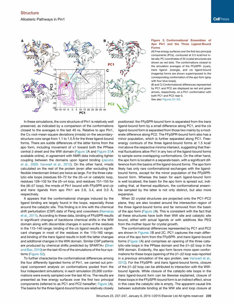
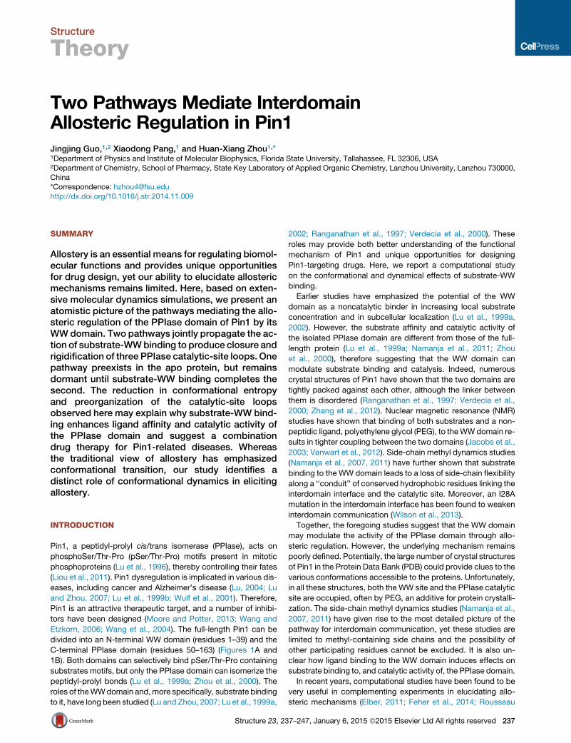
Figure 2. Conformational Ensembles of
Apo Pin1 and the Three Ligand-Bound
Forms
(A) Free energy surfaces over the first two principal
components (PCs), contoured at 0.5 kcal/mol in-
tervals; PC coordinates of 32 crystal structures are
shown as red dots. The conformations closest to
the simulation averages of the FFpSPR- (cyan),
trans ligand- (orange), and cis ligand-bound
(magenta) forms are shown superimposed to the
corresponding conformation of the apo form (gray
with four blue loops).
(B and C) Conformational differences represented
by PC1 and PC2 are displayed as red and green
arrows, respectively, on a Pin1 conformation with
both PC1 and PC2 near 0.
See also Figures S1–S3.
Structure
Allosteric Pathways in Pin1
In these simulations, the core structure of Pin1 is relatively well
preserved, as indicated by a comparison of the conformations
closest to the averages in the last 40 ns. Relative to apo Pin1,
the Ca root-mean-square deviations (rmsds) on the secondary-
structure core range from 1.1 to 1.5 A for the three ligand-bound
forms. There are subtle differences of the latter forms from the
apo form, including movement of a1 toward both the PPIase
central b sheet and the WW domain (Figure 2A and Figure S1A
available online), in agreement with NMR data indicating tighter
coupling between the domains upon ligand binding (Jacobs
et al., 2003; Vanwart et al., 2012). On the other hand, rmsds
calculated on the rest of the protein (even after excluding the
flexible interdomain linker) are twice as large. For the three cata-
lytic-site loops (residues 63–72 for the b5–a4 or catalytic loop,
residues 126–132 for the b5–a4 loop, and residues 151–155 for
the b6–b7 loop), the rmsds of Pin1 bound with FFpSPR and cis
and trans ligands from apo Pin1 are 2.6, 3.4, and 3.0 A,
respectively.
It appears that the conformational changes induced by the
ligand binding are largely found in the loops, especially those
around the catalytic site. This finding is in line with the chemical
shift perturbation (CSP) data of Peng and coworkers (Namanja
et al., 2011). According to these data, binding of FFpSPR results
in significant changes of backbone chemical shifts in the WW
domain along with discernible changes in some of the residues
in the 115–140 range; binding of the cis ligand results in signifi-
cant changes in most of the residues in the 115–160 range;
and binding of the trans ligand results in both the latter changes
and additional changes in the WW domain. Similar CSP patterns
are produced by chemical shifts predicted by SPARTA+ (Shen
andBax, 2010) on the last 40 ns of the simulations of the four sys-
tems (Figure S2).
To further characterize the conformational differences among
the four differently liganded forms of Pin1, we carried out prin-
cipal component analysis on conformations pooled from the
four independent simulations; in each simulation 20,000 confor-
mations were evenly sampled over the last 40 ns. The results are
presented as free energy surfaces over the first two principal
components (referred to as PC1 and PC2 hereafter; Figure 2A).
The basins for the three ligand-bound forms are relatively closely
Structure 23, 23
positioned: the FFpSPR-bound form is separated from the trans
ligand-bound form by a small difference along PC1, and the cis
ligand-bound form is separated from those twomainly by amod-
erate difference along PC2. The FFpSPR-bound form also has a
minor population, which is further separated along PC1. Free-
energy contours of the three ligand-bound forms at 1.5 kcal/
mol above the respective minima intersect, suggesting that ther-
mal fluctuations allow Pin1 in any two of the ligand-bound forms
to sample some overlapping conformations. On the other hand,
the apo form is located in a separate basin, with a significant dif-
ference from the basins of the ligand-bound forms. The apo form
likely has only rare conformational exchanges with the ligand-
bound forms, except for the minor population of the FFpSPR-
bound form. Whereas the basin for each ligand-bound form
is well localized, the basin for the apo form is spread out, indi-
cating that, at thermal equilibrium, the conformational ensem-
ble sampled by the latter is not only distinct, but also more
expansive.
When 32 crystal structures are projected onto the PC1-PC2
plane, they are also located around the intersection region of
the three ligand-bound forms and far removed from the basin
of the apo form (Figure 2A). This is consistent with the fact that
all these structures have both their WW site and catalytic site
bound, either with actual ligands or with additives like PEG
from the mother liquor for crystal growth.
The conformational differences represented by PC1 and PC2
are shown in Figures 2B and 2C. PC1 captures the main differ-
ence of the apo form from the FFpSPR- and trans ligand-bound
forms (Figure 2A) and comprises an opening of the three cata-
lytic-site loops in the PPIase domain and the b1–b2 loop in the
WW domain. Evidently, the apo form favors more open confor-
mations for these loops (opening of the b1–b2 loop was reported
in a previous simulation of the apo protein, see Vanwart et al.,
2012). For the FFpSPR- and trans ligand-bound forms, closure
of the b1–b2 loop can be attributed to interaction with the WW-
bound ligands. While closure of the catalytic-site loops in the
trans ligand-bound form can be likewise explained, closure of
these loops in the FFpSPR-bound form is an indirect effect, since
in this case the catalytic site is empty. The apparent causal link
between substrate binding at the WW site and loop closure at
7–247, January 6, 2015 ª2015 Elsevier Ltd All rights reserved 239
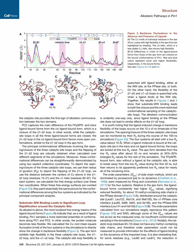
Figure 3. Backbone Fluctuations in the
Absence and Presence of Ligands
(A) The Ca rmsfs of individual residues in the last
40 ns. Loopswith high flexibility in the apo form are
highlighted by shading. The a3 helix, which is a
less stable 310 helix, also shows high flexibility.
(B–D) Differences in rmsfs of the ligand-bound
forms from those of the apo form are colored on
the bound conformations of Pin1. Red and blue
colors represent lower and higher flexibilities,
respectively, in the bound forms.
See also Figures S4–S6 and S8.
Structure
Allosteric Pathways in Pin1
the catalytic site provides the first sign of allosteric communica-
tion between the two domains.
PC2 captures the main difference of the FFpSPR- and trans
ligand-bound forms from the cis ligand-bound form, which is a
closure of the b1–b2 loop. In other words, while the catalytic-
site loops in all the three ligand-bound forms are closed, the
b1–b2 loop in the cis ligand-bound form favors more open con-
formations, similar to the b1–b2 loop in the apo form.
The principal conformational differences involving the open-
ing/closure of the three catalytic-site loops and the flapping of
the b1–b2 loop are robustly obtained when calculated over
different segments of the simulations. Moreover, these confor-
mational differences can be straightforwardly demonstrated by
using two explicit collective coordinates. To depict the open-
ing/closure of the three catalytic-site loops, we use their radius
of gyration (Rg); to depict the flapping of the b1–b2 loop, we
use the distance between the centers of Ca atoms in the b1–
b2 loop (residues 15–21) and the a1 helix (residues 82–97). For
each system, we calculated the free energy surface over these
two coordinates. When these free energy surfaces are overlaid
(Figure S3), they paint essentially the same picture for the confor-
mational differences among the four systems as indicated by the
principal component analysis.
Substrate-WW Binding Leads to Significant LoopRigidification around the Catalytic SiteAs alluded to above, the more localized free-energy basins of the
ligand-bound forms (Figure 2A) indicate that, as a result of ligand
binding, Pin1 samples a more restricted ensemble of conforma-
tions along PC1 and PC2. In other words, the protein molecule
becomes less flexible. We calculated the Ca root-mean-square
fluctuation (rmsf) of the four systems in the simulations to directly
show the change in backbone flexibility (Figure 3). The apo form
exhibits high flexibility in the three catalytic-site loops, the b1–
b2 loop, and the a1–a2 loop. The catalytic-site loop flexibility is
240 Structure 23, 237–247, January 6, 2015 ª2015 Elsevier Ltd All rights reserved
quenched with ligand binding, either at
the WW site, at the PPIase site, or both.
On the other hand, the flexibility of the
b1–b2 and a1–a2 loops is quenched only
when a ligand binds at the WW site.
Together, the results in Figures 2 and 3
show that substrate-WW binding leads
toboth the closure and themore restricted
conformational sampling of the catalytic-
site loops. The allosteric communication
is evidently one-way only, since ligand binding at the PPIase
site does not lead to similar effects on the b1–b2 loop.
It is worth noting that the ligand-induced change in backbone
flexibility of the loops occurs on the 10 s of ns timescale of the
simulations. The opening/closure of the three catalytic-site loops
can be monitored by their Rg during the course of the 100-ns
simulations (Figure S4). The four systems all start with a low Rg
value (about 10 A). When a ligand molecule is bound at the cat-
alytic site (as in the trans and cis ligand-bound forms), the loops
are locked at this low Rg value. The apo form deviates from the
low Rg value after about 50 ns and remains at 20%–30%
enlarged Rg values for the rest of the simulation. The FFpSPR-
bound form, also without a ligand at the catalytic site, is able
to break away from the low Rg value during the first 40 ns, but
then returns to its apparently preferred low Rg value for nearly
all of the remaining time.
The order parameters ðS2axisÞ of side-chain methyls, which are
dominated by picosecond (ps) to ns dynamics (Chatfield et al.,
1998), were measured by Peng and coworkers (Namanja et al.,
2011) for the four systems. Relative to the apo form, the ligand-
bound forms consistently had higher S2axis values, signifying
reduced flexibility, for about 10 methyl-containing side chains in
the PPIase domain (Figure S5A), distributed around the catalytic
site (Leu61, Leu122, Ala124, and Met130), the a1-PPIase core
interface (Leu60, Ile89, Ile93, and Ile156), and the PPIase-WW
interface (Leu141 andVal150) (Figure S5B). The order parameters
calculated from our simulations largely reproduce this pattern
(Figures S5C and S5D), although some of the S2axis values are
not as low as the measured ones, for insufficient conformational
sampling in the simulations (Gasper et al., 2012). Notably, the cat-
alytic loop (residues 63–72) does not have any methyl-containing
side chains, and therefore order parameters could not be
measured to provide information for the effects of ligand binding
on the dynamics of this important loop. It is also interesting that,
for some residues (e.g., Leu60 and Leu61), the reduction in
Figure 4. Allosteric Networks of Four
Systems
(A) Apo Pin1.
(B) FFpSPR-Pin1 complex.
(C) cis ligand-Pin1 complex.
(D) trans ligand-Pin1 complex.
Clusters of residues that persistently form tertiary
contacts between side chains in the simulations
are shown as spheres, either in red or in blue, at Ca
positions. The main chains of the protein and the
ligands are displayed in cartoon and stick,
respectively. Clusters with no more than ten resi-
dues are not displayed. See also Figures S7
and S9.
Structure
Allosteric Pathways in Pin1
flexibility upon ligand binding is limited to the side chains only (as
reported by S2axis), as their Ca rmsfs are unaffected.
In addition to simulations of the four systems described above,
we also carried out simulations of two ‘‘control’’ systems (Fig-
ure S6). The rmsf results for Pin1 with the trans ligand bound
only to the catalytic site are similar to those for the cis ligand-
bound form, thus confirming that the interdomain communica-
tion is one-way only. In addition, a simulation of the isolated
PPIase domain in apo form yields lower rmsfs for the catalytic
and b6–b7 loops than the counterparts in the full-length apo
Pin1, suggesting that, in apo form, the WW domain amplifies
the loop flexibility of the PPIase domain.
Pathways of Interdomain CommunicationHow is the action of substrate-WW binding propagated to affect
the catalytic-site loop conformations and dynamics? Several al-
gorithms (Gerek and Ozkan, 2011; Ghosh and Vishveshwara,
2007; Kannan and Vishveshwara, 1999; Sethi et al., 2009; Van
Wart et al., 2014) have been developed for identifying allosteric
networks, i.e., clusters of linked residues that putatively propa-
gate allosteric signals. Here, we used the original algorithm of
Kannan and Vishveshwara (1999), in which residues are linked
into clusters when tertiary contacts between side chains persist
during a simulation.
For apo Pin1, the simulation produces two major clusters of
linked residues (Figure 4A). The first cluster consists of the WW
backside, the WW-PPIase domain interface, the PPIase domain
core (i.e., b4, b5, b6, b7, and a4), and the b5–a4 and b6–b7 loops.
The second cluster consists of the peripheral a1, the a1-PPIase
core interface, the a1–a2 loop, a2, and the catalytic loop. Cluster
1 is largely maintained in the FFpSPR-Pin1 complex, but cluster
Structure 23, 237–247, January 6, 2015
2 now extends to the WW front pocket
via the bound substrate (Figure 4B). We
propose that these two clusters form
two pathways, referred to as Path1 and
Path2, respectively, that propagate the
allosteric signal from the WW domain to
the PPIase catalytic site. Specifically,
Path1 emanates from the WW backside
and propagates through the interdomain
interface and the PPIase domain core to
the b5–a4 and b6–b7 loops; Path2 ema-
nates from the WW front pocket and
propagates through the bound substrate,
a1, and the a1-core interface to the catalytic loop. Path1 preex-
ists in apo Pin1, but apparently remains dormant until Path2 is
completed by substrate-WW binding.
In the cis ligand-bound complex, Path1 is broken at the WW-
PPIase domain interface because of weak links to the PPIase
domain core; all the remaining PPIase residues coalesce into a
single cluster (Figure 4C). As a result, there is no path that con-
nects the cis ligand to the WW domain, providing an explanation
for the one-way allosteric communication noted above. The allo-
steric networks of the trans ligand-bound complex are very
similar to those of the FFpSPR-Pin1 complex, except that the
catalytic loop is now part of the first cluster, due to links provided
by the ligandmolecule at the catalytic site (Figure 4D). Again, two
pathways tightly couple the two domains.
To demonstrate that the allosteric pathways identified above
are robust, we carried out a community network analysis as
introduced by Sethi et al. (2009). This method accounts for
motional correlation between residues. When applied to the
apo Pin1 simulation, six communities were obtained (Fig-
ure S7A). The WW domain is represented by two communities,
numbered 0 and 1, respectively, for the b1–b2 hairpin and the
rest of WW; the PPIase core is represented by community 2;
and the a1–a2 appendage is represented by communities 3–5,
respectively, for a1 N terminus/catalytic loop, a1 C terminus,
and a2/a3. For the FFpSPR-Pin1 complex (Figure S7B), commu-
nities 0 and 1 coalesce into a single one representing the full WW
domain. Moreover, this community is joined by the N terminus of
the bound substrate, whose C terminus joins community 4.
Therefore, the substrate reinforces the connection between
community 1 (i.e., WW domain) and 4 (i.e., a1 C terminus).
Finally, a new connection is generated between community 1
ª2015 Elsevier Ltd All rights reserved 241
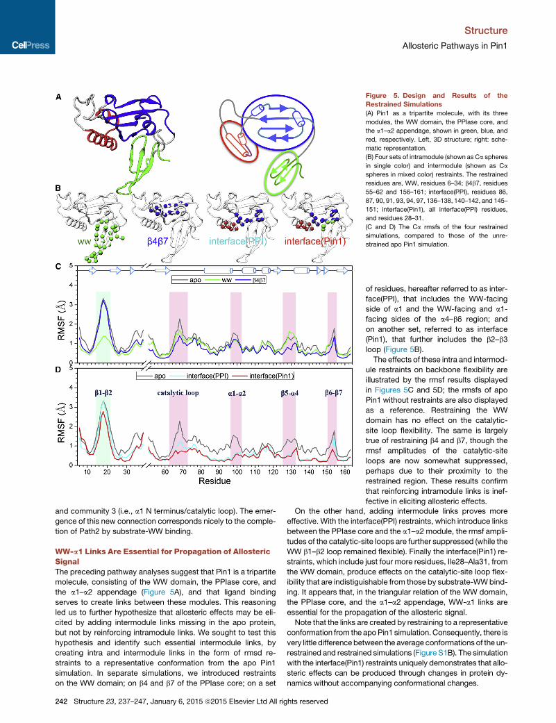
Figure 5. Design and Results of the
Restrained Simulations
(A) Pin1 as a tripartite molecule, with its three
modules, the WW domain, the PPIase core, and
the a1–a2 appendage, shown in green, blue, and
red, respectively. Left, 3D structure; right: sche-
matic representation.
(B) Four sets of intramodule (shown as Ca spheres
in single color) and intermodule (shown as Ca
spheres in mixed color) restraints. The restrained
residues are, WW, residues 6–34; b4b7, residues
55–62 and 156–161; interface(PPI), residues 86,
87, 90, 91, 93, 94, 97, 136–138, 140–142, and 145–
151; interface(Pin1), all interface(PPI) residues,
and residues 28–31.
(C and D) The Ca rmsfs of the four restrained
simulations, compared to those of the unre-
strained apo Pin1 simulation.
Structure
Allosteric Pathways in Pin1
and community 3 (i.e., a1 N terminus/catalytic loop). The emer-
gence of this new connection corresponds nicely to the comple-
tion of Path2 by substrate-WW binding.
WW-a1 Links Are Essential for Propagation of AllostericSignalThe preceding pathway analyses suggest that Pin1 is a tripartite
molecule, consisting of the WW domain, the PPIase core, and
the a1–a2 appendage (Figure 5A), and that ligand binding
serves to create links between these modules. This reasoning
led us to further hypothesize that allosteric effects may be eli-
cited by adding intermodule links missing in the apo protein,
but not by reinforcing intramodule links. We sought to test this
hypothesis and identify such essential intermodule links, by
creating intra and intermodule links in the form of rmsd re-
straints to a representative conformation from the apo Pin1
simulation. In separate simulations, we introduced restraints
on the WW domain; on b4 and b7 of the PPIase core; on a set
242 Structure 23, 237–247, January 6, 2015 ª2015 Elsevier Ltd All rights reserved
of residues, hereafter referred to as inter-
face(PPI), that includes the WW-facing
side of a1 and the WW-facing and a1-
facing sides of the a4–b6 region; and
on another set, referred to as interface
(Pin1), that further includes the b2–b3
loop (Figure 5B).
The effects of these intra and intermod-
ule restraints on backbone flexibility are
illustrated by the rmsf results displayed
in Figures 5C and 5D; the rmsfs of apo
Pin1 without restraints are also displayed
as a reference. Restraining the WW
domain has no effect on the catalytic-
site loop flexibility. The same is largely
true of restraining b4 and b7, though the
rmsf amplitudes of the catalytic-site
loops are now somewhat suppressed,
perhaps due to their proximity to the
restrained region. These results confirm
that reinforcing intramodule links is inef-
fective in eliciting allosteric effects.
On the other hand, adding intermodule links proves more
effective. With the interface(PPI) restraints, which introduce links
between the PPIase core and the a1–a2 module, the rmsf ampli-
tudes of the catalytic-site loops are further suppressed (while the
WW b1–b2 loop remained flexible). Finally the interface(Pin1) re-
straints, which include just four more residues, Ile28–Ala31, from
the WW domain, produce effects on the catalytic-site loop flex-
ibility that are indistiguishable from those by substrate-WWbind-
ing. It appears that, in the triangular relation of the WW domain,
the PPIase core, and the a1–a2 appendage, WW-a1 links are
essential for the propagation of the allosteric signal.
Note that the links are created by restraining to a representative
conformation from the apoPin1 simulation. Consequently, there is
very little differencebetween the average conformations of the un-
restrained and restrained simulations (Figure S1B). The simulation
with the interface(Pin1) restraints uniquely demonstrates that allo-
steric effects can be produced through changes in protein dy-
namics without accompanying conformational changes.
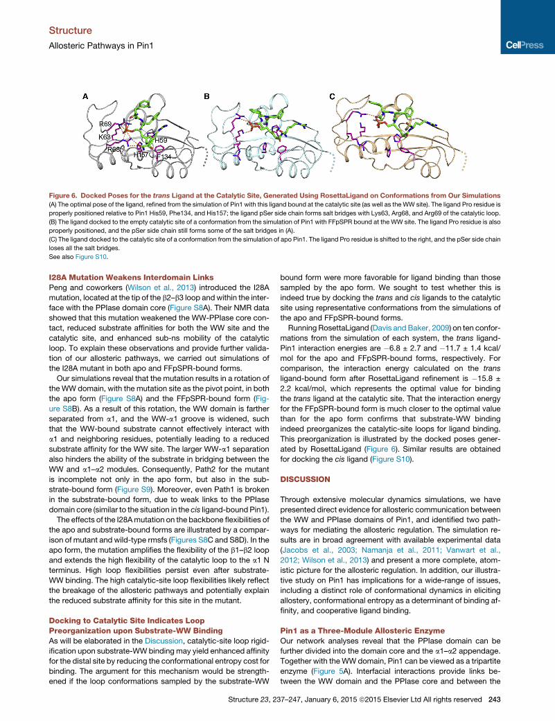
Figure 6. Docked Poses for the trans Ligand at the Catalytic Site, Generated Using RosettaLigand on Conformations from Our Simulations
(A) The optimal pose of the ligand, refined from the simulation of Pin1 with this ligand bound at the catalytic site (as well as the WW site). The ligand Pro residue is
properly positioned relative to Pin1 His59, Phe134, and His157; the ligand pSer side chain forms salt bridges with Lys63, Arg68, and Arg69 of the catalytic loop.
(B) The ligand docked to the empty catalytic site of a conformation from the simulation of Pin1 with FFpSPR bound at the WW site. The ligand Pro residue is also
properly positioned, and the pSer side chain still forms some of the salt bridges in (A).
(C) The ligand docked to the catalytic site of a conformation from the simulation of apo Pin1. The ligand Pro residue is shifted to the right, and the pSer side chain
loses all the salt bridges.
See also Figure S10.
Structure
Allosteric Pathways in Pin1
I28A Mutation Weakens Interdomain LinksPeng and coworkers (Wilson et al., 2013) introduced the I28A
mutation, located at the tip of the b2–b3 loop andwithin the inter-
face with the PPIase domain core (Figure S8A). Their NMR data
showed that this mutation weakened the WW-PPIase core con-
tact, reduced substrate affinities for both the WW site and the
catalytic site, and enhanced sub-ns mobility of the catalytic
loop. To explain these observations and provide further valida-
tion of our allosteric pathways, we carried out simulations of
the I28A mutant in both apo and FFpSPR-bound forms.
Our simulations reveal that the mutation results in a rotation of
the WW domain, with the mutation site as the pivot point, in both
the apo form (Figure S8A) and the FFpSPR-bound form (Fig-
ure S8B). As a result of this rotation, the WW domain is farther
separated from a1, and the WW-a1 groove is widened, such
that the WW-bound substrate cannot effectively interact with
a1 and neighboring residues, potentially leading to a reduced
substrate affinity for the WW site. The larger WW-a1 separation
also hinders the ability of the substrate in bridging between the
WW and a1–a2 modules. Consequently, Path2 for the mutant
is incomplete not only in the apo form, but also in the sub-
strate-bound form (Figure S9). Moreover, even Path1 is broken
in the substrate-bound form, due to weak links to the PPIase
domain core (similar to the situation in the cis ligand-bound Pin1).
The effects of the I28Amutation on the backbone flexibilities of
the apo and substrate-bound forms are illustrated by a compar-
ison of mutant and wild-type rmsfs (Figures S8C and S8D). In the
apo form, the mutation amplifies the flexibility of the b1–b2 loop
and extends the high flexibility of the catalytic loop to the a1 N
terminus. High loop flexibilities persist even after substrate-
WW binding. The high catalytic-site loop flexibilities likely reflect
the breakage of the allosteric pathways and potentially explain
the reduced substrate affinity for this site in the mutant.
Docking to Catalytic Site Indicates LoopPreorganization upon Substrate-WW BindingAs will be elaborated in the Discussion, catalytic-site loop rigid-
ification upon substrate-WWbinding may yield enhanced affinity
for the distal site by reducing the conformational entropy cost for
binding. The argument for this mechanism would be strength-
ened if the loop conformations sampled by the substrate-WW
Structure 23, 23
bound form were more favorable for ligand binding than those
sampled by the apo form. We sought to test whether this is
indeed true by docking the trans and cis ligands to the catalytic
site using representative conformations from the simulations of
the apo and FFpSPR-bound forms.
Running RosettaLigand (Davis and Baker, 2009) on ten confor-
mations from the simulation of each system, the trans ligand-
Pin1 interaction energies are �6.8 ± 2.7 and �11.7 ± 1.4 kcal/
mol for the apo and FFpSPR-bound forms, respectively. For
comparison, the interaction energy calculated on the trans
ligand-bound form after RosettaLigand refinement is �15.8 ±
2.2 kcal/mol, which represents the optimal value for binding
the trans ligand at the catalytic site. That the interaction energy
for the FFpSPR-bound form is much closer to the optimal value
than for the apo form confirms that substrate-WW binding
indeed preorganizes the catalytic-site loops for ligand binding.
This preorganization is illustrated by the docked poses gener-
ated by RosettaLigand (Figure 6). Similar results are obtained
for docking the cis ligand (Figure S10).
DISCUSSION
Through extensive molecular dynamics simulations, we have
presented direct evidence for allosteric communication between
the WW and PPIase domains of Pin1, and identified two path-
ways for mediating the allosteric regulation. The simulation re-
sults are in broad agreement with available experimental data
(Jacobs et al., 2003; Namanja et al., 2011; Vanwart et al.,
2012; Wilson et al., 2013) and present a more complete, atom-
istic picture for the allosteric regulation. In addition, our illustra-
tive study on Pin1 has implications for a wide-range of issues,
including a distinct role of conformational dynamics in eliciting
allostery, conformational entropy as a determinant of binding af-
finity, and cooperative ligand binding.
Pin1 as a Three-Module Allosteric EnzymeOur network analyses reveal that the PPIase domain can be
further divided into the domain core and the a1–a2 appendage.
Together with theWW domain, Pin1 can be viewed as a tripartite
enzyme (Figure 5A). Interfacial interactions provide links be-
tween the WW domain and the PPIase core and between the
7–247, January 6, 2015 ª2015 Elsevier Ltd All rights reserved 243
Structure
Allosteric Pathways in Pin1
PPIase core and the a1–a2 module, but WW-a1 links are largely
missing in the apo protein. The substrate-binding pocket on the
WW domain borders the WW-a1 groove. When a substrate
binds, this groove is filled, and a bridge between the WW and
a1–a2 modules is formed. The WW-bound substrate thus pro-
vides essential links between the WW and a1–a2 modules to
complete the second allosteric pathway from the WW domain
to a catalytic-site loop.
The first allosteric pathway, which connects the WW domain
with two other catalytic-site loops via the WW-PPIase core inter-
face, preexists in apo Pin1, but remains dormant until the second
pathway is completed by substrate-WW binding. That is, allo-
steric regulation is achieved only through the concerted action
of the two pathways. Indeed, the two pathways appear to rein-
force each other, since in some cases (e.g., the cis ligand-bound
Pin1 and the substrate-bound I28Amutant; Figures 4C andS9B),
incompletion of the second pathway is accompanied by
breakage of the first pathway. The essential role of the second
pathway is demonstrated not only by the absence of allosteric
communication in the apo protein, but also by the observation
that the communication is one-way only, i.e., ligand binding at
the PPIase site does not lead to allosteric effects on the b1–b2
loop of the WW domain.
Peng and coworkers (Namanja et al., 2007, 2011) previously
proposed a hydrophobic conduit, consisting of about ten
methyl-containing residues, which lost side-chain flexibility
upon substrate binding (Figure S5B), for interdomain communi-
cation. Our study suggests that these methyl-containing side
chains only provide a partial picture for the allosteric mechanism.
The complete mechanism involves two mutually reinforcing
pathways, one leading to two of the catalytic-site loops, and
the other leading to the third. The latter, i.e., the catalytic loop
(residues 63–72), does not have any methyl-containing side
chains and serves as a reminder of the limitation of order param-
eter measurements. Molecular dynamics simulations also have
limitations, such as the range of accessible timescales. Fortu-
nately for Pin1, much of the conformational dynamics important
for allosteric communication appears to occur at timescales up
to 10 s of ns and have been captured by our simulations.
Restrained Molecular Dynamics as a Tool for DissectingAllosteric EffectsHere, we introduced restrained molecular dynamics simulations
to help dissect the allosteric effects of substrate binding and
identify essential links along allosteric pathways. Our restrained
simulations provide further support to the division of Pin1 into
three modules and to the role of substrate-WW binding in
furnishing essential WW-a1 links. This approach can be applied
to allosteric proteins in general for revealing their modular de-
signs and ascertaining allosteric pathways.
The restrained simulations also provide a unique opportunity
to isolate the role of conformational dynamics in eliciting allo-
stery. The classical view of allostery (Fischer et al., 2011; Monod
et al., 1965) has emphasized the role of conformational transi-
tion. However, there is growing appreciation of the importance
of the change in dynamics, as opposed to the change in confor-
mation, even to the extent that perhaps allostery can be elicited
by the former alone (Cooper and Dryden, 1984; Gasper et al.,
2012; Petit et al., 2009; Tsai et al., 2008; Vashisth et al., 2013).
244 Structure 23, 237–247, January 6, 2015 ª2015 Elsevier Ltd All rig
However, in practice, a change in dynamics is always accompa-
nied by some change in conformation and vice versa. It has thus
been difficult to cleanly separate dynamical effects from confor-
mational effects. Our application of restrained simulations dem-
onstrates their utility in this regard. In these simulations, the
mobility of a selected region of the protein is limited while
imposing the apo conformation, thus generating a change in dy-
namics without an accompanying change in conformation. We
produced allosteric effects similar to those by substrate-WW
binding when the dynamically affected region included the
essential WW-a1 links, thus providing a clear example of allo-
stery by dynamics alone.
Conformational Entropy Reduction and PreorganizationBy CSP mapping, Peng and coworkers (Namanja et al., 2011)
measured the binding affinities of FFpSPR and the trans and
cis ligands for theWWand catalytic sites of Pin1 and the isolated
PPIase domain. The results for the catalytic site were puzzling:
while the cis ligand, which does not bind to the WW site, had a
4-fold lower affinity for the full-length protein than for the PPIase,
both FFpSPR and the trans ligand had 2- to 3-fold higher affin-
ities for the full-length protein. We can now provide an explana-
tion based on the notion that catalytic-site loop rigidification can
enhance affinity by reducing the entropy cost of binding. Confor-
mational entropy has been recognized as a determinant of bind-
ing affinity in general (Frederick et al., 2007; Zhou and Gilson,
2009) and invoked to support the possibility of allostery by dy-
namics in particular (Petit et al., 2009).
Figure S6 shows that, compared to the isolated PPIase
domain, the WW domain in full-length Pin1 amplifies the confor-
mational flexibility of the three catalytic-site loops. We suggest
that there is a corresponding increase in entropy cost for binding,
thus explaining the 4-fold lower affinity of the cis ligand for the
full-length protein than for the PPIase.
We expect the same 4-fold reduction in affinity for the trans
ligand and FFpSPR, were they only able to bind the catalytic
site. However, these two ligands also bind to the WW site, and
our simulations show that FFpSPR-WW binding results in signif-
icant reduction in the catalytic-site loop flexibility (Figure 3).
Therefore, after the WW site is occupied, the conformational en-
tropy cost for binding to the catalytic site of Pin1 is even lower
than for binding to the catalytic site of the isolated PPIase
domain. This explains the 2- to 3-fold higher affinities of FFpSPR
and the trans ligand.
The argument based on reduction in entropy cost is strength-
ened if the rigidified loop conformations of the substrate-WW
bound form are more favorable for ligand binding than the flex-
ible ensemble of the apo form. Our calculations using RosettaLi-
gand show that this is indeed true (Figures 6B and S10B).
Substrate-WWbinding thus preorganizes the catalytic-site loops
for ligand binding, presenting a unique form of cooperativity.
If substrate-WW binding preorganizes the catalytic-site loops
for binding, the preorganization could lead to enhanced catalytic
activity. This might explain the slightly higher kcat/KM value of the
full-length Pin1 relative to the isolated PPIase domain (Lu et al.,
1999a; Zhou et al., 2000).
The above argument can also explain the effects of the I28A
mutation on binding affinity and catalytic activity. Peng and co-
workers (Wilson et al., 2013) found lowered substrate binding
hts reserved
Structure
Allosteric Pathways in Pin1
affinities for both the WW site and the catalytic site, as well as
lowered kcat/KM. We attribute the reduced affinity for the WW
site to a widened WW-a1 groove, due to a rotation of the WW
domain away from a1. Furthermore, we suggest that the reduced
affinity for the catalytic site is due to a disruption of the allosteric
pathways in the WW-substrate bound form of the mutant (Fig-
ure S9B). As a result, the catalytic-site loop flexibility is not
quenched asmuch as in the wild-type protein (Figure S8D), lead-
ing to reduced affinity at the catalytic site and lowered kcat/KM.
Reliability of the Allosteric Picture and Further TestsWe have used multiple analysis methods to characterize the dif-
ferences in conformations, dynamics, and allosteric networks
among the various liganded forms of Pin1. The principal compo-
nent analysis (Figure 2) andRg (Figures S3 and S4) and rmsf (Fig-
ure 3) results show that substrate binding to the WW domain
leads to the closure of, and mobility loss in, the three catalytic-
site loops of the PPIase domain. These remote effects demon-
strate allosteric communication between the domains. Then,
the two network analysis methods both reveal that the WW-
bound substrate acts as a bridge between the WW domain
and the a1 helix to complete the second allosteric pathway to-
ward the catalytic loop. Lastly, our restrained simulations
confirm that theWW-a1 links are essential for the allosteric regu-
lation. These various analyses of the many simulations are
consistent and complementary in generating a robust, detailed
picture of the allosteric behavior of Pin1.
We have already validated our computational results against a
range of experimental data, including CSPs (Figure S2), order
parameters (Figure S5), and the effects of the I28A mutation.
Additional tests can be designed to further interrogate the allo-
steric picture presented here. For example, it is known that a
different substrate, a 10-residue phosphothreonine peptide
from the mitotic phosphatase Cdc25C, has less effect than
FFpSPR in inducing Pin1 interdomain coupling (Jacobs et al.,
2003; Namanja et al., 2007). The Cdc25C peptide lines the bind-
ing pocket on the WW domain in a mode distinct from that of
FFpSPR (Wintjens et al., 2001). Though its affinity for the WW
domain is higher, CSP mapping suggests that the Cdc25C pep-
tide interacts less intimately with the PPIase domain than
FFpSPR (Namanja et al., 2007). The present study leads us to
predict that a reduced ability in forming Path2 explains why the
Cdc25C peptide is less effective in inducing Pin1 interdomain
coupling. It will be interesting to test this prediction in molecular
dynamics simulations.
Another way to directly test the putative essential role of WW-
a1 links in eliciting the allosteric effects is to see whether a
constitutively active variant can be produced by crosslinking
the WW domain and the a1 helix. The Ca-Ca distance between
residues 31 (WW domain) and 93 (a1 helix) is slightly under
10 A, and therefore it may be possible for a disulfide bond or
metal coordination to form when these residues are mutated to
cysteines (or histidines). Such a mutant would be a tantalizing
system for future simulations and experiments.
Potential for Combination Drug TherapyThe cooperative effects delineated above suggest that a
WW-directed ligand can be a positive allosteric modulator for a
catalytic site-directed ligand. Combinations of allosteric and or-
Structure 23, 23
thosteric drugs have been noted for their abilities to enhance
pharmacological action, reduce side effects, and combat drug-
resistant mutants (Epping-Jordan et al., 2007; Nussinov and
Tsai, 2013; Zimmermann et al., 2007). Pin1 dysregulation is
implicated in various diseases (Lu, 2004; Lu and Zhou, 2007;
Lu et al., 1999b; Wulf et al., 2001), and a number of orthosteric
inhibitors have been designed (Moore and Potter, 2013; Wang
and Etzkorn, 2006). The study here suggests the potential for a
combination drug therapy against Pin1-related diseases,
whereby allosteric inhibitors at the WW site enhance the binding
affinities of orthosteric inhibitors at the catalytic site.
EXPERIMENTAL PROCEDURES
System Preparations
Initial models of the systems studied were prepared in Discovery Studio
(Accelrys Software), based on the crystal structures of Pin1 bound with the
cis or trans ligand at the catalytic site (PDB 3TCZ and 3TDB, see Zhang
et al., 2012) and bound with a pSer-Pro containing peptide (PDB 1F8A, see
Verdecia et al., 2000) at the WW site. The systems included Pin1 with FFpSPR
bound at the WW site; Pin1 with the cis ligand bound at the catalytic site; and
Pin1 with two copies of the trans ligand bound, one at the WW site and one at
the catalytic site. For the Pin1-FFpSPR complex, the protein was from 3TDB,
and the substrate wasmodeled from one in 1F8A (after superimposing the pro-
tein molecules in these two PDB entries). The Pin1-cis ligand complex was
taken directly from 3TCZ. The Pin1-trans ligand complex both retained the cat-
alytic site-bound trans ligand in 3TDB and had a second copy of the trans
ligand modeled after the WW-bound substrate in the Pin1-FFpSPR complex.
Missing residues of the ligands and the protein (residues 39–50 in the latter
case, which form the interdomain linker) were added.
The above systems were modified straightforwardly to yield others. For
example, the substrate was removed from the Pin1-FFpSPR complex to pro-
duce apo Pin1; theWWdomain and the interdomain linker were further deleted
to produce the isolated PPIase domain in apo form; and theWW-bound ligand
was removed from the Pin1-trans ligand complex to yield a complex with only
one copy of the trans ligand bound, at the catalytic site.
Molecular Dynamics Simulations
All simulationswerecarriedout using theAMBERsoftwarewith theAMBER99SB
force field (Hornak et al., 2006). The pSer residue wasmodeled as singly proton-
ated, with atomic charges taken from Homeyer et al. (2006). The cis-locked and
trans-locked imide fragmentswere treated as nonstandard residues, denoted as
CIS and TRA, respectively. The atomic charges of ACE-CIS/TRA-NME were
calculated by the R.E.D. server (http://q4md-forcefieldtools.org/RED/) using
Gaussian 03. The net charges of ACE and NME were set to 0, and those of
CIS/TRA to �1. Other missing force-field parameters of the nonstandard resi-
dues were taken from the general AMBER force field (gaff) and parm99 data
set with minor modifications (Homeyer et al., 2006).
Each complex (or protein) was solvated in a cubic box with TIP3P waters
(Jorgensen et al., 1983), with at least 10 A between the solute and nearest
side of the box. Sodium and chloride ions were added to neutralize the system
and yield a 30mM salt concentration. The whole systemwas first energy-mini-
mized, with a series of position restraints on the solute (all atoms, backbone
atoms, Ca atoms, and finally no atoms). Subsequently, the system was heated
from 0 Kelvin (K) to 295 K with backbone atoms restrained for 50 ps, and then
equilibrated with Ca atoms restrained for 50 ps. The restraints excluded the in-
terdomain linker. The simulation was continued at constant pressure (1 bar,
maintained by isotropic position scaling with a 2-ps relaxation time) and con-
stant temperature (using the Langevin thermostat with a 2-ps�1 collision fre-
quency) for 100 ns. The SHAKE algorithm (Ryckaert et al., 1977) was used
to constrain all bonds involving hydrogens, allowing for a 2-fs timestep. Elec-
trostatic interactions were treated by the particle mesh Ewald sum method
(Essmann et al., 1995), with a 10 A cutoff for nonbonded interactions in direct
space.
We also carried out restrained simulations, in which a subset of Pin1 resi-
dues was restrained to their conformation in the frame closest to the average
7–247, January 6, 2015 ª2015 Elsevier Ltd All rights reserved 245
Structure
Allosteric Pathways in Pin1
in the last 40 ns of the apo Pin1 simulation. The restraint was imposed on the
rmsd calculated on the heavy atoms of the restraint set, with a harmonic force
constant of 10 kcal/mol/A2. All restrained simulations started from the last
frame of the apo Pin1 simulation and ran for 100 ns.
The I28A mutant in apo form and FFpSPR-bound form were also simulated.
After introducing the mutation in the last frame of the corresponding wild-type
simulation, each mutant simulation ran for 100 ns.
Principal Component Analysis and RMSF Calculation
Except for the radii of gyration shown in Figure S4, all analyses were made on
the last 40 ns of each simulation. Principal component analysis was carried out
using the Ptraj module in AMBER, over a total of 80,000 Pin1 conformations
(20,000 conformations evenly sampled from the simulation of each of the four
systems shown in Figure 2). For each system, the conformations were binned
into a histogram over the first two principal components, and the histogram
was converted into a free energy surface according to the Boltzmann relation.
Ca rmsfs were calculated for each system after superimposing all of its con-
formations to the simulation average, over the secondary-structure core.
Chemical Shift Prediction
SPARTA+ (Shen and Bax, 2010) was used to predict chemical shifts of back-
bone N and H nuclei, using 2,000 conformations for each system. Then the NH
CSP of each residue (in parts per million) was calculated as
ffiffiffiffiffiffiffiffiffiffiffiffiffiffiffiffiffiffiffiffiffiffiffiffiffiffiffiffiffiffiffiffiffiffiffiffiffiffiffið0:2DdNÞ2 + ðDdHÞ2
q
(Vashisth et al., 2013), where Dd denotes the difference in chemical shift be-
tween a ligand-bound form and the apo form.
Order Parameter Calculation
S2axis values were calculated for all side-chain methyls in 40 1-ns windows. For
each window, S2axis was calculated as (Chatfield et al., 1998)
S2axis =
3
2
��X2
�2+�Y2
�2+�Z2
�2+ 2hXYi2 + 2hXZi2 + 2hYZi2
�� 1
2;
where x, y, and z are Cartesian coordinates of a unit vector along a carbon-
methyl bond, and h:::i denotes the average over the conformations in the
1-ns window, after aligning to the initial model using all Ca atoms to remove
translation and rotation. The averages of S2axis values over the 40 windows
are reported (Figure S5C).
Calculation of Allosteric Networks
The algorithm of Kannan and Vishveshwara (1999), implemented in the
Wordom analysis tool (Seeber et al., 2011), was used to identify allosteric net-
works. In each network, residues are linked by persistent tertiary contacts be-
tween side chains during a simulation. Whether two residues, i and j, are linked
is determined by their interaction percentage, defined as
Iij =nijffiffiffiffiffiffiffiffiffiffiNiNj
p 3 100;
where nij is the number of side chain-side chain heavy atom pairs within a 5 A
cutoff, and Ni and Nj are the normalization factors specified by residue types.
Links are excluded between a residue and its four nearest neighbors in
sequence. In each conformation sampled from a simulation, a provisional
link is formed if Iij exceeds a threshold, Icritic. If a provisional link is formed in
at least 48% of all the conformations from a simulation, then an actual link is
formed. Icritic is chosen so that the largest cluster is approximately half of the
total number of residues.
Docking to Catalytic Site by RosettaLigand
RosettaLigand (Davis and Baker, 2009) was used to dock the trans and cis
ligands into the catalytic site of Pin1 in conformations sampled from the
simulations of the apo form and the FFpSPR-bound form and to refine the
poses of the ligands obtained in the simulations of Pin1 with the trans and
cis ligands bound at the catalytic site. Full flexibility was allowed for the
ligands, but only side-chain flexibility was allowed for the protein, thereby
preserving the backbone conformations. In each docking run, 5,000 poses
were generated.
246 Structure 23, 237–247, January 6, 2015 ª2015 Elsevier Ltd All rig
For each system, ten conformations were selected from the simulation by
clustering based on Ca rmsd. For the two systems with a ligand at the catalytic
site, each of these conformations both provided the initial pose and served as
the reference for selecting the refined pose. The top scoring (i.e., lowest pro-
tein-ligand interaction energy) posewith a ligand rmsd < 2 A from the reference
was selected as the refined pose. The centroid of the ten refined poses is
shown in Figure 6A for the trans ligand-bound system and in Figure S10A for
the cis ligand-bound system.
To dock a ligand to a system that was free of the ligand in the simulation, one
selected conformation of the corresponding ligand-bound systemwas used to
generate the initial pose, based on aligning the protein Ca atoms within 10 A of
the ligand. After the docking run, a refined pose for the ligand-bound system
was used as the reference for selecting the best pose for the ligand-free sys-
tem. The upper bound on ligand rmsd was increased to 2.3 A, since a 2 A
bound did not produce any pose in three of the 20 docking runs for apo
Pin1. The centroid of the ten best poses is shown in Figures 6B and 6C for
the trans ligand docked to FFpSPR-bound Pin1 or apo Pin1; corresponding re-
sults for the cis ligand are shown in Figures S10B and S10C.
SUPPLEMENTAL INFORMATION
Supplemental Information includes ten figures and can be found with this
article online at http://dx.doi.org/10.1016/j.str.2014.11.009.
AUTHOR CONTRIBUTIONS
H.-X.Z. designed the research. J.G. and X.P. performed the research and
analyzed the data. J.G. and H.-X.Z. wrote the manuscript.
ACKNOWLEDGMENTS
We thank Dr. Jeffrey Peng for commenting on the manuscript. This work was
supported by NIH Grant GM58187.
Received: August 19, 2014
Revised: October 24, 2014
Accepted: November 14, 2014
Published: December 24, 2014
REFERENCES
Chatfield, D.C., Szabo, A., and Brooks, B.R. (1998). Molecular dynamics of
staphylococcal nuclease: Comparison of simulation with 15N and 13C NMR
relaxation data. J. Am. Chem. Soc. 120, 5301–5311.
Cooper, A., and Dryden, D.T. (1984). Allostery without conformational change.
A plausible model. Eur. Biophys. J. 11, 103–109.
Davis, I.W., and Baker, D. (2009). RosettaLigand docking with full ligand and
receptor flexibility. J. Mol. Biol. 385, 381–392.
Elber, R. (2011). Simulations of allosteric transitions. Curr. Opin. Struct. Biol.
21, 167–172.
Epping-Jordan, M., Le Poul, E., and Rocher, J.-P. (2007). Innovat.
Pharmaceut. Tech. 24, 22–26.
Essmann, U., Perera, L., Berkowitz, M.L., Darden, T., Lee, H., and Pedersen,
L.G. (1995). A smooth particle mesh Ewald method. J. Chem. Phys. 103,
8577–8593.
Feher, V.A., Durrant, J.D., Van Wart, A.T., and Amaro, R.E. (2014).
Computational approaches to mapping allosteric pathways. Curr. Opin.
Struct. Biol. 25, 98–103.
Fischer, S., Olsen, K.W., Nam, K., and Karplus, M. (2011). Unsuspected
pathway of the allosteric transition in hemoglobin. Proc. Natl. Acad. Sci.
USA 108, 5608–5613.
Frederick, K.K., Marlow, M.S., Valentine, K.G., and Wand, A.J. (2007).
Conformational entropy in molecular recognition by proteins. Nature 448,
325–329.
Gasper, P.M., Fuglestad, B., Komives, E.A., Markwick, P.R., and
McCammon, J.A. (2012). Allosteric networks in thrombin distinguish
hts reserved
Structure
Allosteric Pathways in Pin1
procoagulant vs. anticoagulant activities. Proc. Natl. Acad. Sci. USA 109,
21216–21222.
Gerek, Z.N., and Ozkan, S.B. (2011). Change in allosteric network affects bind-
ing affinities of PDZ domains: analysis through perturbation response scan-
ning. PLoS Comput. Biol. 7, e1002154.
Ghosh, A., and Vishveshwara, S. (2007). A study of communication pathways
in methionyl- tRNA synthetase by molecular dynamics simulations and struc-
ture network analysis. Proc. Natl. Acad. Sci. USA 104, 15711–15716.
Homeyer, N., Horn, A.H., Lanig, H., and Sticht, H. (2006). AMBER force-field
parameters for phosphorylated amino acids in different protonation states:
phosphoserine, phosphothreonine, phosphotyrosine, and phosphohistidine.
J. Mol. Model. 12, 281–289.
Hornak, V., Abel, R., Okur, A., Strockbine, B., Roitberg, A., and Simmerling, C.
(2006). Comparison of multiple Amber force fields and development of
improved protein backbone parameters. Proteins 65, 712–725.
Jacobs, D.M., Saxena, K., Vogtherr, M., Bernado, P., Pons, M., and Fiebig,
K.M. (2003). Peptide binding induces large scale changes in inter-domain
mobility in human Pin1. J. Biol. Chem. 278, 26174–26182.
Jorgensen, W., Chandrasekhar, J., Madura, J., Impey, R., and Klein, M. (1983).
Comparison of simple potential functions for simulating liquid water. J. Chem.
Phys. 79, 926–935.
Kannan, N., and Vishveshwara, S. (1999). Identification of side-chain clusters
in protein structures by a graph spectral method. J. Mol. Biol. 292, 441–464.
Liou, Y.C., Zhou, X.Z., and Lu, K.P. (2011). Prolyl isomerase Pin1 as a molec-
ular switch to determine the fate of phosphoproteins. Trends Biochem. Sci. 36,
501–514.
Lu, K.P. (2004). Pinning down cell signaling, cancer and Alzheimer’s disease.
Trends Biochem. Sci. 29, 200–209.
Lu, K.P., and Zhou, X.Z. (2007). The prolyl isomerase PIN1: a pivotal new twist
in phosphorylation signalling and disease. Nat. Rev. Mol. Cell Biol. 8, 904–916.
Lu, K.P., Hanes, S.D., and Hunter, T. (1996). A human peptidyl-prolyl isom-
erase essential for regulation of mitosis. Nature 380, 544–547.
Lu, P.-J., Zhou, X.Z., Shen, M., and Lu, K.P. (1999a). Function of WW domains
as phosphoserine- or phosphothreonine-binding modules. Science 283,
1325–1328.
Lu, P.J., Wulf, G., Zhou, X.Z., Davies, P., and Lu, K.P. (1999b). The prolyl isom-
erase Pin1 restores the function of Alzheimer-associated phosphorylated tau
protein. Nature 399, 784–788.
Lu, P.J., Zhou, X.Z., Liou, Y.C., Noel, J.P., and Lu, K.P. (2002). Critical role of
WW domain phosphorylation in regulating phosphoserine binding activity and
Pin1 function. J. Biol. Chem. 277, 2381–2384.
Monod, J., Wyman, J., and Changeux, J.-P. (1965). On the nature of allosteric
transitions: a plausible model. J. Mol. Biol. 12, 88–118.
Moore, J.D., and Potter, A. (2013). Pin1 inhibitors: Pitfalls, progress and cellular
pharmacology. Bioorg. Med. Chem. Lett. 23, 4283–4291.
Namanja, A.T., Peng, T., Zintsmaster, J.S., Elson, A.C., Shakour, M.G., and
Peng, J.W. (2007). Substrate recognition reduces side-chain flexibility for
conserved hydrophobic residues in human Pin1. Structure 15, 313–327.
Namanja, A.T., Wang, X.J., Xu, B., Mercedes-Camacho, A.Y., Wilson, K.A.,
Etzkorn, F.A., and Peng, J.W. (2011). Stereospecific gating of functional mo-
tions in Pin1. Proc. Natl. Acad. Sci. USA 108, 12289–12294.
Nussinov, R., and Tsai, C.-J. (2013). Allostery in disease and in drug discovery.
Cell 153, 293–305.
Petit, C.M., Zhang, J., Sapienza, P.J., Fuentes, E.J., and Lee, A.L. (2009).
Hidden dynamic allostery in a PDZ domain. Proc. Natl. Acad. Sci. USA 106,
18249–18254.
Ranganathan, R., Lu, K.P., Hunter, T., and Noel, J.P. (1997). Structural and
functional analysis of the mitotic rotamase Pin1 suggests substrate recogni-
tion is phosphorylation dependent. Cell 89, 875–886.
Structure 23, 23
Rousseau, F., and Schymkowitz, J. (2005). A systems biology perspective on
protein structural dynamics and signal transduction. Curr. Opin. Struct. Biol.
15, 23–30.
Ryckaert, J.P., Ciccotti, G., and Berendsen, H.J.C. (1977). Numerical integra-
tion of the Cartesian equations of motion of a system with constraints: molec-
ular dynamics of n-alkanes. J. Comp. Physiol. 23, 327–341.
Seeber, M., Felline, A., Raimondi, F., Muff, S., Friedman, R., Rao, F., Caflisch,
A., and Fanelli, F. (2011). Wordom: a user-friendly program for the analysis of
molecular structures, trajectories, and free energy surfaces. J. Comput. Chem.
32, 1183–1194.
Sethi, A., Eargle, J., Black, A.A., and Luthey-Schulten, Z. (2009). Dynamical
networks in tRNA:protein complexes. Proc. Natl. Acad. Sci. USA 106, 6620–
6625.
Shen, Y., and Bax, A. (2010). SPARTA+: a modest improvement in empirical
NMR chemical shift prediction by means of an artificial neural network.
J. Biomol. NMR 48, 13–22.
Tsai, C.J., del Sol, A., and Nussinov, R. (2008). Allostery: absence of a change
in shape does not imply that allostery is not at play. J. Mol. Biol. 378, 1–11.
Vanwart, A.T., Eargle, J., Luthey-Schulten, Z., and Amaro, R.E. (2012).
Exploring residue component contributions to dynamical network models of
allostery. J. Chem. Theory Comput. 8, 2949–2961.
Van Wart, A.T., Durrant, J., Votapka, L., and Amaro, R.E. (2014). Weighted im-
plementation of suboptimal paths (WISP): an optimized algorithm and tool for
dynamical network analysis. J. Chem. Theory Comput. 10, 511–517.
Vashisth, H., Storaska, A.J., Neubig, R.R., and Brooks, C.L., 3rd. (2013).
Conformational dynamics of a regulator of G-protein signaling protein reveals
a mechanism of allosteric inhibition by a small molecule. ACS Chem. Biol. 8,
2778–2784.
Verdecia, M.A., Bowman, M.E., Lu, K.P., Hunter, T., and Noel, J.P. (2000).
Structural basis for phosphoserine-proline recognition by group IV WW do-
mains. Nat. Struct. Biol. 7, 639–643.
Wang, X.J., and Etzkorn, F.A. (2006). Peptidyl-prolyl isomerase inhibitors.
Biopolymers 84, 125–146.
Wang, X.J., Xu, B., Mullins, A.B., Neiler, F.K., and Etzkorn, F.A. (2004).
Conformationally locked isostere of phosphoSer-cis-Pro inhibits Pin1 23-
fold better than phosphoSer-trans-Pro isostere. J. Am. Chem. Soc. 126,
15533–15542.
Wilson, K.A., Bouchard, J.J., and Peng, J.W. (2013). Interdomain interactions
support interdomain communication in human Pin1. Biochemistry 52, 6968–
6981.
Wintjens, R., Wieruszeski, J.M., Drobecq, H., Rousselot-Pailley, P., Buee, L.,
Lippens, G., and Landrieu, I. (2001). 1H NMR study on the binding of Pin1
Trp-Trp domain with phosphothreonine peptides. J. Biol. Chem. 276,
25150–25156.
Wulf, G.M., Ryo, A., Wulf, G.G., Lee, S.W., Niu, T., Petkova, V., and Lu, K.P.
(2001). Pin1 is overexpressed in breast cancer and cooperates with Ras
signaling in increasing the transcriptional activity of c-Jun towards cyclin D1.
EMBO J. 20, 3459–3472.
Zhang, M., Wang, X.J., Chen, X., Bowman, M.E., Luo, Y., Noel, J.P., Ellington,
A.D., Etzkorn, F.A., and Zhang, Y. (2012). Structural and kinetic analysis of
prolyl-isomerization/phosphorylation cross-talk in the CTD code. ACS
Chem. Biol. 7, 1462–1470.
Zhou, H.X., and Gilson, M.K. (2009). Theory of free energy and entropy in non-
covalent binding. Chem. Rev. 109, 4092–4107.
Zhou, X.Z., Kops, O., Werner, A., Lu, P.-J., Shen, M., Stoller, G., Kullertz, G.,
Stark, M., Fischer, G., and Lu, K.P. (2000). Pin1-dependent prolyl isomerization
regulates dephosphorylation of Cdc25C and tau proteins. Mol. Cell 6,
873–883.
Zimmermann, G.R., Lehar, J., and Keith, C.T. (2007). Multi-target therapeutics:
when the whole is greater than the sum of the parts. Drug Discov. Today 12,
34–42.
7–247, January 6, 2015 ª2015 Elsevier Ltd All rights reserved 247


