Making new molecules — evolution of structures for novelmetabolites in plantsDaniel J Kliebenstein
Available online at www.sciencedirect.com
Secondary metabolites are essential plant fitness within the
natural environment by providing defense against attacking
and competing organisms including bacteria, fungi, insects,
animals and other plants. These compounds’ defensive
function is frequently intertwined with specific accumulation in
novel developmental structures. While, the biochemical
community is making great strides in identifying the genetic and
biochemical mechanisms that allow these chemicals to be
synthesized there is vastly less progress on understanding the
developmental mechanisms that is equally key to their
defensive function. In this review, I briefly delve into several
novel developmental structures and provide evolutionary
hypothesis for how they may have evolved and how they could
be unique systems for studying key developmental processes
that have heretofore been recalcitrant to study.
Address
Department of Plant Sciences, University of California, Davis, CA 95616,
USA
Corresponding author: Kliebenstein, Daniel J ([email protected])
Current Opinion in Plant Biology 2013, 16:112–117
This review comes from a themed issue on Growth and development
Edited by Michael Scanlon and Marja Timmermans
For a complete overview see the Issue and the Editorial
Available online 5th January 2013
1369-5266/$ – see front matter, # 2012 Elsevier Ltd. All rights
reserved.
http://dx.doi.org/10.1016/j.pbi.2012.12.004
IntroductionA general discussion of plant metabolism is often cen-
tered around two major classes. Primary metabolism is the
metabolism which allows a plant to utilize water, carbon
dioxide and minerals to create metabolites required to
make and maintain cells (sugars, fatty acids, amino acids
and nucleic acids) [1]. These chemicals were slotted into
a biological function early in the evolution of life as
reflected by their synthetic genes being largely conserved
across all known plants. Secondary metabolites (also
referred to as natural products, phytochemicals, defense
chemicals, defense metabolites, specialized metabolites,
among others) are the chemicals required for plant inter-
actions with the environment (e.g., pest and pathogen
defense compounds, ultraviolet-B sunscreens) [1–4]. This
relationship of secondary metabolites with an ever-fluctu-
ating biotic environment imparts an evolutionary pressure
Current Opinion in Plant Biology 2013, 16:112–117
upon plants to constantly create new secondary metab-
olites causing most secondary metabolites to be lineage-
specific. Interestingly this lineage novelty does not mean
that any given secondary chemical is any less essential to
the survival of a plant species within its natural environ-
ment than primary metabolites, plants lacking the appro-
priate secondary metabolism frequently have dramatically
reduced fitness if any residual fitness in the field [5]. Where
primary and secondary chemicals differ is that in the
artificial laboratory environment secondary metabolite
deficient plants have sufficient viability to be easily
manipulated in any genetic, biochemical or cytological
study. This makes systems associated with secondary
metabolites frequently easier to study and manipulate than
those affiliated with primary metabolites.
While secondary metabolites are essential to a plant’s
survival, this is only true under certain conditions when
the appropriate pathogen, herbivore or competitor is
present [6–8]. In the absence of the appropriate pathogen,
herbivore or competitor the compounds are highly
expensive to synthesize. In Arabidopsis thaliana, the syn-
thesis of the glucosinolate defense compounds imparts a
significant cost in utilizing more than 10% of the energy
available in the metabolic network with an associated cost
of decreased growth [9–11]. This conditionality of benefit
imparts a very complex cost/benefit calculation that the
plant species must solve to optimize the benefit of the
compound while diminishing its associated costs. Numer-
ous models for this optimization focus on the induction of
defense compounds in response to the pathogen or insect
[12,13]. Another optimization approach is to developmen-
tally or ontogenically regulate the secondary metabolite
so that it is present only in the tissues or life stages when
the specific attacking organism occurs [14–18]. Together
these regulatory systems can form a complex interaction
of genotype, ontogeny, tissue development and environ-
ment [19,20]. In addition to these macrolevel concepts,
another theorized mechanism for the plant to optimize
the cost/benefit calculation is to restrict the localization
of secondary metabolites to sites/tissues of optimal
activity. This idea is supported by the fact that the
accumulation of key secondary metabolites is often lim-
ited to specialized tissues such as glandular trichomes,
laticifers, secretory cavities, secondary phloem, resin
ducts or specialized cell compartments like the cell wall,
vacuole or cuticular wax layer [21]. This sequestration
from the broader tissues allows the defense chemical to
be generated in highly localized pockets of higher con-
centration, simultaneously increasing the immediate
www.sciencedirect.com
Making new molecules — evolution of structures for novel metabolites in plants Kliebenstein 113
Glossary
Definitions of specialized tissues discussed within the text. This is not
meant as an exhaustive list of all specialized tissues associated with
secondary metabolism in plants.
Laticifer: Elongated cells with altered cytoplasm that accumulate latex
and frequently also accumulate defense compounds.
S-cell: Cells within the Brassicaceae that have developmental properties
similar to the laticifers but accumulate glucosinolates and not latex.
Extrafascicular phloem: An additional phloem network within the
cucurbits that appears to function for the transport of defense
compounds.
Glandular trichomes: Specialized multi-cellular epidermal protrudance
that is often optimized as a metabolic factory to produce and store volatile
oils and other potential defense compounds.
Secretory cavity: Subdermal structures that produce and accumulate
defense compounds. Typically the structure has a storage lumen
surrounded by secretory/synthesis cells and an outer layer of
parenchymatous cells.
Resin duct: A duct within woody tissues that is lined with glandular
epithelium to secrete resin and other defense chemicals.
dose to herbivores and pathogens and decreasing the
overall synthetic cost of making the chemical in every
cell. Another potential benefit to localized accumulation
of secondary metabolites is that this limited distribution
can decrease the ability of herbivores and pathogens to
evolve resistance to the defense chemistry [22,23].
Thus, an essential component of any secondary metab-
olite defense is the generation and proper function of
the specialized accumulation tissue. Yet very little is
known about how these specialized tissues (glandular
trichomes, secondary phloem, laticifers, secretory
cavities, resin ducts, among others) develop or are
regulated. The nescience of these specialized devel-
opmental systems has long been ascribed to the com-
mon knowledge that these tissues are not present in
key model systems and as such not genetically amen-
able. The advent of new genomics technologies is
beginning to erode the model systems technical
advantage [24]. However, this antimodel system argu-
ment also has an underlying presumption that each
specialized developmental system is novel and thus, a
unique, independent and different evolutionary event
with unique genes, among others. At one point, there
was a similar argument about floral development and
the generation of novelty but the past two decades
have shown that the ABC modular model of floral
development simultaneously allows for a common
set of gene functions to create an impressive range
of novelty using gene duplications, gene losses, neo-
functionalization and subfunctionalization events
[25,26]. In this review, I would like to posit the
argument that this reiteration and novelization of
common gene regulatory models for fundamental tis-
sues like vasculature and trichomes could also explain
the vast majority of plant secondary metabolite
specialized tissues. The key difference being that
the complete underlying central developmental
models have yet to be found and described in a way
www.sciencedirect.com
to allow for novelization of the tissue for nonfloral tissues.
In fact it would be easier to identify these underlying gene
regulatory models using the specialized novelty develop-
mental structure that was derived from the original struc-
ture rather than the original structure.
Vasculature specialization and modificationfor defense metabolism?A key specialized tissue for plant secondary metabolism is
the laticifer, specialized cell types dominantly defined as
having latex [27]. Other key characteristics to laticifers are
being highly elongated vascular associated cells with
altered cytoplasm [27]. More critically to this review,
laticifers are highly polyphyletic throughout the plant
kingdom which has suggested that most laticifers represent
independent evolutions with an implicit assumption of
independent genes for each event. However, the fact that
nearly every major plant group has laticifers suggests that
there must be a central module of genes providing the
potential to repeatedly evolve laticifers. The question then
becomes what is this central module of genes enabling
repeated laticifer evolution?
A potential clue comes from the identification of S-cells
within A. thaliana [28,29�]. S-cells are inflorescence
phloem associated elongated cells with altered cytoplasm
and high levels of the glucosinolate secondary metab-
olites. As such, S-cells have been proposed to be identical
developmental structures to laticifers. Removing the
latex requirement for laticifer definition could allow for
more plant species to have laticifer-like structures than
has originally been described. While Arabidopsis leaves
do not have classical S-cells they do have unique single
cells scattered along the vascular bundle that can be seen
when using glucosinolate-specific markers [30]. This
positioning is identical to where an S-cell/laticifer should
be located but these glucosinolate idioblasts do not con-
nect to form a contiguous network. The repeated posi-
tioning near the vasculature for glucosinolate idioblasts,
S-cells and laticifers suggests a hypothesis that laticifers/
S-cells are evolutionarily derived from a vascular devel-
opmental module. This specialized cell type is often
phloem associated suggesting that it may represent a
novelization of the phloem developmental program.
Additionally, laticifers can show tip development which
is also true of xylem during secondary growth [31]. Thus,
if this novel vascular hypothesis is true, it would require
the combination of components from the xylem and
phloem modules. Unfortunately, the absolute require-
ment for an intact vasculature to create a viable plant
has inhibited our understanding of the underlying genetic
modules. However, if this novelization of a vascular
module hypothesis is true it suggests that identifying
the genetic module controlling laticifer/S-cell develop-
ment could be an easier avenue to understanding the
genetics of vascular development. An interesting test of
this model would be to analyze all known Arabidopsis
Current Opinion in Plant Biology 2013, 16:112–117
114 Growth and development
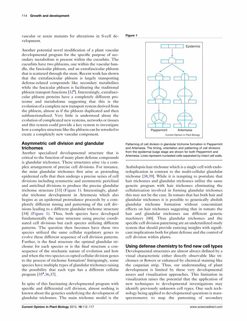
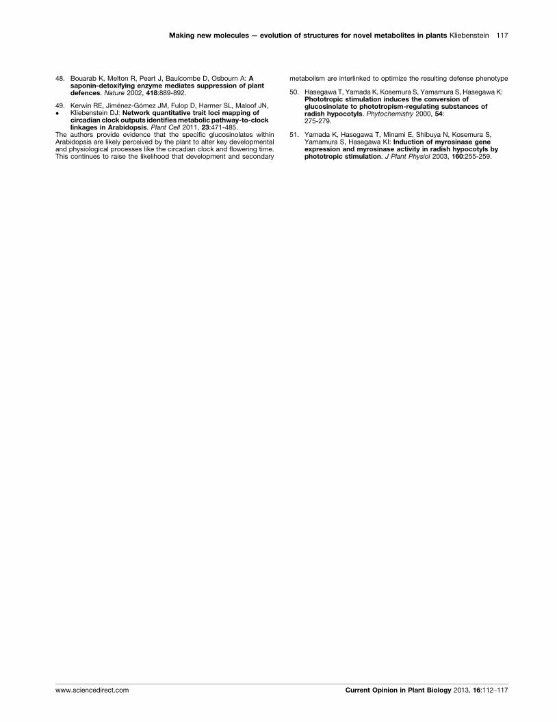
Figure 1
Epidermis
Peppermint ArtemesiaCurrent Opinion in Plant Biology
Patterning of cell division in glandular trichome formation in Peppermint
and Artemesia. The timing, orientation and patterning of cell divisions
from the epidermal bulge stage are shown for both Peppermint and
Artemesia. Lines represent nucleated cells separated by intact cell walls.
vascular or auxin mutants for alterations in S-cell de-
velopment.
Another potential novel modification of a plant vascular
developmental program for the specific purpose of sec-
ondary metabolism is present within the cucurbits. The
cucurbits have two phloems, one within the vascular bun-
dle, the fascicular phloem, and an extrafascicular phloem
that is scattered through the stem. Recent work has shown
that the extrafascicular phloem is largely transporting
defense-related compounds like secondary metabolites
while the fascicular phloem is facilitating the traditional
phloem transport functions [32�]. Interestingly, extrafasci-
cular phloem proteins have a completely different pro-
teome and metabolome suggesting that this is the
evolution of a complete new transport system derived from
the phloem, almost as if the phloem duplicated and then
subfunctionalized. Very little is understood about the
evolution of complicated new systems, networks or tissues
and this system could provide a key system to investigate
how a complex structure like the phloem can be retooled to
create a completely new vascular component.
Asymmetric cell division and glandulartrichomesAnother specialized developmental structure that is
critical to the function of many plant defense compounds
is glandular trichomes. These structures arise via a com-
plex arrangement of precise cell divisions. For instance,
the mint glandular trichomes first arise as protruding
epidermal cells that then undergo a precise series of cell
divisions including symmetric and asymmetric periclinal
and anticlinal divisions to produce the precise glandular
trichome structure [33] (Figure 1). Interestingly, gland-
ular trichome development in Artemesia which also
begins as an epidermal protrudance proceeds by a com-
pletely different timing and patterning of the cell div-
isions leading to a different glandular trichome structure
[34] (Figure 1). Thus, both species have developed
fundamentally the same structure using precise coordi-
nated cell divisions but each species utilizing different
patterns. The question then becomes have these two
species utilized the same cellular regulatory genes to
evolve these different sequence of cell division patterns.
Further, is the final structure the optimal glandular tri-
chome for each species or is the final structure a con-
sequence of the stochastic nature of evolution and how
and when the two species co-opted cellular division genes
to the process of trichome formation? Intriguingly, some
species have multiple types of glandular trichomes raising
the possibility that each type has a different cellular
program [35�,36,37].
In spite of this fascinating developmental program with
specific and differential cell division, almost nothing is
known about the genetics underlying the development of
glandular trichomes. The main trichome model is the
Current Opinion in Plant Biology 2013, 16:112–117
Arabidopsis hair trichome which is a single cell with endo-
reduplication in contrast to the multi-cellular glandular
trichome [38,39]. While it is tempting to postulate that
hair trichomes and glandular trichomes utilize the same
genetic program with hair trichomes eliminating the
cellularization involved in forming glandular trichomes
this may not be the case. In tomato that has both hair and
glandular trichomes it is possible to genetically abolish
glandular trichome formation without concomitant
effects on hair trichomes suggesting that in tomato the
hair and glandular trichomes use different genetic
machinery [40]. Thus glandular trichomes and the
specific cell division patterning are an underutilized study
system that should provide enticing insights with signifi-
cant implications both for plant defense and the control of
cell division within plants.
Using defense chemistry to find new cell typesDevelopmental structures are almost always defined by a
visual characteristic either directly observable like tri-
chomes or flowers or enhanced by chemical staining like
the casparian strip. Thus, our understanding of plant
development is limited by these very developmental
assays and visualization approaches. This limitation in
visualization raises the potential that the application of
new techniques to developmental investigations may
identify previously unknown cell types. One such tech-
nology being applied to developmental questions is mass-
spectrometry to map the patterning of secondary
www.sciencedirect.com
Making new molecules — evolution of structures for novel metabolites in plants Kliebenstein 115
metabolites within specific tissues. Mapping flavonoid
accumulation in Arabidopsis flowers showed that the
petal actually has two different developmental zones
that can only be separated by the differential accumu-
lation of two flavonoids [41]. A similar mapping of glu-
cosinolate accumulation within the Arabidopsis leaf
identified three sites of accumulation, near the vascula-
ture, at the leaf margin and distributed cells within the
leaf [42]. While the vasculature sites are probably unrec-
ognized vascular idioblasts based on gene-fusion studies
[30,43,44] and the leaf margin is a relatively unstudied
tissue, the potentially most interesting accumulation site
are the distributed cells within the leaf. These appear to
be single cells that are previously unrecognized meso-
phyll idioblasts and show a stochastic distribution within
the mesophyll. These glucosinolate mesophyll idioblasts
are solely recognizable by the accumulation of glucosi-
nolates and the expression of specific promoters for
glucosinolate pathway genes [45�,46]. This glucosinolate
mesophyll idioblast would only have been found using
molecular approaches targeted specifically to the gluco-
sinolate pathway. The finding of a new potential tissue
with a single metabolic pathway raises the intriguing
question of how many other unrecognized cell types
may be found when researchers begin targeting each
specific secondary metabolic pathway and its tissue pat-
terning in intact tissues.
Defense chemistry modifying developmentWhile development is usually considered to be the frame-
work upon which secondary metabolism is organized,
there is beginning to be intriguing evidence that plant
secondary metabolites may be perceived by the plant to
control developmental processes. In tomato, a tomatine
derivative can lead to the induction of programmed cell
death in what appears to be a direct recognition rather
than a generic toxicity mechanism [47,48]. Similarly,
glucosinolates have been linked to controlling develop-
mental processes like flowering time in Arabidopsis via
what also appears to be a specific recognition event [49�].This ability of plants to specifically utilize their secondary
metabolites to regulate development is best illustrated in
Raphanus sativa where a specific glucosinolate has taken
over the role of auxin in controlling hypocotyl responses
to light by interacting with the TIR1 receptor [50,51]. In
this system, the plant has evolved a replacement for auxin
in a key developmental process using a chemical that is
specific to R. sativa. Thus, it is possible to rapidly evolve
developmental programs both in terms of their con-
sequence and their cause. The impact of plant secondary
metabolites on plant development is typically not inves-
tigated so it remains to be seen how frequently secondary
metabolites may influence development.
Conclusions and future perspectivesThis review has focused on how secondary metabolites
and their accumulation sites can provide unique
www.sciencedirect.com
opportunities for the study of plant development and
cellular patterning. The very fact that secondary metab-
olites are not conserved may actually simplify these
studies by allowing these developmental systems to be
genetically manipulated. Further, the developmental
structures while apparently independently evolved like
laticifers are probably facilitated by the use of a common
developmental genetic program possibly from a more
essential tissue like the central vasculature. This will
allow for rapid application of information from one lati-
cifer system to another or one glandular trichome to
another. Finally, the development of better metabolite
visualization platforms should greatly help our under-
standing of how many cell types and tissues actually occur
within a plant. Secondary metabolism and developmental
studies have the potential for great future synergism that
will hopefully be quickly tapped.
AcknowledgementsDaniel J Kliebenstein acknowledges funding from NSF DBI grant 0820580and NSF IOS grant 1021861. I apologize to colleagues whose work was notcited due to space constraints.
References and recommended readingPapers of particular interest, published within the period of review,have been highlighted as:
� of special interest
�� of outstanding interest
1. Stahl E: Pflanzen und Schnecken, biologische Studie u ber dieSchutzmittel der Pflanzen gegen Schneckenfraß. Jenaische ZNaturwiss 1888, 15:557-684.
2. Bednarek P, Osbourn A: Plant–microbe interactions: chemicaldiversity in plant defense. Science 2009, 324:746-748.
3. Halkier BA, Gershenzon J: Biology and biochemistry ofglucosinolates. Annu Rev Plant Biol 2006, 57:303-333.
4. Dixon RA: Natural products and plant disease resistance.Nature 2001, 411:843-847.
5. Brown BA, Cloix C, Jiang GH, Kaiserli E, Herzyk P, Kliebenstein DJ,Jenkins GI: A UV-B-specific signaling component orchestratesplant UV protection. Proc Natl Acad Sci U S A 2005, 102:18225-18230.
6. Lankau RA: Specialist and generalist herbivores exertopposing selection on a chemical defense. New Phytol 2007,175:176-184.
7. Lankau RA, Strauss SY: Mutual feedbacks maintain bothgenetic and species diversity in a plant community. Science2007, 317:1561-1563.
8. Zust T, Heichinger C, Grossniklaus U, Harrington R,Kliebenstein DJ, Turnbull LA: Natural enemies drive geographicvariation in plant defenses. Science 2012, 338:116-119.
9. Bekaert M, Edger PP, Hudson CM, Pires JC, Conant GC:Metabolic and evolutionary costs of herbivory defense:Systems biology of glucosinolate synthesis. New Phytologist2012, 196:596-605.
10. Paul-Victor C, Zust T, Rees M, Kliebenstein DJ, Turnbull LA: A newmethod for measuring relative growth rate can uncover thecosts of defensive compounds in Arabidopsis thaliana. NewPhytol 2010, 187:1102-1111.
11. Zust T, Joseph B, Shimizu KK, Kliebenstein DJ, Turnbull LA: Usingknockout mutants to reveal the growth costs of defensivetraits. Proc R Soc B: Biol Sci 2011, 278:2598-2603.
Current Opinion in Plant Biology 2013, 16:112–117
116 Growth and development
12. Karban R, Baldwin IT: Induced Responses to Herbivory. Chicago,IL, USA: University of Chicago Press; 1997.
13. Strauss SY, Agrawal AA: The ecology and evolution of planttolerance to herbivory. Trends Ecol Evol 1999, 14:179-185.
14. Brown PD, Tokuhisa JG, Reichelt M, Gershenzon J: Variation ofglucosinolate accumulation among different organs anddevelopmental stages of Arabidopsis thaliana. Phytochemistry2003, 62:471-781.
15. Byrne PF, McMullen MD, Wiseman BR, Snook ME, Musket TA,Theuri JM, Widstrom NW, Coe EH: Maize silk maysinconcentration and corn earworm antibiosis: QTLs and geneticmechanisms. Crop Sci 1998, 38:461-471.
16. De Luca V, Laflamme P: The expanding universe of alkaloidbiosynthesis. Curr Opin Plant Biol 2001, 4:225-233.
17. Dudt JF, Shure DJ: The influence of light and nutrients on foliarphenolics and insect herbivory. Ecology 1994, 75:86-98.
18. Li C-m, Wang Y, Yu W-x: Dynamic changes of phenoliccompound contents in leaf and bark of poplar during autumntemperature drop. J Forest Res (Harbin) 2011, 22:481-485.
19. Wentzell AM, Boeye I, Zhang ZY, Kliebenstein DJ: Geneticnetworks controlling structural outcome of glucosinolateactivation across development. PLoS Genet 2008:4.
20. Wentzell AM, Kliebenstein DJ: Genotype, age, tissue, andenvironment regulate the structural outcome of glucosinolateactivation. Plant Physiol 2008, 147:415-428.
21. Gershenson J: The cost of plant chemical defense againstherbivory: a biochemical perspective. In Insect–plantInteractions, vol 5. Edited by Barnays EA. CRC Press; 1994:105-173.
22. Shelton AL: Variation in chemical defences of plants mayimprove the effectiveness of defence. Evol Ecol Res 2004,6:709-726.
23. Shelton AL: Within-plant variation in glucosinolateconcentrations of Raphanus sativus across multiple scales. JChem Ecol 2005, 31:1711-1732.
24. Nordborg M, Weigel D: Next-generation genetics in plants.Nature 2008, 456:720-723.
25. Bowman JL: Evolutionary conservation of angiosperm flowerdevelopment at the molecular and genetic levels. J Biosci 1997,22:515-527.
26. Litt A, Kramer EM: The ABC model and the diversification offloral organ identity. Semin Cell Dev Biol 2010, 21:129-137.
27. Hagel JM, Yeung EC, Facchini PJ: Got milk? The secret life oflaticifers. Trends Plant Sci 2008, 13:631-639.
28. Koroleva OA, Davies A, Deeken R, Thorpe MR, Tomos AD,Hedrich R: Identification of a new glucosinolate-rich cell type inArabidopsis flower stalk. Plant Physiol 2000, 124:599-608.
29.�
Koroleva OA, Gibson TM, Cramer R, Stain C: Glucosinolate-accumulating S-cells in Arabidopsis leaves and flower stalksundergo programmed cell death at early stages ofdifferentiation. Plant J 2010, 64:456-469.
The authors provide evidence that the S-cells within Arabidopsis are likelyequivalent structures to laticifers and thus provide a drive to remove therequirement for latex accumulation from the definition of a laticifer
30. Burow M, Rice M, Hause B, Wittstock U, Gershenzon J: Cell- andtissue-specific localization and regulation of theepithiospecifier protein in Arabidopsis thaliana. Plant Mol Biol2007, 64:173-185.
31. Ageeva MV, Petrovska B, Kieft H, Sal’nikov VV, Snegireva AV, vanDam JEG, van Veenendaal WLH, Emons AMC, Gorshkova TA, vanLammeren AAM: Intrusive growth of flax phloem fibers is ofintercalary type. Planta 2005, 222:565-574.
32.�
Zhang B, Tolstikov V, Turnbull C, Hicks LM, Fiehn O: Divergentmetabolome and proteome suggest functional independenceof dual phloem transport systems in cucurbits. Proc Natl AcadSci U S A 2010, 107:13532-13537.
Current Opinion in Plant Biology 2013, 16:112–117
The authors look at the dual phloem system of the cucurbits and showthat while the physical structures of the two are highly similar, themolecular details are nearly completely divergent. This suggests thatplants have an amazing ability to adapt complex developmental struc-tures to new purposes
33. Turner GW, Gerhsenzon J, Croteau RB: Development of peltateglandular trichomes of peppermint. Plant Phys 2000, 124:665-679.
34. Duke SO, Paul RN: Development and fine structure of theglandular trichomes of Atermisia annua L.. Int J Plant Sci 1993,154:107-118.
35.�
Schilmiller AL, Miner DP, Larson M, McDowell E, Gang DR,Wilkerson C, Last RL: Studies of a biochemical factory: tomatotrichome deep expressed sequence tag sequencing andproteomics. Plant Physiol 2010, 153:1212-1223.
Using cell-specific genomics, the authors utilize developmental novelty toenable the cloning of genes critical to the production of specific second-ary metabolites
36. Schilmiller A, Shi F, Kim J, Charbonneau AL, Holmes D, Jones AD,Last RL: Mass spectrometry screening reveals widespreaddiversity in trichome specialized metabolites of tomatochromosomal substitution lines. Plant J 2010, 62:391-403.
37. McDowell E, Kapteyn J, Schmidt A, Li C, Kang J-H, Descour A,Shi F, Larson M, Schilmiller A, An L et al.: Comparative functionalgenomic analysis of Solanum glandular trichome types. PlantPhys 2011, 155:524-539.
38. Larkin JC, Young N, Prigge M, Marks MD: The control oftrichome spacing and number in Arabidopsis. Development1996, 122:997-1005.
39. Zhao M, Morohashi K, Hatlestad G, Grotewold E, Lloyd A: TheTTG1–bHLH–MYB complex controls trichome cell fate andpatterning through direct targeting of regulatory loci.Development 2008, 135:1991-1999.
40. Li L, Zhao YF, McCaig BC, Wingerd BA, Wang JH, Whalon ME,Pichersky E, Howe GA: The tomato homolog of Coronatine-Insensitive1 is required for the maternal control of seedmaturation, jasmonate-signaled defense responses, andglandular trichome development. Plant Cell 2004, 16:126-143.
41. Hoelscher D, Shroff R, Knop K, Gottschaldt M, Crecelius A,Schneider B, Heckel DG, Schubert US, Svatos A: Matrix-free UV-laser desorption/ionization (LDI) mass spectrometric imagingat the single-cell level: distribution of secondary metabolitesof Arabidopsis thaliana and Hypericum species. Plant J 2009,60:907-918.
42. Shroff R, Vergara F, Muck A, Svatos A, Gershenzon J: Nonuniformdistribution of glucosinolates in Arabidopsis thaliana leaveshas important consequences for plant defense. Proc Natl AcadSci U S A 2008, 105:6196-6201.
43. Gigolashvili T, Engqvist M, Yatusevich R, Muller C, Flugge UI:HAG2/MYB76 and HAG3/MYB29 exert a specific andcoordinated control on the regulation of aliphaticglucosinolate biosynthesis in Arabidopsis thaliana. New Phytol2008, 177:627-642.
44. Gigolashvili T, Yatusevich R, Berger B, Muller C, Flugge UI: TheR2R3-MYB transcription factor HAG1/MYB28 is a regulator ofmethionine-derived glucosinolate biosynthesis in Arabidopsisthaliana. Plant J 2007, 51:247-261.
45.�
Nour-Eldin HH, Andersen TG, Burow M, Madsen SR,Jorgensen ME, Olsen CE, Dreyer I, Hedrich R, Geiger D,Halkier BA: NRT/PTR transporters are essential fortranslocation of glucosinolate defence compounds to seeds.Nature 2012, 488:531-534.
The site of synthesis and accumulation in plant secondary metabolism isfrequently in different tissues. This requires a complex transport systemwith numerous specific transporters. The authors begin the process offinding some of the first secondary metabolite-specific transporters
46. Li J, Kristiansen KA, Hansen BG, Halkier BA: Cellular andsubcellular localization of flavin-monooxygenases involved inglucosinolate biosynthesis. J Exp Bot 2011, 62:1337-1346.
47. Quidde T, Osbourn AE, Tudzynski P: Detoxification of alpha-tomatine by Botrytis cinerea. Physiol Mol Plant Path 1998,52:151-165.
www.sciencedirect.com
Making new molecules — evolution of structures for novel metabolites in plants Kliebenstein 117
48. Bouarab K, Melton R, Peart J, Baulcombe D, Osbourn A: Asaponin-detoxifying enzyme mediates suppression of plantdefences. Nature 2002, 418:889-892.
49.�
Kerwin RE, Jimenez-Gomez JM, Fulop D, Harmer SL, Maloof JN,Kliebenstein DJ: Network quantitative trait loci mapping ofcircadian clock outputs identifies metabolic pathway-to-clocklinkages in Arabidopsis. Plant Cell 2011, 23:471-485.
The authors provide evidence that the specific glucosinolates withinArabidopsis are likely perceived by the plant to alter key developmentaland physiological processes like the circadian clock and flowering time.This continues to raise the likelihood that development and secondary
www.sciencedirect.com
metabolism are interlinked to optimize the resulting defense phenotype
50. Hasegawa T, Yamada K, Kosemura S, Yamamura S, Hasegawa K:Phototropic stimulation induces the conversion ofglucosinolate to phototropism-regulating substances ofradish hypocotyls. Phytochemistry 2000, 54:275-279.
51. Yamada K, Hasegawa T, Minami E, Shibuya N, Kosemura S,Yamamura S, Hasegawa KI: Induction of myrosinase geneexpression and myrosinase activity in radish hypocotyls byphototropic stimulation. J Plant Physiol 2003, 160:255-259.
Current Opinion in Plant Biology 2013, 16:112–117
Recommended