Case ReportSt. Marianna Med. J.Vol. 37, pp. 151�157, 2009
Percutaneous Coronary Intervention for the Anomalous Origin
of the Left Anterior Descending and Circumflex Coronary
Arteries in a Patient with Acute Myocardial Infarction
Sadanori Fujita, Osamu Tanaka, Kouichi Mizuno,
Emi Nakano, Hirofumi Wakimoto, Toshio Sasaki,
Tomoo Harada, and Fumihiko Miyake
�Received for Publication: April 1, 2009�
Abstract
This case report presents a patient with acute myocardial infarction �AMI� caused by left coronary arteryanomalies and total occlusion of the left circumflex coronary artery �LCX�. On March 15, 2007, a 73-year-old female was transferred to our emergency room with chest oppression and vomit. Electrocardiogra-
phy showed ST elevation in leads I and aVL, meanwhile, ST depression in leads II, III, aVF and V1�4.
Emergency coronary angiography �CAG� was performed because the patient was suspected as having AMI.Left CAG was unsuccessful; right CAG indicated no left main trunk �LMT� and three separate coronaryarteries arising from the right coronary sinus. LCX �11, which was almost completely stenotic, wasconsidered as a culprit lesion. Percutaneous coronary intervention �PCI� was performed from the rightfemoral artery; however, it failed to reach the diseased area. PCI was successfully performed from the right
brachial artery and the stent was implanted. Follow-up multislice computed tomography �MSCT� showedthe anomalous origin of the left coronary arteries arising from the right coronary sinus. The incidence of
coronary artery anomalies has been reported in only 1� of patients undergoing CAG; especially 0.017� ofthose having anomalous origin of the left coronary arteries arising from the right coronary sinus. Coronary
artery anomalies often require special techniques to perform CAG and PCI. Vascular anomaly with the left
descending coronary artery �LAD� and the LCX being separate and arising from the right coronary sinus isvery rare.
Key Words
Acute myocardial infarction, Coronary intervention, Coronary artery anomaly,
Circumflex coronary artery, Multislice computed tomography
Introduction
Anomalies of the coronary artery are extremely
rare and found in 0.26 to 1.2� of patients undergo-ing coronary angiography �CAG�1�5�. When CAG orpercutaneous coronary intervention �PCI� is per-
formed in such patients, special techniques are often
required. We encountered a patient with acute myo-
cardial infarction �AMI� due to the anomalous ori-gin of the left descending coronary artery �LAD�and the left circumflex coronary artery �LCX� aris-ing from the right coronary sinus. We were forced
Department of Cardiology, Kawasaki Municipal Tama Hospital, Appointed Administrator, St. Marianna UniversitySchool of MedicineDepartment of Cardiology, St. Marianna University School of Medicine
151
75
to change the catheter insertion sites from the right
femoral to brachial artery; PCI was successfully
performed. Here, we report such a case with some
quotations from previous studies.
A case report
The patient was a 73-year-old woman who had
compression fracture of the cervical spine at the age
of 55, no specific family and smoking histories. The
patient felt well until a few months before admis-
sion, when she began to have slight chest oppression
while she climbed up a hill. Around 22 : 00 o’clock
on March 15, 2007, her sleep was disturbed by
severe chest oppression and she started vomiting.
She was immediately transferred to our emergency
room and electrocardiography �ECG� and echocar-diography were performed. She was suspected as
having AMI and was admitted to our hospital.
On admission, the consciousness level was
clear, the height was 147 cm, the weight was 47 kg,
the blood pressure was 111�63 mmHg, the pulse was53 beats�minute, the body temperature was 34.5�C,the oxygen saturation by pulse oximetry �SpO2� was
98� �Room air�, breathing sound was clear, neitherrales nor murmur was heard, and no edema was
observed in the lower limbs. The laboratory data
showed a white blood cell count �WBC� of 8200�ml,C-reactive protein �CRP� of 0.03 mg�ml, creatinekinase �CK� of 172 IU�l, CK-myoglobin binding�CK-MB� of 19 IU�l, asparatate aminotransferase�AST� of 128 IU�l, alkaline phosphatase �ALT� of62 IU�L, lactate dehydrogenase �LDH� of 311 IU�l,and positive heart fatty acid-binding protein �h-FABP�. Chest X-ray showed the cardio-thoracicratio of 57.8�, mild cardiac dilatation and no pul-monary congestion. ECG showed a heart rate of 53
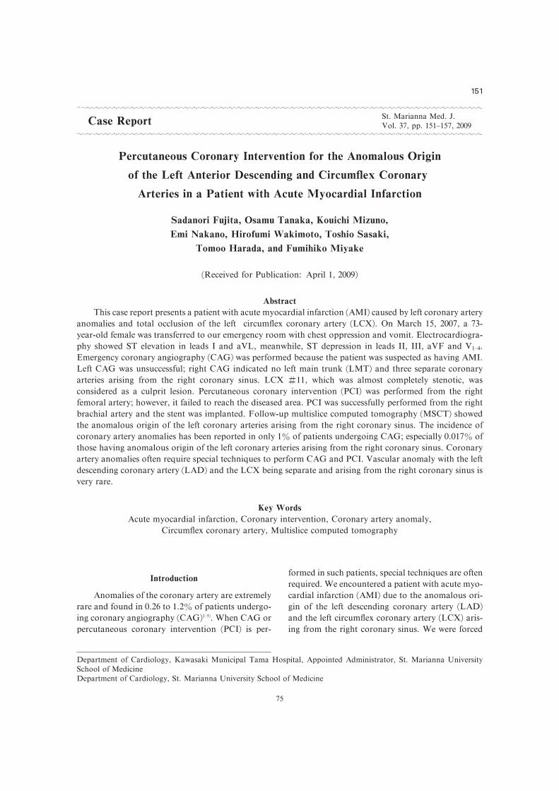
beats�minute during atrioventricular junctionalrhythm and ST elevation in leads I and aVL, mean-
while, ST depression in leads II, III, aVF and V1�4�Fig. 1�. Echocardiography revealed mild hypoki-nesis of left ventricular lateral wall motion and no
myocardial wall thinning.
Accordingly, the patient was diagnosed as hav-
ing AMI and was intravenously administered
400,000 units of monteplase. Emergency CAG was
also performed using a 5-Fr. Judkins left coronary
guiding catheter 3.5 �JL 3.5� inserted from the rightfemoral artery, however, JL3.5 was not adaptable.
Then, a 5-Fr. Judkins right coronary guiding cathe-
ter 3.5 �JR 3.5� was used, which showed the rightcoronary artery �RCA�, LAD and LCX arose fromthe right coronary sinus �Fig 2A�. No significantstenosis was observed in RCA and LAD �Fig 2B,C�, however, �11 LCX had 99� stenosis with
delayed enhancement �Fig 2D�. PCI was performedfrom the right femoral artery, however, the JR 3.5
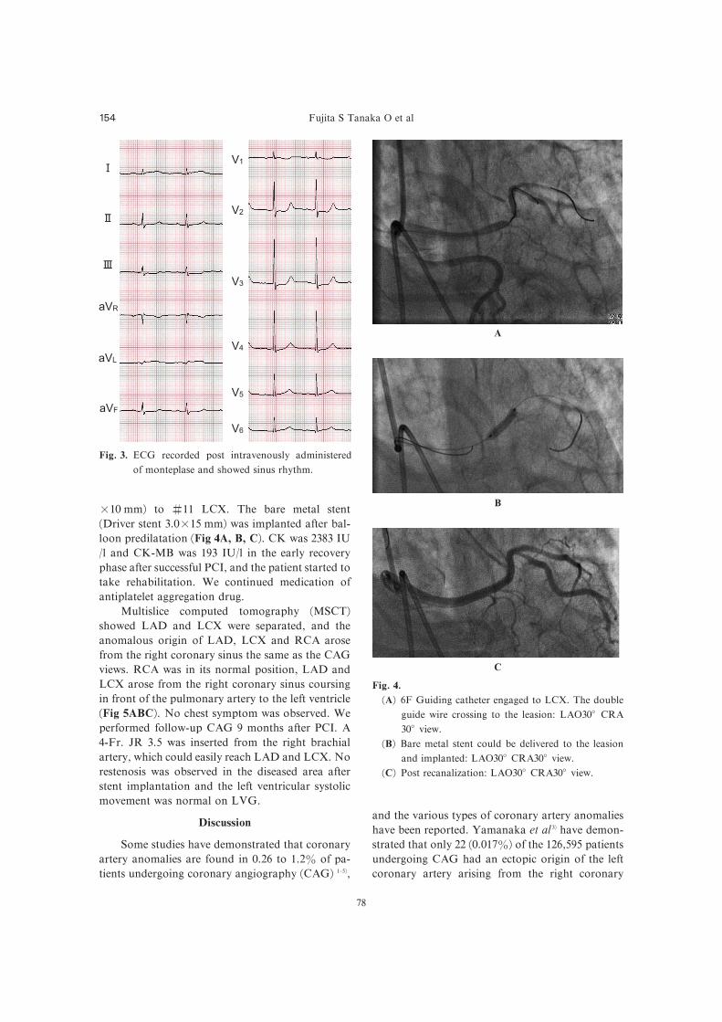
could not reach the diseased area. At this point, ST
elevation decreased to baseline in lead I and aVL
and chest oppression also decreased �Fig 3�. Thepatient was sent to the coronary care unit �CCU�under close observation with continuous infusion of
heparin and nicorandil. Intraaortic balloon pump-
ing �IABP� was not used because severe tortuosityfrom the descending to abdominal aortas was
found. Nine hours later, ECG showed ST elevation
with chest pain. Therefore CAG was performed
again for PCI.
This time, a 6-Fr. JR 3.5 was inserted from the
right brachial artery to the anomalous origin of
LCX arising from the right coronary sinus, which
took a lot of e#ort to insert a balloon catheter
because it could not be maintained coaxial to LCX.
Then, we used Runthrough and Fielder guide wires
and finally sent the balloon catheter �Avion HP 2.75
25.0 mm�sec10 mm�1 mV
Fig. 1. ECG recorded on arrival and showed junctional
rhythm.
Fujita S Tanaka O et al152
76

C
A
B D
Fig. 2. Coronary angiograms before intervention.
�A� The right coronary cusp angiogram showed that RCA, LAD, and LCX arosefrom the right coronary cusp: LAO 45�view.
�B� Right coronary angiogram showed no significant stenosis of the RCA: LAO 45�view.
�C� Left descending coronary angiogram showed no significant stenosis of the LAD :LAO 45�view.
�D� Left circumflex coronary angiogram showed 99� stenosis with delayed
enhancement: LAO30� CRA30� view.
Coronary artery anomaly 153
77
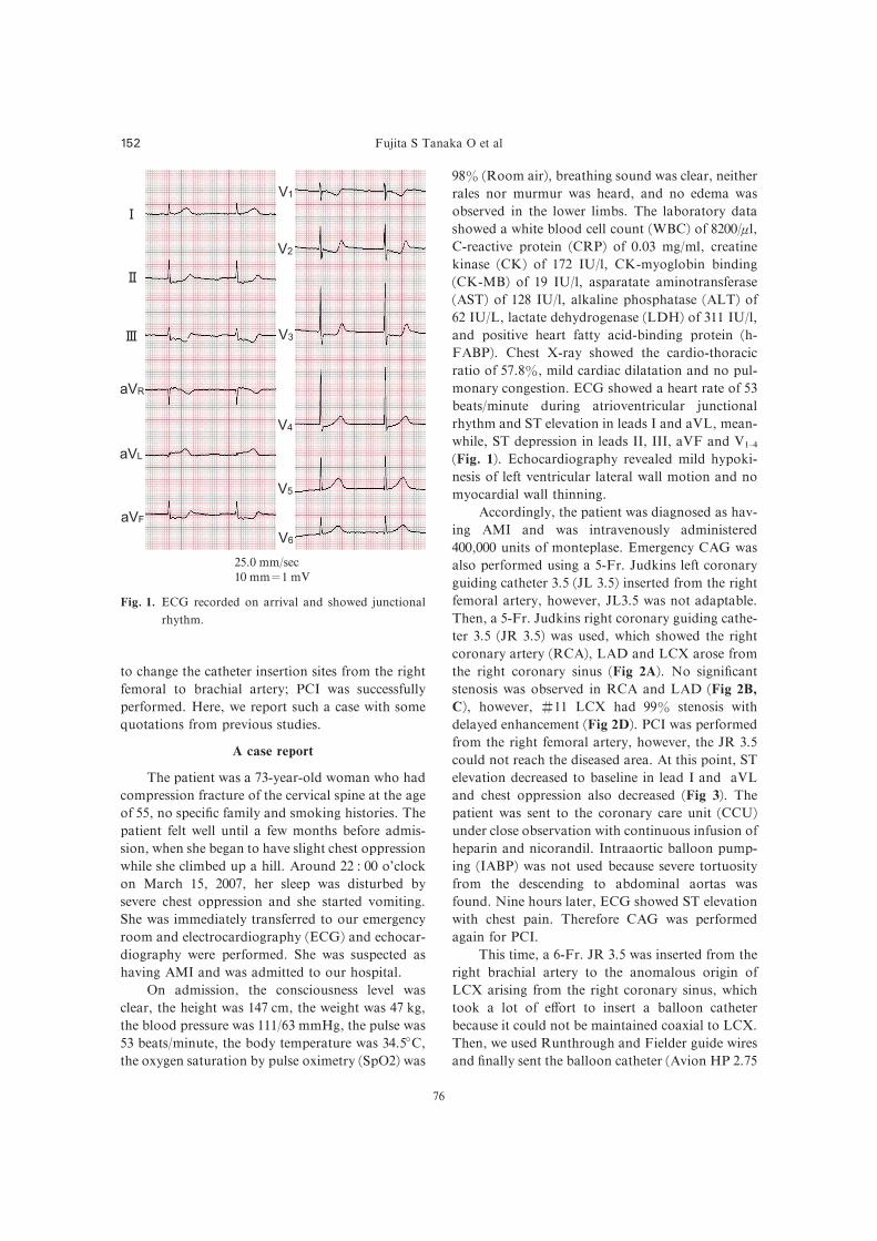
�10 mm� to �11 LCX. The bare metal stent�Driver stent 3.0�15 mm� was implanted after bal-loon predilatation �Fig 4A, B, C�. CK was 2383 IU�l and CK-MB was 193 IU�l in the early recoveryphase after successful PCI, and the patient started to
take rehabilitation. We continued medication of
antiplatelet aggregation drug.
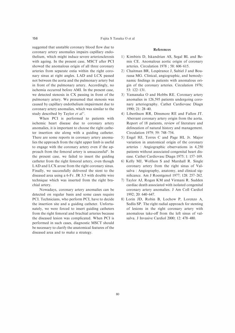
Multislice computed tomography �MSCT�showed LAD and LCX were separated, and the
anomalous origin of LAD, LCX and RCA arose
from the right coronary sinus the same as the CAG
views. RCA was in its normal position, LAD and
LCX arose from the right coronary sinus coursing
in front of the pulmonary artery to the left ventricle
�Fig 5ABC�. No chest symptom was observed. Weperformed follow-up CAG 9 months after PCI. A
4-Fr. JR 3.5 was inserted from the right brachial
artery, which could easily reach LAD and LCX. No
restenosis was observed in the diseased area after
stent implantation and the left ventricular systolic
movement was normal on LVG.
Discussion
Some studies have demonstrated that coronary
artery anomalies are found in 0.26 to 1.2� of pa-tients undergoing coronary angiography �CAG� 1�5�,
and the various types of coronary artery anomalies
have been reported. Yamanaka et al 3� have demon-
strated that only 22 �0.017�� of the 126,595 patientsundergoing CAG had an ectopic origin of the left
coronary artery arising from the right coronary
Fig. 3. ECG recorded post intravenously administered
of monteplase and showed sinus rhythm.
A
B
C
Fig. 4.
�A� 6F Guiding catheter engaged to LCX. The doubleguide wire crossing to the leasion: LAO30� CRA30� view.
�B� Bare metal stent could be delivered to the leasionand implanted: LAO30� CRA30� view.
�C� Post recanalization: LAO30� CRA30� view.
Fujita S Tanaka O et al154
78

sinus. Kelly et al 6� have described the onset mecha-
nism of myocardial ischemia due to coronary artery
anomalies as follows: � an anomalous coronaryartery arises in a sharp angle from a single ostia
which is buried within the aortic wall. On exertion,
the dilated aorta causes valvular stenosis at the ostia
of the coronary artery, or � an anomalous coro-nary artery, which passes between the aorta and the
pulmonary artery, is compressed on exertion due to
the dilation of both arteries. Taylor et al 7� have
reported the association between the onset mecha-
nism of ischemia and arteriosclerosis. They have
A
C
Fig. 5. Multislice computed tomography �MSCT�findings in the patient.
�A� 3-D view shows anomalous left anterior
descending artery �LAD� and left circumflex
artery �LCX� originating from the right sinus ofValsalva.
�B� Sagittal images showing the separate origins of theLAD, LCX, and right coronary artery �RCA�from the right sinus of Valsalva.
�C� The LCX courses in front of the pulmonary artery�PA�.
B
Coronary artery anomaly 155
79
suggested that unstable coronary blood flow due to
coronary artery anomalies impairs capillary endo-
thelium, which might induce severe arteriosclerosis
with ageing. In the present case, MSCT after PCI
showed the anomalous origin of all three coronary
arteries from separate ostia within the right coro-
nary sinus at right angles. LAD and LCX passed
not between the aorta and the pulmonary artery but
in front of the pulmonary artery. Accordingly, no
ischemia occurred before AMI. In the present case,
we detected stenosis in CX passing in front of the
pulmonary artery. We presumed that stenosis was
caused by capillary endothelium impairment due to
coronary artery anomalies, which was similar to the
study described by Taylor et al 7�.
When PCI is performed to patients with
ischemic heart disease due to coronary artery
anomalies, it is important to choose the right cathe-
ter insertion site along with a guiding catheter.
There are some reports in coronary artery anoma-
lies the approach from the right upper limb is useful
to engage with the coronary artery even if the ap-
proach from the femoral artery is unsuccessful8�. In
the present case, we failed to insert the guiding
catheter from the right femoral artery, even though
LAD and LCX arose from the right coronary sinus.
Finally, we successfully delivered the stent to the
diseased area using a 6-Fr. JR 3.5 with double wire
technique which was inserted from the right bra-
chial artery.
Nowadays, coronary artery anomalies can be
detected on regular basis and some cases require
PCI. Technicians, who perform PCI, have to decide
the insertion site and a guiding catheter. Unfortu-
nately, we were forced to insert guiding catheters
from the right femoral and brachial arteries because
the diseased lesion was complicated. When PCI is
performed in such cases, diagnostic MSCT should
be necessary to clarify the anatomical features of the
diseased area and to make a strategy.
References
1� Kimbiris D, Iskandrian AS, Segal BL and Be-mis CE. Anomalous aortic origin of coronary
arteries. Circulation 1978 ; 58: 606�615.2� Chaitman BR, Lesperance J, Saltiel J and Bou-rassa MG. Clinical, angiographic, and hemody-
namic findings in patients with anomalous ori-
gin of the coronary arteries. Circulation 1976;
53: 122�131.3� Yamanaka O and Hobbs RE. Coronary arteryanomalies in 126,595 patients undergoing coro-
nary arteriography. Cathet Cardiovasc Diagn
1990; 21: 28�40.4� Liberthson RR, Dinsmore RE and Fallon JT.Aberrant coronary artery origin from the aorta.
Report of 18 patients, review of literature and
delineation of natural history and management.
Circulation 1979; 59: 748�754.5� Engel HJ, Torres C and Page HL Jr. Majorvariation in anatomical origin of the coronary
arteries : Angiographic observations in 4,250
patients without associated congenital heart dis-
ease. Cathet Cardiovasc Diagn 1975; 1: 157�169.6� Kelly MJ, Wolfson S and Marshall R. Singlecoronary artery from the right sinus of Val-
salva : Angiography, anatomy, and clinical sig-
nificance. Am J Roentgenol 1977; 128: 257�262.7� Taylor AJ, Rogan KM and Virmani R. Suddencardiac death associated with isolated congenital
coronary artery anomalies. J Am Coll Cardiol
1992; 20: 640�647.8� Lorin JD, Robin B, Lochow P, Lorenzo A,Sedlis SP. The right radial approach for stenting
of lesions in the right coronary artery with
anomalous take-o# from the left sinus of val-
salva. J Invasive Cardiol 2000; 12: 478�480.
Fujita S Tanaka O et al156
80

���������� ����������PCI� ��� �������� 1�
���
��
���
���
��
��
���
��
��
��
��
��
��
��
��
��
���
���
���
��
��
��
���
�
���
��
���
�
��
���
���
���
� �ST ����� !"#$%&'()*+,� -./0123 �CAG� �� 4-./56789:� ;<=>?@ABCDEF=(GHIJKLF�MN=>NO�PQ=>RSTUVW NOX 73Y� ZG 2007[ 3\ 15]� !"#$� �^�_`'()*+,> Jab� I, aVL % ST ��� II, III, aVF, V1�4 % ST cd�_`(GHIJKLF�efg(CAG �hi> 4-./01%jk=� l-./m01%&l-./mnoMpVW 3�R-./�_`> 4-./X4qr!Bstuv� wdhA� ?@AX;<=l-./mno56=&f> ?@A�11% 99�xy�_`LFz{|}~��-./����������PCI� ��h=> l��./��R�����SX�h�jS�o� l��./��R�����S������%��=> ��-./ CT%&4-./B;<56=l-./m��Mp=&fW�~B�_+,> -./5678R��X CAG D�R 1�w�~TU+,&¡o� =¢=¢ CAG £ PCI %jk=¤¥¦§¨B©ª%«W ¬%4-./D&Bl-./m��MpVWRX 0.017�~TU+,� «�SwdhA~?@AB;<56=l-./mnoMpVW5678X®`&¯~°±,TU=>
²³´��µ¶·�¸ ¹·¸�º»¼¹·�
Coronary artery anomaly 157
81
Recommended