Role of Fiber Orientation in Atrial Arrythmogenesis
Sanjay Kharche1, 2, Simon Castro1, Belvin Thomas3, Michael Colman1, Jonathan Jarvis3, Bruce Smaill4, Henggui Zhang1, Robert Stephenson3, Jichao Zhao4
1School of Physics and Astronomy, University of Manchester, Manchester, UK 2College of Engineering, Physical and Mathematical Sciences, University of Exeter, Exeter, UK
3Research Institute for Sport and Exercise Sciences, Liverpool John Moores University, UK 4Auckland Bioengineering Institute, University of Auckland, New Zealand
Abstract
Electrical wave-front propagation in the atria is determined largely by local fiber orientation. Recent study suggests that atrial fibrillation (AF) progresses with enhanced anisotropy. In this work, a 3D rabbit atrial anatomical model at 20×20×20 µm3 resolution with realistic fiber orientation was constructed based on the novel contrast-enhanced micro-CT imaging. The Fenton-Karma cellular activation model was adapted to reproduce rabbit atrial action potential period of 80 ms. Diffusivities were estimated for longitudinal and transverse directions of the fiber orientation respectively. Pacing was conducted in the 3D anisotropic atrial model with a reducing S2 interval to facilitate initiation of atrial arrhythmia. Multiple simulations were conducted with varying values of diffusion anisotropy and stimulus locations to evaluate the role of anisotropy in initiating AF. Under physiological anisotropy conditions, a rapid right atrial activation was followed by the left atrial activation. Excitation waves reached the atrio-ventricular border where they terminated. Upon reduction of conduction heterogeneity, re-entry was initiated by the rapid pacing and the activation of both atrial chambers was almost simultaneous. Myofiber orientation is an effective mechanism for regulating atrial activation. Modification of myoarchitecture is proarrhythmic.
1. Introduction
Atrial fibrillation (AF) is associated with structural and electrical remodelling which generates abnormal ectopic triggers [1]. Despite having undergone extensive research over the last few decades, the mechanisms underlying the initiation and sustenance of these perturbing waves remain entangled [1]. It is argued that conduction heterogeneities in tissue coupled with ectopic activity originating from the pulmonary veins are capable of triggering longer episodes of AF, especially for
paroxysmal AF [1-4]. Apart from the macroscopic descriptions of atrial muscular architecture [5], high resolution 3D reconstructions of specific atrial areas [6] are also handy resources to vaguely deduce the atrial myocyte orientation. However, a clear picture was not available until recently, since the well-established DTMRI technique lacks the spatial resolution to capture the intrinsic complexity of atrial wall. Lately, 3D the atrial myofiber orientation has been computed using a novel three dimensional structure tensor based method, which was followed by the visualisation of the detailed architecture in high resolution, thereby setting a standard to perform atrial fiber tracking [7].
Here in this paper, we constructed a model of inducible AF which incorporates a) realistic fiber orientation anisotropy (FOA) based on high resolution Micro CT imaging b) electrical heterogeneity and c) delivered abnormal pacing at pulmonary veins (PVs). The aim of this paper is to evaluate the role of FOA in AF sustenance.
2. Methods
2.1. Cell model
The Fenton-Karma cell action potential (AP) model [8] was specifically developed to reproduce cardiac electrical behaviour in a computationally efficient manner. In brief, it consists of a 3 variable coupled ordinary differential equations (ODEs) system where one variable can be thought to be the cell membrane potential, the other two variables whose combination provides the gating for the inward upstroke initiating current and the repolarisation current. Here, it is modified to simulate rabbit atrial APs [9] as shown in Figure 1A and 1B. Action potential duration (APD90) indicates the time taken to attain 90 percent repolarisation. Diffusivities were estimated for longitudinal (Dp = 0.0065 mm2/ms) and transverse directions (Dp = 0.06 mm2/ms) of the fiber orientation. 1D strands were used
ISSN 2325-8861 Computing in Cardiology 2014; 41:1041-1044.1041
to simulate experimentally measured conduction velocity (CV) of 50 cm/s longitudinally (along the fiber) and 15 cm/s transversely (perpendicular to the fiber) [(10)] (Figure 1C and 1D).
Figure 1. Model rabbit atrial action potential (AP) and conduction velocity (CV). A: AP profile. B: Validation of model AP. Bar shows simulated value (140 ms) while symbols show experimental data. C, D: Simulation of propagation in 1D strands of atrial tissue. The CV was set to 50cm/s in the longitudinal (to the fibers) direction and 15cm/s in the transverse direction. 2.2. Anatomy and fiber orientation anisotropy (FOA)
The rabbit atrial anatomy was constructed based on a 20 µm resolution iodine induced contrast enhanced micro-CT scan [11, 12]. Firstly, 3D atrial structure was segmented using a commercial software Amira. Then the 3D myofiber orientation was reconstructed using structure tensor approach and visualized using a novel fibre tracking approach [7, 13] (Figure 2(A)).
Figure 2. Rabbit atrial fiber orientation anisotropy (FOA) and pacing locations. A: Fiber orientation color coding is according to the inclination angle w .r .t. X-Y plane, i.e., grey denotes vertical and black for horizontal directions. B: Locations of pacing by SAN (dark squares) and locations close to PVs. RA: right atrium, LA: left atrium.
2.3. Pacing locations
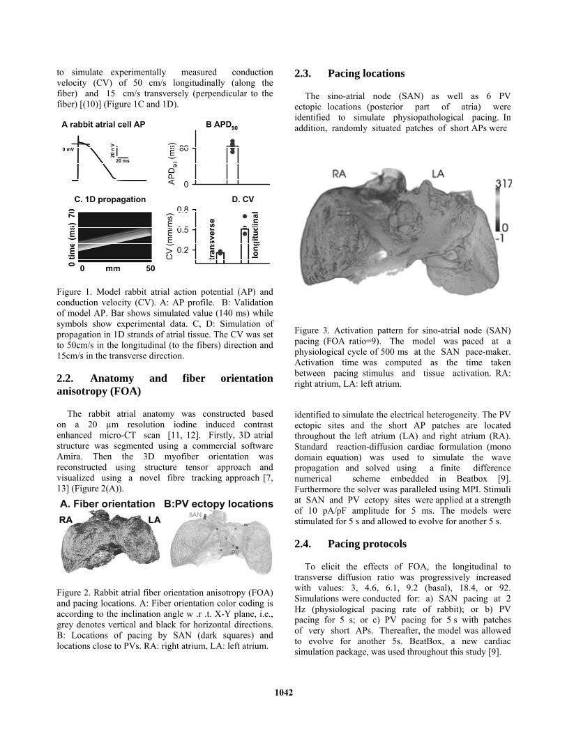
The sino-atrial node (SAN) as well as 6 PV ectopic locations (posterior part of atria) were identified to simulate physiopathological pacing. In addition, randomly situated patches of short APs were
Figure 3. Activation pattern for sino-atrial node (SAN) pacing (FOA ratio=9). The model was paced at a physiological cycle of 500 ms at the SAN pace-maker. Activation time was computed as the time taken between pacing stimulus and tissue activation. RA: right atrium, LA: left atrium. identified to simulate the electrical heterogeneity. The PV ectopic sites and the short AP patches are located throughout the left atrium (LA) and right atrium (RA). Standard reaction-diffusion cardiac formulation (mono domain equation) was used to simulate the wave propagation and solved using a finite difference numerical scheme embedded in Beatbox [9]. Furthermore the solver was paralleled using MPI. Stimuli at SAN and PV ectopy sites were applied at a strength of 10 pA/pF amplitude for 5 ms. The models were stimulated for 5 s and allowed to evolve for another 5 s.
2.4. Pacing protocols
To elicit the effects of FOA, the longitudinal to transverse diffusion ratio was progressively increased with values: 3, 4.6, 6.1, 9.2 (basal), 18.4, or 92. Simulations were conducted for: a) SAN pacing at 2 Hz (physiological pacing rate of rabbit); or b) PV pacing for 5 s; or c) PV pacing for 5 s with patches of very short APs. Thereafter, the model was allowed to evolve for another 5s. BeatBox, a new cardiac simulation package, was used throughout this study [9].
1042
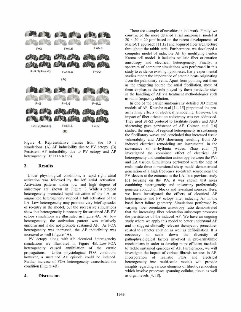
Figure 4. Representative frames from the 10 s simulations. (A) AF inducibility due to PV ectopy. (B) Persistent AF inducibility due to PV ectopy and AP heterogeneity. (F: FOA Ratio)
3. Results
Under physiological conditions, a rapid right atrial activation was followed by the left atrial activation. Activation patterns under low and high degree of anisotropy are shown in Figure 3. While a reduced heterogeneity promoted rapid activation of the LA, an augmented heterogeneity stopped a full activation of the LA. Low heterogeneity may promote very brief episodes of re-entry in the model, but the successive simulations show that heterogeneity is necessary for sustained AF. PV ectopy simulations are illustrated in Figure 4A. At low heterogeneity, the activation pattern was relatively uniform and it did not promote sustained AF. As FOA heterogeneity was increased, the AF inducibility was increased as well (Figure 4A).
PV ectopy along with AP electrical heterogeneity simulations are illustrated in Figure 4B. Low FOA heterogeneity caused annihilation of the erratic propagations. Under physiological FOA conditions however, a sustained AF episode could be induced. Further increase of FOA heterogeneity exacerbated the condition (Figure 4B).
4. Discussion
There are a couple of novelties in this work. Firstly, we constructed the more detailed atrial anatomical model at 20 × 20 × 20 µm3 based on the recent developments of MicroCT approach [11,12] and acquired fiber architecture throughout the rabbit atria. Furthermore, we developed a computer model of inducible AF by modifying Fenton Karma cell model. It includes realistic fiber orientation anisotropy and electrical heterogeneity. Finally, a spectrum of computer simulations was performed in this study to evidence existing hypotheses. Early experimental studies report the importance of ectopic beats originating from the pulmonary veins. Apart from pointing out them as the triggering source for atrial fibrillation, most of them emphasize the role played by these particular sites in the handling of AF via treatment methodologies such as radio frequency ablation.
In one of the earlier anatomically detailed 3D human models of AF, Kharche et.al [14, 15] pinpointed the pro-arrhythmic effects of electrical remodeling. However, the impact of fiber orientation anisotropy was not addressed. They used S1-S2 protocol to facilitate reentry and APD shortening gave persistence of AF. Colman et.al [16] studied the impact of regional heterogeneity in sustaining the fibrillatory waves and concluded that increased tissue vulnerability and APD shortening resulting from AF induced electrical remodeling are instrumental in the sustenance of arrhythmia waves. Zhao et.al [7] investigated the combined effect of electrical AP heterogeneity and conduction anisotropy between the PVs and LA tissues. Simulations performed with the help of multi-scale three dimensional sheep model demonstrated generation of a high frequency re-entrant source near the PV sleeves at the entrance to the LA. In a previous study [6] focusing on the RA, it was shown that areas combining heterogeneity and anisotropy preferentially generate conduction blocks and re-entrant sources. Here, we have investigated the effects of electrical AP heterogeneity and PV ectopy after inducing AF in the basal heart failure geometry. Simulations performed by varying fiber orientation anisotropy ratio demonstrated that the increasing fiber orientation anisotropy promotes the persistence of the induced AF. We have an ongoing study where we apply this model to better understand AF and to suggest clinically relevant therapeutic procedures related to catheter ablation as well as defibrillation. It is necessary to scale down the diversity of pathophysiological factors involved in pro-arrhythmic mechanisms in order to develop more efficient methods to tackle sustained episodes of AF. Furthermore, we will investigate the impact of various fibrosis textures in AF. Incorporation of realistic FOA and electrical heterogeneity into multi-scale models will provide insights regarding various elements of fibrotic remodeling which involve processes spanning cellular, tissue as well as organ levels [4, 18].
1043
5. Conclusions
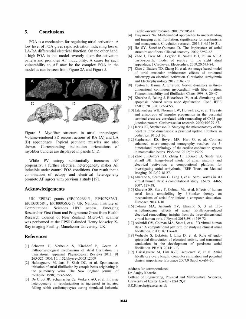
FOA is a mechanism for regulating atrial activation. A low level of FOA gives rapid activation indicating loss of LA-RA differential electrical function. On the other hand, a high FOA in this model severely alters the activation pattern and promotes AF inducibility. A cause for such vulnerability to AF may be the complex FOA in the model as can be seen from Figure 2A and Figure 5.
Figure 5. Myofiber structure in atrial appendages. Volume-rendered 3D reconstructions of RA (A) and LA (B) appendages. Typical pectinate muscles are also shown. Corresponding inclination orientations of myofiber bundles are displayed in panels C, D.
While PV ectopy substantially increases AF
propensity, a further electrical heterogeneity makes AF inducible under control FOA conditions. Our result that a combination of ectopy and electrical heterogeneity promote AF agrees with previous a study [19].
Acknowledgements
UK EPSRC grants (EP/I029664/1, EP/I029826/1, EP/I030158/1, EP/J00958X/1), UK National Institute of Computational Sciences HPC access, Emerging Researcher First Grant and Programme Grant from Health Research Council of New Zealand. Micro-CT scanner was performed at the EPSRC-funded Henry Moseley X-Ray imaging Facility, Manchester University, UK.
References
[1] Schotten U, Verheule S, Kirchhof P, Goette A. Pathophysiological mechanisms of atrial fibrillation : a translational appraisal. Physiological Reviews 2011: 91 265-325. DOI: 10.1152/physrev.00031.2009
[2] Haïssaguerre M, Jaïs P, Shah DC, et al. Spontaneous initiation of atrial fibrillation by ectopic beats originating in the pulmonary veins. The New England journal of medicine. 1998;339:659-66.
[3] De Groot JR, Schumacher Ca, Verkerk AO, et al. Intrinsic heterogeneity in repolarization is increased in isolated failing rabbit cardiomyocytes during simulated ischemia.
Cardiovascular research. 2003;59:705-14. [4] Trayanova Na. Mathematical approaches to understanding
and imaging atrial fibrillation: significance for mechanisms and management. Circulation research. 2014;114:1516-31.
[5] Ho SY, Sanchez-Quintana D. The importance of atrial structure and fibers. Clinical anatomy. 2009;22:52-63.
[6] Zhao J, Trew ML, Legrice IJ, Smaill BH, Pullan AJ. A tissue-specific model of reentry in the right atrial appendage. J Cardiovas. Electrophys. 2009;20:675-84.
[7] Zhao J, Butters TD, Zhang H, et al. An image-based model of atrial muscular architecture: effects of structural anisotropy on electrical activation. Circulation Arrhythmia and Electrophysiology 2012;5:361-70.
[8] Fenton F, Karma A. Erratum: Vortex dynamics in three-dimensional continuous myocardium with fiber rotation: Filament instability and fibrillation Chaos 1998; 8, 20-47.
[9] Kharche S, Beling J, Biktasheva IV, et al. Simulating cell apoptosis induced sinus node dysfunction. Conf. IEEE EMBS. 2013;2013:6842-5.
[10] Litchenberg WH, Norman LW, Holwell aK, et al. The rate and anisotropy of impulse propagation in the postnatal terminal crest are correlated with remodeling of Cx43 gap junction pattern. Cardiovascular research. 2000;45:379-87.
[11] Jarvis JC, Stephenson R. Studying the microanatomy of the heart in three dimensions: a practical update. Frontiers in pediatrics. 2013;1:26.
[12] Stephenson RS, Boyett MR, Hart G, et al. Contrast enhanced micro-computed tomography resolves the 3-dimensional morphology of the cardiac conduction system in mammalian hearts. PloS one. 2012;7:e35299.
[13] Zhao J, Butters TD, Zhang H, LeGrice IJ, Sands GB, Smaill BH. Image-based model of atrial anatomy and electrical activation: a computational platform for investigating atrial arrhythmia. IEEE Trans. on Medical Imaging. 2013;32:18-27.
[14] Kharche S, Seemann G, Leng J, et al. Scroll waves in 3D virtual human atria: a computational study. LNCS. 4466; 2007: 129-38.
[15] Kharche SR, Stary T, Colman Ma, et al. Effects of human atrial ionic remodelling by β-blocker therapy on mechanisms of atrial fibrillation: a computer simulation. Europace 2014:1-10.
[16] Colman MA, Aslanidi OV, Kharche S, et al. Pro-arrhythmogenic effects of atrial fibrillation-induced electrical remodelling: insights from the three-dimensional virtual human atria. J Physiol 2013;591: 4249-72.
[17] Aslanidi OV, Colman MA, Stott J, et al. 3D virtual human atria : A computational platform for studying clinical atrial fibrillation. 2011;107:156-68.
[18] Verheule S, Eckstein J, Linz D, et al. Role of endo-epicardial dissociation of electrical activity and transmural conduction in the development of persistent atrial fibrillation. PBMB. 2014:1-13.
[19] Haissaguerre M, Lim K-T, Jacquemet V, et al. Atrial fibrillatory cycle length: computer simulation and potential clinical importance. Europace 2007;9 Suppl 6:vi64-70.
Address for correspondence Dr. Sanjay Kharche College of Engineering, Physical and Mathematical Sciences, University of Exeter, Exeter - EX4 2QF [email protected]
1044
Recommended