Embed Size (px)
Citation preview
Anatomy of Respiratory System
Consists of an upper respiratory tract (nose to larynx) and a lower respiratory tract ( trachea onwards)
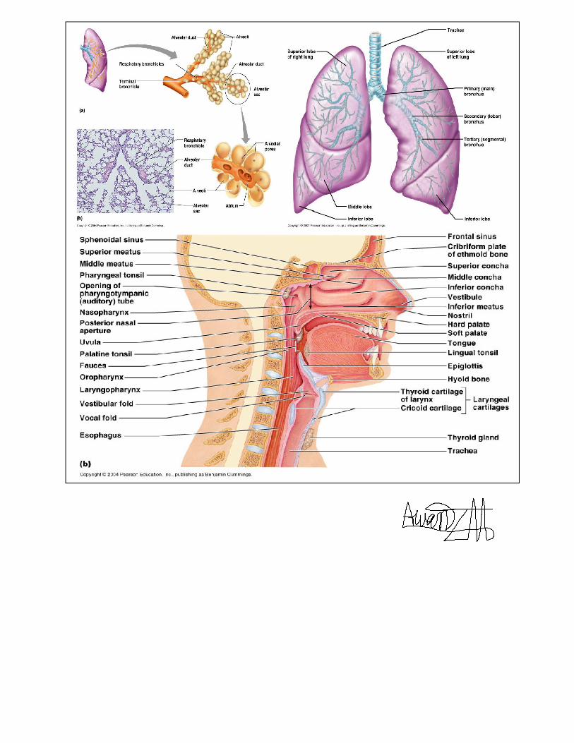
Conducting portion transports air: includes the nose, nasal cavity, pharynx, larynx, trachea, and
progressively smaller airways, from the primary bronchi to the terminal bronchioles. Respiratory
portion carries out gas exchange: composed of small airways called respiratory bronchioles and alveolar
ducts as well as air sacs called alveoli
Respiratory system function:
1) supplies the body with oxygen and disposes of carbon dioxide
2) filters inspired air
3) produces sound
4) contains receptors for smell
5) rids the body of some excess water and heat
6) Helps regulate blood pH.
Breathing (pulmonary ventilation). Consists of two cyclic phases:
Inhalation, also called inspiration - draws gases into the lungs. Exhalation, also called expiration - forces
gases out of the lungs.
Upper Respiratory Tract: Composed of the nose and nasal cavity, paranasal sinuses, pharynx (throat),
larynx. All part of the conducting portion of the respiratory system.
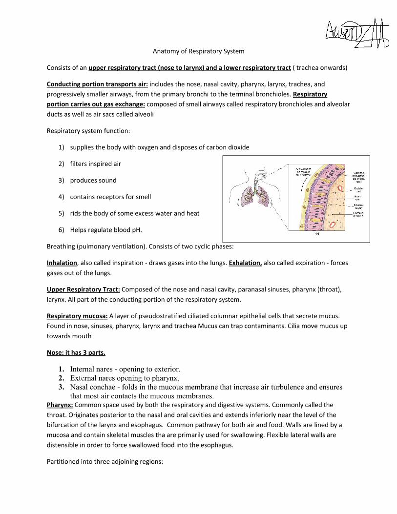
Respiratory mucosa: A layer of pseudostratified ciliated columnar epithelial cells that secrete mucus.
Found in nose, sinuses, pharynx, larynx and trachea Mucus can trap contaminants. Cilia move mucus up
towards mouth
Nose: it has 3 parts.
1. Internal nares - opening to exterior. 2. External nares opening to pharynx. 3. Nasal conchae - folds in the mucous membrane that increase air turbulence and ensures
that most air contacts the mucous membranes. Pharynx: Common space used by both the respiratory and digestive systems. Commonly called the
throat. Originates posterior to the nasal and oral cavities and extends inferiorly near the level of the
bifurcation of the larynx and esophagus. Common pathway for both air and food. Walls are lined by a
mucosa and contain skeletal muscles tha are primarily used for swallowing. Flexible lateral walls are
distensible in order to force swallowed food into the esophagus.
Partitioned into three adjoining regions:
1. nasopharynx 2. oropharynx 3. laryngopharynx
Nasopharynx:
Superior-most region of the pharynx. Covered with pseudostratified ciliated columnar epithelium.
Located directly posterior to the nasal cavity and superior to the soft palate, which separates the oral
cavity. Normally, only air passes through. Material from the oral cavity and oropharynx is typically
blocked from entering the nasopharynx by the uvula of soft palate, which elevates when we swallow. In
the lateral walls of the nasopharynx, paired auditory/eustachian tubes connect the nasopharynx to the
middle ear. Posterior nasopharynx wall also houses a single pharyngeal tonsil (commonly called the
adenoids).
Oropharynx:The middle pharyngeal region.Immediately posterior to the oral cavity.Bounded by the
edge of the soft palate superiorly and the hyoid bone inferiorly. Common respiratory and digestive
pathway through which both air and swallowed food and drink pass. Contains nonkeratinized stratified
squamous epithelim. Lymphatic organs here provide the first line of defense against ingested or inhaled
foreign materials. Palatine tonsils are on the lateral wall between the arches, and the lingual tonsils are
at the base of the tongue.
Laryngopharynx: Inferior, narrowed region of the pharynx. Extends inferiorly from the hyoid bone to the
larynx and esophagus.Terminates at the superior border of the esophagus and the epiglottis of the
larynx.Lined with a nonkeratinized stratified squamous epithelium. Permits passage of both food and air.
Lower Respiratory Tract:
Conducting airways (trachea, bronchi, up to terminal bronchioles).
Respiratory portion of the respiratory system (respiratory bronchioles, alveolar ducts, and
alveoli).
Larynx: Muscular walls aid in voice production and the swallowing reflex. Glottis – the superior opening
of the larynx. Epiglottis – prevents food and drink from entering airway when
swallowingpseudostratified ciliated columnar epithelium
Trachea: A flexible tube also called windpipe. Extends through the mediastinum and lies anterior to the
esophagus and inferior to the larynx. Anterior and lateral walls of the trachea supported by 15 to 20 C-
shaped tracheal cartilages. Cartilage rings reinforce and provide rigidity to the tracheal wall to ensure
that the trachea remains open at all times. Posterior part of tube lined by trachealis muscle. Lined by
ciliated pseudostratified columnar epithelium. At the level of the sternal angle, the trachea bifurcates
into two smaller tubes, called the right and left primary bronchi. Each primary bronchus projects
laterally toward each lung. The most inferior tracheal cartilage separates the primary bronchi at their
origin and forms an internal ridge called the carina
Bronchial tree:A highly branched system of air-conducting passages that originate from the left and
right primary bronchi. Progressively branch into narrower tubes as they diverge throughout the lungs
before terminating in terminal bronchioles. Incomplete rings of hyaline cartilage support the walls of the
primary bronchi to ensure that they remain open. Right primary bronchus is shorter, wider, and more
vertically oriented than the left primary bronchus. Foreign particles are more likely to lodge in the right
primary bronchus. The primary bronchi enter the hilus of each lung together with the pulmonary
vessels, lymphatic vessels, and nerves. Each primary bronchus branches into several secondary bronchi
(or lobar bronchi).The left lung has two secondary bronchi.The right lung has three secondary bronchi.
They further divide into tertiary bronchi. Each tertiary bronchus is called a segmental bronchus because
it supplies a part of the lung called a bronchopulmonary segment.
Secondary bronchi tertiary brnchi bronchioles terminal bronchioles
with successive branching amount of cartilage decreases and amount of smooth muscle increases, this
allows for variation in airway diameter. during exertion and when sympathetic division active
bronchodilation. mediators of allergic reactions like histamine bronchoconstriction . epithelium
gradually changes from ciliated pseudostratified columnar epithelium to simple cuboidal epithelium in
terminal bronchioles
Cells in Alveolus:
1. Type I cells : simple squamous cells forming lining 2. Type II cells : or septal cells secrete surfactant 3. Type 3 cells :Alveolar macrophages
Lungs:
Left lung:divided into 2 lobes by oblique fissure. smaller than the right lung. cardiac notch
accommodates the heart
Right: divided into 3 lobes by oblique and horizontal fissure. located more superiorly in the body due to
liver on right side
Pleura and Pleural Cavities:The outer surface of each lung and the adjacent internal thoracic wall are
lined by a serous membrane called pleura.The outer surface of each lung is tightly covered by the
visceral pleura. while the internal thoracic walls, the lateral surfaces of the mediastinum, and the
superior surface of the diaphragm are lined by the parietal pleura. The parietal and visceral pleural
layers are continuous at the hilus of each lung.
Pleural Cavities:
The potential space between the serous membrane layers is a pleural cavity. The pleural membranes
produce a thin, serous pleural fluid that circulates in the pleural cavity and acts as a lubricant, ensuring
minimal friction during breathing.Pleural effusion – pleuritis with too much fluid