Embed Size (px)
Citation preview

CASE STUDY
BUMPS ON THE NECK AND GROIN OF A
2-YEAR OLD MALE
PRESENTED BY
ANNE SAHITHI S.T
VISHAKHA KUMAR
4/8/2014
1

4/8/2014
2
PATIENT:-
2-year old boy.
CHIEF COMPLAINT:-
Bumps on neck and groin

4/8/2014
3
HISTORY OF THE PATIENT
1- to 2-week of bumps on his neck and
groin.
blood test revealed a high white blood cell
(WBC) count and a low platelet count. The
parents of the patient say that he had
bilateral knee pain and cough for the past
week.
The PCP had mentioned the bumps on his
neck and his cough

4/8/2014
4
MEDICAL & FAMILY HISTORY
The patient’s immunizations are up to date.
He had seasonal allergies, with symptoms that include
itchy eyes and runny nose.
His recurrent ear infections have been resolved by
pressure-equalization tubes and adenoidectomy.
The patient’s maternal grandmother and maternal uncle
have long history of QT syndrome.
o His paternal great-grandfather has a low platelet count.
The patient lives with his parents.
His mother is currently at 31 weeks’ gestation,
originally with triplets but currently with twins (one fetus
recently died in the uterus).

4/8/2014
5
PHYSICAL EXAMINATON:
Symptoms and abnormalities were
found in the:
1. respiratory system,
2.Lymphatic and musculoskeletal
systems.
3. cough, leg and knee pain
4. he also had swollen lymph
glands
4/8/2014
6
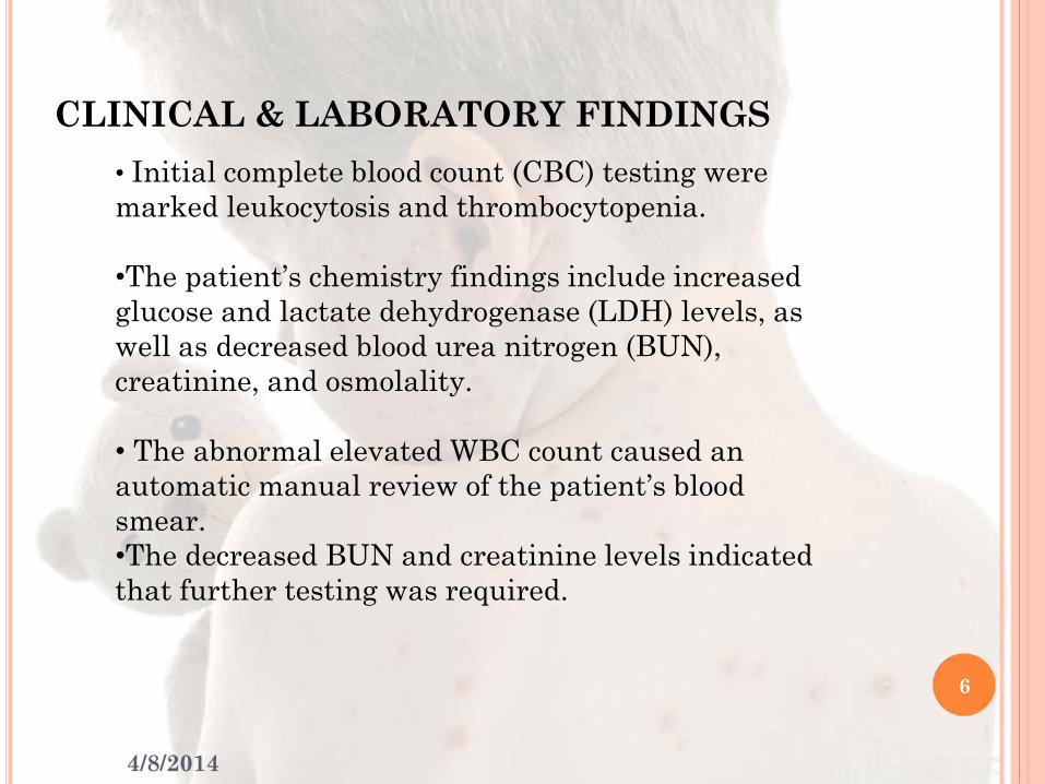
CLINICAL & LABORATORY FINDINGS
• Initial complete blood count (CBC) testing were
marked leukocytosis and thrombocytopenia.
•The patient’s chemistry findings include increased
glucose and lactate dehydrogenase (LDH) levels, as
well as decreased blood urea nitrogen (BUN),
creatinine, and osmolality.
• The abnormal elevated WBC count caused an
automatic manual review of the patient’s blood
smear.
•The decreased BUN and creatinine levels indicated
that further testing was required.

4/8/2014
7
Elevated LDH levels are an indicator for acute or chronic
tissue damage and can be used to monitor the progression
of certain cancers.
4/8/2014
8
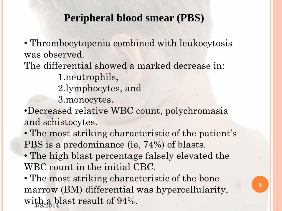
Peripheral blood smear (PBS)
• Thrombocytopenia combined with leukocytosis
was observed.
The differential showed a marked decrease in:
1.neutrophils,
2.lymphocytes, and
3.monocytes.
•Decreased relative WBC count, polychromasia
and schistocytes.
• The most striking characteristic of the patient’s
PBS is a predominance (ie, 74%) of blasts.
• The high blast percentage falsely elevated the
WBC count in the initial CBC.
• The most striking characteristic of the bone
marrow (BM) differential was hypercellularity,
with a blast result of 94%.
4/8/2014
9
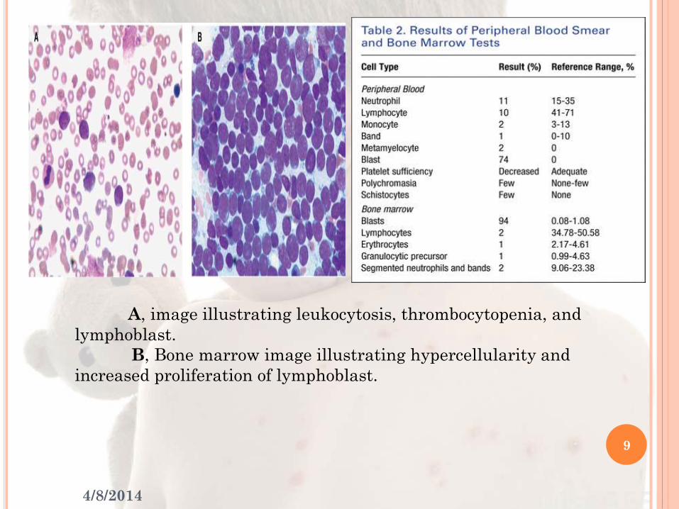
A, image illustrating leukocytosis, thrombocytopenia, and
lymphoblast.
B, Bone marrow image illustrating hypercellularity and
increased proliferation of lymphoblast.
4/8/2014
10
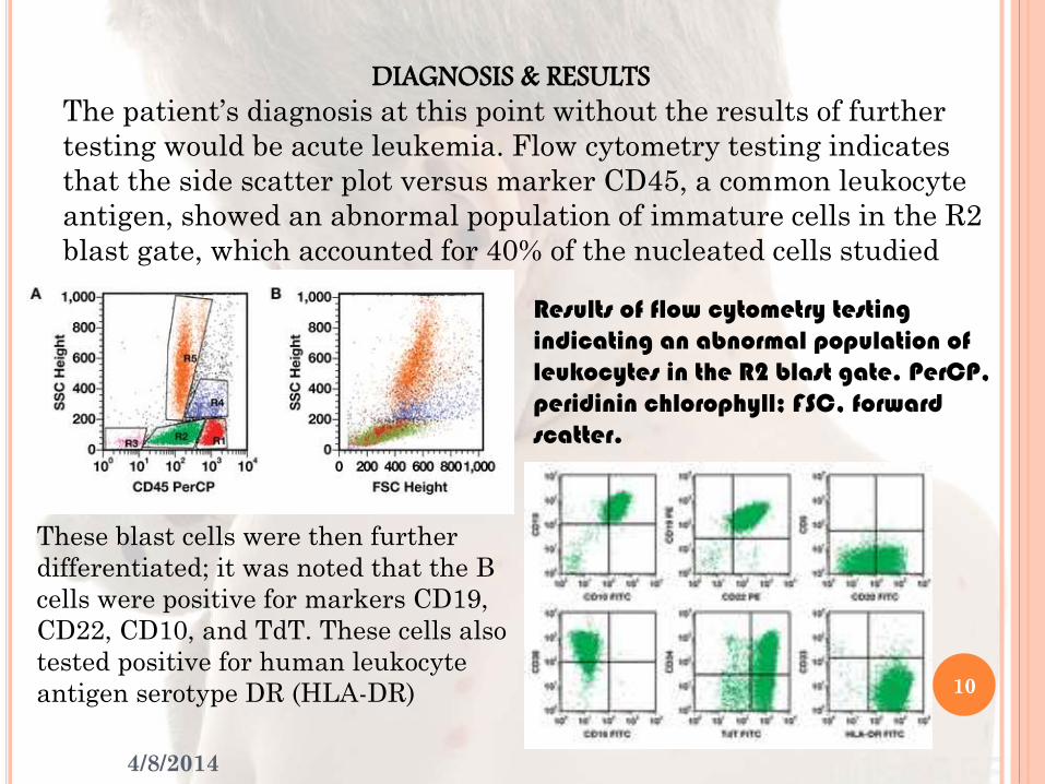
DIAGNOSIS & RESULTS
The patient’s diagnosis at this point without the results of further
testing would be acute leukemia. Flow cytometry testing indicates
that the side scatter plot versus marker CD45, a common leukocyte
antigen, showed an abnormal population of immature cells in the R2
blast gate, which accounted for 40% of the nucleated cells studied
Results of flow cytometry testing indicating an abnormal population of leukocytes in the R2 blast gate. PerCP, peridinin chlorophyll; FSC, forward scatter.
These blast cells were then further
differentiated; it was noted that the B
cells were positive for markers CD19,
CD22, CD10, and TdT. These cells also
tested positive for human leukocyte
antigen serotype DR (HLA-DR)

4/8/2014
11
The test results indicate that the patient’s
acute leukemia was subtyped to pre–B-cell acute lymphoblastic
leukemia (Pre-B ALL).
A fluorescence in situ hybridization (FISH)
panel was performed; the results indicated
no TEL/AML1, BCR/ABL, or MLLgene arrangement. It was
also noted that no trisomies were observed on chromosomes 4,
10, or 17.
Patients often experience fever, bleeding, fatigue,
and severe bone pain. Bone pain can be a possible result of tumor
infiltration or marrow necrosis. Patients may have nodal and
extranodal involvement.
Laboratory findings include a form of cytopenia, such as
anemia or thrombocytopenia. The overall leukocyte count can be
increased, decreased, or normal, with a median count of
approximately 10 to 12 × 109/L

4/8/2014
12
Flow cytometry testing can help
differentiate among the various forms of
B-ALL by identifying the CD markers
that exist on the leukocytes.
For a patient to be diagnosed
with B-ALL, most of the leukocytes
must express specific markers, including
CD19 and/or CD20, which are primarily
found on cells committed to the B-cell
maturation process.
Precursor-B lymphoblasts often
present are TdT+ & HLA-DR+. Moreover,
the expression of CD10, the common ALL
antigen (CALLA), differentiates
intermediate pre-B from pro-B-ALL.

4/8/2014
13
TREATMENT• Includes systemic chemotherapy and CNS therapy. A number
of new treatments are available to newly diagnosed patients through
Children’s Oncology Group (COG)
• The specific therapy recommended for patients is dependent on
the risk group to which the patient belongs, according to the patient’s
genetic features.
Chemotherapy for ALL is divided into 3 stages.
first stage, induction therapy, to eradicate the leukemic
blast population. The drugs administered during this period are usually a
glucocorticoid, vincristine, and asparaginase.
second stage, the CNS prophylactic stage, is administered
because the most common site of relapse is the CNS. The drugs in this
stage are administered at higher doses. Periodically, high-dose
methotrexate (an antimetabolite) and 6-mercaptopurine (an
immunosuppressant) can be used.
final stage of treatment, maintenance chemotherapy, is
designed to eradicate any remaining leukemic cells and to prolong
remission.
• To be cured, patients with pre-B ALL require a 2- to 2½-year
course of therapeutic agents. It has been observed that males need to be
treated longer than females because relapse can occur in the testes.

4/8/2014
14

4/8/2014
15
REFERENCESwerdlow S, Campo E, Harris N, et al. eds. WHO Classification of Tumours
of Haematopoietic and Lymphoid Tissues. Lyon, France: International
Agency for Research on Cancer (IARC); 2008.

4/8/2014
16