Embed Size (px)
Citation preview
AN OVERVIEW OF HEALTH AND DISEASESIN SMALL RUMINANTS
SUSAN SCHOENIAN (Shay-nē-ŭn)Sheep & Goat SpecialistWestern Maryland Research & Education CenterUniversity of Maryland [email protected] - www.sheepandgoat.com
http://www.slideshare.net/schoenian
COMMON DISEASES AND HEALTH PROBLEMSDigestiveHoofParasiticRespiratoryReproductiv
eSkinOther
DIGESTIVE DISORDERS Acidosis Bloat Copper toxicity Enterotoxemia Floppy kid syndrome Milk fever Polioencephalomalica Pregnancy toxemia Scours Urinary calculi White muscle disease
ACIDOSISruminal lactic acidosis, grain overload, grain poisoning, engorgement
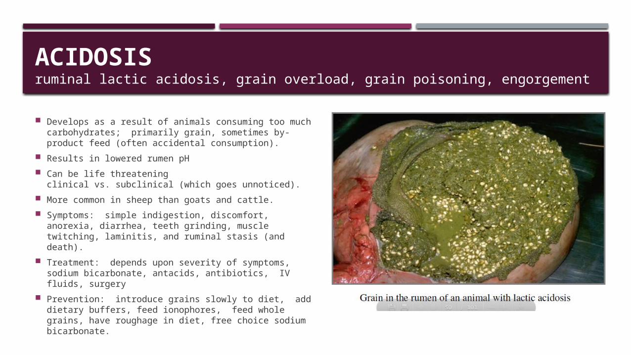
Develops as a result of animals consuming too much carbohydrates; primarily grain, sometimes by-product feed (often accidental consumption).
Results in lowered rumen pH Can be life threatening
clinical vs. subclinical (which goes unnoticed). More common in sheep than goats and cattle. Symptoms: simple indigestion, discomfort, anorexia,
diarrhea, teeth grinding, muscle twitching, laminitis, and ruminal stasis (and death).
Treatment: depends upon severity of symptoms, sodium bicarbonate, antacids, antibiotics, IV fluids, surgery
Prevention: introduce grains slowly to diet, add dietary buffers, feed ionophores, feed whole grains, have roughage in diet, free choice sodium bicarbonate.
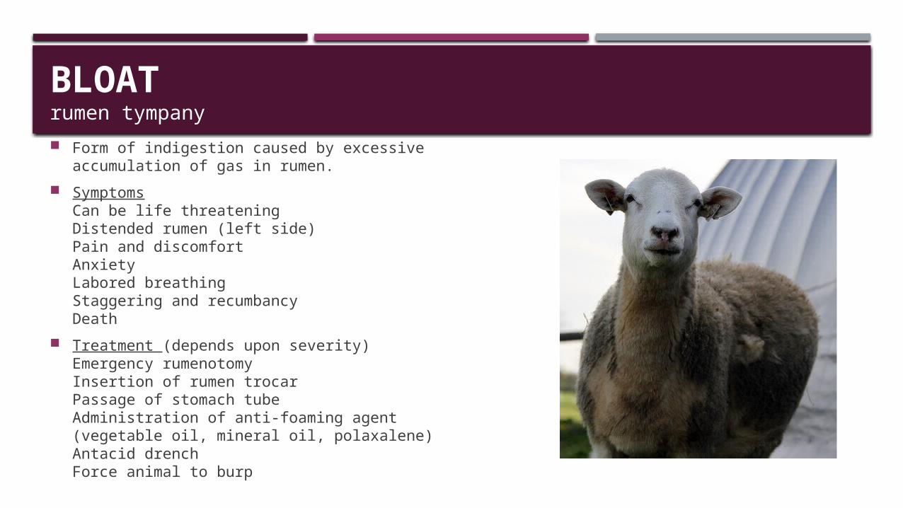
BLOATrumen tympany Form of indigestion caused by excessive accumulation of
gas in rumen. Symptoms
Can be life threateningDistended rumen (left side)Pain and discomfortAnxietyLabored breathingStaggering and recumbancyDeath
Treatment (depends upon severity)Emergency rumenotomyInsertion of rumen trocarPassage of stomach tubeAdministration of anti-foaming agent (vegetable oil, mineral oil, polaxalene)Antacid drenchForce animal to burp
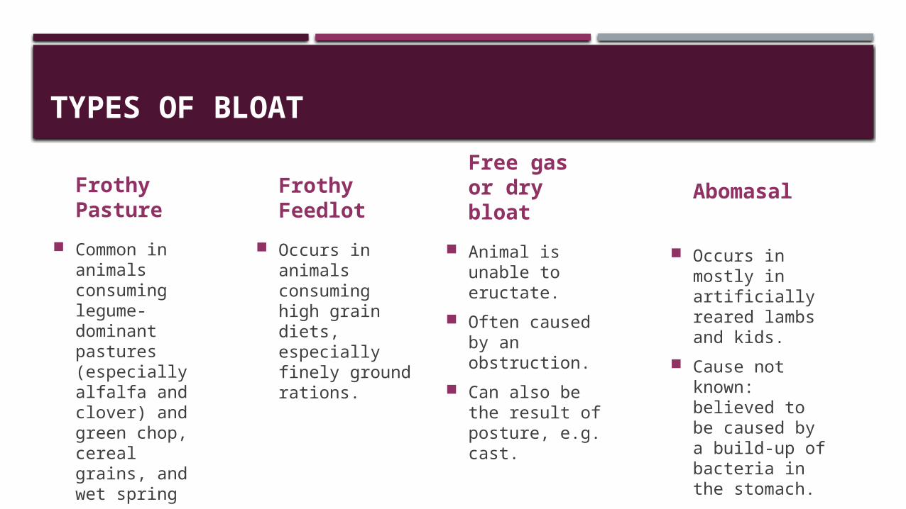
TYPES OF BLOAT
FrothyPasture
Common in animals consuming legume-dominant pastures (especially alfalfa and clover) and green chop, cereal grains, and wet spring grass.
FrothyFeedlot
Occurs in animals consuming high grain diets, especially finely ground rations.
Free gas or dry bloat
Animal is unable to eructate.
Often caused by an obstruction.
Can also be the result of posture, e.g. cast.
Abomasal
Occurs in mostly in artificially reared lambs and kids.
Cause not known: believed to be caused by a build-up of bacteria in the stomach.
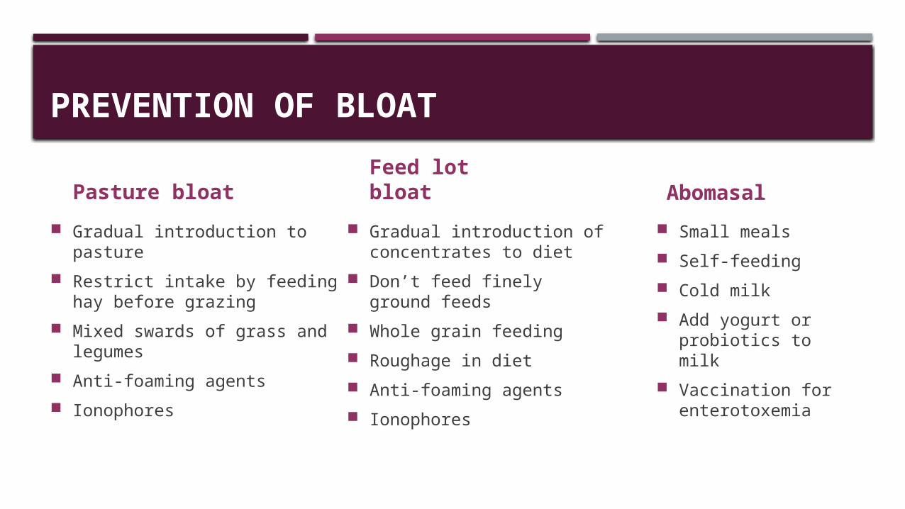
PREVENTION OF BLOAT
Pasture bloat Gradual introduction to pasture Restrict intake by feeding hay
before grazing Mixed swards of grass and
legumes Anti-foaming agents Ionophores
Abomasal
Small meals Self-feeding Cold milk Add yogurt or
probiotics to milk Vaccination for
enterotoxemia
Feed lot bloat Gradual introduction of
concentrates to diet Don’t feed finely ground
feeds Whole grain feeding Roughage in diet Anti-foaming agents Ionophores

COPPER TOXICITY
Sheep are most susceptible livestock species.Goats are more susceptible than cattle and pigs.Breed differences exist.
Can be acute or chronic. Toxicity occurs when copper accumulates in liver to
exceed 1000 mg Cu/kg DM Many factors affect copper metabolism.
Copper has many antagonists: Mo, Su Copper absorption more important than
concentration in feed; influenced by type of diet and level of Mo, S, Fe, and to a lesser extent Ca and Zn. 70-75% absorption rate in newborn ruminants < 10 percent in adults
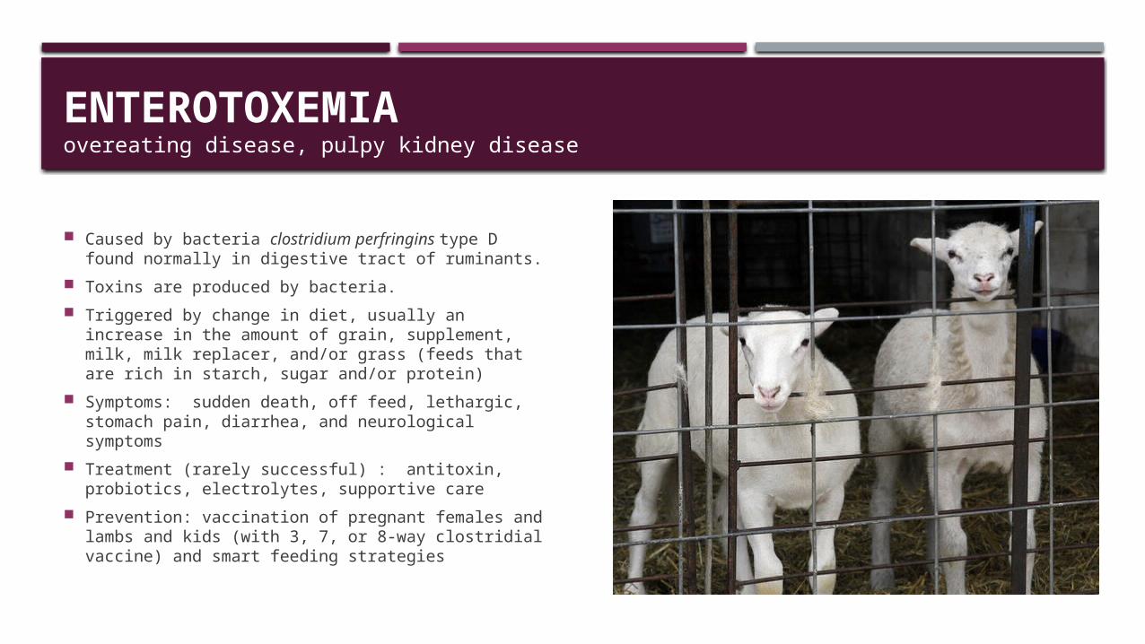
ENTEROTOXEMIA overeating disease, pulpy kidney disease
Caused by bacteria clostridium perfringins type D found normally in digestive tract of ruminants.
Toxins are produced by bacteria. Triggered by change in diet, usually an increase in
the amount of grain, supplement, milk, milk replacer, and/or grass (feeds that are rich in starch, sugar and/or protein)
Symptoms: sudden death, off feed, lethargic, stomach pain, diarrhea, and neurological symptoms
Treatment (rarely successful) : antitoxin, probiotics, electrolytes, supportive care
Prevention: vaccination of pregnant females and lambs and kids (with 3, 7, or 8-way clostridial vaccine) and smart feeding strategies
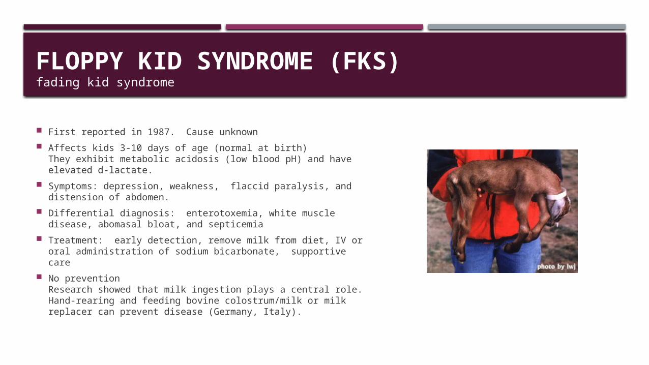
FLOPPY KID SYNDROME (FKS)fading kid syndrome
First reported in 1987. Cause unknown Affects kids 3-10 days of age (normal at birth)
They exhibit metabolic acidosis (low blood pH) and have elevated d-lactate.
Symptoms: depression, weakness, flaccid paralysis, and distension of abdomen.
Differential diagnosis: enterotoxemia, white muscle disease, abomasal bloat, and septicemia
Treatment: early detection, remove milk from diet, IV or oral administration of sodium bicarbonate, supportive care
No prevention Research showed that milk ingestion plays a central role.Hand-rearing and feeding bovine colostrum/milk or milk replacer can prevent disease (Germany, Italy).
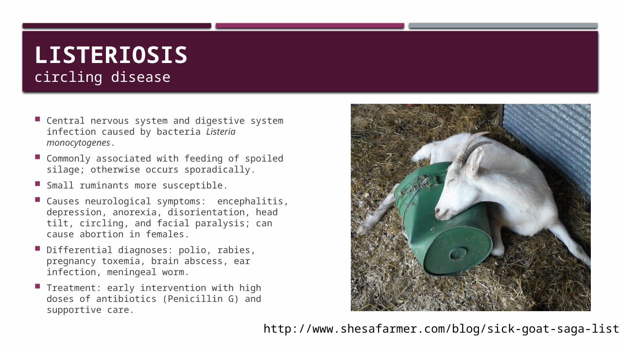
LISTERIOSIScircling disease
Central nervous system and digestive system infection caused by bacteria Listeria monocytogenes.
Commonly associated with feeding of spoiled silage; otherwise occurs sporadically.
Small ruminants more susceptible. Causes neurological symptoms: encephalitis,
depression, anorexia, disorientation, head tilt, circling, and facial paralysis; can cause abortion in females.
Differential diagnoses: polio, rabies, pregnancy toxemia, brain abscess, ear infection, meningeal worm.
Treatment: early intervention with high doses of antibiotics (Penicillin G) and supportive care.
http://www.shesafarmer.com/blog/sick-goat-saga-listeriosis
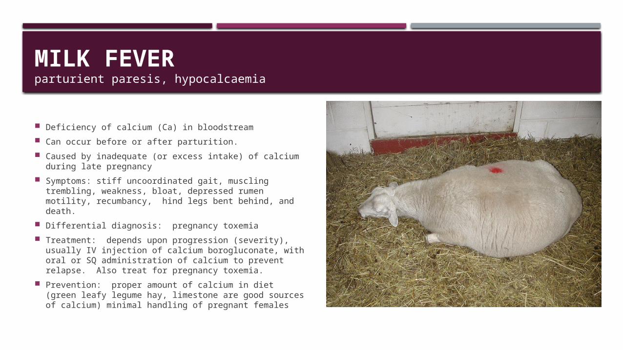
MILK FEVER parturient paresis, hypocalcaemia
Deficiency of calcium (Ca) in bloodstream Can occur before or after parturition. Caused by inadequate (or excess intake) of calcium
during late pregnancy Symptoms: stiff uncoordinated gait, muscling
trembling, weakness, bloat, depressed rumen motility, recumbancy, hind legs bent behind, and death.
Differential diagnosis: pregnancy toxemia Treatment: depends upon progression (severity),
usually IV injection of calcium borogluconate, with oral or SQ administration of calcium to prevent relapse. Also treat for pregnancy toxemia.
Prevention: proper amount of calcium in diet (green leafy legume hay, limestone are good sources of calcium) minimal handling of pregnant females

POLIOENCEPHALOMALACIAPEM, polio, cerebrocortical necrosis, thiamine deficiency
Metabolic disorder with neurological symptoms. Associated with thiamine status and/or high sulfur intake. Thiamine deficiency caused by inadequate production by
rumen or factors that interfere with action of thiamine. Sulfur-related PEM due to high sulfur intake Can occur on pasture, but animals on concentrate diets
(↓ rumen pH) are most susceptible. Can also result from prolonged treatment with Corid®
(thiamine inhibitor). Acute: blindness, star gazing, followed by recumbency,.
Subacute: separation, stop eating, twitching of ears and face, head held upright.
Differential diagnosis: pregnancy toxemia, enterotoxaemia, and listeriosis, rabies, tetanus, CAE, and plant poisoning.
Treatment: thiamine, IV followed by IM.
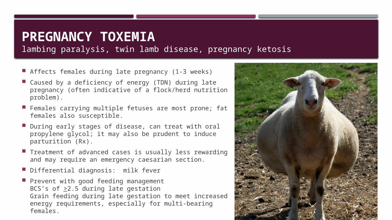
PREGNANCY TOXEMIAlambing paralysis, twin lamb disease, pregnancy ketosis
Affects females during late pregnancy (1-3 weeks) Caused by a deficiency of energy (TDN) during late
pregnancy (often indicative of a flock/herd nutrition problem).
Females carrying multiple fetuses are most prone; fat females also susceptible.
During early stages of disease, can treat with oral propylene glycol; it may also be prudent to induce parturition (Rx).
Treatment of advanced cases is usually less rewarding and may require an emergency caesarian section.
Differential diagnosis: milk fever Prevent with good feeding management
BCS’s of >2.5 during late gestationGrain feeding during late gestation to meet increased energy requirements, especially for multi-bearing females.
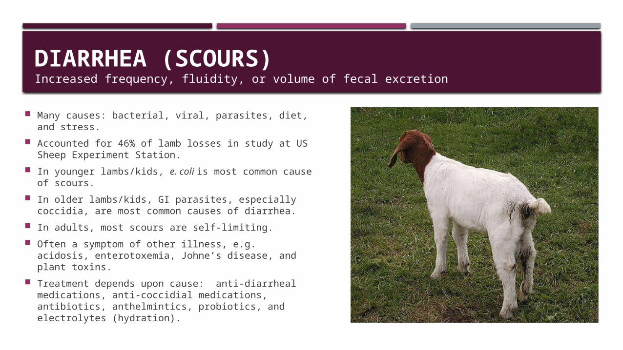
DIARRHEA (SCOURS)Increased frequency, fluidity, or volume of fecal excretion
Many causes: bacterial, viral, parasites, diet, and stress.
Accounted for 46% of lamb losses in study at US Sheep Experiment Station.
In younger lambs/kids, e. coli is most common cause of scours.
In older lambs/kids, GI parasites, especially coccidia, are most common causes of diarrhea.
In adults, most scours are self-limiting. Often a symptom of other illness, e.g. acidosis,
enterotoxemia, Johne’s disease, and plant toxins. Treatment depends upon cause: anti-diarrheal
medications, anti-coccidial medications, antibiotics, anthelmintics, probiotics, and electrolytes (hydration).
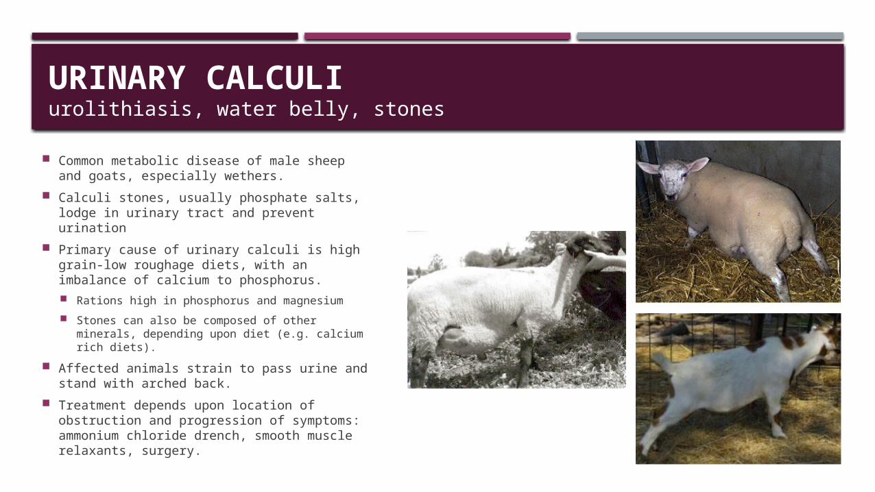
URINARY CALCULIurolithiasis, water belly, stones
Common metabolic disease of male sheep and goats, especially wethers.
Calculi stones, usually phosphate salts, lodge in urinary tract and prevent urination
Primary cause of urinary calculi is high grain-low roughage diets, with an imbalance of calcium to phosphorus. Rations high in phosphorus and magnesium Stones can also be composed of other
minerals, depending upon diet (e.g. calcium rich diets).
Affected animals strain to pass urine and stand with arched back.
Treatment depends upon location of obstruction and progression of symptoms: ammonium chloride drench, smooth muscle relaxants, surgery.
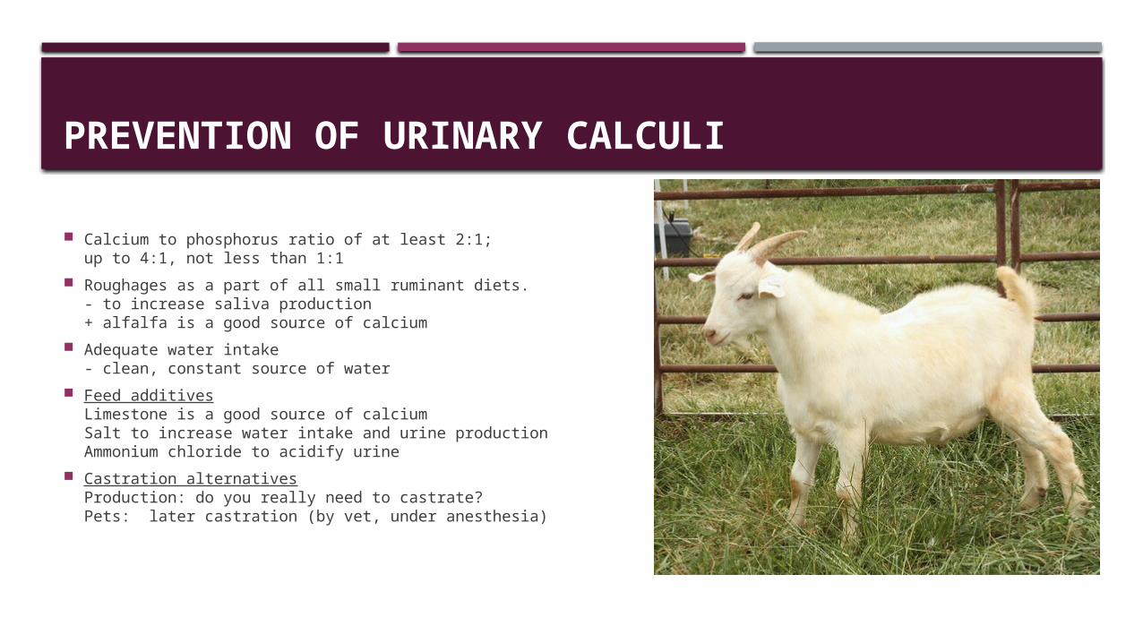
PREVENTION OF URINARY CALCULI
Calcium to phosphorus ratio of at least 2:1; up to 4:1, not less than 1:1
Roughages as a part of all small ruminant diets.- to increase saliva production+ alfalfa is a good source of calcium
Adequate water intake- clean, constant source of water
Feed additivesLimestone is a good source of calcium Salt to increase water intake and urine productionAmmonium chloride to acidify urine
Castration alternativesProduction: do you really need to castrate?Pets: later castration (by vet, under anesthesia)
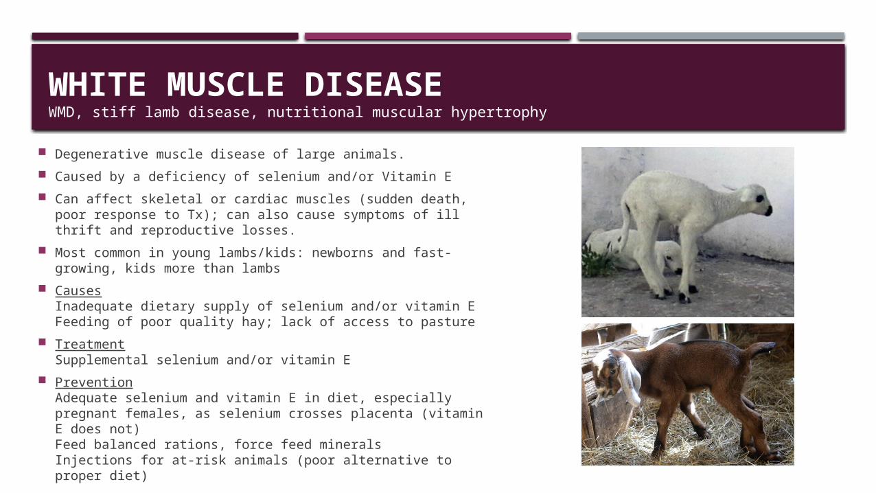
WHITE MUSCLE DISEASEWMD, stiff lamb disease, nutritional muscular hypertrophy
Degenerative muscle disease of large animals. Caused by a deficiency of selenium and/or Vitamin E Can affect skeletal or cardiac muscles (sudden death, poor
response to Tx); can also cause symptoms of ill thrift and reproductive losses.
Most common in young lambs/kids: newborns and fast-growing, kids more than lambs
CausesInadequate dietary supply of selenium and/or vitamin EFeeding of poor quality hay; lack of access to pasture
TreatmentSupplemental selenium and/or vitamin E
PreventionAdequate selenium and vitamin E in diet, especially pregnant females, as selenium crosses placenta (vitamin E does not)Feed balanced rations, force feed mineralsInjections for at-risk animals (poor alternative to proper diet)
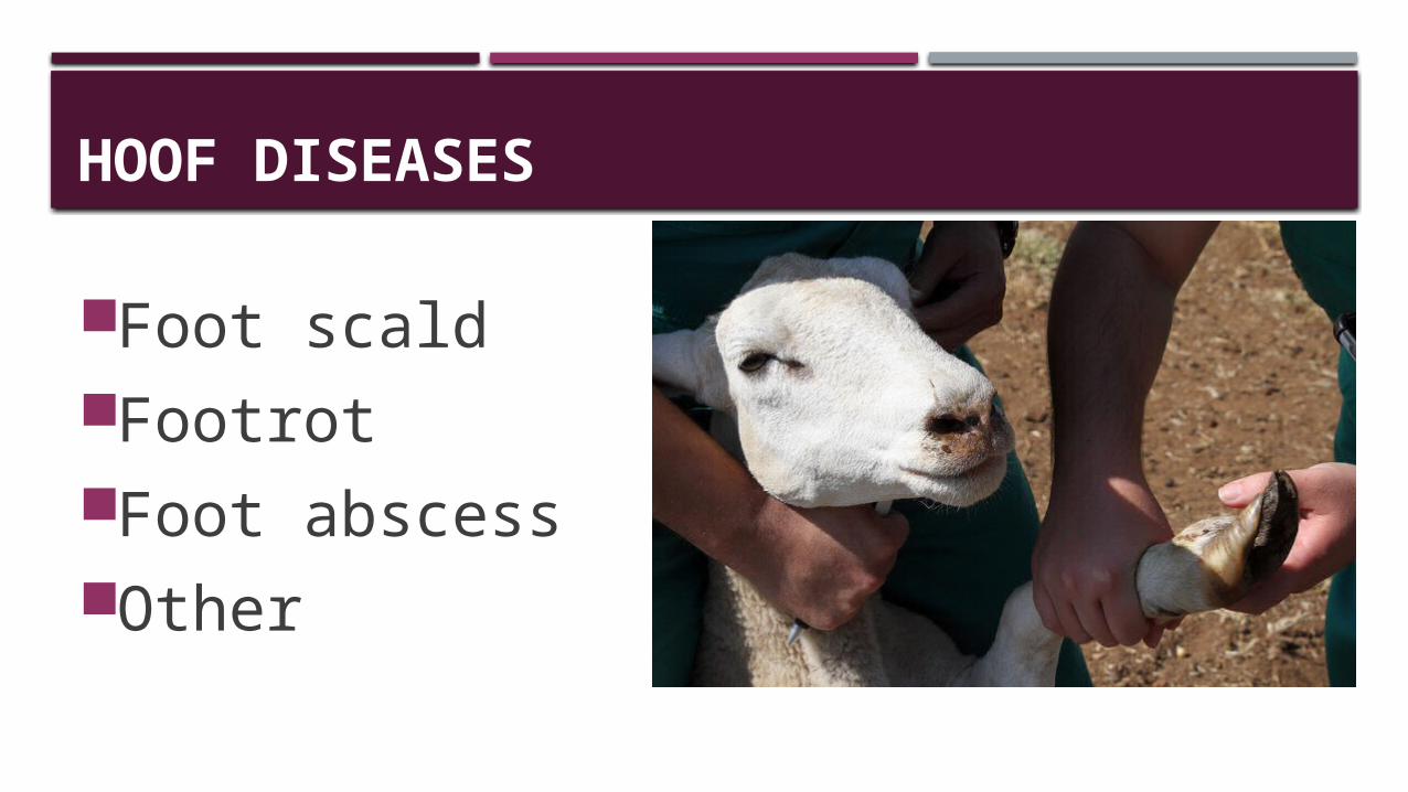
HOOF DISEASES
Foot scaldFootrotFoot abscessOther
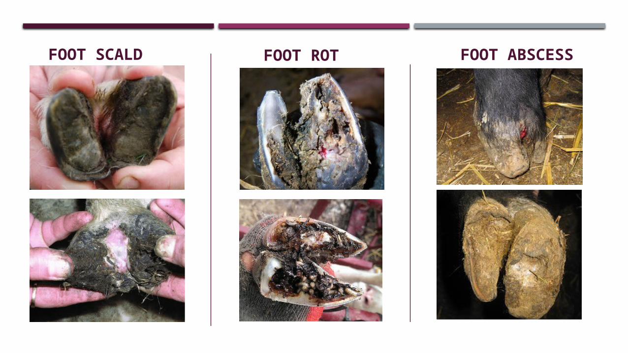
FOOT SCALD FOOT ROT FOOT ABSCESS
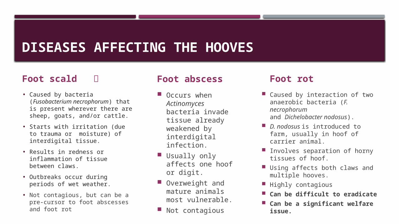
DISEASES AFFECTING THE HOOVES
Foot scald • Caused by bacteria
(Fusobacterium necrophorum) that is present wherever there are sheep, goats, and/or cattle.
• Starts with irritation (due to trauma or moisture) of interdigital tissue.
• Results in redness or inflammation of tissue between claws.
• Outbreaks occur during periods of wet weather.
• Not contagious, but can be a pre-cursor to foot abscesses and foot rot
Foot abscess Occurs when
Actinomyces bacteria invade tissue already weakened by interdigital infection.
Usually only affects one hoof or digit.
Overweight and mature animals most vulnerable.
Not contagious
Foot rot Caused by interaction of two
anaerobic bacteria (F. necrophorum and Dichelobacter nodosus).
D. nodosus is introduced to farm, usually in hoof of carrier animal.
Involves separation of horny tissues of hoof.
Using affects both claws and multiple hooves.
Highly contagious Can be difficult to eradicate Can be a significant welfare
issue.
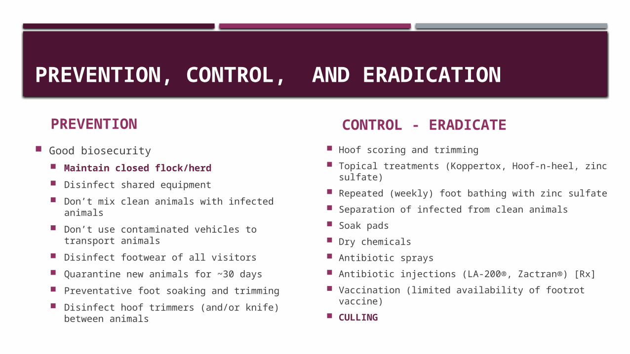
PREVENTION, CONTROL, AND ERADICATION
PREVENTION Good biosecurity
Maintain closed flock/herd Disinfect shared equipment Don’t mix clean animals with infected animals Don’t use contaminated vehicles to transport
animals Disinfect footwear of all visitors Quarantine new animals for ~30 days Preventative foot soaking and trimming Disinfect hoof trimmers (and/or knife) between
animals
CONTROL - ERADICATE Hoof scoring and trimming Topical treatments (Koppertox, Hoof-n-heel, zinc
sulfate) Repeated (weekly) foot bathing with zinc sulfate Separation of infected from clean animals Soak pads Dry chemicals Antibiotic sprays Antibiotic injections (LA-200®, Zactran®) [Rx] Vaccination (limited availability of footrot vaccine) CULLING
REPRODUCTIVE DISEASES AND PROBLEMS
Abortive diseases Dystocia Epididymitis Mastitis Milk fever Pregnancy toxemia Uterine prolapse Vaginal prolapse
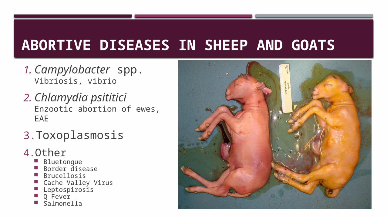
ABORTIVE DISEASES IN SHEEP AND GOATS
1.Campylobacter spp.Vibriosis, vibrio
2.Chlamydia psititiciEnzootic abortion of ewes, EAE
3.Toxoplasmosis4. Other
Bluetongue Border disease Brucellosis Cache Valley Virus Leptospirosis Q Fever Salmonella
ABORTIVE DISEASES IN SHEEP AND GOATS
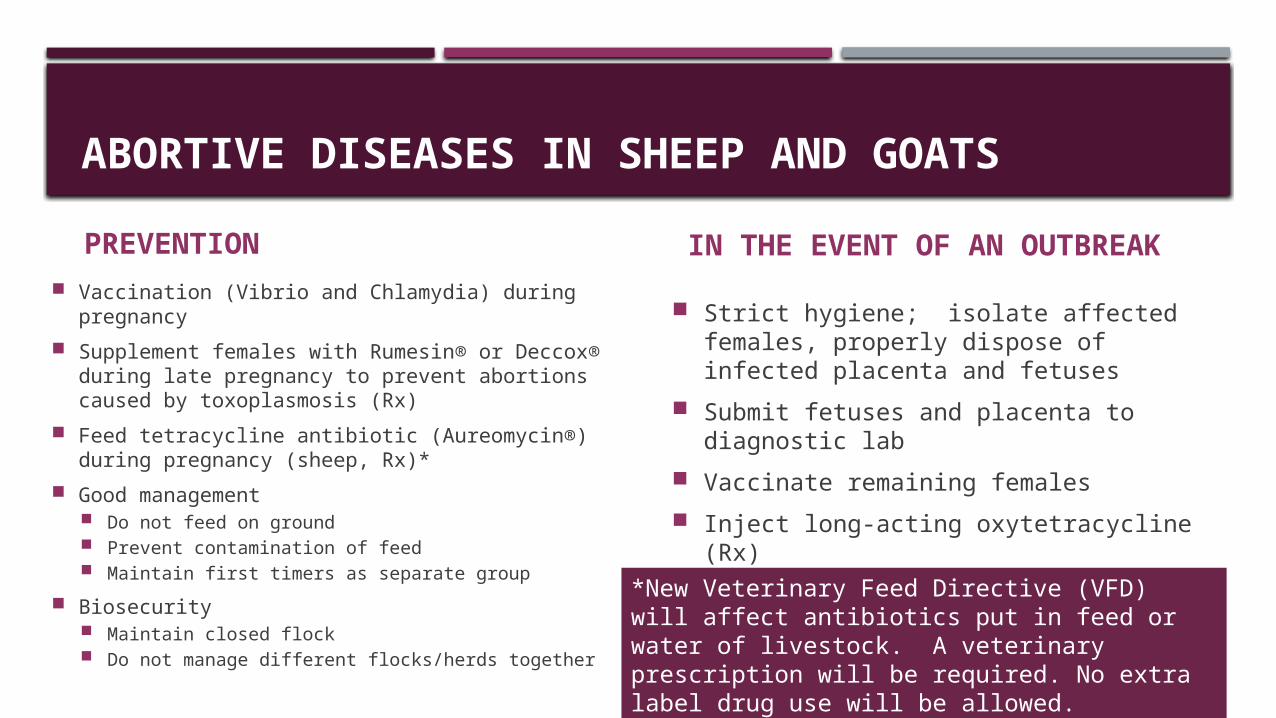
PREVENTION Vaccination (Vibrio and Chlamydia) during
pregnancy Supplement females with Rumesin® or Deccox®
during late pregnancy to prevent abortions caused by toxoplasmosis (Rx)
Feed tetracycline antibiotic (Aureomycin®) during pregnancy (sheep, Rx)*
Good management Do not feed on ground Prevent contamination of feed Maintain first timers as separate group
Biosecurity Maintain closed flock Do not manage different flocks/herds together
IN THE EVENT OF AN OUTBREAK
Strict hygiene; isolate affected females, properly dispose of infected placenta and fetuses
Submit fetuses and placenta to diagnostic lab
Vaccinate remaining females Inject long-acting oxytetracycline (Rx) Begin feeding tetracycline antibiotic
(sheep, Rx)**New Veterinary Feed Directive (VFD) will affect antibiotics put in feed or water of livestock. A veterinary prescription will be required. No extra label drug use will be allowed.
DYSTOCIA (DIFFICULT BIRTHING)
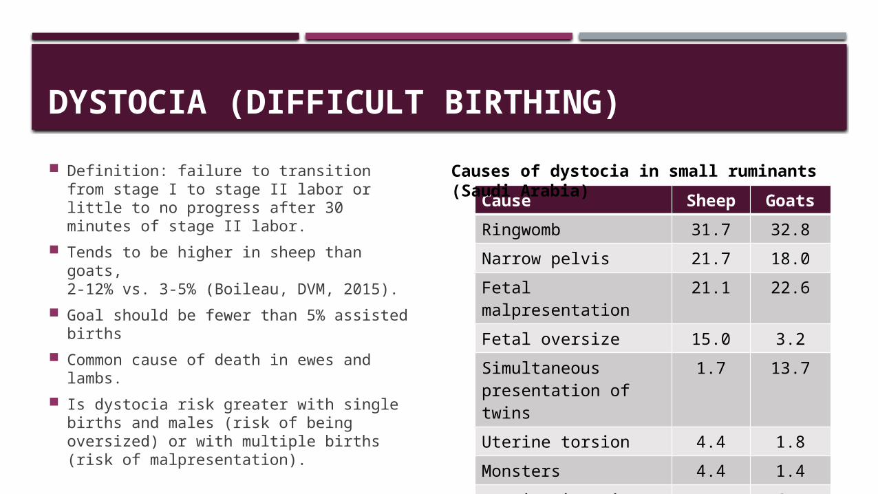
Definition: failure to transition from stage I to stage II labor or little to no progress after 30 minutes of stage II labor.
Tends to be higher in sheep than goats, 2-12% vs. 3-5% (Boileau, DVM, 2015).
Goal should be fewer than 5% assisted births
Common cause of death in ewes and lambs.
Is dystocia risk greater with single births and males (risk of being oversized) or with multiple births (risk of malpresentation).
Cause Sheep GoatsRingwomb 31.7 32.8Narrow pelvis 21.7 18.0Fetal malpresentation 21.1 22.6Fetal oversize 15.0 3.2Simultaneous presentation of twins
1.7 13.7
Uterine torsion 4.4 1.8Monsters 4.4 1.4Uterine inertia 6.7
J. Ag. & Vet Sci (2011)
Causes of dystocia in small ruminants (Saudi Arabia)
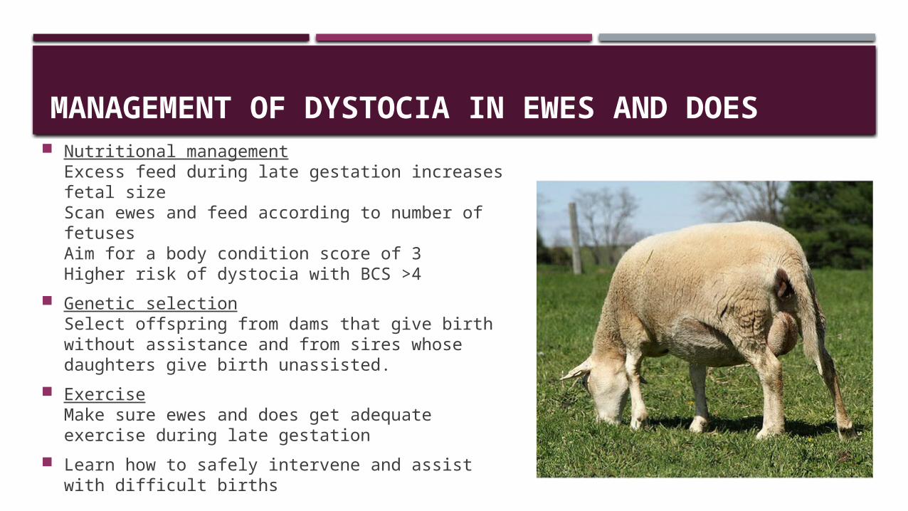
MANAGEMENT OF DYSTOCIA IN EWES AND DOES Nutritional management
Excess feed during late gestation increases fetal sizeScan ewes and feed according to number of fetusesAim for a body condition score of 3Higher risk of dystocia with BCS >4
Genetic selectionSelect offspring from dams that give birth without assistance and from sires whose daughters give birth unassisted.
ExerciseMake sure ewes and does get adequate exercise during late gestation
Learn how to safely intervene and assist with difficult births
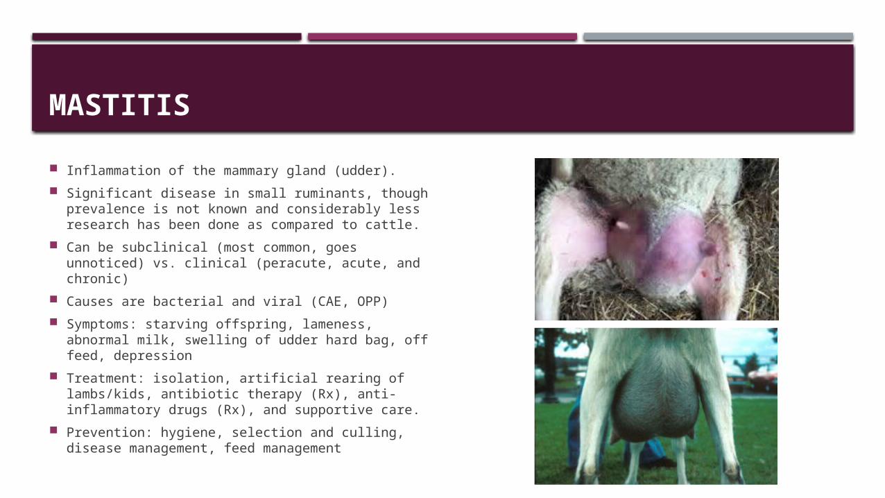
MASTITIS
Inflammation of the mammary gland (udder). Significant disease in small ruminants, though
prevalence is not known and considerably less research has been done as compared to cattle.
Can be subclinical (most common, goes unnoticed) vs. clinical (peracute, acute, and chronic)
Causes are bacterial and viral (CAE, OPP) Symptoms: starving offspring, lameness, abnormal
milk, swelling of udder hard bag, off feed, depression Treatment: isolation, artificial rearing of lambs/kids,
antibiotic therapy (Rx), anti-inflammatory drugs (Rx), and supportive care.
Prevention: hygiene, selection and culling, disease management, feed management
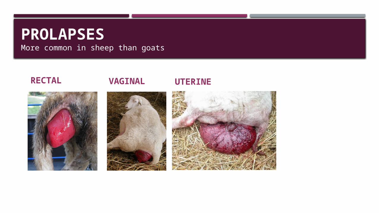
PROLAPSESMore common in sheep than goats
RECTAL VAGINAL UTERINE
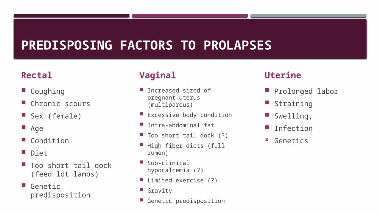
PREDISPOSING FACTORS TO PROLAPSES
Rectal Coughing Chronic scours Sex (female) Age Condition Diet Too short tail dock
(feed lot lambs) Genetic predisposition
Vaginal Increased sized of pregnant
uterus (multiparous) Excessive body condition Intra-abdominal fat Too short tail dock (?) High fiber diets (full rumen) Sub-clinical hypocalcemia
(?) Limited exercise (?) Gravity Genetic predisposition
Uterine Prolonged labor Straining Swelling, Infection Genetics
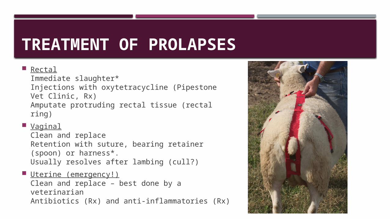
TREATMENT OF PROLAPSES Rectal
Immediate slaughter*Injections with oxytetracycline (Pipestone Vet Clinic, Rx)Amputate protruding rectal tissue (rectal ring)
VaginalClean and replaceRetention with suture, bearing retainer (spoon) or harness*.Usually resolves after lambing (cull?)
Uterine (emergency!)Clean and replace – best done by a veterinarian Antibiotics (Rx) and anti-inflammatories (Rx)
PARASITIC DISEASES
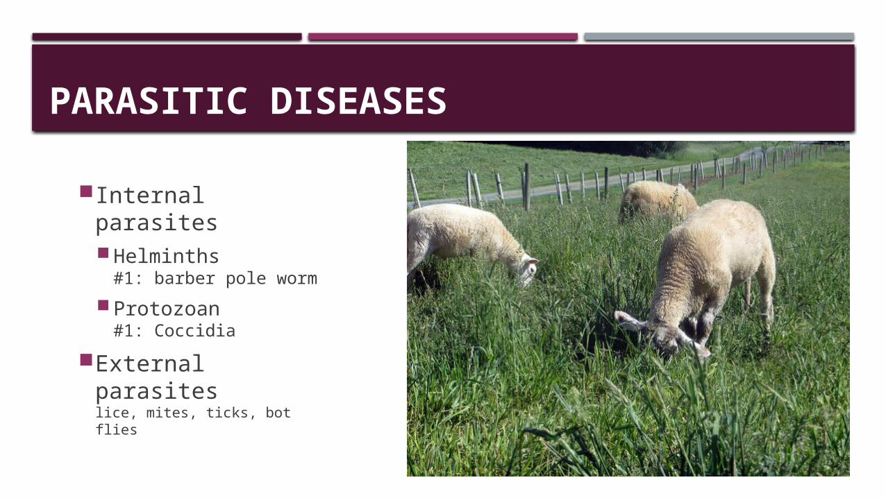
Internal parasitesHelminths
#1: barber pole wormProtozoan
#1: CoccidiaExternal parasites
lice, mites, ticks, bot flies
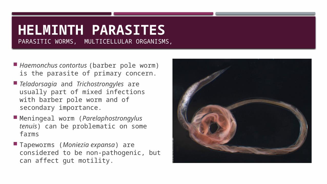
HELMINTH PARASITESPARASITIC WORMS, MULTICELLULAR ORGANISMS,
Haemonchus contortus (barber pole worm) is the parasite of primary concern.
Teladorsagia and Trichostrongyles are usually part of mixed infections with barber pole worm and of secondary importance.
Meningeal worm (Parelaphostrongylus tenuis) can be problematic on some farms
Tapeworms (Moniezia expansa) are considered to be non-pathogenic, but can affect gut motility.
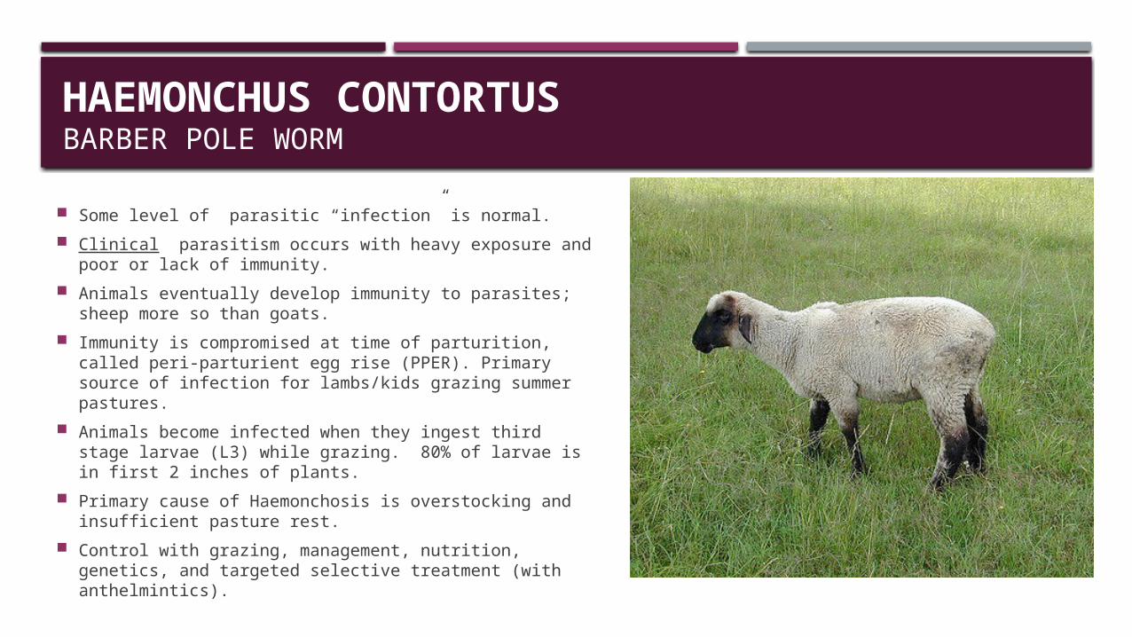
HAEMONCHUS CONTORTUSBARBER POLE WORM
Some level of parasitic “infection” is normal. Clinical parasitism occurs with heavy exposure and
poor or lack of immunity. Animals eventually develop immunity to parasites;
sheep more so than goats. Immunity is compromised at time of parturition, called
peri-parturient egg rise (PPER). Primary source of infection for lambs/kids grazing summer pastures.
Animals become infected when they ingest third stage larvae (L3) while grazing. 80% of larvae is in first 2 inches of plants.
Primary cause of Haemonchosis is overstocking and insufficient pasture rest.
Control with grazing, management, nutrition, genetics, and targeted selective treatment (with anthelmintics).
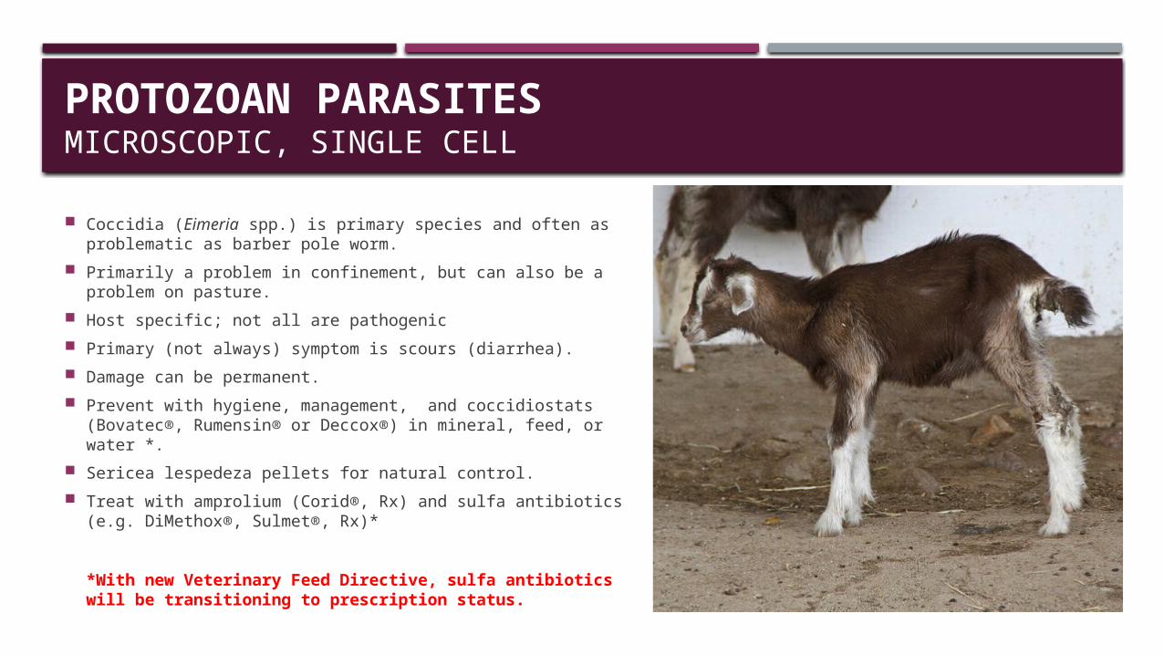
PROTOZOAN PARASITESMICROSCOPIC, SINGLE CELL
Coccidia (Eimeria spp.) is primary species and often as problematic as barber pole worm.
Primarily a problem in confinement, but can also be a problem on pasture.
Host specific; not all are pathogenic Primary (not always) symptom is scours (diarrhea). Damage can be permanent. Prevent with hygiene, management, and coccidiostats
(Bovatec®, Rumensin® or Deccox®) in mineral, feed, or water *. Sericea lespedeza pellets for natural control. Treat with amprolium (Corid®, Rx) and sulfa antibiotics (e.g.
DiMethox®, Sulmet®, Rx)*
*With new Veterinary Feed Directive, sulfa antibiotics will be transitioning to prescription status.
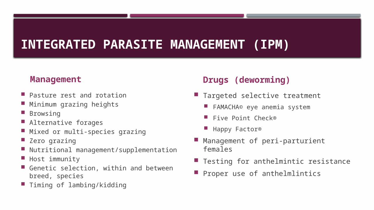
INTEGRATED PARASITE MANAGEMENT (IPM)
Management Pasture rest and rotation Minimum grazing heights Browsing Alternative forages Mixed or multi-species grazing Zero grazing Nutritional management/supplementation Host immunity Genetic selection, within and between
breed, species Timing of lambing/kidding
Drugs (deworming) Targeted selective treatment
FAMACHA© eye anemia system Five Point Check® Happy Factor®
Management of peri-parturient females Testing for anthelmintic resistance Proper use of anthelmlintics
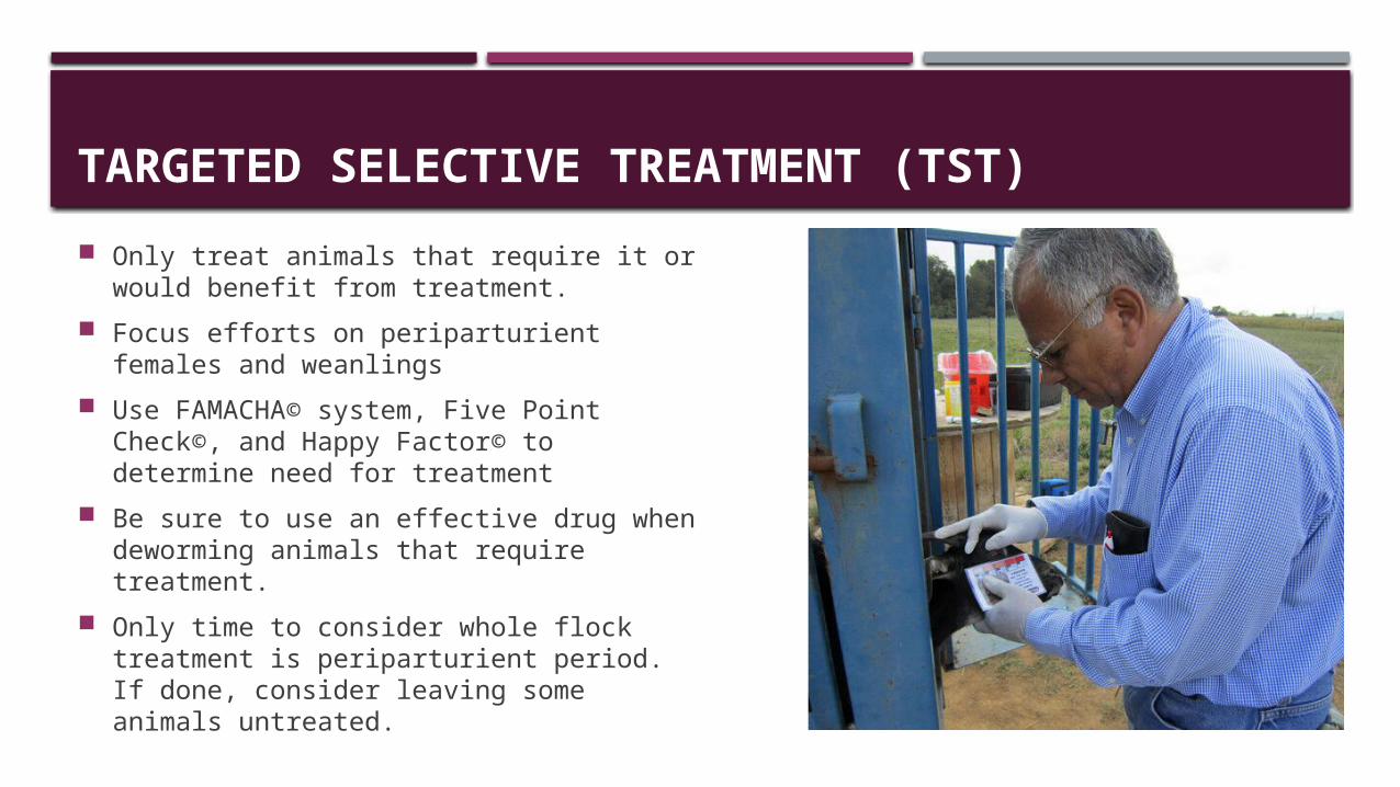
TARGETED SELECTIVE TREATMENT (TST)
Only treat animals that require it or would benefit from treatment.
Focus efforts on periparturient females and weanlings
Use FAMACHA© system, Five Point Check©, and Happy Factor© to determine need for treatment
Be sure to use an effective drug when deworming animals that require treatment.
Only time to consider whole flock treatment is periparturient period. If done, consider leaving some animals untreated.
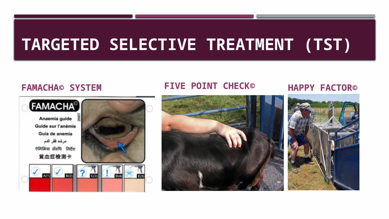
TARGETED SELECTIVE TREATMENT (TST)
FAMACHA© SYSTEM FIVE POINT CHECK© HAPPY FACTOR©
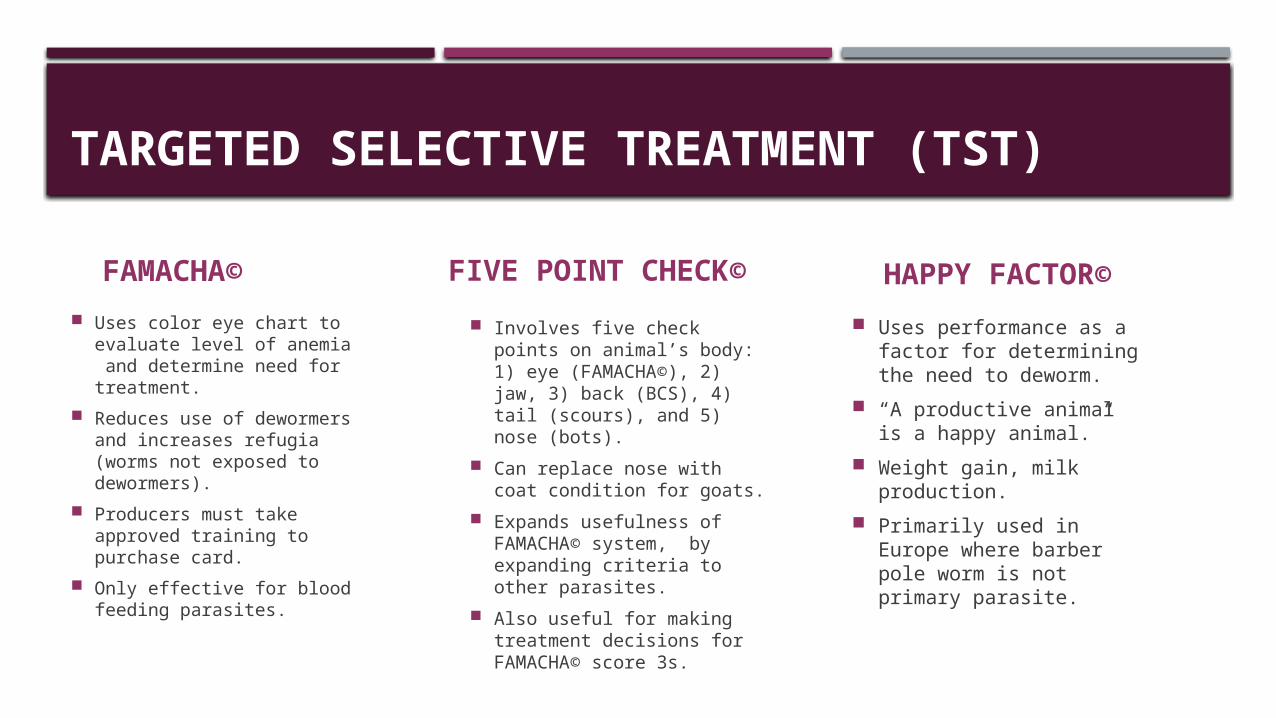
TARGETED SELECTIVE TREATMENT (TST)
FAMACHA© Uses color eye chart to
evaluate level of anemia and determine need for treatment.
Reduces use of dewormers and increases refugia (worms not exposed to dewormers).
Producers must take approved training to purchase card.
Only effective for blood feeding parasites.
FIVE POINT CHECK©
Involves five check points on animal’s body: 1) eye (FAMACHA©), 2) jaw, 3) back (BCS), 4) tail (scours), and 5) nose (bots).
Can replace nose with coat condition for goats.
Expands usefulness of FAMACHA© system, by expanding criteria to other parasites.
Also useful for making treatment decisions for FAMACHA© score 3s.
HAPPY FACTOR© Uses performance as a
factor for determining the need to deworm.
“A productive animal is a happy animal.”
Weight gain, milk production.
Primarily used in Europe where barber pole worm is not primary parasite.
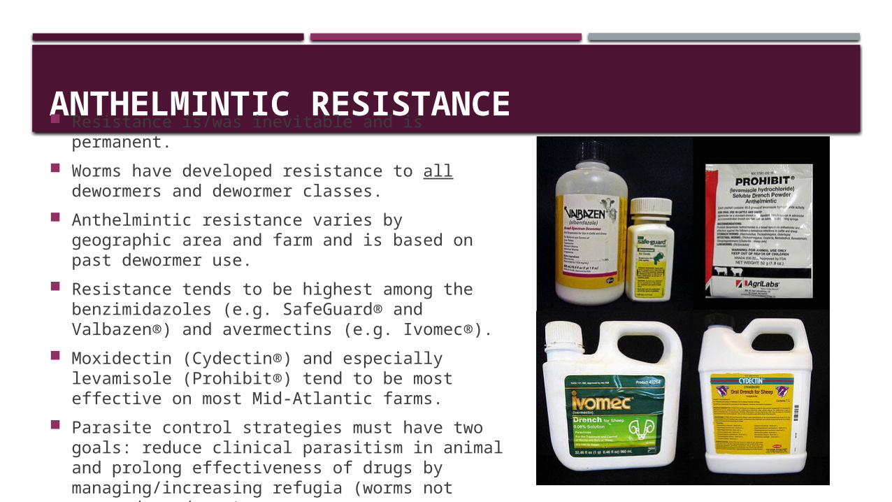
ANTHELMINTIC RESISTANCE Resistance is/was inevitable and is permanent. Worms have developed resistance to all dewormers
and dewormer classes. Anthelmintic resistance varies by geographic area
and farm and is based on past dewormer use. Resistance tends to be highest among the
benzimidazoles (e.g. SafeGuard® and Valbazen®) and avermectins (e.g. Ivomec®).
Moxidectin (Cydectin®) and especially levamisole (Prohibit®) tend to be most effective on most Mid-Atlantic farms.
Parasite control strategies must have two goals: reduce clinical parasitism in animal and prolong effectiveness of drugs by managing/increasing refugia (worms not exposed to drugs).
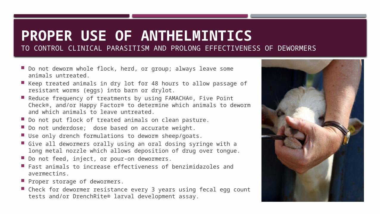
PROPER USE OF ANTHELMINTICSTO CONTROL CLINICAL PARASITISM AND PROLONG EFFECTIVENESS OF DEWORMERS
Do not deworm whole flock, herd, or group; always leave some animals untreated.
Keep treated animals in dry lot for 48 hours to allow passage of resistant worms (eggs) into barn or drylot.
Reduce frequency of treatments by using FAMACHA©, Five Point Check®, and/or Happy Factor® to determine which animals to deworm and which animals to leave untreated.
Do not put flock of treated animals on clean pasture. Do not underdose; dose based on accurate weight. Use only drench formulations to deworm sheep/goats. Give all dewormers orally using an oral dosing syringe with a long metal
nozzle which allows deposition of drug over tongue. Do not feed, inject, or pour-on dewormers. Fast animals to increase effectiveness of benzimidazoles and avermectins. Proper storage of dewormers. Check for dewormer resistance every 3 years using fecal egg count tests
and/or DrenchRite® larval development assay.
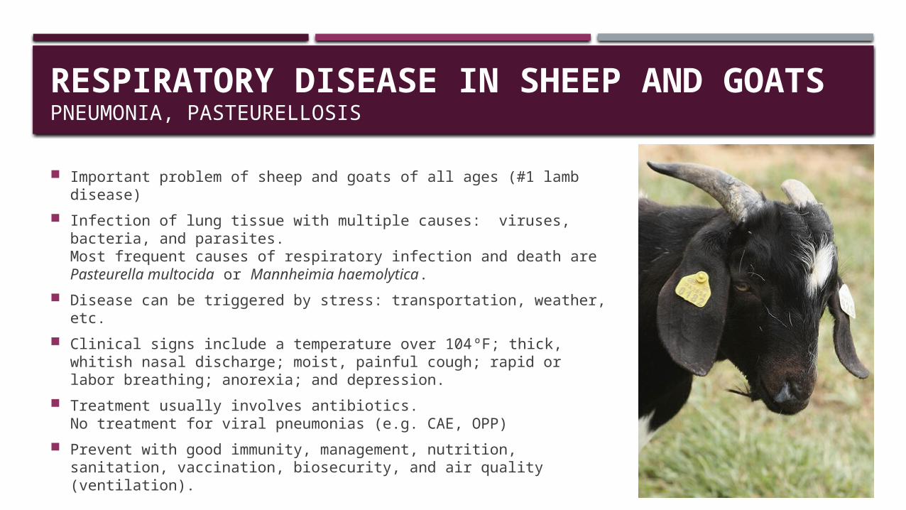
RESPIRATORY DISEASE IN SHEEP AND GOATSPNEUMONIA, PASTEURELLOSIS
Important problem of sheep and goats of all ages (#1 lamb disease)
Infection of lung tissue with multiple causes: viruses, bacteria, and parasites.Most frequent causes of respiratory infection and death are Pasteurella multocida or Mannheimia haemolytica.
Disease can be triggered by stress: transportation, weather, etc. Clinical signs include a temperature over 104ºF; thick, whitish
nasal discharge; moist, painful cough; rapid or labor breathing; anorexia; and depression.
Treatment usually involves antibiotics.No treatment for viral pneumonias (e.g. CAE, OPP)
Prevent with good immunity, management, nutrition, sanitation, vaccination, biosecurity, and air quality (ventilation).
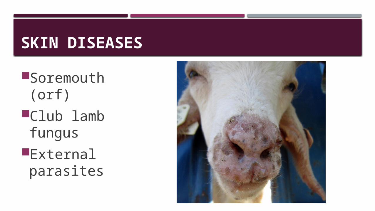
SKIN DISEASES
Soremouth (orf)Club lamb fungus
External parasites
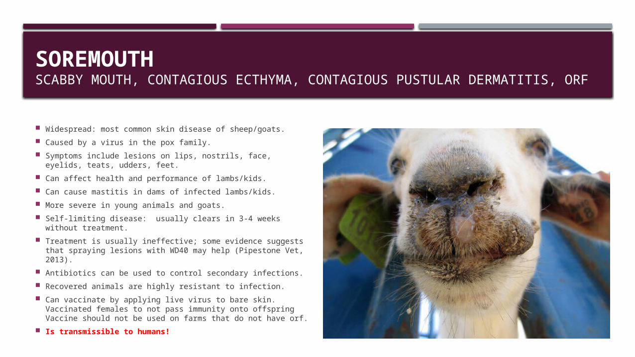
SOREMOUTH SCABBY MOUTH, CONTAGIOUS ECTHYMA, CONTAGIOUS PUSTULAR DERMATITIS, ORF
Widespread: most common skin disease of sheep/goats. Caused by a virus in the pox family. Symptoms include lesions on lips, nostrils, face, eyelids, teats,
udders, feet. Can affect health and performance of lambs/kids. Can cause mastitis in dams of infected lambs/kids. More severe in young animals and goats. Self-limiting disease: usually clears in 3-4 weeks without
treatment. Treatment is usually ineffective; some evidence suggests that
spraying lesions with WD40 may help (Pipestone Vet, 2013). Antibiotics can be used to control secondary infections. Recovered animals are highly resistant to infection. Can vaccinate by applying live virus to bare skin.
Vaccinated females to not pass immunity onto offspringVaccine should not be used on farms that do not have orf.
Is transmissible to humans!
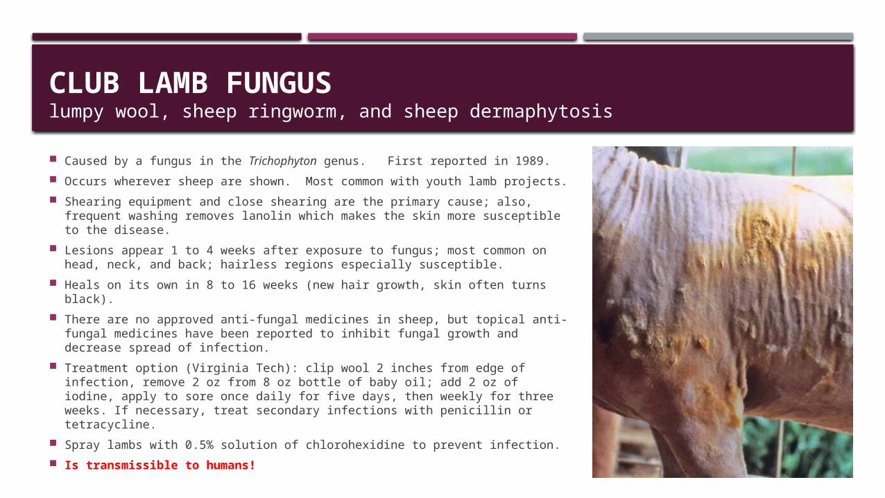
CLUB LAMB FUNGUSlumpy wool, sheep ringworm, and sheep dermaphytosis
Caused by a fungus in the Trichophyton genus. First reported in 1989. Occurs wherever sheep are shown. Most common with youth lamb projects. Shearing equipment and close shearing are the primary cause; also,
frequent washing removes lanolin which makes the skin more susceptible to the disease.
Lesions appear 1 to 4 weeks after exposure to fungus; most common on head, neck, and back; hairless regions especially susceptible.
Heals on its own in 8 to 16 weeks (new hair growth, skin often turns black). There are no approved anti-fungal medicines in sheep, but topical anti-
fungal medicines have been reported to inhibit fungal growth and decrease spread of infection.
Treatment option (Virginia Tech): clip wool 2 inches from edge of infection, remove 2 oz from 8 oz bottle of baby oil; add 2 oz of iodine, apply to sore once daily for five days, then weekly for three weeks. If necessary, treat secondary infections with penicillin or tetracycline.
Spray lambs with 0.5% solution of chlorohexidine to prevent infection. Is transmissible to humans!
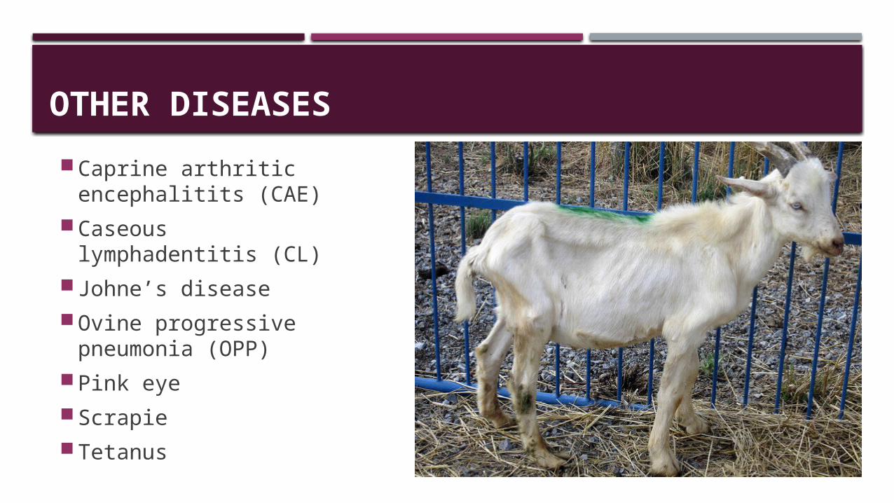
OTHER DISEASES Caprine arthritic
encephalitits (CAE) Caseous
lymphadentitis (CL) Johne’s disease Ovine progressive
pneumonia (OPP) Pink eye Scrapie Tetanus
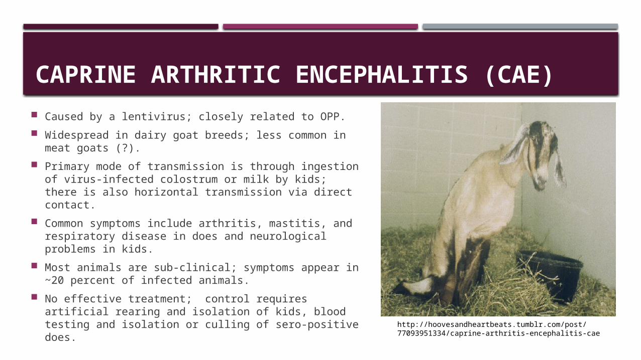
CAPRINE ARTHRITIC ENCEPHALITIS (CAE) Caused by a lentivirus; closely related to OPP. Widespread in dairy goat breeds; less common in
meat goats (?). Primary mode of transmission is through ingestion of
virus-infected colostrum or milk by kids; there is also horizontal transmission via direct contact.
Common symptoms include arthritis, mastitis, and respiratory disease in does and neurological problems in kids.
Most animals are sub-clinical; symptoms appear in ~20 percent of infected animals.
No effective treatment; control requires artificial rearing and isolation of kids, blood testing and isolation or culling of sero-positive does.
http://hoovesandheartbeats.tumblr.com/post/77093951334/caprine-arthritis-encephalitis-cae
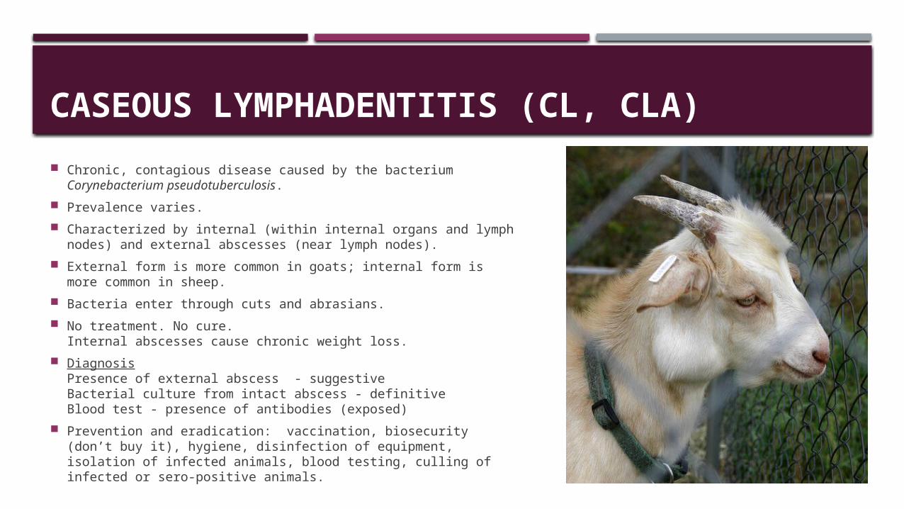
CASEOUS LYMPHADENTITIS (CL, CLA) Chronic, contagious disease caused by the bacterium
Corynebacterium pseudotuberculosis. Prevalence varies. Characterized by internal (within internal organs and lymph
nodes) and external abscesses (near lymph nodes). External form is more common in goats; internal form is more
common in sheep. Bacteria enter through cuts and abrasians. No treatment. No cure.
Internal abscesses cause chronic weight loss. Diagnosis
Presence of external abscess - suggestiveBacterial culture from intact abscess - definitive Blood test - presence of antibodies (exposed)
Prevention and eradication: vaccination, biosecurity (don’t buy it), hygiene, disinfection of equipment, isolation of infected animals, blood testing, culling of infected or sero-positive animals.
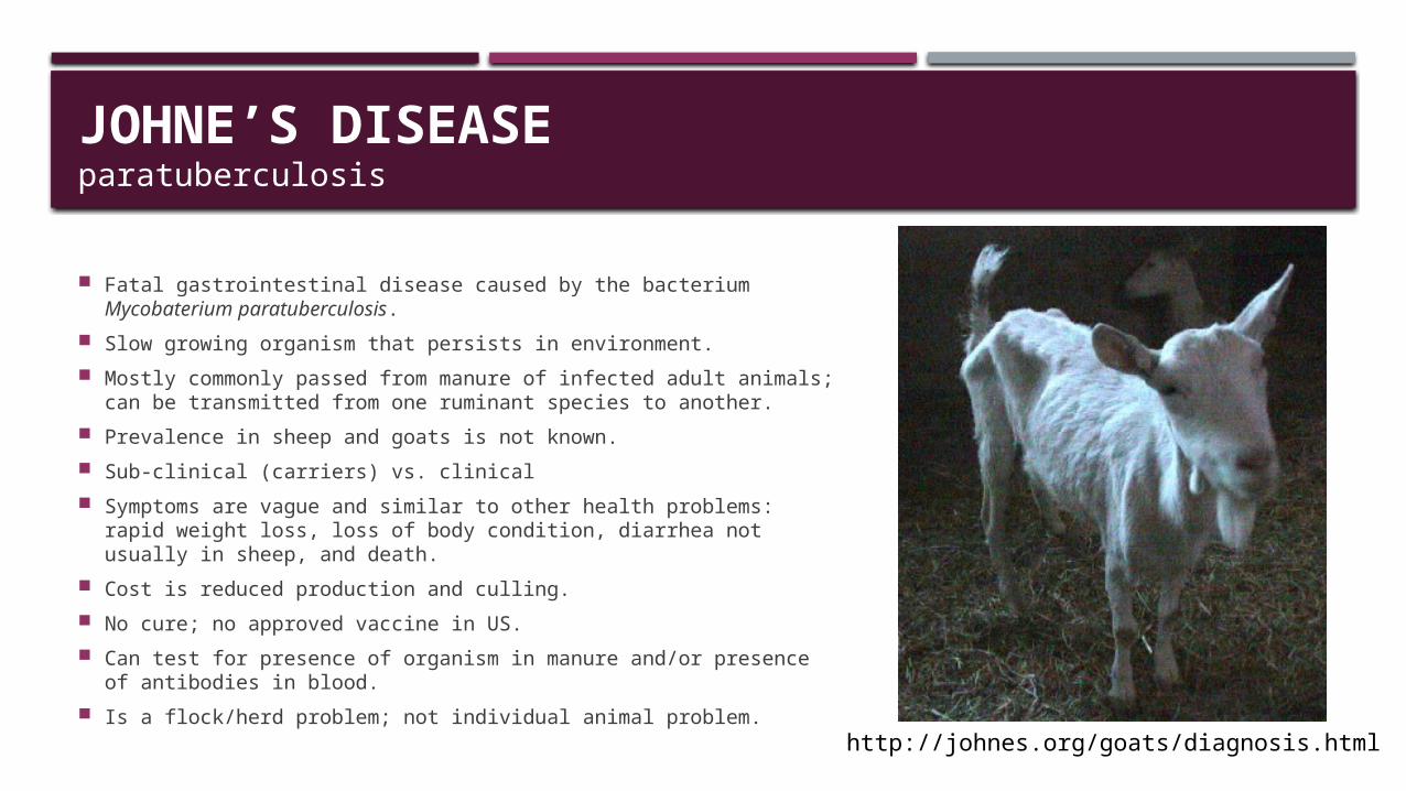
JOHNE’S DISEASE paratuberculosis
Fatal gastrointestinal disease caused by the bacterium Mycobaterium paratuberculosis.
Slow growing organism that persists in environment. Mostly commonly passed from manure of infected adult animals; can
be transmitted from one ruminant species to another. Prevalence in sheep and goats is not known. Sub-clinical (carriers) vs. clinical Symptoms are vague and similar to other health problems: rapid
weight loss, loss of body condition, diarrhea not usually in sheep, and death.
Cost is reduced production and culling. No cure; no approved vaccine in US. Can test for presence of organism in manure and/or presence of
antibodies in blood. Is a flock/herd problem; not individual animal problem. http://johnes.org/goats/diagnosis.html
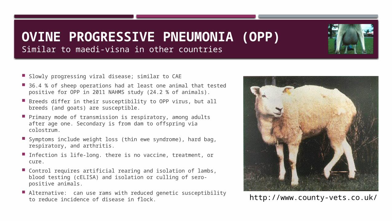
OVINE PROGRESSIVE PNEUMONIA (OPP)Similar to maedi-visna in other countries
Slowly progressing viral disease; similar to CAE 36.4 % of sheep operations had at least one animal that tested
positive for OPP in 2011 NAHMS study (24.2 % of animals). Breeds differ in their susceptibility to OPP virus, but all breeds
(and goats) are susceptible. Primary mode of transmission is respiratory, among adults after
age one. Secondary is from dam to offspring via colostrum. Symptoms include weight loss (thin ewe syndrome), hard bag,
respiratory, and arthritis. Infection is life-long. there is no vaccine, treatment, or cure. Control requires artificial rearing and isolation of lambs, blood
testing (cELISA) and isolation or culling of sero-positive animals. Alternative: can use rams with reduced genetic susceptibility to
reduce incidence of disease in flock. http://www.county-vets.co.uk/
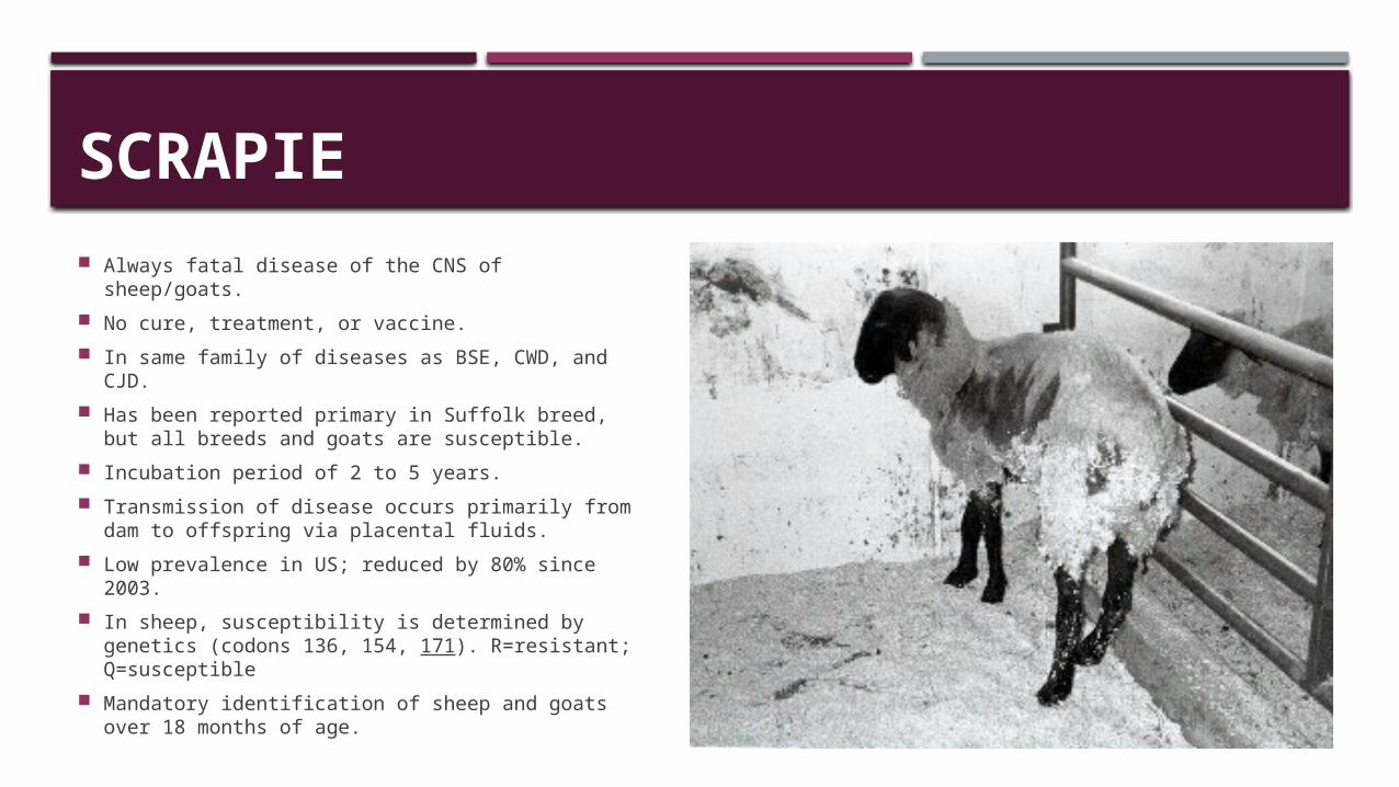
SCRAPIE Always fatal disease of the CNS of sheep/goats. No cure, treatment, or vaccine. In same family of diseases as BSE, CWD, and CJD. Has been reported primary in Suffolk breed, but
all breeds and goats are susceptible. Incubation period of 2 to 5 years. Transmission of disease occurs primarily from
dam to offspring via placental fluids. Low prevalence in US; reduced by 80% since
2003. In sheep, susceptibility is determined by genetics
(codons 136, 154, 171). R=resistant; Q=susceptible
Mandatory identification of sheep and goats over 18 months of age.
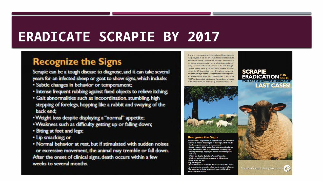
ERADICATE SCRAPIE BY 2017
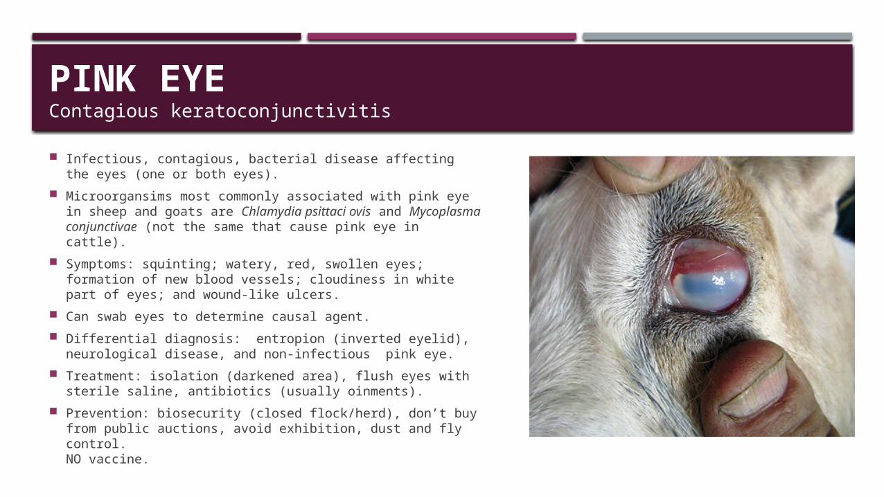
PINK EYEContagious keratoconjunctivitis
Infectious, contagious, bacterial disease affecting the eyes (one or both eyes).
Microorgansims most commonly associated with pink eye in sheep and goats are Chlamydia psittaci ovis and Mycoplasma conjunctivae (not the same that cause pink eye in cattle).
Symptoms: squinting; watery, red, swollen eyes; formation of new blood vessels; cloudiness in white part of eyes; and wound-like ulcers.
Can swab eyes to determine causal agent. Differential diagnosis: entropion (inverted eyelid),
neurological disease, and non-infectious pink eye. Treatment: isolation (darkened area), flush eyes with sterile
saline, antibiotics (usually oinments). Prevention: biosecurity (closed flock/herd), don’t buy from
public auctions, avoid exhibition, dust and fly control.NO vaccine.
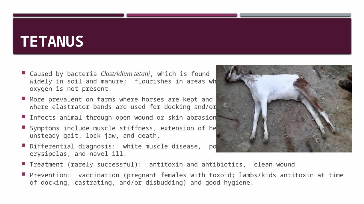
TETANUS Caused by bacteria Clostridium tetani, which is found
widely in soil and manure; flourishes in areas where oxygen is not present.
More prevalent on farms where horses are kept and where elastrator bands are used for docking and/or castrating.
Infects animal through open wound or skin abrasion. Symptoms include muscle stiffness, extension of head and neck,
unsteady gait, lock jaw, and death. Differential diagnosis: white muscle disease, polyarthritis,
erysipelas, and navel ill. Treatment (rarely successful): antitoxin and antibiotics, clean wound Prevention: vaccination (pregnant females with toxoid; lambs/kids antitoxin at time of docking,
castrating, and/or disbudding) and good hygiene.
RECOMMENDED RESOURCES Goat Medicine (2nd edition, 2011)
Mary Smith and David Sherman Sheep & Goat Medicine (2nd edition, 2011)
David Pugh and Nickie Baird Sheep Diseases Directory
http://beefandlamb.ahdb.org.uk/wp/wp-content/uploads/2013/09/brp_l_ol_SheepDiseaseDirectory260913.pdf
Maryland Small Ruminant Pagewww.sheepandgoat.com
American Consortium for Small Ruminant Parasite Control (ACSRPC)www.wormx.info or www.acsrpc.org