Immunity, Vol. 17, 51–62, July, 2002, Copyright 2002 by Cell Press
Blimp-1 Orchestrates Plasma CellDifferentiation by Extinguishing the MatureB Cell Gene Expression Program
and in long-lived plasma cells in the bone marrow.Blimp-1 is not detected in memory B cells but is presentin a subset of germinal center (GC) B cells with a partialplasma cell phenotype (BCL-6�, CD20�, IRF-4�, Synde-can-1�, Blimp-1�), suggesting that these cells may be
A.L. Shaffer,1,4 Kuo-I Lin,2,4 Tracy C. Kuo,2
Xin Yu,1 Elaine M. Hurt,1 Andreas Rosenwald,1
Jena M. Giltnane,1 Liming Yang,1 Hong Zhao,1
Kathryn Calame,2,3 and Louis M. Staudt1,3
1Metabolism Branchcommitted to a plasma cell fate. Of particular interest,National Cancer InstituteBCL-6, a transcriptional repressor required for GC B cellNational Institutes of Healthformation (Chang et al., 1996; Dent et al., 1997; FukudaBethesda, Maryland 20892et al., 1997) is not expressed in the Blimp-1� subset of2 Department of Microbiology andGC B cells (Angelin-Duclos et al., 2000). Consistent withDepartment of Biochemistrythe finding that most GC B cells are BCL-6� andand Molecular BiophysicsBlimp-1�, microarray analyses revealed that PRDM1,Columbia University College ofencoding Blimp-1, is a target of BCL-6 repression (Shaf-Physicians and Surgeonsfer, et al., 2000). This was further confirmed in studiesNew York, New York 10032showing that ectopic BCL-6 inhibited expression ofBlimp-1 and plasmacytic differentiation of B cells in vitro(Reljic et al., 2000). Thus, a primary function of BCL-6Summaryin GC B cells is to repress Blimp-1 and thereby inhibitterminal differentiation.Blimp-1, a transcriptional repressor, drives the termi-
How does a single transcriptional repressor drivenal differentiation of B cells to plasma cells. Using DNAplasma cell development? Three known targets ofmicroarrays, we found that introduction of Blimp-1 intoBlimp-1-dependent repression explain important as-B cells blocked expression of a remarkably large setpects of the plasma cell phenotype. Blimp-1 repressesof genes, while a much smaller number was induced.c-myc transcription (Lin et al., 1997) which explains ces-Blimp-1 initiated this cascade of gene expressionsation of cell cycle in plasma cells as c-myc is requiredchanges by directly repressing genes encoding sev-for proliferation and growth (Eilers, 1999). CIITA is alsoeral transcription factors, including Spi-B and Id3, thatdirectly repressed by Blimp-1 (Piskurich et al., 2000),regulate signaling by the B cell receptor. Blimp-1 alsoleading to the downregulation of MHC Class II genesinhibited immunoglobulin class switching by blockingin plasma cells (Silacci et al., 1994). Finally, Blimp-1expression of AID, Ku70, Ku86, DNA-PKcs, and STAT6.represses PAX5 (Lin et al., 2002), which is required forThese findings suggest that Blimp-1 promotes plas-lineage commitment and B cell development in the bonemacytic differentiation by extinguishing gene expres-marrow (Nutt et al., 2001) as well as for isotype switchingsion important for B cell receptor signaling, germinalin GCs (Liao et al., 1994; Max et al., 1995). Downregula-center B cell function, and proliferation while allowingtion of PAX5 is required for development of antibody-expression of important plasma cell genes such assecreting cells (Lin et al., 2002; Usui et al., 1997), proba-XBP-1.bly because it represses XBP-1 (Reimold et al., 1996,2001), J chain (Rinkenberger et al., 1996; Wallin et al.,
Introduction 1998), and immunoglobulin heavy chain gene transcrip-tion (Singh and Birshtein, 1993).
Plasma cells, the final effectors of humoral immunity, Repression of c-myc, CIITA, and PAX5 alone is un-are nondividing cells devoted to synthesis and secretion likely to explain the entire program of plasma cell devel-of immunoglobulin (Ig). A pivotal regulator of plasma opment activated by Blimp-1. Although repression ofcell development is B lymphocyte-induced maturation c-myc transcription is necessary for differentiation ofprotein-1 (Blimp-1), which was cloned from the BCL1 BCL1 cells, removal of c-myc activity is not sufficientmurine lymphoma upon differentiation to a plasma cell to trigger plasma cell differentiation (Lin et al., 2000).state (Turner et al., 1994). Enforced expression of CIITA activity is primarily limited to transcription of classBlimp-1, in either BCL1 cells or primary mouse spleno- II MHC, invariant chain, and the DM gene in B cellscytes (Piskurich et al., 2000; Schliephake and Schimpl, (Waldburger et al., 2000). Finally, when PAX5 is deleted1996) is sufficient to drive mature B cells to become in mature B cells (Horcher et al., 2001), some changesantibody-secreting plasma cells. Thus, Blimp-1 is a mas- associated with plasma cell differentiation occur, but theseter regulator of terminal B cell development. cells fail to upregulate J chain or secrete immunoglobulin.
The expression pattern of Blimp-1 in vivo (Angelin- Therefore, important aspects of Blimp-1-dependentDuclos et al., 2000) is consistent with its role as a master regulation remain to be discovered.regulator of plasma cell development in vitro. Blimp-1 is To develop an understanding of Blimp-1’s control overpresent in plasma cells formed in primary and secondary terminal B cell differentiation, we used DNA microarraysresponses to T cell-dependent and -independent antigens (Alizadeh et al., 2000) to analyze gene expression
changes caused by manipulation of Blimp-1 activity.We show that Blimp-1 broadly inhibits gene expression3 Correspondence: [email protected] (K.C.), [email protected] controlling mature B cell functions and driving(L.M.S.)
4 These authors contributed equally to this work. proliferation. This is accomplished by direct repression
Immunity52
of several transcription factor genes, including two new pressed by Blimp-1, confirming the ability of these sys-tems to detect Blimp-1 targets. Two hundred twenty-direct targets of Blimp-1, Spi-B and Id3.eight genes were downregulated by Blimp-1, butBlimp-1 did not globally repress transcription since overResults700 microarray elements representing named genes wereunaffected by Blimp-1 expression (data not shown). AMicroarray Analysis of Blimp-1 Expressionsmaller set of 32 genes were induced in the presencein B Cell Linesof Blimp-1 (Figure 1B), and only 15 of these were inducedTo identify genes that are the targets of Blimp-1 action,in two or more cell lines. We did not, however, observeBlimp-1 was introduced acutely or inducibly into cellscertain hallmarks of plasmacytic differentiation such aswhich do not express Blimp-1. For acute expression,Ig secretion (Chilosi et al., 1999; Wijdenes et al., 1996),cells were transduced with Blimp-1-expressing or con-suggesting that Blimp-1 alone is insufficient to activatetrol (puromycin resistance) retroviruses. Several B cellthe complete program of plasmacytic differentiation inlines were chosen for infection to improve the likelihoodtransformed B cells.that a particular Blimp-1 target gene would be ex-
We confirmed the ability of Blimp-1 to downregulatepressed in one of the cell lines: WI-L2, an EBV� maturemany of these microarray targets by independentlymphoblastoid cell line; SUDHL4, an EBV� GC B cell-assays of mRNA and protein expression (see figure atlike diffuse large B cell lymphoma (DLBCL) cell line;http://lymphochip.nih.gov/blimp/). We observed a Blimp-1-BJAB, an EBV� mature B lymphoma cell line; and RAJI,specific decrease in the steady-state levels of targetan EBV� Burkitt’s lymphoma cell line.gene transcripts by RT-PCR, including c-MYC, AID, btk,We also developed a strategy for inducibly expressingCD22, EBF, and others. Western blotting showed reduc-Blimp-1, since attempts to create stable transfectantstion in levels of the target gene proteins c-Myc, PAX5,expressing Blimp-1 were unsuccessful (A.L.S., unpub-and STAT6. Blimp-1 targets encoding cell surface pro-lished data). Neither use of a heavy metal-inducible met-teins were assessed by flow cytometry. Proteins in-allothionein promoter construct (pMEP4) nor fusion ofvolved in BCR signaling (CD19, CD22, CD45) lymphocyteBlimp-1 to the estrogen receptor ligand binding domainhoming, adhesion, and cell-cell interactions (CD11A,(Blimp-1-ERD), a strategy used previously to circumventCXCR5, MHCII, etc.) were downregulated by Blimp-1.the toxicity of BCL-6 (Shaffer et al., 2000), was sufficient
to control Blimp-1 toxicity. We therefore created a dou-bly inducible system in which Blimp-1-ERD expression Blimp-1 Targets in Context
The list of Blimp-1 targets is replete with genes impor-was controlled by the metallothionein promoter andgenerated stable transfectants of three B cell lines: RAJI, tant for B cell function and differentiation (Figure 1). To
put these into a developmental context, we assembledWI-L2, and OCI-Ly7, a GC B cell-like DLBCL cell line.Treatment of these lines with cadmium (Cd) and tamoxi- gene expression data from several primary B cell types,
including resting peripheral blood B cells, mitogenicallyfen (TMX) induced expression of the Blimp-1-ERD pro-tein, allowing us to analyze early transcriptional changes activated peripheral blood B cells, GC B cells, and
chronic lymphocytic leukemia (CLL) cells. Cell lines werecaused by Blimp-1.The acute effects of Blimp-1 on B cell gene expression also analyzed, including GC-like B cell lines and plasma
cell-like multiple myeloma lines, along with several cellwere analyzed using DNA microarrays. The change inexpression for each gene was computed as a ratio of lines representing non-B cells. Relative gene expression
was assessed by comparing each cell type to a refer-expression in Blimp-1-transduced cells versus controlcells (color scale, Figure 1). Genes downregulated in the ence mRNA pool, and genes modulated by Blimp-1 ex-
pression were organized based on hierarchical cluster-presence of Blimp-1 appear green; genes upregulatedby Blimp-1 appear red. Using the doubly inducible system, ing (Eisen et al., 1998; Figure 2A).
This analysis revealed that Blimp-1 plays a major rolegene expression analysis was performed on Blimp-1-ERD-expressing and control cells treated with Cd and in promoting B cell terminal differentiation by turning
off two classes of genes. One class of Blimp-1 targetsTMX (Figure 1). This analysis included only named genesthat were repressed or induced at least 1.8-fold (relative belongs to a gene expression signature of proliferation
and growth (Figure 2A). These target genes were moreto control) in any of the six acute infection experiments.For time course experiments involving inducible Blimp-1, highly expressed in dividing cells (cell lines, mitogeni-
cally activated blood B cells, and GC B cells) than inat least half of the induced samples were required toshow 1.8-fold repression or induction relative to the cor- nondividing cells (resting blood B cells and CLL cells).
Blimp-1 repressed its known target gene c-myc (Lin etresponding control sample. For genes represented bymore than one microarray feature, representative data al., 1997), as well as genes that are transcriptionally
activated by c-myc, including Rcl, DHFR, ODC, and LDH-Ais displayed. Genes meeting these criteria are referredto as Blimp-1 targets, but this is not meant to indicate (Coller et al., 2000; Dang, 1999; Eilers, 1999). Blimp-1
also downregulated genes involved in cell cycle pro-whether Blimp-1 acts directly or indirectly to modulatethe expression of a given gene. gression (CDC2, cdk2, PLK, ckshs, E2F-1) and DNA syn-
thesis and repair (PCNA, Ku70, Ku86, MCM2).Expression of a surprisingly large number of geneswas affected by Blimp-1 (Figure 1A). Of the 260 target Another class of Blimp-1 targets includes genes in-
volved in mature B cell functions (Figure 2A). These weregenes, 180 were targets in two or more cell systems,and 87 were targets in three or more systems (see table generally highly expressed in primary B cells and GC-
like B cell lines compared to multiple myeloma and non-Bat http://lymphochip.nih.gov/blimp/). Three known directtargets of Blimp-1 repression, c-Myc, CIITA, and PAX5 cell samples. Blimp-1 extinguished a gene expression
program specifying B cell identity, including surface(Lin et al., 1997, 2002; Piskurich et al., 2000), were re-
Blimp-1 Blocks B Cell Gene Expression Programs53
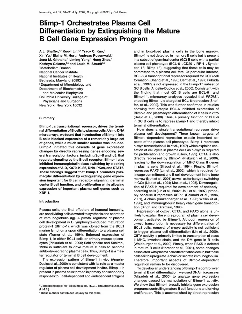
Figure 1. DNA Microarray Analysis of Blimp-1 Target Genes in Human B Cell Lines in Cells Acutely or Inducibly Expressing Blimp-1
Each column represents data from an independent infection of the indicated human B cell line. Each row represents a gene significantlyinduced (red) or repressed (green) following Blimp-1 expression. A color bar shows the magnitude of gene expression changes as a ratio ofexpression in control cells (green) versus Blimp-1-expressing cells (red). To better understand the role of Blimp-1 in controlling B cell geneexpression, analysis was limited to named genes, and genes were organized into broad functional categories (see key) based on analysis ofthe OMIM (http://www.ncbi.nlm.nih.gov:80/entrez/query.fcgi) and GO databases (http://www.geneontology.org/).(A) Genes repressed in the presence of Blimp-1.(B) Genes induced in the presence of Blimp-1.

Immunity54

Blimp-1 Blocks B Cell Gene Expression Programs55
markers (CD19, CD20, CD45, MHCII), B cell receptor ing PAX5, a known repressor of XBP-1 (Reimold et al.,1996).(BCR) signaling components (BLNK, CD79A, syk, btk),
and transcription factors (Spi-B, PAX5, Oct-2, STAT6,EBF). Also in this group are genes induced during B cell Gene Expression Changes Caused by Blimp-1
in Mouse Splenic B Cellsactivation (CD69, MIP-1�, A1) or upregulated at the GCstage of differentiation (AID, JAW1, A-myb). Among the We next investigated whether the target genes identified
by microarray analysis of B cell lines were similarly regu-GC-specific target genes is BCL-6, which encodes atranscription factor that represses Blimp-1 and prevents lated by Blimp-1 in primary B cells. Mouse splenic B cells
were purified and infected with retroviruses expressingplasmacytic differentiation (Reljic et al., 2000; Shafferet al., 2000). Blimp-1 expression reduced both BCL-6 either YFP (yellow fluorescent protein) or a bicistronic
mRNA encoding Blimp-1 and YFP (Piskurich et al., 2000).mRNA and protein levels, as assessed by RT-PCR andWestern blotting, and BCL-6 DNA binding activity, as Blimp-1 expression drove plasmacytic differentiation,
indicated by IgM secretion (data not shown). RNA fromassessed by gel shift analysis (see figure at http://lymphochip.nih.gov/blimp/). The discovery of BCL-6 as YFP� cells was used for semiquantitative RT-PCR which
showed a Blimp-1-dependent decrease in mRNAs fora Blimp-1 target implies a reciprocal regulatory loopin which BCL-6 and Blimp-1 antagonize each other’s numerous microarray targets including BCL-6, Spi-B,
STAT6, Id3, CD79A, CD22, syk, btk, A1, and AID (Figureexpression.Among the genes induced in the presence of Blimp-1, 4A). These data confirm our findings in B cell lines, un-
derscoring the physiological relevance of these targetsa few showed higher expression in plasma cell-like my-eloma cell lines as compared to other cell types, includ- and showing Blimp-1’s functional similarity in human
and mouse B cells (Piskurich et al., 2000).ing genes known to be highly expressed in plasma cells(XBP-1, J chain, Ig light chain; Figure 2B). Of particular We also examined the role of endogenous Blimp-1
during plasmacytic differentiation of mouse splenocytesinterest was the modest induction of XBP-1, which en-codes a critical regulator of plasma cell differentiation in vitro. Semiquantitative RT-PCR showed that Blimp-1
mRNA increased in splenic B cells after 4 days of lipo-(Reimold et al., 2001). We confirmed the induction ofXBP-1 mRNA in RAJI cells expressing Blimp-1-ERD us- polysaccharide (LPS) treatment. Concomitantly, mRNA
for c-myc, as well as several other targets, decreaseding a quantitative RT-PCR assay (Figure 3A) and foundthe induction to be in the same range as detected by following LPS treatment (Figure 4B). To block endoge-
nous Blimp-1, we used a retrovirus to express Tblimpmicroarray analysis (1.7-fold on average). This XBP-1level was substantially lower than the XBP-1 level in a prior to LPS treatment, which prevented the downregu-
lation of the known Blimp-1 targets c-myc, CIITA, andmyeloma cell line (Figure 3A), indicating that Blimp-1alone is insufficient to achieve the high XBP-1 mRNA PAX5 (Figure 4B). In addition, TBlimp prevented the
downregulation of BCL-6, Spi-B, ICSBP, STAT6, CD22,expression characteristic of plasma cells.To further explore the influence of Blimp-1 on XBP-1 syk, btk, CXCR5, and A1, although it failed to block
repression of EBF and CD79A (Figure 4B, and data notexpression, we studied another B cell line, SKW6.4(SKW), which can undergo plasma cell-like differentia- shown). This provides strong evidence that endogenous
Blimp-1 is necessary for the repression of these targetstion when treated with IL-6 (Goldstein and Kim, 1993).Transduction of SKW cells with a Blimp-1-expressing during LPS-driven plasmacytic differentiation.
A similar strategy was used to identify Blimp-1 targetsvirus induced XBP-1 expression (Figure 3B). IL-6 treat-ment of these cells caused IgM secretion and induced that were repressed indirectly as a result of the repres-
sion of PAX5 by Blimp-1. PAX5 can either activate orthe expression of both Blimp-1 and XBP-1 mRNAs (Fig-ure 3C). To test whether induction of the endogenous repress transcription from many target genes (Horcher
et al., 2001; Nutt et al., 1998). Indeed, changes in expres-Blimp-1 gene is necessary for XBP-1 induction, wetransduced these cells with a form of Blimp-1, Tblimp, sion of the PAX5 targets CD19, CD79A, and J chain
(Kozmik et al., 1992; Nutt et al., 2001; Rinkenberger etwhich lacks the repression domain and can dominantlyinterfere with Blimp-1 activity by competing for Blimp-1 al., 1996) were observed upon expression of Blimp-1
(Figure 1). Retroviruses were used to enforce expressionDNA binding sites (Chang et al., 2000; Yu et al., 2000).Tblimp blocked both IL-6-dependent IgM secretion and of PAX5 in splenic B cells prior to LPS treatment, which
interfered with the downregulation of CD19 and the in-induction of XBP-1 mRNA but had no effect on the induc-tion of Blimp-1 mRNA (Figure 3C). Together these data duction of J chain associated with differentiation (Figure
4C). CIITA and STAT6 mRNAs were also higher in PAX5-argue that Blimp-1 is necessary but insufficient for com-plete upregulation of XBP-1 mRNA, possibly by repress- expressing cells, suggesting that part of the regulation
Figure 2. Developmental Context of Blimp-1 Target Genes
(A) Repressed Blimp-1 targets in context. Expression of repressed genes from Figure 1A was analyzed comparing relative gene expressionfrom primary purified B cells (CLL cells, representing quiescent B cells), peripheral memory (CD19�, CD27�) cells, peripheral naive (CD19�,CD27�) cells, resting peripheral B cells (CD19�), peripheral B cells activated with anti-IgM/CD40L/IL-4 for the times indicated, tonsillar GC Bcells (Ma and Staudt, 2001), GC centrocytes (CD77�), GC centroblasts (CD77�), and GC-B-like cell lines along with plasma cell-like multiplemyeloma cell lines and non-B cell lines compared to a common reference mRNA pool. Data were centered, and genes were grouped bysimilarity in expression pattern by hierarchical clustering. Genes related to proliferation are shown in orange; genes related to B cell functionsare shown in red, blue, and purple. Only genes whose data met minimum expression intensity criteria in 50% or more of the arrays are shown.(B) Induced Blimp-1 targets. Shown are those genes that are expressed at a higher level in plasma-like myeloma cell lines than in GC-B celllines.

Immunity56
Figure 3. XBP-1 Expression Depends upon Blimp-1 Expression
(A) Quantitative RT-PCR confirms XBP-1 mRNA increase following Blimp-1 induction. RNA from control (pMEP4, vector only) or Blimp-1-ERD-expressing RAJI cells, treated with cadmium and tamoxifen inducers for 24 hr and RNA from the H929 myeloma line were analyzed for XBP-1(unspliced form) mRNA expression by quantitative Taqman RT-PCR using �-2-microglobulin expression as the normalization control.(B) Blimp-1 induction of XBP-1 mRNA in SKW6.4 cells. Control or Blimp-1-expressing retroviruses were used to infect cells; RNA fromtransduced cells was analyzed by semiquantitative RT-PCR using 4-fold dilutions of cDNAs.(C) Ectopic expression of dominant-negative Blimp-1 (Tblimp) is sufficient to block IgM secretion and XBP-1 mRNA expression in differentiatingSKW cells. Retrovirally infected cells were treated with IL-6 (40 U/ml), and ELISPOT was used to measure IgM secretion after 3 days.Semiquantitative RT-PCR was used to measure endogenous Blimp-1, XBP-1, and GAPDH mRNA using 4-fold dilutions of cDNA.
of these genes by Blimp-1 is secondary to repression evolutionary conservation suggests the sites are func-of PAX5. Other Blimp-1 targets, including c-myc, BCL-6, tionally important.Spi-B, Id3, AID, and others, were mostly unaffected by To determine whether Blimp-1 binds the sites in vivo,PAX5, suggesting that their repression is not secondary we performed chromosomal immunoprecipitation (ChIP)to repression of PAX5. experiments (Lin et al., 2002; Yu et al., 2000). We used
WI-L2 cells expressing a FLAG epitope-tagged Blimp-1-ERD protein, which allowed us to identify genomic sitesDirect Repression of Transcription Factor Genesof Blimp-1 binding by immunoprecipitation (IP) with anImportant for BCR Signalinganti-FLAG antibody. As negative controls, we used WI-L2Blimp-1’s powerful ability to extinguish B cell gene ex-cells transduced with the pMEP4 vector alone or WI-L2pression programs may be explained by its ability tocells expressing a FLAG epitope-tagged form of BCL-6.repress genes encoding other transcription factors (seeSemiquantitative PCR assays were designed for thetable at http://lymphochip.nih.gov/blimp/). All previouslyknown direct Blimp-1 target, CIITA, for the suspectedidentified Blimp-1 target genes encode transcriptiontargets Spi-B and Id3, and for a control genomic locus,factors, as do more than 10% of the genes identified asCSF-1. Each locus was equivalently represented in ge-repressed upon Blimp-1 expression in this study (Figurenomic DNA from the three cell lines prior to IP (Figure1). We therefore suspected that more transcription fac-5C). After IP, the CIITA, Spi-B, and Id3 loci were specifi-tor genes might be direct targets of Blimp-1. Comparingcally enriched in samples from cells expressing Blimp-1-the human and mouse genomic sequences of severalERD, but the control CSF-1 locus was not (Figure 5C).candidate target genes, we found that two genes, Spi-BThus, Blimp-1 binds in vivo to conserved binding sitesand Id3, have evolutionarily conserved sites in their tran-in the Spi-B and Id-3 genes, providing strong evidencescriptional control regions that resemble known Blimp-1that Spi-B and Id3 are direct targets of Blimp-1 repres-binding sites (Figure 5A). Both the Spi-B and Id3 sitession, extending the repertoire of transcription factorcompeted effectively in a gel mobility shift assay forgenes that are repressed by Blimp-1. Interestingly, bothBlimp-1 binding to a site from the c-myc gene (Figure
5B). Thus, Blimp-1 binds these sites in vitro, and their of these transcription factors are required for effective
Blimp-1 Blocks B Cell Gene Expression Programs57
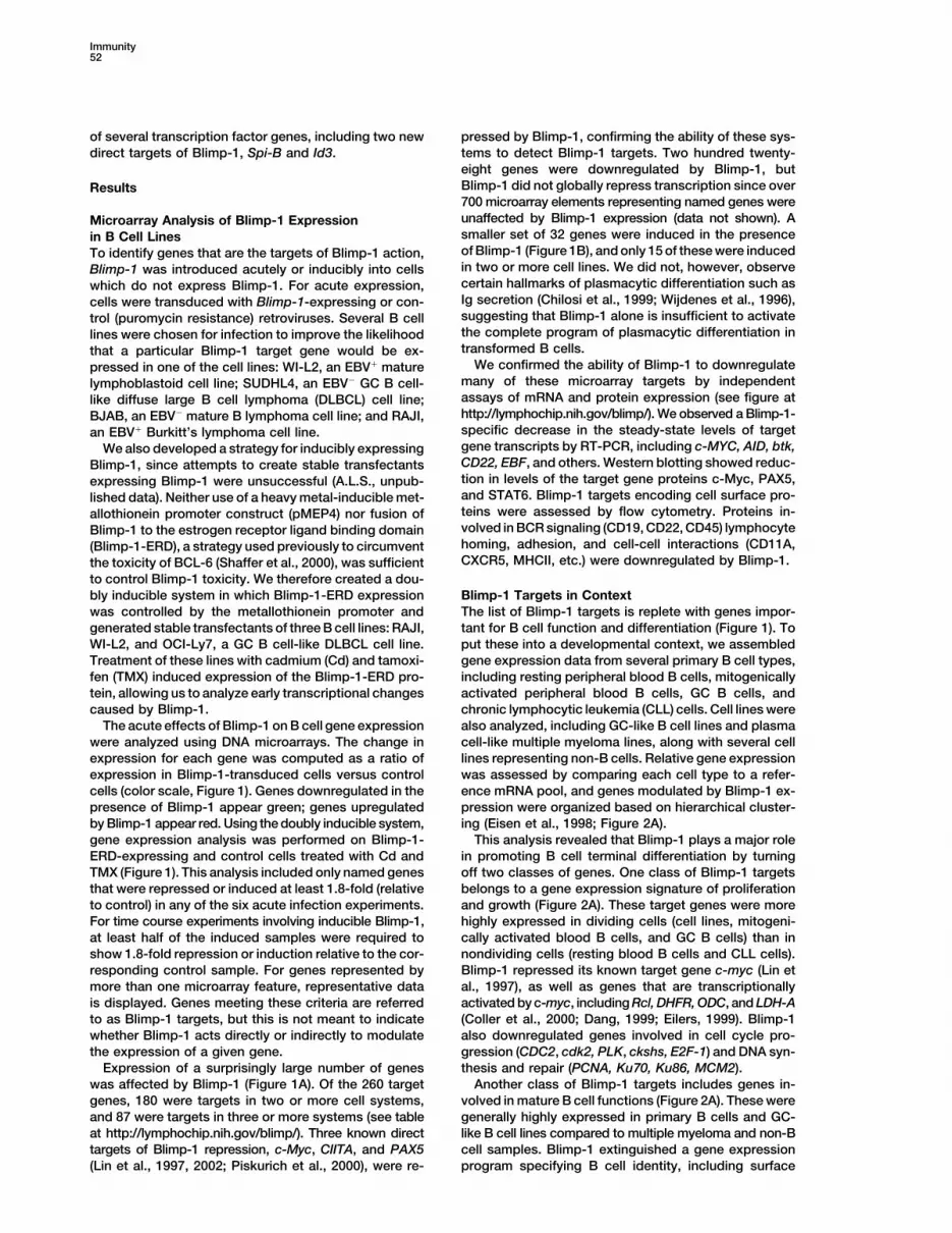
Figure 4. Blimp-1 Is Necessary and Sufficient to Repress Identified Targets during Splenic B Cell Terminal Differentiation
(A) Purified splenic B cells were treated with LPS (10 �g/ml) and anti-F(ab)�2 (5 �g/ml) overnight before retrovirus infection. SemiquantitativeRT-PCR was performed on RNA from sorted YFP� cells, and 4-fold serial dilutions of cDNA were tested.(B) Target gene expression was determined by semiquantitative RT-PCR from purified splenic B cells treated with LPS (10 ug/ml) for 0 and4 days (left panel). Semiquantitative RT-PCR (4-fold dilutions) was performed on RNA from sorted YFP� cells 4 days post-LPS stimulation(right panel).(C) Expression of Blimp-1 targets was determined by semiquantitative RT-PCR on RNA from splenic B cells stimulated with LPS and infectedwith either retrovirus-expressing PAX5 or control virus as above. Signals for STAT6 and A1 in (B) and (C) were detected by blotting withinternal probes.
BCR signaling (Garrett-Sinha et al., 1999, 2001; Pan et Blimp-1 expression in multiple systems using human Bal., 1999), providing one mechanism by which Blimp-1 cell lines. Thirty-four of these genes were confirmed ascan attenuate this central function of mature B cells. Blimp-1 targets in mouse B cells undergoing plasmacytic
differentiation and other complementary systems. Re-Blimp-1 Target Genes: A Role in Class Switching pressed targets fell into two broad functional groups,Microarray analysis (Figure 1) showed that Blimp-1 one specifying B cell identity and function and the otherdownregulates genes required for immunoglobulin class regulating proliferation. From this perspective, it is clearswitching (AID, Ku70, Ku86I, and DNA-PKcs) as well as that Blimp-1 directs plasmacytic differentiation by extin-STAT6, a transcription factor that is essential for IL-4 guishing mature B cell gene expression programs and byinduction of I�1 transcription and switching to IgG1 (Li- causing B cells to exit the cell cycle (Figure 7).nehan et al., 1998). Repression of these genes may ac- It is likely that Blimp-1 initiates this cascade of genecount for the ability of Blimp-1 to suppress class switch- expression changes by directly repressing genes en-ing induced by signaling through the BCR, CD40, and/ coding transcription factors, including three previouslyor IL-4 receptors in mouse splenic B cells (Knodel et identified direct targets, c-myc, PAX5, and CIITA, andal., 2001). To test this possibility, mouse B cells, infected two newly identified direct targets, Spi-B and Id3. Alto-with a control or Blimp-1-expressing retrovirus, were gether, more than 25 transcription factor genes, eachtreated with LPS and IL-4. As reported, a significant
of which has its own set of target genes (Figure 1 andfraction of control cells switched to IgG1 (Figure 6A)
see table at http://lymphochip.nih.gov/blimp/), werewhereas ectopic Blimp-1 blocked surface IgG1 (Knodeldownregulated in the presence of Blimp-1. Many ofet al., 2001). Using semiquantitative RT-PCR (Figure 6B),these transcription factors have been shown by geneticBlimp-1 was shown to block expression of AID, STAT6,disruption to be important for B cell development and/Ku86, and DNA-PKcs as well as STAT6-dependent I�1or mature B cell function (e.g., EBF, E2A, Ikaros, Oct-2,and IgG1 transcripts (Figure 6B). These data supportPAX5, Spi-1/PU.1, Spi-B, Id3, Sox4, and STAT6). Thethe view that Blimp-1 shuts off immunoglobulin isotypedownstream targets of these transcription factors areswitching by reducing expression of factors required foralso modulated by Blimp-1. For example, the directthis type of recombination and by blocking signals thatBlimp-1 target PAX5 activates CD19, CD79A, BLNK, andactivate switch region immunoglobulin transcription.CIITA and represses J chain and IgH (Horcher et al.,2001; Nutt et al., 1998), suggesting Blimp-1-dependentDiscussionregulation of these genes may be secondary to directrepression of PAX5. Indeed, enforced expression ofTaking a genomic approach, we identified 228 genes
that were repressed and 32 that were induced following PAX5 in LPS-treated differentiating B cells maintained

Immunity58
Figure 5. Id3 and Spi-B Are Direct Targets ofBlimp-1
(A) Putative Blimp-1 binding sites in the hu-man and mouse Id3 and SpiB genes alignedwith known sites in CIITA and c-myc (Lin etal., 1997; Piskurich et al., 2000) (underlined).(B) Competitor titrations to measure bindingaffinity of Blimp-1 for Id3 and Spi-B sites.EMSA was performed using nuclear extractfrom P3X, a Blimp-1-expressing plasmacy-toma, and a Blimp-1 binding site from themouse c-myc promoter (PRF). Molar equiva-lents of unlabeled competitor oligonucleo-tides were added as follows: PRF (0.78, 1.56,3.125. 6.25, 12.5, 25), CIITA, hId3, hSpiB(3.125, 6.25, 12.5, 25, 50, 100, 150), and non-specific, N.S. (12.5, 50, 100).(C) ChIP showing Blimp-1 binds to CIITA pro-moter III, hId3 promoter, and hSpi-B intron1(promoter II) in vivo. Chromatin prepared fromWI-L2 cells induced for the expression ofFLAG-tagged Blimp-1-ERD for 20 hr was im-munoprecipitated with anti-FLAG antibody,and regions encompassing the Blimp-1 siteswere amplified by PCR (2-fold dilutions). InputDNA was amplified with the same primersas a control for equivalence of the startingmaterial. Chromatin was prepared from cellswith only control vector and cells expressingan irrelevant FLAG-tagged protein as speci-ficity controls. Amplification of the CSF-1(colony stimulating factor-1) gene was usedas a negative control.
CD19 and CIITA mRNA expression and decreased J mouse splenic B cells (Knodel et al., 2001; Piskurich etal., 2000; Turner et al., 1994). Analysis of Blimp-1 targetchain mRNA levels (Figure 5). Another Blimp-1 target,
STAT6, also increased in response to enforced PAX5, gene expression across a panel of primary B cells andcell lines provides insight into how Blimp-1 drives plas-suggesting its repression by Blimp-1 may be down-
stream of PAX5. However, many other Blimp-1 targets macytic differentiation. Over 80 Blimp-1 target geneswere expressed in mature B cells (peripheral blood andwere insensitive to PAX5 overexpression although their
expression was modulated by TBlimp, suggesting that GC B cells) but were turned off in multiple myelomacells, which represent the plasma cell stage. ProteinsBlimp-1 affects the expression of many genes in a PAX5-
independent manner. encoded by these target genes are critical for the func-tion of mature B cells and include components of theBlimp-1 inhibited a second gene expression program
involving growth and proliferation, and this was likely BCR signaling pathway, cell surface receptors, and thehost of B cell-restricted transcription factors mentioneddue, in part, to direct repression of c-myc (Lin et al.,
2000). In accord with this hypothesis, many known above. Plasma cells can develop as a consequence ofthe germinal center reaction, probably from the smalldownstream targets of c-myc activation were also
downregulated by Blimp-1, including Rcl, ODC, LDH-A, subset of Blimp-1�, BCL-6� cells (Angelin-Duclos et al.,2000). A number of Blimp-1 target genes are GC B cell-and DHFR (Coller et al., 2000; Dang, 1999; Eilers, 1999).
However, Blimp-1 also downregulated other genes in restricted, including BCL-6, A-myb, AID, and TTG-2(Chang et al., 1996; Christoph et al., 1994; Ma andthe proliferation signature involved in DNA synthesis
and repair (PCNA, PMS4, MCM2, primase) and mitosis Staudt, 2001; Muramatsu et al., 2000; Shaffer et al.,2001), suggesting that Blimp-1 may promote plas-(pLK, aurora kinase, ckshs1, ckshs2), perhaps as an
indirect consequence of the cell cycle arrest that follows macytic differentiation by extinguishing the GC B cellgene expression program. Plasmacytic differentiationthe loss of c-myc (Amati et al., 1998; Carey et al., 2000).
The inhibition of these proliferation signature genes illus- can also occur outside of the germinal center and ispreceded by activation through the BCR. Blimp-1 down-trates how Blimp-1 coordinates the cell cycle arrest that
accompanies plasmacytic differentiation. regulated a set of genes that are induced by BCR signal-ing (A1, MIP-1�, CD69, CD83, and spi-1/PU.1), whichmay contribute to its ability to drive plasmacytic differen-Mature B Cell Functions Modulated by Blimp-1
The powerful ability of Blimp-1 to alter B cell identity tiation.Our analyses also suggest that Blimp-1 participatesand function has been shown in the BCL1 line and in

Blimp-1 Blocks B Cell Gene Expression Programs59
Figure 7. Blimp-1 Drives Plasma Cell Differentiation by Extinguish-ing Mature B Cell Gene Expression Programs
Figure 6. Blimp-1 Expression Represses Class Switching to IgG1 An increase in Blimp-1 expression (following relief from BCL-6 re-pression in GC B cells) leads to direct repression of several essential(A) Flow cytometric analysis of surface IgG1 expression in B cellsB cell transcription factors (c-myc, CIITA, Id3, PAX5, and Spi-B) andinfected with control or Blimp-1 viruses after 4 days of LPS�IL-4subsequent repression of several mature B cell functions as welltreatment; the black line divides IgG� and IgG� populations.as proliferation. In this way, Blimp-1, along with the transcription(B) Semiquantitative RT-PCR analysis of gene expression for factorsfactor XBP-1, drives B cells to become terminally differentiatedinvolved in class switching after 4 days of LPS�IL-4 treatment ofplasma cells.control and Blimp-1-expressing cells. STAT6 and DNA-PKcs were
detected by blotting with internal probes.
Blimp-1 expression may concomitantly increase, thusforming a negative regulatory loop.
in a feedback loop that attenuates BCR signaling. BCR Another consequence of Blimp-1 expression is inhibi-signaling suppresses the terminal differentiation of ma- tion of immunoglobulin class switching (Knodel et al.,ture B cells, in part by repressing Blimp-1, and ectopic 1999; Figure 6). Blimp-1 downregulated STAT6, blockingexpression of Blimp-1 reverses the ability of this stimu- expression of the germline I�1 transcript that is requiredlus to suppress plasma cell differentiation (Knodel et al., for switching to IgG1 (Cunningham et al., 1998; Linehan2001; Schliephake and Schimpl, 1996). The ability of et al., 1998). Blimp-1 also downregulated Ku70, Ku86,Blimp-1 to block BCR signaling is likely due to downreg- DNA-PKcs, and AID, four genes that are essential forulation of genes encoding BCR signaling components class switch recombination (Manis et al., 1998; Mura-(CD79A, BLNK, btk, PKC�, lyn, syk, and BRDG-1; Ka- matsu et al., 2000; Zelazowski et al., 1997). Furthermore,tsuta et al., 1998; Kurosaki and Tsukada, 2000; Leitges given the additional role of AID in immunoglobulin so-et al., 1996; Ohya et al., 1999; Reth and Wienands, 1997). matic hypermutation in GC B cells (Muramatsu et al.,Moreover, both Spi-B and Id3, two likely direct targets 2000), repression of AID by Blimp-1 would provide aof Blimp-1 repression, are regulators of BCR signaling mechanism by which this antibody diversification ma-(Garrett-Sinha et al., 1999, 2001; Pan et al., 1999). Spi-B chinery is turned off in plasma cells.cooperates with Spi-1/PU.1 in BCR signaling down-stream of syk phosphorylation (Garrett-Sinha et al., BCL-6 and Blimp-1 Form a Developmental1999) and may also regulate btk transcription (Muller et Feedback Loopal., 1996). Id3 is specifically required for proliferative Blimp-1 represses BCL-6, a GC-restricted transcrip-responses to BCR signaling (Pan et al., 1999). These tional repressor required for GC formation (Dent et al.,observations suggest an intriguing model in which initial 1997; Fukuda et al., 1997; Ye et al., 1997). BCL-6 overex-exposure of a mature B cell to cognate antigen gener- pression was previously shown to repress PRDM1, sup-ates BCR signals that block Blimp-1 expression, thereby pressing plasma cell differentiation (Reljic et al., 2000;
Shaffer et al., 2000). The reciprocal ability of Blimp-1 topromoting clonal expansion. As these signals diminish,

Immunity60
repress BCL-6 creates a feedback loop enforcing strict the regulatory pathways governing XBP-1 function dur-ing plasmacytic differentiation.control over the B cell fate decision to become a plasma
cell: as long as BCL-6 is expressed in a GC B cell, In summary, Blimp-1 plays a powerful role in plas-macytic differentiation by extinguishing mature B cellBlimp-1 expression and plasmacytic differentiation is
blocked, but if Blimp-1 becomes activated, BCL-6 will gene expression programs responsible for several B cellfunctions as well as for proliferation. Blimp-1 partici-be repressed, leading to irreversible plasmacytic differ-
entiation. Significantly, this is precisely the kind of dou- pates in regulatory loops with both BCL-6 and with BCRsignaling components, suggesting that terminal plas-ble negative feedback loop predicted more than forty
years ago by Jacob and Monod to be capable of confer- macytic differentiation can be viewed as an elaboratefeedback and feedforward web of interacting transcrip-ring irreversibility on a biological system (Ferrell, 2002;
Monod and Jacob, 1961). tion factors and signaling modules. Future studies willbe needed to untangle this regulatory web and to learnThis model raises the question of how BCL-6 is initially
downregulated in a GC B cell. Crosslinking the BCR in how Blimp-1 synergizes with other regulators such asXBP-1 to produce the ultimate effector of humoral immu-mature B cells causes a dramatic drop in BCL-6 mRNA
levels (Allman et al., 1996) and in some B cell lines leads nity, the plasma cell.to MAP kinase-mediated phosphorylation of BCL-6 and
Experimental Proceduresits proteosomal degradation (Moriyama et al., 1997; Niuet al., 1998). We speculate that late in the GC response
Cell Culture, Viral Transductions, and Plasmidsstronger BCR-mediated signals, the result of somatic Please refer to http://lymphochip.nih.gov/blimp/.mutations in Ig genes that improve BCR affinity for anti-gen, may exceed a threshold that leads to loss of BCL-6 DNA Microarray
Microarray analysis was performed as described in Shaffer et al.and consequently to Blimp-1 expression. Once the feed-(2000) using 2–5 mg of polyA� RNA (FastTrack, Invitrogen) fromback loop is initiated, Blimp-1 will attenuate BCL-6 ex-each sample. Arrays for each cell system (with the exception ofpression even after the cell leaves the germinal center,WI-L2 and SUDHL4 acute Blimp-1 infections) were repeated two or
and antigenic stimulation through the BCR ends. In B three times with independent pools (for acute infections) or clonescell lymphomas, BCL-6 translocations involve substitu- (for inducible Blimp-1ERD systems). For primary array data fromtion of the BCL-6 promoter with a constitutively active Figures 1 and 2 in a tabular format, please see http://lymphochip.nih.
gov/blimp/.promoter from the translocated gene (Dalla-Favera etal., 1999; Staudt et al., 1999), perhaps creating BCL-6
Human Cell Line RT-PCRalleles that are insensitive to Blimp-1-mediated repres-For nonquantitative confirmation of target genes, polyA� RNA was
sion, blocking terminal differentiation of the malignant isolated (FastTrack, Invitrogen), and 1 mg was converted to cDNAB cell. as described (Shaffer et al., 2000). For quantitative Taqman RTPCR,
serial dilutions of input mRNA (500 ng–2 ng) were analyzed in tripli-cate using a Taqman RT-PCR kit (ABI) and an ABI 7700 sequenceBlimp-1 and Other Factors Controlling Plasmadetector system with primers and probes from Synthegen. PrimerCell Differentiationsequences for all RT-PCRs can be found at http://lymphochip.nih.
Recently, the transcriptional activator XBP-1 was shown gov/blimp/.to play a major role in plasma cell formation and immu-noglobulin secretion (Ono et al., 1991; Reimold et al., Primary Mouse Splenocyte RT-PCR
RNA was isolated by the Trizol method (Gibco, BRL). Subsequent2001; Figure 7). However, Blimp-1 expression is normalRT reactions were performed as described (Piskurich et al., 2000)in XBP-1-deficient cells, implying that Blimp-1 is devel-using 250 ng of RNA. Four-fold serial dilutions of cDNA were usedopmentally upstream of XBP-1 in the plasma cell differ-in order to ensure PCR amplification within the linear range. PCR
entiation program. Since XBP-1 is repressed by PAX5 products were analyzed by electrophoresis in 1.5% agarose gel and(Reimold et al., 1996), Blimp-1-dependent repression of visualized by staining with ethidium bromide or by Southern blottingPAX5 may be necessary for XBP-1 expression and full with a �-P32-labeled internal probe. Primers and PCR conditions can
be found at http://lymphochip.nih.gov/blimp/.plasma cell differentiation. Consistent with this possibil-ity, we observed a modest induction of XBP-1 mRNA in
Protein Expression Analysisthe presence of Blimp-1, although the levels achievedDetection of target gene proteins by Western blotting was as de-
were considerably lower than in multiple myeloma cells. scribed (Shaffer et al., 2000) using antibodies specific for Sp1,In the SKW lymphoma cell line, we found that IL-6 treat- c-Myc, STAT6, and BCL-6 (Santa Cruz Biotechnology). See Lin etment induced XBP-1 mRNA, consistent with previous al. (2000) for details of the ELISPOT assay for secreted Ig. Please
refer to http://lymphochip.nih.gov/blimp/ for details regarding flowresults identifying XPB-1 as a target of IL-6 signalingcytometry.(Wen et al., 1999). Interestingly, blockade of Blimp-1
activity by Tblimp prevented the induction of XBP-1Chromosomal Immunoprecipitation (ChIP)mRNA by IL-6. Taken together, these results suggestFor preparation of chromatin, immunoprecipitation, PCR conditions,
that Blimp-1 repression of PAX5 may be necessary for and primers, please refer to Lin et al. (2002), Yu et al. (2000), andexpression of XBP-1 during plasmacytic differentiation http://lymphochip.nih.gov/blimp/.but may not be sufficient to achieve the high levels of
EMSAXBP-1 expression characteristic of plasma cells. XBP-1Gel mobility shift for BCL-6 was performed using nuclear extractsis also regulated under conditions of endoplasmic retic-and probes as described in Shaffer et al. (2000). EMSA for Id3 andulum stress, which causes activation of XBP-1 transcrip-Spi-B Blimp-1 binding was performed as described (Lin et al., 1997).
tion by ATF6 and splicing of XBP-1 mRNA by IRE1 (Cal- The sequences of double-stranded oligonucleotides used for probefon et al., 2002; Shen et al., 2001; Yoshida et al., 2000). and competitors are: c-myc, CGCGTACAGAAAGGGAAAGGACTAG
CGCG; CIITA, ACAGTAAGGAAGTGAAATTAATTT; hId3, GCGCTGAClearly, much further work is needed to fully elucidate

Blimp-1 Blocks B Cell Gene Expression Programs61
GATTGCAGAAGGAGGAGGGAAAGGGGGTTTGAG; mId3, GCGCTG Dang, C.V. (1999). c-Myc target genes involved in cell growth, apo-ptosis, and metabolism. Mol. Cell. Biol. 19, 1–11.AGATTGCAGAAGAAGGAGGGAAAGGGAGGTCTAA; hSpiB, GGTG
GGAGGGGCAGGGAAAGTGAG; mSpiB, CGCTGGGGGAGGCAGGG Dent, A.L., Shaffer, A.L., Yu, X., Allman, D., and Staudt, L.M. (1997).AAAGTAGGG; hBCL-6, GACACACCTTGCAAAGAAAAAGGGAAACT Control of inflammation, cytokine expression, and germinal centerGGCAGT; nonspecific, AGGAAGCAGGTCATGTGGCAAGG. formation by BCL-6. Science 276, 589–592.
Eilers, M. (1999). Control of cell proliferation by Myc family genes.ELISPOT Mol. Cells 9, 1–6.For details, please refer to Lin et al. (2002).
Eisen, M.B., Spellman, P.T., Brown, P.O., and Botstein, D. (1998).Cluster analysis and display of genome-wide expression patterns.
Acknowledgments Proc. Natl. Acad. Sci. USA 95, 14863–14868.
Ferrell, J.E. (2002). Self-perpetuating states in signal transduction:A.L.S. thanks the members of the Staudt lab for the productionpositive feedback, double-negative feedback and bistability. Curr.and analysis of microarrays, as well as for helpful comments andOpin. Cell Biol. 14, 140–148.criticism. We also thank Sarah Henrickson for assistance with theFukuda, T., Yoshida, T., Okada, S., Hatano, M., Miki, T., Ishibashi,web companion. K.-I.L. thanks members of the Calame lab for help-K., Okabe, S., Koseki, H., Hirosawa, S., Taniguchi, M., et al. (1997).ful discussions and Jerry Liao for excellent technical assistance.Disruption of the Bcl6 gene results in an impaired germinal centerThis work was supported by the grants GM29361 and AI43576 to K.C.formation. J. Exp. Med. 186, 439–448.K.-I.L. is a fellow of the Leukemia and Lymphoma Society (5332-00).Garrett-Sinha, L.A., Su, G.H., Rao, S., Kabak, S., Hao, Z., Clark, M.R.,and Simon, M.C. (1999). PU.1 and Spi-B are required for normal BReceived: October 4, 2001cell receptor-mediated signal transduction. Immunity 10, 399–408.Revised: June 3, 2002Garrett-Sinha, L.A., Dahl, R., Rao, S., Barton, K.P., and Simon, M.C.(2001). PU.1 exhibits partial functional redundancy with Spi-B, butReferencesnot with Ets-1 or Elf-1. Blood 97, 2908–2912.
Alizadeh, A.A., Eisen, M.B., Davis, R.E., Ma, C., Lossos, I.S., Rosen- Goldstein, H., and Kim, A. (1993). Immunoglobulin secretion andwald, A., Boldrick, J.C., Sabet, H., Tran, T., Yu, X., et al. (2000). phosphorylation of common proteins are induced by IL-2, IL-4, andDistinct types of diffuse large B-cell lymphoma identified by gene IL-6 in the factor responsive human B cell line, SKW6.4. J. Immunol.expression profiling. Nature 403, 503–511. 151, 6701–6711.Allman, D., Jain, A., Dent, A., Maile, R.R., Selvaggi, T., Kehry, M.R., Horcher, M., Souabni, A., and Busslinger, M. (2001). Pax5/BSAPand Staudt, L.M. (1996). BCL-6 expression during B-cell activation. maintains the identity of B cells in late B lymphopoiesis. ImmunityBlood 87, 5257–5268. 14, 779–790.Amati, B., Alevizopoulos, K., and Vlach, J. (1998). Myc and the cell Katsuta, H., Tsuji, S., Niho, Y., Kurosaki, T., and Kitamura, D. (1998).cycle. Front. Biosci. 3, D250–D268. Lyn-mediated down-regulation of B cell antigen receptor signaling:
inhibition of protein kinase C activation by Lyn in a kinase-indepen-Angelin-Duclos, C., Cattoretti, G., Lin, K.I., and Calame, K. (2000).dent fashion. J. Immunol. 160, 1547–1551.Commitment of B lymphocytes to a plasma cell fate is associated
with Blimp-1 expression in vivo. J. Immunol. 165, 5462–5471. Knodel, M., Kuss, A.W., Lindemann, D., Berberich, I., and Schimpl,A. (1999). Reversal of Blimp-1-mediated apoptosis by A1, a memberCalfon, M., Zeng, H., Urano, F., Till, J.H., Hubbard, S.R., Harding,of the Bcl-2 family. Eur. J. Immunol. 29, 2988–2998.H.P., Clark, S.G., and Ron, D. (2002). IRE1 couples endoplasmic
reticulum load to secretory capacity by processing the XBP-1 Knodel, M., Kuss, A.W., Berberich, I., and Schimpl, A. (2001). Blimp-1mRNA. Nature 415, 92–96. over-expression abrogates IL-4- and CD40-mediated suppression
of terminal B cell differentiation but arrests isotype switching. Eur.Carey, G.B., Donjerkovic, D., Mueller, C.M., Liu, S., Hinshaw, J.A.,J. Immunol. 31, 1972–1980.Tonnetti, L., Davidson, W., and Scott, D.W. (2000). B-cell receptor
and Fas-mediated signals for life and death. Immunol. Rev. 176, Kozmik, Z., Wang, S., Dorfler, P., Adams, B., and Busslinger, M.105–115. (1992). The promoter of the CD19 gene is a target for the B-cell-
specific transcription factor BSAP. Mol. Cell. Biol. 12, 2662–2672.Chang, C.C., Ye, B.H., Chaganti, R.S., and Dalla-Favera, R. (1996).Kurosaki, T., and Tsukada, S. (2000). BLNK: connecting Syk andBCL-6, a POZ/zinc-finger protein, is a sequence-specific transcrip-Btk to calcium signals. Immunity 12, 1–5.tional repressor. Proc. Natl. Acad. Sci. USA 93, 6947–6952.
Leitges, M., Schmedt, C., Guinamard, R., Davoust, J., Schaal, S.,Chang, D.H., Angelin-Duclos, C., and Calame, K. (2000). BLIMP-1:Stabel, S., and Tarakhovsky, A. (1996). Immunodeficiency in proteintrigger for differentiation of myeloid lineage. Nat. Immunol. 1,kinase cbeta-deficient mice. Science 273, 788–791.169–176.
Liao, F., Birshtein, B.K., Busslinger, M., and Rothman, P. (1994). TheChilosi, M., Adami, F., Lestani, M., Montagna, L., Cimarosto, L.,transcription factor BSAP (NF-HB) is essential for immunoglobulinSemenzato, G., Pizzolo, G., and Menestrina, F. (1999). CD138/synde-germ-line epsilon transcription. J. Immunol. 152, 2904–2911.can-1: a useful immunohistochemical marker of normal and neoplas-
tic plasma cells on routine trephine bone marrow biopsies. Mod. Lin, Y., Wong, K., and Calame, K. (1997). Repression of c-myc tran-Pathol. 12, 1101–1106. scription by Blimp-1, an inducer of terminal B cell differentiation.
Science 276, 596–599.Christoph, T., Rickert, R., and Rajewsky, K. (1994). M17: a novelgene expressed in germinal centers. Int. Immunol. 6, 1203–1211. Lin, K.I., Lin, Y., and Calame, K. (2000). Repression of c-myc is
necessary but not sufficient for terminal differentiation of B lympho-Coller, H.A., Grandori, C., Tamayo, P., Colbert, T., Lander, E.S.,cytes in vitro. Mol. Cell. Biol. 20, 8684–8695.Eisenman, R.N., and Golub, T.R. (2000). Expression analysis with
oligonucleotide microarrays reveals that MYC regulates genes in- Lin, K.-I., Angelin-Duclos, C., Kuo, T.C., and Calame, K. (2002).volved in growth, cell cycle, signaling, and adhesion. Proc. Natl. Blimp-1-dependent repression of Pax-5 is required for differentia-Acad. Sci. USA 97, 3260–3265. tion of B cells to IgM secreting plasma cells. Mol. Cell. Biol. 22,
4771–4780.Cunningham, K., Ackerly, H., Claflin, L., Collins, J., Wu, P., Ford, C.,Lansford, R., Alt, F., and Dunnick, W.A. (1998). Germline transcription Linehan, L.A., Warren, W.D., Thompson, P.A., Grusby, M.J., andand recombination of a murine VDJmudeltagamma1 transgene. Int. Berton, M.T. (1998). STAT6 is required for IL-4-induced germlineImmunol. 10, 1027–1037. Ig gene transcription and switch recombination. J. Immunol. 161,
302–310.Dalla-Favera, R., Migliazza, A., Chang, C.C., Niu, H., Pasqualucci,L., Butler, M., Shen, Q., and Cattoretti, G. (1999). Molecular patho- Ma, C., and Staudt, L.M. (2001). Molecular definition of the germinal
centre stage of B-cell differentiation. Philos. Trans. R. Soc. Lond.genesis of B cell malignancy: the role of BCL-6. Curr. Top. Microbiol.Immunol. 246, 257–263. B Biol. Sci. 356, 83–89.

Immunity62
Manis, J.P., Gu, Y., Lansford, R., Sonoda, E., Ferrini, R., Davidson, block in IgM secretion in lipopolysaccharide/anti- mu F(ab�)2-co-stimulated B lymphocytes. Eur. J. Immunol. 26, 268–271.L., Rajewsky, K., and Alt, F.W. (1998). Ku70 is required for late B
cell development and immunoglobulin heavy chain class switching. Shaffer, A.L., Yu, X., He, Y., Boldrick, J., Chan, E., and Staudt, L.J. Exp. Med. 187, 2081–2089. (2000). BCL-6 represses genes that function in lymphocyte differen-
tiation, inflammation, and cell cycle control. Immunity 13, 199–212.Max, E.E., Wakatsuki, Y., Neurath, M.F., and Strober, W. (1995).The role of BSAP in immunoglobulin isotype switching and B-cell Shaffer, A.L., Rosenwald, A., Hurt, E.M., Giltnane, J.M., Lam, L.T.,proliferation. Curr. Top. Microbiol. Immunol. 194, 449–458. Pickeral, O.K., and Staudt, L.M. (2001). Signatures of the immune
response. Immunity 15, 375–385.Monod, J., and Jacob, F. (1961). General conclusions: telonomicmechanisms in cellular metabolism, growth and differentiation. Cold Shen, X., Ellis, R.E., Lee, K., Liu, C.Y., Yang, K., Solomon, A., Yoshida,Spring Harbor Symp. Quant. Biol. 26, 389–401. H., Morimoto, R., Kurnit, D.M., Mori, K., and Kaufman, R.J. (2001).
Complementary signaling pathways regulate the unfolded proteinMoriyama, M., Yamochi, T., Semba, K., Akiyama, T., and Mori, S.response and are required for C. elegans development. Cell 107,(1997). BCL-6 is phosphorylated at multiple sites in its serine- and893–903.proline-clustered region by mitogen-activated protein kinaseSilacci, P., Mottet, A., Steimle, V., Reith, W., and Mach, B. (1994).(MAPK) in vivo. Oncogene 14, 2465–2474.Developmental extinction of major histocompatibility complex classMuller, S., Sideras, P., Smith, C.I., and Xanthopoulos, K.G. (1996).II gene expression in plasmocytes is mediated by silencing of theCell specific expression of human Bruton’s agammaglobulinemiatransactivator gene CIITA. J. Exp. Med. 180, 1329–1336.tyrosine kinase gene (Btk) is regulated by Sp1- and Spi-1/PU.1-Singh, M., and Birshtein, B.K. (1993). NF-HB (BSAP) is a repressorfamily members. Oncogene 13, 1955–1964.of the murine immunoglobulin heavy-chain 3� alpha enhancer atMuramatsu, M., Kinoshita, K., Fagarasan, S., Yamada, S., Shinkai, Y.,early stages of B-cell differentiation. Mol. Cell. Biol. 13, 3611–3622.and Honjo, T. (2000). Class switch recombination and hypermutationStaudt, L.M., Dent, A.L., Shaffer, A.L., and Yu, X. (1999). Regulationrequire activation-induced cytidine deaminase (AID), a potentialof lymphocyte cell fate decisions and lymphomagenesis by BCL-6.RNA editing enzyme. Cell 102, 553–563.Int. Rev. Immunol. 18, 381–403.
Niu, H., Ye, B.H., and Dalla-Favera, R. (1998). Antigen receptor sig-Turner, C.A., Jr., Mack, D.H., and Davis, M.M. (1994). Blimp-1, analing induces MAP kinase-mediated phosphorylation and degrada-novel zinc finger-containing protein that can drive the maturationtion of the BCL-6 transcription factor. Genes Dev. 12, 1953–1961.of B lymphocytes into immunoglobulin-secreting cells. Cell 77,
Nutt, S.L., Morrison, A.M., Dorfler, P., Rolink, A., and Busslinger, M. 297–306.(1998). Identification of BSAP (Pax-5) target genes in early B-cell
Usui, T., Wakatsuki, Y., Matsunaga, Y., Kaneko, S., Koseki, H., Kita,development by loss- and gain-of-function experiments. EMBO J.T., and Kosek, H. (1997). Overexpression of B cell-specific activator17, 2319–2333.protein (BSAP/Pax-5) in a late B cell is sufficient to suppress differ-
Nutt, S.L., Eberhard, D., Horcher, M., Rolink, A.G., and Busslinger, entiation to an Ig high producer cell with plasma cell phenotype. J.M. (2001). Pax5 determines the identity of B cells from the beginning Immunol. 158, 3197–3204.to the end of B-lymphopoiesis. Int. Rev. Immunol. 20, 65–82.
Waldburger, J.M., Masternak, K., Muhlethaler-Mottet, A., Villard, J.,Ohya, K., Kajigaya, S., Kitanaka, A., Yoshida, K., Miyazato, A., Ya- Peretti, M., Landmann, S., and Reith, W. (2000). Lessons from themashita, Y., Yamanaka, T., Ikeda, U., Shimada, K., Ozawa, K., and bare lymphocyte syndrome: molecular mechanisms regulating MHCMano, H. (1999). Molecular cloning of a docking protein, BRDG1, class II expression. Immunol. Rev. 178, 148–165.that acts downstream of the Tec tyrosine kinase. Proc. Natl. Acad. Wallin, J.J., Gackstetter, E.R., and Koshland, M.E. (1998). Depen-Sci. USA 96, 11976–11981. dence of BSAP repressor and activator functions on BSAP concen-Ono, S.J., Liou, H.C., Davidon, R., Strominger, J.L., and Glimcher, tration. Science 279, 1961–1964.L.H. (1991). Human X-box-binding protein 1 is required for the tran- Wen, X.Y., Stewart, A.K., Sooknanan, R.R., Henderson, G., Hawley,scription of a subset of human class II major histocompatibility T.S., Reimold, A.M., Glimcher, L.H., Baumann, H., Malek, L.T., andgenes and forms a heterodimer with c-fos. Proc. Natl. Acad. Sci. Hawley, R.G. (1999). Identification of c-myc promoter-binding pro-USA 88, 4309–4312. tein and X-box binding protein 1 as interleukin-6 target genes inPan, L., Sato, S., Frederick, J.P., Sun, X.H., and Zhuang, Y. (1999). human multiple myeloma cells. Int. J. Oncol. 15, 173–178.Impaired immune responses and B-cell proliferation in mice lacking Wijdenes, J., Vooijs, W.C., Clement, C., Post, J., Morard, F., Vita, N.,the Id3 gene. Mol. Cell. Biol. 19, 5969–5980. Laurent, P., Sun, R.X., Klein, B., and Dore, J.M. (1996). A plasmocyte
selective monoclonal antibody (B-B4) recognizes syndecan-1. Br.Piskurich, J.F., Lin, K.I., Lin, Y., Wang, Y., Ting, J.P., and Calame,J. Haematol. 94, 318–323.K. (2000). BLIMP-I mediates extinction of major histocompatibility
class II transactivator expression in plasma cells. Nat. Immunol. 1, Ye, B.H., Cattoretti, G., Shen, Q., Zhang, J., Hawe, N., de Waard,526–532. R., Leung, C., Nouri-Shirazi, M., Orazi, A., Chaganti, R.S., et al. (1997).
The BCL-6 proto-oncogene controls germinal-centre formation andReimold, A.M., Ponath, P.D., Li, Y.S., Hardy, R.R., David, C.S., Strom-Th2-type inflammation. Nat. Genet. 16, 161–170.inger, J.L., and Glimcher, L.H. (1996). Transcription factor B cell
lineage-specific activator protein regulates the gene for human Yoshida, H., Okada, T., Haze, K., Yanagi, H., Yura, T., Negishi, M.,X-box binding protein 1. J. Exp. Med. 183, 393–401. and Mori, K. (2000). ATF6 activated by proteolysis binds in the pres-
ence of NF-Y (CBF) directly to the cis-acting element responsibleReimold, A.M., Iwakoshi, N.N., Manis, J., Vallabhajosyula, P., Szo-for the mammalian unfolded protein response. Mol. Cell. Biol. 20,molanyi-Tsuda, E., Gravallese, E.M., Friend, D., Grusby, M.J., Alt,6755–6767.F., and Glimcher, L.H. (2001). Plasma cell differentiation requiresYu, J., Angelin-Duclos, C., Greenwood, J., Liao, J., and Calame, K.the transcription factor XBP-1. Nature 412, 300–307.(2000). Transcriptional repression by blimp-1 (PRDI-BF1) involvesReljic, R., Wagner, S.D., Peakman, L.J., and Fearon, D.T. (2000).recruitment of histone deacetylase. Mol. Cell. Biol. 20, 2592–2603.Suppression of signal transducer and activator of transcriptionZelazowski, P., Max, E.E., Kehry, M.R., and Snapper, C.M. (1997).3-dependent B lymphocyte terminal differentiation by BCL-6. J. Exp.Regulation of Ku expression in normal murine B cells by stimuli thatMed. 192, 1841–1848.promote switch recombination. J. Immunol. 159, 2559–2562.
Reth, M., and Wienands, J. (1997). Initiation and processing of sig-nals from the B cell antigen receptor. Annu. Rev. Immunol. 15,453–479.
Rinkenberger, J.L., Wallin, J.J., Johnson, K.W., and Koshland, M.E.(1996). An interleukin-2 signal relieves BSAP (Pax5)-mediated re-pression of the immunoglobulin J chain gene. Immunity 5, 377–386.
Schliephake, D.E., and Schimpl, A. (1996). Blimp-1 overcomes the
Recommended


