
Vascular Biology, Atherosclerosis and Endothelium Biology
Involvement of Endothelial CD44 during in VivoAngiogenesis
Gaoyuan Cao,* Rashmin C. Savani,†
Melane Fehrenbach,* Chris Lyons,* Lin Zhang,‡
George Coukos,‡ and Horace M. DeLisser*From the Pulmonary, Allergy and Critical Care Division,*
Department of Medicine, Division of Neonatology,† Children’s
Hospital of Philadelphia, Philadelphia; Department of Obstetrics
and Gynecology,‡ University of Pennsylvania School of Medicine,
Philadelphia, Pennsylvania
CD44, a cell-surface receptor for hyaluronan, hasbeen implicated in endothelial cell functions, but itsrole in the formation of blood vessels in vivo has notbeen established. In CD44-null mice, vascularizationof Matrigel implants and tumor and wound angiogen-esis were inhibited. Leukocyte accumulation duringtumor growth and wound healing in wild-type andCD44-null mice were comparable, and reconstitutionof CD44-null mice with wild-type bone marrow didnot restore the wild-type phenotype, suggesting thatimpairments in angiogenesis in CD44-deficient miceare due to the loss of endothelial CD44. Although thecell proliferation, survival, and wound-induced mi-gration of CD44-null endothelial cells were intact,these cells were impaired in their in vitro ability toform tubes. Nascent vessels in Matrigel implants fromCD44-null mice demonstrated irregular luminal sur-faces characterized by retracted cells and thinned en-dothelia. Further, an anti-CD44 antibody that dis-rupted in vitro tube formation induced hemorrhagearound Matrigel implants, suggesting that antago-nism of endothelial CD44 undermined the integrity ofthe endothelium of nascent vessels. These data estab-lish a role for CD44 during in vivo angiogenesis andsuggest that CD44 may contribute to the organiza-tion and/or stability of developing endothelial tu-bular networks. (Am J Pathol 2006, 169:325–336; DOI:10.2353/ajpath.2006.060206)
Hyaluronan (HA), an important glycosaminoglycan con-stituent of the extracellular matrix, is composed of repeat-ing disaccharide units of D-glucuronic acid and N-acetyl-D-glucosamine.1 This widely distributed molecule
regulates cellular events such as cell proliferation andlocomotion that are required for a variety of biologicalprocesses, including morphogenesis, tumorigenesis, in-flammation, and host responses to injury (reviewed inRef. 2). HA has also been implicated in the formation ofvessels, but its effects on in vivo angiogenesis and endo-thelial cell (EC) function are complex and depend on HAconcentration and molecular size.3 High molecularweight HA (at concentrations of �100 �g/ml) inhibits ECproliferation and disrupts confluent endothelial monolay-ers.4 These findings are consistent with the fact thatavascular regions in chick embryo limb buds are rich innative high molecular weight HA and that expression ofthis form of HA in normally vascular areas results indecreased vascularity.5 In contrast, low molecular weightHA stimulates EC proliferation,4,6 wound-induced migra-tion,6 in vitro endothelial tube formation,7 and neovascu-larization in chick chorioallantoic membranes8 and cuta-neous wounds.9,10
HA mediates its biological effects through binding in-teractions with specific cell-associated receptors.11 Anumber of HA-binding proteins (so-called hyaladherins)have been identified, with CD44 and Receptor for HA-Mediated Motility (RHAMM) being the two best charac-terized cell-surface receptors for HA.2 Although severalother binding interactions for CD44 and RHAMM havebeen reported,12,13 currently their interactions with HAappear to be the ones most likely to directly activateintracellular signals required to stimulate processes rele-vant to angiogenesis.14 With respect to EC functions,previous studies have implicated CD44 in EC prolifera-tion, migration, and adhesion to HA; RHAMM in EC mo-tility; and both receptors in EC tube formation.15–22
Although there is evidence for the activity of RHAMMduring in vivo angiogenesis,16 the involvement of CD44 inthe formation of blood vessels has not been estab-
Supported by the Department of Defense (grant PR043482 to H.M.D.), theNational Institutes of Health (grant HL079090 to H.M.D.; grants HL62472,HL62868, and HL075930 to R.C.S.), and the Philadelphia Veterans Med-ical Center (to H.M.D.).
Accepted for publication April 11, 2006.
Address reprint requests to Horace M. DeLisser, M.D., Pulmonary,Allergy and Critical Care Division, BRB II/III, 421 Curie Blvd., Philadelphia,PA 19104-6160. E-mail: [email protected].
American Journal of Pathology, Vol. 169, No. 1, July 2006
Copyright © American Society for Investigative Pathology
DOI: 10.2353/ajpath.2006.060206
325

lished.16 We therefore investigated in vivo angiogenesisin CD44-null mice and found that vascularization of sub-cutaneous Matrigel (Collaborative Biomedical Products,Bedford, MA) plugs, as well as tumor and wound angio-genesis, was inhibited in CD44-null animals. Leukocyterecruitment during tumor growth and wound healing inwild-type and CD44-null mice were similar, and reconsti-tution of CD44-deficient mice with wild-type bone marrowdid not restore the wild-type phenotype, suggesting thatimpairments in angiogenesis in CD44-null mice resultfrom the absence of endothelial and not leukocyte CD44.ECs were isolated from wild-type and CD44-null mice.Although the cell proliferation, survival, and wound-in-duced migration of the CD44-null ECs were intact, thesecells were impaired in their in vitro ability to form tubes onMatrigel as compared to wild-type EC controls. Electronmicroscopic analysis of Matrigel implants in the CD44-null mice revealed nascent vessels with irregular luminalsurfaces characterized by retracted cells and thinnedendothelia. Treatment of wild-type mice with an anti-CD44 antibody that disrupted in vitro tube formation in-duced vessel hemorrhage around subcutaneous Matri-gel implants, suggesting that antagonism of endothelialCD44 undermined the integrity of the endothelium ofnascent vessels. These data establish for the first time theinvolvement CD44 in the in vivo formation of blood vesselsand suggest that CD44 may be involved in morphologicalevents required for the organization and/or stability ofendothelial tubular networks during in vivo angiogenesis.
Materials and Methods
Reagents and Chemicals
All reagents and chemicals were obtained from Sigma-Aldrich (St. Louis, MO) unless otherwise specified.
Antibodies
The following antibodies against murine surface recep-tors were used: IM7.8.1,22 a rat anti-CD44 antibody fromAmerican Type Culture Collection (Rockville, MD); 390,rat anti-PECAM-1 antibody,23 and F4/80 antibody againstmurine macrophages (Serotec, Raleigh, NC); anti-ICAM-2 antibody (Southern Biotech, Birmingham, AL);anti-murine CD11b antibody (Chemicon, Temecula, CA)and anti-CD8 and anti-CD45 antibodies (BD Pharmingen,San Jose, CA). Cell-surface antibody binding was deter-mined by fluorescence-activated cell sorting (FACS)analysis using previously described procedures.16
Cell Lines
The H5V murine EC line,23 B16 murine melanoma line(obtained from the American Type Culture Collection)and ID8-VEGF (vascular endothelial growth factor) tumorline, developed in the laboratory of Dr. George Coukos,24
were cultured in Dulbecco’s modified Eagle’s mediumcontaining 10% fetal bovine serum, penicillin/streptomy-cin, and 2 mmol/L L-glutamine, with insulin also added to
the medium for the culturing of the ID8-VEGF cells. LungECs were isolated immunomagnetically from wild-typeand CD44-null mice using published protocols25 andwere cultured in EGM-2 MV medium from Cambrex Bio-science Inc. (Walkersville, MD).
Animals
The Institutional Animal Care and Utilization Committeesat both the Wistar Institute and the University of Pennsyl-vania School of Medicine approved all animal care pro-cedures. CD44-null mice, on a C57BL/6 background,were the kind gift of Dr. Tak Mak (University of Toronto,Toronto, ON, Canada). Wild-type mice, also on aC57BL/6 background, were obtained from Taconic (Ger-mantown, NY).
Matrigel Neovascularization Model
Wild-type and CD44-null mice in the C57BL/6 back-ground were injected subcutaneously with 0.5 ml of Ma-trigel containing 1 � 106 B16 melanoma cells to inducethe growth of vessels into the gel. After 5 to 7 days, theanimals were sacrificed, and the gels were harvestedand processed for hemoglobin analysis as a measure ofthe vascularization the gels. For the antibody studies, fourinjections (200 �g) of antibody were administered via thetail vein, beginning 2 days after Matrigel implantation.
Tumor Growth and Angiogenesis
2 � 106 B16 melanoma or ID8-VEGF ovarian tumor cellsin a total volume of 50 �l were injected into wild-type andCD44-null mice. After 14 days (B16 melanoma) or 9weeks (ID8-VEGF ovarian tumor) the mice were sacri-ficed, and the tumors were harvested, measured, andprocessed for staining.
Murine Model of Wound-Induced Angiogenesis
Wounding was performed as previously described.26
Briefly, 1-cm2 wounds were made in the skin on the upperback of the mice with the wounds placed so as to notinjure the underlying muscle. Digital images of thewounds were captured at the time of wounding and thenon days 1, 3, 5, 7, and 10 after placement of the wounds.At these time points the wounds and surrounding tissuewere harvested and processed for immunohistochemicalstaining for vessel density and leukocyte infiltration.
Generation of Bone Marrow Chimeric Animals
Bone marrow chimeric mice were generated as previ-ously described.27 Briefly, to generate recipient animals,6-week-old wild-type or CD44-null mice were irradiatedwith 1000 rads from a Cs-137 irradiation source. Within24 hours after irradiation, donor marrow was obtainedfrom the femur and tibia of non-irradiated mice and 5 �106 cells were injected via the tail vein into the irradiated
326 Cao et alAJP July 2006, Vol. 169, No. 1

recipient mice. Experiments were subsequently con-ducted 4 to 6 weeks after transplantation. FACS analysisof leukocytes using an anti-mouse CD44 antibody con-firmed the phenotype of each chimeric mouse.
Immunohistochemical Staining
Immunohistochemistry was performed using a commer-cially available kit according to the manufacturer’s in-structions (ABC Immunodetection kit, Vector Laborato-ries, Burlingame, CA). Briefly, 6-�m-thick sections wereprepared by cryostat, transferred to glass slides, andfixed in ice-cold acetone and rinsed in phosphate-buff-ered saline (PBS). The sections were then permeabilizedwith 0.3% Triton X-100 in PBS for 10 minutes, treated with0.5% H2O2 in PBS for 30 minutes, and then blocked with0.5% bovine serum albumin for 30 minutes. The sectionswere incubated with primary antibody for 1 hour, washed,and then incubated with biotinylated secondary antibodyfor 1 hour. The reaction was developed with an avidin-biotin complex reaction, and the sections were lightlycounterstained with hematoxylin.
Quantitation of Angiogenesis and LeukocyteAccumulation
For quantitation of angiogenesis, frozen tissue sectionswere stained with monoclonal antibody 390 or ICAM-2antibodies to identify murine blood vessels. To assess thetumor angiogenic response, serial sections were ob-tained from different levels within the tumor separated by�100 �m. A total of four to eight levels per tumor wereanalyzed. For the B16 tumors, the vessel density at themargins of the tumors was determined by counting thenumber of vessels per 40� field. For the ID8-VEGF tu-mors, computer-assisted image analysis was used todetermine the vessel area of a defined optical field of3.6 � 105 �m2 positioned at serial locations along themargins of the tumor. For each section eight to 12 opticalfields were analyzed. To assess wound angiogenesiscomputer-assisted image analysis was used to deter-mine the vessel density within 300 �m of the edge of thewound, expressed as the percentage of tissue at woundedge occupied by vessels. Staining for leukocytes andmacrophages in wounded tissue was achieved with anti-CD11b and F4/80 antibodies, respectively, and the num-ber of cells/40� field at the base of the wound wasdetermined. FACS analysis of cells from minced andfiltered tumors was used to determine the percentage ofcells expressing CD8, CD11b, or CD45 leukocytemarkers.
Light and Electron Microscopy
Tissues were fixed in 10% formalin (for paraffin sections)or 1% paraformaldehyde and 2.5% glutaraldehyde in 0.1mol/L cacodylate buffer (pH 7.4) (for electron micros-copy). Paraffin blocks were sectioned for hematoxylinand eosin staining, while electron microscopy sections
were obtained after fixation in 2% osmium tetroxide andembedding in epoxy resin. Sufficient sections for repre-sentative serial sampling were produced and reviewed.
In Vitro Cell Proliferation
Endothelial cells were cultured for 24 hours in 96-wellplates (4000 cells/well), and the number of viable cellswas determined using the Cell Titer 96� Aqueous Non-Radioactive Cell Proliferation Assay (Promega Madison,WI). This assay is composed of solutions of a noveltetrazolium compound (designated MTS) and an electroncoupling (phenazine methosulfate) agent. MTS is biore-duced by dehydrogenase enzymes in metabolically ac-tive cells into a soluble formazan product. The absor-bance of the formazan at 490 nm can be measureddirectly from 96-well assay plates without further process-ing and is directly proportional to the number of livingcells in culture. As per the manufacture, the linear rangefor this assay is between 1000 and 200,000/well. In ourhands we found a similar linear range for human umbilicalvein endothelial cells with this assay (data not shown).Cell doubling was determined for isolated murine ECs byculturing the cells in T-25 flasks (25,000 cells/flask ini-tially) over 6 days with the number of cells determined byCoulter counter.
HA Binding
Wild-type and CD44-null ECs were incubated with HA(Healon; Pharmacia and Upjohn, Kalamazoo, MI) for 30minutes at 4°C, then washed two times with PBS. Thecells were then lysed, and HA binding was determined byenzyme-linked immunosorbent assay according to themanufacturer’s instructions (Echelon Bioscience, Inc.,Salt Lake City, UT). HA binding was also assessed byFACS analysis in which wild-type and CD44-null cellswere incubated with biotinylated HA for 60 minutes at4°C, followed by incubation with fluorescein-conjugatedstreptavidin (Vector Laboratories).
HA Adhesion Assay
96-well plates were coated with HA (as Healon) usingpreviously described procedures,16 and 40,000 H5Vcells in 200 �l of serum-free Dulbecco’s modified Eagle’smedium were added to the plates for 30 minutes. Aftergentle washing, the number of adherent cells was deter-mined using a commercially available nonradioactive col-orimetric assay according to the manufacturer’s instruc-tions (Titer 96 Aqueous Non-Radioactive Cell ProliferationAssay).
In Vitro Wounding-Induced Migration Assay
EC wounding was performed as previously described.28
20,000 ECs were added to 24-well tissue culture platesand allowed to grow to confluence. Linear defects werethen made in the monolayer. The wounded culture was
CD44 and Angiogenesis 327AJP July 2006, Vol. 169, No. 1

washed with PBS and then incubated for 24 hours inmedium (with 1% serum). Using computer-assisted im-age analysis, and the Image-Pro Plus program (MediaCybernetics, Silver Spring, MD), images were obtainedimmediately after wounding and 24 hours later, and thenthe distance migrated by cells at the wound edge wasdetermined. For each condition three to five wounds wereanalyzed.
In Vitro Tube Formation Assay
In vitro tube formation was studied using previously de-scribed procedures.23 Fifty microliters of Matrigel wasadded to each well of a 96-well plate and allowed to forma gel at 37°C for 30 minutes. Cells (20,000) in 200 �l ofcomplete medium were subsequently added to eachwell, and the mixture was incubated for 6 hours at 37°C in5% CO2. The wells were washed, and the gel and its cellswere fixed with 3% paraformaldehyde. Total tube lengthper well was determined by computer-assisted imageanalysis using the Image-Pro Plus program.
In Vitro Cell Death Detection
For the studies of apoptosis, confluent cells were ex-posed for 5 hours to serum-free medium or completemedium, with or without antibody. Apoptosis was thenassessed using the APOPercentage Apoptosis Assay (Bi-color Ltd., Belfast, Ireland).
Statistical Analyses
Differences among groups were analyzed using one-wayanalysis of variance. Results are presented as means �SE. When statistically significant differences were found(P � 0.05), individual comparisons were made using theBonferroni/Dunn test.
Results
Reduced Vascularization of Matrigel Implants inCD44-Null Mice
To investigate the involvement of CD44 in the formation ofblood vessels, in vivo angiogenesis was studied in micedeficient in CD44.29 Initial studies were performed with amodel in which vessels develop around and within sub-cutaneously implanted Matrigel plugs containing B16 tu-mor cells as a source of angiogenic growth factors. After5 to 7 days, a “blush” of vessel proliferation was seensurrounding plugs in the wild-type animals that was notevident in the CD44-null mice (Figure 1, A and B). Con-sistent with this, vascularization of the plugs, as assessedby hemoglobin concentration, was significantly reducedin CD44-deficient mice compared to wild-type animals(Figure 1C).
Decreased Tumor Growth and Angiogenesis inCD44-Null Mice
To confirm and extend the findings of the Matrigel stud-ies, the subcutaneous growth and associated tumor an-giogenesis of the B16 melanoma line and an ovariantumor line overexpressing VEGF (ID8-VEGF) were stud-ied in CD44-deficient mice. The growth of both tumors inthe CD44-null mice was significantly reduced (�70%)compared to that of tumors in wild-type animals (Figure 2,A, B, C, and E). The vessel densities of tumors of com-parable sizes from wild-type and mutant mice were sub-sequently determined and found to be significantly re-duced in the CD44-null mice (Figure 2, D and F). Further,histological analysis of B16 melanoma tumors fromCD44-null animals revealed a paucity of the large sinu-soidal vessels characteristic of the tumors harvested fromwild-type mice (Figure 2G). These data suggest that theinhibition of tumor growth in the CD44-null mice resultedat least in part from a reduced angiogenic response.
Delayed Wound Healing with ReducedAngiogenesis in CD44-Null Mice
With the data above implicating CD44 in the pathologicalformation of blood vessels additional studies were per-formed to determine whether CD44 might also be in-volved in physiological angiogenesis. The closure of1-cm2 skin wounds was therefore studied in wild-typeand CD44-null mice. We observed that wound closure inthe CD44-mutant animals was delayed during days 1 to 3after wounding but by day 7 had recovered and wassimilar to that of wild-type animals (Figure 3A). The den-sity of vessels at the edge of the wounds on day 3 wasreduced by 20% in the CD44-null animals compared towild-type mice (Figure 3B). Thus, the absence of CD44results in an early delay in the closure of skin wounds thatis associated with a modest but significant reduction inthe neovascularization of the wounded tissue.
Figure 1. Neovascularization of Matrigel implants in wild-type and CD44-null mice. Shown are images of Matrigel implants (star) containing B16melanoma cells as a source of angiogenic factors harvested after 5 days fromwild-type (A) and CD44-null (CD44 KO) (B) animals. A “blush” of vesselproliferation (circle) was evident around the implants from the wild-typeanimal that was not present in the CD44-null mice. The vascularization of thegels, as assessed by hemoglobin concentration (C), was significantly reducedin CD44-null mice compared to wild-type mice. Data are presented asmeans � SE (n � 20, *P � 0.02).
328 Cao et alAJP July 2006, Vol. 169, No. 1

Intact Leukocyte Recruitment during TumorGrowth and Wound Healing in CD44-Null Mice
HA receptors are expressed on leukocytes and havebeen implicated in leukocyte trafficking.30–32 Conse-quently, the absence of CD44 could lead to less leu-kocyte recruitment and thus a loss of a source ofpro-angiogenic factors. We, however, found that leu-kocyte accumulation in wild-type and CD44-mutantmice in the B16 and ID8-VEGF tumors (Figure 2H; datanot shown for ID8-VEGF tumors) and during woundhealing were comparable (Figure 3C). This suggeststhat the diminished tumor and wound angiogenesis
arising from the loss of CD44 was not the result ofreduced leukocyte recruitment.
Murine Angiogenesis Involves Endothelial CD44
The recruitment data presented above do not excludefunctional consequences arising from the loss of CD44expression on leukocytes or on circulating bone marrow-derived progenitor endothelial cells, both of which havebeen identified along with ECs as cellular participantsduring in vivo angiogenesis.33–38 To identify the possibleinvolvement of CD44 expressed on recruited leukocytes,
Figure 2. Tumor growth and angiogenesis in CD44-null mice. The growth of the B16 melanoma line (A and C) and the ID8-VEGF ovarian tumor line (B and E),as accessed by tumor weight, were significantly inhibited in the CD44-null (CD44 KO) mice (n � 10, *P � 0.01). The vessel densities of tumors of comparablesizes, as assessed by vessel number/40� field for the B16 tumor (D) and vessel area, �m2/optical field for the ID8-VEGF tumor (F), were significantly reducedin the CD44-null mice (n � 4–6, *P � 0.002). G: Hematoxylin and eosin sections of B16 melanomas from CD44-null mice revealed a paucity of the large vascularspaces (arrows) that were readily observable in the wild-type animals. H: The percentage of cells from B16 tumors expressing leukocyte surface markers (CD11b,CD45, or CD8) was determined by FACS analysis and found to be comparable in wild-type and CD44-null animals. Data are presented as means � SE.
CD44 and Angiogenesis 329AJP July 2006, Vol. 169, No. 1

tissue macrophages, and/or circulating endothelial pro-genitor cells in vessel formation, bone marrow chimericanimals were generated to selectively propagate bonemarrow-derived wild-type vascular cells against a back-ground of CD44-deficient endothelium. Experimentswere performed in which the following chimeric (donor-recipient) mice were generated: wild-type into wild-typemice (WT-WT; wild-type control), wild-type into CD44-nullmice (WT-KO), CD44-null into wild-type mice (KO-WT),and CD44-null into CD44-null mice (KO-KO; null control).FACS analysis confirmed that the blood leukocytes fromthe WT-WT and WT-KO mice expressed CD44, whereasthe leukocytes from the KO-WT and KO-KO animals weredevoid of CD44 (data not shown). The vascularization ofsubcutaneous implanted Matrigel plugs was subse-quently studied (Figure 4). The angiogenic responses inthe WT-KO and KO-KO mice were comparable but weresignificantly reduced compared to the WT-WT andKO-WT responses, which were similar to each other. Theinability of wild-type CD44-expressing leukocytes and/orbone marrow-derived endothelial progenitor cells to re-store the wild-type phenotype in the null animals (WT-KO)and the failure of the corresponding CD44-null cells toinduce an impaired angiogenic response (KO-WT) areconsistent with the primary involvement of endothelialCD44 during in vivo angiogenesis.
CD44-Null ECs Demonstrate Preserved HAAdhesion, Proliferation, Migration, andSusceptibility to Apoptotic Stress but ImpairedTube Formation on Matrigel
The studies of the bone marrow chimeric animals sug-gested that the loss of endothelial CD44 function inhib-
Figure 3. Wound healing and angiogenesis in CD44-null mice. A: Theclosure of 1-cm2 skin wounds, as measured by percentage of initial wound,was less at days 1 and 3 after wounding in CD44-null (CD44 KO) animalscompared to wild-type mice, but not at later time points (n � 6, *P � 0.02).B: Vessel density at the edge of the wound (percentage of tissue at woundedge occupied by vessels) on day 3 was reduced (by 20%) in the CD44-nullmice (B) (n � 8, *P � 0.03). C: Leukocyte and macrophage accumulations(number of cells/40� field) in the two mice strains were comparable (n � 4;*P � 0.1). Data are presented as means � SE.
Figure 4. Neovascularization of Matrigel implants in bone marrow chimericmice. Vascularization of Matrigel implants was studied in the followingchimeric (donor-recipient) mice: wild-type into wild-type mice (WT-WT),wild-type into CD44-null mice (WT-KO), CD44-null into wild-type mice(KO-WT), and CD44-null into CD44-null mice (KO-KO). The angiogenicresponses (assessed by hemoglobin concentration) in the WT-KO andKO-KO mice were very similar but were significantly reduced compared tothe WT-WT and KO-WT responses, which were similar. Data are presentedas means � SE (n � 8, *P � 0.02).
330 Cao et alAJP July 2006, Vol. 169, No. 1

its in vivo angiogenesis. To further investigate this, theactivities of ECs isolated from wild-type and CD44-nullanimals were studied. Although CD44 has long beenrecognized as a major surface receptor for the bindingof HA,11,12 we found that HA binding to CD44-null ECswas preserved and actually may have been greaterthan that of wild-type EC (Figure 5A). These findingswere confirmed by FACS analysis (Table 1), althoughthe overall level of HA binding was low. Cell prolifera-tion for the two cell types was similar whether assessedby a colorimetric assay (Figure 5B) or by the number ofcell doublings over 6 days (6.8 � 0.2 for WT EC vs.6.7 � 0.1 for CD44-null EC). Further, motility in wild-type and CD44-deficient ECs was comparable, andboth cell types were similarly susceptible to serumdeprivation-induced apoptosis (Figure 5, C and D).Studies of in vitro tube formation on Matrigel, however,
demonstrated that the loss of CD44 severely compro-mised the ability of the CD44-null ECs to form tubularnetworks on this substrate (Figure 5E).
Table 1. HA Binding to Murine ECs Assessed by FACSAnalysis
BackgroundWild-type
ECCD44-null
EC
Mean fluorescenceintensity
0.217 0.315 0.424
Murine ECs isolated from wild-type and CD44-null mice weretreated with biotinylated HA, incubated with fluorescein-conjugatedstreptavidin, and then subjected to FACS analysis. Shown are the meanfluorescence intensities. Background staining is that of WT cells stainedonly with secondary antibody.
Figure 5. In vitro function of murine ECs iso-lated from wild-type and CD44-null mice. A:Binding of HA to ECs isolated from wild-typeand CD44-null (CD44 KO) mice was determinedby enzyme-linked immunosorbent assay (n �2). Before the addition of exogenous HA, thebackground levels of HA associated with cellsurface of the wild-type and the CD44-null cellswere 384 and 295 ng/ml, respectively. HA bind-ing to CD44-null cells was preserved. B: Theproliferation of wild-type and CD44-null ECscultured for 24 hours in the presence of serumwas assessed using a colorimetric assay andmeasurement of the reaction mixture at 490 nm.The proliferative responses of the two cell typeswere comparable (n � 4). C: Liner defects weremade in confluent cell monolayers, and closureof the wounds after 24 hours was assessed bycomputer-assisted image analysis (n � 12).Wound-induced migration was similar in thetwo cell types. D: Apoptosis was assessed after5 hours in the presence or absence of serum.Wild-type and CD44 KO ECs were comparablein their susceptibility to serum deprivation (n �15). E: Shown are representative images of wild-type and CD44-null ECs plated on Matrigel,demonstrating impaired tube formation by theCD44-null ECs.
CD44 and Angiogenesis 331AJP July 2006, Vol. 169, No. 1

Electron Microscopic Analysis of ActivelyForming Vessels Reveals MorphologicallyDifferent Microvessels in CD44-Null Mice
The impairment of in vitro tube formation by CD44-nullECs led to further investigations of whether the structureof actively forming vessels might be affected by the ab-sence of CD44. Electron microscopic analyses weretherefore performed on the vessels invading subcutane-ous Matrigel implants harvested from wild-type andCD44-deficient mice (Figure 6). Compared to the wild-type animals, the endothelia of these invading vessels inthe CD44-null mice were characterized by an absence ofcellular ruffling and an irregular surface punctuated byretracted cells and/or very thin or flattened endothelialcells. Further, numerous pinocytotic vesicles were readilydetected in the microvessels of the wild-type capillariesbut were less evident in the CD44-null vessels. Togetherthese data suggest that the assembly, organization,and/or maturation of the endothelium of actively formingvessels are altered by the loss of CD44.
Anti-CD44 Antibody (IM7.8.1) Inhibits TubeFormation and Induces Hemorrhage aroundMatrigel Implants in Wild-Type Mice
In prior studies, neovascularization of Matrigel implantswas not inhibited by KM81, an anti-CD44 antibody.16 Ourfinding that the angiogenic response in the Matrigel as-say was decreased in the CD44-null mice, led us tore-explore the effects of acute antibody inhibition bystudying the activity of another well characterized anti-CD44 antibody, IM7.8.1.22 The specificity of this antibodywas confirmed by the fact it bound to wild-type but notCD44-null ECs (data not shown). IM7.8.1 inhibited thebinding of murine ECs to HA (Figure 7A) but had noeffects on cell proliferation, migration, or susceptibility toapoptotic stress (Figure 7, B–D). In vitro tube formation onMatrigel, however, was significantly inhibited by IM7.8.1(Figure 7E). Further, grossly visible hemorrhage was ob-served around the implants of mice treated with IM7.8.1but not in animals treated with a control antibody (Figure8). The antibody did not induce thrombocytopenia orhemorrhage in other organs (data not shown). Given this,and the fact that the antibody inhibits in vitro tube forma-tion, these in vivo data suggest that the acute inhibition ofendothelial CD44 function during the active formation ofvessels may undermine the integrity of these nascentvessels, making them more likely to rupture.
Discussion
In this report, studies were performed to define the in vivoinvolvement of CD44 in blood vessel formation. We foundthat vascularization of Matrigel implants as well as tumorand wound angiogenesis were inhibited in CD44-nullmice. Leukocyte accumulation during tumor growth andwound healing were comparable in wild-type and CD44-null animals, suggesting that impairments in leukocyte
recruitment did not contribute to the angiogenic pheno-type of the CD44-null mice. Reciprocal bone marrowtransplants involving wild-type and CD44-deficient micedemonstrated that the impaired angiogenic response re-sulted from a loss of endothelial, not leukocyte, CD44. Insubsequent studies of ECs isolated from these animals,we found that in vitro tube formation was significantlycompromised in the ECs isolated from CD44-deficientmice. Consistent with these in vitro data, the nascentvessels of Matrigel implants from CD44-null mice exhib-ited abnormal morphology. Lastly, hemorrhage devel-oped around Matrigel implants treated with anti-CD44antibody that inhibits in vitro tube formation, suggestingthat inhibition of CD44 function may undermine the sta-bility of newly formed vessels. Together these data pro-vide evidence of the involvement of endothelial CD44 inthe formation of vessels, possibly by mediating morpho-logical events required for the organization and/or stabil-ity of endothelial tubular networks.
CD44 is a type 1 transmembrane glycoprotein that isexpressed by most cell types, including ECs,16 and isconsidered the major cell-surface receptor for HA.2,11,12
With respect to the molecule’s interaction with low mo-lecular weight HA, studies using HA oligomers of definedsizes have demonstrated that oligomers of six sugars arethe minimal size fragments required for monovalent bind-ing to CD44, with oligomers bearing 20 or more sugarshaving the apparent capacity for divalent binding.39 Thismultifunctional transmembrane receptor is capable of in-ducing signal transduction pathways14 and is involved incell-cell and cell-matrix interactions that mediate anchor-ing to HA-rich pericellular matrices, internalization of HA,cell locomotion, growth factor activation, leukocyte re-cruitment, and lymphocyte homing and activation.12 Thisdiversity of function arises from a large number of iso-forms generated by alternative splicing and their subse-quent variable glycosylation.11,12
For a growing tumor or wounded tissue, the local “an-giogenic” environment is one that is characterized byincreased expression of hyaluronidases,40–43 and oxida-tive and nitrative stresses,44 factors known to promote thedegradation of high molecular weight species of HA intopro-angiogenic lower molecular weight fragments. It hasbeen presumed that CD44 mediates HA-dependent cel-lular events involved in regulating EC functions requiredfor angiogenesis. In line with this are studies of human orbovine ECs that implicate CD44 in HA-dependent adhe-sion, proliferation, migration, and/or tube formation.15–22
Somewhat surprisingly, however, treatment of mice withKM81, a bioactive anti-murine CD44 antibody, failed toconsistently inhibit the neovascularization of Matrigel im-plants containing basic fibroblast growth factor as theangiogenic factor.16 These results may have been re-lated to the assay used, a possible lack of activity ofKM81 against EC functions and/or compensation byRHAMM, and thus the question of CD44’s involvementduring in vivo angiogenesis remained an open one. Inlight of this, our findings are important because theyprovide, for the first time, clear evidence of the activity ofCD44 during in vivo angiogenesis in the adult animal(Figure 1), including both pathological (Figure 2) and
332 Cao et alAJP July 2006, Vol. 169, No. 1
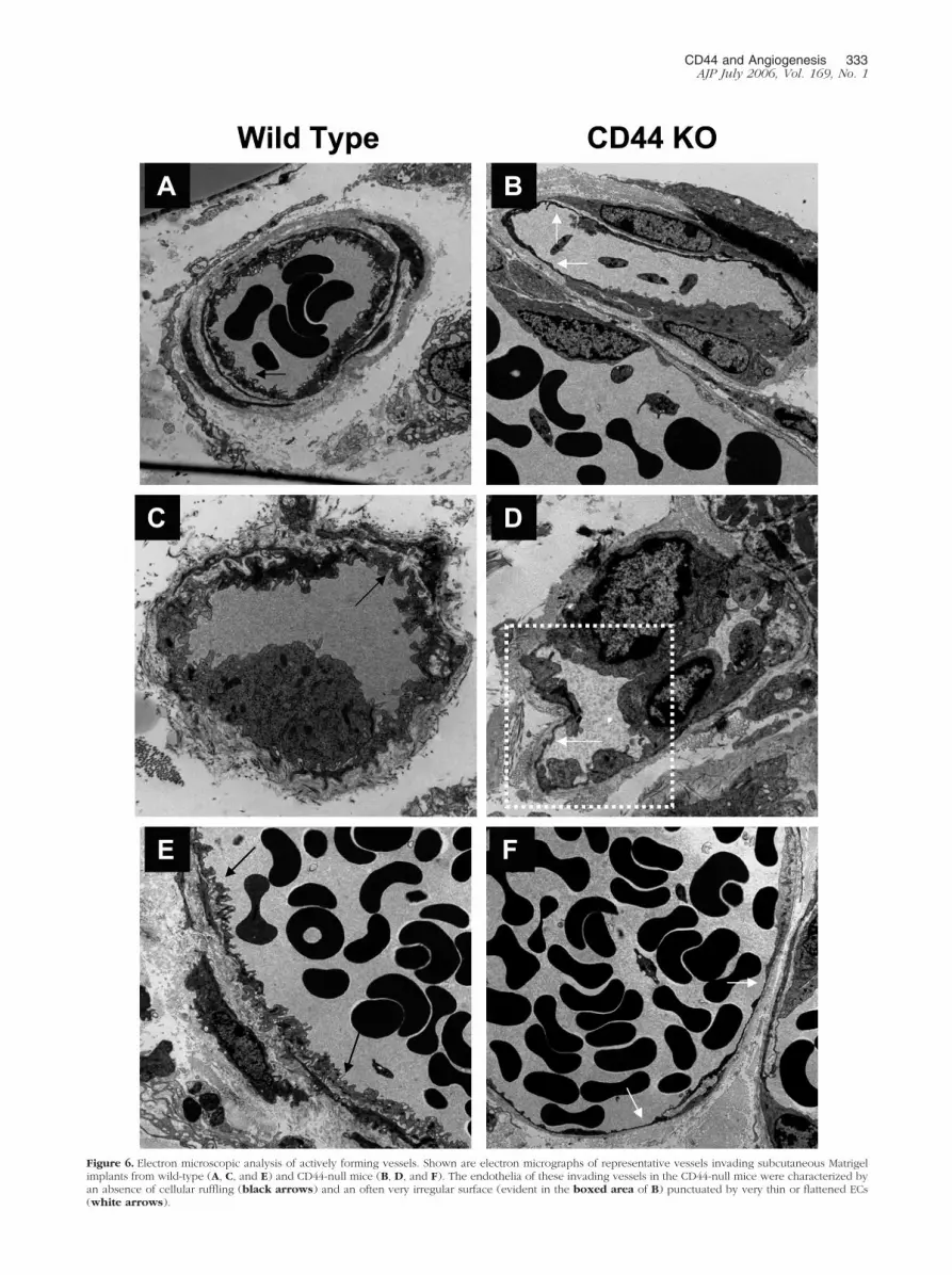
Figure 6. Electron microscopic analysis of actively forming vessels. Shown are electron micrographs of representative vessels invading subcutaneous Matrigelimplants from wild-type (A, C, and E) and CD44-null mice (B, D, and F). The endothelia of these invading vessels in the CD44-null mice were characterized byan absence of cellular ruffling (black arrows) and an often very irregular surface (evident in the boxed area of B) punctuated by very thin or flattened ECs(white arrows).
CD44 and Angiogenesis 333AJP July 2006, Vol. 169, No. 1

physiological (Figure 3) settings. The fact, however, thatCD44-null mice are viable, along with the absence of aperinatal cardiovascular phenotype, suggests that vas-cular development in the absence of CD44 is sufficient topermit adequate embryogenesis.29 Whether this reflectscompensation by RHAMM (or another cell-surface hyal-adherin) will be addressed in future studies whenRHAMM and CD44 double knockout mice are generatedand analyzed.
CD44 is expressed not only on ECs but on bone marrow-derived cells such as recruited leukocytes, tissue macro-phages, and circulating endothelial progenitor cells thatalso participate in angiogenesis.33–38 The loss of CD44expression on these bone marrow-derived cells, in additionto the absence of CD44 on ECs, could contribute to the
angiogenic phenotype of the CD44 mutant mice. The inabil-ity of wild-type CD44-expressing leukocytes and/or bonemarrow-derived endothelial progenitor cells to restore thewild-type phenotype in the null animals (WT-KO) and thefailure of the corresponding CD44-null cells to induce animpaired angiogenic response in the wild-type mice (KO-WT) provide strong evidence for the endothelial involve-ment of CD44 during in vivo angiogenesis (Figure 4). Fur-ther, leukocyte recruitment into tumors (Figure 2H) orwounded tissue (Figure 3C) was preserved in CD44 mutantmice, suggesting that the diminished angiogenic responsewas not the result of compromised CD44-dependent leuko-cyte trafficking. Together, these data point to the impor-tance of endothelial CD44, in contrast to leukocyte CD44, inthe formation of blood vessels.
Figure 7. Effects of IM7.8.1 antibody on the invitro function of murine ECs. Studies were per-formed of the effects of anti-CD44 antibody(IM7.8.1) on various functions of the H5V mu-rine EC line. A: The adhesion of the H5V cells toHA-coated surfaces was inhibited by IM7.8.1(n � 4, *P � 0.001). B: The proliferation of H5Vcells cultured for 24 hours in the presence ofserum was assessed using a colorimetric assayand measurement of the reaction mixture at 490nm. IM7.8.1 did not inhibit the proliferative re-sponse (n � 4). C: Linear defects were made inconfluent cell monolayers, and closure of thewounds after 24 hours was assessed by comput-er-assisted image analysis (n � 6). Wound-in-duced migration was not inhibited by IM7.8.1.D: Apoptosis was assessed after 5 hours in thepresence or absence of serum. Wild-type andCD44 KO ECs were comparable in their suscep-tibility to serum deprivation (n � 24). E: Shownare representative images of H5V cells plated onMatrigel that demonstrate that IM7.8.1 impairstube formation.
334 Cao et alAJP July 2006, Vol. 169, No. 1

With respect to the mechanism of action of CD44 dur-ing angiogenesis, our data demonstrating impairments intube formation in vitro by CD44-null ECs (Figure 5E) orfollowing treatment of murine ECs with anti-CD44 anti-body (Figure 7E) provide evidence of a role for CD44 inthe assembly of EC-lined channels. The abnormal mor-phology of the CD44-null neovessels in the Matrigelmodel (Figure 6) and the susceptibility of the wild-typeform of these vessels to hemorrhage during treatmentwith anti-CD44 antibody (Figure 8) are consistent withthese in vitro data and implicate CD44 in the organizationand/or stabilization of the endothelia of forming or newlyformed vessels. Whether these morphological differ-ences are present in normal vessels during developmentis unknown but is currently under investigation.
Although the role of CD44 in the morphogenesis ofendothelial tubes is still unknown, one or more processesmay be involved. First, epithelial cell HA acts as a mod-ulator of cell adhesion that mediates the early stages ofthe cell-substrate interaction.45 If comparable processesexist for ECs, and are mediated by CD44, the loss of theexpression of this molecule could disrupt initial adhesiveevents required for tube assembly. Second, CD44through association with matrix metalloproteinases maypromote remodeling of the endothelial pericellular matrixthat is required for endothelial tube formation.17,46 Third,certain isoforms of CD44 have heparan sulfate sidechains that could provide for the attachment of heparin-binding angiogenic factors such as VEGF and basicfibroblast growth factor.47 The binding of these factors tothe endothelium by CD44 could facilitate their activity inthe stabilization and maturation of newly formed vessels.Last, as newly formed vessels begin to mature, CD44may locate and organize high molecular species in thebasement membrane, which, given their inhibitory effectson EC proliferation and migration,4,5 may induce vesselquiescence. These possible mechanisms are the subjectof ongoing investigation.
In considering further the possible in vivo actions ofendothelial CD44, two additional processes should be
noted. First, although we found that the cell-proliferativeresponses of murine ECs appear to be independent ofCD44 (Figures 5B and 7B), other studies have implicatedCD44 in the proliferation of human umbilical vein endo-thelial cells.15,16,21,22 Second, CD44 has been reportedto be involved in the migration and/or invasion of humanand bovine ECs through matrix-coated filters, apparentlyby mechanisms that involve activation of Rho GTPasesand a cooperative interaction with the matrix metallopro-teinase-9.17,19,20 In contrast to this possible involvementof CD44 in cell motility, we found that loss of CD44expression (Figure 5C) or antibody antagonism of itsfunction (Figure 7B; see also Ref. 16) did not impairwound-induced migration. These differences in the re-quirement of CD44 for EC proliferation and migration mayreflect species-dependent differential expression of en-dothelial CD44 isoforms. Also, the differences noted withrespect to cell migration may reflect differences in theassays used, as published reports have used chemotaxisstudies while we have relied on a model of wound-in-duced migration. Studies of single cell migration are on-going to more clearly define the involvement of CD44 inendothelial cell motility using ECs isolated from wild-typeand CD44-null animals. Thus, although our data implicatea role for CD44 in tube formation, it may also mediate ECproliferation and migration in vivo.
The finding that IM7.8.1 inhibited the adhesion of mu-rine EC to HA (Figure 7A) is consistent with previousstudies of human ECs,16 as well as the fact that CD44 haslong been considered the major cell-surface receptor forHA binding.2,11,12 In contrast to this, we found that HAbinding to CD44-null ECs was not reduced and in factmay have been increased (Figure 5A and Table 1). Thepreservation of HA binding suggests that other HA-bind-ing receptors may be able to compensate for the loss ofCD44-mediated adhesion. More importantly, however,these data suggest that there may well be aspects ofCD44-dependent signaling that are either independent ofHA21 and/or cannot be reconstituted by HA interactionswith other receptors.
In summary, the loss of CD44 impairs in vivo angiogen-esis. This effect is due principally to the loss of expres-sion of endothelial CD44, an effect that may be related tocompromises in the ability of nascent vessels to properlyassemble endothelium-lined tubes, although our data donot exclude impairments in EC proliferation and migra-tion. Understanding its potential multiple roles in the for-mation of vessels may provide insights into CD44 as atherapeutic target.
References
1. Laurent TC, Fraser JR: Hyaluronan. FASEB J 1992, 6:2397–24042. Savani RC, DeLisser: Hyaluronan and its receptors in lung health and
disease. Proteoglycans in Lung Disease. Edited by HG Garg, PJRoughley, and CA Hales. New York, Marcel Dekker, Inc., 2002, pp73–106
3. Rooney P, Kumar S, Ponting J, Wang M: The role of hyaluronan intumour neovascularization. Int J Cancer 1995, 60:632–636
4. West DC, Kumar S: The effect of hyaluronate and its oligosaccharideson endothelial cell proliferation and monolayer integrity. Exp Cell Res1989, 183:179–196
Figure 8. Effects of IM7.8.1 antibody on the neovascularization of Matrigelimplants. Shown are representative images of Matrigel implants containingB16 melanoma cells as a source of angiogenic factors harvested after 5 daysfrom animals injected with saline (A), a control antibody (B), and the IM7.8.1antibody (C). Hemorrhage (arrows) was observed around the gels fromanimals treated with IM7.8.1 antibody.
CD44 and Angiogenesis 335AJP July 2006, Vol. 169, No. 1

5. Feinberg RN, Beebe DC: Hyaluronate in vasculogenesis. Science1983, 220:1177–1179
6. Slevin M, Kumar S, Gaffney J: Angiogenic oligosaccharides of hya-luronan induce multiple signaling pathways affecting vascular endo-thelial cell mitogenic and wound healing responses. J Biol Chem2002, 277:41046–41059
7. Rahmanian M, Pertoft H, Kanda S, Christofferson R, Claesson-WelshL, Heldin P: Hyaluronan oligosaccharides induce tube formation of abrain endothelial cell line in vitro. Exp Cell Res 1997, 237:223–230
8. West DC, Hampson IN, Arnold F, Kumar S: Angiogenesis inducedby degradation products of hyaluronic acid. Science 1985,228:1324–1326
9. Sattar A, Rooney P, Kumar S, Pye D, West DC, Scott I, Ledger P:Application of angiogenic oligosaccharides of hyaluronan increasesblood vessel numbers in rat skin. J Invest Dermatol 1994,103:576–579
10. Lees VC, Fan TP, West DC: Angiogenesis in a delayed revascular-ization model is accelerated by angiogenic oligosaccharides of hya-luronan. Lab Invest 1995, 73:259–266
11. Sherman L, Sleeman J, Herrlich P, Ponta H: Hyaluronate receptors:key players in growth, differentiation, migration and tumor progres-sion. Curr Opin Cell Biol 1994, 6:726–733
12. Naor D, Sionov RV, Ish-Shalom D: CD44: structure, function, andassociation with the malignant process. Adv Cancer Res 1997,71:241–319
13. Yang B, Hall CL, Yang BL, Savani RC, Turley EA: Identification of anovel heparin binding domain in RHAMM and evidence that it mod-ifies HA mediated locomotion of ras-transformed cells. J Cell Bio-chem 1994, 56:455–468
14. Turley EA, Noble PW, Bourguignon LY: Signaling properties of hya-luronan receptors. J Biol Chem 2002, 277:4589–4592
15. Trochon V, Mabilat C, Bertrand P, Legrand Y, Smadja-Joffe F, SoriaC, Delpech B, Lu H: Evidence of involvement of CD44 in endothelialcell proliferation, migration and angiogenesis in vitro. Int J Cancer1996, 66:664–668
16. Savani RC, Cao G, Pooler PM, Zaman A, Zhou Z, DeLisser HM:Differential involvement of the hyaluronan (HA) receptors CD44 andreceptor for HA-mediated motility in endothelial cell function andangiogenesis. J Biol Chem 2001, 276:36770–36778
17. Singleton PA, Bourguignon LY: CD44v10 interaction with Rho-kinase(ROK) activates inositol 1,4,5-triphosphate (IP3) receptor-mediatedCa2� signaling during hyaluronan (HA)-induced endothelial cell mi-gration. Cell Motil Cytoskeleton 2002, 53:293–316
18. Rahmanian M, Heldin P: Testicular hyaluronidase induces tubularstructures of endothelial cells grown in three-dimensional collagengel through a CD44-mediated mechanism. Int J Cancer 2002,97:601–607
19. Abecassis I, Olofsson B, Schmid M, Zalcman G, Karniguian A: RhoAinduces MMP-9 expression at CD44 lamellipodial focal complexesand promotes HMEC-1 cell invasion. Exp Cell Res 2003, 291:363–376
20. Forster-Horvath C, Meszaros L, Raso E, Dome B, Ladanyi A, Morini M,Albini A, Timar J: Expression of CD44v3 protein in human endothelialcells in vitro and in tumoral microvessels in vivo. Microvasc Res 2004,68:110–118
21. Pall T, Gad A, Kasak L, Drews M, Stromblad S, Kogerman P: Recom-binant CD44-HABD is a novel and potent direct angiogenesis inhib-itor enforcing endothelial cell-specific growth inhibition independentlyof hyaluronic acid binding. Oncogene 2004, 23:7874–7881
22. Murphy JF, Lennon F, Steele C, Kelleher D, Fitzgerald D, Long AC:Engagement of CD44 modulates cyclooxygenase induction, VEGFgeneration, and proliferation in human vascular endothelial cells.FASEB J 2005, 19:446–448
23. Zhou Z, Christofidou-Solomidou M, Garlanda C, DeLisser HM: Anti-body against murine PECAM-1 inhibits tumor angiogenesis in mice.Angiogenesis 1999, 3:181–188
24. Zhang L, Yang N, Garcia JR, Mohamed A, Benencia F, Rubin SC,Allman D, Coukos G: Generation of a syngeneic mouse model tostudy the effects of vascular endothelial growth factor in ovariancarcinoma. Am J Pathol 2002, 161:2295–2309
25. Bowden RA, Ding ZM, Donnachie EM, Petersen TK, Michael LH,Ballantyne CM, Burns AR: Role of alpha4 integrin and VCAM-1 inCD18-independent neutrophil migration across mouse cardiac endo-thelium. Circ Res 2002, 90:562–569
26. Livant DL, Brabec RK, Kurachi K, Allen DL, Wu Y, Haaseth R, An-
drews P, Ethier SP, Markwart S: The PHSRN sequence inducesextracellular matrix invasion and accelerates wound healing in obesediabetic mice. J Clin Invest 2000, 105:1537–1545
27. Mahooti S, Graesser D, Patil S, Newman P, Duncan G, Mak T, MadriJA: PECAM-1 (CD31) expression modulates bleeding time in vivo.Am J Pathol 2000, 157:75–81
28. Cao G, O’Brien CD, Zhou Z, Sanders SM, Greenbaum JN, Makrigian-nakis A, DeLisser HM: Involvement of human PECAM-1 in angiogen-esis and in vitro endothelial cell migration. Am J Physiol 2002,282:C1181–C1190
29. Teder P, Vandivier RW, Jiang D, Liang J, Cohn L, Pure E, Henson PM,Noble PW: Resolution of lung inflammation by CD44. Science 2002,296:155–158
30. Katayama Y, Hidalgo A, Chang J, Peired A, Frenette PS: CD44 is aphysiological E-selectin ligand on neutrophils. J Exp Med 2005,201:1183–1189
31. Khan AI, Kerfoot SM, Heit B, Liu L, Andonegui G, Ruffell B, JohnsonP, Kubes P: Role of CD44 and hyaluronan in neutrophil recruitment.J Immunol 2004, 173:7594–7601
32. Zaman A, Cui Z, Foley JP, Zhao H, Grimm PC, Delisser HM, SavaniRC: Expression and role of the hyaluronan receptor RHAMM in in-flammation after bleomycin injury. Am J Respir Cell Mol Biol 2005,33:447–454
33. Schruefer R, Lutze N, Schymeinsky J, Walzog B: Human neutrophilspromote angiogenesis by a paracrine feedforward mechanism involv-ing endothelial interleukin-8. Am J Physiol 2005, 288:H1186–H1192
34. Chavakis T, Cines DB, Rhee JS, Liang OD, Schubert U, Hammes HP,Higazi AA, Nawroth PP, Preissner KT, Bdeir K: Regulation of neo-vascularization by human neutrophil peptides (alpha-defensins):a link between inflammation and angiogenesis. FASEB J 2004,18:1306–1308
35. Asahara T, Murohara T, Sullivan A, Silver M, van der Zee R, Li T,Witzenbichler B, Schatteman G, Isner JM: Isolation of putativeprogenitor endothelial cells for angiogenesis. Science 1997,275:964–967
36. Crowther M, Brown NJ, Bishop ET, Lewis CE: Microenvironmentalinfluence on macrophage regulation of angiogenesis in wounds andmalignant tumors. J Leukoc Biol 2001, 70:478–490
37. Coukos G, Benencia F, Buckanovich RJ, Conejo-Garcia JR: The roleof dendritic cell precursors in tumour vasculogenesis. Br J Cancer2005, 92:1182–1187
38. Conejo-Garcia JR, Buckanovich RJ, Benencia F, Courreges MC,Rubin SC, Carroll RG, Coukos G: Vascular leukocytes contribute totumor vascularization. Blood 2005, 105:679–681
39. Lesley J, Hascall VC, Tammi M, Hyman R: Hyaluronan binding by cellsurface CD44. J Biol Chem 2000, 275:26967–26975
40. Liu D, Pearlman E, Diaconu E, Guo K, Mori H, Haqqi T, Markowitz S,Willson J, Sy MS: Expression of hyaluronidase by tumor cells inducesangiogenesis in vivo. Proc Natl Acad Sci USA 1996, 93:7832–7837
41. Franzmann EJ, Schroeder GL, Goodwin WJ, Weed DT, Fisher P,Lokeshwar VB: Expression of tumor markers hyaluronic acid andhyaluronidase (HYAL1) in head and neck tumors. Int J Cancer 2003,106:438–445
42. Udabage L, Brownlee GR, Nilsson SK, Brown TJ: The over-expres-sion of HAS2, Hyal-2 and CD44 is implicated in the invasiveness ofbreast cancer. Exp Cell Res 2005, 310:205–217
43. Lokeshwar VB, Cerwinka WH, Lokeshwar BL: HYAL1 hyaluronidase:a molecular determinant of bladder tumor growth and invasion. Can-cer Res 2005, 65:2243–2250
44. Deguine V, Menasche M, Ferrari P, Fraisse L, Pouliquen Y, Robert L:Free radical depolymerization of hyaluronan by Maillard reactionproducts: role in liquefaction of aging vitreous. Int J Biol Macromol1998, 22:17–22
45. Zimmerman E, Geiger B, Addadi L: Initial stages of cell-matrix adhe-sion can be mediated and modulated by cell-surface hyaluronan.Biophys J 2002, 82:1848–1857
46. Yu Q, Stamenkovic I: Cell surface-localized matrix metalloprotein-ase-9 proteolytically activates TGF-beta and promotes tumor invasionand angiogenesis. Genes Dev 2000, 14:163–176
47. Hibino S, Shibuya M, Hoffman MP, Engbring JA, Hossain R, Mochi-zuki M, Kudoh S, Nomizu M, Kleinman HK: Laminin alpha5 chainmetastasis- and angiogenesis-inhibiting peptide blocks fibroblastgrowth factor 2 activity by binding to the heparan sulfate chains ofCD44. Cancer Res 2005, 65:10494–10501
336 Cao et alAJP July 2006, Vol. 169, No. 1
Recommended


