View Article
Keeping an Eye on I1: I1 Dynein as a Modelfor Flagellar Dynein Assembly and Regulation
Maureen Wirschell,1* Triscia Hendrickson,2 and Winfield S. Sale1
1Department of Cell Biology, Emory University School of Medicine,Atlanta, Georgia
2Biology Department, Morehouse College, Atlanta, Georgia
Among the major challenges in understanding ciliary and flagellar motility is todetermine how the dynein motors are assembled and localized and how dynein-driven outer doublet microtubule sliding is controlled. Diverse studies, particu-larly in Chlamydomonas, have determined that the inner arm dynein I1 is targetedto a unique structural position and is critical for regulating the microtubule slidingrequired for normal ciliary/flagellar bending. As described in this review, I1dynein offers additional opportunities to determine the principles of assembly andtargeting of dyneins to cellular locations and for studying the mechanisms thatregulate dynein activity and control of motility by phosphorylation. Cell Motil.Cytoskeleton 64:569–579, 2007. ' 2007 Wiley-Liss, Inc.
Key words: I1 dynein; f-dynein; cilia; flagella; axoneme
INTRODUCTION AND OVERVIEW
Genetic, proteomic, and computational analyseshave recently shed much light on the importance of ciliaand flagella in human health [El Zein et al., 2003;Avidor-Reiss et al., 2004; Li et al., 2004; Pazour, 2004;Snell et al., 2004; Pazour et al., 2005]. Failure to assem-ble these structures or failure in their motility can lead tosevere consequences in development or adult functionsin many organs [Snell et al., 2004; Davenport and Yoder,2005; Pan et al., 2005; Quarmby and Parker, 2005; Daviset al., 2006; Christensen et al., 2007]. Therefore, under-standing how cilia and flagella assemble and the pro-cesses that drive motility—particularly the dyneinmotors—are of great interest. Here, we focus on a con-served dynein motor, inner arm dynein I1, and how stud-ies, primarily in Chlamydomonas, have revealed newfeatures of dynein assembly and regulation of axonemalmotility by phosphorylation.
Chlamydomonas is a unicellular, biflagellate, greenalga that is an excellent model system for the discoveryof conserved genes required for flagellar assembly and
function [Silflow and Lefebvre, 2001] and in particularfor study of the flagellar dyneins-the microtubule motorsthat drive flagellar motility [DiBella and King, 2001;Kamiya, 2002; Oiwa and Sakakibara, 2005]. Structuraland biochemical studies of wild-type and mutant Chla-mydomonas have revealed the axoneme bears at leastseven different inner arm dyneins that differ in composi-
*Correspondence to: Maureen Wirschell, Department of Cell Biology,
Emory University School of Medicine, Whitehead Biomedical
Research Building, Suite 465, 615 Michael St., Atlanta, GA 30322,
USA. E-mail: [email protected]
Contract grant sponsor: NIH; Contract grant numbers: GM075446,
GM051173, GM067078; Contract grant sponsor: March of Dimes;
Contract grant number: FY04-115.
Received 28 March 2007; Accepted 17 April 2007
Published online 4 June 2007 in Wiley InterScience (www.interscience.
wiley.com).
DOI: 10.1002/cm.20211
' 2007 Wiley-Liss, Inc.
Cell Motility and the Cytoskeleton 64:569–579 (2007)
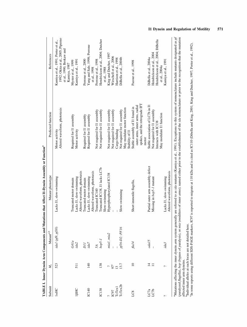
tion and structural location [DiBella and King, 2001].For example, in the axoneme, I1-dynein is a trilobedstructure originally identified as the ‘‘triad’’ [Goode-nough and Heuser, 1985a]. Mutations affecting certainI1 subunits often result in a complete failure in I1-dyneinassembly [ida1, ida2, ida7-See Table I for nomenclature,Kamiya et al., 1991] leaving a gap in the I1 position thatrepeats every 96-nm and does not affect the assembly ofthe other flagellar dynein isoforms [Piperno et al., 1990;Kamiya et al., 1991; Porter et al., 1992; Smith and Sale,1992b; Perrone et al., 1998]. Thus I1-dynein must be tar-geted to a unique position in the axoneme by specializedproteins or domains that repeat every 96-nm.
Phenotypic analysis has revealed that the inner armdyneins are responsible for determining the size andshape of the flagellar bend, parameters that define wave-form [Brokaw and Kamiya, 1987; Kamiya et al., 1991;Kamiya, 1995]. In some cases, mutations that affectI1-dynein assembly suppress paralysis in a central pairmutant [Porter et al., 1992]. Also, I1-dynein assemblymutants exhibit defects in phototaxis—a behavioralresponse to a light stimulus that requires precise controlof flagellar bending indicating I1 is important, althoughnot absolutely required, in this process [King andDutcher, 1997; Okita et al., 2005]. These phenotypicdata demonstrate that I1 dynein is a critical componentof a complex system, involving multiple axonemaldyneins, which controls axonemal bending. Extensive invitro functional and pharmacological data also indicateI1 dynein is required for control of axonemal sliding[Habermacher and Sale, 1997; King and Dutcher, 1997;Yang and Sale, 2000; Hendrickson et al., 2004], and thatthe mechanism of regulation involves reversible phos-phorylation of the I1 subunit IC138 in a process that alsomay be controlled by calcium [Hennessey et al., 2002;Smith, 2002a; Dymek and Smith, 2006].
Phylogenetic analyses have revealed that I1-dyneinproteins are highly conserved, thus the study of I1 inChlamydomonas is relevant to a wide range of organ-isms. For example, not only is I1-dynein activity regu-lated by phosphorylation in Chlamydomonas, recent datasuggests that I1 activity plays a role in regulating motil-ity in Tetrahymena thermophila [Hennessey et al., 2002;Deckman and Pennock, 2004].
In this commentary, we discuss mechanisms for as-sembly and transport of I1, anchoring I1-dynein to thedoublet microtubule, and localizing I1-dynein within the96-nm repeat. This review also incorporates very recentand exciting unpublished work that has been presentedin Abstract form at the 2006 American Society for CellBiology annual meeting. We describe a model for howlocalized control of I1-dynein phosphorylation operatesto locally control microtubule sliding and adjust axone-mal waveform. In particular, we summarize a model in
which I1 dynein locally inhibits microtubule slidingdriven by other dynein isoforms.
ORGANIZATION OF THE INNER ARMS: THE 96-NMREPEATAND SUBUNIT COMPOSITION OF I1DYNEIN
Compared to the outer dynein arms, the inner armdyneins are heterogeneous with respect to their organiza-tion [Porter and Sale, 2000; DiBella and King, 2001;Kamiya, 2002]. Analysis of mutant phenotypes, bio-chemical identification of the inner arm dynein subtypes,and eloquent ultrastructural studies have helped to definethe composition and arrangement of the complex innerdynein arm system, particularly the I1-dynein. The com-plexity of composition and structure has led to differentnomenclatures, but on the basis of heavy chain (HC)number, there appear to be two types of inner armdyneins. I1-dynein, also called f-dynein, contains twoHCs, whereas the inner arm dyneins containing one HCare designated a, b, c, d, e, and g, and were initiallythought to be organized in structures referred to as I2and I3 [Piperno et al., 1990; Kagami and Kamiya, 1992,1995].
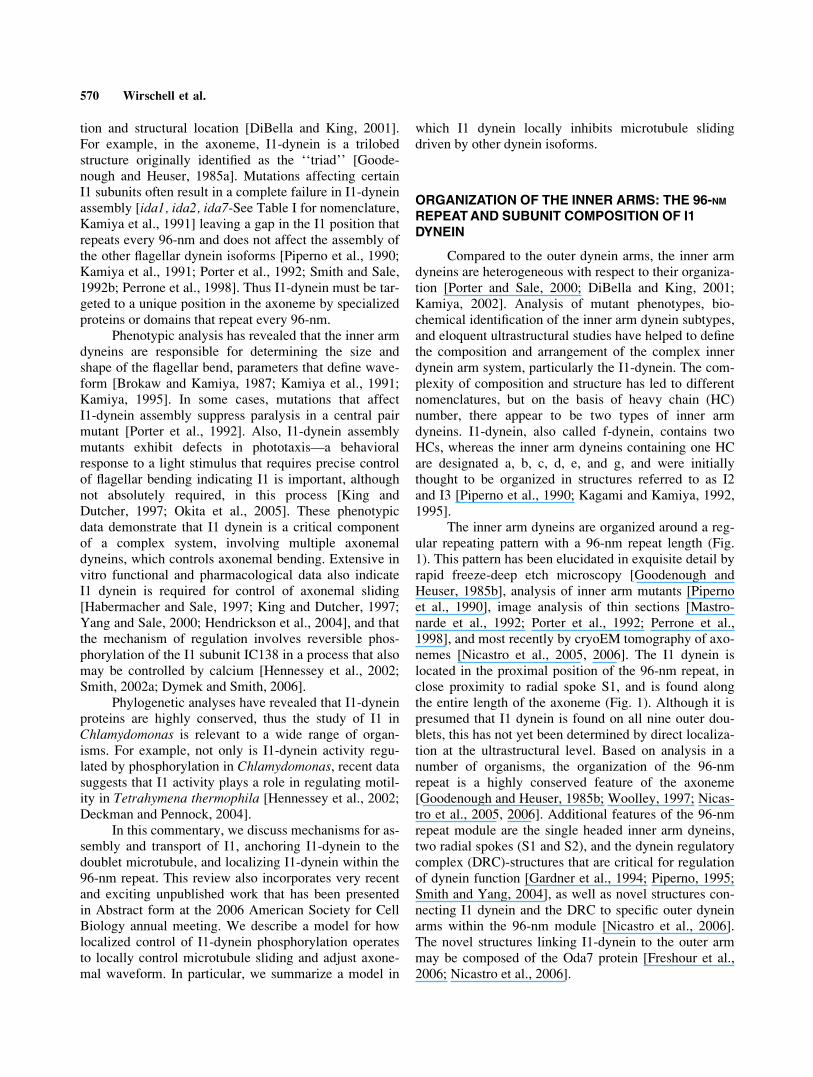
The inner arm dyneins are organized around a reg-ular repeating pattern with a 96-nm repeat length (Fig.1). This pattern has been elucidated in exquisite detail byrapid freeze-deep etch microscopy [Goodenough andHeuser, 1985b], analysis of inner arm mutants [Pipernoet al., 1990], image analysis of thin sections [Mastro-narde et al., 1992; Porter et al., 1992; Perrone et al.,1998], and most recently by cryoEM tomography of axo-nemes [Nicastro et al., 2005, 2006]. The I1 dynein islocated in the proximal position of the 96-nm repeat, inclose proximity to radial spoke S1, and is found alongthe entire length of the axoneme (Fig. 1). Although it ispresumed that I1 dynein is found on all nine outer dou-blets, this has not yet been determined by direct localiza-tion at the ultrastructural level. Based on analysis in anumber of organisms, the organization of the 96-nmrepeat is a highly conserved feature of the axoneme[Goodenough and Heuser, 1985b; Woolley, 1997; Nicas-tro et al., 2005, 2006]. Additional features of the 96-nmrepeat module are the single headed inner arm dyneins,two radial spokes (S1 and S2), and the dynein regulatorycomplex (DRC)-structures that are critical for regulationof dynein function [Gardner et al., 1994; Piperno, 1995;Smith and Yang, 2004], as well as novel structures con-necting I1 dynein and the DRC to specific outer dyneinarms within the 96-nm module [Nicastro et al., 2006].The novel structures linking I1-dynein to the outer armmay be composed of the Oda7 protein [Freshour et al.,2006; Nicastro et al., 2006].
570 Wirschell et al.
TABLEI.
InnerDynein
Arm
ComponentsandMutationsthatAffectI1-D
ynein
Assembly
orFunctiona
Subunit
Mr
Mutanta,b
Mutantphenotype
Predictedfunction
References
1aHC
523
ida1(pf9,pf30)
LacksI1,slow-swim
ming
Motoractivity
Kam
iyaetal.,1991;Porter
etal.,
1992;Okitaetal.,2005;Piperno
etal.,1990;Brokaw
and
Kam
iya,1987
Altered
waveform
,phototaxis
G41
aTruncatedmotordomain
Required
forI1
assembly
Myster
etal.,1999
1bHC
511
ida2
LacksI1,slow-swim
ming
Motoractivity
Kam
iyaetal.,1991
Altered
waveform
,phototaxis
D11
Truncatedmotordomain
Required
forI1
assembly
Perroneetal.,2000
IC140
140
ida7
LacksI1,slow-swim
ming
Required
forI1
assembly
YangandSale,1998;Perrone
etal.,1998
Altered
waveform
,phototaxis
5A
TruncatedIC140
Notrequired
forI1
assembly
Perroneetal.,1998
IC138
138
bop
5-1
TruncatedIC138,I1
lacksLC7b
Slow-swim
ming
Notrequired
forI1
assembly
Hendricksonetal.,2004;Dutcher
etal.,1988
??
mia1,m
ia2
HyperphosphorylatedIC138
Notrequired
forI1
assembly
KingandDutcher,1997
IC97
97c
––
Notrequired
forI1
assembly
Wirschelletal.,2006
TcT
ex1
13
––
Cargobinding
Harrisonetal.,1998
TcT
ex2b
13.7
pf16-D2::PF16
Slow-swim
ming
Notrequired
forI1
assembly
DiBellaetal.,2004b
May
modulatemotorfunction
StabilityofI1
LC8
10
fla14
Shortim
motileflagella,
Stableassembly
ofI1
foundin
outerarms,inner
arms,radial
spokes,andtheretrogradeIFT
motor
Pazouretal.,1998
LC7a
14
oda1
5Partialouterarm
assembly
defect
StableassociationofLC7bin
I1DiBellaetal.,2004a
LC7b
11
–Missingin
bop5
-1mutant
Notrequired
forI1
assembly
Hendricksonetal.,2004
InteractswithIC138
May
modulateI1
function
Hendricksonetal.,2004;DiBella
etal.,2004a
??
ida3
LacksI1,slow-swim
ming
Kam
iyaetal.,1991
Altered
waveform
,phototaxis
aMutationsaffectingtheinner
dyneinarm
system
generally
arereferred
toas
idamutants(K
amiyaetal.,1991).Exceptionsto
thissystem
ofnomenclature
includemutantsdesignated
aspf
(paralyzedflagella),bo
p(bypassofparalysis),ormia
(modifiersofinner
arms);nam
edeither
priorto
theestablishmentoftheidanomenclature
orpriorto
therecognitionthatthemutation
affected
inner
arm
dyneins.
bIndividualallelesateach
locusarenotdetailedhere.
cIn
somereportsusingdifferentSDS-PAGEmarkers,IC97isreported
tomigrateat110kDaandiscitedas
IC110(D
ibellaandKing,2001;KingandDutcher,1997;Porter
etal.,1992).
I1 Dynein and Regulation of Motility 571
I1-deficient axonemes reveal that I1 is localized toa unique position in the 96-nm repeat and that failure inI1 assembly does not affect assembly of other axonemalstructures [Piperno et al., 1990; Kamiya et al., 1991; Por-ter et al., 1992; Smith and Sale, 1992b; Nicastro et al.,2006]. This result is important in several ways. First, theother axonemal dyneins assemble normally indicatingmutations in I1 assembly do not disrupt assembly ofother dynein isoforms. Second, the results also implythat I1-dynein is localized in the axoneme by a dockingmechanism that resides near the S1 radial spoke, at theproximal end of the 96-nm repeat (Fig. 1). Consistentwith this prediction, in vitro reconstitution of I1-deficientaxonemes results in a restoration of functional I1 struc-tures to the original position in the 96-nm repeat [Smithand Sale, 1992b; Yamamoto et al., 2006]. However, todate, no docking complex has been identified.
I1 is the only inner dynein arm with two HCs, des-ignated 1a and 1b, [Piperno et al., 1990]. I1 dyneinforms a two headed structure as revealed by electron mi-croscopy of isolated I1 particles [Goodenough et al.,1987; Smith and Sale, 1991; Sakakibara et al., 2006].
Along with the HCs, are three intermediate chains(IC140, IC138, and IC97), and five known light chains(LC) (Tctex1, Tctex2b, LC8, LC7b, and LC7a) [Porteret al., 1992; Harrison et al., 1998; Perrone et al., 1998;Yang and Sale, 1998; DiBella et al., 2001, 2004a,b; Hen-drickson et al., 2004; Wirschell et al., 2006] (Fig. 1 insetand Table I). The HCs form stem domains which connectto an IC/LC complex that forms the cargo-binding do-main of the dynein motor as well as globular headdomains that possess the motor activity. For dyneins ingeneral, the intermediate and LCs play important roles inmotor assembly and regulation [Perrone et al., 1998;Yang and Sale, 1998; DiBella et al., 2001, 2004a,b].Several of the LCs are not unique to I1, notably LC8,LC7a, LC7b, and Tctex1. Rather, they are also LCs inother dyneins and additional enzymes and structures[Pazour et al., 1998; DiBella et al., 2004a,b]. These LCsmay have common functions in stable assembly ofdynein structures (LC8), cargo binding (Tctex1-rhodop-sin), or regulation (LC7b) [King et al., 1996; Harrisonet al., 1998; Pazour et al., 1998; Tai et al., 1999; Hen-drickson et al., 2004].
Fig. 1. Schematic of the 96-nm axonemal dynein repeat. The
arrangement of the inner dynein arms, outer dynein arms, radial
spokes, and DRC on the A-tubule is shown [adapted from Porter and
Sale, 2000; Nicastro et al., 2006]. These structures repeat every 96-nm
along the length of the axoneme (the outer dynein arms repeat every
24-nm). The proximal end (closest to the cell body) faces left. I1-
dynein is the trilobed inner arm just proximal to radial spoke S1 in
each repeat (green); it is followed by a series of single headed inner
arms (labeled 2–6). PKA is thought to be localized at the base of the
radial spokes [Gaillard et al., 2006]; CK1 and PP2A are predicted to
be anchored near I1 dynein; a fraction of PP1 located on the outer
doublets. The inset shows a diagram of the I1-dynein and its individ-
ual subunits [Harrison et al., 1998; Perrone et al., 1998; Yang and
Sale, 1998; Porter and Sale, 2000; DiBella et al., 2004a,b; Hendrick-
son et al., 2004]. I1-dynein is composed of two motor subunits (1a-and 1b-HC), three intermediate chains (IC140, IC138, and IC97), and
five LC subunits (Tctex1, Tctex2b, LC8, LC7a, and LC7b). IC138 is a
phosphoprotein in I1 and is one of the key regulatory subunits critical
for I1-dynein activity.
572 Wirschell et al.
I1-DYNEIN MUTANTS REVEAL NEW FEATURESOFASSEMBLYAND DOCKING
Mutations that disrupt specific I1 subunits or spe-cific domains of those subunits often result in assemblyof incomplete, or partial, I1 dynein complexes (Table I).For example, mutant strains containing truncated HCgenes, lacking the motor domains, still assemble an I1complex [Myster et al., 1999; Perrone et al., 2000].These results are consistent with previous studies indi-cating that N-terminal fragments between 140- and 160-kDa are capable of complex assembly [Sakakibara et al.,1993; Koonce and Knecht, 1998; Iyadurai et al., 1999].Importantly, electron microscopy of these HC trunca-tions revealed the location of the globular motor domainsof the 1a and 1b-HCs in the axoneme [Myster et al.,1999; Perrone et al., 2000]. They also indirectly revealthe location of the IC/LC complex at the base of I1dynein where it is anchored to the A-tubule (Fig. 1). Theassembly of I1 dynein complexes that lack one of theHC motor domains provides an opportunity to dissectthe individual roles of the 1a- and 1b-HCs [Fox et al.,2006; Toba et al., 2006].
The ICs typically contain WD repeats and are inte-gral structural components of dynein motors. This indeedseems to be the case for the IC140 subunit (see below).Like the ICs of the outer arm, IC140 is required for as-sembly of the I1 dynein complex and thus is implicated
in anchoring of I1 to the axoneme [Perrone et al., 1998;
Yang and Sale, 1998]. In support of this, the C-terminus
of IC140 interacts with the axoneme directly [Yang and
Sale, 1998] and thus may interact with components that
localize I1 in the 96-nm axonemal repeat structure.
However, while IC140 is required for I1 assembly in the
axoneme, the N-terminal region is not critical for its as-
sembly function (Table I-strain 5A, [Perrone et al.,
1998]). Alternatively, in the absence of IC140, I1-dynein
components in the cytoplasm may be unstable and
degraded, thus contributing to the failure of I1-dynien to
assemble in the axoneme.The bop5-1 mutant expresses a truncated IC138
protein that lacks the C-terminal WD repeat domain (Ta-ble I). All I1 subunits assemble in this mutant, except forLC7b indicating that LC7b and IC138 interact [DiBellaet al., 2004a; Hendrickson et al., 2004]. A second IC138mutation indicates that, unlike IC140 and the outer armICs, IC138 is not required for assembly of the entire I1-dynein complex, but may be required for assembly of theIC97 and LC7b subunits [Hendrickson et al., 2004;Bower et al., 2006]. These studies indicate that the pri-mary role of IC138 is not in assembly and is consistentwith the known regulatory role of IC138 in control ofmicrotubule sliding (see below). The results also revealthat IC97 is not required for assembly of I1, but they do
not reveal the exact function of IC97 within I1. Thismutation further indicates that IC140 along with theN-terminal HC domains are principally responsible fortargeting and anchoring I1 to its specific location in the96-nm repeat.
Based on crosslinking studies, IC138 and IC140interact directly with axonemal proteins [Yang and Sale,1998; Hendrickson et al., 2004] and at least some ofthese interactions may reflect interactions with tubulin(Wirschell, Hendrickson, and Sale, unpublished observa-tions). IC97 interacts directly with both a- and b-tubulinsubunits [Wirschell et al., 2006]. Given that all three ICsmay interact directly with tubulin, anchoring of I1dynein to the doublet microtubule involves multipleinteractions. However, IC-tubulin interactions cannotexplain the exact placement of I1 within the 96-nmrepeat. One idea is that I1 interacts with an intrinsic com-ponent of the outer doublet that establishes the 96-nmrepeat structure that governs placement of the innerdynein arms, radial spokes, and DRC. Proteins thatmight be part of such a complex could include the tektins[Norrander et al., 1996; Yanagisawa and Kamiya, 2004;Setter et al., 2006]. Consistent with this idea, tektins mayact as a molecular ruler that defines the axonemal 96-nmrepeat [Setter et al., 2006] and in Chlamydomonas,defects in tektin assembly are associated with defectiveinner dynein arm assembly [Yanagisawa and Kamiya,2004].
Structural analyses of axonemes from dyneinmutants have demonstrated that each dynein isoform istargeted to its own unique position along the length andaround the circumference of the A-microtubule of mostouter doublets. Anchoring of the outer dynein arm ismediated in part through additional complexes calledouter dynein arm–docking complexes (ODA-DC and theOda5 complex) [Takada and Kamiya, 1994; Wakabaya-shi et al., 2001; Casey et al., 2003; Wirschell et al.,2004]. Moreover, additional mutations have revealed theouter dynein arm is transported into the axoneme specifi-cally through an interaction with the intraflagellar trans-port component IFT46 and Oda16p—a novel non-dyneintransport factor required for outer dynein arm assembly[Ahmed and Mitchell, 2005; Hou et al., 2007]. Evidencesuggests that single-headed inner arms are also trans-ported into the flagellum by IFT [Piperno et al., 1996].Given that other dyneins are transported by IFT, it islikely that I1 is as well. However, to date, similar IFTtransport mechanisms have not been defined for I1dynein. Predictably, recovery of additional mutants thatfail to assemble I1 will reveal new genes required for as-sembly, transport, targeting, or docking of I1 dynein.The ida3 mutant may be an example. The ida3 mutationcauses a complete I1 assembly defect, but the IDA3 geneproduct remains unknown (Table I).
I1 Dynein and Regulation of Motility 573
FUNCTIONAL ROLE OF I1 IN REGULATION OFFLAGELLAR BENDING: A MODEL FOR LOCALREGULATION OF MICROTUBULE SLIDING BYPHOSPHORYLATION
Among the major challenges in understanding themechanism of ciliary motility is to determine howdynein-driven microtubule sliding is regulated. Based onmodels, oscillatory bending and bend propagationrequire mechanical feedback mechanisms that controlthe timing and location of active sliding [Brokaw, 1985,1994; Lindemann, 2002]. However, the experimentalevidence for the identity and nature of such a mechanicalfeedback mechanism is lacking.
In contrast, direct experimental evidence, through
genetic and in vitro functional studies using Chlamydo-monas axonemes, has revealed I1-dynein is important
for control of microtubule sliding and is likely designed
for modulating the size and shape of the axonemal bend
[Brokaw and Kamiya, 1987]. We summarize data indi-
cating I1-dynein is important for control of flagellar
waveform and that the mechanism involves regulation of
phosphorylation of the I1-dynein subunit IC138 and con-
trol of microtubule sliding [Habermacher and Sale,
1997; King and Dutcher, 1997; Yang and Sale, 2000;
Hendrickson et al., 2004]. We also discuss a model in
which localized inhibition of microtubule sliding [Smith,
2002b] is mediated by I1-dynein and IC138 phosphoryl-
ation and is required for normal control of bending
behavior.Mutations in I1-dynein subunits or mutations that
alter I1 phosphorylation result in failure of control ofnormal axonemal waveform and phototaxis in Chlamy-domonas [Brokaw and Kamiya, 1987; King and Dutcher,1997; Okita et al., 2005]. For example, the mia mutantswere isolated in a screen for phototaxis mutations andthe mutant axonemes exhibit hyperphosphorylatedIC138 [King and Dutcher, 1997]. Based on microtubulesliding assays and pharmacological analysis, dynein-driven sliding is inhibited in mia mutant axonemes.However, addition of kinase inhibitors rescues themicrotubule sliding defects. The data revealed a correla-tion between IC138 phosphorylation and regulation ofdynein; phosphorylated IC138 inhibited microtubulesliding and dephosphorylation rescued microtubule slid-ing (Fig. 2). Okita et al. have further examined the roleof I1 in phototaxis and suggest that I1 may be involvedin one of many pathways required for generating photo-tactic behavior [Okita et al., 2005]. Additionally, muta-tions in I1 subunits can suppress paralysis in a centralpair mutant, indicating a functional regulatory interac-tion between I1-dynein activity and the central pair-radial spoke structures for control of microtubule slidingand axonemal motility [Porter et al., 1992]. Thus, I1
dynein appears to regulate axonemal motility through amechanism that controls microtubule sliding.
Consistent with the idea that I1-dynein plays a role incontrol, in vitro functional assays, using isolated axonemesand microtubule sliding, have also revealed that I1-dyneinplays a central role in regulation of microtubule sliding andthat the mechanism depends upon changes in phosphoryla-tion of IC138. Figure 2 illustrates the role of CK1 and PKAin phosphorylation of IC138. Interestingly, a matinginduced elevation in intraflagellar cAMP [Hasegawa et al.,1987; Pasquale and Goodenough, 1987] levels is presum-ably responsible for quiescence of flagella during mating(observations by Ursula Goodenough, personal communi-cation). An interesting and testable idea is that theincreased intraflagellar cAMP levels affect IC138 phospho-rylation and thus globally inhibits microtubule sliding lead-ing to cessation or slowing of flagellar motility during themating response. Moreover, dynein-driven microtubulesliding in paralyzed axonemes, lacking either the radialspokes or the central pair, is inhibited presumably through-out the axoneme [Smith and Sale, 1992a]. The inhibitionof microtubule sliding in these paralyzed mutant axonemescan be rescued by addition of kinase inhibitors indicatingabnormal phosphorylation in the axoneme inhibits microtu-bule sliding [Howard et al., 1994; Habermacher and Sale,1995, 1997; Yang and Sale, 2000; Smith, 2002b]. Theresults are consistent with the model shown in Fig. 2 indi-cating that inhibition of dynein in the paralyzed mutants isdue, in part, to phosphorylation of IC138 and that dephos-phorylation rescues microtubule sliding [Habermacher andSale, 1997; King and Dutcher, 1997; Yang and Sale, 2000;Smith, 2002b; Hendrickson et al., 2004]. However, the invitro experiments were performed in a paralyzed flagella
Fig. 2. Regulation of IC138 phosphorylation. IC138 is a critical
component of I1 involved in the radial spoke-central pair mechanism
for regulating I1 dynein activity [adapted from Habermacher and Sale,
1997; Porter and Sale, 2000]. This regulatory process involves
changes in IC138 phosphorylation such that in the phosphorylated
state, I1 is inactive; when IC138 is dephosphorylated, I1-dynein activ-
ity is restored. The critical axonemal kinases and phosphatases
involved in this pathway are indicated [Porter and Sale, 2000; Smith
and Yang, 2004].
574 Wirschell et al.
mutants, lacking either the central pair or radial spokes, inwhich axonemal kinase activity is misregulated, revealinga predicted global change in dynein activity [Gaillard et al.,2001, 2006; Gokhale et al., 2006]. The same in vitro meas-urements of microtubule sliding in wild-type axonemeshave not been informative, most likely because the axone-mal kinases are tightly and locally controlled. Thus, amajor challenge is to determine how I1-dynein operates inwild-type axonemes for control of movement.
One idea, illustrated in Fig. 3 is that in wild-typeaxonemes, I1-dynein is locally and asymmetrically regu-lated in response to chemical or mechanical signals thatimpinge on the central pair apparatus. In this model, sig-nals originating from the central pair, an inherentlyasymmetric structure, are directed to specific doubletmicrotubules through the radial spokes [Smith and Yang,2004; Smith, 2007]. The idea is that an asymmetric sig-nal, for example from one of the central pair microtu-bules, is directed at doublet ‘‘N’’ (Fig. 3) and locallyinduces phosphorylation of IC138 on doublet ‘‘N’’. Incontrast, signals from the other central pair microtubulewould have a different affect on the phosphorylation ofIC138 on other outer doublet microtubules. Thus, fol-lowing local changes in phosphorylation, sliding be-tween doublets ‘‘N’’ and ‘‘N + 1’’ will be different rela-tive to sliding between other doublets, therefore alteringbending. As discussed before [Yang et al., 2004], theinput for the asymmetric central pair-radial spoke signalcould be a second messenger, such as calcium impingingupon the central pair [Wargo et al., 2005; Dymek andSmith, 2006], and/or a mechanical interaction with theradial spoke [Warner and Satir, 1974; Smith and Yang,
2004; Yang et al., 2004]. The output signal, presumablymediated by a radial spoke on one doublet microtubule,would be the activation of axonemal kinases locatednear I1-dynein [Yang et al., 2004; Gaillard et al., 2006].
Consistent with these ideas, the central pair appara-tus is asymmetric in structure and composition [Mitchelland Sale, 1999; Mitchell and Yokoyama, 2003; Mitchell,2003a; Wargo and Smith, 2003; Smith and Yang, 2004;Lechtreck and Witman, 2007] and the position of thecentral pair correlates with sites of active sliding [Wargoand Smith, 2003; Wargo et al., 2004] and the position ofdoublets relative to the bend [Mitchell, 2003a; Smith andYang, 2004]. The results have also revealed a functionalrole for kinases (PKA, CK1) and phosphatases (PP1,PP2A) that are physically localized in the axoneme[Habermacher and Sale, 1996, 1997; King and Dutcher,1997; Porter and Sale, 2000; Yang et al., 2000; Gaillardet al., 2001, 2006; Gokhale et al., 2006]. Much effort isunderway to identify molecular mechanisms for target-ing otherwise ubiquitous signaling molecules to preciselocations in the axoneme [Gaillard et al., 2001, 2006].Further tests of these ideas will require new assays link-ing radial spoke function to the direct control of axone-mal kinases located on the outer doublet microtubule.
Related challenges include determining the mecha-nism for how IC138 can alter microtubule sliding. Theimplication of the model is the I1-dynein locally operatesto inhibit microtubule sliding [Smith, 2002b]. Consistentwith this idea, isolated I1-dyneins generate relatively slowmicrotubule translocation indicating that I1 dynein isunlike the outer arm dyneins or other inner arm dy-neins [Smith and Sale, 1991; Kagami and Kamiya, 1992;
Fig. 3. A model for localized regulation of I1 and axonemal bending.
The axoneme is shown in cross section illustrating the 9 + 2 arrange-
ment of the axoneme and the asymmetric central pair apparatus, the
radial spokes, and the dynein arms [Smith, 2007]. The black line indi-
cates the plane of bending [Mitchell, 2003b; Mitchell and Yokoyama,
2003]. The current model for regulation of I1-dynein is that chemical
and/or mechanical signals from the central pair are transmitted
through the radial spokes to affect IC138 phosphorylation on specific
outer doublets (e.g., outer doublets N, N + 1, N � 1). Thus, phospho-
rylation of IC138 in an asymmetrical manner is predicted to result in
local inhibition of sliding. This in turn would alter the form of the
bend.
I1 Dynein and Regulation of Motility 575
Sakakibara and Nakayama, 1998; Sakakibara et al., 1999;Yagi et al., 2005]. Although it is possible that the I1-dyneinis not well suited to such in vitro assays that involveabsorption to inert surfaces, recent advances in the K.Oiwa lab (Kobe Advanced ICT Research Center, Kobe, Ja-pan) have confirmed that I1-dynein generates only slowmicrotubule translocation compared to other dyneins[Sakakibara et al., 2006; Toba et al., 2006].
When IC138 is phosphorylated, microtubule slidingis inhibited in the axoneme [Habermacher and Sale, 1997;King and Dutcher, 1997; Yang and Sale, 2000; Hendrick-son et al., 2004]. One hypothesis is that phosphorylation ofIC138 inhibits I1-dynein motor activity. However, usingpurified I1-dynein and in vitro motility assays, it has beendetermined that motor activity of isolated I1 dynein is notaltered by IC138 phosphorylation, yet in the axonememicrotubule sliding is inhibited by the same changes inIC138 [Sakakibara et al., 2006]. Thus further progress willrequire measurements of single I1 motor characteristicsincluding assessment of processivity and duty cycle as hasbeen done for other inner arm dyneins and further structuralanalyses of I1 [Sakakibara et al., 2006, 1999]. Additionally,our collaborative studies with M. Porter (University of Min-nesota), Toba, Oiwa, Sakakibara, and colleagues (KobeAdvanced ICT Research Center, Kobe, Japan) are address-ing models of I1-dynein mechanism using single motoranalysis and dyneins isolated from wild-type and I1-mutantaxonemes [Myster et al., 1999; Perrone et al., 2000; Foxet al., 2006; Toba et al., 2006]. The goal is to further testthe hypothesis that I1 dynein acts as a ‘‘brake’’ to locallyinhibit microtubule sliding driven by other dynein isoformsand thus control axonemal waveform.
ACKNOWLEDGMENTS
We are grateful to Dr. Elizabeth Smith (DartmouthCollege) and Dr. Steven King (University of ConnecticutHealth Science Center) for helpful discussions of I1dynein and this manuscript.
REFERENCES
Ahmed NT, Mitchell DR. 2005. ODA16p, a Chlamydomonas flagellarprotein needed for dynein assembly. Mol Biol Cell 16(10):
5004–5012.
Avidor-Reiss T, Maer AM, Koundakjian E, Polyanovsky A, Keil T,
Subramaniam S, Zuker CS. 2004. Decoding cilia function:
Defining specialized genes required for compartmentalized
cilia biogenesis. Cell 117(4):527–539.
Bower R, Perrone CA, O’Toole E, Fox L, Wirschell M, Sale WS, Por-
ter ME. 2006. IC138 is required for regulation but not assembly
of the I1 inner arm dynein. San Diego, CA: Abstracts from the
2006 Annual Meeting of the American Society for Cell Biol-
ogy and published at www.ASCB.org.
Brokaw CJ. 1985. Computer simulation of flagellar movement. VI.
Simple curvature-controlled models are incompletely specified.
Biophys J 48(4):633–642.
Brokaw CJ. 1994. Control of flagellar bending: A new agenda based
on dynein diversity. Cell Motil Cytoskeleton 28(3):199–204.
Brokaw CJ, Kamiya R. 1987. Bending patterns of Chlamydomonasflagella: IV. Mutants with defects in inner and outer dynein
arms indicate differences in dynein arm function. Cell Motil
Cytoskeleton 8(1):68–75.
Casey DM, Inaba K, Pazour GJ, Takada S, Wakabayashi K, Wilker-
son CG, Kamiya R, Witman GB. 2003. DC3, the 21-kDa sub-
unit of the outer dynein arm-docking complex (ODA-DC), is a
novel EF-hand protein important for assembly of both the outer
arm and the ODA-DC. Mol Biol Cell 14(9):3650–3663.
Christensen ST, Pedersen LB, Schneider L, Satir P. 2007. Sensory
cilia and integration of signal transduction in human health and
disease. Traffic 8(2):97–109.
Davenport JR, Yoder BK. 2005. An incredible decade for the primary
cilium: A look at a once-forgotten organelle. Am J Physiol
Renal Physiol 289(6):F1159–1169.
Davis EE, Brueckner M, Katsanis N. 2006. The emerging complexity
of the vertebrate cilium: New functional roles for an ancient or-
ganelle. Dev Cell 11(1):9–19.
Deckman CM, Pennock DG. 2004. Dephosphorylation of inner arm 1
is associated with ciliary reversals in Tetrahymena thermo-phila. Cell Motil Cytoskeleton 57(2):73–83.
DiBella LM, Benashski SE, Tedford HW, Harrison A, Patel-King RS,
King SM. 2001. The Tctex1/Tctex2 class of dynein light
chains. Dimerization, differential expression, and interaction
with the LC8 protein family. J Biol Chem 276(17):14366–
14373.
DiBella LM, King SM. 2001. Dynein motors of the Chlamydomonasflagellum. Int Rev Cytol 210:227–268.
DiBella LM, Sakato M, Patel-King RS, Pazour GJ, King SM. 2004a.
The LC7 light chains of Chlamydomonas flagellar dyneins
interact with components required for both motor assembly and
regulation. Mol Biol Cell 15(10):4633–4646.
DiBella LM, Smith EF, Patel-King RS, Wakabayashi K, King SM.
2004b. A novel Tctex2-related light chain is required for stabil-
ity of inner dynein arm I1 and motor function in the Chlamydo-monas flagellum. J Biol Chem 279(20):21666–21676.
Dymek EE, Smith EF. 2006. Regulation of axonemal dynein activity
by a calmodulin and radial spoke associated complex (CRC).
San Diego, CA: Abstracts from the 2006 Annual Meeting of
the American Society for Cell Biology and published at
www.ASCB.org.
El Zein L, Omran H, Bouvagnet P. 2003. Lateralization defects and
ciliary dyskinesia: Lessons from algae. Trends Genet 19(3):
162–167.
Fox LA, Tritschler D, Porter ME, Sale WS. 2006. Regulation of flag-
ellar inner arm dynein I1: The 1b heavy chain motor domain is
necessary for control of microtubule sliding by phosphoryla-
tion. San Diego, CA: Abstracts from the 2006 Annual Meeting
of the American Society for Cell Biology and published at
www.ASCB.org.
Freshour J, Yokoyama R, Mitchell DR. 2006. Chlamydomonas flagel-lar outer row dynein assembly protein Oda7 interacts with both
outer row and I1 inner row dyneins. J Biol Chem 282(8):5404–
5412.
Gaillard AR, Diener DR, Rosenbaum JL, Sale WS. 2001. Flagellar ra-
dial spoke protein 3 is an A-kinase anchoring protein (AKAP).
J Cell Biol 153(2):443–448.
Gaillard AR, Fox LA, Rhea JM, Craige B, Sale WS. 2006. Disruption
of the A-kinase anchoring domain in flagellar radial spoke pro-
tein 3 results in unregulated axonemal cAMP-dependent pro-
tein kinase activity and abnormal flagellar motility. Mol Biol
Cell 17(6):2626–2635.
576 Wirschell et al.
Gardner LC, O’Toole E, Perrone CA, Giddings T, Porter ME. 1994.
Components of a ‘‘dynein regulatory complex’’ are located at
the junction between the radial spokes and the dynein arms in
Chlamydomonas flagella. J Cell Biol 127(5):1311–1325.Gokhale A, Wirschell M, Sale WS. 2006. Characterization of the
protein kinase CK1 that regulates dynein-driven microtubule
sliding and is anchored in Chlamydomonas flagellar axonemes.
San Diego, CA: Abstracts from the 2006 Annual Meeting of
the American Society for Cell Biology and published at
www.ASCB.org.
Goodenough UW, Gebhart B, Mermall V, Mitchell DR, Heuser JE.
1987. High-pressure liquid chromatography fractionation of
Chlamydomonas dynein extracts and characterization of inner-
arm dynein subunits. J Mol Biol 194(3):481–494.
Goodenough UW, Heuser JE. 1985a. Outer and inner dynein arms of
cilia and flagella. Cell 41(2):341–342.
Goodenough UW, Heuser JE. 1985b. Substructure of inner dynein
arms, radial spokes, and the central pair/projection complex of
cilia and flagella. J Cell Biol 100(6):2008–2018.
Habermacher G, Sale WS. 1995. Regulation of dynein-driven micro-
tubule sliding by an axonemal kinase and phosphatase in Chla-mydomonas flagella. Cell Motil Cytoskeleton 32(2):106–109.
Habermacher G, Sale WS. 1996. Regulation of flagellar dynein by an
axonemal type-1 phosphatase in Chlamydomonas. J Cell Sci
109(Part 7):1899–1907.
Habermacher G, Sale WS. 1997. Regulation of flagellar dynein by
phosphorylation of a 138-kD inner arm dynein intermediate
chain. J Cell Biol 136(1):167–176.
Harrison A, Olds-Clarke P, King SM. 1998. Identification of the t
complex-encoded cytoplasmic dynein light chain tctex1 in
inner arm I1 supports the involvement of flagellar dyneins in
meiotic drive. J Cell Biol 140(5):1137–1147.
Hasegawa E, Hayashi H, Asakura S, Kamiya R. 1987. Stimulation of
in vitro motility of Chlamydomonas axonemes by inhibition of
cAMP-dependent phosphorylation. Cell Motil Cytoskeleton
8(4):302–311.
Hendrickson TW, Perrone CA, Griffin P, Wuichet K, Mueller J, Yang
P, Porter ME, Sale WS. 2004. IC138 is a WD-repeat dynein in-
termediate chain required for light chain assembly and regula-
tion of flagellar bending. Mol Biol Cell 15(12):5431–5442.
Hennessey TM, Kim DY, Oberski DJ, Hard R, Rankin SA, Pennock
DG. 2002. Inner arm dynein 1 is essential for Ca++-dependent
ciliary reversals in Tetrahymena thermophila. Cell Motil Cyto-
skeleton 53(4):281–288.
Hou Y, Qin H, Follit JA, Pazour GJ, Rosenbaum JL, Witman GB.
2007. Functional analysis of an individual IFT protein: IFT46
is required for transport of outer dynein arms into flagella.
J Cell Biol 176(5):653–665.
Howard DR, Habermacher G, Glass DB, Smith EF, Sale WS. 1994.
Regulation of Chlamydomonas flagellar dynein by an axonemal
protein kinase. J Cell Biol 127(6, Part 1):1683–1692.
Iyadurai SJ, Li MG, Gilbert SP, Hays TS. 1999. Evidence for coopera-
tive interactions between the two motor domains of cytoplas-
mic dynein. Curr Biol 9(14):771–774.
Kagami O, Kamiya R. 1992. Translocation and rotation of microtu-
bules caused by multiple species of Chlamydomonas inner-armdynein. J Cell Sci 103(3):653–664.
Kagami O, Kamiya R. 1995. Separation of dynein species by high-pres-
sure liquid chromatography. Methods Cell Biol 47:487–489.
Kamiya R. 1995. Exploring the function of inner and outer dynein
arms with Chlamydomonas mutants. Cell Motil Cytoskeleton
32(2):98–102.
Kamiya R. 2002. Functional diversity of axonemal dyneins as studied
in Chlamydomonas mutants. Int Rev Cytol 219:115–155.
Kamiya R, Kurimoto E, Muto E. 1991. Two types of Chlamydomonasflagellar mutants missing different components of inner-arm
dynein. J Cell Biol 112(3):441–447.
King SJ, Dutcher SK. 1997. Phosphoregulation of an inner dynein arm
complex in Chlamydomonas reinhardtii is altered in phototacticmutant strains. J Cell Biol 136(1):177–191.
King SM, Barbarese E, Dillman JF III, Patel-King RS, Carson JH,
Pfister KK. 1996. Brain cytoplasmic and flagellar outer arm
dyneins share a highly conserved Mr 8,000 light chain. J Biol
Chem 271(32):19358–19366.
Koonce MP, Knecht DA. 1998. Cytoplasmic dynein heavy chain is an
essential gene product in Dictyostelium. Cell Motil Cytoskele-
ton 39(1):63–72.
Lechtreck K-F, Witman GB. 2007. Chlamydomonas reinhardtii hydinis a central pair protein required for flagellar motility. J Cell
Biol 176(4):473–482.
Li JB, Gerdes JM, Haycraft CJ, Fan Y, Teslovich TM, May-Simera H,
Li H, Blacque OE, Li L, Leitch CC, Lewis RA, Green JS, Parfrey
PS, Leroux MR, Davidson WS, Beales PL, Guay-Woodford LM,
Yoder BK, Stormo GD, Katsanis N, Dutcher SK. 2004. Compara-
tive genomics identifies a flagellar and basal body proteome that
includes the BBS5 human disease gene. Cell 117(4):541–552.
Lindemann CB. 2002. Geometric Clutch model version 3: The role of
the inner and outer arm dyneins in the ciliary beat. Cell Motil
Cytoskeleton 52(4):242–254.
Mastronarde DN, O’Toole ET, McDonald KL, McIntosh JR, Porter ME.
1992. Arrangement of inner dynein arms in wild-type and mutant
flagella of Chlamydomonas. J Cell Biol 118(5):1145–1162.Mitchell DR. 2003a. Orientation of the central pair complex during
flagellar bend formation in Chlamydomonas. Cell Motil Cyto-
skeleton 56(2):120–129.
Mitchell DR. 2003b. Reconstruction of the projection periodicity and
surface architecture of the flagellar central pair complex. Cell
Motil Cytoskeleton 55(3):188–199.
Mitchell DR, Sale WS. 1999. Characterization of a Chlamydomonasinsertional mutant that disrupts flagellar central pair microtu-
bule-associated structures. J Cell Biol 144(2):293–304.
Mitchell DR, Yokoyama R. 2003. Structural analysis of central pair
function in Chlamydomonas flagella. Mol Biol Cell 14:436a.
Myster SH, Knott JA, Wysocki KM, O’Toole E, Porter ME. 1999.
Domains in the 1alpha dynein heavy chain required for inner
arm assembly and flagellar motility in Chlamydomonas. J CellBiol 146(4):801–818.
Nicastro D, McIntosh JR, Baumeister W. 2005. 3D structure of eu-
karyotic flagella in a quiescent state revealed by cryo-electron
tomography. Proc Natl Acad Sci USA 102(44):15889–15894.
Nicastro D, Schwartz C, Pierson J, Gaudette R, Porter M, McIntosh J.
2006. The molecular architecture of axonemes revealed by cry-
oelectron tomography. Science 313(5789):944–948.
Norrander JM, Perrone CA, Amos LA, Linck RW. 1996. Structural
comparison of tektins and evidence for their determination of
complex spacings in flagellar microtubules. J Mol Biol
257(2):385–397.
Oiwa K, Sakakibara H. 2005. Recent progress in dynein structure and
mechanism. Curr Opin Cell Biol 17(1):98–103.
Okita N, Isogai N, Hirono M, Kamiya R, Yoshimura K. 2005. Photo-
tactic activity in Chlamydomonas ‘non-phototactic’ mutants
deficient in Ca2+-dependent control of flagellar dominance or
in inner-arm dynein. J Cell Sci 118(Part 3):529–537.
Pan J, Wang Q, Snell WJ. 2005. Cilium-generated signaling and cilia-
related disorders. Lab Invest 85(4):452–463.
Pasquale SM, Goodenough UW. 1987. Cyclic AMP functions as a
primary sexual signal in gametes of Chlamydomonas reinhardtii.J Cell Biol 105(5):2279–2292.
I1 Dynein and Regulation of Motility 577
Pazour GJ. 2004. Intraflagellar transport and cilia-dependent renal dis-
ease: The ciliary hypothesis of polycystic kidney disease. J Am
Soc Nephrol 15(10):2528–2536.
Pazour GJ, Agrin N, Leszyk J, Witman GB. 2005. Proteomic analysis
of a eukaryotic cilium. J Cell Biol 170(1):103–113.
Pazour GJ, Wilkerson CG, Witman GB. 1998. A dynein light chain is
essential for the retrograde particle movement of intraflagellar
transport (IFT). J Cell Biol 141(4):979–992.
Perrone CA, Myster SH, Bower R, O’Toole ET, Porter ME. 2000.
Insights into the structural organization of the I1 inner arm
dynein from a domain analysis of the 1b dynein heavy chain.
Mol Biol Cell 11(7):2297–2313.
Perrone CA, Yang P, O’Toole E, Sale WS, Porter ME. 1998. The
Chlamydomonas IDA7 locus encodes a 140-kDa dynein inter-
mediate chain required to assemble the I1 inner arm complex.
Mol Biol Cell 9(12):3351–3365.
Piperno G. 1995. Regulation of dynein activity within Chlamydomo-nas flagella. Cell Motil Cytoskeleton 32(2):103–105.
Piperno G, Mead K, Henderson S. 1996. Inner dynein arms but not
outer dynein arms require the activity of kinesin homologue
protein KHP1(FLA10) to reach the distal part of flagella in
Chlamydomonas. J Cell Biol 133(2):371–379.Piperno G, Ramanis Z, Smith EF, Sale WS. 1990. Three distinct
inner dynein arms in Chlamydomonas flagella: Molecular
composition and location in the axoneme. J Cell Biol 110(2):
379–389.
Porter ME, Power J, Dutcher SK. 1992. Extragenic suppressors of
paralyzed flagellar mutations in Chlamydomonas reinhardtiiidentify loci that alter the inner dynein arms. J Cell Biol
118(5):1163–1176.
Porter ME, Sale WS. 2000. The 9 + 2 axoneme anchors multiple inner
arm dyneins and a network of kinases and phosphatases that
control motility. J Cell Biol 151(5):F37–F42.
Quarmby LM, Parker JD. 2005. Cilia and the cell cycle? J Cell Biol
169(5):707–710.
Sakakibara H, Burgess S, Sakai Y. 2006. Conformational changes of
inner-arm dynein-f from Chlamydomonas coupled with
phosphorylation of its intermediated chain IC138. San Diego,
CA: Abstracts from the 2006 Annual Meeting of the Ameri-
can Society for Cell Biology and published at www.ASCB.
org.
Sakakibara H, Kojima H, Sakai Y, Katayama E, Oiwa K. 1999. Inner-
arm dynein c of Chlamydomonas flagella is a single-headed
processive motor. Nature 400(6744):586–590.
Sakakibara H, Nakayama H. 1998. Translocation of microtubules
caused by the alphabeta, beta and gamma outer arm dynein
subparticles of Chlamydomonas. J Cell Sci 111(Part 9):1155–1164.
Sakakibara H, Takada S, King SM, Witman GB, Kamiya R. 1993. A
Chlamydomonas outer arm dynein mutant with a truncated beta
heavy chain. J Cell Biol 122(3):653–661.
Setter PW, Malvey-Dorn E, Steffen W, Stephens RE, Linck RW.
2006. Tektin interactions and a model for molecular functions.
Exp Cell Res 312(15):2880–2896.
Silflow CD, Lefebvre PA. 2001. Assembly and motility of eukaryotic
cilia and flagella. Lessons from Chlamydomonas reinhardtii.Plant Physiol 127(4):1500–1507.
Smith EF. 2002a. Regulation of flagellar dynein by calcium and a role
for an axonemal calmodulin and calmodulin-dependent kinase.
Mol Biol Cell 13(9):3303–3313.
Smith EF. 2002b. Regulation of flagellar dynein by the axonemal cen-
tral apparatus. Cell Motil Cytoskeleton 52(1):33–42.
Smith EF. 2007. Hydin seek: Finding a function in ciliary motility.
J Cell Biol 176(4):403–404.
Smith EF, Sale WS. 1991. Microtubule binding and translocation by
inner dynein arm subtype I1. Cell Motil Cytoskeleton 18(4):
258–268.
Smith EF, Sale WS. 1992a. Regulation of dynein-driven microtubule
sliding by the radial spokes in flagella. Science 257(5076):
1557–1559.
Smith EF, Sale WS. 1992b. Structural and functional reconstitution of
inner dynein arms in Chlamydomonas flagellar axonemes.
J Cell Biol 117(3):573–581.
Smith EF, Yang P. 2004. The radial spokes and central apparatus:
Mechano-chemical transducers that regulate flagellar motility.
Cell Motil Cytoskeleton 57(1):8–17.
Snell WJ, Pan J, Wang Q. 2004. Cilia and flagella revealed: From flag-
ellar assembly in Chlamydomonas to human obesity disorders.
Cell 117(6):693–697.
Tai AW, Chuang JZ, Bode C, Wolfrum U, Sung CH. 1999. Rhodop-
sin’s carboxy-terminal cytoplasmic tail acts as a membrane re-
ceptor for cytoplasmic dynein by binding to the dynein light
chain Tctex-1. Cell 97(7):877–887.
Takada S, Kamiya R. 1994. Functional reconstitution of Chlamy-domonas outer dynein arms from alpha-beta and gamma sub-
units: Requirement of a third factor. J Cell Biol 126(3):737–
745.
Toba S, Sakakibara H, Porter ME, Sale WS, Oiwa K. 2006. Motility
of the 1a and 1b heavy chains of the inner arm dynein I1 of
Chlamydomonas flagella. San Diego, CA: Abstracts from the
2006 Annual Meeting of the American Society for Cell Biol-
ogy and published at www.ASCB.org.
Wakabayashi K, Takada S, Witman GB, Kamiya R. 2001. Transport
and arrangement of the outer-dynein-arm docking complex in
the flagella of Chlamydomonas mutants that lack outer dynein
arms. Cell Motil Cytoskeleton 48(4):277–286.
Wargo MJ, Dymek EE, Smith EF. 2005. Calmodulin and PF6 are
components of a complex that localizes to the C1 microtubule
of the flagellar central apparatus. J Cell Sci 118(Part 20):4655–
4665.
Wargo MJ, McPeek MA, Smith EF. 2004. Analysis of microtubule
sliding patterns in Chlamydomonas flagellar axonemes reveals
dynein activity on specific doublet microtubules. J Cell Sci
117(Part 12):2533–2544.
Wargo MJ, Smith EF. 2003. Asymmetry of the central apparatus
defines the location of active microtubule sliding in Chlamydo-monas flagella. Proc Natl Acad Sci USA 100(1):137–142.
Warner FD, Satir P. 1974. The structural basis of ciliary bend forma-
tion. Radial spoke positional changes accompanying microtu-
bule sliding. J Cell Biol 63(1):35–63.
Wirschell M, Pazour G, Yoda A, Hirono M, Kamiya R, Witman
GB. 2004. Oda5p, a novel axonemal protein required for as-
sembly of the outer dynein arm and an associated adenylate
kinase 10.1091/mbc.E03-11-0820. Mol Biol Cell 15(6):2729–
2741.
Wirschell M, Yanagisawa HA, Kamiya R, Witman GB, Porter ME,
Sale WS. 2006. The role of the IC97 in I1-dynein assembly
and axonemal anchoring. San Diego, CA: Abstracts from the
2006 Annual Meeting of the American Society for Cell Biol-
ogy and published at www.ASCB.org.
Woolley DM. 1997. Studies on the eel sperm flagellum. I. The struc-
ture of the inner dynein arm complex. J Cell Sci 110(Part 1):
85–94.
Yagi T, Minoura I, Fujiwara A, Saito R, Yasunaga T, Hirono M,
Kamiya R. 2005. An axonemal dynein particularly important
for flagellar movement at high viscosity. Implications from a
new Chlamydomonas mutant deficient in the dynein heavy
chain gene DHC9. J Biol Chem 280(50):41412–41420.
578 Wirschell et al.
Yamamoto R, Yagi T, Kamiya R. 2006. Functional binding of inner-
arm dyneins with demembranated flagella of Chlamydomonasmutants. Cell Motil Cytoskeleton 63(5):258–265.
Yanagisawa H-A, Kamiya R. 2004. A tektin homologue is decreased
in Chlamydomonas mutants lacking an axonemal inner-arm
dynein. Mol Biol Cell 15(5):2105–2115.
Yang P, Fox L, Colbran RJ, Sale WS. 2000. Protein phosphatases
PP1 and PP2A are located in distinct positions in the
Chlamydomonas flagellar axoneme. J Cell Sci 113(Part 1):91–
102.
Yang P, Sale WS. 1998. The Mr 140,000 intermediate chain of Chla-mydomonas flagellar inner arm dynein is a wd-repeat protein
implicated in dynein arm anchoring. Mol Biol Cell 9(12):
3335–3349.
Yang P, Sale WS. 2000. Casein kinase I is anchored on axonemal
doublet microtubules and regulates flagellar dynein phospho-
rylation and activity. J Biol Chem 275(25):18905–18912.
Yang P, Yang C, Sale WS. 2004. Flagellar radial spoke protein 2 is a
calmodulin binding protein required for motility in Chlamydo-monas reinhardtii. Eukaryot Cell 3(1):72–81.
I1 Dynein and Regulation of Motility 579
Recommended


