Modifying Ligand-Induced and Constitutive Signaling ofthe Human 5-HT4 ReceptorWei Chun Chang1,4¤, Jennifer K. Ng1, Trieu Nguyen1, Lucie Pellissier2,3, Sylvie Claeysen2,3, Edward C. Hsiao1,5, Bruce R. Conklin1,5,6*
1 Gladstone Institute of Cardiovascular Disease, University of California at San Francisco, San Francisco, California, United States of America, 2 Institutde Genomique Fonctionnelle, Universites de Montpellier, CNRS UMR 5203, Montpellier, France, 3 INSERM U661, Montpellier, France, 4 GraduateProgram in Pharmaceutical Sciences and Pharmacogenomics, University of California at San Francisco, San Francisco, California, United States ofAmerica, 5 Department of Medicine, University of California at San Francisco, San Francisco, California, United States of America, 6 Department ofCellular and Molecular Pharmacology, University of California at San Francisco, San Francisco, California, United States of America
G protein–coupled receptors (GPCRs) signal through a limited number of G-protein pathways and play crucial roles in manybiological processes. Studies of their in vivo functions have been hampered by the molecular and functional diversity of GPCRsand the paucity of ligands with specific signaling effects. To better compare the effects of activating different G-proteinsignaling pathways through ligand-induced or constitutive signaling, we developed a new series of RASSLs (receptorsactivated solely by synthetic ligands) that activate different G-protein signaling pathways. These RASSLs are based on thehuman 5-HT4b receptor, a GPCR with high constitutive Gs signaling and strong ligand-induced G-protein activation of the Gs
and Gs/q pathways. The first receptor in this series, 5-HT4-D100A or Rs1 (RASSL serotonin 1), is not activated by its endogenousagonist, serotonin, but is selectively activated by the small synthetic molecules GR113808, GR125487, and RO110-0235. Allagonists potently induced Gs signaling, but only a few (e.g., zacopride) also induced signaling via the Gq pathway. Zacopride-induced Gq signaling was enhanced by replacing the C-terminus of Rs1 with the C-terminus of the human 5-HT2C receptor.Additional point mutations (D66A and D66N) blocked constitutive Gs signaling and lowered ligand-induced Gq signaling.Replacing the third intracellular loop of Rs1 with that of human 5-HT1A conferred ligand-mediated Gi signaling. This Gi-coupledRASSL, Rs1.3, exhibited no measurable signaling to the Gs or Gq pathway. These findings show that the signaling repertoire ofRs1 can be expanded and controlled by receptor engineering and drug selection.
Citation: Chang WC, Ng JK, Nguyen T, Pellissier L, Claeysen S, et al (2007) Modifying Ligand-Induced and Constitutive Signaling of the Human 5-HT4
Receptor. PLoS ONE 2(12): e1317. doi:10.1371/journal.pone.0001317
INTRODUCTIONHeptahelical G protein–coupled receptors (GPCRs) are the largest
family of human cell-surface receptors, encompassing more than
340 hormone receptors and 350–460 olfactory receptors [1,2].
They are activated by peptide hormones, odorants, photons,
biogenic amines, phospholipids, and many other extracellular
signals. Upon activation, GPCRs undergo conformational changes
that allow active and reversible signaling through a limited
number of G-protein pathways (Gs, Gi, Gq, G12/13). These signals
mediate a wide variety of physiological responses, including heart
rate, chemotaxis, cell proliferation, neurotransmission, and
hormonal responses. Owing to their physiological importance,
GPCRs are of great medical interest. Indeed, they are targets for
at least 40% of modern pharmaceuticals [3].
Although many drugs target GPCRs, studies of GPCR signaling
in vivo have been hampered by the lack of specific agonists and
antagonists for many of the receptors. GPCRs display molecular
and functional diversity, such as the type of G-protein signaling
pathway associations, different levels of constitutive activity, and
different signaling responses due to different ligand-selective
conformations [4,5]. This diversity enables the receptors to
transmit unique extracellular signals but hampers efforts to sort
out the relative contributions of each signaling pathway or the
roles of constitutive signaling for each receptor.
To better study the diversity of GPCRs, we developed
receptors activated solely by synthetic ligands (RASSLs) by
modifying their structures to render them unresponsive to
endogenous hormones. Instead, RASSLs are activated by small-
molecule drugs [6], allowing them to be used to activate specific
G-protein pathways rapidly and reversibly and to mimic the speed,
localization, regulation, and amplification of endogenous GPCR
signals [7].
Since it is impractical to convert all GPCRs into RASSLs, we
and others have focused on representative Gs-, Gi-, and Gq-
coupled GPCRs, which stimulate adenylyl cyclase, inhibit adenylyl
cyclase, and stimulate phospholipase-C, respectively. Ro1 (RASSL
opioid 1), the prototype RASSL based on a Gi-coupled k-opioid
receptor [8], provided a proof of concept for this strategy. Its Gi
response to natural ligands is 0.001% of that of the wildtype
receptor, but it is potently activated by the synthetic agonist
spiradoline [8–10]. Ro1 decreases heart rate in mice [11] and
affects taste sensation in the tongue [9]. In addition to serving as
powerful tools to dissect the G-protein signaling in vivo, RASSLs
can yield insights into fundamental aspects of receptor diversity
[12]. For instance, constitutive signaling of Ro1 led to cardiomy-
Academic Editor: Hany El-Shemy, Cairo University, Egypt
Received July 27, 2007; Accepted November 18, 2007; Published December 19,2007
Copyright: � 2007 Chang et al. This is an open-access article distributed underthe terms of the Creative Commons Attribution License, which permitsunrestricted use, distribution, and reproduction in any medium, provided theoriginal author and source are credited.
Funding: This study was supported by NIH grant HL-60664 and American HeartAssociation Predoctoral Fellowship 0415005Y (to WCC), NIH Fellowship TrainingGrant 2T32DK07418-26 (to ECH), J David Gladstone CIRM Fellowship (to ECH), andKirschstein NRSA Fellowship F32A624044-02 (to JKN).
Competing Interests: The authors have declared that no competing interestsexist.
* To whom correspondence should be addressed. E-mail: [email protected]
¤ Current address: Lexicon Pharmaceuticals, The Woodlands, Texas, UnitedStates of America
PLoS ONE | www.plosone.org 1 December 2007 | Issue 12 | e1317
opathy [11], diminished bone formation [13], and induced
hydrocephalus [10]. These constitutive signaling phenotypes
would have been difficult or impossible to identify by studying
endogenous receptors.
Multiple RASSLs have since been made, including a Gs-
coupled RASSL based on the melanocortin-4 receptor [14], a Gq-
coupled RASSL based on the histamine 1 receptor [15], and a
series of RASSLS based on muscarinic receptors [16]. These
RASSLs are useful tools; however, it is still advantageous to derive
a series of RASSLs with distinct G-protein signaling from the same
parental GPCR. It can be difficult to compare the effects of
RASSLs based on different parental GPCRs since these RASSLs
could have different pharmacokinetics, constitutive activity,
desensitization kinetics, and cellular localization.
To better study GPCRs, we built a series of RASSLs based on
the human 5-HT4 receptor (Figure 1), which has several
advantages over other serotonin receptors. First, its pharmacolog-
ical properties are well established [17]. Second, its agonists have
milder effects (increased gastrokinesis [18], augmented memory
acquisition and retention [19], increased chronotropic and
inotropic cardiostimulation [20], and enhanced cortisol release
[21]) than other serotonergic drugs. Third, it has a large number
of synthetic ligands, which allows us to identify differences in their
effects on that receptor. Fourth, a single mutation (D100A) in the
mouse 5-HT4 receptor dramatically reduced its affinity for
serotonin, its endogenous ligand. This mutation also allows
synthetic agonists and antagonists for the wildtype receptor to
activate the mutant, turning 5-HT4-D100A into a RASSL [22].
Finally, we reasoned that novel receptors coupling to other
signaling pathways could be created by making chimeras of the
5-HT4 receptor with other family members. Altering the G-
protein selectivity of GPCRs is often difficult because it is based
on receptor conformation determined by multiple regions of the
receptor [23]. Changing multiple regions involves large internal
mutations that often lead to receptor instability. A better strategy
for altering G-protein signaling characteristics is to swap domains
between structurally similar receptors within the same family.
The 5-HT4 receptor belongs to a family of at least 15 receptors,
each with different subfamilies that engage different G-protein
signaling pathways. The 5-HT4, 5-HT6, and 5-HT7 subfamilies
are Gs coupled, the 5-HT1 subfamily is Gi coupled, and the 5-
HT2 subfamily is Gq coupled [24]. These characteristics could
expedite our efforts to make purely Gs-, Gi-, and Gq-coupled
RASSLs.
A Y L F P N L G S N I Y G L W L F A T
W V
Q G
P V T Y D
I F
P D V I N T V F F P A
C L C F C G M I I C L L Y A L V M L L F P I Y F A V V S C T I A I P L F S I F T P I V W C G G L M L A I
R L P T M K
N R
Y V
Y R D L
I C C L H F I S A T T L L V L S T R V L L
C F V E
G Y I W I D
Q V L E I
A A G F P M V L V S V L L A F A L S V I L L L N G L I A M L I V T S L F T L L V
V V
YA
IC
C Q P L
F Y N T K I
K R L Q
A V M V
VC
WD R
Y M Q
GW
NNI
GI
IDL
I EK
R KF
N Q NS
NS
TY
CV
FM
VNKP
S
SVKEVSGFGEESSVNAD L K D M R C P P D M D R G A D D D D K Y D
FLAG
A F V L FC I Y S L A I I R K M
Signal Peptide
S
G
S
N
SK
FR R A
VLIILC C D D
ERYRRPSILGQTVPCSTTTINGSTHL VRDAVECGGQS E WC QHPPATSPLVAAQPSDT
DDW
T
T K A A K
T E T R
Y I R
Y V T
H A
HE
KA
I Q
Q M
LQ
R A G A S S E S
R
R P Q S A
DQ
QHS
H
M
Y FL
A
66
100
A
272
S
Spacer
D
A
Rs1.1
IYR
R A F S NYL
RCNYKVEKKP
5-HT4
-NH2
COOH-
Rs1.3junction
Rs1
Rs1.2
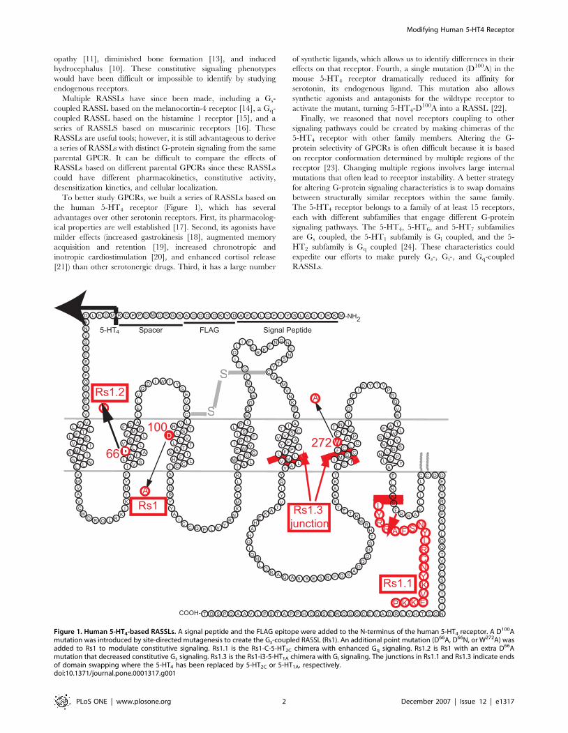
Figure 1. Human 5-HT4-based RASSLs. A signal peptide and the FLAG epitope were added to the N-terminus of the human 5-HT4 receptor. A D100Amutation was introduced by site-directed mutagenesis to create the Gs-coupled RASSL (Rs1). An additional point mutation (D66A, D66N, or W272A) wasadded to Rs1 to modulate constitutive signaling. Rs1.1 is the Rs1-C-5-HT2C chimera with enhanced Gq signaling. Rs1.2 is Rs1 with an extra D66Amutation that decreased constitutive Gs signaling. Rs1.3 is the Rs1-i3-5-HT1A chimera with Gi signaling. The junctions in Rs1.1 and Rs1.3 indicate endsof domain swapping where the 5-HT4 has been replaced by 5-HT2C or 5-HT1A, respectively.doi:10.1371/journal.pone.0001317.g001
Modifying Human 5-HT4 Receptor
PLoS ONE | www.plosone.org 2 December 2007 | Issue 12 | e1317
Here, we describe a new series of RASSLs developed to modify
the ligand-induced and constitutive signaling of the human 5-HT4
receptor. These modified GPCRs will help us better study the
effect of constitutive Gs signaling and ligand-induced Gs, Gs/Gq,
and Gi signaling in vivo.
RESULTS
Human 5-HT4 D100A is a Gs-coupled RASSLThe D100A mutation in the mouse 5-HT4 receptor converts it
into a RASSL [22], but its effects on the human 5-HT4 receptor
have not been tested. We now extend these findings to the
human 5-HT4 receptor (Figure 1). To determine if antagonists
for the wildtype 5-HT4 receptor also activate the human 5-HT4-
D100A mutant, we tested a variety of compounds. The mutant
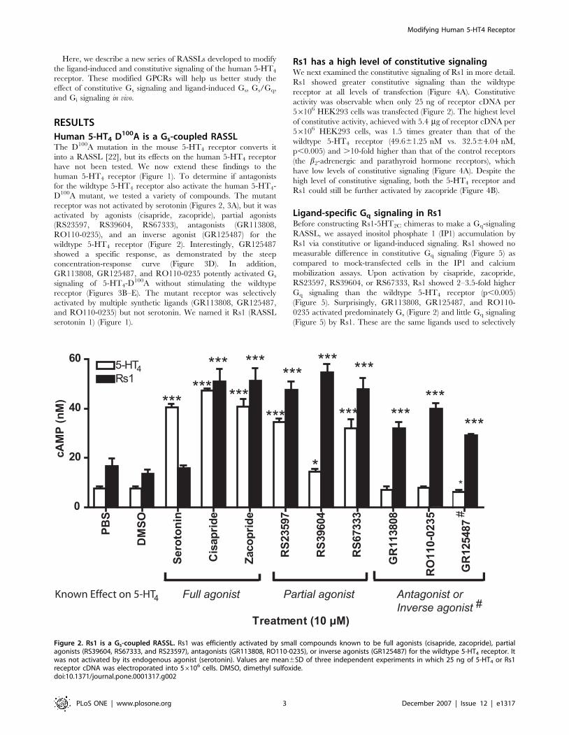
receptor was not activated by serotonin (Figures 2, 3A), but it was
activated by agonists (cisapride, zacopride), partial agonists
(RS23597, RS39604, RS67333), antagonists (GR113808,
RO110-0235), and an inverse agonist (GR125487) for the
wildtype 5-HT4 receptor (Figure 2). Interestingly, GR125487
showed a specific response, as demonstrated by the steep
concentration-response curve (Figure 3D). In addition,
GR113808, GR125487, and RO110-0235 potently activated Gs
signaling of 5-HT4-D100A without stimulating the wildtype
receptor (Figures 3B–E). The mutant receptor was selectively
activated by multiple synthetic ligands (GR113808, GR125487,
and RO110-0235) but not serotonin. We named it Rs1 (RASSL
serotonin 1) (Figure 1).
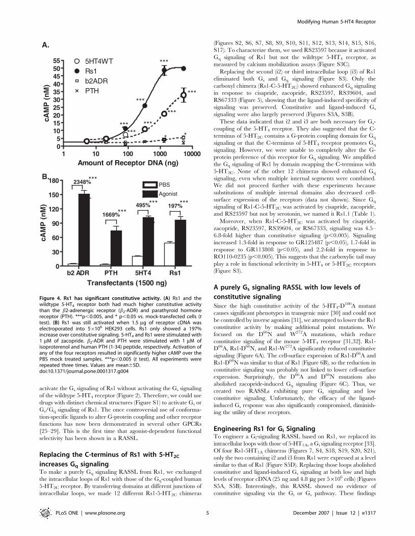
Rs1 has a high level of constitutive signalingWe next examined the constitutive signaling of Rs1 in more detail.
Rs1 showed greater constitutive signaling than the wildtype
receptor at all levels of transfection (Figure 4A). Constitutive
activity was observable when only 25 ng of receptor cDNA per
56106 HEK293 cells was transfected (Figure 2). The highest level
of constitutive activity, achieved with 5.4 mg of receptor cDNA per
56106 HEK293 cells, was 1.5 times greater than that of the
wildtype 5-HT4 receptor (49.661.25 nM vs. 32.564.04 nM,
p,0.005) and .10-fold higher than that of the control receptors
(the b2-adrenergic and parathyroid hormone receptors), which
have low levels of constitutive signaling (Figure 4A). Despite the
high level of constitutive signaling, both the 5-HT4 receptor and
Rs1 could still be further activated by zacopride (Figure 4B).
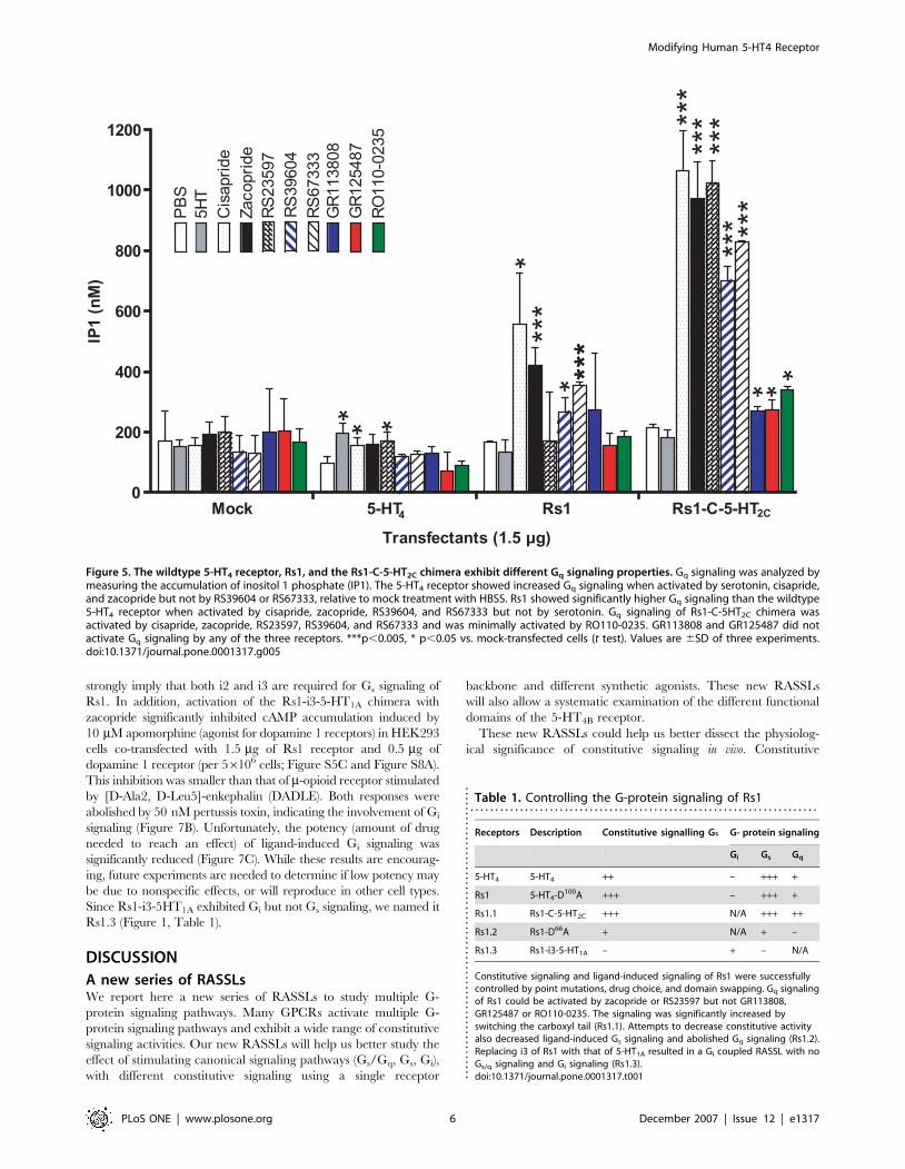
Ligand-specific Gq signaling in Rs1Before constructing Rs1-5HT2C chimeras to make a Gq-signaling
RASSL, we assayed inositol phosphate 1 (IP1) accumulation by
Rs1 via constitutive or ligand-induced signaling. Rs1 showed no
measurable difference in constitutive Gq signaling (Figure 5) as
compared to mock-transfected cells in the IP1 and calcium
mobilization assays. Upon activation by cisapride, zacopride,
RS23597, RS39604, or RS67333, Rs1 showed 2–3.5-fold higher
Gq signaling than the wildtype 5-HT4 receptor (p,0.005)
(Figure 5). Surprisingly, GR113808, GR125487, and RO110-
0235 activated predominately Gs (Figure 2) and little Gq signaling
(Figure 5) by Rs1. These are the same ligands used to selectively
Treatment (10 μM)
Full agonist Partial agonist Antagonist orInverse agonist
PBS
DM
SO
Sero
toni
n
Cis
aprid
e
Zac
oprid
e
RS2
3597
RS3
9604
RS6
7333
GR
1138
08
GR
1254
87
RO
110-
0235
0
20
40
60 5-HTRs1
cAM
P (n
M)
Known Effect on 5-HT
4
4
#
****** ***
***
**
***
*** ******
*** ***
******
***
#
Figure 2. Rs1 is a Gs-coupled RASSL. Rs1 was efficiently activated by small compounds known to be full agonists (cisapride, zacopride), partialagonists (RS39604, RS67333, and RS23597), antagonists (GR113808, RO110-0235), or inverse agonists (GR125487) for the wildtype 5-HT4 receptor. Itwas not activated by its endogenous agonist (serotonin). Values are mean6SD of three independent experiments in which 25 ng of 5-HT4 or Rs1receptor cDNA was electroporated into 56106 cells. DMSO, dimethyl sulfoxide.doi:10.1371/journal.pone.0001317.g002
Modifying Human 5-HT4 Receptor
PLoS ONE | www.plosone.org 3 December 2007 | Issue 12 | e1317
= Does not activate
B. A. 5-HTRs1
4
D.
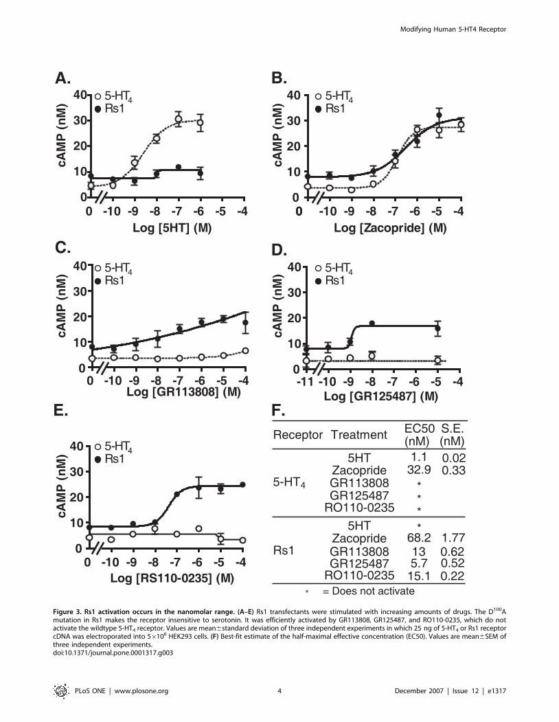
Receptor Treatment EC50 (nM)
S.E. (nM)
5HT 1.1 0.02Zacopride 32.9 0.33GR113808 *GR125487 *
RO110-0235 *5HT *
Zacopride 68.2 1.77GR113808GR125487 5.7 0.52
RO110-0235 15.1 0.22
5-HT
Rs1
F.
4
C.
13 0.62
*
-10 -9 -8 -7 -6 -5 -400
10
20
30
40
Log [5HT] (M)
cAM
P (n
M)
0 -10 -9 -8 -7 -6 -5 -400
10
20
30
40
Log [Zacopride] (M)
cAM
P (n
M) 5-HT
Rs14
-10 -9 -8 -7 -6 -5 -400
10
20
30
40
Log [RS110-0235] (M)
cAM
P (n
M) 5-HT
Rs14
-11 -10 -9 -8 -7 -6 -5 -40
10
20
30
40
Log [GR125487] (M)
cAM
P (n
M) 5-HT
Rs14
-10 -9 -8 -7 -6 -5 -400
10
20
30
40
Log [GR113808] (M)
cAM
P (n
M) 5-HT
Rs14
E.
Figure 3. Rs1 activation occurs in the nanomolar range. (A–E) Rs1 transfectants were stimulated with increasing amounts of drugs. The D100Amutation in Rs1 makes the receptor insensitive to serotonin. It was efficiently activated by GR113808, GR125487, and RO110-0235, which do notactivate the wildtype 5-HT4 receptor. Values are mean6standard deviation of three independent experiments in which 25 ng of 5-HT4 or Rs1 receptorcDNA was electroporated into 56106 HEK293 cells. (F) Best-fit estimate of the half-maximal effective concentration (EC50). Values are mean6SEM ofthree independent experiments.doi:10.1371/journal.pone.0001317.g003
Modifying Human 5-HT4 Receptor
PLoS ONE | www.plosone.org 4 December 2007 | Issue 12 | e1317
activate the Gs signaling of Rs1 without activating the Gs signaling
of the wildtype 5-HT4 receptor (Figure 2). Therefore, we could use
drugs with distinct chemical structures (Figure S1) to activate Gs or
Gs/Gq signaling of Rs1. The once controversial use of conforma-
tion-specific ligands to alter G-protein coupling and other receptor
functions has now been demonstrated in several other GPCRs
[25–29]. This is the first time that agonist-dependent functional
selectivity has been shown in a RASSL.
Replacing the C-terminus of Rs1 with 5-HT2C
increases Gq signalingTo make a purely Gq signaling RASSL from Rs1, we exchanged
the intracellular loops of Rs1 with those of the Gq-coupled human
5-HT2C receptor. By transferring domains at different junctions of
intracellular loops, we made 12 different Rs1-5-HT2C chimeras
(Figures S2, S6, S7, S8, S9, S10, S11, S12, S13, S14, S15, S16,
S17). To characterize them, we used RS23597 because it activated
Gq signaling of Rs1 but not the wildtype 5-HT4 receptor, as
measured by calcium mobilization assays (Figure S3C).
Replacing the second (i2) or third intracellular loop (i3) of Rs1
eliminated both Gs and Gq signaling (Figure S3). Only the
carboxyl chimera (Rs1-C-5-HT2C) showed enhanced Gq signaling
in response to cisapride, zacopride, RS23597, RS39604, and
RS67333 (Figure 5), showing that the ligand-induced specificity of
signaling was preserved. Constitutive and ligand-induced Gs
signaling were also largely preserved (Figures S3A, S3B).
These data indicated that i2 and i3 are both necessary for Gs-
coupling of the 5-HT4 receptor. They also suggested that the C-
terminus of 5-HT2C contains a G-protein coupling domain for Gq
signaling or that the C-terminus of 5-HT4 receptor promotes Gq
signaling. However, we were unable to completely alter the G-
protein preference of this receptor for Gq signaling. We amplified
the Gq signaling of Rs1 by domain swapping the C-terminus with
5-HT2C. None of the other 12 chimeras showed enhanced Gq
signaling, even when multiple internal segments were combined.
We did not proceed further with these experiments because
substitutions of multiple internal domains also decreased cell-
surface expression of the receptors (data not shown). Since Gq
signaling of Rs1-C-5-HT2C was activated by cisapride, zacopride,
and RS23597 but not by serotonin, we named it Rs1.1 (Table 1).
Moreover, when Rs1-C-5-HT2C was activated by cisapride,
zacopride, RS23597, RS39604, or RS67333, signaling was 4.5–
6.8-fold higher than constitutive signaling (p,0.005). Signaling
increased 1.3-fold in response to GR125487 (p,0.05), 1.7-fold in
response to GR113808 (p,0.05), and 2.2-fold in response to
RO110-0235 (p,0.005). This suggests that the carboxylic tail may
play a role in functional selectivity in 5-HT4 or 5-HT2C receptors
(Figure S3).
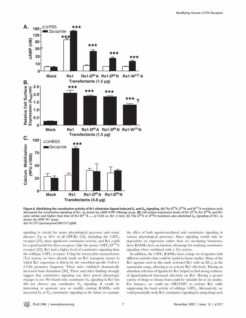
A purely Gs signaling RASSL with low levels of
constitutive signalingSince the high constitutive activity of the 5-HT4-D100A mutant
causes significant phenotypes in transgenic mice [30] and could not
be controlled by inverse agonists [31], we attempted to lower the Rs1
constitutive activity by making additional point mutations. We
focused on the D66N and W272A mutations, which reduce
constitutive signaling of the mouse 5-HT4 receptor [31,32]. Rs1-
D66A, Rs1-D66N, and Rs1-W272A significantly reduced constitutive
signaling (Figure 6A). The cell-surface expression of Rs1-D66A and
Rs1-D66N was similar to that of Rs1 (Figure 6B), so the reduction in
constitutive signaling was probably not linked to lower cell-surface
expression. Surprisingly, the D66A and D66N mutations also
abolished zacopride-induced Gq signaling (Figure 6C). Thus, we
created two RASSLs exhibiting pure Gs signaling and low
constitutive signaling. Unfortunately, the efficacy of the ligand-
induced Gs response was also significantly compromised, diminish-
ing the utility of these receptors.
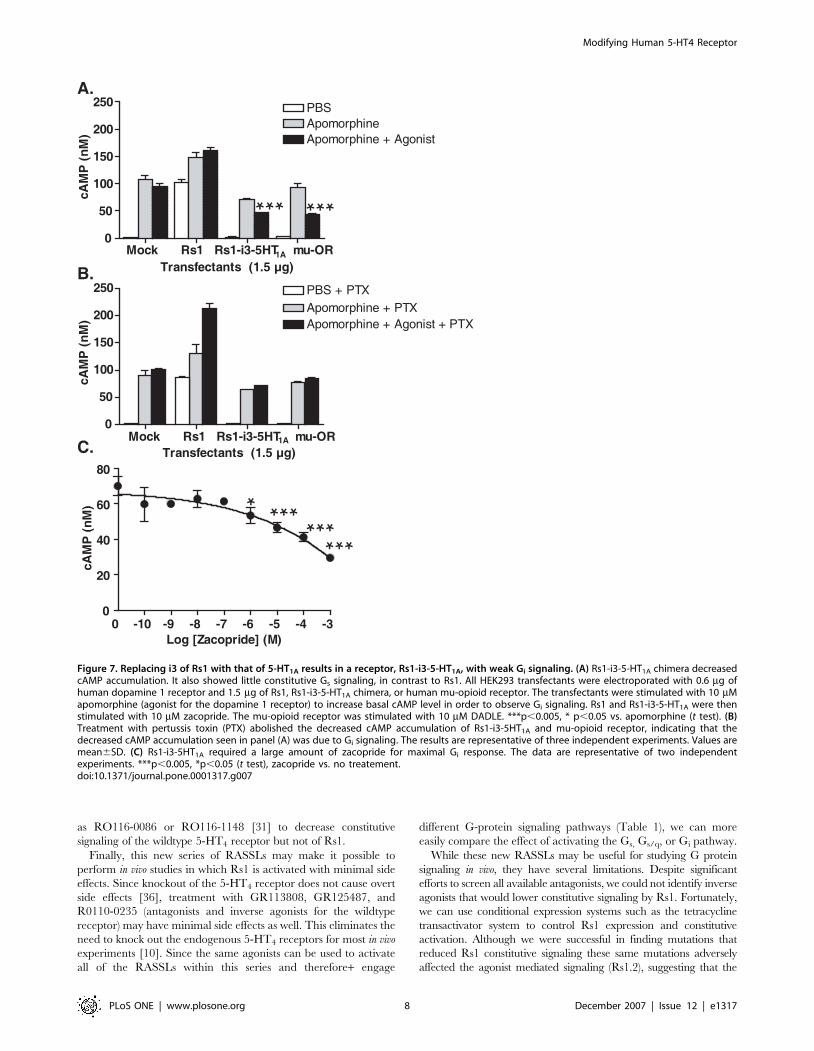
Engineering Rs1 for Gi SignalingTo engineer a Gi-signaling RASSL based on Rs1, we replaced its
intracellular loops with those of 5-HT1A, a Gi signaling receptor [33].
Of four Rs1-5HT1A chimeras (Figures 7, S4, S18, S19, S20, S21),
only the two containing i2 and i3 from Rs1 were expressed at a level
similar to that of Rs1 (Figure S5D). Replacing those loops abolished
constitutive and ligand-induced Gs signaling at both low and high
levels of receptor cDNA (25 ng and 4.8 mg per 56106 cells) (Figures
S5A, S5B). Interestingly, this RASSL showed no evidence of
constitutive signaling via the Gi or Gs pathway. These findings
A.
B.
197%
PBSAgonist
495%1669%
2348%
***
****** ***
b2 ADR PTH 5HT4 Rs10
30
60
90
120
150
180
Transfectants (1500 ng)
cAM
P (n
M)
1 10 100 1000 1000005
10152025303540455055 5HT4WT
Rs1b2ADRPTH
Amount of Receptor DNA (ng)
cAM
P (n
M)
***
***
***
***
******
*** **
Figure 4. Rs1 has significant constitutive activity. (A) Rs1 and thewildtype 5-HT4 receptor both had much higher constitutive activitythan the b2-adrenergic receptor (b2-ADR) and parathyroid hormonereceptor (PTH). ***p,0.005, and * p,0.05 vs. mock-transfected cells (ttest). (B) Rs1 was still activated when 1.5 mg of receptor cDNA waselectroporated into 56106 HEK293 cells. Rs1 only showed a 197%increase over constitutive signaling. 5-HT4 and Rs1 were stimulated with1 mM of zacopride. b2-ADR and PTH were stimulated with 1 mM ofisoproterenol and human PTH (1-34) peptide, respectively. Activation ofany of the four receptors resulted in significantly higher cAMP over thePBS mock treated samples. ***p,0.005 (t test). All experiments wererepeated three times. Values are mean6SD.doi:10.1371/journal.pone.0001317.g004
Modifying Human 5-HT4 Receptor
PLoS ONE | www.plosone.org 5 December 2007 | Issue 12 | e1317
strongly imply that both i2 and i3 are required for Gs signaling of
Rs1. In addition, activation of the Rs1-i3-5-HT1A chimera with
zacopride significantly inhibited cAMP accumulation induced by
10 mM apomorphine (agonist for dopamine 1 receptors) in HEK293
cells co-transfected with 1.5 mg of Rs1 receptor and 0.5 mg of
dopamine 1 receptor (per 56106 cells; Figure S5C and Figure S8A).
This inhibition was smaller than that of m-opioid receptor stimulated
by [D-Ala2, D-Leu5]-enkephalin (DADLE). Both responses were
abolished by 50 nM pertussis toxin, indicating the involvement of Gi
signaling (Figure 7B). Unfortunately, the potency (amount of drug
needed to reach an effect) of ligand-induced Gi signaling was
significantly reduced (Figure 7C). While these results are encourag-
ing, future experiments are needed to determine if low potency may
be due to nonspecific effects, or will reproduce in other cell types.
Since Rs1-i3-5HT1A exhibited Gi but not Gs signaling, we named it
Rs1.3 (Figure 1, Table 1).
DISCUSSION
A new series of RASSLsWe report here a new series of RASSLs to study multiple G-
protein signaling pathways. Many GPCRs activate multiple G-
protein signaling pathways and exhibit a wide range of constitutive
signaling activities. Our new RASSLs will help us better study the
effect of stimulating canonical signaling pathways (Gs/Gq, Gs, Gi),
with different constitutive signaling using a single receptor
backbone and different synthetic agonists. These new RASSLs
will also allow a systematic examination of the different functional
domains of the 5-HT4B receptor.
These new RASSLs could help us better dissect the physiolog-
ical significance of constitutive signaling in vivo. Constitutive
***
Mock 5-HT Rs1 Rs1-C-5-HT0
200
400
600
800
1000
1200
PBS
5HT
Cis
aprid
eZa
copr
ide
RS23
597
RS39
604
RS67
333
GR1
1380
8G
R125
487
RO11
0-02
35
Transfectants (1.5 μg)
IP1
(nM
)
2C4
** *
*
***
* ***
***
***
***
***
*** **
*** *
Figure 5. The wildtype 5-HT4 receptor, Rs1, and the Rs1-C-5-HT2C chimera exhibit different Gq signaling properties. Gq signaling was analyzed bymeasuring the accumulation of inositol 1 phosphate (IP1). The 5-HT4 receptor showed increased Gq signaling when activated by serotonin, cisapride,and zacopride but not by RS39604 or RS67333, relative to mock treatment with HBSS. Rs1 showed significantly higher Gq signaling than the wildtype5-HT4 receptor when activated by cisapride, zacopride, RS39604, and RS67333 but not by serotonin. Gq signaling of Rs1-C-5HT2C chimera wasactivated by cisapride, zacopride, RS23597, RS39604, and RS67333 and was minimally activated by RO110-0235. GR113808 and GR125487 did notactivate Gq signaling by any of the three receptors. ***p,0.005, * p,0.05 vs. mock-transfected cells (t test). Values are 6SD of three experiments.doi:10.1371/journal.pone.0001317.g005
Table 1. Controlling the G-protein signaling of Rs1. . . . . . . . . . . . . . . . . . . . . . . . . . . . . . . . . . . . . . . . . . . . . . . . . . . . . . . . . . . . . . . . . . . . . .
Receptors Description Constitutive signalling Gs G- protein signaling
Gi Gs Gq
5-HT4 5-HT4 ++ – +++ +
Rs1 5-HT4-D100A +++ – +++ +
Rs1.1 Rs1-C-5-HT2C +++ N/A +++ ++
Rs1.2 Rs1-D66A + N/A + –
Rs1.3 Rs1-i3-5-HT1A – + – N/A
Constitutive signaling and ligand-induced signaling of Rs1 were successfullycontrolled by point mutations, drug choice, and domain swapping. Gq signalingof Rs1 could be activated by zacopride or RS23597 but not GR113808,GR125487 or RO110-0235. The signaling was significantly increased byswitching the carboxyl tail (Rs1.1). Attempts to decrease constitutive activityalso decreased ligand-induced Gs signaling and abolished Gq signaling (Rs1.2).Replacing i3 of Rs1 with that of 5-HT1A resulted in a Gi coupled RASSL with noGs/q signaling and Gi signaling (Rs1.3).doi:10.1371/journal.pone.0001317.t001..
....
....
....
....
....
....
....
....
....
....
....
....
....
....
.
Modifying Human 5-HT4 Receptor
PLoS ONE | www.plosone.org 6 December 2007 | Issue 12 | e1317
signaling is crucial for many physiological processes and many
diseases. Up to 40% of all GPCRs [34], including the 5-HT4
receptor [35], show significant constitutive activity, and Rs1 could
be a good model for these receptors. Like the mouse 5-HT4-D100A
receptor [22], Rs1 had a higher level of constitutive signaling than
the wildtype 5-HT4 receptor. Using the tetracycline transactivator
(Tet) system, we have already made an Rs1 transgenic mouse in
which Rs1 expression is driven by the osteoblast-specific Col1a-1
2.3-kb promoter fragment. These mice exhibited dramatically
increased bone formation [30]. These and other findings strongly
suggest that constitutive signaling can drive potent phenotypic
changes in vivo. We found only constitutive Gs signaling in Rs1 but
did not observe any constitutive Gq signaling. It would be
interesting to generate new or modify existing RASSLs with
increased Gi or Gq constitutive signaling in the future to examine
the effect of both agonist-mediated and constitutive signaling in
various physiological processes. Since signaling would only be
dependent on expression rather than on circulating hormones,
these RASSLs have an intrinsic advantage for studying constitutive
signaling when combined with a Tet system.
In addition, the 5-HT4 RASSLs have a large set of agonists with
different activities that could be useful in future studies. Many of the
Rs1 agonists used in this study activated Rs1 with an EC50 in the
nanomolar range, allowing to us activate Rs1 effectively. Having an
abundant selection of ligands for Rs1 helped us find strong evidence
of ligand-induced functional selectivity on Rs1. Having a greater
variety of drugs to choose from could be valuable for in vivo studies.
For instance, we could use GR125487 to activate Rs1 while
suppressing the basal activity of wildtype 5-HT4. Alternatively, we
could potentially study Rs1 constitutive signaling by using drugs such
C.
A.
B.
Mock Rs1 Rs1-D A Rs1-D N0
25
50
75
100 HBSSZacopride
Transfectants (4.8 µg)
Cal
cium
Mob
iliza
tion
(RFU
x10
00)
66 66
*
Mock Rs1 Rs1-D A Rs1-D N Rs1-W A0
0.5
1.0
1.5
2.0
2.5
Transfectants (1.5 µg)
Rel
ativ
e C
ell S
urfa
ceEx
pres
sion
(A
nm
)
66 66 272
***PBSZacopride
0
10
20
30
80130180
cAM
P (n
M)
Mock Rs1 Rs1-D A Rs1-D N Rs1-W A66 66 272
Transfectants (1.5 µg)
405
***
***
*** ***
*** *** ******
***
?
Figure 6. Abolishing the constitutive activity of Rs1 eliminates ligand-induced Gs and Gq signaling. (A) The D66A, D66N, and W272A mutations eachdecreased the constitutive signaling of Rs1, as shown by cAMP HTRF HiRange assay. (B) Cell-surface expression levels of Rs1-D66A, Rs1-D66N, and Rs1were similar and higher than that of Rs1-W272A. *** p,0.05 vs. Rs1 (t test). (C) The D66A or D66N mutations also abolished Gq signaling of Rs1, asshown by HTRF IP1 assay.doi:10.1371/journal.pone.0001317.g006
Modifying Human 5-HT4 Receptor
PLoS ONE | www.plosone.org 7 December 2007 | Issue 12 | e1317
as RO116-0086 or RO116-1148 [31] to decrease constitutive
signaling of the wildtype 5-HT4 receptor but not of Rs1.
Finally, this new series of RASSLs may make it possible to
perform in vivo studies in which Rs1 is activated with minimal side
effects. Since knockout of the 5-HT4 receptor does not cause overt
side effects [36], treatment with GR113808, GR125487, and
R0110-0235 (antagonists and inverse agonists for the wildtype
receptor) may have minimal side effects as well. This eliminates the
need to knock out the endogenous 5-HT4 receptors for most in vivo
experiments [10]. Since the same agonists can be used to activate
all of the RASSLs within this series and therefore+ engage
different G-protein signaling pathways (Table 1), we can more
easily compare the effect of activating the Gs, Gs/q, or Gi pathway.
While these new RASSLs may be useful for studying G protein
signaling in vivo, they have several limitations. Despite significant
efforts to screen all available antagonists, we could not identify inverse
agonists that would lower constitutive signaling by Rs1. Fortunately,
we can use conditional expression systems such as the tetracycline
transactivator system to control Rs1 expression and constitutive
activation. Although we were successful in finding mutations that
reduced Rs1 constitutive signaling these same mutations adversely
affected the agonist mediated signaling (Rs1.2), suggesting that the
C.
A.
B.Mock Rs1 Rs1-i3-5HT mu-OR
0
50
100
150
200
250 PBSApomorphineApomorphine + Agonist
Transfectants (1.5 µg)
cAM
P (n
M)
0
50
100
150
200
250Apomorphine + PTXApomorphine + Agonist + PTX
PBS + PTX
cAM
P (n
M)
1A
Mock Rs1 Rs1-i3-5HT mu-ORTransfectants (1.5 µg)
1A
*** ***
-10 -9 -8 -7 -6 -5 -4 -300
20
40
60
80
Log [Zacopride] (M)
cAM
P (n
M)
**********
Figure 7. Replacing i3 of Rs1 with that of 5-HT1A results in a receptor, Rs1-i3-5-HT1A, with weak Gi signaling. (A) Rs1-i3-5-HT1A chimera decreasedcAMP accumulation. It also showed little constitutive Gs signaling, in contrast to Rs1. All HEK293 transfectants were electroporated with 0.6 mg ofhuman dopamine 1 receptor and 1.5 mg of Rs1, Rs1-i3-5-HT1A chimera, or human mu-opioid receptor. The transfectants were stimulated with 10 mMapomorphine (agonist for the dopamine 1 receptor) to increase basal cAMP level in order to observe Gi signaling. Rs1 and Rs1-i3-5-HT1A were thenstimulated with 10 mM zacopride. The mu-opioid receptor was stimulated with 10 mM DADLE. ***p,0.005, * p,0.05 vs. apomorphine (t test). (B)Treatment with pertussis toxin (PTX) abolished the decreased cAMP accumulation of Rs1-i3-5HT1A and mu-opioid receptor, indicating that thedecreased cAMP accumulation seen in panel (A) was due to Gi signaling. The results are representative of three independent experiments. Values aremean6SD. (C) Rs1-i3-5HT1A required a large amount of zacopride for maximal Gi response. The data are representative of two independentexperiments. ***p,0.005, *p,0.05 (t test), zacopride vs. no treatement.doi:10.1371/journal.pone.0001317.g007
Modifying Human 5-HT4 Receptor
PLoS ONE | www.plosone.org 8 December 2007 | Issue 12 | e1317
D66N and W272A mutations are critical for all aspects of Gs signaling.
Other mutations could be explored that may provide a more optimal
reduction in Gs basal activity without affecting ligand-induced
receptor activation. In addition, the relatively small Gi signaling of
Rs1.3 can only be activated by a relatively high concentration of
zacopride, thus limiting its usefulness in vivo.
Insights into G-protein signaling by Rs1Our study yielded insights into the G-protein selectivity and
functional selectivity (differential effects of ligands on the same
receptor) of Rs1. Of the 12 Rs1-5-HT2C chimeras that are
expressed on the cell surface, none of the i2 or i3 chimeras showed
any Gs or Gq signaling. Evidently, these intracellular loops of Rs1
are crucial for signaling via those pathways. The importance of i2
and i3 for Gs signaling of Rs1 is further supported by the lack of Gs
signaling by the Rs1-i2-5-HT1A and Rs1-i3-5-HT1A chimeras.
This is the first study showing the importance of i2 and i3 in both
Gs and Gq signaling of the human 5-HT4-D100A receptor.
We also found that i3 domain swapping abolished all Gs
signaling and enabled Rs1 to stimulate Gi signaling of 5-HT1A.
The role of i2 and i3 in the Gi signaling of 5-HT1A receptor has
been extensively reported. The entire N-terminus [37] and C-
terminus of i2 [38] of 5-HT1A are thought to be sufficient to
support G-protein coupling, but not signaling. On the other hand,
the N-terminus [39] and C-terminus of i3 of 5-HT1A [40,41] seem
to be essential for the Gi signaling of 5-HT1A. In fact, replacing the
N-terminus of the i3 of the a2-adrenergic receptor with that of 5-
HT1A resulted in a chimera that signals like a 5-HT1A receptor
when stimulated by a a2-adrenergic receptor agonist [42]. Since
Rs1-5HT1A chimeras with multiple internal domains replaced are
not significantly expressed on the cell surface (data not shown), it
may be difficult to further improve the potency of the Rs1.3 using
our current approach. We hypothesize that replacing the N- and
C-terminal portions of i2 and i3 instead of the whole i2 and i3
loops may increase the potency of Rs1.
We also showed that various drugs can differentially activate G-
protein signaling of Rs1. Functional selectivity has been reported
for many receptors. It led to divergent fates of internalization for
the dopamine D1 receptor [25], various binding specificities for
gonadotropin-releasing hormone receptors, and different levels of
activation of G proteins for the b2-adrenergic [26], mu-opioid
[27], dopamine D2 [28], and human 5-HT2A [29] receptors.
The indoleamine derivatives GR113808, GR125487, and
RO110-0235 did not fully activate Gq signaling of Rs1, Rs1.1,
or the 5-HT4 receptor. On the other hand, the benzamide
derivatives cisapride, zacopride, RS23597, RS39604, and
RS67333 activated the Gq signaling of Rs1. These findings may
reflect distinct conformational changes caused by indoleamine and
benzamide derivatives.
The possibility of functional selectivity is further supported by
the results obtained with Rs1-C-5HT2c and Rs1 point mutants
(Rs1-D66A and Rs1-D66N). The D100A mutation and replacement
of the C-terminus amplified Gq signaling by the 5-HT4 receptor.
The addition of D66A and D66N abolished Gq signaling. Since
D100A is located in the binding pocket of the 5-HT4 receptor, this
mutation in Rs1 may have changed the configuration of the
binding pocket, making the receptor more susceptible to Gq
activation by zacopride and RS23597. This response was even
more pronounced when the D100A mutation was combined with
domain swapping of the C-terminus with that of 5-HT2C. Thus, it
is reasonable to hypothesize that these changes modified the
ligand-selective receptor conformation [5], changing the receptor
susceptibility to functional selectivity.
ConclusionsOur studies with Rs1 provide a proof-of-concept for making a
series of RASSLs with different signaling properties. Recently,
Armbruster et al. made a series of RASSLs based on the
muscarinic M3 and M4 receptors, which have low constitutive
activity. These RASSLs each couple different G-protein signaling
pathways and can be activated by clozapine-N-oxide, an inert
ligand with high bioavailability [16]. These RASSLs nicely
complement our Rs1 RASSLs with varying constitutive activity.
In addition, we predict that some RASSLs with the same
canonical G-protein signaling (Gs, Gi, or Gq) will have different
in vivo phenotypes due to noncanonical signaling. This growing
collection of RASSLs will greatly facilitate our efforts to
understand the physiological significance of the inherent signaling
diversity of GPCRs.
An ideal series of RASSLs would have receptors with different
combinations of low and high basal signaling, with robust ligand-
induced effects for each major pathway, and potent inverse
agonists. Although we have not achieved this goal with the Rs1
series, we are hopeful that it can be achieved with other receptors
in the future. Indeed, RASSLs based on the muscarinic receptors
[16] show great promise, as there are naturally occurring, or
published mutants of the muscarinic reports that activate each of
the major G protein signaling pathways.
MATERIALS AND METHODS
Constructing human 5-HT4 mutant cDNA and Rs1-5-
HT1A, and Rs1-5-HT2C chimerasThe human 5-HT4 receptor cDNA (a gift from Dr. Bryan Roth,
University of North Carolina) was used in all experiments. To
improve expression and allow detection of the receptor, we added
a signal peptide from influenza hemagglutinin [43] and a FLAG
epitope (DYKDDDDA) at the N-terminus. 5-HT4 was then
subcloned by PCR; the primers, ATCGATCGgcggccgcGTGAG-
CAAGGGCGAGGAGCTGTTC and ATCGATCG gcggccgc-
CTAAGTGTCACTGGGCTGAGCAGCC, were inserted into
the Not1 restriction site (gcggccgc) of the pUNIV-5-HT2C-INI
plasmid to replace the 5-HT2C-INI (a gift from Dr. Bryan Roth) in
frame with the signal peptide and the FLAG epitope. The receptor
was then mutated (D100A) with a Quick-Change site-directed
mutagenesis kit (Stratagene, La Jolla, CA) with primer GTCTT-
GTTCGGACATCTCTGgccGT CCTGCTCACAACGGCAT-
CG (Figures 1, S2). The following mutant sense primers were used:
5-HT4-D66A, TTCATTGTATCTCTTGCTTTTGCGgcaCT-
GCT GGTTTCGGTGCTGGT-GATG; 5-HT4-D66N, TTCA-
TTGTATCTCTTGCTTTTGCGaacC TGCTGGTTTCGGT-
GCT-GGTGATG; and 5-HT4-W272A, GTTGCTTCTGCC-
TCTGCT GGGCGCCAgccTTTGTCA-CCAATATTGTGG.
The sense primer used to replace the carboxyl chimera for
Rs1.1 (Table 1) was AGTTACTCTTCCgcgGCCGCGAATT-
CAG TGGATCCACTAGTAAC. The Rs1-5-HT1A and Rs1-5-
HT2C chimeras were made by PCR fusion. The mutations are
indicated by boldface, lower-case letters.
HEK293 maintenance and electroporationEarly-passage (#20) HEK293 cells were maintained in high-
glucose DMEM (Invitrogen, Carlsbad, CA) supplemented with
sodium pyruvate (Invitrogen) and 10% Fetalplex (Gemini Bio-
Products, West Sacramento, CA). Receptors were electroporated
into HEK293 cells as described [44]. The electroporated cells
were reconstituted into a suspension using DMEM with 10% heat-
inactivated, dialyzed fetal bovine serum (Thermo-Fisher Scientific,
Modifying Human 5-HT4 Receptor
PLoS ONE | www.plosone.org 9 December 2007 | Issue 12 | e1317
Logan, UT). The transfection efficiency was monitored by flow
cytometry, and the cell-surface expression of the receptor was
determined by FLAG ELISA (enzyme-linked immunosorbent
assay) the next day.
Drugs5-HT, isoproterenol, and 3-isobutyl-1-methylxanthine (IBMX)
were purchased from Sigma-Aldrich (St. Louis, MO). Cisapride,
zacopride, GR113808, GR1254875, RS23597-190 HCl,
RS39604 HCl, and RS67333 HCl were from Tocris (Bristol,
UK). Human parathyroid hormone peptide (amino acids 1–34)
was from Bachem Biosciences (King of Prussia, PA). RO110-
0235 was generously donated by Renee Martin (Roche, Palo Alto,
CA).
Measuring cell-surface expression by FLAG ELISACell-surface receptor expression was measured with a FLAG
ELISA as described [45]. Cells seeded in poly-D-lysine-coated 96-
well plates were fixed with 100 ml of 4% paraformaldehyde for
10 min at room temperature, washed, and stained with 100 ml of
staining buffer (DMEM, 10% FBS, and 1 mM CaCl2) containing
anti-FLAG M1 antibody (1:1000; Sigma-Aldrich) for 1 h at 25uC.
The samples were washed three times with wash buffer (PBS and
1 mM CaCl2) and stained with 100 ml of staining buffer with rat
anti-mouse IgG antibody conjugated with horseradish peroxidase
(1:1000; Bio-Rad Laboratories, Hercules, CA). After 30 min, the
samples were washed with wash buffer, placed on a rocker for
10 min, and washed again. This process was repeated two more
times. Then, 2,2-azino-bis(3-ethylbenzthiazoline-6-sulfonic acid)
liquid substrate (200 ml; Sigma-Aldrich) was added to the samples.
After rocking for 30 min, 200 ml of the substrate was transferred to
new 96-well plates, and optical density was measured with Victor 3
(PerkinElmer, Waltham, MA) at 405 nm. All samples contain
three replicates, and all experiments were repeated at least three
times.
Measurement of cAMP production in intact cellsTo improve assay consistency and minimize pipetting error in the
384-well plates, we modified the high-range HTRF assay (CisBio
International, Bagnols-sur-Ceze, France) by seeding, stimulating,
and lysing the cells in 96-well plates and using the lysate instead
of live cells to determine cAMP production. The remainder of
the analysis was performed according to the manufacturer’s
instructions
Gi AssayGi signaling was examined in cells transfected with 1.5 mg of
receptor cDNA, 0.6 mg of human dopamine 1 receptor cDNA,
and pcDNA3 (up to 6 mg). The co-transfectants were stimulated
first with 100 ml of KRBG buffer containing IBMX for 10 min at
room temperature and then with 50 ml of PBS containing 10 mM
apomorphine (agonist for the dopamine 1 receptor) and 10 mM
zacopride for 10 min at 37uC. The cells were lysed in 50 ml of lysis
buffer, and 5 ml of lysate was used in the HiRange HTRF assay.
Fluorometric imaging plate reader assay to measure
calcium mobilizationTo measure calcium mobilization, 4.8 mg of receptor cDNA, 0.6 mg
of DsRed plasmid, and 0.6 mg of human bombesin receptor cDNA
were electroporated into 56106 HEK293 cells as above [44]. Hank’s
balanced salt solution (10 ml) with 20 mM HEPES, 0.25 mM
probenecid acid (Sigma-Aldrich), and 2% pluronic acid (Sigma-
Aldrich) was added to each bottle of Calcium 4 (Molecular Devices,
Sunnyvale, CA), and 100 ml of the resulting solution was added to
each well for 1 h at 37uC before measurement. Assays were
performed with a FLEX Station (Molecular Devices), with excitation
of 485 nm, emission of 525 nm, and cut-off of 515 nm, as
recommended by the manufacturer.
Determination of IP1 production in intact cellsA modified version of the IP1 protocol was used (CisBio
International). HEK293 cells were washed once with calcium-free
PBS and dissociated from flasks with cell dissociation buffer
(Invitrogen). Cells (56106) were electroporated as described above.
Then, 105 cells were placed in DMEM supplemented with 10%
decomplemented, dialyzed against FBS, and seeded onto 96-well
plates coated with poly-D-lysine. The next day, the cells were
stimulated with agonists in 50 ml of 16 stimulation buffer for
30 min at 37uC and lysed for 10 min with 9 ml of lysis/detection
buffer. Then, 14 ml of lysate was added to 384-well plates and
subjected to High-Range HTRF assay as described above, except
that 3 ml of cAMP-d2 and anti-cAMP-cryptate solution were
added to each well.
Data analysiscAMP and IP1 values were analyzed with GraphPad Prism 4
(GraphPad Software, San Diego, CA). Calcium mobilization
results were analyzed with SoftMax Pro v5 (Molecular Devices).
Statistical significance was determined with paired Student’s t tests.
SUPPORTING INFORMATION
Figure S1 Chemical structures of the compounds used in the
study. All the chemicals used in the experimental are shown.
Found at: doi:10.1371/journal.pone.0001317.s001 (1.92 MB
EPS)
Figure S2 Rs1-5-HT2C chimeras. All modifications made to
Rs1 (Figure 1). Red lines indicate the junctions of chimeras. The
amino acids exchanged are shown by the amino acid alignment.
Found at: doi:10.1371/journal.pone.0001317.s002 (12.17 MB
EPS)
Figure S3 The second and third intracellular loops (i2 and i3) of
Rs1 are crucial for Gs and Gq signaling. (A, B) Rs1-5-HT2C
chimeras with swaps of i2 and i3 could no longer process Gs
signals, at either 25 ng or 4.8 mg of receptor cDNA per 56106
HEK293 cells. (C) Gq signaling of Rs1 was abolished when i2 and
i3 of Rs1 were replaced with those of 5-HT2C. The Gq signaling
was measured by calcium mobilization assay. (D) Only chimeras
with a single domain swap were expressed on the cell surface. The
results represent three independent experiments. All figures were
representative of three independent experiments.
Found at: doi:10.1371/journal.pone.0001317.s003 (2.03 MB EPS)
Figure S4 Rs1-5-HT1A chimeras. All modifications were made
on Rs1 (Figure 1). Red lines indicate the junctions of chimeras.
The amino acids exchanged are shown by the amino acid
alignment.
Found at: doi:10.1371/journal.pone.0001317.s004 (3.82 MB EPS)
Figure S5 Replacing the second or third intracellular loop (i2 or
i3) of Rs1 with 5-HT1A alters G protein signaling. (A, B) Rs1-5-
HT1A chimeras with swaps of the second (i2) and third
intracellular (i3) loops no longer signal via the Gs pathway,
regardless of whether cells were transfected with 25 ng or 4.8 mg of
receptor cDNA per 56106 HEK293 cells. This suggests that the
second and third intracellular loops are crucial for acute Gs
Modifying Human 5-HT4 Receptor
PLoS ONE | www.plosone.org 10 December 2007 | Issue 12 | e1317
signaling of Rs1. ***p,0.001 vs. mock transfected (t test). (C)
Replacing i3 of Rs1 resulted in a Gi signaling receptor. All
HEK293 transfectants were electroporated with 0.6 mg of the
human dopamine 1 receptor and 1.5 mg of Rs1, Rs1-5HT1A
chimeras, or the mu-opioid receptor. Rs1 and Rs1-5HT1a
chimeras were treated with 10 mM apomorphine (an agonist for
dopamine 1 receptor) or with 10 mM apomorphine and 10 mM
zacopride. Transfectants with 0.6 mg of the human dopamine 1
receptor and 1.5 mg of the mu-opioid receptor served as positive
controls. 10 mM DADLE was used in place of zacopride to
stimulate mu-opioid receptors. ***p,0.005, *p,0.05 vs. apomor-
phine (t test). (D) Rs1, (i1, i2 and i30 were expressed on the cell
surface. Cell-surface expression and calcium mobilization of the
chimeras were examined at 4.8 mg of receptor DNA per 56106
HEK293 cells. The results are representative of three independent
experiments. Values are mean6SD. ***p,0.001 vs. mock
transfected (t test).
Found at: doi:10.1371/journal.pone.0001317.s005 (1.67 MB EPS)
Figure S6 High-resolution representation of Rs1-5HT1A and
Rs1-5HT2C chimeras. All modifications made to Rs1 (Figure 1).
Red lines indicate the junctions of chimeras. The amino acids
exchanged are shown by the amino acid alignment.
Found at: doi:10.1371/journal.pone.0001317.s006 (2.98 MB EPS)
Figure S7 High-resolution representation of Rs1-5HT1A and
Rs1-5HT2C chimeras. All modifications made to Rs1 (Figure 1).
Red lines indicate the junctions of chimeras. The amino acids
exchanged are shown by the amino acid alignment.
Found at: doi:10.1371/journal.pone.0001317.s007 (2.96 MB EPS)
Figure S8 High-resolution representation of Rs1-5HT1A and
Rs1-5HT2C chimeras. All modifications made to Rs1 (Figure 1).
Red lines indicate the junctions of chimeras. The amino acids
exchanged are shown by the amino acid alignment.
Found at: doi:10.1371/journal.pone.0001317.s008 (3.00 MB EPS)
Figure S9 High-resolution representation of Rs1-5HT1A and
Rs1-5HT2C chimeras. All modifications made to Rs1 (Figure 1).
Red lines indicate the junctions of chimeras. The amino acids
exchanged are shown by the amino acid alignment.
Found at: doi:10.1371/journal.pone.0001317.s009 (2.99 MB EPS)
Figure S10 High-resolution representation of Rs1-5HT1A and
Rs1-5HT2C chimeras. All modifications made to Rs1 (Figure 1).
Red lines indicate the junctions of chimeras. The amino acids
exchanged are shown by the amino acid alignment.
Found at: doi:10.1371/journal.pone.0001317.s010 (2.99 MB EPS)
Figure S11 High-resolution representation of Rs1-5HT1A and
Rs1-5HT2C chimeras. All modifications made to Rs1 (Figure 1).
Red lines indicate the junctions of chimeras. The amino acids
exchanged are shown by the amino acid alignment.
Found at: doi:10.1371/journal.pone.0001317.s011 (2.99 MB EPS)
Figure S12 High-resolution representation of Rs1-5HT1A and
Rs1-5HT2C chimeras. All modifications made to Rs1 (Figure 1).
Red lines indicate the junctions of chimeras. The amino acids
exchanged are shown by the amino acid alignment.
Found at: doi:10.1371/journal.pone.0001317.s012 (3.00 MB EPS)
Figure S13 High-resolution representation of Rs1-5HT1A and
Rs1-5HT2C chimeras. All modifications made to Rs1 (Figure 1).
Red lines indicate the junctions of chimeras. The amino acids
exchanged are shown by the amino acid alignment.
Found at: doi:10.1371/journal.pone.0001317.s013 (2.99 MB EPS)
Figure S14 High-resolution representation of Rs1-5HT1A and
Rs1-5HT2C chimeras. All modifications made to Rs1 (Figure 1).
Red lines indicate the junctions of chimeras. The amino acids
exchanged are shown by the amino acid alignment.
Found at: doi:10.1371/journal.pone.0001317.s014 (3.00 MB
DOC)
Figure S15 High-resolution representation of Rs1-5HT1A and
Rs1-5HT2C chimeras. All modifications made to Rs1 (Figure 1).
Red lines indicate the junctions of chimeras. The amino acids
exchanged are shown by the amino acid alignment.
Found at: doi:10.1371/journal.pone.0001317.s015 (2.99 MB EPS)
Figure S16 High-resolution representation of Rs1-5HT1A and
Rs1-5HT2C chimeras. All modifications made to Rs1 (Figure 1).
Red lines indicate the junctions of chimeras. The amino acids
exchanged are shown by the amino acid alignment.
Found at: doi:10.1371/journal.pone.0001317.s016 (3.00 MB EPS)
Figure S17 High-resolution representation of Rs1-5HT1A and
Rs1-5HT2C chimeras. All modifications made to Rs1 (Figure 1).
Red lines indicate the junctions of chimeras. The amino acids
exchanged are shown by the amino acid alignment.
Found at: doi:10.1371/journal.pone.0001317.s017 (2.96 MB EPS)
Figure S18 High-resolution representation of Rs1-5HT1A and
Rs1-5HT2C chimeras. All modifications made to Rs1 (Figure 1).
Red lines indicate the junctions of chimeras. The amino acids
exchanged are shown by the amino acid alignment.
Found at: doi:10.1371/journal.pone.0001317.s018 (3.00 MB EPS)
Figure S19 High-resolution representation of Rs1-5HT1A and
Rs1-5HT2C chimeras. All modifications made to Rs1 (Figure 1).
Red lines indicate the junctions of chimeras. The amino acids
exchanged are shown by the amino acid alignment.
Found at: doi:10.1371/journal.pone.0001317.s019 (2.96 MB
DOC)
Figure S20 High-resolution representation of Rs1-5HT1A and
Rs1-5HT2C chimeras. All modifications made to Rs1 (Figure 1).
Red lines indicate the junctions of chimeras. The amino acids
exchanged are shown by the amino acid alignment.
Found at: doi:10.1371/journal.pone.0001317.s020 (3.40 MB EPS)
Figure S21 High-resolution representation of Rs1-5HT1A and
Rs1-5HT2C chimeras. All modifications made to Rs1 (Figure 1).
Red lines indicate the junctions of chimeras. The amino acids
exchanged are shown by the amino acid alignment.
Found at: doi:10.1371/journal.pone.0001317.s021 (3.03 MB EPS)
ACKNOWLEDGMENTSWe thank Kimberly Scearce-Levie and Renee Martin for insightful review
of the manuscript, Bryan Roth for the human 5-HT4B and 5-HT2C
receptors cDNA, Robert J. Lefkowitz for the human 5-HT1A receptor
cDNA, Mark von Zastrow for the human b2-adrenergic receptor cDNA,
human m-opioid receptor cDNA, and DADLE, Renee Martin for RO110-
0235 (Roche Pharmaceuticals Division, Palo Alto, CA), Mary Weglarz for
manuscript preparation and Gary Howard and Stephen Ordway for
editorial review.
Author Contributions
Conceived and designed the experiments: BC WC JN TN LP SC EH.
Performed the experiments: WC. Analyzed the data: BC WC JN TN LP
SC EH. Contributed reagents/materials/analysis tools: BC. Wrote the
paper: WC.
Modifying Human 5-HT4 Receptor
PLoS ONE | www.plosone.org 11 December 2007 | Issue 12 | e1317
REFERENCES1. Karchin R, Karplus K, Haussler D (2002) Classifying G-protein coupled
receptors with support vector machines. Bioinformatics 18: 147–159.2. Fredriksson R, Lagerstrom MC, Lundin LG, Schioth HB (2003) The G-protein-
coupled receptors in the human genome form five main families. Phylogenetic
analysis, paralogon groups, and fingerprints. Mol Pharmacol 63: 1256–1272.3. Brink CB, Harvey BH, Bodenstein J, Venter DP, Oliver DW (2004) Recent
advances in drug action and therapeutics: relevance of novel concepts in G-protein-coupled receptor and signal transduction pharmacology. Br J Clin
Pharmacol 57: 373–387.4. Maudsley S, Martin B, Luttrell LM (2005) The origins of diversity and specificity
in g protein-coupled receptor signaling. J Pharmacol Exp Ther 314: 485–494.
5. Kenakin T (2003) Ligand-selective receptor conformations revisited: the promiseand the problem. Trends Pharmacol Sci 24: 346–354.
6. Scearce-Levie K, Coward P, Redfern CH, Conklin BR (2001) Engineeringreceptors activated solely by synthetic ligands (RASSLs). Trends Pharmacol Sci
22: 414–420.
7. Srinivasan S, Santiago P, Lubrano C, Vaisse C, Conklin BR. Engineering themelancortin-4 receptor to control constitutive and ligand-mediated Gs signaling
in vivo. PLoS One. In press.8. Coward P, Wada HG, Falk MS, Chan SD, Meng F, et al. (1998) Controlling
signaling with a specifically designed Gi-coupled receptor. Proc Natl Acad SciUSA 95: 352–357.
9. Zhao GQ, Zhang Y, Hoon MA, Chandrashekar J, Erlenbach I, et al. (2003) The
receptors for mammalian sweet and umami taste. Cell 115: 255–266.10. Sweger EJ, Casper KB, Scearce-Levie K, Conklin BR, McCarthy KD (2007)
Development of hydrocephalus in mice expressing the Gi-coupled GPCR Ro1RASSL receptor in astrocytes. J Neurosci 27: 2309–2317.
11. Redfern CH, Coward P, Degtyarev MY, Lee EK, Kwa AT, et al. (1999)
Conditional expression and signaling of a specifically designed Gi-coupledreceptor in transgenic mice. Nat Biotechnol 17: 165–169.
12. Pauwels PJ (2003) Unravelling multiple ligand-activation binding sites usingRASSL receptors. Trends Pharm Sci 24: 504–507.
13. Peng J, Bencsik M, Lu W, Storer A, Burghardt A, et al. (2006) Osteopeniaproduced by expression of a Gi-activating receptor in osteoblasts. J Bone Miner
Res 21: s1–s530.
14. Srinivasan S, Vaisse C, Conklin BR (2003) Engineering the melanocortin-4receptor to control G(s) signaling in vivo. Ann N Y Acad Sci 994: 225–232.
15. Bruysters M, Jongejan A, Akdemir A, Bakker RA, Leurs R (2005) A G(q/11)-coupled mutant histamine H(1) receptor F435A activated solely by synthetic
ligands (RASSL). J Biol Chem 280: 34741–34746.
16. Armbruster BN, Li X, Pausch MH, Herlitze S, Roth BL (2007) Evolving the lockto fit the key to create a family of GPCRs potentially activated by an inert ligand.
Proc Natl Acad Sci USA..17. Claeysen S, Sebben M, Becamel C, Eglen RM, Clark RD, et al. (2000)
Pharmacological properties of 5-Hydroxytryptamine(4) receptor antagonists on
constitutively active wild-type and mutated receptors. Mol Pharmacol 58:136–144.
18. Crowell MD, Mathis C, Schettler VA, Yunus T, Lacy BE (2005) The effects oftegaserod, a 5-HT receptor agonist, on gastric emptying in a murine model of
diabetes mellitus. Neurogastroenterol Motil 17: 738–743.19. Orsetti M, Dellarole A, Ferri S, Ghi P (2003) Acquisition, retention, and recall of
memory after injection of RS67333, a 5-HT4 receptor agonist, into the nucleus
basalis magnocellularis of the rat. Learn Mem 10: 420–426.20. Kaumann AJ, Levy FO (2006) 5-Hydroxytryptamine receptors in the human
cardiovascular system. Pharmacol Ther 111: 674–706.21. Lefebvre H, Cartier D, Duparc C, Lihrmann I, Contesse V, et al. (2002)
Characterization of serotonin(4) receptors in adrenocortical aldosterone-
producing adenomas: in vivo and in vitro studies. J Clin Endocrinol Metab87: 1211–1216.
22. Claeysen S, Joubert L, Sebben M, Bockaert J, Dumuis A (2003) A singlemutation in the 5-HT4 receptor (5-HT4-R D100(3.32)A) generates a Gs-
coupled receptor activated exclusively by synthetic ligands (RASSL). J BiolChem 278: 699–702.
23. Wong SK (2003) G protein selectivity is regulated by multiple intracellular
regions of GPCRs. Neurosignals 12: 1–12.24. Hoyer D, Hannon JP, Martin GR (2002) Molecular, pharmacological and
functional diversity of 5-HT receptors. Pharmacol Biochem Behav 71: 533–554.
25. Ryman-Rasmussen JP, Griffith A, Oloff S, Vaidehi N, Brown JT, et al. (2007)Functional selectivity of dopamine D1 receptor agonists in regulating the fate of
internalized receptors. Neuropharmacology 52: 562–575.
26. Ghanouni P, Gryczynski Z, Steenhuis JJ, Lee TW, Farrens DL, et al. (2001)
Functionally different agonists induce distinct conformations in the G proteincoupling domain of the beta 2 adrenergic receptor. J Biol Chem 276:
24433–24436.
27. Saidak Z, Blake-Palmer K, Hay DL, Northup JK, Glass M (2006) Differential
activation of G-proteins by mu-opioid receptor agonists. Br J Pharmacol 147:
671–680.
28. Gay EA, Urban JD, Nichols DE, Oxford GS, Mailman RB (2004) Functional
selectivity of D2 receptor ligands in a Chinese hamster ovary hD2L cell line:evidence for induction of ligand-specific receptor states. Mol Pharmacol 66:
97–105.
29. Urban JD, Clarke WP, Zastrow M von, Nichols DE, Kobilka B, et al. (2007)
Functional selectivity and classical concepts of quantitative pharmacology.J Pharmacol Exp Ther 320: 1–13.
30. Hsiao EC, Boudignon B, Chang PW, Bencsik M, Peng J, Manalac C,Halloran BP, Conklin BR, Nissenson RA (2007) A stone mouse: transgenic
expression of an engineered Gs-coupled receptor in osteoblasts produces
increased bone mass. J Bone Miner Res 22 (Suppl 1): S6.
31. Joubert L, Claeysen S, Sebben M, Bessis AS, Clark RD, et al. (2002) A 5-HT4
receptor transmembrane network implicated in the activity of inverse agonistsbut not agonists. J Biol Chem 277: 25502–25511.
32. Rivail L, Giner M, Gastineau M, Berthouze M, Soulier JL, et al. (2004) Newinsights into the human 5-HT4 receptor binding site: exploration of a
hydrophobic pocket. Br J Pharmacol 143: 361–370.
33. Liu YF, Ghaharemani MH, Raesnik MM, Jakobs KH, Albert PR (1999)
Stimulation of cAMP synthesis by Gi-coupled receptors upon ablation of distinctGai protein expression. Gi subtype specificity of the 5-HT1A receptor. J Biol
Chem 274: 16444–16450.
34. Seifert R, Wenzel-Seifert K (2002) Constitutive activity of G-protein-coupled
receptors: cause of disease and common property of wild-type receptors. Naunyn
Schmiedebergs Arch Pharmacol 366: 381–416.
35. Claeysen S, Sebben M, Becamel C, Parmentier ML, Dumuis A, et al. (2001)
Constitutively active mutants of 5-HT4 receptors are they in unique activestates? EMBO Rep 2: 61–67.
36. Compan V, Charnay Y, Dusticier N, Daszuta A, Hen R, et al. (2004) Feedingdisorders in 5-HT4 receptor knockout mice. J Soc Biol 198: 37–49.
37. Thiagaraj HV, Ortiz TC, Devereaux MC Jr, Seaver B, Hall B, et al. (2007)Regulation of G proteins by human 5-HT1a receptor TM3/i2 and TM5/i3
loop peptides. Neurochem Int 50: 109–118.
38. Kushwaha N, Harwood SC, Wilson AM, Berger M, Tecott LH, et al. (2006)
Molecular determinants in the second intracellular loop of the 5-hydroxytryp-tamine-1A receptor for G-protein coupling. Mol Pharmacol 69: 1518–1526.
39. Ortiz TC, Devereaux MC, Parker KK (2000) Structural variants of a human 5-HT1A receptor intracellular loop 3 peptide. Pharmacology 60: 195–202.
40. Hayataka K, O’Connor MF, Kinzler N, Weber JT, Parker KK (1998) Abioactive peptide from the transmembrane 5-intracellular loop 3 region of the
human 5HT1a receptor. Biochem Cell Biol 76: 657–660.
41. Malmberg A, Strange PG (2000) Site-directed mutations in the third
intracellular loop of the serotonin 5-HT1A receptor alter G protein coupling
from Gi to Gs in a ligand-dependent manner. J Neurochem 75: 1283–1293.
42. Eason MG, Liggett SB (1995) Identification of a Gs coupling domain in the
amino terminus of the third intracellular loop of the alpha 2A-adrenergicreceptor. Evidence for distinct structural determinants that confer Gs versus Gi
coupling. J Biol Chem 270: 24753–24760.
43. Guan XM, Kobilka TS, Kobilka BK (1992) Enhancement of membrane
insertion and function in a type IIIb membrane protein following introduction ofa cleavable signal peptide. J Biol Chem 267: 21995–21998.
44. Dumuis A, Bouhelal R, Sebben M, Cory R, Bockaert J (1988) A nonclassical 5-hydroxytryptamine receptor positively coupled with adenylate cyclase in the
central nervous system. Mol Pharmacol 34: 880–887.
45. Scearce-Levie K, Lieberman M, Elliott HH, Conklin BR (2005) Engineered G
protein coupled receptors reveal independent regulation of internalization,
desensitization and acute signaling. BMC Biol 3: 3.
Modifying Human 5-HT4 Receptor
PLoS ONE | www.plosone.org 12 December 2007 | Issue 12 | e1317
Recommended


