
Angius et al. BMC Microbiology (2015) 15:74 DOI 10.1186/s12866-015-0415-7
RESEARCH ARTICLE Open Access
Neutral lipid alterations in Human Herpesvirus8-infected HUVEC cells and their possibleinvolvement in neo-angiogenesisFabrizio Angius*, Sabrina Uda, Enrica Piras, Stefano Spolitu, Angela Ingianni, Barbara Batetta† and Raffaello Pompei†
Abstract
Background: Human Herpesvirus 8 (HHV8), the causative agent of Kaposi’s sarcoma, induces an intense modificationof lipid metabolism and enhances the angiogenic process in endothelial cells. In the present study, neutral lipid (NL)metabolism and angiogenesis were investigated in HHV8-infected HUVEC cells. The viral replication phases were verifiedby rtPCR and also by K8.1 and LANA immunostaining.
Results: Lipid droplets (Nile Red) were higher in all phases and NL staining (LipidTOX) combined with viral-antigendetection (immunofluorescence) demonstrated a NL content increase in infected cells. In particular, triglyceride synthesisincreases in the lytic phase, whereas cholesteryl ester synthesis rises in the latent one. Moreover, the inhibition ofcholesterol esterification reduces neo-tubule formation mainly in latently infected cells.
Conclusions: We suggest that a reprogramming of cholesteryl ester metabolism is involved in regulatingneo-angiogenesis in HHV8-infected cells and plays a likely role in the high metastatic potential of derived-tumours.
Keywords: Herpesvirus 8, Endothelial cells, Viral infection, Cholesteryl esters, Triglycerides, Lipid droplets, Angiogenesis
BackgroundThe Human Herpesvirus 8 (HHV8) is well known for itspeculiar tropism for endothelial cells and B lymphocytes[1-5]. In fact, it is the causative agent of Kaposi’s sar-coma (KS) and of several lympho-proliferative diseases,namely primary effusion lymphoma (PEL) and multi-centric Castleman’s disease. KS lesions are characterizedby neo-angiogenesis and the production of typical spin-dle cells of endothelial origin [6,7]. HHV8-infection ofcultured endothelial cells induces profound changes inthe cellular phenotype, which resembles the spindleshape of KS lesion cells [1]. Moreover, the virus inducesrelevant modifications in the behaviour of both primaryand immortalized endothelial cells and causes an intensetranscriptional reprogramming in human umbilical veinendothelial cells (HUVEC) [8]. It also stimulates theWarburg effect in latently infected TIME endothelialcells with an increase of glycolysis and glucose consump-tion [9] and an activation of hypoxia-induced factors [10].
* Correspondence: [email protected]†Equal contributorsDepartment of Biomedical Sciences, University of Cagliari, via Porcell 4,Cagliari 09124, Italy
© 2015 Angius et al.; licensee BioMed Central.Commons Attribution License (http://creativecreproduction in any medium, provided the orDedication waiver (http://creativecommons.orunless otherwise stated.
HHV8 is believed to establish persistent infection for theduration of the host’s lifespan, with occasional switching tothe lytic phase of infection [11,12]. In addition, it has beendemonstrated that the HHV8 latency-associated nuclearantigen (LANA) prolongs the lifespan of primary HUVECand enhances cell survival in both serum-deprived condi-tions and in the presence of apoptotic inducers [13,14].There are numerous examples in the literature that de-scribe the biochemical and metabolic alterations in infectedcells [2,3,10,15]. It has recently been found that PEL andother non-viral lymphoma cells synthesize fatty acids fromglucose at a higher rate and contain more lipid droplets(LDs) as compared to primary B cells. To sustain the re-quirements of the proliferating lymphoma cells, the newlysynthesized fatty acids are rapidly stored in triglycerides(TGs) and/or incorporated into membrane lipids [16].Other authors have demonstrated that the increase of lipidsynthesis and accumulation in LDs is dependent uponHHV8-gene expression, even in the latent phase [17].Cholesteryl esters (CEs) as well as TGs are a common com-ponent of LDs. They have been found in great numbers inseveral diseases, such as tumours, infections [18] and ath-erosclerosis. However, CE synthesis, their content, and
This is an Open Access article distributed under the terms of the Creativeommons.org/licenses/by/4.0), which permits unrestricted use, distribution, andiginal work is properly credited. The Creative Commons Public Domaing/publicdomain/zero/1.0/) applies to the data made available in this article,

Angius et al. BMC Microbiology (2015) 15:74 Page 2 of 10
possible role during HHV8-infection have never been in-vestigated [19-21]. For this reason, this study focuses onboth CE and TG synthesis and their content during bothlytic and latent HHV8-infection phases. We extended ourinvestigation to the late latent phase because HHV8-latencyleads to Kaposi’s sarcoma and to other lymphoproliferativemalignancies in humans, characterized by higher CE con-tent. Furthermore, we also investigated whether the inhib-ition of CE synthesis could affect neo-angiogenesis, whichrepresents the main biological property acquired by in-fected cells, and is responsible for the remarkable meta-static potential of Kaposi’s sarcoma.
MethodsCells and virusesA pool stock of HUVEC cells (Invitrogen, Life Technolo-gies, UK) was grown in a M200 medium (Gibco, LifeTechnologies, UK) with low serum growth supplement(LSGS, Invitrogen, Life Technologies, UK). HUVEC cellswere always kept in a semi-confluent state and weresub-cultured at least once a week. Prior to the experi-ments, the cells had been sub-cultured no more than 3to 5 times. HHV8 permanently infected BC3 cells werekindly donated by Dr. Caselli [6] and were grown in aRPMI-1640 medium supplemented with 10% fetal calfserum (FCS) (Invitrogen, Life Technologies, UK). BC3cells were used to produce 100x concentrated stocks ofHHV8, as previously described [22,23]. The virus pelletwas suspended in RPMI, filtered through a 0.22 μm filterand kept at -80°C until use. The quantitative analysis ofvirus genomes present in the stock preparation was ob-tained by a real-time polymerase chain reaction (qPCR).The purified cell-free inoculums contained an averagenumber of 4.7 × 10⁵ copies of viral DNA/ml (data notshown). For cell infection, about 5.0 × 10⁴/ml HUVECcells were seeded in 12 multi-well plates. The cells wereinfected with HHV8, concentrated at a multiplicity of atleast 10-20 genomes per cell in a M200 mediumcontaining 2 μg/ml of polybrene for 2 h at 37°C. After24-48 h the infected cells were observed with a lightmicroscope to detect the typical spindle cell morph-ology. Only cell monolayers with at least 70-80% ofHHV8-infected cells were used for the experiments.
RT-PCRThe presence of HHV8 transcription in HUVEC cellswas analyzed by RT-PCR for the amplification of theorf26, orf50 and orf73 (LANA) genes. Total RNA wasextracted with a pureLink RNA Mini kit (Life Technolo-gies, UK) and treated with TURBO DNase (Applied Bio-systems, UK) before the synthesis of cDNA with a SuperScript VILO kit (Invitrogen, Life Technologies, UK).RT-PCR amplification was performed using 200 ng oftotal RNA extracted from the infected cells as previously
indicated [23-25]. Primers and conditions for RT-PCRamplification were as follows: orf26 took place with primersorf26 fw 5′-GCCGAAAGGATTCCACCATTGTGCT-3′and orf26 rev 5′- GGGCCCCGGCCGATATTTTGG-3′for 40 cycles (15'' at 95°C, 1' at 60°C and 15'' at 72°C) plus10' at 72°C of extension; orf50 amplification took place withprimers orf50 fw 5′-CATGCAGCGGGGTGAGCCTG-3′and orf50 rev 5′- AGCAGCCCGGCGGTATCGTA-3′ for40 cycles (15'' at 95°C, 1′ at 60°C and 15′ at 72°C); orf73amplification took place with primers orf73 fw 5′- ATCCTCGGGAAATCTGGTCT-3′ and orf73 rev 5′-TTCAGCGTTTCAGTGTCTGC-3′ for 40 cycles (15'' at95°C, 1′ at 60°C and 15′ at 72°C) plus 10′ at 72°C of ex-tension. The amplification of the housekeeping ß-actingene (actb) was used as a control with primers actb fw5′-CACCATTGGCAATGAGCGGTTC-3′ and actb rev5′- AGGTCTTTGCGGATGTCCACGT-3′ for 40 cycles(15'' at 95°C, 1′ at 60°C and 15′ at 72°C). RT-PCRproducts were run in 2% agarose gels and visualized byethidium bromide staining. Acquisition and image pro-cessing were performed by PhotoDoc-It Imaging SystemDigital (UVP, Cambridge, MA) and PhotoPaint expres-sion (Corel, Ottawa, Canada).
Western blot analysisThe cells were lysed at 4°C in a PBS buffer containing10% SDS, 50 μg TRIS, 1 μM EDTA, pH 7.5, 50 μM DTTand a protease/phosphatase inhibitor cocktail, homoge-nized by a UP100H Compact Ultrasonic LaboratoryDevice (Hielscher Ultrasonic GmbH, Teltow, Germany).The protein content of each sample was determined bythe BCA assay (Sigma-Aldrich, Milan, Italy) [26] andprocessed as previously described [27]. In particular,protein samples (12 μg/lane) were separated by electro-phoresis (acrylamide precast gel; Bio-Rad, LaboratoriesInc., Milan, Italy) and transferred to nitrocellulose,0.45 μm pore size (Millipore, Milan, Italy) by standardelectro-blotting procedure. The blots were pre-treatedwith a blocking solution, ChemiBLOCKER™ (Millipore,Milan, Italy) diluted 1:3 v/v with TBST (50 μM TRIS-HCl, pH 7.6, 0.15 M NaCl and 0.05% Tween-20) for atleast 1 h at RT before the addition of the primaryantibodies (dilution 1:200) for K8.1 A/B (mouse mono-clonal), LN53 (rat monoclonal) and β-actin (goat poly-clonal). After overnight incubation at 4°C, the primaryantibodies were removed and appropriate horseradishperoxidase-conjugated secondary antibodies were addedin a dilution range of 1:5000 for at least 1 h at RT. Allthe antibodies were purchased from SantaCruz Biotech-nologies (Dallas, TX, USA). Proteins were detected byenhanced chemiluminescence (Millipore, Milan, Italy)and by exposure to X-ray film (Sigma-Aldrich, Milan,Italy) for various times. Densitometric quantification of

Angius et al. BMC Microbiology (2015) 15:74 Page 3 of 10
the protein bands was then accomplished by Image Jsoftware (NIH Bethesda, MA, USA).
Viral antigen immunodetectionThe cells were processed for microscopy experiments 3,14 and 24 days after viral infection. In order to carry outthe experiments, both the controls and the HHV8-infected HUVEC cells were diluted from stock cultures,seeded (in triplicate) at a density of 2.0 × 10⁵ in 35 mmglass-bottomed dishes (MatTek, Ashland, MA, USA)and cultured at 37°C in a 5% CO2 incubator in a growthmedium for 24 h. After fixation with methanol for10 min, cells were processed for viral immunodetectionusing a validated immunofluorescence kit assay (Sci-medx Corp., Denville, NJ, USA) as indicated by the man-ufacturer’s instructions. BC3 cells were used as thepositive control.
Immunocytofluorescence and neutral lipid stainingThe cells were processed for microscopy experiments 3,14 and 24 days after viral infection. Two series of experi-ments were carried out. Both the controls and HHV8-infected HUVEC cells were diluted from stock culturesand seeded at a density of 2.0 × 10⁵ in 35 mm glass-bottomed dishes (MatTek, Ashland, MA, USA) and cul-tured at 37°C in a 5% CO2 incubator in a growthmedium. After fixation with 4% paraformaldehyde (PFA)for 10 min, some cell cultures were stained with 300 nMNile Red (9-diethylamino-5H-benzo[α]phenoxazine-5-one; Fluka, Buchs, SG, Switzerland) in PBS [28-30] andobserved, in order to visualize neutral lipids in situ. NileRed is a fluorescent dye that differentially stains polarlipids (i.e. phospholipids) and neutral lipids such as CEsand TGs. Polar lipids display a red emission, while neu-tral lipids have a green emission. Red emission was ob-served with 540 ± 12.5 nm excitation and 590 LP nmemission filters. Green emission was observed with 460± 25 nm excitation and 535 ± 20 nm emission filters.Furthermore, the other series of cell cultures was usedto perform composite staining by firstly using a newneutral lipid probe called HCS LipidTOX™ Red NeutralLipid (Life Technologies Corporation, UK), followed byan immunocytofluorescence method for identifying K8.1(mouse monoclonal) or LANA (rat monoclonal) anti-gens (Santa Cruz Biotechnology, Santa Cruz, CA) withFITC-conjugated secondary antibodies counterstained inthe same cells with Hoechst 33258 (Sigma-Aldrich,Milan, Italy) for nuclei. HCS LipidTOX™ Red NeutralLipid stain has an extremely high affinity for neutrallipid droplets and is best imaged with filter sets appro-priate for Alexa Fluor® 594 dye or Texas Red® dye(ex577-em609 nm). The XY coordinates were annotatedin order to recognize the same microscopic field. Treblestaining allowed us to evaluate possible differences in
neutral lipid content between the control and infectedcells.
Microscopy and imagingMicroscope observations were performed with an Olym-pus IX71 inverted wide-field fluorescence microscope(Olympus, Tokyo, Japan) fitted with a 20×/0.7 or 2.5×/0.075 plan apochromatic objective. Twelve bit-imageswere captured using a cooled CCD camera (PCO Sensi-cam, Kelheim, Germany), electronically coupled to amechanical shutter interposed between the 100 W Hglamp and the microscope so as to limit photo bleaching.In some cases the excitation light was attenuated with a6% neutral density filter. Nominal image resolution was0.3 and 2.4 μm/pixel for 20 and 2.5× objectives, respect-ively. Quantitative analysis of images was performedwith the Image Pro Plus package (Media Cybernetics,Silver Springs, MD, USA). At least 10 microscopic fieldsand 200 cells were individually selected and measuredfor each experimental group. Calculations were madewith Excel (Microsoft Co., Redmond, WA, USA). Normal-ized data represent the percentage of the mean densityvalue (intensity per pixel) ± standard error (SE).
Determination of CE and TG synthesisHUVEC cells were incubated for 4 h in a medium con-taining [14C]-oleate bound to bovine serum albumin(BSA). To prepare the oleate-BSA complex, 3.7 MBq of[14C]-oleic acid in ethanol (specific activity 2.035 GBq/mmol) were mixed with 1.4 mg KOH, after which theethanol was evaporated. PBS (1.5 ml) without Ca2+ andMg2+, containing 4.24 mg BSA (fatty acid-free) wasadded and the mixture shaken vigorously. This solutionwas added to each well to a final concentration of74KBq/ml. After incubation, the cells were washed withice-cold PBS and lipids extracted with acetone. The celllysate was prepared for measuring protein content(Lowry method). Neutral lipids were separated by thinlayer chromatography (TLC), visualized by iodine va-pors, and the incorporation of [14C]-oleate into CEs andTGs was measured in a liquid scintillation counter.
Angiogenic activity assayThe capillary-like micro-tubule formation in both thecontrol and HHV8-infected cells was performed as re-ported in the literature [6]. 100 μl of Geltrex matrix(Gibco, Life technologies, UK) were poured into the bot-tom of 24 well plates and left to solidify for 1 h at 37°Cas indicated by the manufacturer. After this, 3.0 × 10⁴
cells were seeded in a complete M200 medium and incu-bated for 24 h in a CO2 incubator at 37°C. In some ex-periments the cells were incubated in a serum-freemedium. The formation of capillary-like micro-tubuleswas checked under a light microscope after 6 and 24 h.

Table 1 SZ inhibits the CE synthesis in HHV8-infected andcontrol cells
HUVEC HHV8-HUVEC BC3
untreated 100.00 ± 19.51 100.00 ± 12.39 100.00 ± 13.65
SZ *13.16 ± 8.90 *15.19 ± 7.22 *8.42 ± 3.77
Control and HHV8-infected HUVEC and BC3 cells (1.0x10⁶) were treated withSandoz 58035 (SZ, 4 μM) for 24 h. Before the last 4 h, [14C]-oleate bound toBSA was added to the medium and cells were incubated for the remainingtime. Subsequently, cells were washed with ice-cold PBS and lipids extractedwith acetone. Neutral lipids were separated by thin layer chromatography(TLC), and the incorporation of [14C]-oleate into CEs was determined as describedin Methods. Data were reported as percentage of DPM/106 cells mean ± SE.Significance was set up when p < 0.05 (*) vs. respective control (t-test).
Angius et al. BMC Microbiology (2015) 15:74 Page 4 of 10
The angiogenic index was calculated according to theliterature [13]. In several experiments, the specificACAT inhibitor, Sandoz 58035 (SZ; Sigma-Aldrich,Milan, Italy), was used at a concentration of 4 μMwhich, as previously reported for other cell types[18,27], was the dose that also totally inhibits choles-terol esterification in HUVEC cells (Table 1) withoutaltering viability and cell growth. All the samples were
Figure 1 Characterization of lytic and latent phases during long termHHV8, concentrated at a multiplicity of at least 10-20 genomes per cell in aPanel A: On days 3, 14 and 24 post infection, 1.0 × 10⁶ cells were harveste(orf73) viral genes by RT-PCR. HHV8-infected BC3 cells were used as a posit(for details see Methods). Panel B: At the indicated times (3, 14 and 24 dayrequired for western blotting (for details see Methods). Panel C: 24 h beforseeded at a density of 2.0 × 10⁵ in 35 mm glass-bottomed dishes and cultcells were washed and fixed for the immunofluorescence detection of lyticThe bar in the figure is 30 μm.
prepared in triplicate and the experiments were re-peated at least twice.
Statistical analysisStatistical analysis was performed with GraphPad Prism(GraphPad Software Inc. La Jolla, CA, USA) softwareand Statistica (StatSoft, Tulsa, OK, USA). All data wereexpressed as the mean ± SE of experiments in triplicateand analyzed by the t-student test or ANOVA, and LSD-Fisher as a post-hoc test when required. Data were con-sidered significant when p < 0.05.
ResultsCharacterization of lytic and latent phases during longterm HHV8 infection of HUVEC cellsHHV8-infected cells were in good shape and continuedto replicate for up to 6-8 weeks after infection, whereascontrol cells started to slow their growth and show signsof senescence after 4 weeks of culture. For this reason,
HHV8 infection of HUVEC cells. HUVEC cells were infected withM200 medium containing 2 μg/ml of polybrene for 2 h at 37°C.d and RNA-extracted for detection of lytic (orf26 and orf50) and latentive control. The housekeeping β-actin gene was used as a controls post infection) sub-confluent cells were harvested and processed ase the indicated times (3, 14 and 24 days post infection), cells wereured at 37°C in a 5% CO2 incubator in a growth medium. Thereafter,(K8.1) and latent (LANA) viral-antigens (green) as reported in Methods.
Angius et al. BMC Microbiology (2015) 15:74 Page 5 of 10
our experiments were performed from 3 to 24 days afterinfection (Figure 1). On days 3 and 14, lytic genes orf26and orf50 were both clearly expressed, whilst latent geneorf73, slightly expressed on day 14, was the only genetranscribed on day 24. Gene expression data were con-firmed by western blotting analysis (Figure 1B) and im-munofluorescence detection for lytic (K8.1) and latent(LANA) viral-antigens (Figure 1C). Indeed, on day 3 atleast 70-80% of cells were K8.1-positive; on day 14 amixed population of either K8.1- or LANA-positive cellswas present, whereas on day 24 about 40-60% of cellswere LANA-positive.
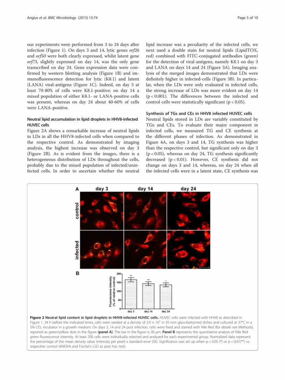
Neutral lipid accumulation in lipid droplets in HHV8-infectedHUVEC cellsFigure 2A shows a remarkable increase of neutral lipidsin LDs in all the HHV8-infected cells when compared tothe respective control. As demonstrated by imaginganalysis, the highest increase was observed on day 3(Figure 2B). As is evident from the images, there is aheterogeneous distribution of LDs throughout the cells,probably due to the mixed population of infected/unin-fected cells. In order to ascertain whether the neutral
Figure 2 Neutral lipid content in lipid droplets in HHV8-infected HUVFigure 1. 24 h before the indicated times, cells were seeded at a density of5% CO2 incubator in a growth medium. On days 3, 14 and 24 post infectioreported as green/yellow dots in the figure (panel A). The bar in the figuregreen fluorescence intensity. At least 200 cells were individually selected anthe percentage of the mean density value (intensity per pixel) ± standard erespective control (ANOVA and Fischer’s LSD as post hoc test).
lipid increase was a peculiarity of the infected cells, wenext used a double stain for neutral lipids (LipidTOX,red) combined with FITC-conjugated antibodies (green)for the detection of viral-antigens, namely K8.1 on day 3and LANA on days 14 and 24 (Figure 3A). Imaging ana-lysis of the merged images demonstrated that LDs weredefinitely higher in infected-cells (Figure 3B). In particu-lar, when the LDs were only evaluated in infected cells,the strong increase of LDs was more evident on day 14(p < 0.001). The differences between the infected andcontrol cells were statistically significant (p < 0.05).
Synthesis of TGs and CEs in HHV8 infected HUVEC cellsNeutral lipids stored in LDs are variably constituted byTGs and CEs. To evaluate their major component ininfected cells, we measured TG and CE synthesis atthe different phases of infection. As demonstrated inFigure 4A, on days 3 and 14, TG synthesis was higherthan the respective control, but significant only on day 3(p < 0.05), whereas on day 24, TG synthesis significantlydecreased (p < 0.01). However, CE synthesis did notchange on days 3 and 14, whereas, on day 24 when allthe infected cells were in a latent state, CE synthesis was
EC cells. HUVEC cells were infected with HHV8 as described in2.0 × 105 in 35 mm glass-bottomed dishes and cultured at 37°C in an, cells were fixed and stained with Nile Red (for details see Methods),is 30 μm. Panel B represents the quantitative analysis of Nile Redd analyzed for each experimental group. Normalized data representrror (SE). Significance was set up when p < 0.05 (*) or p < 0.01(**) vs.

Figure 3 (See legend on next page.)
Angius et al. BMC Microbiology (2015) 15:74 Page 6 of 10

(See figure on previous page.)Figure 3 Neutral lipid detection and quantification in HHV8-infected HUVEC cells by LipidTOX dye. HUVEC cells were infected withHHV8 as described in Figure 1. 24 h before the indicated times, cells were seeded at a density of 2.0 × 10⁵ in 35 mm glass-bottomed dishes andcultured at 37°C in a 5% CO2 incubator in a growth medium. On days 3, 14 and 24 post infection, cells were fixed and treble-stained with firstlyHCS LipidTOX™ Red Neutral Lipid stain (red), followed by an immunocytofluorescence method for identifying lytic (K8.1) and latent (LANA)viral-antigens with FITC-conjugated secondary antibodies (green) in the same cells counterstained with Hoechst 33258 for nuclei (blue), for detailssee Methods (panel A). The bar in the figure is 30 μm. Panel B represents the quantitative analysis of HCS LipidTOX™ Red Neutral Lipid stainfluorescence intensity. This method allowed the quantification of the neutral lipids in LDs in HHV8-infected cells alone. At least 10 microscopicfields were individually analysed for each experimental group. Normalized data represent the percentage of the mean density value (intensity perpixel) ± SE. Significance was set up when p < 0.05 (*) or p < 0.001 (***) vs. respective control (ANOVA and Fischer’s LSD as post hoc test).
Angius et al. BMC Microbiology (2015) 15:74 Page 7 of 10
about 69% higher than the respective control (Figure 4B,p < 0.001).
CE synthesis inhibition induces impairment of HUVEC cellneo-angiogenic activityIn order to verify whether neutral lipids, specifically CEs,could somehow also be involved in the peculiar modifi-cations induced by lytic or latent HHV8 infection, weevaluated their possible role in neo-angiogenesis, whichis typically enhanced in HHV8-infected cells. In fact, theneo-angiogenic properties of HHV8 are necessary forthe formation of the characteristic lesions of Kaposi’sangiosarcoma [1-6]. In the angiogenic activity assay,both control and lytic (day 3) or latent (day 24) HHV8-infected cells produced micro-tubules within 24 h(Figure 5A and B). The specific inhibitor of CE synthesisSZ 58035 significantly reduced tubule formation in in-fected cells on day 24 (p < 0.001) but not during the lyticinfection (day 3). Interestingly, control cells grown in aserum-free M200 medium were not able to producecomplete capillary micro-tubules, whilst HHV8-infectedcells still formed regular and almost normal tubules (p <0.05). Furthermore, in these conditions (Figure 5A and B)SZ was also able to dramatically reduce tubule formation(p < 0.001) in both lytic and latent infection, as confirmedby evaluation of the angiogenic index (Figure 5B) [13].
Figure 4 TG and CE synthesis in HHV8-infected and control HUVEC ceOn days 3, 14 and 24 post infection, 1.0 × 10⁶ cells were incubated for 4 h(BSA). Subsequently, cells were washed with ice-cold PBS and lipids extracted w(TLC), and the incorporation of [14C]-oleate into TGs (panel A) and CEs (panel B± SE. Significance was set up when p< 0.05 (*) vs. respective control (t-test).
DiscussionHHV8 is known for its endothelial tropism and is gener-ally related to proliferative diseases, mainly KS. Recently,dysfunctional glucose metabolism in vitro [23], and ahigh prevalence of HHV8 infection in subjects with dia-betes mellitus [24] have been reported. Neutral lipid in-crease in LDs, mainly TGs, has been reported in severalherpes virus infections, and considered an effect of thereprogramming induced by the viral infection to consentvirion production [31]. A few reports have described thepresence of HHV8 in plaque lesions, but the possiblecorrelation with atherosclerosis has yet to be clarified[32,33]. In the present study, we investigated neutrallipid metabolism and LD content during the lytic and la-tent phases of endothelial HHV8-infection. In fact, moststudies have investigated the effect of cell reprogram-ming on lipids in the first days of infection. We presenta model that allows the HHV8-infectious effect on cellmetabolism to be investigated from the lytic to the latelatent phase for up to more than three weeks, which isparticularly suitable for HHV8-infection of endothelialcells. Even though, as shown by immunofluorescence,40-60% of cells still appeared to be infected on day 24after infection, it was not possible to prolong the experi-ment any further because the cells gradually lost their viralepisome and rapidly started to show some signs of senes-cence (data not shown). TG synthesis was increased in the
lls. HUVEC cells were infected with HHV8 as described in Figure 1.in a medium containing [14C]-oleate bound to bovine serum albuminith acetone. Neutral lipids were separated by thin layer chromatography) was determined as described in Methods. Data were reported as mean

Figure 5 (See legend on next page.)
Angius et al. BMC Microbiology (2015) 15:74 Page 8 of 10

(See figure on previous page.)Figure 5 Capillary-like vascular micro-tubule formation in HHV8-infected and control HUVEC cells. HUVEC cells were infected with HHV8as described in Figure 1. 24 h before the indicated times, 3.0 × 10⁴ cells were seeded in plates previously coated with Geltrex matrix and culturedin a complete or serum-free M200 medium in a CO2 incubator at 37°C. The specific ACAT inhibitor Sandoz 58035 (SZ, 4 μM) was added in someexperimental groups. Capillary-like microtubule formation in the control and HHV8-infected HUVEC cells was performed as described in theMethods section. Panel A shows the light microscope images of micro-tubule formation in control, lytic and latent HHV8-infected HUVEC cellscultured in medium with or without (W/O) fetal calf serum (FCS). The bar in the figure is 240 μm. Panel B represents the angiogenic indexquantification (for details, see Methods). All the samples were prepared in triplicate and the experiments were repeated at least twice. Data werereported as mean ± SE. Significance was set up when p < 0.05 (*) or p < 0.001 (***) vs. respective control (t-test).
Angius et al. BMC Microbiology (2015) 15:74 Page 9 of 10
lytic phase. The fact that cholesterol esterification wasnot significantly modified for up to 14 days suggeststhat the higher LD content was due to TG content.Conversely, TG synthesis was lower, whereas choles-terol esterification underwent a remarkable increase inthe latent phase, suggesting that, while TGs are prom-inent in the lytic phase, CEs are the prominent neutrallipid fraction in the late latent phase. It is worthnoting that we performed our lipid synthesis analysisin a mixed population of infected/uninfected cells, asdemonstrated by immunofluorescence. Based on thisconsideration, it is likely that our results couldbe underestimated. To overcome this problem, wedouble-stained neutral lipids (LipidTOX) and viral-antigens (immunofluorescence) in the same cells, andthe results confirmed that neutral lipids increased onlyin infected cells. An increase of TG-enriched LDs dur-ing the lytic phase of viral infection has often been re-ported for HCV virus. In fact, it has been suggestedthat the virus uses TG-enriched LDs as a platform forthe assembly of nascent virions, and that the HCVcore protein is a main player in the manipulation ofthese organelles [34,35]. Overall, these observationshighlight that a range of viruses (HBV, GBV-B,Dengue virus) have evolved mechanisms to interactwith LDs and possibly to subvert the function of theseorganelles, so as to use them as a platform for viralparticle assembly [31]. Accordingly, our results sup-port the fact that TG synthesis and LD increase aremainly necessary during active virus replication. Onthe contrary, in the latent phase of HHV8-infection,LD content seems to be mainly constituted by CEs, assuggested by the high rate of cholesterol esterification.High CE content and cholesterol esterification in-crease are frequently reported in cancer cells and tu-mours [18,27]. The storage of CEs has been related tothe large amount of cholesterol needed by malignantcells to sustain membrane biogenesis. Although thereal significance of CEs in cancer cells is far frombeing clarified, CEs have recently been related to theaggressiveness and metastatic properties of prostatecancer [36,37]. For the first time, we observed theincrease of cholesterol esterification in the latentphase, which can be considered equivalent to persistent
infection in humans, and as leading to KS. The mostcommon pathogenic effect expressed by HHV8 in KSis represented by the angiogenic activity of infectedcells. Accordingly, angiogenesis is one of the majoreffects produced by HHV8-chronic infection in vitroand, just as in cancer cells, still present even in serum-deprived conditions [13]. For these reasons, we inhib-ited CE formation using a specific ACAT inhibitor(Sandoz 58035), which is able to inhibit cholesterolesterification up to 90%, but does not affect TG synthe-sis. In contrast with the control, HHV8-infected cellsproduced micro-tubules even in serum-deprived con-ditions. Interestingly, the inhibition of CE formationwas accompanied by a remarkable reduction of micro-tubules in HHV8-infected cells.
ConclusionsHHV8-infection induces an enhancement of neutrallipid synthesis and the accumulation of LDs in HUVECcells. The TG increase in the lytic phase is probably re-lated to peculiar metabolic viral requirements. On theother hand, CEs seem to be closely related to the angio-genic properties of the infected cells, mainly in the latentphase of infection, suggesting that these neutral lipidsmay, in some way, be functionally involved in regulatingthe malignant process. These findings suggest that theuse of cholesterol esterification inhibitors on HHV8-derived tumours could be a useful therapeutic tool inKS. In fact, angiogenesis inhibition may help to preventthe occurrence of the diffusion of angiosarcoma andmetastasis.
Competing interestsThe authors declare that they have no competing interests.
Authors’ contributionsFA, BB and RP conceived and equally contributed to the design and theexperimental coordination of the study as well as to the drafting of themanuscript. EP and AI carried out the molecular genetic studies. FA, SU andSS carried out biochemical, microscopy and imaging analysis. All authorsread and approved the final manuscript.
AcknowledgementsThe authors thank Sally Davies for English language editing and Mrs. AnnaSaba for technical assistance. This work was supported by the FondazioneBanco di Sardegna 2013-14.
Angius et al. BMC Microbiology (2015) 15:74 Page 10 of 10
Received: 26 September 2014 Accepted: 12 March 2015
References1. Ablashi DV, Chatlynne LG, Whitman Jr JE, Cesarman E. Spectrum of Kaposi's
sarcoma-associated herpesvirus, or human herpesvirus 8, diseases. ClinMicrobiol Rev. 2002;15(3):439–64.
2. Dourmishev LA, Dourmishev AL, Palmeri D, Schwartz RA, Lukac DM.Molecular genetics of Kaposi's sarcoma-associated herpesvirus (humanherpesvirus-8) epidemiology and pathogenesis. Microbiol Mol Biol Rev.2003;67(2):175–212.
3. Ganem D. KSHV and the pathogenesis of Kaposi sarcoma: listening tohuman biology and medicine. J Clin Invest. 2010;120(4):939–49.
4. Hengge UR, Ruzicka T, Tyring SK, Stuschke M, Roggendorf M, Schwartz RA,et al. Update on Kaposi's sarcoma and other HHV8 associated diseases. Part1: epidemiology, environmental predispositions, clinical manifestations, andtherapy. Lancet Infect Dis. 2002;2(5):281–92.
5. Wen KW, Damania B. Kaposi sarcoma-associated herpesvirus (KSHV):molecular biology and oncogenesis. Cancer Lett. 2010;289(2):140–50.
6. Caselli E, Fiorentini S, Amici C, Di Luca D, Caruso A, Santoro MG. Humanherpesvirus 8 acute infection of endothelial cells induces monocytechemoattractant protein 1-dependent capillary-like structure formation:role of the IKK/NF-kappaB pathway. Blood. 2007;109(7):2718–26.
7. Gregory SM, Wang L, West JA, Dittmer DP, Damania B. Latent Kaposi'ssarcoma-associated herpesvirus infection of monocytes downregulatesexpression of adaptive immune response costimulatory receptors andproinflammatory cytokines. J Virol. 2012;86(7):3916–23.
8. Wang HW, Trotter MW, Lagos D, Bourboulia D, Henderson S, Makinen T,et al. Kaposi sarcoma herpesvirus-induced cellular reprogrammingcontributes to the lymphatic endothelial gene expression in Kaposisarcoma. Nat Genet. 2004;36(7):687–93.
9. Delgado T, Carroll PA, Punjabi AS, Margineantu D, Hockenbery DM,Lagunoff M. Induction of the Warburg effect by Kaposi's sarcomaherpesvirus is required for the maintenance of latently infected endothelialcells. Proc Natl Acad Sci U S A. 2010;107(23):10696–701.
10. Carroll PA, Kenerson HL, Yeung RS, Lagunoff M. Latent Kaposi'ssarcoma-associated herpesvirus infection of endothelial cells activateshypoxia-induced factors. J Virol. 2006;80(21):10802–12.
11. Douglas JL, Gustin JK, Viswanathan K, Mansouri M, Moses AV, Fruh K. Thegreat escape: viral strategies to counter BST-2/tetherin. PLoS Pathog.2010;6(5):e1000913.
12. Parravicini C, Chandran B, Corbellino M, Berti E, Paulli M, Moore PS, et al.Differential viral protein expression in Kaposi's sarcoma-associatedherpesvirus-infected diseases: Kaposi's sarcoma, primary effusion lymphoma,and multicentric Castleman's disease. Am J Pathol. 2000;156(3):743–9.
13. Wang L, Damania B. Kaposi's sarcoma-associated herpesvirus confers asurvival advantage to endothelial cells. Cancer Res. 2008;68(12):4640–8.
14. Watanabe T, Sugaya M, Atkins AM, Aquilino EA, Yang A, Borris DL, et al.Kaposi's sarcoma-associated herpesvirus latency-associated nuclear antigenprolongs the life span of primary human umbilical vein endothelial cells.J Virol. 2003;77(11):6188–96.
15. Guilluy C, Zhang Z, Bhende PM, Sharek L, Wang L, Burridge K, et al. LatentKSHV infection increases the vascular permeability of human endothelialcells. Blood. 2011;118(19):5344–54.
16. Bhatt AP, Jacobs SR, Freemerman AJ, Makowski L, Rathmell JC, Dittmer DP,et al. Dysregulation of fatty acid synthesis and glycolysis in non-Hodgkinlymphoma. Proc Natl Acad Sci U S A. 2012;109(29):11818–23.
17. Delgado T, Sanchez EL, Camarda R, Lagunoff M. Global metabolic profilingof infection by an oncogenic virus: KSHV induces and requires lipogenesisfor survival of latent infection. PLoS Pathog. 2012;8(8):e1002866.
18. Uda S, Spolitu S, Angius F, Collu M, Accossu S, Banni S, et al. Role of HDLin cholesteryl ester metabolism of lipopolysaccharide-activated P388D1macrophages. J Lipid Res. 2013;54(11):3158–69.
19. McAllister SC, Moses AV. Endothelial cell- and lymphocyte-based in vitrosystems for understanding KSHV biology. Curr Top Microbiol Immunol.2007;312:211–44.
20. Ottensmeyer FP, Beniac DR, Luo RZ, Yip CC. Mechanism of transmembranesignaling: insulin binding and the insulin receptor. Biochemistry.2000;39(40):12103–12.
21. Raggo C, Ruhl R, McAllister S, Koon H, Dezube BJ, Fruh K, et al. Novelcellular genes essential for transformation of endothelial cells by Kaposi'ssarcoma-associated herpesvirus. Cancer Res. 2005;65(12):5084–95.
22. Cerimele F, Curreli F, Ely S, Friedman-Kien AE, Cesarman E, Flore O. Kaposi'ssarcoma-associated herpesvirus can productively infect primary humankeratinocytes and alter their growth properties. J Virol. 2001;75(5):2435–43.
23. Ingianni A, Piras E, Laconi S, Angius F, Batetta B, Pompei R. LatentHerpesvirus 8 infection improves both insulin and glucose uptake inprimary endothelial cells. New Microbiol. 2013;36(3):257–65.
24. Ingianni A, Carta F, Reina A, Manai M, Desogus A, Pompei R. Prevalence ofHerpesvirus 8 infection in type 2 diabetes mellitus patients. Am J Infect DIs.2007;3(3):123–7.
25. Ingianni A, Madeddu MA, Carta F, Reina A, Lai C. Epidemiology of humanherpesvirus type 8 infection in cardiopathic patients. OnLine J BiomedSci. 2009;9(2):36–9.
26. Smith PK, Krohn RI, Hermanson GT, Mallia AK, Gartner FH, Provenzano MD,et al. Measurement of protein using bicinchoninic acid. Anal Biochem.1985;150(1):76–85.
27. Uda S, Accossu S, Spolitu S, Collu M, Angius F, Sanna F, et al. A lipoproteinsource of cholesteryl esters is essential for proliferation of CEM-CCRFlymphoblastic cell line. Tumour Biol. 2012;33(2):443–53.
28. Diaz G, Batetta B, Sanna F, Uda S, Reali C, Angius F, et al. Lipid dropletchanges in proliferating and quiescent 3 T3 fibroblasts. Histochem Cell Biol.2008;129(5):611–21.
29. Diaz G, Melis M, Batetta B, Angius F, Falchi AM. Hydrophobiccharacterization of intracellular lipids in situ by Nile Red red/yellow emissionratio. Micron. 2008;39(7):819–24.
30. Greenspan P, Fowler SD. Spectrofluorometric studies of the lipid probe, nilered. J Lipid Res. 1985;26(7):781–9.
31. Saka HA, Valdivia R. Emerging roles for lipid droplets in immunity andhost-pathogen interactions. Annu Rev Cell Dev Biol. 2012;28:411–37.
32. Carletti F, Mandolini C, Rossi A, Capobianchi MR, Borgia MC. Prevalence ofhuman herpesvirus (HHV)-8 infection among carriers of cardiovasculardisease. J Biol Regul Homeost Agents. 2002;16(2):110–3.
33. Ye D, Nichols TC, Dehmer GJ, Tate DA, Wehbie RS, Quinlivan EB. Absence ofhuman herpesvirus 8 genomes in coronary atherosclerosis inimmunocompetent patients. Am J Cardiol. 1997;79(9):1245–7.
34. Boulant S, Targett-Adams P, McLauchlan J. Disrupting the association ofhepatitis C virus core protein with lipid droplets correlates with a loss inproduction of infectious virus. J Gen Virol. 2007;88(Pt 8):2204–13.
35. Miyanari Y, Atsuzawa K, Usuda N, Watashi K, Hishiki T, Zayas M, et al. Thelipid droplet is an important organelle for hepatitis C virus production. NatCell Biol. 2007;9(9):1089–97.
36. Peck B, Schulze A. Cholesteryl esters: fueling the fury of prostate cancer.Cell Metab. 2014;19(3):350–2.
37. Yue S, Li J, Lee SY, Lee HJ, Shao T, Song B, et al. Cholesteryl esteraccumulation induced by PTEN loss and PI3K/AKT activation underlieshuman prostate cancer aggressiveness. Cell Metab. 2014;19(3):393–406.
Submit your next manuscript to BioMed Centraland take full advantage of:
• Convenient online submission
• Thorough peer review
• No space constraints or color figure charges
• Immediate publication on acceptance
• Inclusion in PubMed, CAS, Scopus and Google Scholar
• Research which is freely available for redistribution
Submit your manuscript at www.biomedcentral.com/submit
Recommended


