Orientational Disorder and Entropy of Water in Protein Cavities
Vladimir P. Denisov,† Kandadai Venu,†,‡ Jo1rg Peters,§ Hans Dietrich Ho1rlein,§ andBertil Halle* ,†
Condensed Matter Magnetic Resonance Group, Department of Chemistry, Lund UniVersity, P.O. Box 124,S-22100 Lund, Sweden, and Bayer AG, PH-TO Biotechnologie, D-42096 Wuppertal, Germany
ReceiVed: April 9, 1997X
Water 17O, 2H, and 1H nuclear magnetic relaxation dispersion (NMRD) data are presented for the Y35Gmutant of bovine pancreatic trypsin inhibitor (BPTI) in aqueous solution. The NMRD data show that thethree buried water molecules in this protein exchange with bulk water on the time scale of 15 ns to 1µs andundergo librational motion of considerable amplitude in the protein cavities. By analysis of the threeindependent order parameters provided by the NMRD data in terms of an anisotropic harmonic librationmodel, the amplitude and anisotropy of water rotation in protein cavities is quantitatively assessed. All sevendistinct buried water molecules in wild-type BPTI and in the G36S and Y35G mutants are examined in thisway. The harmonic libration model also allows the rotational entropy of buried water molecules to be deducedfrom the experimental order parameters. Although each of the investigated buried water molecules is engagedin three or four hydrogen bonds, their entropies span the full range from ice to bulk water, suggesting that thehydration of protein cavities with weaker hydrogen-bonding capacity may be entropically driven. This mightbe true also for weakly hydrogen-bonded water molecules in surface clefts, thus reversing the conventionalview that ligand binding to proteins is entropically favored by release of ordered water.
Introduction
Internal cavities large enough to accomodate at least one watermolecule are found in nearly all globular proteins, typicallyaccounting for 1 % of the protein volume.1-4 In crystalstructures, some of these cavities are occupied by translationallyordered water molecules, while other cavities appear to beempty.5-7 Protein cavities may be viewed as structural defects,resulting from the conflicting requirements of hydrogen-bondedsecondary structure elements and a uniformly high packingdensity. It is becoming increasingly clear, however, that cavitiescan be directly involved in protein function. Empty cavitiescan facilitate transport of small substrates through the proteinmatrix8,9 or can enable large-scale domain movements.10,11
Hydrated protein cavities, i.e., internal (or buried) watermolecules, can be directly involved in enzyme catalysis12 andligand binding.13,14 An improved understanding of internalhydration should also have important implications for thebroader issues of folding and stability of globular proteins.The current knowledge about buried water molecules in
proteins is largely derived from high-resolution X-ray diffractiondata.5,6 In recent years, information about buried watermolecules has also come from high-resolution1H NMR studiesof intermolecular nuclear Overhauser effects (NOEs)15-17 andfrom water17O and2H nuclear magnetic relaxation dispersion(NMRD) studies.18-20 Although not competitive with crystal-lography in terms of structural detail, the NMR methods havedistinct advantages in other respects. First, they report onproteins in aqueous solution, which is usually the physiologicalmedium. Second, the NOE method can detect cavity water atoccupancies as low as 10%,21 whereas the detection limit ofX-ray diffraction is typically 30-50%. (Owing to a strongcorrelation between the occupancy and the thermalB factor,
occupancies of all detected buried water moelcules are usuallyfixed at 100% during refinement.5,6) Third, the NMRD methodprovides intramolecular order parameters reflecting the orien-tational fluctuations of buried water molecules, information thatis not readily extracted even from neutron diffraction data.7
Fourth, both NMR methods provide information about the rateof exchange of water molecules between protein cavities andbulk water, whereas conventional crystallography can provideonly equilibrium averages. The dynamic information is mostdirectly manifested in NMRD data, which probe the fluctuationspectrum over a wide frequency range. The rate at which awater molecule escapes from a protein cavity is of interestmainly because it reflects large-scale structural fluctuations thatare thought to be rate-limiting for exchange of deeply buriedwater molecules.22
Another important aspect of cavity hydration concerns therelation between structure and thermodynamics. When thisrelationship is understood, it should be possible, given thestructure of a protein cavity, to predict its occupancy and theconsequent stabilization of the native protein structure. Con-sidering the marginal stability with respect to unfolding of mostglobular proteins under physiological conditions, cavity hydra-tion may well be an important stabilizing factor. The availablecrystallographic data show that most buried water moleculesengage in three or four hydrogen bonds to protein O and Natoms lining the cavity,3,5 suggesting rather stringent require-ments for cavity hydration. This picture is supported by freeenergy calculations based on atomic force fields23,24 and byconsiderations of the solubility of water in nonpolar solvents.25
Yet, water molecules have also been detected, by X-raycrystallography3,6,26and by the NOE method,21,27 in essentiallynonpolar cavities, suggesting rather less stringent hydrogen-bondrequirements. Since positional disorder (largeB factor) and/orpartial occupancy may render such buried water moleculesX-ray-invisible, their prevalence may have been underestimated.Experimental data on the thermodynamics of cavity hydration
in proteins are scarce. The order parameters obtained from
† Lund University.‡ Permanent address: School of Physics, University of Hyderabad,
Hyderabad 500046, India.§ Bayer AG.X Abstract published inAdVance ACS Abstracts,October 1, 1997.
9380 J. Phys. Chem. B1997,101,9380-9389
S1089-5647(97)01221-2 CCC: $14.00 © 1997 American Chemical Society
NMRD studies, however, reflect the local orientational distribu-tion function that governs the configurational part of therotational entropy.28,29 This link between NMR-derived orderparameters and rotational entropy has previously been exploredfor orientational fluctuations of N-H and C-H bonds in thepolypeptide backbone30,31 and in side chains.32 The case ofwater is different in several respects. First, the entropy of aburied water molecule directly affects an observable property,the cavity occupancy. Second, since the nuclear spin couplingtensors of17O, 2H, and1H are differently oriented in the watermolecule, multinuclear NMRD data can provide three indepen-dent order parameters, characterizing the anisotropy of the localwater reorientation.33,34 Third, the translational degrees offreedom of a buried water molecule also contribute to theentropy. Our analysis indicates, however, that this contributionis less important than the rotational one, at least for the buriedwater molecules considered here.Among the several globular proteins so far studied by
multinuclear water NMRD,20 the best characterized is bovinepancreatic trypsin inhibitor (BPTI). NMRD data for all threewater nuclei have been reported for wild-type BPTI,18,34,35withfour buried water molecules,36 and for the mutant G36S,19,22,34
where one of the four is replaced by the hydroxyl group of Ser-36.37 Here, we report a complete set of water NMRD curvesfor another BPTI mutant, Y35G, which has three buried watermolecules in cavities distinct from those in wild-type BPTI.38
The combined NMRD data for wild-type BPTI and the twomutants, comprising seven distinct buried water molecules, arethen analyzed in terms of an anisotropic harmonic libration(AHL) model, allowing us to convert the17O, 2H, and1H orderparameters to fluctuation amplitudes for the rock, wag, and twistlibration modes. The theoretical basis for this analysis ispresented in the Appendix. Although the singly buried watermolecule (W122) in wild-type BPTI has an orientational ordercomparable to that in ice, the other buried water molecules arefound to be more disordered, with rms libration amplitudes aslarge as 30°. Moreover, the three buried water molecules inY35G are found to be more disordered than the three in G36S.In the final stage of the analysis, we use the AHL model to
convert the libration amplitudes to rotational entropy contribu-tions. The results indicate that the entropy of the buried watermolecules in Y35G is comparable to that of bulk water,suggesting that the hydration of cavities with weaker hydrogen-bonding capacity may be entropically driven. Enthalpy-entropycompensation thus appears to be an important feature of cavityhydration, as in many other processes that involve restructuringof hydrogen bonds in aqueous media at ambient temperature.39,40
It is widely believed that the release of ordered water contributessignificantly to the driving force for protein-ligand andprotein-receptor association in aqueous solution. This is alsothe conventional view of the hydrophobic effect. The scenariothat we propose for weakly hydrogen-bonded cavity water isthe opposite one, where dehydration reduces the entropy ofwater. Similarly, ligand binding to pockets and crevices at theprotein surface may be entropically disfavored by the displace-ment of weakly hydrogen-bonded water molecules.
Materials and Methods
Materials. The BPTI mutant Met(-1)-Y35G was preparedaccording to the procedure described earlier for the G36Smutant.37 A cell-free yeast culture was adjusted to pH 3 withcitric acid and diluted with water to a final conductivity of 11mS cm-1, whereafter 200 mL SP-Sepharose FF (Pharmacia,Uppsala) was added and incubated for 50 min. The resin waswashed first with 3 L of 50 mMcitrate-NaOH buffer (pH 3),
then with 2 L of 50 mMTris-HCl (pH 9), and finally with 2 Lof 20 mM HEPES (pH 6). After the gel was packed into acolumn, the Y35G mutant was eluted by a linear salt gradient(0-1 M NaCl, 20 mM HEPES, pH 6). Fractions (20 mL) weretaken and pooled according to reversed-phase HPLC analysis.Fractions containing the product were pooled, adjusted to pH 3(citric acid), diluted to a final conductivity of 23 mS cm-1, andpassed through a 0.22µm filter (Sartorius, Go¨ttingen). Thissolution was pumped onto a 100 mL S-Sepharose HP column(Pharmacia, Uppsala) previously equilibrated with 3 columnvolumes of citrate-NaOH buffer (50 mM, pH 3). The adsorbedproduct was washed with 470 mL of starting buffer and thenwith 20 mM HEPES (pH 6). Elution of the Y35G mutant wasperformed using a linear salt gradient (20 mM HEPES/0-1 MNaCl, pH 6). Fractions (20 mL) were taken for RP-HPLCanalysis, and those containing the product were pooled andfiltered (0.22 µm). As the final purification step, the S-Sepharose-HP pool was chromatographed on a preparativeVydac C18-HPLC column (40 mL) previously equilibrated with0.1% trifluoroacetic acid. The product was eluted using a lineargradient of acetonitrile. Fractions containing the Y35G mutantwere pooled, diluted with water, and lyophilized. The finalproduct was characterized by N-terminal sequencing over 39cycles, capillary electrophoresis (yielding a homogeneous peak),amino acid analysis (in agreement with calculated values), andnative PAGE (yielding a homogeneous peak). The purifiedprotein preparation, lyophilized as a salt of trifluoroacetic acid,contained 99.7% Y35G according to results of SDS-PAGE andRP-HPLC.Protein solutions were made by dissolving the lyophilized
protein in heavy water (molar mass 21.5 g mol-1), enriched in17O (Ventron, 21.9 atom %17O, 61.9 atom %18O, 99.95 atom% 2H), or doubly distilled water. The operational pH* (withoutisotope correction) was adjusted to 5.2 by adding small amountsof concentrated NaOH or KOH. Protein concentrations weredetermined on the basis of complete amino acid analysis,yielding 23.1( 0.5 mM for the D2O sample.Relaxation Dispersion Measurements.Oxygen-17, deu-
teron, and proton relaxation rates were measured at differentmagnetic field strengths as described previously,19,34using fourBruker and Varian NMR spectrometers and an iron magnet(Drusch EAR-35N) equipped with field-variable lock and fluxstabilizer. The sample temperature was maintained at 27.0(0.1 °C by a thermostated air flow.
Results and Discussion
Water Relaxation Dispersions. The longitudinal magneticrelaxation rates of the water17O, 2H, and1H nuclei in aqueousprotein solutions typically exhibit a dispersion in the megahertzrange, with a dispersion frequency that reflects the rotationaldiffusion (correlation timeτR) of the protein. The17O dispersionis produced by a small number of water molecules withresidence times longer thanτR but shorter than the intrinsic spinrelaxation time.18-20 When17O NMRD data from a variety ofproteins are correlated with high-resolution crystal structures,it is found that such long-lived water molecules are eithertrapped in cavities or deep crevices or coordinated to protein-bound multivalent metal ions.20 The 2H and 1H dispersionsgenerally contain a contribution from labile protein hydrogens,in addition to that from long-lived water molecules.34,35 In thecase of BPTI, the labile hydrogen contribution to the2Hdispersion is negligible at pH 5-6 35 but is substantial at allpH values for the more slowly relaxing1H nucleus.34
Wild-type BPTI contains four buried water molecules.36 Oneof these (denoted W122) resides in an isolated cavity near the
Water in Protein Cavities J. Phys. Chem. B, Vol. 101, No. 45, 19979381
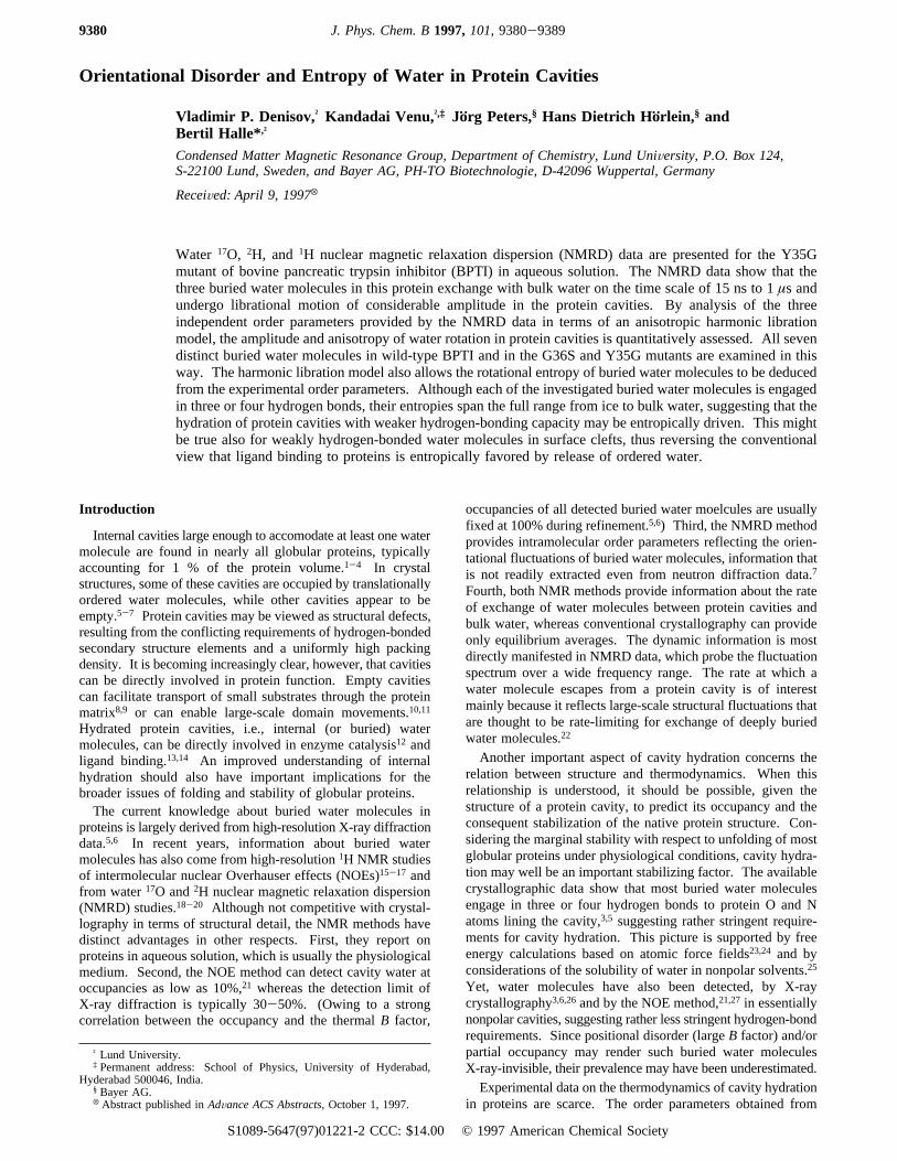
14-38 disulfide bridge, while the other three (W111-W113)occupy a porelike cavity between the two major loops of thepolypeptide chain (see Figure 1). In the mutant G36S, W122is replaced by the hydroxyl group of the Ser-36 side chain withlittle structural perturbation elsewhere in the protein.37 Previousdifference-NMRD studies of wild-type BPTI and the G36Smutant have established that W122 exchanges relatively slowlywith a residence time of 170( 20µs at 300 K.22 Since this iscomparable to the intrinsic relaxation time for2H but muchshorter/longer than that for1H/17O, W122 contributes fully onlyto the1H dispersion at this temperature.22,34 After subtractionof the labile-proton contribution to the1H relaxation, the17O,2H, and1H dispersions from G36S were found to be consistentwith a contribution from three water molecules, with residencetimes in the range of 15 ns of 1µs and with fast librationalmotions of modest amplitude.19,22,34 Although not demonstrateddirectly by a difference-NMRD experiment (as for W122), thecrystal structure strongly suggests that this contribution is dueto the buried water molecules W111-W113. The only otherplausible candidate is W143, residing in a surface pocket withthree hydrogen bonds to the protein. Compared to W111 (atthe mouth of the pore), however, W143 is more exposed tobulk solvent (the pocket is not as deep) and has longer (weaker)hydrogen bonds. Both these factors should reduce the residencetime of W143 compared to the case of W111.20 Indeed, high-resolution1H NMR studies of intermolecular NOEs betweenBPTI and water indicate that W143 has a subnanosecondresidence time even at 4°C.16 In conclusion, only three watermolecules, viz. W111-W113, contribute to the17O, 2H, and1H dispersions from the G36S mutant.The BPTI mutant Y35G, with the bulky side chain of Tyr-
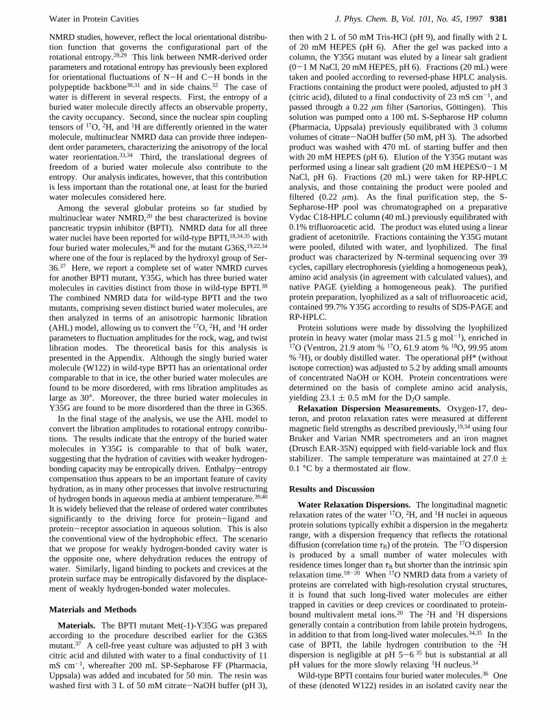
35 replaced by a hydrogen atom, shows an unusually largestructural perturbation for a single-site mutation.38 Althoughthe main fold of the polypeptide chain and about two-thirds ofthe protein structure (including the hydrophobic core) are highlyconserved, the two major loops are substantially rearrangedcompared to that of wild-type BPTI (rms deviations of 2 and 4Å).38 As a result, none of the four buried water molecules inwild-type BPTI is strictly conserved in Y35G. A new cavityoccupied by two buried water molecules (W301 and W302) iscreated near the mutation site, while a third buried watermolecule (W303) corresponds roughly (rms deviation 1.3 Å)to W111 in wild-type BPTI (cf. Figures 1 and 2b).38 The
solution structure of Y35G has not been determined but isprobably close to the crystal structure as for wild-type BPTIand G36S.37,41 On the other hand, chemical shift, hydrogenexchange, and15N relaxation studies on Y35G in solutionindicate that Y35G has a more flexible structure than wild-typeBPTI.42,72 The unfolding temperature is lowered by nearly 20°C, corresponding to ca. 5 kcal mol-1 destablization of the nativestructure.42 The hydrogen-bond geometries for W111-W113in wild-type BPTI (assumed to be the same in G36S) and forW301-W303 in Y35G are shown in Figures 1 and 2, whilethe relevant hydrogen-bond lengths are compiled in Table 1.Figure 3 shows the water17O, 2H, and 1H relaxation
dispersion curves from solutions of Y35G at pH* 5.2. For
Figure 1. Hydrogen-bond geometry for the buried water moleculesW111-W113 in the crystal structure 5pti of wild-type BPTI,36 whichis nearly identical to the solution structures of BPTI and G36S.37,41
Figure 2. Hydrogen-bond geometry for the buried water moleculesW301-W302 (A) and W303 (B) in the crystal structure 8pti of theBPTI mutant Y35G.38 Hydrogen atoms have been added assumingstandard intramolecular geometry and optimal hydrogen bonds.
TABLE 1: Hydrogen Bond Lengths for Buried WaterMolecules in Wild-Type BPTI and in the Y35G Mutant
BPTIwatera partner
ROX(Å)
Y35Gwaterb partner
ROX(Å)
W113 W112 2.6 W301 Ile-18 CO 2.9Tyr-10 CO 2.9 Phe-33 CO 3.0Asn-44 NH2 2.9 Gly-36 NH 2.7Lys-41 NH 3.0
W112 W111 2.9 W302 Ile-18 CO 2.8Asn-43 CO 2.8 Ala-16 CO 2.7W113 2.6 Gly-35 NH 3.0Tyr-10 NH 2.9 Gly-37 NH 3.0
W111 Glu-7 CO2 2.6 W303 Glu-7 CO2 2.7Pro-8 CO 2.7 Pro-8 CO 3.0W112 2.9 Asn-43 NH 3.1
a Based on PDB file 5pti (wild-type BPTI in crystal form II).36
b Based on PDB file 8pti (Y35G mutant).38
9382 J. Phys. Chem. B, Vol. 101, No. 45, 1997 Denisov et al.
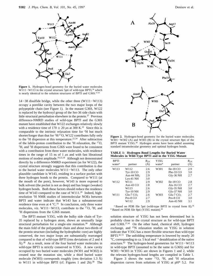
comparison, we have included the corresponding dispersioncurves for G36S19,34 (scaled to the Y35G concentration). Thedata are well represented by the theoretical expression43-45
where ω0 is the Larmor frequency andτc is the effectivecorrelation time for the buried water molecules, which here canbe identified with the rotational correlation timeτR of the protein(vide infra). The dispersion amplitudeâ is due to buried watermolecules (and, for1H, to labile protons), while the high-frequency relaxation rateRhf ≡ R1(ω0 . 1/τc) represents thecontribution from all external water molecules in the bulk andat the protein surface. The parametersRhf, â, andτc, determinedfrom the nonlinear least-squares fits shown in Figure 3, arecollected in Table 2, together with the corresponding valuesfor G36S.19,34
The similarity of the two mutants with respect to the high-frequency plateau (Rhf) and the dispersion frequency (τc) isconsistent with a similar surface (on average) and shape for thetwo proteins, as expected from the crystal structures. Thecorrelation timesτc obtained from the17O and1H dispersionsdiffer by a factor of 1.23( 0.08, close to the ratio of the solventviscosities (1.27) in the D2O and H2O samples. This is
consistent with the identification ofτc as the rotational correla-tion time τR of the protein.For the analysis of the amplitudes of the17O, 2H, and 1H
dispersions, it is convenient to scale the relaxation ratesaccording to19,34
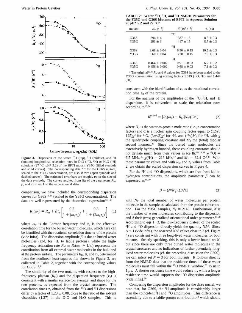
whereNT is the water-to-protein mole ratio (i.e., a concentrationfactor) andC is a nuclear spin coupling factor equal to (12π2/125)ø2 for 17O, (3π2/2)ø2 for 2H, and (10/3)M2 for 1H, with øthe quadrupole coupling constant andM2 the (total) dipolarsecond moment.43 Since the buried water molecules areextensively hydrogen bonded, these coupling constants shouldnot deviate much from their values in ice Ih:18,19,34ø(17O) )6.5 MHz,46 ø(2H) ) 213 kHz,47 andM2 ) 32.4 G2.48 Withthese parameter values and withRhf andτc values from Table2, we obtain the scaled dispersion curves in Figure 4.For the2H and17O dispersions, which are free from labile-
hydrogen contributions, the amplitude parameterâ can beexpressed as18,20
with NT the total number of water molecules per proteinmolecule in the sample as calculated from the protein concentra-tion. For the Y35G samples,NT ) 2140. Furthermore,N isthe number of water molecules contributing to the dispersionandA their (rms) generalized orientational order parameter.18,20
According to eqs 1-3, the low-frequency plateau of the scaled2H and17O dispersion directly yields the quantityNA2. SinceA< 1 (vide infra), the observedNA2 values close to 2 (cf. Figure4) are consistent with three long-lived water molecules for bothmutants. Strictly speaking, this is only a lower bound onN,but since there are only three buried water molecules in thecrystal structures and no indications of further potentially long-lived water molecules (cf. the preceding discussion for G36S),we can safely setN ) 3 for both mutants. It follows directlyfrom the NMRD data that the residence times of these watermolecules must fall within the17O NMRD window,20 15 ns to1 µs. A shorter residence time would reduceτc, while a longerresidence time would suppress the17O dispersion amplitude(vide infra).20
Comparing the dispersion amplitudes for the three nuclei, wenote that, for G36S, the1H amplitude is considerably largerthan the coincident2H and17O amplitudes. This difference isessentially due to a labile-proton contribution,34 which should
Figure 3. Dispersion of the water17O (top), 2H (middle), and1H(bottom) longitudinal relaxation rates in D2O (17O, 2H) or H2O (1H)solutions (27°C, pH* 5.2) of the BPTI mutant Y35G (filled symbolsand solid curves). The corresponding data19,34 for the G36S mutant,scaled to the Y35G concentration, are also shown (open symbols anddashed curves). The estimated error bars are roughly twice the size ofthe data symbols. The curves resulted from fits of the parametersRhf,â, andτc in eq 1 to the experimental data.
R1(ω0) ) Rhf + âτc [ 0.2
1+ (ω0τc)2
+ 0.8
1+ (2ω0τc)2] (1)
TABLE 2: Water 17O, 2H, and 1H NMRD Parameters forthe Y35G and G36S Mutants of BPTI in Aqueous Solutionat pH* 5.2 and 27 °Ca
mutant Rhf (s-1) â (108 s-2) τc (ns)17O
G36S 294( 4 387( 15 8.3( 0.3Y35G 291( 3 417( 15 8.7( 0.3
2HG36S 3.68( 0.04 6.50( 0.15 10.5( 0.3Y35G 3.60( 0.04 3.70( 0.15 7.9( 0.3
1HG36S 0.464( 0.002 0.91( 0.03 6.2( 0.2Y35G 0.456( 0.002 0.68( 0.02 7.1( 0.2
a The original19,34Rhf andâ values for G36S have been scaled to theY35G concentration using scaling factors 1.019 (17O, 2H) and 1.444(1H).
R1scaled≡ [R1(ω0) - Rhf]NT/(Cτc) (2)
â ) (N/NT)⟨CA2⟩ (3)
Water in Protein Cavities J. Phys. Chem. B, Vol. 101, No. 45, 19979383
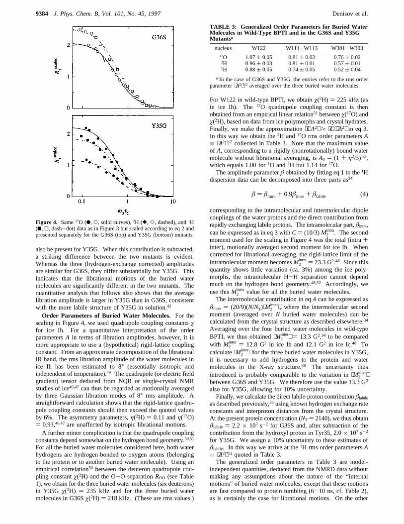
also be present for Y35G. When this contribution is subtracted,a striking difference between the two mutants is evident.Whereas the three (hydrogen-exchange corrected) amplitudesare similar for G36S, they differ substantially for Y35G. Thisindicates that the librational motions of the buried watermolecules are significantly different in the two mutants. Thequantitative analysis that follows also shows that the averagelibration amplitude is larger in Y35G than in G36S, consistentwith the more labile structure of Y35G in solution.42
Order Parameters of Buried Water Molecules. For thescaling in Figure 4, we used quadrupole coupling constantsøfor ice Ih. For a quantitative interpretation of the orderparametersA in terms of libration amplitudes, however, it ismore appropriate to use a (hypothetical) rigid-lattice couplingconstant. From an approximate decomposition of the librationalIR band, the rms libration amplitude of the water molecules inice Ih has been estimated to 8° (essentially isotropic andindependent of temperature).49 The quadrupole (or electric fieldgradient) tensor deduced from NQR or single-crystal NMRstudies of ice46,47can thus be regarded as motionally averagedby three Gaussian libration modes of 8° rms amplitude. Astraightforward calculation shows that the rigid-lattice quadru-pole coupling constants should then exceed the quoted valuesby 6%. The asymmetry parameters,η(2H) ) 0.11 andη(17O)) 0.93,46,47 are unaffected by isotropic librational motions.A further minor complication is that the quadrupole coupling
constants depend somewhat on the hydrogen bond geometry.50,51
For all the buried water molecules considered here, both waterhydrogens are hydrogen-bonded to oxygen atoms (belongingto the protein or to another buried water molecule). Using anempirical correlation50 between the deuteron quadrupole cou-pling constantø(2H) and the O-O separationROO (see Table1), we obtain for the three buried water molecules (six deuterons)in Y35G ø(2H) ) 235 kHz and for the three buried watermolecules in G36Sø(2H) ) 218 kHz. (These are rms values.)
For W122 in wild-type BPTI, we obtainø(2H) ) 225 kHz (asin ice Ih). The 17O quadrupole coupling constant is thenobtained from an empirical linear relation51 betweenø(17O) andø(2H), based on data from ice polymorphs and crystal hydrates.Finally, we make the approximation⟨CA2⟩ ≈ ⟨C⟩⟨A2⟩ in eq 3.In this way we obtain the2H and17O rms order parametersA≡ ⟨A2⟩1/2 collected in Table 3. Note that the maximum valueof A, corresponding to a rigidly (nonrotationally) bound watermolecule without librational averaging, isA0 ) (1 + η2/3)1/2,which equals 1.00 for1H and2H but 1.14 for17O.The amplitude parameterâ obtained by fitting eq 1 to the1H
dispersion data can be decomposed into three parts as34
corresponding to the intramolecular and intermolecular dipolecouplings of the water protons and the direct contribution fromrapidly exchanging labile protons. The intramolecular part,âintra,can be expressed as in eq 3 withC) (10/3)M2
intra. The secondmoment used for the scaling in Figure 4 was the total (intra+inter), motionally averaged second moment for ice Ih. Whencorrected for librational averaging, the rigid-lattice limit of theintramolecular moment becomesM2
intra ) 23.3 G2.48 Since thisquantity shows little variation (ca. 3%) among the ice poly-morphs, the intramolecular H-H separation cannot dependmuch on the hydrogen bond geometry.48,52 Accordingly, weuse thisM2
intra value for all the buried water molecules.The intermolecular contribution in eq 4 can be expressed as
âinter ) (20/9)(N/NT)⟨M2inter⟩, where the intermolecular second
moment (averaged overN buried water molecules) can becalculated from the crystal structure as described elsewhere.34
Averaging over the four buried water molecules in wild-typeBPTI, we thus obtained⟨M2
inter⟩ ) 13.3 G2,34 to be comparedwith M2
inter ) 12.8 G2 in ice Ih and 12.1 G2 in ice Ic.48 Tocalculate⟨M2
inter⟩ for the three buried water molecules in Y35G,it is necessary to add hydrogens to the protein and watermolecules in the X-ray structure.38 The uncertainty thusintroduced is probably comparable to the variation in⟨M2
inter⟩between G36S and Y35G. We therefore use the value 13.3 G2
also for Y35G, allowing for 10% uncertainty.Finally, we calculate the direct labile-proton contributionâlabile
as described previously,34 using known hydrogen exchange rateconstants and interproton distances from the crystal structure.At the present protein concentration (NT ) 2140), we thus obtainâlabile ) 2.2× 10⁷ s-2 for G36S and, after subtraction of thecontribution from the hydroxyl proton in Tyr35, 2.0× 10⁷ s-2
for Y35G. We assign a 10% uncertainty to these estimates ofâlabile. In this way we arrive at the1H rms order parametersA≡ ⟨A2⟩1/2 quoted in Table 3.The generalized order parameters in Table 3 are model-
independent quantities, deduced from the NMRD data withoutmaking any assumptions about the nature of the “internalmotions” of buried water molecules, except that these motionsare fast compared to protein tumbling (6-10 ns, cf. Table 2),as is certainly the case for librational motions. On the other
Figure 4. Same17O (b, O, solid curves),2H ([, ], dashed), and1H(9, 0, dash-dot) data as in Figure 3 but scaled according to eq 2 andpresented separately for the G36S (top) and Y35G (bottom) mutants.
TABLE 3: Generalized Order Parameters for Buried WaterMolecules in Wild-Type BPTI and in the G36S and Y35GMutantsa
nucleus W122 W111-W113 W301-W30317O 1.07( 0.05 0.81( 0.02 0.76( 0.022H 0.96( 0.03 0.81( 0.01 0.57( 0.011H 0.88( 0.05 0.74( 0.05 0.52( 0.04
a In the case of G36S and Y35G, the entries refer to the rms orderparameter⟨A2⟩1/2 averaged over the three buried water molecules.
â ) âintra + 0.9âinter + âlabile (4)
9384 J. Phys. Chem. B, Vol. 101, No. 45, 1997 Denisov et al.
hand, water motions that are slower than protein tumbling donot affect the spin relaxation rate. The flipping of a buried watermolecule 180° around itsC2 axis is usually in this category asare the motions that constitute the actual exchange event.Nevertheless, buried water exchange can affect the measuredspin relaxation rate even if it is much slower than proteintumbling. This is the case if the mean residence time (theinverse exchange rate) is not much shorter than the intrinsicspin relaxation time at zero frequency,20 about 5µs, 0.3 ms,and 5 ms for17O, 2H, and1H, respectively, under the presentconditions. The fast-exchange limit assumed in the derivationeq 3 is then no longer applicable. If eq 3 is used under suchconditions, it will produce an apparent order parameter that issmaller than the true value. If NMRD profiles are recorded atdifferent temperatures, it can be established whether a buriedwater molecule is in the fast-exchange limit. For the deeplyburied water molecule W122 in wild-type BPTI, it was thusfound that the residence time is between the intrinsic relaxationtimes of 17O and2H at room temperature.22 The 17O and2Horder parameters for W122 included in Table 3 were obtainedfrom temperature-dependent difference dispersions and by takingintermediate-exchange effects into account.22 For 1H, the fast-exchange limit is applicable at 27°C even for W122.34 In usingeq 3 to derive order parameters, we assume that the six buriedwater molecules in the G36S and Y35G mutants are in the fast-exchange limit for all three nuclei at 27°C. Although notdemonstrated explicitly, this assumption is supported by thecrystal structures, suggesting that W122 exchanges more slowlythan the other buried water molecules. Furthermore, a deviationfrom the fast-exchange limit would mainly suppress the (ap-parent) order parameter for17O (since this nucleus has theshortest intrinsic relaxation time). Such a trend is not evidentin Table 3.Libration Amplitudes for Buried Water Molecules. If the
orientational disorder of the buried water molecules is aniso-tropic, we expect theA/A0 ratios for the three nuclei to differ.The results in Table 3 show that this is the case for both mutants.Moreover, the buried waters in Y35G appear to be significantlymore disordered than those in G36S.To obtain a quantitative measure of the degree and anisotropy
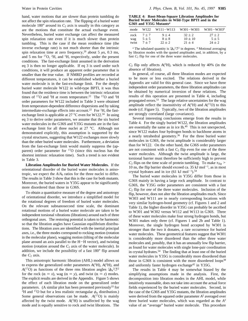
of orientational disorder, we introduce a simplified model forthe rotational degrees of freedom of buried water molecules.On the relevant subnanosecond time scale, the dominantrotational motions of a buried water molecule are taken to beindependent torsional vibrations (librations) around each of threeorthogonal axes. The restoring potential is taken to be harmonicso that the libration angles have Gaussian equilibrium distribu-tions. The libration axes are identified with the inertial principalaxes, i.e., the three modes correspond to rocking motion (rotationin the molecular plane), wagging motion (tilting of the molecularplane around an axis parallel to the H-H vector), and twistingmotion (rotation around theC2 axis of the water molecule). Inaddition, we include the possibility of a fast 180° flip aroundtheC2 axis.This anisotropic harmonic libration (AHL) model allows us
to express the generalized order parametersA(1H), A(2H), andA(17O) as functions of the three rms libration angles⟨φR
2⟩1/2for the rock (R ) x), wag (R ) y), and twist (R ) z) modes.The explicit results are derived in the Appendix. Figure 5 showsthe effect of each libration mode on the generalized orderparameters. (A similar plot has been presented previously33 for2H and17O but for a less realistic rectangularφR distribution.)Some general observations can be made.A(17O) is mainlyaffected by the twist mode.A(1H) is unaffected by the wagmode and is equally sensitive to rock and twist librations. The
C2 flip only affectsA(2H), which is reduced by 40% (in theabsence of libration).In general, of course, all three libration modes are expected
to be more or less excited. The relations derived in theAppendix are valid for this general case. Since we have threeindependent order parameters, the three libration amplitudes canbe obtained by numerical inversion of these relations. Theresults of this operation are presented in Table 4, includingpropagated errors.53 The large relative uncertainties for the wagamplitude reflect the insensitivity ofA(1H) andA(17O) to thismode (cf. Figure 5). Typically, two of the libration amplitudesare strongly correlated (large covariance).Several interesting conclusions emerge from the results in
Table 4. For the singly buried W122, the libration amplitudesare essentially the same as in ice Ih.49 This is not unexpected,since W122 makes four hydrogen bonds to backbone atoms ina nearly tetrahedral geometry.36 For the three buried watermolecules in G36S, the twist amplitude is considerably largerthan for W122. On the other hand, the G36S order parametersare not consistent with a fastC2 flip even for one of the threewater molecules. Although the twist amplitude is large, thetorsional barrier must therefore be sufficiently high to preventC2 flips on the time scale of protein tumbling. To makeτflip >10 ns, the flip barrier should exceed ca. 35 kJ mol-1, as in mostcrystal hydrates and in ice (61 kJ mol-1).54
The buried water molecules in Y35G differ from those inG36S mainly in having a large rock amplitude. In contrast toG36S, the Y35G order parameters are consistent with a fastC2 flip for oneof the three water molecules. Inclusion of theflip, however, does not alter the libration amplitudes much. SinceW303 and W111 are in nearly corresponding locations withvery similar hydrogen-bond geometry (cf. Figures 1 and 2 andTable 1), the higher disorder in Y35G can probably be ascribedto W301 and W302 versus W112 and W113 in G36S. Threeof these water molecules make four strong hydrogen bonds, butW301 makes only three (cf. Figures 1 and 2b and Table 1).Moreover, the single hydrogen bond accepted by W301 isstronger than the two it donates, a rare occurrence for buriedwater molecules. These geometrical features suggest that W301is considerably more disordered than the other three watermolecules and, possibly, that it has an unusually low flip barrier,as found for water molecules with single lone-pair coordinationin crystal hydrates.54 The finding that at least one of the buriedwater molecules in Y35G is considerably more disordered thanthose in G36S is consistent with the more disordered loops38
and uniformly faster hydrogen exchange42 in Y35G.The results in Table 4 may be somewhat biased by the
simplifying assumptions made in the analysis. First, thedecomposition into libration modes in the AHL model, whileintuitively reasonable, does not take into account the actual forcefields experienced by the buried water molecules. Second, inthe case of the G36S and Y35Gmutants, the libration amplitudeswere derived from the squared order parameterA2 averaged overthree buried water molecules, which was regarded as theA2
value of an “average” buried water molecule. This procedure
TABLE 4: Root-Mean-Square Libration Amplitudes forBuried Water Molecules in Wild-Type BPTI and in theG36S and Y35G Mutantsa
mode W122 W111-W113 W301-W303 W301-W303b
rock 7( 7 9( 4 32( 2 27( 2wag 5( 5 8( 8 10( 10 5( 5twist 7( 7 22( 2 21( 4 24( 2
a The tabulated quantity is⟨φR2⟩1/2 in degrees.bMotional averaging
by libration modes with the quoted amplitudes and, in addition, by afastC2 flip for one of the three water molecules.
Water in Protein Cavities J. Phys. Chem. B, Vol. 101, No. 45, 19979385
is clearly not rigorous. Nevertheless, we believe that neitherthe general conclusions nor the rotational entropy estimatespresented below are seriously compromised by these simplifica-tions.Thermodynamics of Cavity Hydration. According to
classical statistical mechanics,55 the orientational distributionsf(φR) that determine the order parametersA(N) are associatedwith a configurational entropy (per molecule)
Owing to the small moments of inertia of the water molecule,quantum corrections are important and the rotational entropySR (per molecule) for the AHL model is given more accuratelyby31,56
with the dimensionless quantityCR ) pωR/(kBT) related to themode amplitudes⟨φR
2⟩1/2 and the moments of inertiaIR through
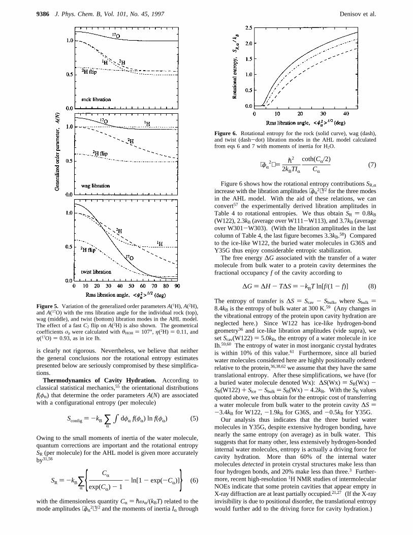
Figure 6 shows how the rotational entropy contributionsSR,Rincrease with the libration amplitudes⟨φR
2⟩1/2 for the three modesin the AHL model. With the aid of these relations, we canconvert57 the experimentally derived libration amplitudes inTable 4 to rotational entropies. We thus obtainSR ) 0.8kB(W122), 2.3kB (average over W111-W113), and 3.7kB (averageover W301-W303). (With the libration amplitudes in the lastcolumn of Table 4, the last figure becomes 3.3kB.58) Comparedto the ice-like W122, the buried water molecules in G36S andY35G thus enjoy considerable entropic stabilization.The free energy∆G associated with the transfer of a water
molecule from bulk water to a protein cavity determines thefractional occupancyf of the cavity according to
The entropy of transfer is∆S ) Scav - Sbulk, whereSbulk )8.4kB is the entropy of bulk water at 300 K.59 (Any changes inthe vibrational entropy of the protein upon cavity hydration areneglected here.) Since W122 has ice-like hydrogen-bondgeometry36 and ice-like libration amplitudes (vide supra), wesetScav(W122)) 5.0kB, the entropy of a water molecule in iceIh.59,60 The entropy of water in most inorganic crystal hydratesis within 10% of this value.61 Furthermore, since all buriedwater molecules considered here are highly positionally orderedrelative to the protein,36,38,62we assume that they have the sametranslational entropy. After these simplifications, we have (fora buried water molecule denoted Wx):∆S(Wx) ) SR(Wx) -SR(W122)+ Sice- Sbulk ) SR(Wx) - 4.2kB. With theSR valuesquoted above, we thus obtain for the entropic cost of transferringa water molecule from bulk water to the protein cavity∆S)-3.4kB for W122,-1.9kB for G36S, and-0.5kB for Y35G.Our analysis thus indicates that the three buried water
molecules in Y35G, despite extensive hydrogen bonding, havenearly the same entropy (on average) as in bulk water. Thissuggests that for many other, less extensively hydrogen-bondedinternal water molecules, entropy is actually a driving force forcavity hydration. More than 60% of the internal watermoleculesdetectedin protein crystal structures make less thanfour hydrogen bonds, and 20% make less than three.3 Further-more, recent high-resolution1H NMR studies of intermolecularNOEs indicate that some protein cavities that appear empty inX-ray diffraction are at least partially occupied.21,27 (If the X-rayinvisibility is due to positional disorder, the translational entropywould further add to the driving force for cavity hydration.)
Figure 5. Variation of the generalized order parametersA(1H), A(2H),andA(17O) with the rms libration angle for the individual rock (top),wag (middle), and twist (bottom) libration modes in the AHL model.The effect of a fastC2 flip on A(2H) is also shown. The geometricalcoefficientsσp were calculated withθHOH ) 107°, η(2H) ) 0.11, andη(17O) ) 0.93, as in ice Ih.
Sconfig ) -kB ∑R∫ dφR f(φR) ln f(φR) (5)
SR ) -kB∑R CR
exp(CR) - 1- ln[1 - exp(-CR)] (6)
Figure 6. Rotational entropy for the rock (solid curve), wag (dash),and twist (dash-dot) libration modes in the AHL model calculatedfrom eqs 6 and 7 with moments of inertia for H2O.
⟨φR2⟩ ) p2
2kBTIR
coth(CR/2)
CR(7)
∆G) ∆H - T∆S) -kBT ln[f/(1- f)] (8)
9386 J. Phys. Chem. B, Vol. 101, No. 45, 1997 Denisov et al.
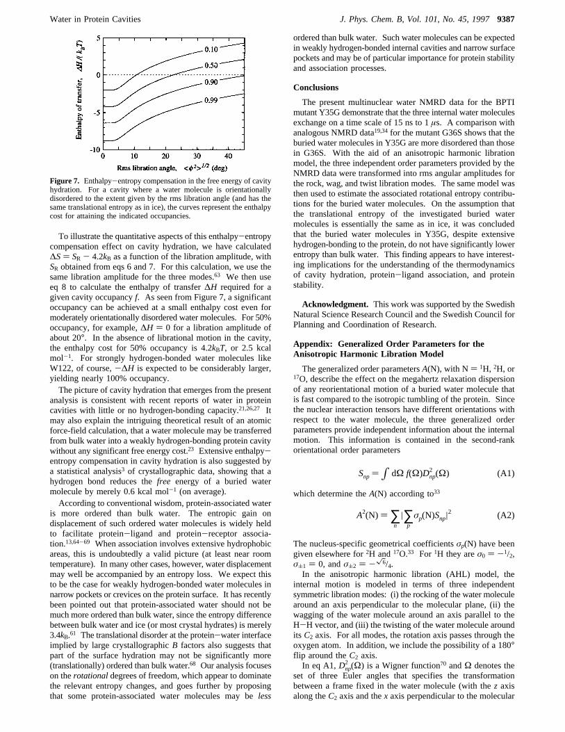
To illustrate the quantitative aspects of this enthalpy-entropycompensation effect on cavity hydration, we have calculated∆S) SR - 4.2kB as a function of the libration amplitude, withSR obtained from eqs 6 and 7. For this calculation, we use thesame libration amplitude for the three modes.63 We then useeq 8 to calculate the enthalpy of transfer∆H required for agiven cavity occupancyf. As seen from Figure 7, a significantoccupancy can be achieved at a small enthalpy cost even formoderately orientationally disordered water molecules. For 50%occupancy, for example,∆H ) 0 for a libration amplitude ofabout 20°. In the absence of librational motion in the cavity,the enthalpy cost for 50% occupancy is 4.2kBT, or 2.5 kcalmol-1. For strongly hydrogen-bonded water molecules likeW122, of course,-∆H is expected to be considerably larger,yielding nearly 100% occupancy.The picture of cavity hydration that emerges from the present
analysis is consistent with recent reports of water in proteincavities with little or no hydrogen-bonding capacity.21,26,27 Itmay also explain the intriguing theoretical result of an atomicforce-field calculation, that a water molecule may be transferredfrom bulk water into a weakly hydrogen-bonding protein cavitywithout any significant free energy cost.23 Extensive enthalpy-entropy compensation in cavity hydration is also suggested bya statistical analysis3 of crystallographic data, showing that ahydrogen bond reduces thefree energy of a buried watermolecule by merely 0.6 kcal mol-1 (on average).According to conventional wisdom, protein-associated water
is more ordered than bulk water. The entropic gain ondisplacement of such ordered water molecules is widely heldto facilitate protein-ligand and protein-receptor associa-tion.13,64-69 When association involves extensive hydrophobicareas, this is undoubtedly a valid picture (at least near roomtemperature). In many other cases, however, water displacementmay well be accompanied by an entropy loss. We expect thisto be the case for weakly hydrogen-bonded water molecules innarrow pockets or crevices on the protein surface. It has recentlybeen pointed out that protein-associated water should not bemuch more ordered than bulk water, since the entropy differencebetween bulk water and ice (or most crystal hydrates) is merely3.4kB.61 The translational disorder at the protein-water interfaceimplied by large crystallographicB factors also suggests thatpart of the surface hydration may not be significantly more(translationally) ordered than bulk water.68 Our analysis focuseson therotationaldegrees of freedom, which appear to dominatethe relevant entropy changes, and goes further by proposingthat some protein-associated water molecules may beless
ordered than bulk water. Such water molecules can be expectedin weakly hydrogen-bonded internal cavities and narrow surfacepockets and may be of particular importance for protein stabilityand association processes.
Conclusions
The present multinuclear water NMRD data for the BPTImutant Y35G demonstrate that the three internal water moleculesexchange on a time scale of 15 ns to 1µs. A comparison withanalogous NMRD data19,34for the mutant G36S shows that theburied water molecules in Y35G are more disordered than thosein G36S. With the aid of an anisotropic harmonic librationmodel, the three independent order parameters provided by theNMRD data were transformed into rms angular amplitudes forthe rock, wag, and twist libration modes. The same model wasthen used to estimate the associated rotational entropy contribu-tions for the buried water molecules. On the assumption thatthe translational entropy of the investigated buried watermolecules is essentially the same as in ice, it was concludedthat the buried water molecules in Y35G, despite extensivehydrogen-bonding to the protein, do not have significantly lowerentropy than bulk water. This finding appears to have interest-ing implications for the understanding of the thermodynamicsof cavity hydration, protein-ligand association, and proteinstability.
Acknowledgment. This work was supported by the SwedishNatural Science Research Council and the Swedish Council forPlanning and Coordination of Research.
Appendix: Generalized Order Parameters for theAnisotropic Harmonic Libration Model
The generalized order parametersA(N), with N ) 1H, 2H, or17O, describe the effect on the megahertz relaxation dispersionof any reorientational motion of a buried water molecule thatis fast compared to the isotropic tumbling of the protein. Sincethe nuclear interaction tensors have different orientations withrespect to the water molecule, the three generalized orderparameters provide independent information about the internalmotion. This information is contained in the second-rankorientational order parameters
which determine theA(N) according to33
The nucleus-specific geometrical coefficientsσp(N) have beengiven elsewhere for2H and17O.33 For 1H they areσ0 ) -1/2,σ(1 ) 0, andσ(2 ) -x6/4.In the anisotropic harmonic libration (AHL) model, the
internal motion is modeled in terms of three independentsymmetric libration modes: (i) the rocking of the water moleculearound an axis perpendicular to the molecular plane, (ii) thewagging of the water molecule around an axis parallel to theH-H vector, and (iii) the twisting of the water molecule aroundits C2 axis. For all modes, the rotation axis passes through theoxygen atom. In addition, we include the possibility of a 180°flip around theC2 axis.In eq A1,Dnp
2 (Ω) is a Wigner function70 andΩ denotes theset of three Euler angles that specifies the transformationbetween a frame fixed in the water molecule (with thez axisalong theC2 axis and thex axis perpendicular to the molecular
Figure 7. Enthalpy-entropy compensation in the free energy of cavityhydration. For a cavity where a water molecule is orientationallydisordered to the extent given by the rms libration angle (and has thesame translational entropy as in ice), the curves represent the enthalpycost for attaining the indicated occupancies.
Snp )∫ dΩ f(Ω)Dnp2 (Ω) (A1)
A2(N) ) ∑n
|∑p
σp(N)Snp|2 (A2)
Water in Protein Cavities J. Phys. Chem. B, Vol. 101, No. 45, 19979387
plane) and a frame that coincides with the molecular frame inthe equilibrium configuration (zero libration angles). In theAHL model, the angular variables are the libration anglesφx,φy, andφz for the rock, wag, and twist modes, respectively. Theorder parametersSnp can be expressed in terms of these variablesas
where the angular brackets denote averages over the appropriateequilibrium distributionf(φR), R ) x, y, or z. Owing to thenoncommutability of finite rotations, the order parameters inthe AHL model depend on the order in which the rotations areapplied. (The result in eq A3 corresponds to the orderφx firstand φz last.) For the libration amplitudes of interest here,however, this dependence is very weak and can be neglected.On account of the symmetry of the libration modes (with
respect toφR ) 0), there are only five (rather than 25)independent order parameters, namely,
In the presence of aC2 flip, the order parametersSnpmust alsoreflect theC2V symmetry of the water molecule, which requiresp to be even. The only effect of the flip is thus to makeS11 +S1-1 ) 0.In the AHL model, the five order parameters in eq A4 are
not independent. They are all determined by three parameters,which we take as the rms amplitudes⟨φR
2⟩1/2 of the librationmodes. The orientational distribution function for each modeis of the form
This distribution is normalized on the unrestricted interval-∞< φR < ∞, rather than on-π e φR e π. The error introducedby this approximation is negligible for the libration amplitudesof interest here (say,⟨φR
2⟩1/2 < 45°). For the Gaussiandistribution A5, the five order parameters in eq A4 can beexpressed in terms of the orientational averages
with n ) 1 or 2.In the rigid-lattice limit, i.e., in the absence of librational
motion, we haveSnp ) δnp and the preceding results for the
AHL model reduce correctly to
with η the asymmetry parameter of the nuclear interactiontensor.In the free-rotation limit, with a uniformφR distribution, the
AHL model yieldsA2(N) ) [σ0(N) - x6σ2(N)]2/4 for the rock
mode,A2(N) ) [σ0(N) + x6σ2(N)]2/4 for the wag mode, and
A2(N) ) [σ0(N)]2 for the twist mode.33 If all three librationangles are uniformly distributed, the AHL model yieldsA2(N)) [σ0(N) - x6σ2(N)]
2/64, i.e.,A(1H) ) 1/8, A(2H) ) (1 + η)/8, andA(17O)) 1/4. This differs from the isotropic limit, whereA(N) ) 0. The difference is unimportant, however, for thelibration amplitudes of interest here.
References and Notes
(1) Connolly, M. L. Int. J. Pept. Protein Res. 1985, 28, 360-363.(2) Rashin, A. A.; Iofin, M.; Honig, B.Biochemistry1986, 25, 3619-
3625.(3) Williams, M. A.; Goodfellow, J. M.; Thornton, J. M.Protein Sci.
1994, 3, 1224-1235.(4) Hubbard, S. J.; Gross, K.-H.; Argos, P.Protein Eng. 1994, 7, 613-
626. Hubbard, S. J.; Argos, P.Protein Eng. 1995, 8, 1011-1015.(5) Baker, E. N. InProtein-SolVent Interactions; Gregory, R. B., Ed.;
M. Dekker: New York, 1995; Chapter 2.(6) Schoenborn, B. P.; Garcia, A.; Knott, R.Prog. Biophys.Mol. Biol.
1995, 64, 105-119.(7) McDowell, R. S.; Kossiakoff, A. A.J.Mol. Biol. 1995, 250, 553-
570.(8) Elber, R.; Karplus, M.J. Am. Chem. Soc. 1990, 112, 9161-9175.(9) Carlson, M. L.; Regan, R. M.; Gibson, Q. H.Biochemistry1996,
35, 1125-1136.(10) Gerstein, M.; Lesk, A. M.; Chothia, C.Biochemistry1994, 33,
6739-6749.(11) Hubbard, S. J.; Argos, P.J. Mol. Biol. 1996, 261, 289-300.(12) Meyer, E.Protein Sci. 1992, 1, 1543-1562.(13) Ladbury, J. E.Chem. Biol. 1996, 3, 973-980.(14) Raymer, M. L.; Sanschagrin, P. C.; Punch, W. F.; Venkataraman,
S.; Goodman, E. D.; Kuhn, L. A.J. Mol. Biol. 1997, 265, 445-464.(15) Otting, G.; Wuthrich, K. J. Am. Chem. Soc. 1989, 111, 1871-
1875.(16) Otting, G.; Liepinsh, E.; Wu¨thrich, K. Science1991, 254, 974-
980.(17) Otting, G.; Liepinsh, E.Acc. Chem. Res. 1995, 28, 171-177.(18) Denisov, V. P.; Halle, B.J. Mol. Biol. 1995, 245, 682-697.(19) Denisov, V. P.; Halle, B.; Peters, J.; Ho¨rlein, H. D.Biochemistry
1995, 34, 9046-9051.(20) Denisov, V. P.; Halle, B.Faraday Discussions1996, 103, 227-
244.(21) Otting, G.; Liepinsh, E.; Halle, B.; Frey, U.Nature Struct. Biol.
1997, 4, 396-404.(22) Denisov, V. P.; Peters, J.; Ho¨rlein, H. D.; Halle, B.Nature Struct.
Biol. 1996, 3, 505-509.(23) Wade, R. C.; Mazor, M. H.; McCammon, J. A.; Quiocho, F. A.
Biopolymers1991, 31, 919-931.(24) Zhang, L.; Hermans, J.Proteins: Struct. Funct.Gen. 1996, 24, 433-
438.(25) Wolfenden, R.; Radzicka, A.Science1994, 265, 936-937.(26) Buckle, A. M.; Cramer, P.; Fersht, A. R.Biochemistry1996, 35,
4298-4305.(27) Ernst, J. A.; Clubb, R. T.; Zhou, H.-X.; Gronenborn, A. M.; Clore,
G. M. Science1995, 267, 1813-1817.(28) Berne, B. J.; Pechukas, P.; Harp, G. D.J. Chem. Phys. 1968, 49,
3125-3129.(29) Zannoni, C. InThe Molecular Dynamics of Liquid Crystals;
Luckhurst, G. R., Veracini, C. A., Eds.; Kluwer: Dordrecht, 1994; Chapter2.
(30) Akke, M.; Bruschweiler, R.; Palmer, A. G.J. Am.Chem. Soc. 1993,115, 9832-9833.
(31) Yang, D.; Kay, L. E.J. Mol. Biol. 1996, 263, 369-382.(32) Li, Z.; Raychaudhuri, S.; Wand, A. J.Protein Sci. 1996, 5, 2647-
2650.(33) Denisov, V. P.; Halle, B.J. Am. Chem. Soc. 1995, 117, 8456-
8465.(34) Venu, K.; Denisov, V. P.; Halle, B.J. Am. Chem. Soc. 1997, 119,
3122-3134.(35) Denisov, V. P.; Halle, B.J. Mol. Biol. 1995, 245, 698-709.
Snp ) δn+p,even⟨cos(nφz)⟩∑q
δq+p,even(-1)(p-q)/2×
⟨dnq2 (φy)⟩⟨dqp
2 (φx)⟩ (A3)
S00 ) 1- 32[⟨sin2 φx⟩ + ⟨sin2 φy⟩ - ⟨sin2 φx⟩⟨sin
2φy⟩] (A4a)
S02 ) -x64[-⟨sin2 φx⟩ + ⟨sin2 φy⟩ + ⟨sin2 φx⟩⟨sin
2φy⟩]
(A4b)
S20 )x64[1 - 2⟨sin2 φz⟩] ×[-⟨sin2 φx⟩ + ⟨sin2 φy⟩ - ⟨sin2 φx⟩⟨sin
2φy⟩] (A4c)
S22 + S2-2 ) 12[1 - 2⟨sin2 φz⟩] ×
[2 - ⟨sin2 φx⟩ - ⟨sin2 φy⟩ - ⟨sin2 φx⟩⟨sin2φy⟩] (A4d)
S11 + S1-1 ) ⟨cosφz⟩⟨cosφy⟩[1 - 2⟨sin2 φx⟩] (A4e)
f(φR) ) 1
(2π⟨φR2⟩)1/2
exp(-φR
2
2⟨φR2⟩) (A5)
⟨cos(nφR)⟩ ) exp(- n2
2⟨φR
2⟩) (A6)
A2(N) ) 1+ η2(N)/3 (A7)
9388 J. Phys. Chem. B, Vol. 101, No. 45, 1997 Denisov et al.
(36) Wlodawer, A.; Walter, J.; Huber, R.; Sjo¨lin, L. J.Mol. Biol. 1984,180, 301-329.
(37) Berndt, K. D.; Beunink, J.; Schro¨der, W.; Wuthrich, K.Biochemistry1993, 32, 4564-4570.
(38) Housset, D.; Kim, K.-S.; Fuchs, J.; Woodward, C.; Wlodawer, A.J. Mol. Biol. 1991, 220, 757-770.
(39) Lumry, R.; Rajender, S.Biopolymers1970, 9, 1125-1227.(40) Dunitz, J. D.Chem. Biol. 1995, 2, 709-712.(41) Berndt, K. D.; Gu¨ntert, P.; Orbons, L. P. M.; Wu¨thrich, K. J.Mol.
Biol. 1992, 227, 757-775.(42) Kim, K.-S.; Fuchs, J. A.; Woodward, C. K.Biochemistry1993,
32, 9600-9608.(43) Abragam, A.The Principles of Nuclear Magnetism: Clarendon
Press: Oxford, 1961.(44) Halle, B.; Wennerstro¨m, H. J. Magn. Reson. 1981, 44, 89-100.(45) Halle, B.; Wennerstro¨m, H. J. Chem. Phys. 1981, 75, 1928-1943.(46) Spiess, H. W.; Garrett, B. B.; Sheline, R. K.; Rabideau, S. W.J.
Chem. Phys. 1969, 51, 1201-1205. Edmonds, D. T.; Zussman, A.Phys.Lett. 1972, 41A, 167-169.
(47) Edmonds, D. T.; Mackay, A. L.J. Magn. Reson. 1975, 20, 515-519.
(48) Whalley, E.Mol. Phys. 1974, 28, 1105-1108.(49) Sceats, M. G.; Rice, S. A.J. Chem. Phys. 1980, 72, 3236-3247.(50) Berglund, B.; Lindgren, J.; Tegenfeldt, J.J.Mol. Struct. 1978, 43,
179-191.(51) Poplett, I. J. F.J. Magn. Reson. 1982, 50, 397-408.(52) Kuhs, W. F.; Lehmann, M. S.Water Sci. ReV. 1986, 2, 1-65.(53) The libration amplitude bounds in Table 4 are mainly determined
by A(2H) andA(17O) and depend only weakly on the less precisely definedA(1H). If the estimated uncertainties in⟨M2
inter⟩ andâlabile are taken to be20% rather than 10%, the propagated error inA(1H) is doubled but theeffect on the libration amplitudes is barely significant. For Y35G (withoutC2 flip), for example, we obtain 32( 2°, 13( 13°, and 20( 5° for 20%uncertainty in⟨M2
inter⟩ andâlabile, compared with the values in the fourthcolumn of Table 4.
(54) Larsson, K.; Tegenfeldt, J.; Hermansson, K.J.Chem. Soc., FaradayTrans. 1991, 87, 1193-1200.
(55) Hill, T. L. An Introduction to Statistical Thermodynamics; Dover:New York, 1986.
(56) Landau, L. D.; Lifshitz, E. M.Statistical Physics, 3rd ed.;Pergamon: Oxford, 1980; Part 1.
(57) All thermodynamic quantities refer to H2O. With its twice as largemoments of inertia, D2O should have 10-15% larger libration amplitudes.Since this difference is comparable to or smaller than the propagateduncertainties in Table 4, we ignore the isotope effect.
(58) Since theC2 flip interconverts two indistinguishable states, thereis no associated configurational entropy.
(59) Eisenberg, D.; Kauzmann, W.The Structure and Properties ofWater; Clarendon: Oxford, 1969.
(60) The entropy of ice Ih at 273 K is 5.0kB from which a contributionof 0.4kB, due to residual proton disorder, should be subtracted and anothercontribution of 0.4kB, from the extrapolation to 300 K (using the heatcapacity of ice Ih), should be added.59
(61) Dunitz, J. D.Science1994, 264, 670.(62) The Debye-Waller factors for the water oxygens are 11.1 Å2
(W122), 13.4 Å2 (average for W111-W113), and 9.1 Å2 (average forW301-W303).36,38 To compensate for differences in lattice disorderbetween the BPTI and Y35G crystals, the difference, 7- 2) 5 Å2, betweenthe smallestB factors in the two proteins should be added to the lattervalue.71
(63) For relatively large libration amplitudes, this is actually norestriction, since the classical limit of eq 6 depends only on the geometricaverage of the three libration amplitudes.
(64) Finkelstein, A. V.; Janin, J.Protein Eng. 1989, 3, 1-3.(65) Searle, M. S.; Williams, D. H.J. Am. Chem. Soc. 1992, 114,
10690-10697.(66) Gilli, P.; Ferretti, V.; Gilli, G.; Borea, P. A.J. Phys. Chem. 1994,
98, 1515-1518.(67) Chervenak, M. C.; Toone, E. J.J. Am. Chem. Soc. 1994, 116,
10533-10539.(68) Ringe, D.Curr. Opin. Struct. Biol. 1995, 5, 825-829.(69) Tame, J. R. H.; Sleigh, S. H.; Wilkinson, A. J.; Ladbury, J. E.Nature
Struct. Biol. 1996, 3, 998-1001.(70) Brink, D. M.; Satchler, G. R.Angular Momentum, 2nd ed.;
Clarendon: Oxford, 1968.(71) Ringe, D.; Petsko, G. A.Prog. Biophys.Mol. Biol. 1985, 45, 197-
235.(72) Beeser, S. A.; Goldenberg, D. P.; Oas, T. G.J. Mol. Biol. 1997,
269, 154-164.
Water in Protein Cavities J. Phys. Chem. B, Vol. 101, No. 45, 19979389
Recommended


