www.impactjournals.com/oncotarget/ Oncotarget, July 2010, Vol. 1, No 3
www.impactjournals.com/oncotarget 175 Oncotarget 2010; 1: 175-184
INTRODUCTION
POLQ (DNA Polymerase Theta) is a member of the A family of DNA polymerases, which, unusually for this class of polymerases, synthesizes DNA with very low fidelity [1, 2]. The precise physiological functions of this protein are currently unclear. It has previously been suggested that mice deficient in POLQ had a substantially decreased frequency of mutations in immunoglobulin genes [3, 4]. However a recent study found that mutation types and frequencies were similar in wild type, POLQ-/-, POLH-/-, and POLQ-/- POLH-/- mice [5]. Accordingly this group suggested that
POLQ does not have a significant role in the hypermutation pathway.
It has been suggested that POLQ has a role in base excision repair (BER) but this also remains unresolved. It has previously been shown in the DT40 chicken B cell lymphoma line, that POLQ/POLβ mutants had significantly higher sensitivity to methyl methanesulfonate than either single mutant. Extracts obtained from this cell line were used to show that POLQ mutant cells have markedly reduced single nucleotide BER capacity in vitro and that this reduction was of a similar magnitude to cells deficient in POLβ
Overexpression of POLQ Confers a Poor Prognosis in Early Breast Cancer Patients Geoff S Higgins1, Adrian L Harris2, Remko Prevo1, Thomas Helleday1, W Gillies McKenna*1 and Francesca M Buffa*2
1 Gray Institute for Radiation Oncology and Biology 2 Molecular Oncology Laboratories, University of Oxford, UK Correspondence to: Professor W. Gillies McKenna, e-mail: [email protected] Dr Francesca M Buffa, e-mail: [email protected] Keywords: Translational research, POLQ, breast cancer, prognosis, radiotherapy Received: June 7, 2010, Accepted: June 27, 2010, Published: July 9, 2010 Copyright: C 2010 Higgins et al. This is an open-access article distributed under the terms of the Creative Commons Attribution License,
which permits unrestricted use, distribution, and reproduction in any medium, provided the original author and source are credited.
ABSTRACT: Depletion of POLQ (DNA polymerase theta) has recently been shown to render tumour cells more sensitive to radiotherapy whilst having little or no effect on normal tissues. This finding led us to investigate whether tumours that overexpress POLQ are associated with an adverse outcome. We therefore correlated the clinical outcomes of two retrospective series of patients with early breast cancer with the expression levels of POLQ, as determined by microarray gene expression analysis. We found that a significant number of tumours overexpressed POLQ and that overexpression was correlated with ER negative disease (p=0.047) and high tumour grade (p=0.004), both of which are associated with poor clinical outcomes. POLQ overexpression was associated with poor relapse free survival rates on both univariate (HR 5.80; 95% CI, 2.220 to 15.159; p<0.001) and multivariate analysis (HR 8.086; 95% CI 2.340 to 27.948 p=0.001). Analysis of other published clinical series confirmed that POLQ overexpression is associated with adverse clinical outcomes. The poor prognosis associated with POLQ is independent of other clinical or pathological features. The mechanism that causes this adverse outcome remains to be elucidated but may in part arise from resistance to adjuvant treatment. These findings, combined with the limited normal tissue expression of POLQ, make it a very appealing target for possible clinical exploitation.
www.impactjournals.com/oncotarget 176 Oncotarget 2010; 1: 175-184
[6]. These findings led to the suggestion that POLQ and POLβ cooperate in BER.
Recent biochemical analysis has shown that cloned full-length human POLQ as well as a C-terminal fragment of POLQ, have 5’-deoxyribose phosphate (5’-dRP) lyase activity. The full-length protein and the C-terminal fragment were shown to have BER activity in vitro [7]. Although these findings have been used to support the argument that POLQ may have a role in BER in vivo, it should be noted that the rate of 5’-dRP lyase activity of POLQ is approximately 40 fold slower than that of POLβ. Cells with deficiencies in the BER pathway have been shown to have increased sensitivity to temozolomide [8]. Since cells depleted of POLQ do not show hypersensitivity to this drug, it has been questioned as to whether POLQ has any physiologically significant role in BER [9].
We have recently published a siRNA screen that aimed to identify molecular determinants of tumour radiosensitivity [9]. This study demonstrated that POLQ siRNA transfection resulted in radiosensitisation of a panel of tumour cell lines but had little or no effect on normal tissue lines. These differences reflect previous work showing significant disparity in expression between normal tissues and tumour cells [10]. Normal tissue expression appears to be mainly limited to lymphoid tissues such as the fetal liver, thymus, and bone marrow. However POLQ is known to be overexpressed in a large proportion of tumours derived from patients with colon, lung, and gastric cancer.
In view of the in vitro evidence linking POLQ expression to tumour cell radioresistance, we hypothesised that POLQ overexpression may increase the likelihood of treatment failure in cancer patients, and therefore confer an adverse clinical prognosis.
We therefore correlated the clinical outcomes of two series of breast cancer patients (n=279 in total) with the expression levels of POLQ as determined by microarray gene expression analysis. We also analysed the pathways associated with POLQ expression in vivo by data-mining gene expression data from published breast cancer studies (n=1015 samples). To the best of our knowledge this is the first study to demonstrate that POLQ is overexpressed in breast cancer, that its overexpression confers a significant adverse prognosis, and that it is associated with key cancer pathways.
MATERIALS AND METHODS. Ethics Statement
Informed consent was obtained and all clinical investigations were conducted according to the ethical standards and principles expressed in the Declaration of Helsinki. Ethical approval was obtained from the local research ethics committee.
Patient Details
Individual tumour samples were obtained from
retrospective series of patients with early primary breast cancer who were treated in Oxford, UK, between 1989 and 1998. Patients received adjuvant chemotherapy and/or adjuvant hormone therapy, or no adjuvant treatment. Tamoxifen was used as endocrine therapy for 5 years in estrogen receptor (ER) positive
patients. Patients who were 50 years of age, with lymph node positive tumors, or ER– and/or a primary tumor >3 cm in diameter, received adjuvant cyclophosphamide, methotrexate, and 5-fluorouracil (CMF) for six cycles, in a three weekly intravenous
regimen. Patients 50 years of age with ER–, lymph node–positive tumors also received CMF. Two series of 152 (Series 1) and 127 (Series 2) samples respectively were analysed. Series 1 has been described previously [11]; this series had completed 7 years of follow-up for all but 4 patients, and the median follow-up time for patients leaving the study alive and without a relapse was 12 years. Series 2 is part of a published series [12]; the published cohort had 93 cases in common with Series 1, these have been excluded from this study so that Series 1 and 2 have no overlapping cases. Series 2 had completed 10 year of follow-up apart from one case. Patient demographic details of Series 1 and 2 as analysed in this study are summarised in supplementary table 1.
RNA Extraction and Gene Expression Profiling
Total RNA was isolated by Trizol method
(Invitrogen, Carlsbad, CA) according to the manufacturer’s instructions. mRNA expression was measured using Affymetrix U133 arrays for Series 1 and Illumina Human RefSeq-8 arrays (Illumina inc., San Diego, CA, USA) for Series 2. RNA was amplified using Ambion Illumina Amplification Kit. Methods for both protocols have been previously described [12, 13]. Affymetrix data were pre-processed using gcrma [14]; signal from Illumina arrays was background subtracted with local background subtraction (BeadStudio). Data from both series were quantile normalized in Bioconductor (www.bioconductor.org) and logged
www.impactjournals.com/oncotarget 177 Oncotarget 2010; 1: 175-184
(base 2). The target sequence of the probes that corresponded to POLQ expression in Affymetrix and Illumina arrays are shown in supplementary table 2. Two additional published datasets of patients with early breast cancer were accessed to validate the findings observed in the Oxford datasets [15, 16].
Published Clinical Series
NCBI Gene Expression Omnibus
(http://www.ncbi.nlm.nih.gov/geo/) was searched for gene expression studies in cancer, published in peer-reviewed journals, where microarrays were performed on frozen material extracted before treatment with either chemotherapy, radiotherapy or endocrine treatment. Five data sets [11, 15, 17] of 1015 samples in total (supplementary table 3) were selected that used latest generation Affymetrix 3’ array platforms (Affymetrix U133 and plus2, www.affymetrix.com). All handling and processing of the downloaded data was performed as previously described [18]. Data-mining of Gene Expression Data
Seed-clustering with bootstrap resampling was
applied as previously described [18] to obtain genes co- and inversely expressed with POLQ in the 1015 published breast cancer samples. In short, the two probesets targeting POLQ (supplementary table 2) were chosen as initial seeds. Transcripts on the arrays showing significant association (Spearman Rank Test, Bonferroni multiple test correction) with each seed after bootstrap resampling of the breast cancer samples
were considered. Amongst these, transcripts showing a concordant association with both seeds that was significantly higher than observed by random simulation were selected as POLQ co-/inversely expressed genes. A pathway enrichment analysis was thus performed using GeneCodis2 [19] to study the Gene Ontology classes and the KEGG pathways which are over-represented in POLQ co-/inversely expressed genes. Survival Analysis
Endpoints were relapse free survival for Series 1;
and distant-relapse free survival and recurrence free survival as defined by the STEEP criteria [20] for Series 2. Endpoints as published were considered for the other datasets. Univariate and multivariate analysis was performed. Cox multivariate models were reduced using stepwise backward likelihood selection. In univariate analyses, expression of POLQ and other genes was considered either as binary variable, with median expression as binary cut-off, or as continuous variable, ranked and normalised between 0 and 1. In multivariate analysis the latter was always considered.
RESULTS
POLQ is overexpressed in breast cancer compared to normal breast tissue
In order to assess POLQ expression, we identified two independent gene expression datasets that were obtained using arrays from different manufacturers. Series 1 and 2 were obtained using Affymetrix and
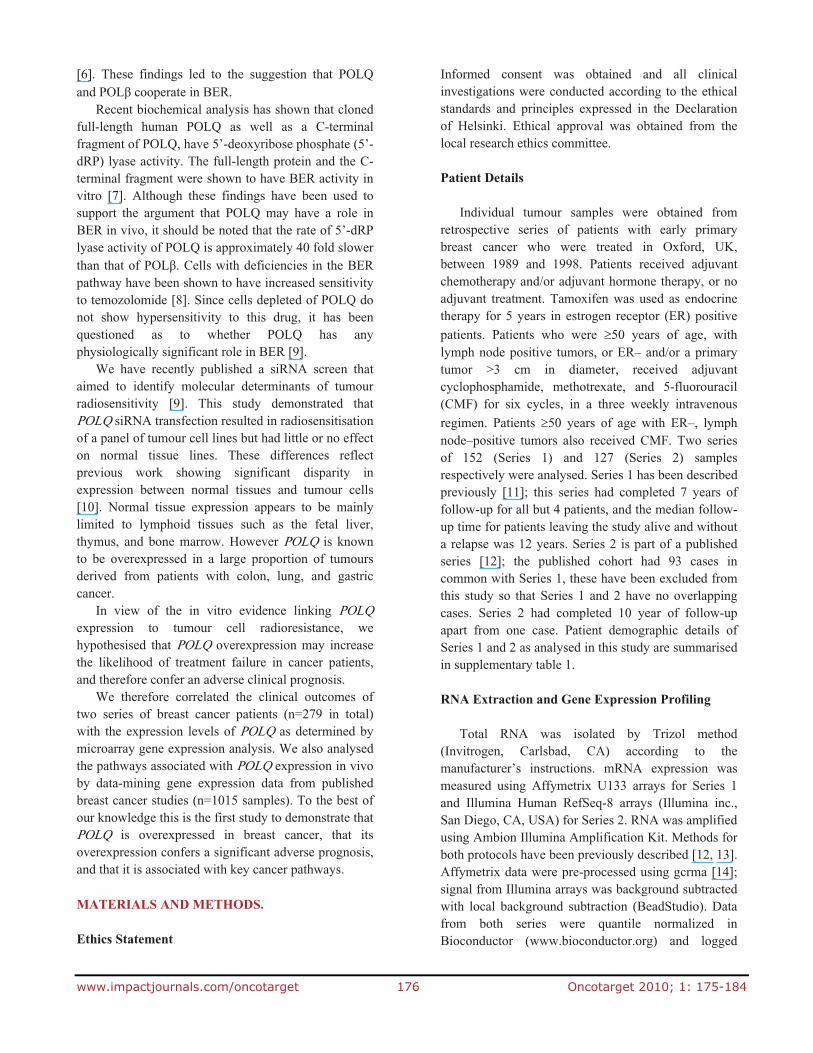
Fig 1: POLQ Expression in Breast Cancer A) Breast cancer samples, Series 1, described in this study (N=152). No normal breast tissue samples wereavailable for Series 1 so POLQ data were normalised to the sample with the lowest expression of POLQ (named T0).Expression fold change (FC) between all other tumours and T0 is shown for POLQ (207746_at). Expression ismeasured by Affymetrix array and quantile normalized. B) Breast cancer samples, Series 2, (N=127) described in this study. The FC between POLQ (ILMN_1450687)expression in each tumour and the median expression of 10 normal pools is shown. Expression is measured byIllumina arrays and quantile normalized.
www.impactjournals.com/oncotarget 178 Oncotarget 2010; 1: 175-184
Illumina arrays respectively. POLQ expression was normalised to the lowest level of tumour expression in the Affymetrix series, and to a panel of normal breast tissue samples for the Illumina series. POLQ expression is upregulated in a large proportion of breast tumour samples (Fig 1).
POLQ overexpression is independently associated with significantly worse relapse free survival (RFS) rates
The samples from Series 1 were divided into the
top and bottom 50th centiles and a univariate analysis
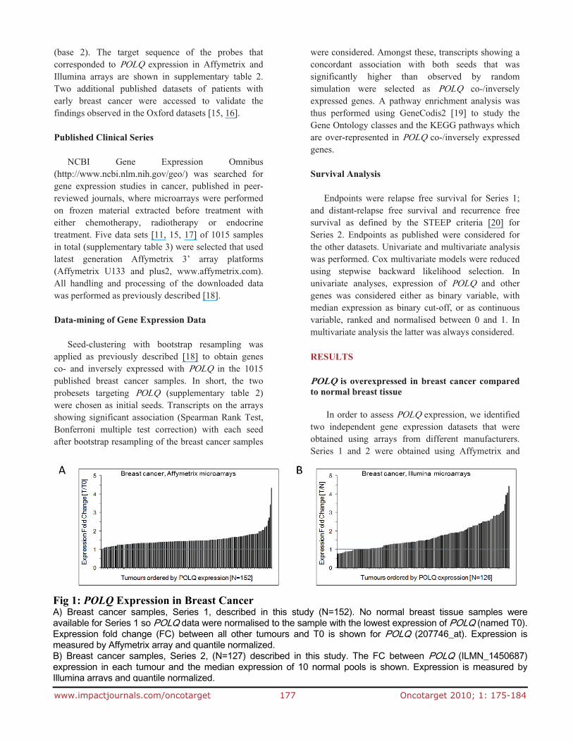
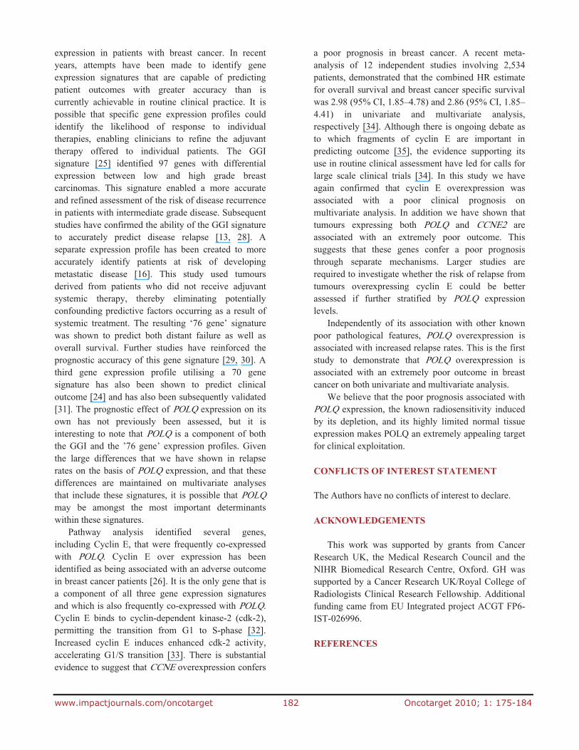
Fig. 2: POLQ expression is prognostic in breast cancer independently from clinico-pathological variables.A) Univariate analysis in 152 breast cancers (Series 1). POLQ expression is divided in two groups by medianvalue. B) POLQ expression is associated with tumour grade (left) and ER status (right) in Series 1 and 2 described inthis study (Affymetrix and Illumina arrays respectively) and two published series (Affymetrix arrays, seeMethods), although grade information was not available for GSE2034. Boxes summarize the median, quartilesand extreme values of POLQ expression in the different categories. One outlier is shown (circle), defined ascase with values between 1.5-3 box lengths from the edge of the box. Mann-Whitney and Spearman RankAssociation significance levels for the null hypotheses of POLQ expression not varying with ER and Graderespectively, are indicated on the highest category of each plot:*=p<0.05, **=p<0.01, ***=p<0.001.
C) Forest plot of POLQ Hazard Ratio for Survival Endpoints in univariate and multivariate analysis in the 2series described in this study and 2 published datasets (GEO Ids shown). Dots represent Hazard Ratios ofPOLQ expression and grey bars the 95% confidence intervals. Dot dimensions are proportional to dataset size.The expression of POLQ is entered in this model as a continuous ranked variable, normalised between 0(lowest rank) and 1 (highest rank). RFS= Recurrence Free Survival, DRFS=Distant Relapse Free Survival,DSS= Disease Specific Survival.
A
B
C
Hazard Ratio for Survival Endpoint
Univariate
Series 1 [RFS]Series 2 [DRFS]GSE3494 [DSS]GSE2034 [RFS]
Multivariate
Series 1 [RFS]Series 2 [DRFS]GSE3494 [DSS]GSE2034 [RFS]
1 50.1 0.5
www.impactjournals.com/oncotarget 179 Oncotarget 2010; 1: 175-184
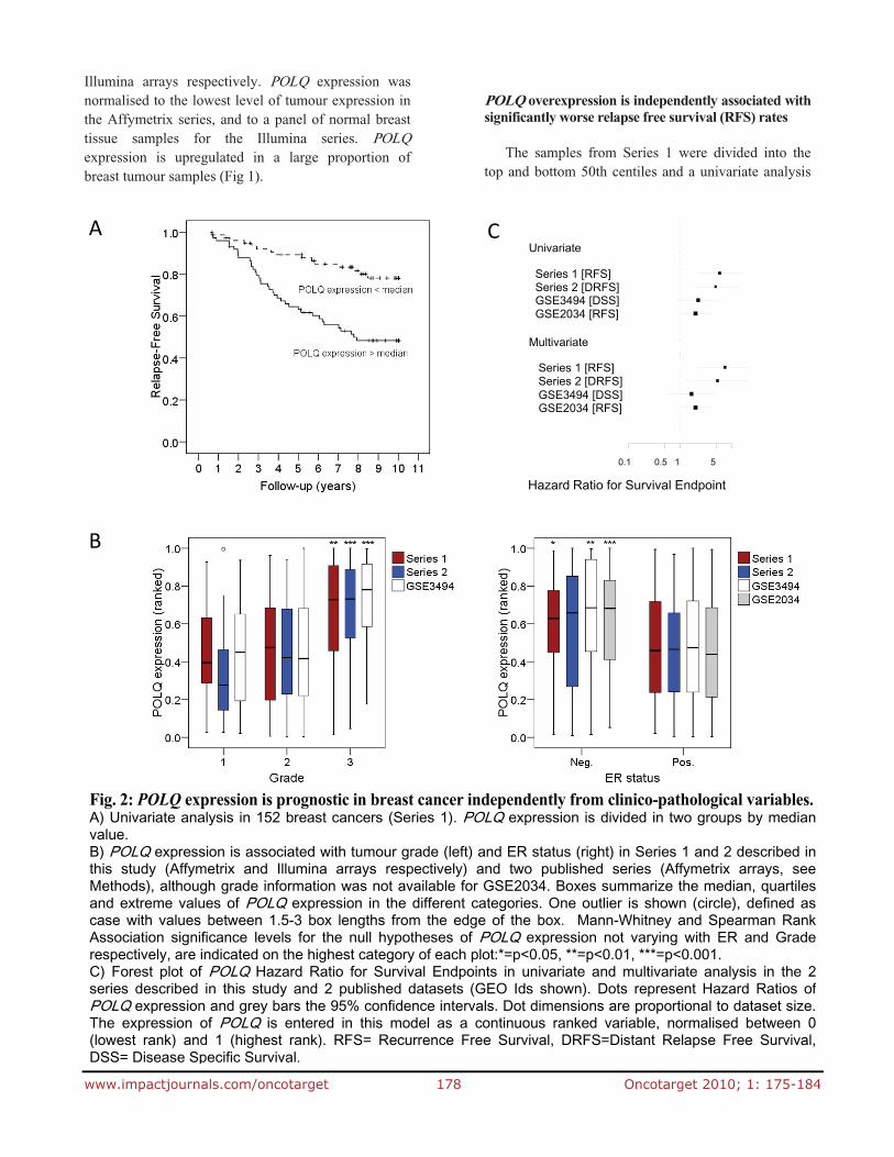
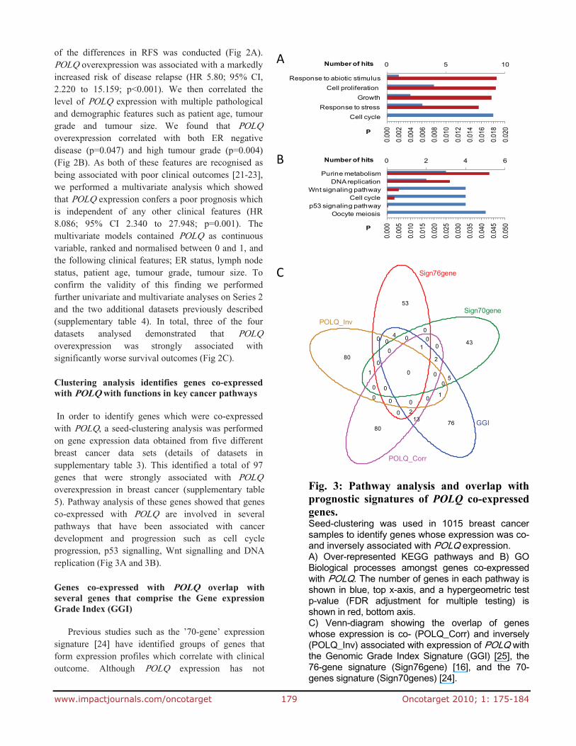
Fig. 3: Pathway analysis and overlap withprognostic signatures of POLQ co-expressedgenes. Seed-clustering was used in 1015 breast cancersamples to identify genes whose expression was co-and inversely associated with POLQ expression. A) Over-represented KEGG pathways and B) GOBiological processes amongst genes co-expressedwith POLQ. The number of genes in each pathway isshown in blue, top x-axis, and a hypergeometric testp-value (FDR adjustment for multiple testing) isshown in red, bottom axis. C) Venn-diagram showing the overlap of geneswhose expression is co- (POLQ_Corr) and inversely(POLQ_Inv) associated with expression of POLQ withthe Genomic Grade Index Signature (GGI) [25], the76-gene signature (Sign76gene) [16], and the 70-genes signature (Sign70genes) [24].
of the differences in RFS was conducted (Fig 2A). POLQ overexpression was associated with a markedly increased risk of disease relapse (HR 5.80; 95% CI, 2.220 to 15.159; p<0.001). We then correlated the level of POLQ expression with multiple pathological and demographic features such as patient age, tumour grade and tumour size. We found that POLQ overexpression correlated with both ER negative disease (p=0.047) and high tumour grade (p=0.004) (Fig 2B). As both of these features are recognised as being associated with poor clinical outcomes [21-23], we performed a multivariate analysis which showed that POLQ expression confers a poor prognosis which is independent of any other clinical features (HR 8.086; 95% CI 2.340 to 27.948; p=0.001). The multivariate models contained POLQ as continuous variable, ranked and normalised between 0 and 1, and the following clinical features; ER status, lymph node status, patient age, tumour grade, tumour size. To confirm the validity of this finding we performed further univariate and multivariate analyses on Series 2 and the two additional datasets previously described (supplementary table 4). In total, three of the four datasets analysed demonstrated that POLQ overexpression was strongly associated with significantly worse survival outcomes (Fig 2C).
Clustering analysis identifies genes co-expressed with POLQ with functions in key cancer pathways
In order to identify genes which were co-expressed with POLQ, a seed-clustering analysis was performed on gene expression data obtained from five different breast cancer data sets (details of datasets in supplementary table 3). This identified a total of 97 genes that were strongly associated with POLQ overexpression in breast cancer (supplementary table 5). Pathway analysis of these genes showed that genes co-expressed with POLQ are involved in several pathways that have been associated with cancer development and progression such as cell cycle progression, p53 signalling, Wnt signalling and DNA replication (Fig 3A and 3B). Genes co-expressed with POLQ overlap with several genes that comprise the Gene expression Grade Index (GGI)
Previous studies such as the ’70-gene’ expression
signature [24] have identified groups of genes that form expression profiles which correlate with clinical outcome. Although POLQ expression has not
53
43
7680
80
04
0
0
5
0
1
13
10
0 0
0
2
0
0
2
00
0
10
0
0
00
Sign76gene
Sign70gene
GGI
POLQ_Corr
POLQ_Inv
0.00
00.
002
0.00
40.
006
0.00
80.
010
0.01
20.
014
0.01
60.
018
0.02
0
0 5 10
Cell cycleResponse to stress
GrowthCell proliferation
Response to abiotic stimulus
P
Number of hits
0.00
00.
005
0.01
00.
015
0.02
00.
025
0.03
00.
035
0.04
00.
045
0.05
0
0 2 4 6
Oocyte meiosisp53 signaling pathway
Cell cycleWnt signaling pathway
DNA replicationPurine metabolism
P
Number of hits
A
B
C
www.impactjournals.com/oncotarget 180 Oncotarget 2010; 1: 175-184
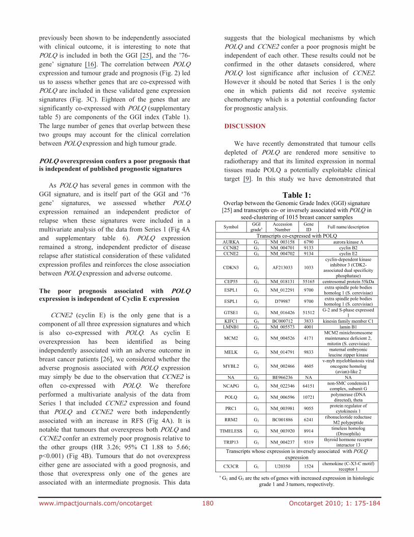
previously been shown to be independently associated with clinical outcome, it is interesting to note that POLQ is included in both the GGI [25], and the ’76-gene’ signature [16]. The correlation between POLQ expression and tumour grade and prognosis (Fig. 2) led us to assess whether genes that are co-expressed with POLQ are included in these validated gene expression signatures (Fig. 3C). Eighteen of the genes that are significantly co-expressed with POLQ (supplementary table 5) are components of the GGI index (Table 1). The large number of genes that overlap between these two groups may account for the clinical correlation between POLQ expression and high tumour grade.
POLQ overexpression confers a poor prognosis that is independent of published prognostic signatures
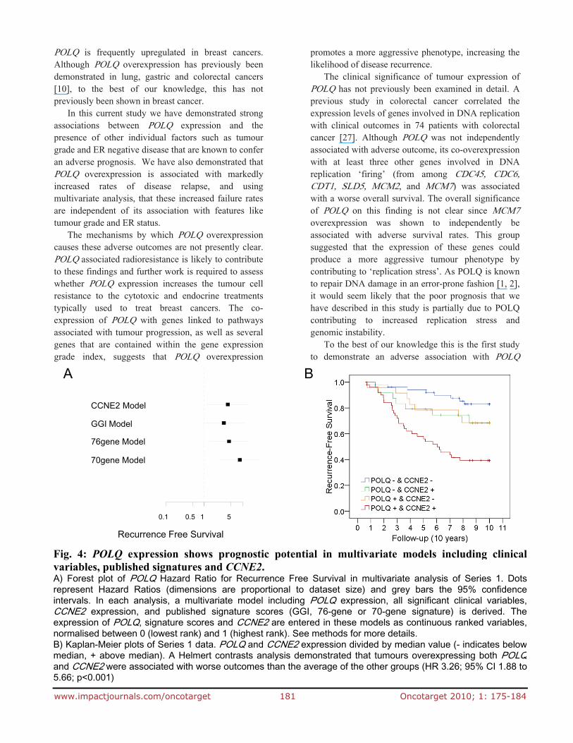
As POLQ has several genes in common with the GGI signature, and is itself part of the GGI and ‘76 gene’ signatures, we assessed whether POLQ expression remained an independent predictor of relapse when these signatures were included in a multivariate analysis of the data from Series 1 (Fig 4A and supplementary table 6). POLQ expression remained a strong, independent predictor of disease relapse after statistical consideration of these validated expression profiles and reinforces the close association between POLQ expression and adverse outcome.
The poor prognosis associated with POLQ expression is independent of Cyclin E expression
CCNE2 (cyclin E) is the only gene that is a
component of all three expression signatures and which is also co-expressed with POLQ. As cyclin E overexpression has been identified as being independently associated with an adverse outcome in breast cancer patients [26], we considered whether the adverse prognosis associated with POLQ expression may simply be due to the observation that CCNE2 is often co-expressed with POLQ. We therefore performed a multivariate analysis of the data from Series 1 that included CCNE2 expression and found that POLQ and CCNE2 were both independently associated with an increase in RFS (Fig 4A). It is notable that tumours that overexpress both POLQ and CCNE2 confer an extremely poor prognosis relative to the other groups (HR 3.26; 95% CI 1.88 to 5.66; p<0.001) (Fig 4B). Tumours that do not overexpress either gene are associated with a good prognosis, and those that overexpress only one of the genes are associated with an intermediate prognosis. This data
suggests that the biological mechanisms by which POLQ and CCNE2 confer a poor prognosis might be independent of each other. These results could not be confirmed in the other datasets considered, where POLQ lost significance after inclusion of CCNE2. However it should be noted that Series 1 is the only one in which patients did not receive systemic chemotherapy which is a potential confounding factor for prognostic analysis.
DISCUSSION
We have recently demonstrated that tumour cells
depleted of POLQ are rendered more sensitive to radiotherapy and that its limited expression in normal tissues made POLQ a potentially exploitable clinical target [9]. In this study we have demonstrated that
Table 1: Overlap between the Genomic Grade Index (GGI) signature
[25] and transcripts co- or inversely associated with POLQ in seed-clustering of 1015 breast cancer samples
Symbol GGI
grades Accession Number
Gene ID
Full name/description
Transcripts co-expressed with POLQ AURKA G3 NM_003158 6790 aurora kinase A CCNB2 G3 NM_004701 9133 cyclin B2 CCNE2 G3 NM_004702 9134 cyclin E2
CDKN3 G3 AF213033 1033
cyclin-dependent kinase inhibitor 3 (CDK2-
associated dual specificity phosphatase)
CEP55 G3 NM_018131 55165 centrosomal protein 55kDa
ESPL1 G3 NM_012291 9700 extra spindle pole bodies homolog 1 (S. cerevisiae)
ESPL1 G3 D79987 9700 extra spindle pole bodies homolog 1 (S. cerevisiae)
GTSE1 G3 NM_016426 51512 G-2 and S-phase expressed
1 KIFC1 G3 BC000712 3833 kinesin family member C1
LMNB1 G3 NM_005573 4001 lamin B1
MCM2 G3 NM_004526 4171 MCM2 minichromosome maintenance deficient 2, mitotin (S. cerevisiae)
MELK G3 NM_014791 9833 maternal embryonic leucine zipper kinase
MYBL2 G3 NM_002466 4605 v-myb myeloblastosis viral
oncogene homolog (avian)-like 2
NA G3 BE966236 NA NA
NCAPG G3 NM_022346 64151 non-SMC condensin I complex, subunit G
POLQ G3 NM_006596 10721 polymerase (DNA
directed), theta
PRC1 G3 NM_003981 9055 protein regulator of
cytokinesis 1
RRM2 G3 BC001886 6241 ribonucleotide reductase
M2 polypeptide
TIMELESS G3 NM_003920 8914 timeless homolog
(Drosophila)
TRIP13 G3 NM_004237 9319 thyroid hormone receptor
interactor 13 Transcripts whose expression is inversely associated with POLQ
expression
CX3CR G1 U20350 1524 chemokine (C-X3-C motif)
receptor 1
s G1 and G3 are the sets of genes with increased expression in histologic grade 1 and 3 tumors, respectively.
www.impactjournals.com/oncotarget 181 Oncotarget 2010; 1: 175-184
POLQ is frequently upregulated in breast cancers. Although POLQ overexpression has previously been demonstrated in lung, gastric and colorectal cancers [10], to the best of our knowledge, this has not previously been shown in breast cancer.
In this current study we have demonstrated strong associations between POLQ expression and the presence of other individual factors such as tumour grade and ER negative disease that are known to confer an adverse prognosis. We have also demonstrated that POLQ overexpression is associated with markedly increased rates of disease relapse, and using multivariate analysis, that these increased failure rates are independent of its association with features like tumour grade and ER status.
The mechanisms by which POLQ overexpression causes these adverse outcomes are not presently clear. POLQ associated radioresistance is likely to contribute to these findings and further work is required to assess whether POLQ expression increases the tumour cell resistance to the cytotoxic and endocrine treatments typically used to treat breast cancers. The co-expression of POLQ with genes linked to pathways associated with tumour progression, as well as several genes that are contained within the gene expression grade index, suggests that POLQ overexpression
promotes a more aggressive phenotype, increasing the likelihood of disease recurrence.
The clinical significance of tumour expression of POLQ has not previously been examined in detail. A previous study in colorectal cancer correlated the expression levels of genes involved in DNA replication with clinical outcomes in 74 patients with colorectal cancer [27]. Although POLQ was not independently associated with adverse outcome, its co-overexpression with at least three other genes involved in DNA replication ‘firing’ (from among CDC45, CDC6, CDT1, SLD5, MCM2, and MCM7) was associated with a worse overall survival. The overall significance of POLQ on this finding is not clear since MCM7 overexpression was shown to independently be associated with adverse survival rates. This group suggested that the expression of these genes could produce a more aggressive tumour phenotype by contributing to ‘replication stress’. As POLQ is known to repair DNA damage in an error-prone fashion [1, 2], it would seem likely that the poor prognosis that we have described in this study is partially due to POLQ contributing to increased replication stress and genomic instability.
To the best of our knowledge this is the first study to demonstrate an adverse association with POLQ
Fig. 4: POLQ expression shows prognostic potential in multivariate models including clinicalvariables, published signatures and CCNE2. A) Forest plot of POLQ Hazard Ratio for Recurrence Free Survival in multivariate analysis of Series 1. Dotsrepresent Hazard Ratios (dimensions are proportional to dataset size) and grey bars the 95% confidenceintervals. In each analysis, a multivariate model including POLQ expression, all significant clinical variables,CCNE2 expression, and published signature scores (GGI, 76-gene or 70-gene signature) is derived. Theexpression of POLQ, signature scores and CCNE2 are entered in these models as continuous ranked variables,normalised between 0 (lowest rank) and 1 (highest rank). See methods for more details. B) Kaplan-Meier plots of Series 1 data. POLQ and CCNE2 expression divided by median value (- indicates belowmedian, + above median). A Helmert contrasts analysis demonstrated that tumours overexpressing both POLQand CCNE2 were associated with worse outcomes than the average of the other groups (HR 3.26; 95% CI 1.88 to5.66; p<0.001)
A B
Recurrence Free Survival
CCNE2 Model
GGI Model
76gene Model
70gene Model
1 50.1 0.5
www.impactjournals.com/oncotarget 182 Oncotarget 2010; 1: 175-184
expression in patients with breast cancer. In recent years, attempts have been made to identify gene expression signatures that are capable of predicting patient outcomes with greater accuracy than is currently achievable in routine clinical practice. It is possible that specific gene expression profiles could identify the likelihood of response to individual therapies, enabling clinicians to refine the adjuvant therapy offered to individual patients. The GGI signature [25] identified 97 genes with differential expression between low and high grade breast carcinomas. This signature enabled a more accurate and refined assessment of the risk of disease recurrence in patients with intermediate grade disease. Subsequent studies have confirmed the ability of the GGI signature to accurately predict disease relapse [13, 28]. A separate expression profile has been created to more accurately identify patients at risk of developing metastatic disease [16]. This study used tumours derived from patients who did not receive adjuvant systemic therapy, thereby eliminating potentially confounding predictive factors occurring as a result of systemic treatment. The resulting ‘76 gene’ signature was shown to predict both distant failure as well as overall survival. Further studies have reinforced the prognostic accuracy of this gene signature [29, 30]. A third gene expression profile utilising a 70 gene signature has also been shown to predict clinical outcome [24] and has also been subsequently validated [31]. The prognostic effect of POLQ expression on its own has not previously been assessed, but it is interesting to note that POLQ is a component of both the GGI and the ’76 gene’ expression profiles. Given the large differences that we have shown in relapse rates on the basis of POLQ expression, and that these differences are maintained on multivariate analyses that include these signatures, it is possible that POLQ may be amongst the most important determinants within these signatures.
Pathway analysis identified several genes, including Cyclin E, that were frequently co-expressed with POLQ. Cyclin E over expression has been identified as being associated with an adverse outcome in breast cancer patients [26]. It is the only gene that is a component of all three gene expression signatures and which is also frequently co-expressed with POLQ. Cyclin E binds to cyclin-dependent kinase-2 (cdk-2), permitting the transition from G1 to S-phase [32]. Increased cyclin E induces enhanced cdk-2 activity, accelerating G1/S transition [33]. There is substantial evidence to suggest that CCNE overexpression confers
a poor prognosis in breast cancer. A recent meta-analysis of 12 independent studies involving 2,534 patients, demonstrated that the combined HR estimate for overall survival and breast cancer specific survival was 2.98 (95% CI, 1.85–4.78) and 2.86 (95% CI, 1.85–4.41) in univariate and multivariate analysis, respectively [34]. Although there is ongoing debate as to which fragments of cyclin E are important in predicting outcome [35], the evidence supporting its use in routine clinical assessment have led for calls for large scale clinical trials [34]. In this study we have again confirmed that cyclin E overexpression was associated with a poor clinical prognosis on multivariate analysis. In addition we have shown that tumours expressing both POLQ and CCNE2 are associated with an extremely poor outcome. This suggests that these genes confer a poor prognosis through separate mechanisms. Larger studies are required to investigate whether the risk of relapse from tumours overexpressing cyclin E could be better assessed if further stratified by POLQ expression levels.
Independently of its association with other known poor pathological features, POLQ overexpression is associated with increased relapse rates. This is the first study to demonstrate that POLQ overexpression is associated with an extremely poor outcome in breast cancer on both univariate and multivariate analysis.
We believe that the poor prognosis associated with POLQ expression, the known radiosensitivity induced by its depletion, and its highly limited normal tissue expression makes POLQ an extremely appealing target for clinical exploitation.
CONFLICTS OF INTEREST STATEMENT The Authors have no conflicts of interest to declare. ACKNOWLEDGEMENTS
This work was supported by grants from Cancer Research UK, the Medical Research Council and the NIHR Biomedical Research Centre, Oxford. GH was supported by a Cancer Research UK/Royal College of Radiologists Clinical Research Fellowship. Additional funding came from EU Integrated project ACGT FP6-IST-026996.
REFERENCES
www.impactjournals.com/oncotarget 183 Oncotarget 2010; 1: 175-184
1. Arana ME, Seki M, Wood RD, Rogozin IB, Kunkel TA. Low-fidelity DNA synthesis by human DNA polymerase theta. Nucleic Acids Res 2008; 36:3847-3856.
2. Seki M, Masutani C, Yang LW, Schuffert A, Iwai S, Bahar I, Wood RD. High-efficiency bypass of DNA damage by human DNA polymerase Q. EMBO J 2004; 23:4484-4494.
3. Zan H, Shima N, Xu Z, Al-Qahtani A, Evinger Iii AJ, Zhong Y, Schimenti JC, Casali P. The translesion DNA polymerase theta plays a dominant role in immunoglobulin gene somatic hypermutation. EMBO J 2005; 24:3757-3769.
4. Masuda K, Ouchida R, Hikida M, Nakayama M, Ohara O, Kurosaki T, O-Wang J. Absence of DNA polymerase theta results in decreased somatic hypermutation frequency and altered mutation patterns in Ig genes. DNA Repair (Amst) 2006; 5:1384-1391.
5. Martomo SA, Saribasak H, Yokoi M, Hanaoka F, Gearhart PJ. Reevaluation of the role of DNA polymerase theta in somatic hypermutation of immunoglobulin genes. DNA Repair (Amst) 2008; 7:1603-1608.
6. Yoshimura M, Kohzaki M, Nakamura J, Asagoshi K, Sonoda E, Hou E, Prasad R, Wilson SH, Tano K, Yasui A, Lan L, Seki M, Wood RD, Arakawa H, Buerstedde JM, Hochegger H, Okada T, Hiraoka M, Takeda S. Vertebrate POLQ and POLbeta cooperate in base excision repair of oxidative DNA damage. Mol Cell 2006; 24:115-125.
7. Prasad R, Longley MJ, Sharief FS, Hou EW, Copeland WC, Wilson SH. Human DNA polymerase theta possesses 5'-dRP lyase activity and functions in single-nucleotide base excision repair in vitro. Nucleic Acids Res 2009; 37:1868-1877.
8. Trivedi RN, Almeida KH, Fornsaglio JL, Schamus S, Sobol RW. The role of base excision repair in the sensitivity and resistance to temozolomide-mediated cell death. Cancer Res 2005; 65:6394-6400.
9. Higgins GS, Prevo R, Lee YF, Helleday T, Muschel RJ, Taylor S, Yoshimura M, Hickson ID, Bernhard EJ, McKenna WG. A small interfering RNA screen of genes involved in DNA repair identifies tumor-specific radiosensitization by POLQ knockdown. Cancer Res 2010; 70:2984-2993.
10. Kawamura K, Bahar R, Seimiya M, Chiyo M, Wada A, Okada S, Hatano M, Tokuhisa T, Kimura H, Watanabe S, Honda I, Sakiyama S, Tagawa M, O-Wang J. DNA polymerase theta is preferentially expressed in lymphoid tissues and upregulated in human cancers. Int J Cancer 2004; 109:9-16.
11. Loi S, Haibe-Kains B, Desmedt C, Wirapati P, Lallemand F, Tutt AM, Gillet C, Ellis P, Ryder K, Reid JF, Daidone MG, Pierotti MA, Berns EM, Jansen MP, Foekens JA, Delorenzi M, Bontempi G, Piccart MJ, Sotiriou C. Predicting prognosis using molecular
profiling in estrogen receptor-positive breast cancer treated with tamoxifen. BMC Genomics 2008; 9:239.
12. Camps C, Buffa FM, Colella S, Moore J, Sotiriou C, Sheldon H, Harris AL, Gleadle JM, Ragoussis J. hsa-miR-210 Is induced by hypoxia and is an independent prognostic factor in breast cancer. Clin Cancer Res 2008; 14:1340-1348.
13. Loi S, Haibe-Kains B, Desmedt C, Lallemand F, Tutt AM, Gillet C, Ellis P, Harris A, Bergh J, Foekens JA, Klijn JG, Larsimont D, Buyse M, Bontempi G, Delorenzi M, Piccart MJ, Sotiriou C. Definition of clinically distinct molecular subtypes in estrogen receptor-positive breast carcinomas through genomic grade. J Clin Oncol 2007; 25:1239-1246.
14. Wu Z IR, Gentleman R, Martinez-Murillo F, Spencer F. A Model-Based Background Adjustment for Oligonucleotide Expression Arrays. Journal of the American Statistical Association 2004; 99:909–917.
15. Miller LD, Smeds J, George J, Vega VB, Vergara L, Ploner A, Pawitan Y, Hall P, Klaar S, Liu ET, Bergh J. An expression signature for p53 status in human breast cancer predicts mutation status, transcriptional effects, and patient survival. Proc Natl Acad Sci U S A 2005; 102:13550-13555.
16. Wang Y, Klijn JG, Zhang Y, Sieuwerts AM, Look MP, Yang F, Talantov D, Timmermans M, Meijer-van Gelder ME, Yu J, Jatkoe T, Berns EM, Atkins D, Foekens JA. Gene-expression profiles to predict distant metastasis of lymph-node-negative primary breast cancer. Lancet 2005; 365:671-679.
17. Carroll JS, Meyer CA, Song J, Li W, Geistlinger TR, Eeckhoute J, Brodsky AS, Keeton EK, Fertuck KC, Hall GF, Wang Q, Bekiranov S, Sementchenko V, Fox EA, Silver PA, Gingeras TR, Liu XS, Brown M. Genome-wide analysis of estrogen receptor binding sites. Nat Genet 2006; 38:1289-1297.
18. Buffa FM, Harris AL, West CM, Miller CJ. Large meta-analysis of multiple cancers reveals a common, compact and highly prognostic hypoxia metagene. Br J Cancer 2010; 102:428-435.
19. Nogales-Cadenas R, Carmona-Saez P, Vazquez M, Vicente C, Yang X, Tirado F, Carazo JM, Pascual-Montano A. GeneCodis: interpreting gene lists through enrichment analysis and integration of diverse biological information. Nucleic Acids Res 2009; 37:W317-322.
20. Hudis CA, Barlow WE, Costantino JP, Gray RJ, Pritchard KI, Chapman JA, Sparano JA, Hunsberger S, Enos RA, Gelber RD, Zujewski JA. Proposal for standardized definitions for efficacy end points in adjuvant breast cancer trials: the STEEP system. J Clin Oncol 2007; 25:2127-2132.
21. Parl FF, Schmidt BP, Dupont WD, Wagner RK. Prognostic significance of estrogen receptor status in breast cancer in relation to tumor stage, axillary node metastasis, and histopathologic grading. Cancer 1984; 54:2237-2242.
www.impactjournals.com/oncotarget 184 Oncotarget 2010; 1: 175-184
22. Kinne DW, Butler JA, Kimmel M, Flehinger BJ, Menendez-Botet C, Schwartz M. Estrogen receptor protein of breast cancer in patients with positive nodes. High recurrence rates in the postmenopausal estrogen receptor-negative group. Arch Surg 1987; 122:1303-1306.
23. Elston CW, Ellis IO. Pathological prognostic factors in breast cancer. I. The value of histological grade in breast cancer: experience from a large study with long-term follow-up. Histopathology 1991; 19:403-410.
24. van 't Veer LJ, Dai H, van de Vijver MJ, He YD, Hart AA, Mao M, Peterse HL, van der Kooy K, Marton MJ, Witteveen AT, Schreiber GJ, Kerkhoven RM, Roberts C, Linsley PS, Bernards R, Friend SH. Gene expression profiling predicts clinical outcome of breast cancer. Nature 2002; 415:530-536.
25. Sotiriou C, Wirapati P, Loi S, Harris A, Fox S, Smeds J, Nordgren H, Farmer P, Praz V, Haibe-Kains B, Desmedt C, Larsimont D, Cardoso F, Peterse H, Nuyten D, Buyse M, Van de Vijver MJ, Bergh J, Piccart M, Delorenzi M. Gene expression profiling in breast cancer: understanding the molecular basis of histologic grade to improve prognosis. J Natl Cancer Inst 2006; 98:262-272.
26. Nielsen NH, Arnerlov C, Emdin SO, Landberg G. Cyclin E overexpression, a negative prognostic factor in breast cancer with strong correlation to oestrogen receptor status. Br J Cancer 1996; 74:874-880.
27. Pillaire MJ, Selves J, Gordien K, Gourraud PA, Gentil C, Danjoux M, Do C, Negre V, Bieth A, Guimbaud R, Trouche D, Pasero P, Mechali M, Hoffmann JS, Cazaux C. A 'DNA replication' signature of progression and negative outcome in colorectal cancer. Oncogene; 29:876-887.
28. Desmedt C, Giobbie-Hurder A, Neven P, Paridaens R, Christiaens MR, Smeets A, Lallemand F, Haibe-Kains B, Viale G, Gelber RD, Piccart M, Sotiriou C. The Gene expression Grade Index: a potential predictor of relapse for endocrine-treated breast cancer patients in the BIG 1-98 trial. BMC Med Genomics 2009; 2:40.
29. Foekens JA, Atkins D, Zhang Y, Sweep FC, Harbeck N, Paradiso A, Cufer T, Sieuwerts AM, Talantov D, Span PN, Tjan-Heijnen VC, Zito AF, Specht K, Hoefler H, Golouh R, Schittulli F, Schmitt M, Beex LV, Klijn JG, Wang Y. Multicenter validation of a gene expression-based prognostic signature in lymph node-negative primary breast cancer. J Clin Oncol 2006; 24:1665-1671.
30. Desmedt C, Piette F, Loi S, Wang Y, Lallemand F, Haibe-Kains B, Viale G, Delorenzi M, Zhang Y, d'Assignies MS, Bergh J, Lidereau R, Ellis P, Harris AL, Klijn JG, Foekens JA, Cardoso F, Piccart MJ, Buyse M, Sotiriou C. Strong time dependence of the 76-gene prognostic signature for node-negative breast cancer patients in the TRANSBIG multicenter independent validation series. Clin Cancer Res 2007; 13:3207-3214.
31. van de Vijver MJ, He YD, van't Veer LJ, Dai H, Hart AA, Voskuil DW, Schreiber GJ, Peterse JL, Roberts C, Marton MJ, Parrish M, Atsma D, Witteveen A, Glas A, Delahaye L, van der Velde T, Bartelink H, Rodenhuis S, Rutgers ET, Friend SH, Bernards R. A gene-expression signature as a predictor of survival in breast cancer. N Engl J Med 2002; 347:1999-2009.
32. Koff A, Giordano A, Desai D, Yamashita K, Harper JW, Elledge S, Nishimoto T, Morgan DO, Franza BR, Roberts JM. Formation and activation of a cyclin E-cdk2 complex during the G1 phase of the human cell cycle. Science 1992; 257:1689-1694.
33. Resnitzky D, Gossen M, Bujard H, Reed SI. Acceleration of the G1/S phase transition by expression of cyclins D1 and E with an inducible system. Mol Cell Biol 1994; 14:1669-1679.
34. Wang L, Shao ZM. Cyclin e expression and prognosis in breast cancer patients: a meta-analysis of published studies. Cancer Invest 2006; 24:581-587.
35. Harris L, Fritsche H, Mennel R, Norton L, Ravdin P, Taube S, Somerfield MR, Hayes DF, Bast RC, Jr. American Society of Clinical Oncology 2007 update of recommendations for the use of tumor markers in breast cancer. J Clin Oncol 2007; 25:5287-5312.
Recommended


