F
SGFPSCB
Aaab
oaam(gCa
S1
2
Journal of the American College of Cardiology Vol. 46, No. 9, 2005© 2005 by the American College of Cardiology Foundation ISSN 0735-1097/05/$30.00P
OCUS ISSUE: CARDIAC REGENERATION
afety and Efficacy of Subcutaneous-Onlyranulocyte-Macrophage Colony-Stimulatingactor for Collateral Growth Promotion inatients With Coronary Artery Disease
tephan Zbinden, MD, Rainer Zbinden, MD, Pascal Meier, MD, Stephan Windecker, MD,hristian Seiler, MD, FACC, FESCern, Switzerland
OBJECTIVES This study was designed to investigate the safety and efficacy of a short-term subcutaneous-only granulocyte-macrophage colony-stimulating factor (GM-CSF) protocol for coronarycollateral growth promotion.
BACKGROUND The safety and efficacy of an exclusively systemic application of GM-CSF in patients withcoronary artery disease (CAD) and collateral artery promotion has not been studied so far.
METHODS In 14 men (age 61 � 11 years) with chronic stable CAD, the effect of GM-CSF(molgramostim) on quantitatively assessed collateral flow was tested in a randomized,double-blind, placebo-controlled fashion. The study protocol consisted of an invasivecollateral flow index (CFI) measurement in a stenotic as well as a normal coronary arterybefore and after a two-week period with subcutaneous GM-CSF (10 �g/kg; n � 7) orplacebo (n � 7). Collateral flow index was determined by simultaneous measurement of meanaortic, distal coronary occlusive, and central venous pressure.
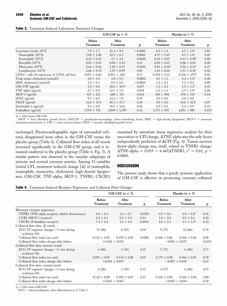
RESULTS Collateral flow index in all vessels changed from 0.116 � 0.05 to 0.159 � 0.07 in theGM-CSF group (p � 0.028) and from 0.166 � 0.06 to 0.166 � 0.04 in the placebo group(p � NS). The treatment-induced difference in CFI was �0.042 � 0.05 in the GM-CSFgroup and �0.001 � 0.04 in the placebo group (p � 0.035). Among 11 determinedcytokines, chemokines, and their monocytic receptor concentrations, the treatment-inducedchange in CFI was predicted by the respective change in tumor necrosis factor-alphaconcentration. Two of seven patients in the GM-CSF group and none in the placebo groupsuffered an acute coronary syndrome during the treatment period.
CONCLUSIONS A subcutaneous-only, short-term protocol of GM-CSF is effective in promoting coronarycollateral artery growth among patients with CAD. However, the drug’s safety regarding theoccurrence of acute coronary syndrome is questionable. (J Am Coll Cardiol 2005;46:
ublished by Elsevier Inc. doi:10.1016/j.jacc.2005.01.068
1636–42) © 2005 by the American College of Cardiology Foundation
(liascehbtnlg
petim
revascularization strategy alternative to bypass surgery orngioplasty is warranted, both to control symptoms and tolter the course of severe coronary artery disease (CAD),ecause the traditional approaches are not suitable in 1 out
See page 1649
f 5 to 3 patients. The promotion of preformed collateralrteries to a myocardial territory jeopardized by a blockedrtery is such an option. Experimentally, there have beenany candidates with angiogenic or arteriogenic properties
1), but in only one controlled clinical investigation usingranulocyte-macrophage colony-stimulating factor (GM-SF) has the promotion of large, conductive collateral
rteries (arteriogenesis) been documented to be attainable
From the Department of Cardiology, University Hospital, Bern, Switzerland.upported by a grant from the Swiss National Science Foundation, #3200BO-00065/1. The first two authors contributed equally to this work.
(Manuscript received October 20, 2004; revised manuscript received January 19,
005, accepted January 25, 2005.
2). Conversely, inducing the growth of capillary-like col-ateral vessels (angiogenesis) in patients with CAD has notnfluenced myocardial perfusion (3,4). With pro-angiogenicnd -arteriogenic substances, the risk of advancing athero-clerosis while promoting vascular detours has been criti-ized (5). During arteriogenesis, monocytes seem to prefer-ntially adhere to the endothelium of vessels subject toigh-velocity blood flow owing to a pressure gradientetween a collateral supplying artery and a blocked vascularerritory (i.e., homing). In this context, it is probably notecessary to deliver growth factors for monocyte stimulation
ocally, as performed in our recent study (2), but they can beiven systemically.
On the basis of these considerations, the goal of theresent controlled study was to investigate the safety andfficacy of a short-term subcutaneous-only GM-CSF pro-ocol for coronary collateral growth promotion. The follow-ng hypotheses were tested: GM-CSF is safe and it aug-
ents directly obtained coronary collateral flow index (CFI)
Fig. 1).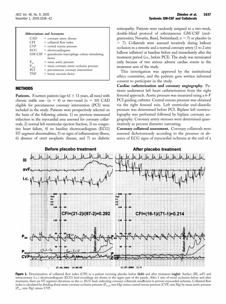
M
PceitietS6
rdg�obtet
ecCtfPvplgtCas
Fiti(
1637JACC Vol. 46, No. 9, 2005 Zbinden et al.November 1, 2005:1636–42 Systemic GM-CSF and Collaterals
ETHODS
atients. Fourteen patients (age 61 � 11 years, all men) withhronic stable one- (n � 4) or two-vessel (n � 10) CADligible for percutaneous coronary intervention (PCI) werencluded in the study. Patients were prospectively selected onhe basis of the following criteria: 1) no previous transmuralnfarction in the myocardial area assessed for coronary collat-rals, 2) normal left ventricular ejection fraction, 3) no conges-ive heart failure, 4) no baseline electrocardiogram (ECG)T-segment abnormalities, 5) no signs of inflammatory illness,) absence of overt neoplastic disease, and 7) no diabetic
Abbreviations and AcronymsCAD � coronary artery diseaseCFI � collateral flow indexCVP � central venous pressureECG � electrocardiogramGM-CSF � granulocyte-macrophage colony-stimulating
factorPao � mean aortic pressurePoccl � mean coronary artery occlusive pressurePCI � percutaneous coronary interventionTNF � tumor necrosis factor
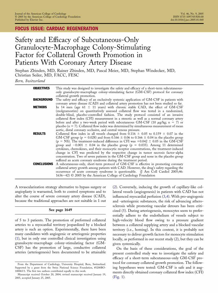
igure 1. Determination of collateral flow index (CFI) in a patient recentracoronary (i.c.) electrocardiogram (ECG) lead recordings are shown inreatment, there are ST-segment elevations on the i.c. ECG lead, indicatin
ndex is calculated by dividing distal mean coronary occlusive pressure (Poccl, mmPao, mm Hg) minus CVP.etinopathy. Patients were randomly assigned to a two-week,ouble-blind protocol of subcutaneous GM-CSF (mol-ramostim; Novartis, Basel, Switzerland; n � 7) or placebo (n
7). Collaterals were assessed invasively during ballooncclusion in a stenotic and a normal coronary artery (1 to 2 atmalloon inflation) at baseline before and immediately after thereatment period (i.e., before PCI). The study was terminatedarly because of two serious adverse cardiac events in thereatment arm of the study.
This investigation was approved by the institutionalthics committee, and the patients gave written informedonsent to participate in the study.ardiac catheterization and coronary angiography. Pa-
ients underwent left heart catheterization from the rightemoral approach. Aortic pressure was measured using a 6-FCI guiding catheter. Central venous pressure was obtainedia the right femoral vein. Left ventricular end-diastolicressure was determined before PCI. Biplane left ventricu-
ography was performed followed by biplane coronary an-iography. Coronary artery stenoses were determined quan-itatively as percent diameter narrowing.
oronary collateral assessment. Coronary collaterals weressessed dichotomously according to the presence or ab-ence of ECG signs of myocardial ischemia at the end of a
placebo before (left) and after treatment (right). Surface (III, avF) andupper part of the panels. After 1 min of vessel occlusion before and afternary collaterals insufficient to prevent myocardial ischemia. Collateral flow
ivingthe
g coro
Hg) minus central venous pressure (CVP, mm Hg) by mean aortic pressure
oM�Prvm(saCscHCbDmpDkqroG(
C
Cfifttcistwwbi
C
crraStaWcdpstc
ssap
R
PTtmpqsHeopobnwhPir
o(Snsd
T
AMBHAD
SHHOFDABCNA
SD
1638 Zbinden et al. JACC Vol. 46, No. 9, 2005Systemic GM-CSF and Collaterals November 1, 2005:1636–42
ne-minute balloon occlusion of the vessel of interest.yocardial ischemia was defined as ST-segment changes0.1 mV (Fig. 1).rimary end point of the study. Coronary collateral flow
elative to normal antegrade flow through the non-occludedessel (CFI) was determined using coronary pressure measure-ents. A 0.014-in pressure monitoring angioplasty guidewire
Pressure Wave, Endosonics, Mountain View, California) waset at zero, calibrated, advanced through the guiding catheter,nd positioned in the distal part of the vessel of interest.ollateral flow index was determined by simultaneous mea-
urement of mean aortic pressure (Pao, mm Hg), the distaloronary artery pressure during balloon occlusion (Poccl, mmg), and the central venous pressure (CVP, mm Hg) (Fig. 1).ollateral flow index was calculated as (Poccl � CVP) dividedy (Pao � CVP). The accuracy of pressure in comparison tooppler-derived CFI measurements and to ECG signs ofyocardial ischemia during occlusion has been documented
reviously (6).etermination of progenitor cells, cytokines, chemo-
ines, and their receptors. PROGENITOR CELLS. Foruantitation of the CD34 messenger ribonucleic acid, aeal-time quantitative polymerase chain reaction assay basedn a specific set of primers and probe (Assays-on-Demand,ene Expression Products) supplied by Applied Biosystems
Rotkreuz, Switzerland) was used.
YTOKINES AND CHEMOKINES. Concentrations of GM-SF, monocyte chemoattractant protein-1 (MCP-1), basicbroblast growth factor, and vascular endothelial growthactor were determined as immunoreactivity using a quan-itative sandwich enzyme immunoassay technique (Quan-ikine, R and D Systems, Minneapolis, Minnesota). Con-entrations of tumor necrosis factor (TNF)-alpha andnterleukin (IL)-6 were determined by immunometric as-ays (Immulite, DPC, Los Angeles, California) according tohe manufacturer’s guidelines. Fractalkine concentrationas assessed using enzyme-linked immunosorbent assayith mouse anti-human Fractalkine capture antibody andiotinylated mouse anti-human Fractalkine detection antibod-es (DuoSet; R and D Systems, Minneapolis, Minnesota).
YTOKINE AND CHEMOKINE RECEPTORS. Cytokine andhemokine monocyte receptor concentrations (TNF-alphaeceptor, TNFR1; MCP-1 receptor, CCR2; Fractalkineeceptor, CXCR1) were determined by fluorescent-ctivated cell sorting analysis on CD14� mononuclear cells.tatistical analysis. Between-group comparisons of con-
inuous clinical, hemodynamic, angiographic, blood analysisnd collateral flow data were performed by a Mann-
hitney test. A chi-square test was used for comparison ofategorical variables among the two study groups. Intrain-ividual comparison of baseline versus follow-up data waserformed using Wilcoxon signed-rank test. Linear regres-ion analysis was performed to assess an association betweenreatment-induced alterations of CD34� cells, cytokines,
hemokines, their receptors, and CFI changes. ParametersC�
ignificantly related to CFI changes in this univariate regres-ion analysis were entered in a multivariate stepwise regressionnalysis model for the determination of factors independentlyredicting CFI. Mean values � SD are given.
ESULTS
atient characteristics and clinical data at baseline.here were no statistically significant differences between
he two groups regarding age of the patients, gender, bodyass index, heart rate, or severity and duration of angina
ectoris. There were no statistical differences in the fre-uency of cardiovascular risk factors and the use of acetyl-alicylic acid, vasoactive drugs, statins, or diuretics (Table 1).
emodynamic, coronary structure, function, and collat-ral data at baseline. Mean blood pressure during vesselcclusion, left ventricular ejection fraction, end-diastolicressure, central venous pressure, pressure during coronarycclusion in the stenotic and the normal vessel were similaretween the study groups at baseline (Table 2). There wereo differences among the groups in the number of vesselsith relevant stenotic lesions, in the total number ofemodynamically relevant stenoses, in the vessel selected forCI, in the angiographic severity of the treated stenosis, and
n the hemodynamic degree of obstruction (fractional floweserve) of the stenotic or normal vessel.
Qualitative and quantitative variables for the assessmentf the collateral circulation were similar among the groupsTable 2).ide effects. Two patients in the GM-CSF group andone in the placebo group suffered an acute coronaryyndrome with proximal occlusion of the left anteriorescending coronary artery (at day 12 of the treatment
able 1. Patient Characteristics and Clinical Data at Baseline
GM-CSF(n � 7)
Placebo(n � 7) p
ge (yrs) 65 � 9 57 � 13 0.22ale gender (%) 7 (100) 7 (100) —
ody mass index (kg/m2) 29 � 4 27 � 6 0.55eart rate (beats/min) 64 � 9 73 � 22 0.32ngina pectoris CCS class 1.6 � 1.4 1.4 � 0.8 0.75uration of angina pectoris(months)
19 � 27 8 � 14 0.40
moking (%) 4 (57) 3 (43) 0.78ypercholesterolemia (%) 6 (86) 4 (57) 0.46ypertension (%) 3 (43) 3 (43) 0.83besity (%) 6 (86) 4 (57) 0.18amily history for CAD (%) 6 (86) 3 (43) 0.20iabetes mellitus (%) 0 (0) 1 (14) 0.27cetylsalicylic acid (%) 6 (86) 7 (100) 0.48eta-blockers (%) 7 (100) 4 (57) 0.18alcium antagonists (%) 1 (14) 0 (0) 0.33itrates (%) 1 (14) 2 (29) 0.44ngiotensin-converting enzymeinhibitor (%)
2 (29) 1 (14) 0.60
tatin (%) 7 (100) 3 (43) 0.07iuretica (%) 2 (29) 1 (14) 0.60
AD � coronary artery disease; CCS � Canadian Cardiovascular Society; GM-CSFgranulocyte macrophage-colony stimulating factor.
pavuwct0i(7(TflpC
ittcwttigTvngtiT
path
Fbts
1639JACC Vol. 46, No. 9, 2005 Zbinden et al.November 1, 2005:1636–42 Systemic GM-CSF and Collaterals
rotocol) and proximal occlusion of the right coronaryrtery (at day 9 of the treatment protocol) (Fig. 2). Theessel could be successfully recanalized in both individ-als. Maximum creatine kinase levels following the eventere 64 and 622 U/l. Patients of the GM-CSF group
omplained about any side effect in 6 of 7 instances andhose in the placebo group did so in 2 of 7 cases (p �.05). Low fever temperatures occurred in 3 of 7 patientsn the GM-CSF group and in none in the placebo groupp � NS). Skin rashes during treatment appeared in 7 ofcases in the GM-CSF group and in 2 of 7 placebo cases
p � 0.01).reatment-induced laboratory parameter and collateralow changes. Total leucocyte count, neutrophils, eosino-hils, and monocytes increased significantly in the GM-SF group, whereas they remained statistically unchanged
Table 2. Hemodynamic, Coronary Structural,
Hemodynamic dataMean blood pressure during occlusion (Pao, mm HLeft ventricular ejection fraction (%)Left ventricular end-diastolic pressure (mm Hg)Central venous pressure during occlusion (mm HgCoronary occlusive pressure, path. (Poccl)Coronary occlusive pressure, norm. (Poccl)
Coronary structure and functionNumber of vessels diseasedNumber of stenoses �50% in diameterVessel (PCI): LAD/LCX/RCAPercent diameter stenosis of treated lesionFractional flow reserve, stenotic vesselFractional flow reserve, normal vessel
Collateral assessmentAngina pectoris during occlusion (%)ECG ST-segment changes �1 mm during occlusCollateral flow index, path. (no unit)Collateral flow index, norm. (no unit)
GM-CSF � granulocyte-macrophage colony-stimulating faccircumflex coronary artery; norm. � normal artery; path. �intervention; RCA � right coronary artery.
igure 2. Angiograms showing the right coronary artery of a patient receefore treatment (day 0), multiple proximal stenoses with hazy appearance
o the hospital with an acute coronary syndrome; the extensively calcified righuccessfully recanalized (right panel).n the placebo group (Table 3). CD34� cells showed arend to decrease in the treatment group and to increase inhe placebo group. Total and high-density lipoproteinholesterol decreased significantly in the GM-CSF group,hereas they remained stable in the placebo group. Among
he growth factor, cytokine, and chemokine concentra-ions obtained, the following increased significantly dur-ng follow-up in the treatment but not in the placeboroup: GM-CSF, TNF-alpha, and MCP-1 (Table 3).he concentrations of basic fibroblast growth factor,
ascular endothelial growth factor, and fractalkine didot alter relevantly either in the treatment or the placeboroup. TNFR1 and CXCR1 monocyte receptor concentra-ion was significantly up- and down-regulated, respectivelyn the GM-CSF but not in the placebo group (Table 4).he CCR2 receptor concentration remained statistically
tional, and Collateral Data at Baseline
GM-CSF(n � 7)
Placebo(n � 7) p
91 � 10 93 � 24 0.8561 � 11 66 � 6 0.299 � 2 11 � 8 0.554 � 1 5 � 4 0.78
18 � 6 22 � 7 0.3321 � 11 18 � 5 0.60
2.1 � 0.6 1.7 � 0.8 0.282.4 � 1.3 2.1 � 1.8 0.78
3/2/2 2/2/3 0.6962 � 22 65 � 32 0.82
0.68 � 0.19 0.72 � 0.11 0.630.89 � 0.14 0.94 � 0.02 0.41
7 (100) 6 (86) 0.75) 6 (86) 5 (71) 0.45
0.091 � 0.050 0.173 � 0.080 0.380.142 � 0.089 0.163 � 0.058 0.54
AD � left anterior descending coronary artery; LCX � leftologic, i.e., stenotic artery; PCI � percutaneous coronary
granulocyte-macrophage colony-stimulating factor. On the image takenisible (1). At day 9 of the treatment protocol, the patient was admitted
Func
g)
)
ion (%
tor; L
ivingare v
t coronary artery was proximally occluded (middle panel), and could be
uepimss(et
eaif�0
D
To
T
L
CTHGTMbVIF
*e colo
c elial g
T
M
C
C
C
*
1640 Zbinden et al. JACC Vol. 46, No. 9, 2005Systemic GM-CSF and Collaterals November 1, 2005:1636–42
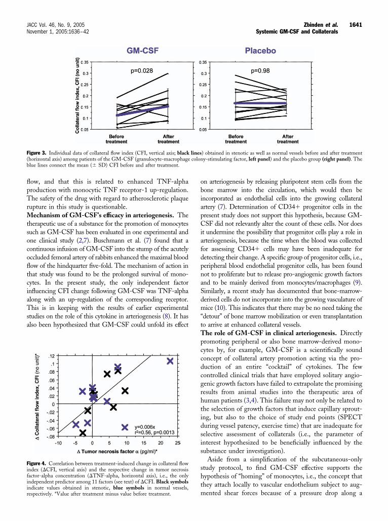
nchanged. Electrocardiographic signs of myocardial isch-mia disappeared more often in the GM-CSF versus thelacebo group (Table 4). Collateral flow index of all vesselsncreased significantly in the GM-CSF group, and it re-
ained unaltered in the placebo group (Table 4, Fig. 3). Aimilar pattern was observed in the vascular subgroups oftenotic and normal coronary arteries. Among 11 variablesinitial CFI, treatment-induced change [�] of neutrophils,osinophils, monocytes, cholesterol, high-density lipopro-ein, GM-CSF, TNF-alpha, MCP-1, TNFR1, CXCR1)
able 3. Treatment-Induced Laboratory Parameter Changes
GM-C
BeforeTreatment
eucocytes (total; 109/l) 7.9 � 1.7 3Neutrophils (109/l) 5.04 � 1.00 2Eosinophils (109/l) 0.15 � 0.10Basophils (109/l) 0.03 � 0.03 0Monocytes (109/l) 0.64 � 0.16 1Lymphocytes (109/l) 2.06 � 0.72 1
D34� cells (% expression of CD34 cell line) 0.051 � 0.04 0.0otal serum cholesterol (mmol/l) 4.0 � 1.0DL cholesterol (mmol/l) 1.2 � 0.3M-CSF (pg/ml) 3.0 � 0.0 1NF-alpha (pg/ml) 2.7 � 0.9 1CP-1 (pg/ml) 415 � 233 6
FGF (pg/ml) 9.7 � 6.9 1EGF (pg/ml) 16.6 � 15.9 4
nterleukin-6 (pg/ml) 2.0 � 0.0 1raktalkine (pg/ml) 1,954 � 742 2,8
p � 0.02 versus GM-CSF.bFGF � basic fibroblast growth factor; GM-CSF � granulocyte-macrophag
hemoattractant protein-1; TNF � tumor necrosis factor; VEGF � vascular endoth
able 4. Treatment-Induced Receptor Expression and Collateral
G
BeforeTreatment
onocyte receptor expressionTNFR1 (TNF-alpha receptor; relative fluorescence) 0.6 � 0.4CCR2 (MCP-1 receptor) 0.2 � 0.1CXCR1 (Fraktalkine receptor) 7.4 � 2.6ollateral flow data: all vesselsECG ST-segment changes �1 mm during
occlusion (%)12 (86)
Collateral flow index (no unit) 0.116 � 0.05Collateral flow index change after-before �0.04
ollateral flow data: stenotic vesselsECG ST-segment changes �1 mm during
occlusion (%)6 (86)
Collateral flow index (no unit) 0.091 � 0.05Collateral flow index change after-before �0.04
ollateral flow data: normal vesselsECG ST-segment changes �1 mm during
occlusion (%)6 (86)
Collateral flow index (no unit) 0.142 � 0.08Collateral flow index change after-before �0.04
p � 0.02 versus GM-CSF.ECG � electrocardiogram; other abbreviations as in Table 3.
xamined by univariate linear regression analysis for theirssociation to CFI change, �TNF-alpha was the only factorndependently predictive of �CFI (Fig. 4). Tumor necrosisactor-alpha change was, itself, related to TNFR1 change:TNF-alpha � 0.053 � 6.447�TNFR1; r2 � 0.61, p �.0001.
ISCUSSION
he present study shows that a purely systemic applicationf GM-CSF is effective in promoting coronary collateral
n � 7) Placebo (n � 7)
terment p
BeforeTreatment
AfterTreatment p
9.9 �0.0001 6.5 � 1.4 6.5 � 1.9* 0.816.3 0.0001 4.37 � 1.65 4.3 � 1.6* 0.452.6 0.0028 0.10 � 0.07 0.13 � 0.08* 0.060.14 0.41 0.04 � 0.03 0.06 � 0.04 0.200.53 0.0101 0.41 � 0.16* 0.56 � 0.31* 0.270.89 0.05 1.63 � 0.64 1.51 � 0.38 0.44.010 0.13 0.103 � 0.11 0.126 � .073* 0.410.3 0.0002 4.5 � 1.1 4.4 � 1.4* 0.400.2 �0.0001 1.2 � 0.2 1.3 � 0.4 0.8810.9 0.037 3.1 � 0.1 3.5 � 1.1* 0.417.0 0.039 3.3 � 1.1 2.7 � 1.0* 0.26353 0.014 560 � 494 670 � 319 0.147.0 0.59 3.0 � 0.0 3.0 � 0.0 —57.7 0.38 9.0 � 0.0 44.0 � 42.8 0.0714.6 0.26 2.0 � 0.1 2.4 � 0.9 0.331,495 0.12 1,731 � 945 2,851 � 883 0.033
ny-stimulating factor; HDL � high-density lipoprotein; MCP-1 � monocyterowth factor.
Changes
SF (n � 7) Placebo (n � 7)
AfterTreatment p
BeforeTreatment
AfterTreatment p
2.2 � 0.7 0.0392 0.9 � 0.6 0.8 � 0.4* 0.160.5 � 0.3 0.14 1.8 � 2.8 0.5 � 0.6 0.301.1 � 1.0 0.0011 4.6 � 2.3 5.1 � 3.3* 0.33
6 (43) 0.04 9 (75) 12 (86) 0.78
.159 � 0.07 0.0281 0.166 � 0.06 0.166 � 0.04 0.98.051 �0.001 � 0.037 0.0351
3 (43) 0.15 5 (71) 6 (86) 0.77
.134 � 0.08 0.05 0.173 � 0.08 0.166 � 0.03 0.78.037 �0.007 � 0.047 0.11
3 (43) 0.15 4 (57) 6 (86) 0.55
.183 � 0.07 0.23 0.163 � 0.05 0.166 � 0.04 0.80.067 �0.003 � 0.034 0.20
SF (
AfTreat
1.4 �2.1 �4.7 �.08 �.18 �.37 �11 �
3.0 �0.9 �8.0 �2.7 �68 �2.2 �5.3 �0.5 �49 �
Flow
M-C
02 � 0
03 � 0
01 � 0
flpTrMtsocofltciaTsa
obiapCiafdpnaSdm“tTpccdcgrhtidsis
sht
F(b
Fifiir
1641JACC Vol. 46, No. 9, 2005 Zbinden et al.November 1, 2005:1636–42 Systemic GM-CSF and Collaterals
ow, and that this is related to enhanced TNF-alpharoduction with monocytic TNF receptor-1 up-regulation.he safety of the drug with regard to atherosclerotic plaque
upture in this study is questionable.echanism of GM-CSF’s efficacy in arteriogenesis. The
herapeutic use of a substance for the promotion of monocytesuch as GM-CSF has been evaluated in one experimental andne clinical study (2,7). Buschmann et al. (7) found that aontinuous infusion of GM-CSF into the stump of the acutelyccluded femoral artery of rabbits enhanced the maximal bloodow of the hindquarter five-fold. The mechanism of action inhat study was found to be the prolonged survival of mono-ytes. In the present study, the only independent factornfluencing CFI change following GM-CSF was TNF-alphalong with an up-regulation of the corresponding receptor.his is in keeping with the results of earlier experimental
tudies on the role of this cytokine in arteriogenesis (8). It haslso been hypothesized that GM-CSF could unfold its effect
igure 3. Individual data of collateral flow index (CFI, vertical axis; blackhorizontal axis) among patients of the GM-CSF (granulocyte-macrophagelue lines connect the mean (� SD) CFI before and after treatment.
igure 4. Correlation between treatment-induced change in collateral flowndex (�CFI, vertical axis) and the respective change in tumor necrosisactor-alpha concentration (�TNF-alpha, horizontal axis), i.e., the onlyndependent predictor among 11 factors (see text) of �CFI. Black symbols
mndicate values obtained in stenotic, blue symbols in normal vessels,espectively. *Value after treatment minus value before treatment.
n arteriogenesis by releasing pluripotent stem cells from theone marrow into the circulation, which would then bencorporated as endothelial cells into the growing collateralrtery (7). Determination of CD34� progenitor cells in theresent study does not support this hypothesis, because GM-SF did not relevantly alter the count of these cells. Nor does
t undermine the possibility that progenitor cells play a role inrteriogenesis, because the time when the blood was collectedor assessing CD34� cells may have been inadequate foretecting their change. A specific group of progenitor cells, i.e.,eripheral blood endothelial progenitor cells, has been foundot to proliferate but to release pro-angiogenic growth factorsnd to be mainly derived from monocytes/macrophages (9).imilarly, a recent study has documented that bone-marrow-erived cells do not incorporate into the growing vasculature ofice (10). This indicates that there may be no need taking the
detour” of bone marrow mobilization or even transplantationo arrive at enhanced collateral vessels.
he role of GM-CSF in clinical arteriogenesis. Directlyromoting peripheral or also bone marrow-derived mono-ytes by, for example, GM-CSF is a scientifically soundoncept of collateral artery promotion acting via the pro-uction of an entire “cocktail” of cytokines. The fewontrolled clinical trials that have employed solitary angio-enic growth factors have failed to extrapolate the promisingesults from animal studies into the therapeutic area ofuman patients (3,4). This failure may not only be related tohe selection of growth factors that induce capillary sprout-ng, but also to the choice of study end points (SPECTuring vessel patency, exercise time) that are inadequate forelective assessment of collaterals (i.e., the parameter ofnterest hypothesized to be beneficially influenced by theubstance under investigation).
Aside from a simplification of the subcutaneous-onlytudy protocol, to find GM-CSF effective supports theypothesis of “homing” of monocytes, i.e., the concept thathey attach locally to vascular endothelium subject to aug-
) obtained in stenotic as well as normal vessels before and after treatmenty-stimulating factor, left panel) and the placebo group (right panel). The
linescolon
ented shear forces because of a pressure drop along a
p“ocaaGCamapipplatplemobaG
ATOz
RPCi
R
1
1642 Zbinden et al. JACC Vol. 46, No. 9, 2005Systemic GM-CSF and Collaterals November 1, 2005:1636–42
reformed collateral vessel. Alternatively, monocytes mayhome” at vascular sites exposed to high shear forces forther reasons than a perfusion pressure gradient betweenollateral supplying and receiving artery, for example, at antherosclerotic plaque obstructing the epicardial coronaryrtery.
M-CSF and atherogenesis/atherogenic plaque rupture.onsidering in this context the role of monocytes in
therogenesis, it is conceivable that the use of GM-CSFay translate into the rupture of atherosclerotic plaques
longside the growth of collateral arteries. Theoretically, aro-atherogenic action of GM-CSF could be imagined viats MCP-1 elevating effect, which could be observed in theresent study. Van Royen et al. (5) demonstrated in apoli-oprotein E-deficient mice that local MCP-1 therapy in the
igated femoral artery augmented collateral artery formationnd atherosclerotic plaque progression. In the present study,wo patients in the GM-CSF group, but none in thelacebo group, had had an acute coronary syndrome, very
ikely because of an atherosclerotic plaque rupture. Consid-ring that “no-option” patients with extensive CAD are theost likely candidates for a therapy with GM-CSF, safety
f the drug in relation to acute coronary syndromes cannote guaranteed. This interpretation of the study results canlso be extended to patients with subclinical CAD receivingM-CSF for other indications.
cknowledgmentshe authors thank Caroline Zwicky, MD, and Elisabethppliger, PhD, Hematology, University Hospital, Bern, Swit-
eprint requests and correspondence: Dr. Christian Seiler,rofessor and Co-Chairman of Cardiology, University Hospital,H-3010 Bern, Switzerland. E-mail: christian.seiler.cardio@
nsel.ch.
EFERENCES
1. Carmeliet P. Mechanisms of angiogenesis and arteriogenesis. NatMed 2000;6:389–95.
2. Seiler C, Pohl T, Wustmann K, et al. Promotion of collateral growthby granulocyte-macrophage colony-stimulating factor in patients withcoronary artery disease. A randomized, double-blind, placebo-controlled study. Circulation 2001;104:2012–27.
3. Henry TD, Annex BH, McKendall GR, et al. The VIVA trial:vascular endothelial growth factor in ischemia for vascular angiogen-esis. Circulation 2003;107:1359–65.
4. Grines CL, Watkins MW, Helmer G, et al. Angiogenic gene therapy(AGENT) trial in patients with stable angina pectoris. Circulation2002;105:1291–7.
5. van Royen N, Hoefer I, Boettinger M, et al. Local monocytechemoattractant protein-1 therapy increases collateral artery formationin apolipoprotein E-deficient mice but induces systemic monocyticCD11b expression, neointimal formation, and plaque progression.Circ Res 2003;92:218–25.
6. Seiler C, Fleisch M, Garachemani AR, Meier B. Coronary collateralquantitation in patients with coronary artery disease using intravascularflow velocity or pressure measurements. J Am Coll Cardiol 1998;32:1272–9.
7. Buschmann I, Hoefer I, van Royen N, et al. GM-CSF: a strongarteriogenic factor acting by amplification of monocyte function.Atherosclerosis 2001;159:343–56.
8. Arras M, Wulf DI, Scholz D, Winkler B, Schaper J, Schaper W.Monocyte activation in angiogenesis and collateral growth in the rabbithindlimb. J Clin Invest 1998:40–50.
9. Rehman J, Li J, Orschell CM, March KL. Peripheral blood“endothelial progenitor cells” are derived from monocyte/macrophages and secrete angiogenic growth factors. Circulation2003;107:1164 –9.
0. Ziegelhoeffer T, Fernandez B, Kostin S, et al. Bone marrow-derived
erland, for their support of the analysis of CD34 cells.cells do not incorporate into the adult growing vasculature. Circ Res2004;94:230–8.
Recommended


