Biol. Rev. (2012), 87, pp. 330–345. 330doi: 10.1111/j.1469-185X.2011.00199.x
The influence of fundamental traitson mechanisms controlling appendageregeneration
Ashley W. Seifert1,2,∗, James R. Monaghan1,2, Matthew D. Smith1,2, Bret Pasch1,2,Adrian C. Stier1,2, Francois Michonneau1,2 and Malcolm Maden2
1 Nexus Biology Group, University of Florida, 223 Bartram Hall, P.O. Box 118525, Gainesville, FL 32611, USA2 Department of Biology, University of Florida, 223 Bartram Hall, P.O. Box 118525, Gainesville, FL 32611, USA
ABSTRACT
One of the most compelling questions in evolutionary biology is why some animals can regenerate injured struc-tures while others cannot. Appendage regeneration appears to be common when viewed across the metazoanphylogeny, yet this ability has been lost in many taxa to varying degrees. Within species, the capacity for regen-eration also can vary ontogenetically among individuals. Here we argue that appendage regeneration along thesecondary body axis may be constrained by fundamental traits such as body size, aging, life stage, and growthpattern. Studies of the molecular mechanisms affecting regeneration have been conducted primarily with smallorganisms at early life stages. Such investigations disregard the dramatic shifts in morphology and physiology thatorganisms undergo as they age, grow, and mature. To help explain interspecific and intraspecific constraints onregeneration, we link particular fundamental traits to specific molecular mechanisms that control regeneration. Wepresent a new synthesis for how these fundamental traits may affect the molecular mechanisms of regenerationat the tissue, cellular, and genomic levels of biological organization. Future studies that explore regeneration inorganisms across a broad phylogenetic scale, and within an ontogenetic framework, will help elucidate the proxi-mate mechanisms that modulate regeneration and may reveal new biomedical applications for use in regenerativemedicine.
Key words: regeneration, appendage, growth, development, metamorphosis, fundamental trait, phylogenetic constraint,regenerative medicine, body size.
CONTENTS
I. Introduction . . . . . . . . . . . . . . . . . . . . . . . . . . . . . . . . . . . . . . . . . . . . . . . . . . . . . . . . . . . . . . . . . . . . . . . . . . . . . . . . . . . . . . . . . . . . . . . . 331(1) General introduction . . . . . . . . . . . . . . . . . . . . . . . . . . . . . . . . . . . . . . . . . . . . . . . . . . . . . . . . . . . . . . . . . . . . . . . . . . . . . . . . . . . 331(2) Types of regeneration . . . . . . . . . . . . . . . . . . . . . . . . . . . . . . . . . . . . . . . . . . . . . . . . . . . . . . . . . . . . . . . . . . . . . . . . . . . . . . . . . . 331(3) Appendage regeneration . . . . . . . . . . . . . . . . . . . . . . . . . . . . . . . . . . . . . . . . . . . . . . . . . . . . . . . . . . . . . . . . . . . . . . . . . . . . . . . 332(4) Fundamental traits and regeneration: patterns and assumptions . . . . . . . . . . . . . . . . . . . . . . . . . . . . . . . . . . . . . . . 333
II. Tissue-level effects . . . . . . . . . . . . . . . . . . . . . . . . . . . . . . . . . . . . . . . . . . . . . . . . . . . . . . . . . . . . . . . . . . . . . . . . . . . . . . . . . . . . . . . . . . 334(1) Wound healing and re-epithelialization . . . . . . . . . . . . . . . . . . . . . . . . . . . . . . . . . . . . . . . . . . . . . . . . . . . . . . . . . . . . . . . . 335(2) Histolysis and blastema formation . . . . . . . . . . . . . . . . . . . . . . . . . . . . . . . . . . . . . . . . . . . . . . . . . . . . . . . . . . . . . . . . . . . . . 335(3) Nerve dependence . . . . . . . . . . . . . . . . . . . . . . . . . . . . . . . . . . . . . . . . . . . . . . . . . . . . . . . . . . . . . . . . . . . . . . . . . . . . . . . . . . . . . 336(4) Angiogenesis . . . . . . . . . . . . . . . . . . . . . . . . . . . . . . . . . . . . . . . . . . . . . . . . . . . . . . . . . . . . . . . . . . . . . . . . . . . . . . . . . . . . . . . . . . . 336(5) Growth and differentiation . . . . . . . . . . . . . . . . . . . . . . . . . . . . . . . . . . . . . . . . . . . . . . . . . . . . . . . . . . . . . . . . . . . . . . . . . . . . . 337
III. Cellular effects . . . . . . . . . . . . . . . . . . . . . . . . . . . . . . . . . . . . . . . . . . . . . . . . . . . . . . . . . . . . . . . . . . . . . . . . . . . . . . . . . . . . . . . . . . . . . . 337
* Address for correspondence at address 2 (Tel.: (352) 273-7876; Fax: (352) 392-3704; E-mail: [email protected]).
Biological Reviews 87 (2012) 330–345 © 2011 The Authors. Biological Reviews © 2011 Cambridge Philosophical Society
The influence of fundamental traits on mechanisms controlling appendage regeneration 331
(1) Cell cycle re-entry and de-differentiation . . . . . . . . . . . . . . . . . . . . . . . . . . . . . . . . . . . . . . . . . . . . . . . . . . . . . . . . . . . . . . 337(2) Progenitor cells . . . . . . . . . . . . . . . . . . . . . . . . . . . . . . . . . . . . . . . . . . . . . . . . . . . . . . . . . . . . . . . . . . . . . . . . . . . . . . . . . . . . . . . . . 339(3) Cell proliferation and growth . . . . . . . . . . . . . . . . . . . . . . . . . . . . . . . . . . . . . . . . . . . . . . . . . . . . . . . . . . . . . . . . . . . . . . . . . . 339
IV. Genomic-level effects . . . . . . . . . . . . . . . . . . . . . . . . . . . . . . . . . . . . . . . . . . . . . . . . . . . . . . . . . . . . . . . . . . . . . . . . . . . . . . . . . . . . . . . 340(1) Regeneration-specific genes . . . . . . . . . . . . . . . . . . . . . . . . . . . . . . . . . . . . . . . . . . . . . . . . . . . . . . . . . . . . . . . . . . . . . . . . . . . . 340(2) Epigenetic control of regeneration . . . . . . . . . . . . . . . . . . . . . . . . . . . . . . . . . . . . . . . . . . . . . . . . . . . . . . . . . . . . . . . . . . . . . 340
V. Future perspectives . . . . . . . . . . . . . . . . . . . . . . . . . . . . . . . . . . . . . . . . . . . . . . . . . . . . . . . . . . . . . . . . . . . . . . . . . . . . . . . . . . . . . . . . . 341VI. Conclusions . . . . . . . . . . . . . . . . . . . . . . . . . . . . . . . . . . . . . . . . . . . . . . . . . . . . . . . . . . . . . . . . . . . . . . . . . . . . . . . . . . . . . . . . . . . . . . . . 343
VII. Acknowledgements . . . . . . . . . . . . . . . . . . . . . . . . . . . . . . . . . . . . . . . . . . . . . . . . . . . . . . . . . . . . . . . . . . . . . . . . . . . . . . . . . . . . . . . . . 343VIII. References . . . . . . . . . . . . . . . . . . . . . . . . . . . . . . . . . . . . . . . . . . . . . . . . . . . . . . . . . . . . . . . . . . . . . . . . . . . . . . . . . . . . . . . . . . . . . . . . . . 343
I. INTRODUCTION
(1) General introduction
The ability of organisms to regenerate tissues and structuresis one of the most captivating phenomena in biology.Regeneration is a ubiquitous feature of metazoans (Brockes& Kumar, 2008), although there is substantial variationacross taxa from the complete regeneration of an entireorganism (e.g. hydra, cnidarians, planarians) to a restrictedregeneration of certain tissues in birds and mammals. Amajor goal of regeneration research is to understand if thesame molecular mechanisms control regeneration in distantlyrelated taxa, or if the capacity to regenerate damagedtissue has evolved multiple times and with different controlmechanisms. Additionally, there is substantial variation inregeneration among individuals within regenerating species.For example, older zebrafish (Danio rerio) regenerate theirfins more poorly compared to young individuals (Anchelinet al., 2011). What are the changes that occur throughoutthe lifetime of an individual to constrain regeneration?The possible discovery of conserved molecular mechanismsthat control regeneration would have a profound impacton regenerative medicine. Is it possible to activate suchmechanisms and reawaken latent regenerative capacity inolder individuals or in mammals? Contemporary advancesin molecular biology have begun to uncover the cellular,molecular, and genetic mechanisms governing regeneration,however, our understanding of what explains variation inregenerative ability within and across taxa is lacking.
One possible explanation for our limited understanding ofwhy regenerative capacity fluctuates across taxa is the biasof regeneration research towards experiments conductedon small-sized model organisms when they are youngin age, and early in life stage. This approach ignoresconsiderable shifts in growth, development, and physiologythat animals experience in their lifetime, and thus masks theinfluence of numerous fundamental traits on regenerativecapacity. Therefore, we need to consider both the abilityto regenerate across species, and alterations in regenerativecapacity throughout an individual’s lifetime (within species).Fundamental traits are organismal properties (e.g. life-historytraits, developmental traits, physiological traits) that areinherent to individuals and species; and that influence theirecology, behaviour, and physiology. As such, fundamentaltraits have the potential to constrain regenerative ability
or the rate of regeneration. Identifying constraints onregenerative ability that operate both within and acrossspecies will help elucidate if regeneration is inherently linkedto development and growth. Exploring relationships betweenfundamental traits and regeneration at the mechanistic levelcan serve as a platform for investigating why some animalshave reduced powers of regeneration.
Herein we address how a suite of fundamental traits caninfluence regeneration. These traits can influence the abilityto regenerate, the rate of regeneration, or the quality of theregenerated appendage (i.e. heteromorphy) both within andacross species. Specifically, we focus on reparative regener-ation of appendages along the secondary body axis, and thefollowing fundamental traits: (1) body size, (2) aging, (3) lifestage (pre- or post-metamorphosis; larva, juvenile or adult),and (4) mode of post-embryonic growth (determinate ver-sus indeterminate). We first review the literature and presentpublished viewpoints regarding how fundamental traits affectregeneration. We then present hypotheses about how specificfundamental traits can influence the mechanisms of regener-ation at the tissue, cellular, and genomic levels of biologicalorganization. Along with these hypotheses we suggest multi-ple avenues for future research. In order to maximize read-ability, we avoid reiterating molecular mechanisms for eachtrait by organizing this review according to biological level,and discussing fundamental traits within each section. Ourgoal is to spark an interest in the underexplored relationshipsbetween fundamental traits and mechanisms of regenerationtowards a better understanding of how and why regen-eration is curtailed in more derived vertebrates, includinghumans.
(2) Types of regeneration
Regeneration is traditionally viewed in two contexts; physio-logical and reparative (Table 1) (Morgan, 1901). Physiologi-cal regeneration is the regular and repeated regeneration ofa particular structure that is normally replaced throughoutthe life of an organism and examples exist in every meta-zoan species (Table 1). By contrast, reparative regenerationis induced by injury, and leads to replacement of the missingstructure. Although this definition distinguishes repair fromreplacement, some traditional examples of reparative regen-eration blur the lines between them because restoration ofthe missing part occurs with structural alterations and with
Biological Reviews 87 (2012) 330–345 © 2011 The Authors. Biological Reviews © 2011 Cambridge Philosophical Society

332 Ashley W. Seifert and others
Table 1. Examples of regular and recurring regeneration(physiological) and regeneration stimulated via injury or damage(reparative)
Type ofregeneration Examples
Physiological Cervid antlers, replacement of blood, epidermis,endometrium, gut lining, arthropodexoskeleton (moulting)
Reparative Incomplete: fish barbels, lizard tails, larvalurodele tails, young mammalian digit tips
Complete: urodele limbs, adult urodele tails,some fish fins, mollusc eye stalks, arthropodand crustacean limbs, antennae
reductions in function. Thus, reparative regeneration can becomplete or incomplete (Table 1). Examples of incompleteregeneration include tail regeneration in lizards (which donot regenerate neurons or vertebrae) (Simpson, 1964), andbarbel regeneration in fish (which do not regenerate thestructural mesodermal core) (LeClair & Topczewski, 2010).Interestingly, when the larval urodele tail is amputated thenotochord fails to regenerate, and is replaced by cartilagi-nous vertebrae that will not develop until months later in therest of the tail (Goss, 1969). In this case of regeneration thereplacement is not perfect, but rather is a precursor of whatwill develop around the structure in the future. This finedistinction is important as it may reveal early compromiseson regenerative ability in some lineages where regenerationand repair occur in tandem. Here we define regeneration asthe complete replacement of the original form.
We also note a fundamental distinction between the abilityto generate an entirely new organism from a severed piece(e.g. Hydra) and regeneration of a limb in arthropods orsalamanders (Fig. 1). Whereby, if you cut a planarian in two,or sever the arm of a starfish, each piece has the ability to forma complete, autonomous animal (Fig. 1A). By contrast, thesevered tail of a lizard or the limb of a cockroach cannot giverise to a new individual, and this piece fails to survive apartfrom the animal (Fig. 1B). Mechanistically, the ability toreplace a missing structure in asexually reproducing animalsstems from a continuous source of pluripotent cells withlimitless proliferative potential and with the ability to formevery cell type in the new animal (Wagner, Wang & Reddien,2011). By contrast, cells involved in vertebrate, crustacean, orarthropod regeneration cannot form every part of an animalin vivo, and are limited in their developmental potency (Kraglet al., 2009; Truby, 1983). Herein, we focus on appendageregeneration along the secondary body axis.
(3) Appendage regeneration
The early events of appendage regeneration can occur fromthe production of new tissue through cell proliferation (epi-morphosis), or through the rearrangement of existing cells toform the new part in the initial absence of proliferation (mor-phallaxis) (Morgan, 1901). More recently this distinction has
BA
Fig. 1. Distinguishing between modes of regeneration.(A) After a seastar loses an arm, both the body (missing thearm) and the arm (apart from the body) can regenerate themissing part. (B) A lizard, following autotomy or injury, canregenerate a new tail, but the tail has no capacity to regeneratemore of itself, let alone another lizard.
been argued to be purely historical, and that all instances ofregeneration share the feature of re-specification of positionalinformation at the injury site followed by intercalation andre-growth of the regenerating tissue (Agata, Saito & Naka-jima, 2007). The important distinction is that in all examplesof appendage regeneration along the secondary body axiscell proliferation is required to supply material for the initialregenerating appendage, and this process couples growth ofthe appendage with regeneration. For this reason, epimor-phosis mechanistically unites leg or antennae regenerationin hemimetabolous insects, eye-stalk regeneration in snails,and limb, fin, and tail regeneration in vertebrates.
Following extirpation, epimorphic appendage regener-ation proceeds through a series of similar events (Fig. 2)(Bryant, Endo & Gardiner, 2002). First, epithelial cellsmigrate over the wound surface (under in the case wherea scab is formed) to re-epithelialize the injury site. Next,cells at the amputation plane re-enter the cell cycle and de-differentiate. Local cell populations comprised of these newlydedifferentiated cells, progenitor cells, and fibroblasts form aregeneration blastema. The blastema proliferates, and as inembryonic development, these cells acquire spatial pattern-ing information as they expand, ultimately re-differentiatingto restore the injured structure through intercalation (French,Bryant & Bryant, 1976). An extended growth period followsregeneration once patterning is complete. Full replacementof the appendage can take years in some large terrestrial sala-manders (Young, Bailey & Dalley, 1983b), and is restricted bymoulting times in arthropods such that a small limb emergesand grows after each successive moult (Maruzzo et al., 2005).That epimorphic regeneration of appendages appears highlyconserved is fascinating given the great phylogenetic dis-tance among these species, although it remains unclear ifthe molecular mechanisms underlying these processes arehomologous across taxa.
Biological Reviews 87 (2012) 330–345 © 2011 The Authors. Biological Reviews © 2011 Cambridge Philosophical Society

The influence of fundamental traits on mechanisms controlling appendage regeneration 333
Arthropod regeneration
De-Diff. Blastema
Blastema
Proliferation
Proliferation
Proliferation
Re-Ep.
Re-Ep.
Injury or Amputation
Differentiation
Differentiation
Patterning GrowthDe-differentiationWound healing
Cutical reformation
Muscle Cuticle
Nerves Epidermis
Cartilage/Bone
Vertebrate regeneration
Fig. 2. Epimorphic regeneration in vertebrates and arthropods. Following injury or amputation, appendage regeneration in thesetaxa proceeds through a generally similar set of stages. First, re-epithelialization (Re-Ep.) occurs as the edges of the epidermis migrateto cover the wound surface. In taxa with an exoskeleton, the cuticle reforms after the epidermis has finished covering the wound. Cellcycle re-entry and de-differentiation (De-Diff.) of local cell populations leads to the formation of a regeneration blastema. Blastemalcells proliferate and acquire patterning information as they re-differentiate to replace the missing structure.
(4) Fundamental traits and regeneration: patternsand assumptions
Fundamental traits are key components of the ecology,behaviour, physiology, and life-history strategy of anorganism. The critical role of fundamental traits in modifyingform and function begs the question whether or not theymay also influence mechanisms that control appendageregeneration. Surprisingly, the effects of such fundamentaltraits have been largely ignored in rigorous experimentalstudies, and this oversight stems partly from the difficulty inseparating traits that are correlated with one another (Goss,1969; Pritchett & Dent, 1972; Scott, 1909; Speakman, 2005;Wagner & Misof, 1992; Young et al., 1983b).
Nonetheless, some studies have examined the influence ofbody size in vertebrates (but not arthropods), which has ledto the perception that rate of regeneration and replacementtime of a structure are negatively affected by size (Pritchett& Dent, 1972). Unfortunately, only one study has addressedbody size alone (controlling for age): Pritchett & Dent (1972)found that in newts, the larger hindlimbs took longer to regen-erate than did the smaller forelimbs in the same individuals.From this they inferred a negative relationship between thesize of adult newts and the rate of limb regeneration (Pritch-ett & Dent, 1972). In practice, the study was limited by thesize variation in limbs, inherent differences in the structureof hindlimbs and forelimbs, and that the patterning phaseand the growth phase were analyzed together. Analyzing
regeneration and growth together confounds how the size ofa structure might directly influence the development and pat-terning phases of regeneration (Fig. 2). For instance, studiesin salamanders and fish have shown that body size neg-atively correlates with growth of the regenerate followingmorphogenesis, although not necessarily for development ofthe regenerating anlagen (Fig. 2) (Scott, 1909; Tank, Carlson& Connelly, 1976; Young et al., 1983b). Thus, the influenceof size on regeneration remains unclear, and we can con-clude only that larger organisms take longer to re-grow anappendage following regeneration of a small replacement.
Aging and life stage also strongly influence organismalperformance (Lakatta, 1983; Marden et al., 2003; Pough &Kamel, 1984). Among vertebrates, the general assumptionis that regeneration will take longer at older ages (Wagner& Misof, 1992; Wallace, 1981). Although regeneration hasbeen documented to occur in a few mature salamanders andfish, little information actually exists on regenerative abilityor rate in aged animals. Underscoring the importance of con-ducting regeneration experiments in older animals, a recentstudy found no difference in the rate or quality of regen-erating lenses in very old newts between 16 and 30 yearsof age (Eguchi et al., 2011). Still, at a mechanistic level,there are many reasons why aging may affect regeneration.When decoupled from body size, aging becomes relevant tocell differentiation, cell cycle re-entry (de-differentiation),metabolic stress, and the capacity for cell proliferation.
Biological Reviews 87 (2012) 330–345 © 2011 The Authors. Biological Reviews © 2011 Cambridge Philosophical Society

334 Ashley W. Seifert and others
Similarly, metamorphosis represents a major transition in lifewhere structure, function, and physiology change dramati-cally. Life-stage-limited regeneration has been demonstratedin anurans (Dent, 1962) and holometabolous insects (see ref-erences in Maginnis, 2006; Maruzzo et al., 2005) where thecapacity to regenerate is lost after metamorphosis. Regener-ative capacity in hemimetabolous insects, crustaceans, andsome chelicerates is dependent on remaining moulting peri-ods (see references in Goss, 1969; Maginnis, 2006; Maruzzoet al., 2005); whether or not these organisms can regeneratean appendage beneath the cuticle following their final instarremains unknown. Assuming that the cellular and geneticstructure of these animals does not change at metamorphosis(or moult), then why there is a change in their regenerativeability is puzzling, and warrants further inquiry in a varietyof pre- and post-metamorphic animals.
While the relationship between regeneration and growthhas long been appreciated (Goss, 1965; Voit, Anton &Blecker, 1995), the interaction between regeneration andmode of growth (determinate versus indeterminate) remainspoorly studied. Some evidence suggests that species whichcontinue to grow after reaching sexual maturity (indeter-minate growth) may be more likely to retain the capacityto regenerate throughout their life (Kara, 1994; Klapperet al., 1998), whereas this capacity may be limited to juvenilegrowth periods in species that attain maximum size at matu-rity (determinate growth). In newts and other amphibians, theyearly addition of skeletal bone (measured as growth rings) isa key factor in attributing indeterminate growth to these ani-mals (Homan, Reed & Windmiller, 2003; Jakob et al., 2002).Despite continued skeletal addition, overall growth rates formany species of indeterminate growers often decrease withage, metamorphosis, or sexual maturity, and this may greatlyreduce regenerative capacity. For example, some effects ofaging that may inhibit regeneration have been observed inmammalian cells, but appear to be absent from fishes and
amphibians with indeterminate growth. Interestingly, arthro-pods that do not exhibit post-larval cell division (e.g. mites) areincapable of regeneration because they are incapable of fur-ther growth (Rockett & Woodring, 1972). While regenerationin determinate growers remains to be tested at stages wheregrowth plateaus, the current evidence supports a relationshipbetween growth potential and the capacity for regeneration.
After considering previous attempts to explain howfundamental traits can affect appendage regeneration, weare left with conflicting results, and with no clear set ofhypotheses for how these traits may affect the specificmechanisms that control regeneration. Below we discuss howfundamental traits can modulate mechanisms of regenerationat three levels of biological organization: tissue, cellular, andgenomic. Some traits may have diverse effects at multiplelevels, while others (e.g. size) may have effects at only onelevel (Fig. 3). Because of the complex interaction betweentraits and mechanisms, we present hypotheses for each levelof organization and reference traits where applicable.
II. TISSUE-LEVEL EFFECTS
An appendage is comprised of multiple tissues that caninclude combinations of nerves, muscle, cartilage, and bonesurrounded by epidermis. At the tissue level, fundamentaltraits of an organism may limit regeneration through effectson these tissue components alone or in combination (Fig. 3).Appendage size, which is correlated with body size, dictatesthe area of the injury plane. In large-bodied animals, the areaof this surface may delay re-epithelialization to such a degreethat it prevents the formation of a blastema. Also, injuryabove a critical size could lead to lethal loss of vital fluids (e.g.blood, haemolymph, water). The inability to recover fromsuch a large injury would negate the need for regenerating
Life stage
Growth pattern
Tissue level properties Cellular level properties
Genomic level properties
Catastrophic loss of limb
Re-epithelialization
Appendage growth
Nerve dependence
Angiogenic dependence
Cell cycle re-entry
De-differentiation
Progenitor cell availability
DNA methylation state
Fundamental Trait
Body size
AgeAEC formation
Cell proliferation
Telomerase activity
Histone modifications
Histolysis
Blastema formation
Fig. 3. Fundamental traits can exert their effect on regenerative processes at multiple levels of biological organization. Biologicalprocesses are listed for the level of biological organization at which they can exert their effect on regenerative capacity. Linesconnecting a fundamental trait to a biological process indicate a hypothesized interaction that can affect regeneration. Theseinteractions are discussed in the text. AEC, apical epithelial cap.
Biological Reviews 87 (2012) 330–345 © 2011 The Authors. Biological Reviews © 2011 Cambridge Philosophical Society

The influence of fundamental traits on mechanisms controlling appendage regeneration 335
the appendage, and would provide a natural limit for theinjury size an animal could sustain. Provided the injury isnot life threatening, how does the size of the appendage, orits developmental stage impact particular requirements tomount a successful regenerative response?
(1) Wound healing and re-epithelialization
In order to stabilize an injury, animals need to closethe wound surface to protect against infection, desiccation(terrestrial animals), or osmotic imbalance (aquatic animals).Both initiation and the rate of re-epithelialization canregulate the regenerative response, and may be dependenton size, aging, or life stage. All animals capable of appendageregeneration completely re-epithelialize the wound surface.A survey of limb regeneration among amphibians showedthat species exhibiting some form of regenerative outgrowthhave rapid epithelial migration, whereas species withdelayed migration exhibit no regeneration (Scadding, 1981).Moreover, the largest individuals in this survey showed areduced regenerative capacity (i.e. slower rates, incompletepatterning, incomplete regeneration); but variation in sizeamong species, and variation in age within species were notrigorously controlled for. This suggests that size may haveincreased the time needed to re-epithelialize the wound,which in turn inhibited a successful regenerative response.Delayed re-epithelialization contributes to scarring and canlead to chronic, non-healing wounds (Sivamani, Garcia &Isseroff, 2007). By contrast, rapid re-epithelialization occursduring scar-free healing in fetal mammals and an increasedrate of re-epithelialization can reduce scarring (Whitbyet al., 1991).
Among arthropods, re-epithelialization follows haemoly-mph coagulation which initially protects the amputationplane by forming a scab (similar to mammals) (Adidoyi, 1972;Rockett & Woodring, 1972). The rate of re-epithelializationin arthropods is much slower compared to amphibians, buthas not been examined across an age or size gradient; andwhether or not re-epithelialization occurs in non-moultingadults following amputation is not known.
The neo-epidermis also plays an important role indirecting regeneration. Following re-epithelialization, thenew epidermal layer becomes a specialized signaling centrecalled the apical epithelial cap (AEC). The AEC directsgrowth of the underlying tissue and is necessary for successfulappendage regeneration (Wallace, 1981). Formation of thisstructure appears to depend partially on the life stage whenthe amputation occurs. The inability to regenerate a tailduring frog metamorphosis is correlated with a lack of AECformation, and post-metamorphic anuran species that showheteromorphic regeneration do not form an AEC as thick asthat seen in urodeles capable of complete limb regeneration(Wolfe, Nye & Cameron, 2000).
The loss of limb regeneration ability in anuransalso may be due to a loss of competency in themesenchyme as opposed to the epidermis. Yokoyamaet al. (2000) created recombinant limbs with epider-mis collected from regeneration-incompetent limbs and
mesenchyme from regeneration-competent limbs (and vice-versa). They showed that only the regeneration-competentmesenchyme can rescue a regeneration-incompetent epider-mis (i.e. regeneration-competent epidermis did not rescueregeneration-incompetent mesenchyme). Thus, some con-troversy remains about whether changes in the epidermisare the cause of regenerative loss. However, complete for-mation of the AEC is required for complete limb regenerationin salamanders; and available evidence shows a correlationbetween life stage and AEC formation.
Life stage may affect the regenerative response due toalterations in tissue physiology of the epidermis. Duringregeneration, changes in ion flux across the wound bed causea shift in electric potential that correlates with a regeneration-permissive environment (Borgens, Vanable & Jaffe, 1977).Xenopus laevis tadpoles are normally capable of tail regener-ation but undergo a short refractory period (stages 45-47)preceding metamorphosis where they become regenerationincompetent (Beck, Christen & Slack, 2003). Regenerationprior to this phase is partly due to the movement of protons(via an ion pump) across the new epithelium and to the sub-sequent rapid physiological restoration of a polarized plasmamembrane that covers the wound site (Adams, Masi & Levin,2007). An inability to rapidly re-polarize during the refrac-tory period leads to loss of regeneration while mis-expressionof a heterologous cell membrane H+ pump in refractoryepidermis can rescue tail regeneration in tadpoles (Adamset al., 2007). Though previous research has been conductedonly on tail regeneration, appendage regeneration followingmetamorphosis may be constrained in a similar manner.The involvement of tissue-specific changes that alter ionmovements underscores the role that life stage may playin the control of regeneration through changes in tissuephysiology.
(2) Histolysis and blastema formation
Following re-epithelialization, histolysis of underlying stumptissue takes place to break down injured tissue and thesurrounding extracellular matrix (ECM). This, in turn, freesup cells to migrate and form a blastema (Fig. 2). The processof histolysis and blastema formation may be influenced bystructural changes that occur to the ECM during aging.Collagen is the major component of the ECM in skin,and must be degraded for cell migration to proceed. Asvertebrates age, cross-linking of collagen increases tensilestrength and decreases solubility of the ECM (Baum, Faris &Franzblau, 1975; Kara, 1994). Thus, aged animals likely havea more difficult time breaking down the ECM, which wouldconsequently delay or inhibit blastema formation. In additionto the ECM, bone and cartilage at the amputation plane mustundergo histolysis prior to replacement during regeneration(Wallace, 1981). While the ratio of bone to cartilage increaseswith age, the effects of these ratios on regeneration rates arenot known. Further testing of these observations is necessaryto understand if adult appendages with mineralized skeletonsexhibit reduced regenerative capacity.
Biological Reviews 87 (2012) 330–345 © 2011 The Authors. Biological Reviews © 2011 Cambridge Philosophical Society

336 Ashley W. Seifert and others
(3) Nerve dependence
One of the most extensively studied phenomena in regenera-tion is the dependence on an intact nerve supply as observedin hydra, echinoderms, planarians, annelids (reviewed inBrockes & Kumar, 2008), teleost fins (Geraudie & Singer,1985), Xenopus laevis froglet limbs (Suzuki et al., 2005), urodelelimbs (Singer, 1952), lizard tails (Simpson, 1970), and fetalwound healing in chickens and mammals (Harsum, Clarke &Martin, 2001; Stelnicki et al., 2000). Nerves play a supportive(rather than instructive) role by secreting nerve-derived fac-tors that facilitate appendage regeneration (Carlson, 2007).Body size, aging, and life stage each may alter the nerve-limbrelationship and lead to the loss of regenerative capacity.
In vertebrates, the number of nerve fibres or the cross-sectional area of nerve bundles (expressed as a fraction ofthe cross-sectional area of the limb) is often negatively cor-related with increasing body size (Peadon & Singer, 1965;Wallace, 1981). In a large animal, an insufficient concen-tration of nerves may constrain regeneration in a largeappendage that would normally be able to regenerate if itwas smaller. The hypothesis that nerve concentration con-strains regenerative capacity was championed by MarcusSinger, based primarily on experiments showing that newtlimbs must have a minimum number of nerve fibres in orderto regenerate (Singer, 1952). Also, there may be an inter-specific relationship between regeneration rate and degreeof innervation. However, the evidence for this is equivocal:nerve abundance appears to be positively correlated withthe rate of regeneration across Ambystoma species (Younget al., 1983b), but other studies have shown that regenerativecapacity among species does not correlate with innervation(Kurabuchi, 1990; Scadding, 1982; Van Stone, 1964) or withregeneration rate within the same species (Scadding, 1983).Furthermore, the Mexican axolotl has a relatively fast rate ofregeneration, despite having one of the lowest nerve densitiesamong amphibians (Scadding, 1982). More experiments thatcompare innervation parameters across larger interspecificranges in appendage size are necessary to better resolve theconnection between nerve fibre abundance and regenerativeability or rate.
In addition to body size, aging and metamorphosis mayconstrain regenerative capacity by inhibiting the ability of anerve to produce the vital nerve-derived factor(s). Comparedto old individuals, spinal cord extracts from young axolotlsare more mitogenic than extracts from old axolotls whenadded to blastemal cells (in vitro) from a regenerating limb(Boilly & Albert, 1988). This suggests that there is somedecrease in the neurotrophic influence of spinal cordsacross an age gradient in salamanders. Nerves may alsolose the ability to re-grow over ontological time, makingthem unable to provide support for regeneration. The abilityof mammals to regenerate peripheral nerves decreases withage, and all vertebrates regenerate nerves better when theyare younger (Tanaka & Ferretti, 2009; Verdu et al., 2000).Additionally, some forms of axon re-growth are lost atmetamorphosis (Gibbs, Chittur & Szaro, 2010). For example,X. laevis axons can regenerate across a spinal cord lesion
when metamorphosis is pharmacologically inhibited, butthis ability is lost when metamorphosis is precociously forced.Thus, during aging and after metamorphosis, there is a cleardecrease in the regenerative capacity of the nervous systemand this likely contributes to the reduced ability of an animalto support peripheral tissue regeneration.
An exception to the nerve-dependency phenomenonis that larval salamander limbs can regenerate withoutinnervation if the limb was never innervated duringdevelopment (termed aneurogenic) (Yntema, 1959). Duringdevelopment, limb buds are formed before nerve fibres enterthe limb, meaning that early limb development is nerveindependent. Limb buds and surgically created aneurogeniclimbs may produce their own neurotrophic-like factors (Filoniet al., 1999), or they may not need the neurotrophic factorfor growth (Tassava & Olsen-Winner, 2003). Regardless ofthe mechanism, innervation of developing or aneurogeniclimbs leads to nerve dependency (Thornton & Thornton,1970). Surgically creating aneurogenic limbs may simplyextend the time in which the limb can produce its ownneurotrophic-like factors, or the time in which the limb doesnot need the neurotrophic-like factors for growth. Recentstudies have shown that denervated limbs can regenerate(albeit without muscle) if a single molecule, anterior gradient2, is introduced into the denervated limb blastema (Kumaret al., 2007). An explanation for why aneurogenic limbscan regenerate may be provided through functional studiesduring limb development and in aneurogenic limbs. The keypoint is that once limbs are innervated for some time, theybecome nerve dependent for the life of the animal, and thusmay be affected by fundamental traits later in life.
The role of the nervous system during arthropod regen-eration has received little attention, partly due to difficultiesassociated with denervating arthropod appendages. Somestudies have established that muscle regeneration is depen-dent upon innervation after moulting (Consoulas & Levine,1997), while others suggest that innervation plays only a par-tial role in the support of appendage regeneration (Nuesch,1968). Although the available evidence supports a role fornerves, the conflicting results and experimental hurdles fromvarious arthropod studies make it difficult to test hypothesesabout how changes in the nervous system across ontogenymay influence regeneration (Goss, 1969; Nuesch, 1968).
(4) Angiogenesis
Similar to nerve requirements, vascular support is also a likelyrequisite for proper appendage regeneration. The formationof new blood vessels (angiogenesis) is necessary for organdevelopment and tissue homeostasis in adulthood. Uponinjury, hypoxic conditions produced by the local destructionof tissue are thought to induce the formation of angiogenicfactors including vascular endothelial growth factor (VEGF)(Ferrara, 2002). During fin regeneration in zebrafish, block-ing angiogenesis by inhibiting VEGF signaling does not affectblastema formation or early tissue outgrowth (∼800 μm),but does block further growth of the regenerate (Bayliss et al.,2006). This suggests that while vascularization does not play
Biological Reviews 87 (2012) 330–345 © 2011 The Authors. Biological Reviews © 2011 Cambridge Philosophical Society

The influence of fundamental traits on mechanisms controlling appendage regeneration 337
a role in blastema formation or early cell proliferation, it isnecessary for growth and morphogenesis of the regenerat-ing appendage (Fig. 2). The age and size of a regeneratingstructure likely affects angiogenesis in a similar manner tonerve regeneration, because growth of the two tissues is inti-mately connected. Angiogenic potential decreases with agein mammals (that do not regenerate), and affects the injuryresponse (Edelberg & Reed, 2003), but whether animals withregenerative abilities also have decreased angiogenesis withincreased age is unknown.
(5) Growth and differentiation
The previous sections discussed influences on the ability ofan organism to mount a regenerative response (i.e. prior todifferentiation of the regenerate; Fig. 2). Alternatively, fun-damental traits may affect the growth phase of regeneration.Intriguingly, the rate of regeneration prior to differentiationmay be relatively constant across body size, and only varyas the newly regenerated appendage grows (which would bea function of growth rate). Observations from regeneratinglimbs of large and old Ambystoma mexicanum demonstrate thatthe digit-stage regenerate is actually a small limb in thecentre of the amputation plane, surrounded by a collar oftissue (unpublished data). In contrast to previous research(Scadding, 1977), a study on large adult Ambystoma annulatum,A. maculatum, A. texanum, and A. tigranum found they werecapable of regenerating their limbs, although in some casescomplete replacement took nearly two years (Young, Bailey& Dalley, 1983a; Young et al., 1983b). In hemimetabolousinsects and crustaceans, regeneration is wholly dependenton moulting (and thus on growth). For many arthropodsand crustaceans, the regenerate following the first moult isalways a small version of the missing appendage, and sub-sequent moults are necessary in order to completely replacethe missing part (Maruzzo et al., 2005). The formation ofa miniature replacement suggests that fundamental traitsmay constrain regeneration after patterning, and that givenenough time and energy, a small appendage will eventuallyregenerate to the normal size. Hence, claims that larger andolder animals regenerate more slowly may have nothing todo with regeneration per se, but instead be a consequenceof growth of the replacement part. To understand morefully how growth and regeneration are coupled, future stud-ies examining this relationship should consider: (1) if factorspermit a regenerative response but preclude growth to fullyrestore the missing tissue; and (2) how various factors affectthe rate of regeneration during the development phase andgrowth phase independently.
III. CELLULAR EFFECTS
Next, we address how fundamental traits may affect regen-eration at the cellular level. During embryonic developmentof an appendage, the proliferation, patterning, and differen-tiation of dividing cells are integrated to produce the final
structure. Appendage regeneration following injury appearsto recapitulate the developmental process. But in contrast todevelopment, differentiated cells must re-enter the cell cycleduring regeneration, and are induced to proliferate amongstpopulations of cells that necessarily must remain in the differ-entiated state. The delicate regulation of de-differentiation,proliferation, and re-differentiation is critical to the regener-ation process. The effects of aging and growth on the abilityof cells to de-differentiate and proliferate likely contribute toconstraints on regeneration. Aging in some animals is inti-mately tied to cellular senescence and a decrease in telomeresize. The role of cellular senescence and telomere length inrelation to regeneration has been almost completely unex-plored outside of mammals and a few fish. A lack of cellularsenescence in cold-blooded vertebrates and some crustaceansmay provide for prolonged regenerative capacity throughoutlife (Elmore et al., 2008; Gomes, Shay & Wright, 2010). Herewe explore how these cellular mechanisms can be affectedduring the aging process, and how indeterminate growersmight escape the effects of senescence, thereby exhibitingnear-limitless regenerative potential.
(1) Cell cycle re-entry and de-differentiation
Following appendage amputation in vertebrates and arthro-pods, local signals at the site of injury stimulate differentiatedcells to ‘‘rewind’’ their cellular history and re-enter thecell cycle (Echeverri, Clarke & Tanaka, 2001; Hay & Fis-chman, 1961; Lentz, 1969; Truby, 1983). Simultaneously,progenitor cells are mobilized and contribute to regenerat-ing tissue (Morrison et al., 2006). Both differentiated cells andlocal progenitor cells participate in appendage regeneration,although the relative contribution of these cells to the sub-sequent tissue components remains unclear (Morrison et al.,2006). Recent research examining axolotl limb regenerationconfirmed that blastemal cells respect strict cell lineages, sug-gesting that de-differentiation does not produce a blastemaof pluripotent stem cells, but rather a blastema of lineage-committed cells that respect their embryonic origin (Kraglet al., 2009). Following activation or de-differentiation, bothcell types must proliferate, acquire positional identity, andthen differentiate to populate the regenerated tissue (Fig. 2).Subsequent regeneration will not proceed correctly if cells atthe injury site are unable to complete all of these processes.Research on the cellular mechanisms of de-differentiationhas mainly focused on lens regeneration in newts (Eguchi& Shingai, 1971; Imokawa & Brockes, 2003), muscle regen-eration in amphibians and mammals (Carlson, 2007; Slack,2006), and heart regeneration in the zebrafish (Jopling et al.,2010; Kikuchi et al., 2010). The most well-studied exampleof de-differentiation is the re-entry of myonuclei into thecell cycle during urodele appendage regeneration (Fig. 4).Following amputation, muscle fibres located near the ampu-tation plane fragment into mononucleate cells, divide, andcontribute to new muscle in the regenerate (Calve & Simon,2011; Echeverri et al., 2001; Hay & Fischman, 1961; Kumaret al., 2004; Lentz, 1969; Morrison et al., 2006; Thornton,1938).
Biological Reviews 87 (2012) 330–345 © 2011 The Authors. Biological Reviews © 2011 Cambridge Philosophical Society
338 Ashley W. Seifert and others
Non-amniotic vertebrate Mammal
Injury
Injury
Exogenous
Exogenoushuman
?Injury
Injury
Knockdown of pRb and ARF
In vivo conditions
Quiescent cells
Experimental manipulations
Cell cycle re-entry
Cell cycle re-entryNO cell cycle re-entry
cell cycle re-entrygrowth arrest and NO
S
quiescent cell
de-diff. cell
G2
M
G1
cdk4,6
P P
PP
P
quiescent cell
Rbp
MCM2 p53
Rbp
de-diff. cell
MCM2 53p
growth arrrested cell
ARF MCM253p
growth arrrested cell
ARF +
A
B
C
S G2
M
G1
S G2
M
G1
S G2
M
G1
S G2
M
G1
S G2
M
G1
cdk4,6
cdk4,6
cdk4,6Rbp
cdk4,6
Rbp
pInk4a/b
pInk4a/b
pInk4a/b
Ink4ap16
Ink4b15p
Ink4b15p
Ink4b15p
Ink4a16p
Ink4a16p
Ink4a16p
Rbp
cdk4,6
Rbp
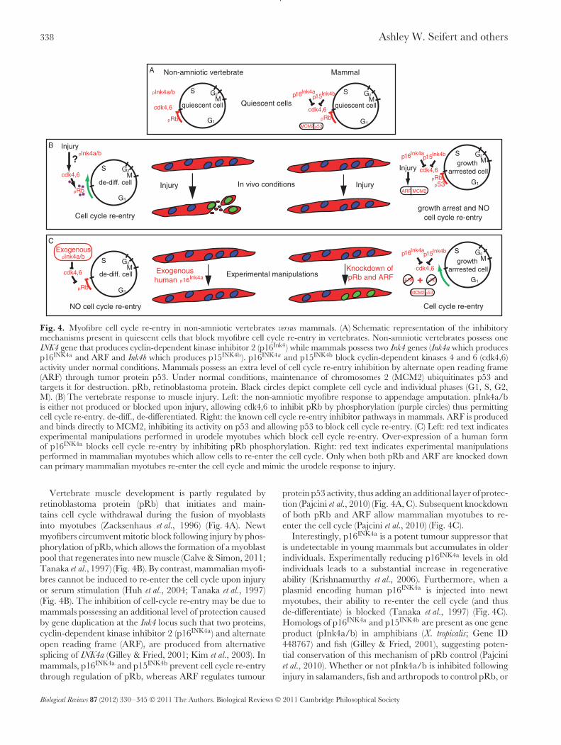
Fig. 4. Myofibre cell cycle re-entry in non-amniotic vertebrates versus mammals. (A) Schematic representation of the inhibitorymechanisms present in quiescent cells that block myofibre cell cycle re-entry in vertebrates. Non-amniotic vertebrates possess oneINK4 gene that produces cyclin-dependent kinase inhibitor 2 (p16Ink4) while mammals possess two Ink4 genes (Ink4a which producesp16INK4a and ARF and Ink4b which produces p15INK4b). p16INK4a and p15INK4b block cyclin-dependent kinases 4 and 6 (cdk4,6)activity under normal conditions. Mammals possess an extra level of cell cycle re-entry inhibition by alternate open reading frame(ARF) through tumor protein p53. Under normal conditions, maintenance of chromosomes 2 (MCM2) ubiquitinates p53 andtargets it for destruction. pRb, retinoblastoma protein. Black circles depict complete cell cycle and individual phases (G1, S, G2,M). (B) The vertebrate response to muscle injury. Left: the non-amniotic myofibre response to appendage amputation. pInk4a/bis either not produced or blocked upon injury, allowing cdk4,6 to inhibit pRb by phosphorylation (purple circles) thus permittingcell cycle re-entry. de-diff., de-differentiated. Right: the known cell cycle re-entry inhibitor pathways in mammals. ARF is producedand binds directly to MCM2, inhibiting its activity on p53 and allowing p53 to block cell cycle re-entry. (C) Left: red text indicatesexperimental manipulations performed in urodele myotubes which block cell cycle re-entry. Over-expression of a human formof p16INK4a blocks cell cycle re-entry by inhibiting pRb phosphorylation. Right: red text indicates experimental manipulationsperformed in mammalian myotubes which allow cells to re-enter the cell cycle. Only when both pRb and ARF are knocked downcan primary mammalian myotubes re-enter the cell cycle and mimic the urodele response to injury.
Vertebrate muscle development is partly regulated byretinoblastoma protein (pRb) that initiates and main-tains cell cycle withdrawal during the fusion of myoblastsinto myotubes (Zacksenhaus et al., 1996) (Fig. 4A). Newtmyofibers circumvent mitotic block following injury by phos-phorylation of pRb, which allows the formation of a myoblastpool that regenerates into new muscle (Calve & Simon, 2011;Tanaka et al., 1997) (Fig. 4B). By contrast, mammalian myofi-bres cannot be induced to re-enter the cell cycle upon injuryor serum stimulation (Huh et al., 2004; Tanaka et al., 1997)(Fig. 4B). The inhibition of cell-cycle re-entry may be due tomammals possessing an additional level of protection causedby gene duplication at the Ink4 locus such that two proteins,cyclin-dependent kinase inhibitor 2 (p16INK4a) and alternateopen reading frame (ARF), are produced from alternativesplicing of INK4a (Gilley & Fried, 2001; Kim et al., 2003). Inmammals, p16INK4a and p15INK4b prevent cell cycle re-entrythrough regulation of pRb, whereas ARF regulates tumour
protein p53 activity, thus adding an additional layer of protec-tion (Pajcini et al., 2010) (Fig. 4A, C). Subsequent knockdownof both pRb and ARF allow mammalian myotubes to re-enter the cell cycle (Pajcini et al., 2010) (Fig. 4C).
Interestingly, p16INK4a is a potent tumour suppressor thatis undetectable in young mammals but accumulates in olderindividuals. Experimentally reducing p16INK4a levels in oldindividuals leads to a substantial increase in regenerativeability (Krishnamurthy et al., 2006). Furthermore, when aplasmid encoding human p16INK4a is injected into newtmyotubes, their ability to re-enter the cell cycle (and thusde-differentiate) is blocked (Tanaka et al., 1997) (Fig. 4C).Homologs of p16INK4a and p15INK4b are present as one geneproduct (pInk4a/b) in amphibians (X. tropicalis; Gene ID448767) and fish (Gilley & Fried, 2001), suggesting poten-tial conservation of this mechanism of pRb control (Pajciniet al., 2010). Whether or not pInk4a/b is inhibited followinginjury in salamanders, fish and arthropods to control pRb, or
Biological Reviews 87 (2012) 330–345 © 2011 The Authors. Biological Reviews © 2011 Cambridge Philosophical Society

The influence of fundamental traits on mechanisms controlling appendage regeneration 339
operates in aged individuals, awaits detailed investigation ofpInk4a/b to shed light on this mechanism of cellular control.
An open question is whether de-differentiation in othercell types may be controlled via regulation of alternatecell cycle control genes that permit entry into S-phase.The nucleolar protein nucleostemin can regulate stem cellself-renewal and proliferation in a p53-dependent manner,and regulates cell-cycle re-entry during regeneration (Ma &Pederson, 2007; Maki et al., 2007; Tsai, Kittappa & McKay,2002). In newts, upregulation of nucleostemin precedescell-cycle re-entry and it co-localizes with de-differentiatingpigmented retinal epithelium during lens regeneration, andwith blastema cells during limb regeneration (Maki et al.,2007). Interestingly, ARF expression (from the Ink4a locus)downregulates nucleostemin which leads to p53-mediatedcell cycle arrest (Ma & Pederson, 2007). These findingssuggest that nucleostemin may contribute to cell cycle re-entry in other cell types, and supports a role for the Ink4a locusin restricting de-differentiation in mammalian cells throughmultiple pathways. In cockroaches, epidermal cells re-enterthe cell cycle following amputation, although the mechanismsof this action are unclear (Truby, 1983). Hypothesizing thatsimilar events occur in these and other arthropods capable ofregeneration is reasonable and warrants further investigationinto the control of cell cycle re-entry during appendageregeneration. Future research should address: (1) whetheror not de-differentiation in other organs and appendagesinvolves removing similar inhibition of cell cycle re-entry;(2) if this blockade is maintained in an age-dependent fashion;and (3) whether other metazoans also have such mechanisms.
(2) Progenitor cells
Progenitor cells are lineage-restricted stem cells that can dif-ferentiate into specific cell types a limited number of times,and serve to replenish various cell types throughout the body.Along with de-differentiating cells, progenitor cells accountfor an unknown fraction of regenerating tissue. Data frommammalian and avian taxa indicate that the number andproliferative ability of progenitor cells declines with age (Carl-son, 1995; Renault et al., 2000). Furthermore, evidence fromboth newts and mammals has demonstrated the involve-ment of resident progenitor cells from a variety of tissuesduring regeneration (Carlson, 2007). Progenitor cells arethought to re-enter the cell cycle following injury in a similarmanner to de-differentiating cells (Dhawan & Rando, 2005;Morrison et al., 2006). Evidence in favour of a repressiverole for progenitor cell cycle re-entry as cells age comes frominvestigations examining the Notch/Delta signaling pathwayand transforming growth factor-ß induced phosphorylationof Smad3 (Tgf-ß/pSmad3) in satellite cells during muscleregeneration (Carlson, Hsu & Conboy, 2008; Hjiantoniouet al., 2008; Odelberg, 2002). Inhibiting the Notch pathwayin young muscle inhibits regeneration, while forced acti-vation of Notch in old muscle rejuvenates repair (Conboyet al., 2003). Additionally, heterochronic parabiotic pairingsbetween old and young mice have shown that systemic fac-tors are capable of rescuing the proliferative and regenerative
ability of aged satellite cells through activation of the Notchpathway (Conboy et al., 2005). Conversely, young regenera-tive muscle has low levels of Tgf-ß/pSmad3, and this balanceshifts towards higher levels in old, non-regenerative muscle(Carlson et al., 2008). Whether or not these pathways interactdirectly to convey regenerative capacity remains unclear. Atleast in mammalian cells, aging does change the way thatthese progenitor cells respond to injury cues through theseand other cell cycle modulators. Because these pathwaysare involved in mammalian muscle regeneration, they areintriguing candidates to explore in non-mammalian verte-brates and arthropods (especially in cases where regenerationis curtailed because of metamorphosis or life stage).
(3) Cell proliferation and growth
Telomeres are regions of repetitive DNA at the ends ofchromosomes that function to prevent cellular degradation.Telomeres progressively shorten during successive celldivisions, eventually leading to cellular senescence andthe cessation of cell proliferation (Lee et al., 1998). Theenzyme telomerase acts to combat this shortening. Thus, theability of cells to remain in the cell cycle and proliferate ispartially due to telomerase (Bousman, Schneider & Shampay,2003; Greider, 1998; Klapper et al., 1998; Lee et al., 1998).Telomerase activity also appears to function independentlyof telomere maintenance to maintain cell proliferation viaan unknown mechanism (Smith, Coller & Roberts, 2003).Telomerase expression and subsequent activity may beinfluenced during aging, following metamorphosis, or bygrowth patterns.
Following de-differentiation and blastema formation, cellsmust proliferate for morphogenesis to proceed. The molec-ular pathways described above confer the ability of cells tore-enter the cell cycle primarily through a loss of repressionon cell cycle control genes (or signaling pathways that interactwith them). As regenerative capacity declines, either with ageor following metamorphosis, these repressive states becomeharder to overcome. Following de-differentiation, mitoticdivision follows and proliferation must be maintained forregeneration to proceed. While maintaining cells in a prolif-erative state is clearly a nerve-dependent process (see SectionII.3), blastemal cells must also retain the ability to dividerepeatedly (instead of differentiating) in order to produceenough cells to replace the missing tissue. Examining the roleof telomerase during appendage regeneration seems appro-priate given the importance for maintaining cell proliferationin regenerating tissues. In fact, an examination of telomeraseactivity across taxa suggests a strong correlation betweenregeneration, aging, and growth pattern (Gomes et al., 2010).
In addition to being used as a marker of cellular age,telomerase activity also can be used as an indicator forgrowth capacity (Klapper et al., 1998). Telomerase activityincreases to restore telomere length and remains high duringproliferation in regenerating tissues (Elmore et al., 2008;Klapper et al., 1998). Although not examined in the contextof metamorphosis, evidence from Xenopus laevis suggests thattelomerase activity is highest in embryonic tissue and in
Biological Reviews 87 (2012) 330–345 © 2011 The Authors. Biological Reviews © 2011 Cambridge Philosophical Society

340 Ashley W. Seifert and others
adult tissues with high regenerative capacity (e.g. testis, liver,spleen) (Bousman et al., 2003). Telomerase activity is high inboth larval tissue and in fully differentiated adult tissue ofarthropods that are capable of appendage regeneration (e.g.lobsters) (Gomes et al., 2010; Klapper et al., 1998). The samepattern holds for a diverse array of fish, and an upregulationof telomerase activity has been detected in fin tissue duringregeneration (Elmore et al., 2008). In birds and mammals thatexhibit cellular aging and senescence, telomerase activity ispredictably high in embryonic tissues and germ cells, butlow in fully differentiated tissues incapable of regeneration.Taken together, these studies demonstrate a correlationbetween regenerative capacity, high telomerase activity,‘‘young’’ tissue, and high growth capacity. This correlationsuggests that telomerase activity might also predict theability to regenerate both intraspecifically (as an animal ages)and interspecifically. In addition, cells capable of indefinitegrowth have high telomerase activity, but there is little to noactivity in terminally differentiated cells from animals that age(Gomes et al., 2010; Greider, 1998). Some urodeles, like manyfish, are indeterminate growers whose cells may not senesce(Goss, 1994; Kara, 1994). Thus, indeterminate growth mayconfer a high degree of regenerative ability, and this can betested in a diverse array of animals (Klapper et al., 1998).
IV. GENOMIC-LEVEL EFFECTS
Lastly, we examine the involvement of a single gene familythat may permit regeneration regardless of fundamentaltraits; and then address how fundamental traits may affectregenerative capacity at the level of the epigenome. Genetictools (e.g. microarrays, high-throughput sequencing) andproteomics have identified a number of key signals that actin time and space to coordinate a regenerative response,yet the factors that govern activation of these genes remainunclear (Monaghan et al., 2009; Rao et al., 2009; Whiteheadet al., 2005).
At the genomic level, the ability to respond to injury byexpressing key genes could be affected by fundamental traitsif such traits alter the ability of genes to become activated(either directly or through a loss of inhibition) in response toinjury. Access to promoter or enhancer sites (and thus theability to activate or repress transcription) can be maintainedthrough epigenetic mechanisms. Epigenetic control overchromatin structure could in turn govern the ability tomount a regenerative response. Thus, it seems plausible toask if regenerative capacity is ultimately controlled by cellularchanges that are directed by epigenetics, and if these changescould be influenced by fundamental traits of an organism.
(1) Regeneration-specific genes
As discussed throughout this review, several key pro-cesses are necessary for complete appendage regenerationto occur. Some researchers have proposed the involve-ment of regeneration-specific genes that coordinate these
processes and ultimately permit appendage regeneration(Garza-Garcia et al., 2009; Garza-Garcia, Driscoll & Brockes,2010). The existence of regeneration-specific genes was basedon the discovery of Prod1, a cell surface protein of theCD59/Ly6 protein family that regulates proximodistal cellidentity (Blassberg et al., 2011). Furthermore, the subsequentdiscovery that the Prod1 ligand, newt anterior gradient pro-tein (nAG), was sufficient to partly rescue limb regenerationin denervated newt limbs, suggested that Prod1 coordinatedboth growth and patterning to regulate the regenerativeresponse (Kumar et al., 2007). That Prod1 is found only insalamanders led to the suggestion that the lack of regenera-tive ability in certain vertebrate lineages stems partly from alack of this gene (Garza-Garcia et al., 2010).
Subsequent research has identified axolotl Prod1, althoughit is a secreted molecule instead of being linked to the cellmembrane as in newts (Blassberg et al., 2011). A differentmember of the CD59/Ly6 protein family, CD59, was alsofound to play a role in proximodistal cell identity during geckotail regeneration, suggesting that the CD59/Ly6 family ofproteins may have been adapted by different vertebratelineages to regulate patterning during regeneration (Wanget al., 2011). It will be interesting to see if the CD59/Ly6protein family plays a similar role in all regenerating species,and if the activity of these proteins is lost in non-regeneratingspecies. Ultimately, the existence of mammalian CD59 andadditional CD59/Ly6 family members, along with the roleof CD59 during gecko tail regeneration, suggests that it isnot the presence or absence of a particular gene that controlsregenerative ability, but rather an inability to coordinategene expression to induce a regenerative response.
(2) Epigenetic control of regeneration
During the regenerative response, both the ability of cellsto re-enter the cell cycle, and the ability to activatespecific genetic networks can be controlled through cell-cycle control genes and transcription factors. Epigeneticmodification refers to changes in non-sequence DNA thatalters transcription and gene function, resulting in phenotypicchanges at the cellular level (Fraga, 2009). Various epigeneticmechanisms can regulate gene expression, and changesin these mechanisms due to age or metamorphosis mayinfluence the regenerative ability of an organism.
A number of potential epigenetic alterations can affectgene expression. First, a change in methylation state at CpGislands (genomic regions of >50% same strand CG asso-ciations) or in promoter and enhancer regions can affectthe chromatin state of DNA. Alterations to chromatin statecan subsequently render genes in a transcriptionally silentor active state (reviewed in Bird & Wolffe, 1999). Second,modifications to histones (through selective methylation,acetylation, sumolation, or phosphorylation at specific aminoacid residues) can directly affect the ability of genes to beactively transcribed either directly or through higher orderchromatin rearrangement (reviewed in Jenuwein & Allis,2001). Both of these modifications to DNA architecture aregoverned through the enzymatic activity of various proteins
Biological Reviews 87 (2012) 330–345 © 2011 The Authors. Biological Reviews © 2011 Cambridge Philosophical Society

The influence of fundamental traits on mechanisms controlling appendage regeneration 341
such as the polycomb-group (PcG), trithorax-group (TcG),histone methyltranferases (HMTases), histone deacetylases(HDACs), and lysine demethylases. Each of these proteins hasa high specificity for particular modifications that alone or incombination can regulate cell behaviour (reviewed in Berger,2007). Clearly epigenetic alterations that affect cell cycle re-entry and proliferation (de-differentiation), or that affectthe transcriptional networks underlying positional informa-tion would have profound effects on regenerative ability(Yakushiji, Yokoyama & Tamura, 2009b). Although fewstudies examining structural regeneration have accountedfor the possibility of epigenetic modifications, recent workhas just begun to address this issue. Moreover, we understandlittle about how fundamental traits may influence epigeneticmodifications.
Can life stage (i.e. metamorphosis) influence the modifi-cation of DNA architecture? In anurans, pre-metamorphictadpoles are capable of limb regeneration, whereas post-metamorphic froglets are not (Dent, 1962; Polezhayev, 1946).Using Xenopus laevis, Yakushiji et al., (2007) examined methy-lation states at CpG islands of an enhancer region for theSonic hedgehog (Shh) gene that plays an important role duringembryonic limb development and limb regeneration. Theyfound low levels of methylation in the Shh enhancer region,and correspondingly high expression of Shh in the limbcells of tadpoles. By contrast, they found a higher degreeof methylation in froglets, correlating with a failure to acti-vate Shh and incomplete regeneration. Further, when Shh
expression was experimentally activated (exogenously, usingsmall molecules) following amputation in froglets, there wasan increase in cell proliferation and regenerative capacity(Yakushiji et al., 2009a). These findings suggest an increasedmethylation in control regions of genes that participate inregeneration, and therefore may help explain the loss ofregenerative ability following metamorphosis in X. laevis.Interestingly, axolotls and newts show relatively low lev-els of methylation in the Shh enhancer in both intact andregenerating limbs (Yakushiji et al., 2007). This suggests aspecies-specific component to methylation at this enhancer,which might contribute to retention of regenerative abilityin urodeles. These examples suggest a strong correlationbetween regeneration and growth that may be dependent onthe drastic physiological and morphological changes thatoccur during metamorphosis. They also underscore theimportance of multiple genetic inputs for successfully res-cuing regeneration, and cast doubt on a ‘‘magic bullet’’ inthe form of one lone gene or pathway. These studies offertantalizing clues to how life stage might alter methylationstates of key regenerative genes to suppress transcription, andimply that methylation may also be regulated at the specieslevel in the context of regeneration.
While methylation state at CpG islands is most often asso-ciated with gene silencing, histone modifications provide anattractive model for how a regeneration program can becontrolled to affect regeneration. In another study examin-ing caudal fin regeneration in zebrafish, (Stewart, Tsun &Izpisua Belmonte, 2009) the ‘‘bivalent loci’’ control of gene
expression through selective trimethylation of lysine 27 his-tone 3 (me3K27 H3) by polycomb group proteins (PcGs) andtrimethylation of lysine 4 histone 3 (me3K4 H3) by trithoraxgroup proteins (TcGs) was examined. Based on previouswork in embryonic stem cells, the authors proposed thatmaintenance of both histone marks act to prime genes foractivation, and that subsequent loss of the repressive me3K27H3 mark can lead to gene activation. When they examinedgenes involved in fin regeneration they observed a significantdecrease in the me3K27 H3 mark, but not in the me3K4H3 mark that also correlated with a significant increasein expression of some genes involved in the regenerativeresponse. Underscoring the complexity of these modifica-tions, this pattern does not hold for many of the genesknown to be important for regeneration, and the authorssuggest that alternative control mechanisms might have con-founded their results. Nonetheless, their data suggests thepotential for these types of epigenetic modifications to affectlimb regeneration and should spur future experiments inmore relevant systems, particularly after metamorphosis inanurans.
V. FUTURE PERSPECTIVES
With the re-emergence of regeneration research in the con-text of regenerative medicine, the prospects for discoveryhave never been richer. The last twenty years has seenhuge progress in our ability to replace failing organs withbioengineered replacements based on biological scaffoldsand autologous human cells. Damaged blood vessels, heartvalves, tracheas, bone fragments, and even bladders can allnow be replaced through advances in regenerative medicine.Despite these successes, bioengineering approaches havefailed to find a way to regenerate skin, appendages, or partsof complex organs containing specialized cell types. Thefuture lies in the ability of science to coax damaged tis-sue to repair itself, thus regenerating a perfect replacementin situ. If we are to succeed in this task, researchers mustlook towards organisms that regenerate damaged tissues inorder to understand the mechanisms that naturally regulateand constrain regeneration at various levels of biologicalorganization.
Underlying the hypotheses presented above is an implicitsuggestion that regenerative ability is fundamentally cou-pled to development and growth. Although not a new idea,discovering how these processes interact is tantamount tounderstanding how and why some animals can regener-ate but others cannot. Traditional evolutionary comparisonsof regenerative ability across metazoans have used sim-ple presence or absence (of regeneration) when comparingspecies, rather than comparing whether regenerative capac-ity changes across life stage. This has led to the misconceptionthat adult appendage regeneration is widespread. In fact,when life stage is considered, the available evidence sug-gests otherwise. Very few species that reproduce sexuallyare capable of appendage regeneration along the secondary
Biological Reviews 87 (2012) 330–345 © 2011 The Authors. Biological Reviews © 2011 Cambridge Philosophical Society

342 Ashley W. Seifert and others
Appendage Regeneration and Growth
Art
hro
po
da CONTINUOUS MOULTING
CONTINUOUS MOULTING
HEMIMETABOLOUS
HOLOMETABOLOUS
INDETERMINATE
DETERMINATE
INDETERMINATE
INDETERMINATE
DETERMINATE
INDETERMINATE
DETERMINATE
DETERMINATE
TERMINAL MOULTING
TERMINAL MOULTING
MODE OF GROWTH
DEVELOPMENT
?
Non-Amniotes
Amniotes
Crustacea
Chelicerata
Hexapoda
Fishes
Urodeles
Anurans
Reptiles
Aves
Mammals
Ver
teb
rata
Embryo Larvae Juvenile
Juvenile
AdultUnknown age-limit toregenerative ability
Unknown age limit toregenerative ability
Unknown age limit toregenerative ability
Regenerative ability disappears during metamorphosis
Regenerative ability disappears during metamorphosis
Birds cannot regenerate appendages even as embryos
Regenerative ability disappears early in embryonic developmentLizard tail regeneration
Regenerative ability declines towards final moult
Regenerative ability declines towards final moult
Fig. 5. Appendage regeneration is restricted among adult forms of Arthropoda and Vertebrata. Regenerative ability appearshighly correlated with development and growth capacity. Shaded bars represent regenerative ability. Green represents completeregeneration and red, no or partial regeneration. Species from each phyla are grouped according to their mode of growth (growthpatterns for many species are unknown). All amphibians are considered indeterminate growers (see Section I.4). Within amniotes,appendage regeneration either does not occur (birds) or is lost during embryogenesis. Tail regeneration in certain reptiles is theexception and is incomplete as the spinal cord is not regenerated.
body axis as adults (Fig. 5). Instead, the data suggest a near-ubiquity of appendage regeneration in embryos and larvae,with a general decline as juveniles move towards adulthood(Fig. 5). Exceptions occur in some salamanders, fish, andcrustaceans that exhibit seemingly boundless regenerativeability, even as adults. Interestingly, indeterminate growthand neoteny (some salamanders) occur in conjunction withthis extended regenerative capacity, suggesting a mechanismthat hijacks aspects of juvenile development to block cellularsenescence or to provide a constant source of progenitor cellsto the continually growing organism. An important advancein understanding the relationship between indeterminategrowth (or neoteny) and extended regeneration will be to findthe upper age boundaries at which regeneration fails (if at all).This relationship also makes revisiting regeneration acrossmetamorphosis appealing as it provides an experimental sys-tem where regenerative capacity is naturally lost in the sametissue over a short time period. Future research attempting toidentify the cellular and molecular mechanisms underlyingregeneration through comparative studies will benefit from
experiments conducted across various developmental stages,and from species with different modes of growth.
The discussion and hypotheses in this paper have aimedto frame future research by emphasizing the importance offundamental traits in the context of appendage regeneration.This stems partly from a need to understand if regener-ation fluctuates within a species, or is an all-or-nothingprocess within taxa. Researchers must continue to explorenew species and gather data on the presence and absence ofregeneration. Simultaneously, they will need to uncover how(if at all) regenerative capacity changes over variable bodysizes, during aging within species, across metamorphosis,and with respect to growth patterns. While time consuming,this approach is vital to elucidating mechanisms that dis-rupt regeneration, and will provide insight into mammalianrepair processes. We conclude that increasing phylogeneticsampling and exploring these ideas from multiple levels ofbiological organization is vital to finding an answer to thequestion that continues to elude science: why can someanimals regenerate but others cannot?
Biological Reviews 87 (2012) 330–345 © 2011 The Authors. Biological Reviews © 2011 Cambridge Philosophical Society

The influence of fundamental traits on mechanisms controlling appendage regeneration 343
VI. CONCLUSIONS
(1) This review summarizes our current knowledge of howcertain fundamental traits (e.g. body size, aging, life stage, andgrowth pattern) affect appendage regeneration in metazoans.We highlight the need for future experiments to decouplethese traits to examine if they constrain regeneration withinand across species.
(2) We posit specific ways in which fundamental traits canmodify mechanisms controlling regeneration at the tissue,cellular, and genomic levels. These include: age-dependentloss of telomerase expression and activity (which restrictsproliferative capacity and thus regeneration); prolongedregenerative capacity with indeterminate growth (throughthe lack of cellular senescence, sustained access to progenitorcells, and maintenance of high proliferative capacity); andlife-stage-associated changes in the epigenetic control of geneexpression (which can restrict the ability to mount a completeregenerative response).
(3) We review possible taxon-specific differences over themolecular control of cell-cycle re-entry (e.g. Ink4a/b locusgenes, nucleostemin) and highlight the need to investigatethese interactions across an array of regenerating species andtissue types.
(4) Appendage regeneration in metazoans is stronglycoupled to developmental stage. When viewed across taxa,regeneration appears widespread among larval and juvenilestates but becomes restricted among adult forms. Limiteddata suggest that some taxa may escape this developmentalconstraint through extended growth capacity, although moreinformation on the presence and absence of regeneration inlarger and older organisms is necessary.
VII. ACKNOWLEDGEMENTS
We thank Jamie Gillooly for discussions and helpful sugges-tions on the manuscript and two anonymous reviewers forhelpful comments. We thank Luther D. Smith for stimulat-ing discussions on the topic of indeterminate growth and aregrateful to Michael Samuels for providing the owl house forour working group. We apologize to those authors whoseoriginal work was not directly cited due to space constraints.Our work is supported by the Michael A. Singer BiologyFund through the Department of Biology at the Universityof Florida.
VIII. REFERENCES
Adams, D. S., Masi, A. & Levin, M. (2007). H+ pump-dependent changes inmembrane voltage are an early mechanism necessary and sufficient to induceXenopus tail regeneration. Development 134, 1323–35.
Adidoyi, R. G. (1972). Wound healing and regeneration in the crab Paratelphusahydrodromous. International Review of Cytology 32, 257–289.
Agata, K., Saito, Y. & Nakajima, E. (2007). Unifying principles of regenerationI: Epimorphosis versus morphallaxis. Development Growth and Differentiation 49, 73–8.
Anchelin, M., Murcia, L., Alcaraz-Perez, F., Garcia-Navarro, E. M. &Cayuela, M. L. (2011). Behaviour of telomere and telomerase during aging andregeneration in zebrafish. PLoS One 6, e16955.
Baum, B. J., Faris, B. & Franzblau, C. (1975). Further studies on newt tail collagen:description of lysine-derived crosslinks. Comparative Biochemistry and Physiology Part B
51, 51–5.Bayliss, P. E., Bellavance, K. L., Whitehead, G. G., Abrams, J. M.,
Aegerter, S., Robbins, H. S., Cowan, D. B., Keating, M. T., O’Reilly, T.,Wood, J. M., Roberts, T. M. & Chan, J. (2006). Chemical modulation of receptorsignaling inhibits regenerative angiogenesis in adult zebrafish. Nature Chemical Biology
2, 265–73.Beck, C. W., Christen, B. & Slack, J. M. (2003). Molecular pathways needed for
regeneration of spinal cord and muscle in a vertebrate. Developmental Cell 5, 429–39.Berger, S. L. (2007). The complex language of chromatin regulation during
transcription. Nature 447, 407–12.Bird, A. P. & Wolffe, A. P. (1999). Methylation-induced repression–belts, braces,
and chromatin. Cell 99, 451–4.Blassberg, R. A., Garza-Garcia, A., Janmohamed, A., Gates, P. B. &
Brockes, J. P. (2011). Functional convergence of signalling by GPI-anchoredand anchorless forms of a salamander protein implicated in limb regeneration.Journal of Cell Science 124, 47–56.
Boilly, B. & Albert, P. (1988). Blastema cell proliferation in vitro: effects of limbamputation on the mitogenic activity of spinal cord extracts. Biology of the Cell 62,183–7.
Borgens, R. B., Vanable, J. W., Jr. & Jaffe, L. F. (1977). Bioelectricity andregeneration: large currents leave the stumps of regenerating newt limbs. Proceedings
of the National Academies of Sciences of the United Statesof America 74, 4528–32.Bousman, S., Schneider, G. & Shampay, J. (2003). Telomerase activity is
widespread in adult somatic tissues of Xenopus. Journal of Experimental Zoology
Part B Molecular and Developmental Evolution 295, 82–6.Brockes, J. P. & Kumar, A. (2008). Comparative aspects of animal regeneration.
Annu Rev Cell Developmental Biology 24, 525–49.Bryant, S. V., Endo, T. & Gardiner, D. M. (2002). Vertebrate limb regeneration
and the origin of limb stem cells. International Journal of Developmental Biology 46,887–96.
Calve, S. & Simon, H. G. (2011). High resolution three-dimensional imaging:Evidence for cell cycle reentry in regenerating skeletal muscle. Developmental Dynamics
240, 1233–9.Carlson, B. M. (1995). Factors influencing the repair and adaptation of muscles in
aged individuals: satellite cells and innervation. The Journals of Gerontology. Series A,
Biological Sciences and Medical Sciences 50 Spec No, 96–100.Carlson, B. M. (2007). Principles of Regenerative Biology. Elsevier.Carlson, M. E., Hsu, M. & Conboy, I. M. (2008). Imbalance between pSmad3 and
Notch induces CDK inhibitors in old muscle stem cells. Nature 454, 528–32.Conboy, I. M., Conboy, M. J., Smythe, G. M. & Rando, T. A. (2003). Notch-
mediated restoration of regenerative potential to aged muscle. Science 302, 1575–7.Conboy, I. M., Conboy, M. J., Wagers, A. J., Girma, E. R., Weissman, I. L. &
Rando, T. A. (2005). Rejuvenation of aged progenitor cells by exposure to a youngsystemic environment. Nature 433, 760–4.
Consoulas, C. & Levine, R. B. (1997). Accumulation and proliferation of adult legmuscle precursors in Manduca are dependent on innervation. Journal of Neurobiology
32, 531–53.Dent, J. N. (1962). Limb regeneration in larvae and metamorphosing individuals of
the South African clawed toad. Journal of Morphology 110, 61–77.Dhawan, J. & Rando, T. A. (2005). Stem cells in postnatal myogenesis: molecular
mechanisms of satellite cell quiescence, activation and replenishment. Trends in Cell
Biology 15, 666–73.Echeverri, K., Clarke, J. D. & Tanaka, E. M. (2001). In vivo imaging indicates
muscle fiber dedifferentiation is a major contributor to the regenerating tail blastema.Developmental Biology 236, 151–64.
Edelberg, J. M. & Reed, M. J. (2003). Aging and angiogenesis. Frontiers in Bioscience
8, s1199–209.Eguchi, G., Eguchi, Y., Nakamura, K., Yadav, M. C., Millan, J. L. &
Tsonis, P. A. (2011). Regenerative capacity in newts is not altered by repeatedregeneration and ageing. Nature Communications 2, 384.
Eguchi, G. & Shingai, R. (1971). Cellular analysis on localization of lens formingpotency in the newt iris epithelium. Development Growth and Differentiation 13, 337–49.
Elmore, L. W., Norris, M. W., Sircar, S., Bright, A. T., McChesney, P. A.,Winn, R. N. & Holt, S. E. (2008). Upregulation of telomerase function duringtissue regeneration. Experimental Biology and Medicine (Maywood) 233, 958–67.
Ferrara, N. (2002). Role of vascular endothelial growth factor in physiologic andpathologic angiogenesis: therapeutic implications. Seminars in Oncology 29, 10–4.
Filoni, S., Bernardini, S., Cannata, S. M. & Ghittoni, R. (1999). Nerve-independence of limb regeneration in larval Xenopus laevis is related to thepresence of mitogenic factors in early limb tissues. Journal of Experimental Zoology 284,188–96.
Fraga, M. F. (2009). Genetic and epigenetic regulation of aging. Current Opinions in
Immunology 21, 446–53.
Biological Reviews 87 (2012) 330–345 © 2011 The Authors. Biological Reviews © 2011 Cambridge Philosophical Society
344 Ashley W. Seifert and others
French, V., Bryant, P. J. & Bryant, S. V. (1976). Pattern regulation in epimorphicfields. Science 193, 969–81.
Garza-Garcia, A., Harris, R., Esposito, D., Gates, P. B. & Driscoll, P. C.(2009). Solution structure and phylogenetics of Prod1, a member of the three-fingerprotein superfamily implicated in salamander limb regeneration. PLoS One 4, e7123.
Garza-Garcia, A. A., Driscoll, P. C. & Brockes, J. P. (2010). Evidence forthe local evolution of mechanisms underlying limb regeneration in salamanders.Integrative and Comparative Biology 50, 528–35.
Geraudie, J. & Singer, M. (1985). Necessity of an adequate nerve supply forregeneration of the amputated pectoral fin in the teleost Fundulus. Journal of
Experimental Zoology 234, 367–74.Gibbs, K. M., Chittur, S. V. & Szaro, B. G. (2010). Metamorphosis and the
regenerative capacity of spinal cord axons in Xenopus laevis. The European Journal of
Neuroscience 33, 9–25.Gilley, J. & Fried, M. (2001). One INK4 gene and no ARF at the Fugu equivalent of
the human INK4A/ARF/INK4B tumour suppressor locus. Oncogene 20, 7447–52.Gomes, N. M., Shay, J. W. & Wright, W. E. (2010). Telomere biology in Metazoa.
FEBS Letters 584, 3741–51.Goss, R. J. (1965). Kinetics of Compensatory Growth. Quarterly Review of Biology 40,
123–46.Goss, R. J. (1969). Principles of Regeneration. Academic Press, New York.Goss, R. J. (1994). Why study ageing in cold-blooded vertebrates? Gerontology 40, 65–9.Greider, C. W. (1998). Telomerase activity, cell proliferation, and cancer. Proceedings
of the National Academies of Sciences of the United States of America 95, 90–2.Harsum, S., Clarke, J. D. & Martin, P. (2001). A reciprocal relationship between
cutaneous nerves and repairing skin wounds in the developing chick embryo.Developmental Biology 238, 27–39.
Hay, E. D. & Fischman, D. A. (1961). Origin of the blastema in regenerating limbs ofthe newt Triturus viridescens. An autoradiographic study using tritiated thymidineto follow cell proliferation and migration. Developmental Biology 3, 26–59.
Hjiantoniou, E., Anayasa, M., Nicolaou, P., Bantounas, I., Saito, M.,Iseki, S., Uney, J. B. & Phylactou, L. A. (2008). Twist induces reversal ofmyotube formation. Differentiation 76, 182–92.
Homan, R. N., Reed, J. M. & Windmiller, B. S. (2003). Analysis of spottedsalamander (Ambystoma maculatum) growth rates based on long-bone growthrings. Journal of Herpetology 37, 617–621.
Huh, M. S., Parker, M. H., Scime, A., Parks, R. & Rudnicki, M. A. (2004). Rb isrequired for progression through myogenic differentiation but not maintenance ofterminal differentiation. Journal of Cell Biology 166, 865–76.
Imokawa, Y. & Brockes, J. P. (2003). Selective activation of thrombin is a criticaldeterminant for vertebrate lens regeneration. Current Biology 13, 877–81.
Jakob, C., Poizat, G., Crivelli, A. J. & Veith, M. (2002). Larval growth variationin marbled newts (Triturus marmoratus) from temporary Mediterranean ponds.Amphibia-Reptilia 23, 359–362.
Jenuwein, T. & Allis, C. D. (2001). Translating the histone code. Science 293,1074–80.
Jopling, C., Sleep, E., Raya, M., Marti, M., Raya, A. & Belmonte, J. C.(2010). Zebrafish heart regeneration occurs by cardiomyocyte dedifferentiation andproliferation. Nature 464, 606–9.
Kara, T. C. (1994). Ageing in amphibians. Gerontology 40, 161–73.Kikuchi, K., Holdway, J. E., Werdich, A. A., Anderson, R. M., Fang, Y.,
Egnaczyk, G. F., Evans, T., Macrae, C. A., Stainier, D. Y. & Poss, K. D.(2010). Primary contribution to zebrafish heart regeneration by gata4(+)cardiomyocytes. Nature 464, 601–5.
Kim, S. H., Mitchell, M., Fujii, H., Llanos, S. & Peters, G. (2003). Absence ofp16INK4a and truncation of ARF tumor suppressors in chickens. Proceedings of the
National Academies of Sciences of the United States of America 100, 211–6.Klapper, W., Kuhne, K., Singh, K. K., Heidorn, K., Parwaresch, R. &
Krupp, G. (1998). Longevity of lobsters is linked to ubiquitous telomerase expression.FEBS Letters 439, 143–6.
Kragl, M., Knapp, D., Nacu, E., Khattak, S., Maden, M., Epperlein, H. H. &Tanaka, E. M. (2009). Cells keep a memory of their tissue origin during axolotllimb regeneration. Nature 460, 60–5.
Krishnamurthy, J., Ramsey, M. R., Ligon, K. L., Torrice, C., Koh, A.,Bonner-Weir, S. & Sharpless, N. E. (2006). p16INK4a induces an age-dependent decline in islet regenerative potential. Nature 443, 453–7.
Kumar, A., Godwin, J. W., Gates, P. B., Garza-Garcia, A. A. & Brockes, J. P.(2007). Molecular basis for the nerve dependence of limb regeneration in an adultvertebrate. Science 318, 772–7.
Kumar, A., Velloso, C. P., Imokawa, Y. & Brockes, J. P. (2004). The regenerativeplasticity of isolated urodele myofibers and its dependence on MSX1. PLoS Biology
2, E218.Kurabuchi, S. (1990). Loss of limb regenerative capacity during postmetamorphic
growth in the tree frog, Hyla arborea japonica.Growth Development and Aging 54,17–22.
Lakatta, E. G. (1983). Determinants of Cardiovascular Performance - ModificationDue to Aging. Journal of Chronic Diseases 36, 15–30.
LeClair, E. E. & Topczewski, J. (2010). Development and regeneration of thezebrafish maxillary barbel: a novel study system for vertebrate tissue growth andrepair. PLoS One 5, e8737.
Lee, H. W., Blasco, M. A., Gottlieb, G. J., Horner, J. W., 2nd, Greider, C. W.& DePinho, R. A. (1998). Essential role of mouse telomerase in highly proliferativeorgans. Nature 392, 569–74.
Lentz, T. L. (1969). Cytological studies of muscle dedifferentiation and differentiationduring limb regeneration of the newt Triturus.American Journal of Anatomy 124,447–79.
Ma, H. & Pederson, T. (2007). Depletion of the nucleolar protein nucleostemincauses G1 cell cycle arrest via the p53 pathway. Molecular Biology of the Cell 18,2630–5.
Maginnis, T. L. (2006). The costs of autotomy and regeneration in animals: a reviewand framework for future research. Behavioral Ecology 17, 857–872.
Maki, N., Takechi, K., Sano, S., Tarui, H., Sasai, Y. & Agata, K. (2007). Rapidaccumulation of nucleostemin in nucleolus during newt regeneration. Developmental
Dynamics 236, 941–50.Marden, J. H., Rogina, B., Montooth, K. L. & Helfand, S. L. (2003). Conditional
tradeoffs between aging and organismal performance of Indy long-lived mutant flies.Proceedings of the National Academy of Sciences of the United States of America 100, 3369–3373.
Maruzzo, D., Bonato, L., Brena, C., Fusco, G. & Minelli, A. (2005). Appendageloss and regeneration in arthropods: A comparative view. In Crustacea and arthropod
relationships (eds S. Koenemann and R A. Jenner). CRC Press, Boca RatonMonaghan, J. R., Epp, L. G., Putta, S., Page, R. B., Walker, J. A.,
Beachy, C. K., Zhu, W., Pao, G. M., Verma, I. M., Hunter, T., Bryant, S. V.,Gardiner, D. M., Harkins, T. T. & Voss, S. R. (2009). Microarray and cDNAsequence analysis of transcription during nerve-dependent limb regeneration. BMC
Biology 7, 1.Morgan, T. H. (1901). Regeneration. The Macmillan Company, New York.Morrison, J. I., Loof, S., He, P. & Simon, A. (2006). Salamander limb regeneration
involves the activation of a multipotent skeletal muscle satellite cell population.Journal of Cell Biology 172, 433–40.
Nuesch, H. (1968). Role of Nervous System in Insect Morphogenesis andRegeneration. Annual Review of Entomology 13, 27–44.
Odelberg, S. J. (2002). Inducing cellular dedifferentiation: a potential method forenhancing endogenous regeneration in mammals. Seminars in Cell and Developmental
Biology 13, 335–43.Pajcini, K. V., Corbel, S. Y., Sage, J., Pomerantz, J. H. & Blau, H. M. (2010).
Transient inactivation of Rb and ARF yields regenerative cells from postmitoticmammalian muscle. Cell Stem Cell 7, 198–213.
Peadon, A. M. & Singer, M. (1965). A quantitative study of forelimb innervation inrelation to regenerative capacity in the larval, land stage, and adult forms of Triturusviridescens. Journal of Exp Zool 159, 337–45.
Polezhayev, L. W. (1946). The loss and restoration of regenerative capacity in thelimbs of tailless Amphibia. Biological Reviews of Cambridge Philosophical Society 21, 141–7.
Pough, F. H. & Kamel, S. (1984). Post-Metamorphic Change in Activity Metabolismof Anurans in Relation to Life-History. Oecologia 65, 138–144.
Pritchett, W. H. & Dent, J. N. (1972). The role of size in the rate of limbregeneration in the adult newt. Growth 36, 275–89.
Rao, N., Jhamb, D., Milner, D. J., Li, B., Song, F., Wang, M., Voss, S. R.,Palakal, M., King, M. W., Saranjami, B., Nye, H. L., Cameron, J. A. &Stocum, D. L. (2009). Proteomic analysis of blastema formation in regeneratingaxolotl limbs. BMC Biology 7, 83.
Renault, V., Piron-Hamelin, G., Forestier, C., DiDonna, S., Decary, S.,Hentati, F., Saillant, G., Butler-Browne, G. S. & Mouly, V. (2000). Skeletalmuscle regeneration and the mitotic clock. Experimental Gerontology 35, 711–9.
Rockett, C. L. & Woodring, J. P. (1972). Comparative Studies of Acarine LimbRegeneration, Apolysis, and Ecdysis. Journal of Insect Physiology 18, 2319-&.
Scadding, S. R. (1977). Phylogenic Distribution of Limb Regeneration Capacity inAdult Amphibia. Journal of Experimental Zoology 202, 57–67.
Scadding, S. R. (1981). Limb Regeneration in Adult Amphibia. Canadian Journal of
Zoology-Revue Canadienne De Zoologie 59, 34–46.Scadding, S. R. (1982). Can differences in limb regeneration ability between
amphibian species be explained by differences in quantity of innervation?Journal of
Experimental Zoology 219, 81–85.Scadding, S. R. (1983). Can differences in limb regeneration ability between
individuals within certain amphibian species be explained by differences in thequantity of innervation? Journal of Experimental Zoology 226, 75–80.
Scott, G. G. (1909). Regeneration in fundulus and its relation to the size of the fish.Biological Bulletin 17, 343–353.
Simpson, S. B. (1964). Analysis of Tail Regeneration in Lizard Lygosoma Laterale .1.Initiation of Regeneration + Cartilage Differentiation - Role of Ependyma. Journal
of Morphology 114, 425-&.Simpson, S. B., Jr. (1970). Studies on regeneration of the lizard’s tail. American Zoologist
10, 157–65.Singer, M. (1952). The influence of the nerve in regeneration of the amphibian
extremity. Quarterly Review of Biology 27, 169–200.
Biological Reviews 87 (2012) 330–345 © 2011 The Authors. Biological Reviews © 2011 Cambridge Philosophical Society

The influence of fundamental traits on mechanisms controlling appendage regeneration 345
Sivamani, K., Garcia, M. S. & Isseroff, R. R. (2007). Wound re-epithelialization:modulating keratinocyte migration in wound healing. Frontiers in Bioscience 12,2849–68.
Slack, J. M. (2006). Amphibian muscle regeneration–dedifferentiation or satellitecells? Trends in Cell Biology 16, 273–5.
Smith, L. L., Coller, H. A. & Roberts, J. M. (2003). Telomerase modulatesexpression of growth-controlling genes and enhances cell proliferation. Nature Cell
Biology 5, 474–9.Speakman, J. R. (2005). Correlations between physiology and lifespan–two widely
ignored problems with comparative studies. Aging Cell 4, 167–75.Stelnicki, E. J., Doolabh, V., Lee, S., Levis, C., Baumann, F. G.,
Longaker, M. T. & Mackinnon, S. (2000). Nerve dependency in scarless fetalwound healing. Plastic and Reconstructive Surgery 105, 140–7.
Stewart, S., Tsun, Z. Y. & Izpisua Belmonte, J. C. (2009). A histone demethylaseis necessary for regeneration in zebrafish. Proceedings of the National Academiesof Sciences
of the United States of America 106, 19889–94.Suzuki, M., Satoh, A., Ide, H. & Tamura, K. (2005). Nerve-dependent and -
independent events in blastema formation during Xenopus froglet limb regeneration.Developmental Biology 286, 361–75.
Tanaka, E. M. & Ferretti, P. (2009). Considering the evolution of regeneration inthe central nervous system. Nature Reviews Neuroscience 10, 713–23.
Tanaka, E. M., Gann, A. A., Gates, P. B. & Brockes, J. P. (1997). Newt myotubesreenter the cell cycle by phosphorylation of the retinoblastoma protein. Journal of
Cell Biology 136, 155–65.Tank, P. W., Carlson, B. M. & Connelly, T. G. (1976). A staging system for
forelimb regeneration in the axolotl, Ambystoma mexicanum. Journal of Morphology
150, 117–28.Tassava, R. A. & Olsen-Winner, C. L. (2003). Responses to amputation of
denervated ambystoma limbs containing aneurogenic limb grafts. Journal of
Experimental Zoology Part A Comparative and Experimental Biology 297, 64–79.Thornton, C. S. (1938). The histogenesis of muscle in the regenerating fore limb of
larval Amblystoma punctatum. Journal of Morphology 62, 17–47.Thornton, C. S. & Thornton, M. T. (1970). Recuperation of regeneration in
denervated limbs of Ambystoma larvae. Journal of Experimental Zoology 173, 293–301.Truby, P. R. (1983). Blastema formation and cell division during cockroach limb
regeneration. Journal of Embryology and Experimental Morphology 75, 151–64.Tsai, R. Y., Kittappa, R. & McKay, R. D. (2002). Plasticity, niches, and the use of
stem cells. Developmental Cell 2, 707–12.Van Stone, J. M. (1964). The relationship of nerve number to regenerative capacity
in hind limbs of Rana sylvatica.Journal of Experimental Zoology 155, 293–302.Verdu, E., Ceballos, D., Vilches, J. J. & Navarro, X. (2000). Influence of aging
on peripheral nerve function and regeneration. Journal of the Peripheral Nervous System
5, 191–208.Voit, E. O., Anton, H. J. & Blecker, J. (1995). Regenerative Growth Curves.
Mathematical Bioscience 73, 253–269.
Wagner, D. E., Wang, I. E. & Reddien, P. W. (2011). Clonogenic neoblastsare pluripotent adult stem cells that underlie planarian regeneration. Science 332,811–6.
Wagner, G. P. & Misof, B. Y. (1992). Evolutionary Modification of RegenerativeCapability in Vertebrates - a Comparative-Study on Teleost Pectoral FinRegeneration.Journal of Experimental Zoology 261, 62–78.
Wallace, H. (1981). Vertebrate Limb Regeneration. John Wiley and Sons.Wang, Y., Wang, R., Jiang, S., Zhou, W., Liu, Y., Gu, Q., Gu, Y., Dong, Y.,
Liu, M., Gu, X. & Ding, F. (2011). Gecko CD59 is implicated in proximodistalidentity during tail regeneration. PLoS One 6, e17878.
Whitby, D. J., Longaker, M. T., Harrison, M. R., Adzick, N. S. &Ferguson, M. W. (1991). Rapid epithelialisation of fetal wounds is associatedwith the early deposition of tenascin. Journal of Cell Science 99 (Pt 3), 583–6.
Whitehead, G. G., Makino, S., Lien, C. L. & Keating, M. T. (2005). fgf20 isessential for initiating zebrafish fin regeneration. Science 310, 1957–60.
Wolfe, A. D., Nye, H. L. & Cameron, J. A. (2000). Extent of ossification atthe amputation plane is correlated with the decline of blastema formationand regeneration in Xenopus laevis hindlimbs. Developmental Dynamics 218,681–97.
Yakushiji, N., Suzuki, M., Satoh, A., Ide, H. & Tamura, K. (2009a). Effectsof activation of hedgehog signaling on patterning, growth, and differentiation inXenopus froglet limb regeneration. Developmental Dynamics 238, 1887–96.
Yakushiji, N., Suzuki, M., Satoh, A., Sagai, T., Shiroishi, T., Kobayashi, H.,Sasaki, H., Ide, H. & Tamura, K. (2007). Correlation between Shh expressionand DNA methylation status of the limb-specific Shh enhancer region during limbregeneration in amphibians. Developmental Biology 312, 171–82.
Yakushiji, N., Yokoyama, H. & Tamura, K. (2009b). Repatterning in amphibianlimb regeneration: A model for study of genetic and epigenetic control of organregeneration. Seminars in Cell and Developmental Biology 20, 565–74.
Yntema, C. L. (1959). Blastema formation in sparsely innervated and aneurogenicforelimbs of amblystoma larvae Journal of Experimental Zoology 142, 423–39.
Yokoyama, H., Yonei-Tamura, S., Endo, T., Izpisua Belmonte, J. C.,Tamura, K. & Ide, H. (2000). Mesenchyme with fgf-10 expression is responsiblefor regenerative capacity in Xenopus limb buds. Developmental Biology 219,18–29.
Young, H. E., Bailey, C. F. & Dalley, B. K. (1983a). Environmental-ConditionsPrerequisite for Complete Limb Regeneration in the Postmetamorphic Adult Land-Phase Salamander, Ambystoma. Anatomical Record 206, 289–294.
Young, H. E., Bailey, C. F. & Dalley, B. K. (1983b). Gross Morphological Analysisof Limb Regeneration in Postmetamorphic Adult Ambystoma. Anatomical Record
206, 295–306.Zacksenhaus, E., Jiang, Z., Chung, D., Marth, J. D., Phillips, R. A. &
Gallie, B. L. (1996). pRb controls proliferation, differentiation, and death ofskeletal muscle cells and other lineages during embryogenesis. Genes and Development
10, 3051–64.
(Received 8 March 2011; revised 21 July 2011; accepted 9 August 2011; published online 19 September 2011)
Biological Reviews 87 (2012) 330–345 © 2011 The Authors. Biological Reviews © 2011 Cambridge Philosophical Society
Recommended


