Embed Size (px)
Citation preview
Anaesthetic considerations for posterior
fossa surgery
BY
Dr. Chamika Huruggamuwa
• The posterior fossa or the infratentorial fossais a rigid compartment with poor compliance and houses important structures such as
the brainstem and cerebellum.
• Tumours are the commonest pathology affecting the posterior fossa and they account for up to two-thirds of brain tumours in children.
• The sitting position offers better surgical access particularly in midline tumours but can be a significant challenge for anaesthetists.
• Venous air embolism is a potentially life-threatening complication associated with
surgery on the posterior fossa in the sitting position.
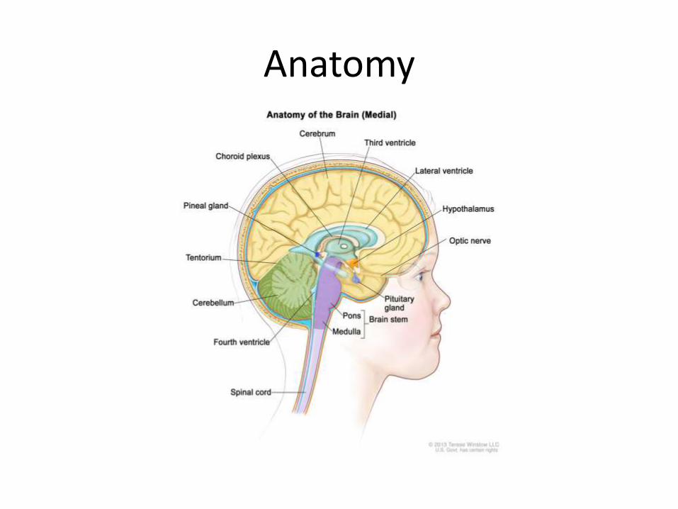
Anatomy
• The posterior fossa is the deepest cranial fossa.• It contains many important structures including
the brainstem, cerebellum and lower cranial nerves.
• The sigmoid, transverse and occipital sinuses all traverse the fossa.
• The cerebrospinal fluid (CSF) pathway is very narrow through the cerebral aqueduct and any obstruction can cause hydrocephalus which can result in a significant increase in intracranial pressure .
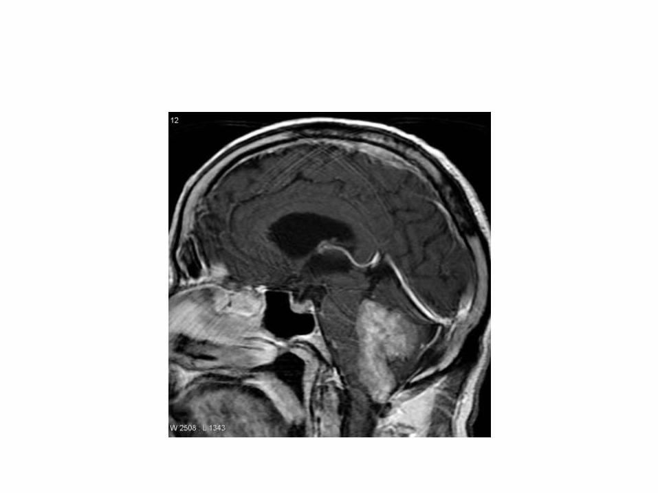
Pathology
• Tumours are the commonest pathology affecting the posterior fossa.
• 15% of intracranial aneurysms occur in the posterior fossa vasculature.
• Cysts
• Cranial nerve lesions
Intraoperative positioning
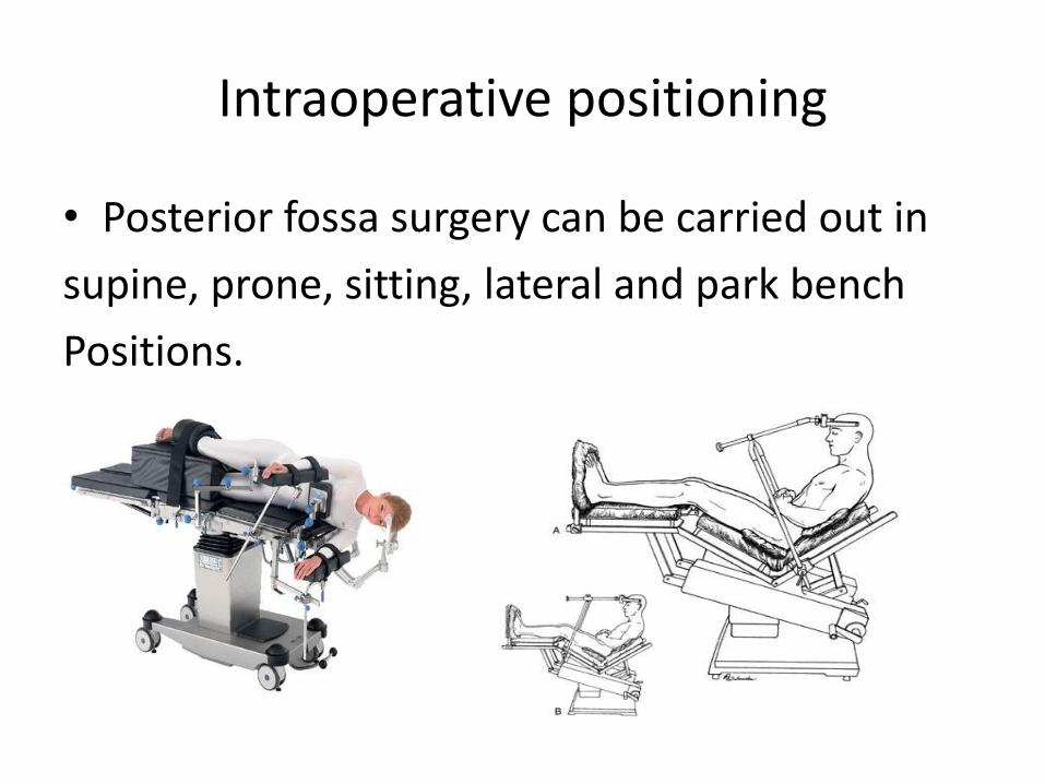
• Posterior fossa surgery can be carried out in
supine, prone, sitting, lateral and park bench
Positions.
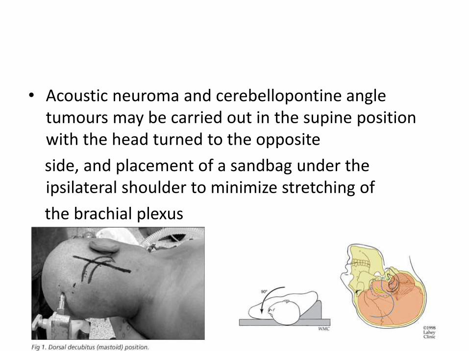
• Acoustic neuroma and cerebellopontine angle tumours may be carried out in the supine position with the head turned to the opposite
side, and placement of a sandbag under the ipsilateral shoulder to minimize stretching of
the brachial plexus
Park bench position - This facilitates greater access to midline structures and, in selected patients, avoids the need for the prone position.
• Meticulous care should be taken during positioning to avoid dislodgement of lines and the tracheal tube, and protection of pressure areas.
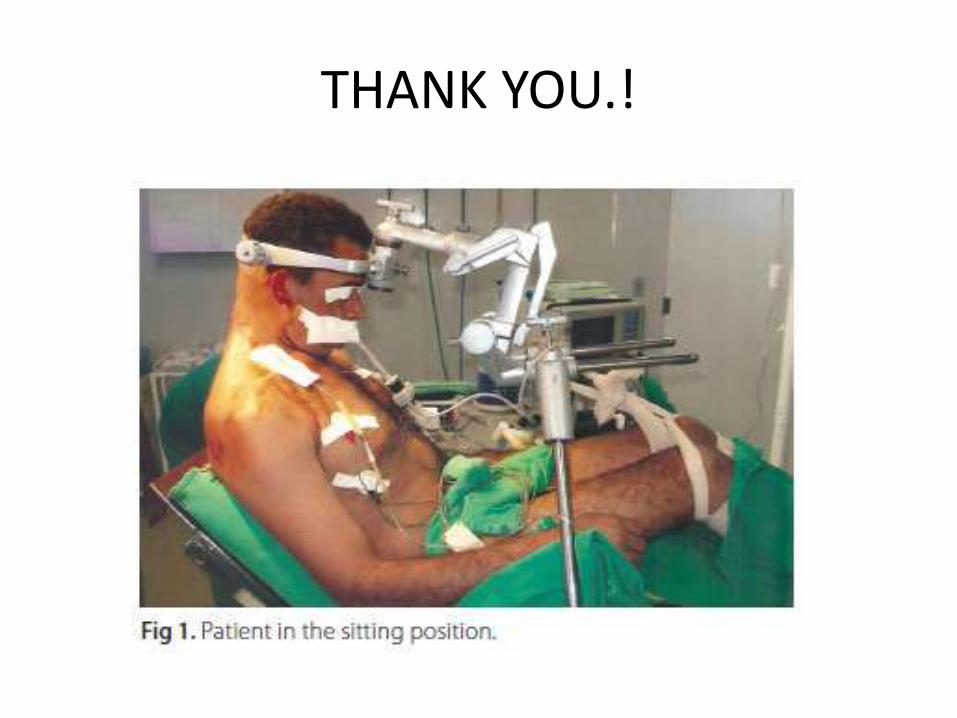
• Sitting position - improves surgical access to
the posterior fossa by facilitating gravity assisted
drainage of blood and CSF and decreasing ICP.
• Patients in the sitting position must be returned to the supine position rapidly for resuscitative measures in case of an acute cardiovascular collapse.
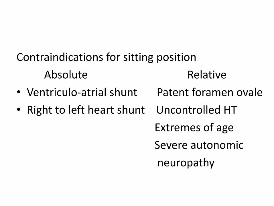
Contraindications for sitting position
Absolute Relative
• Ventriculo-atrial shunt Patent foramen ovale
• Right to left heart shunt Uncontrolled HT
Extremes of age
Severe autonomic
neuropathy
Complications of the sitting position
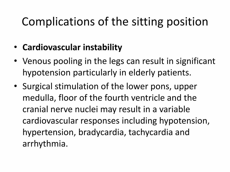
• Cardiovascular instability
• Venous pooling in the legs can result in significant hypotension particularly in elderly patients.
• Surgical stimulation of the lower pons, upper medulla, floor of the fourth ventricle and the cranial nerve nuclei may result in a variable cardiovascular responses including hypotension, hypertension, bradycardia, tachycardia and arrhythmia.
• Changes in ECG and blood pressure during
surgery on the posterior fossa may be signs to the surgeon of their proximity to vital structures.
• Venous air embolism
• Venous air embolism (VAE) is a potentially life-threatening complication associated with all surgery in the steep head-up position, including posterior fossa surgery in the sitting position.
• Air entrainment usually occurs through the diploic veins and open venous sinuses, but entrainment through head pin sites has also been reported
• Dehydration exacerbates the low venous pressure and increases the risk of air entrainment, so normovolaemia must be maintained at all times.
• When air passes into the pulmonary circulation, it causes an increase in pulmonary vascular resistance and pulmonary hypertension. This results in elevated right heart pressure and the risk of paradoxical air embolism.
• Clinical features of VAE.
• The spectrum of manifestations includes cardiovascular, respiratory and neurological changes
• Elevated right atrial pressure results in decreased venous return, hypotension and shock.
• A large embolus obstructing the outlet of the right ventricle can result in a sudden onset of right heart failure and cardiac arrest.
• Pulmonary signs of VAE include wheeze, crepitations, and sudden decrease in endtidal
carbon dioxide .
• Arterial blood gas analysis may reveal hypoxia and hypercapnia.
• Neurological manifestations include cerebral hypoperfusion as a result of shock and stroke in the event of a paradoxical embolus.
• How to minimise risk of VAE?
• Use of Trendelenburg tilt and leg elevation minimize the gradient between the surgical field and the right heart.
• Hydration status should be carefully optimized.
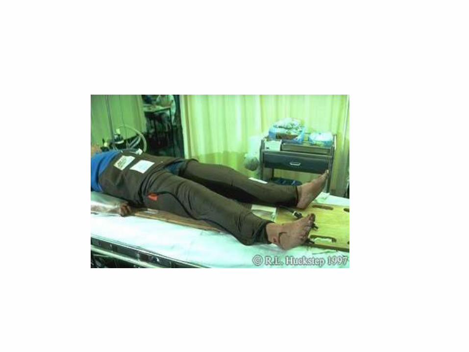
• Military Anti Shock Trousers can be used to elevate right atrial pressure.
• When VAE is suspected,
• the surgical field should immediately be covered with saline soaked swabs to prevent further air entrainment.
• Any suspected air entry point should be sealed.
• If possible, the surgical field should be positioned below the level of heart.
• Jugular venous compression reduces air entrainment.
• The management of VAE,• Generally supportive.• Oxygen should be administered in high
concentration• Nitrous oxide if used must be discontinued
immediately.• Cardiovascular stability should be maintained
with i.v. fluid loading and a vasopressor.• A carefully positioned central venous catheter
can be used to aspirate air from the right atrium.
• In the event of massive VAE with cardiac standstill, immediate initiation of chest compression can result in the breaking down of a large air bubble obstructing the right ventricular outflow tract with return of spontaneous circulation.
• Pneumocephalus
• Pneumocephalus is a recognized complication of surgery in the sitting position.
• The condition is worsened with the use of nitrous oxide but avoiding its use does not eliminate the problem.
• Features include - delayed recovery, neurological deficit, headache, confusion, agitation or convulsion.
• High-flow oxygen reduces pneumocephalus
• In severe cases, neurosurgical treatment by means of burr hole and aspiration of air should be considered.
• Untreated tension pneumocephalus can result in brain herniation and cardiac arrest.
• Macroglossia
• Obstruction to the venous and lymphatic drainage of the tongue because of a flexed neck during prolonged surgery in the sitting position.
• It may cause postoperative respiratory obstruction, particularly in children.
• Quadriplegia
• Quadriplegia is a rare but potentially disastrous complication that is caused by prolonged focal pressure on the spinal cord secondary to the acute flexion of the head in the sitting position.
• Regional spinal cord blood flow can be compromised particularly during episodes of significant hypotension resulting in ischaemicdamage to the spinal cord.
• Meticulous attention during positioning and avoiding significant and prolonged hypotension during surgery can help avoid this complication.
Preoperative evaluation
• Evaluation for cerebellar and cranial nerve dysfunction.
• Evaluation for the presence of elevated ICP
• Evaluation of hydration status and electrolyte disturbance.
• Evaluation for intraoperative positioning.
• Evaluation of the airway.
Monitoring
• Routine monitoring should include pulse oximetry, ECG, capnography and temperature.
• Invasive arterial monitoring will allow the measurement of beat-to-beat variability and accurate control of blood pressure, and is mandatory.
• The arterial transducer should be placed at the level of external auditory meatus to correlate with cerebral perfusion.
• central venous catheter in the right internal jugular vein.
• Measurement of urine output.
• Monitoring for venous air embolism• Precordial Doppler - sensitive /non-invasive• Transoesophageal echocardiography and Doppler is
more sensitive than precordial Doppler but is invasive, expensive and can be associated with complications such as oesophageal injury.
• VAE results in an increase in dead space ventilation and a decrease in the level of EtCO2.
• Measurement of end-tidal nitrogen -more sensitive and specific
• An oesophageal stethoscope
• Neurological monitoring
• The electroencephalogram
• somatosensory evoked potentials
• Brainstem auditory-evoked potentials
• continuous electromyographic monitoring of the VII cranial nerve
Anaesthetic technique
• The goals of anaesthetic management are to, Avoid significant increase in ICP
maintain cerebral perfusion pressure
Avoid haemodynamic instability,
Enable intraoperative neuro-monitoring and
Ensure the early detection and management of complications.
• Any unexpected haemodynamic change or instability should be notified to the surgeon immediately because it may indicate close surgical proximity to vital centres.
• Normothermia should be maintained throughout.
• Careful observation of blood loss and volume status of the patient should be ensured.
Postoperativemanagement
• Extubation depends on the preoperative condition of the patient and the intraoperativecourse
• In a patient who is neurologically intact and who has had uneventful surgery, smooth emergence and extubation should be carried out and the patient monitored for signs of neurological changes in a high dependency area.
• lower cranial nerve dysfunction and potential for aspiration pneumonia- post op ventilation
• Extensive intraoperative dissection may result in postoperative airway compromise after extubation.
• Airway oedema after prolonged prone positioning and tongue swelling after the sitting position.
• Postoperative hypertension should be carefully managed to avoid bleeding complications.
• Postoperative nausea and vomiting (PONV) and pain are important considerations after posterior fossa surgery.
THANK YOU.!