Embed Size (px)
Citation preview
Dr. Shashwat Jani.M. S. ( Obs – Gyn )
Diploma in Advance Laparoscopy.
Consultant Assistant Professor,Smt. N.H.L. Municipal Medical College.
Sheth V. S. General Hospital , Ahmedabad.
Mobile : +91 99099 44160.E-mail : [email protected]
WHOClassification Of GTD
• Hydatidiform mole- Complete- Partial
• Invasive hydatidiform mole• Choriocarcinoma • Placental site trophoblastic tumor• Trophoblastic lesions, miscellaneous
Exaggerated placental sitePlacental site nodule
• Unclassified trophoblastic lesions6/5/2017
Dr Shashwat Jani. 99099 44160.
2
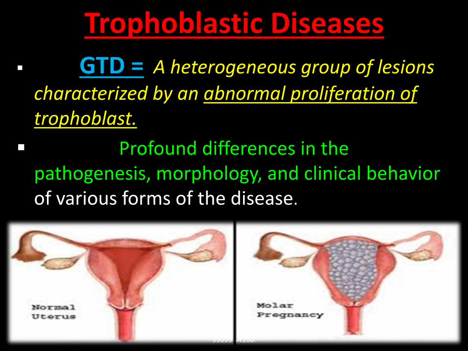
GTD = A heterogeneous group of lesions
characterized by an abnormal proliferation of trophoblast.
Profound differences in the pathogenesis, morphology, and clinical behavior of various forms of the disease.
Trophoblastic Diseases
6/5/2017Dr Shashwat Jani.
99099 44160.3
Hydatidiform moles (complete, partial, and invasive) represent abnormally formed placentas with specific genetic abnormalities that are related to villous trophoblast.
Choriocarcinoma and the placental site trophoblastic tumor are true neoplasms and are related to previllous and extravilloustrophoblast.
6/5/2017Dr Shashwat Jani.
99099 44160.4
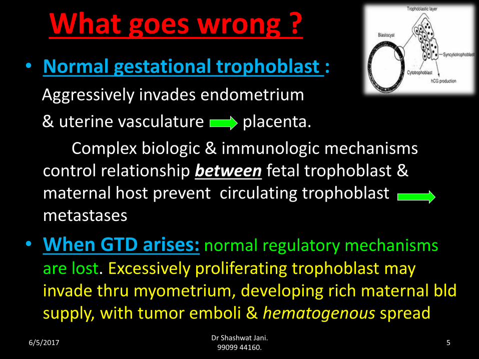
What goes wrong ?• Normal gestational trophoblast :
Aggressively invades endometrium
& uterine vasculature placenta.
Complex biologic & immunologic mechanisms control relationship between fetal trophoblast & maternal host prevent circulating trophoblast metastases
• When GTD arises: normal regulatory mechanisms are lost. Excessively proliferating trophoblast may invade thru myometrium, developing rich maternal bldsupply, with tumor emboli & hematogenous spread
6/5/2017Dr Shashwat Jani.
99099 44160.5
They are characterized by pregnancy associated trophoblastic proliferations.
They range from tumor like conditions to malignancy.
H. mole is a common complication of gestation (1 in 1000 to 2000).
They can be monitored by measuring HCG levels (to detect early recurrence and response to Tx)
Choriocarcinomas are highly responsive to chemotherapy.
Trophoblastic Diseases
6/5/2017Dr Shashwat Jani.
99099 44160.6
6/5/2017Dr Shashwat Jani.
99099 44160.7
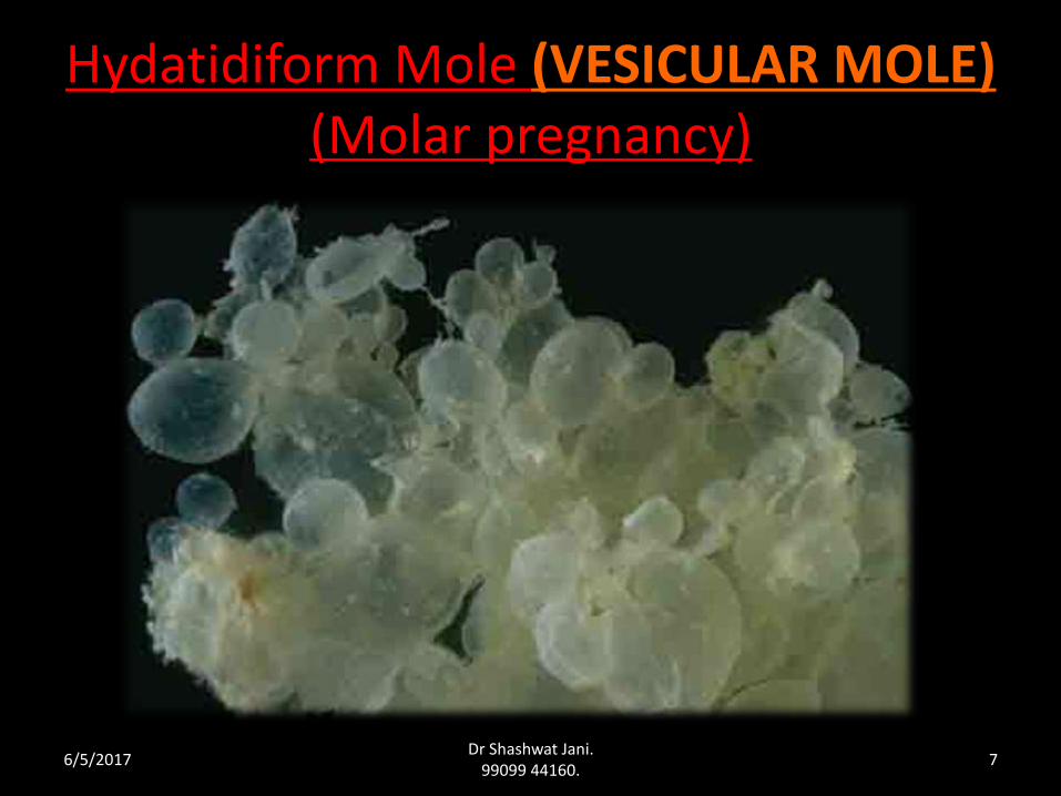
Hydatidiform Mole (VESICULAR MOLE)(Molar pregnancy)
Definition
• In latin
"hydatid" means "drop of water”
"mole" means "spot”.
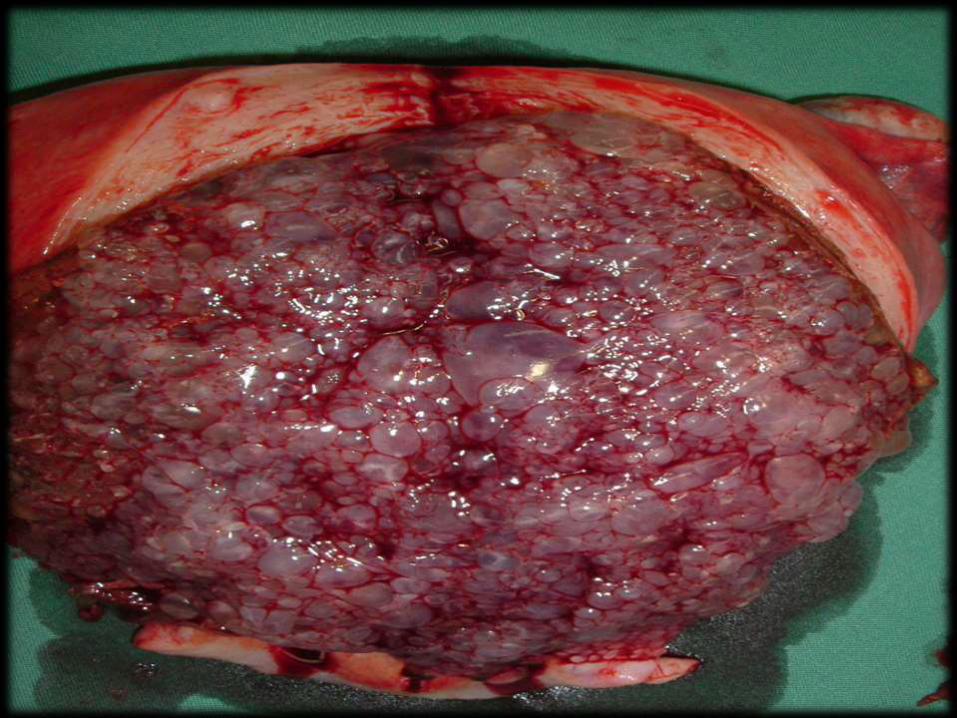
• H. mole is a pregnancy characterized by vesicular swelling of placental villi and usually the absence of an intact fetus.
6/5/2017Dr Shashwat Jani.
99099 44160.8
Incidence
• Wide range in geographical & ethnic variation of prevalence.
• Common in oriental countries.
• Highest in Philippines 1 : 80
• Lowest in European countries 1 : 750
• India 1 : 400 .
6/5/2017Dr Shashwat Jani.
99099 44160.9
• Approximately 10-17% of
H. moles will result in Invasive mole.
• Approximately 2-3% of
H. Moles progress to choriocarcinoma
( most of them are curable)
Not definitely benign disease , has a tight relationship with GTT
6/5/2017Dr Shashwat Jani.
99099 44160.10
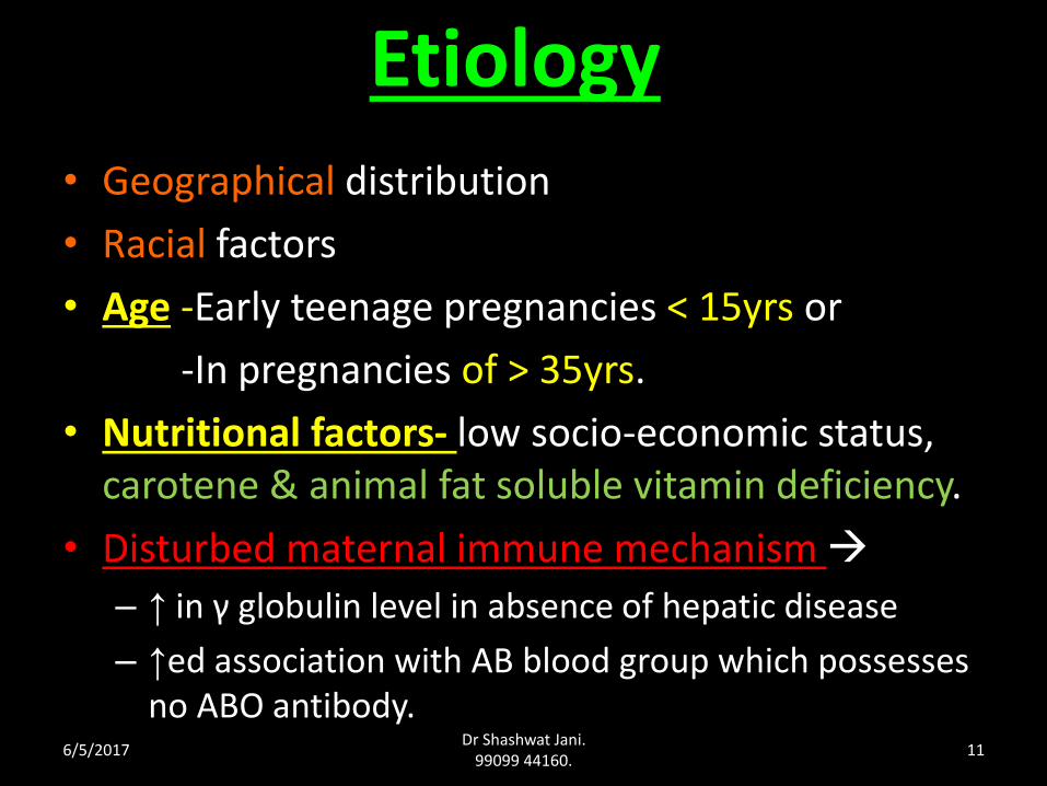
Etiology • Geographical distribution
• Racial factors
• Age -Early teenage pregnancies < 15yrs or
-In pregnancies of > 35yrs.
• Nutritional factors- low socio-economic status, carotene & animal fat soluble vitamin deficiency.
• Disturbed maternal immune mechanism
– ↑ in γ globulin level in absence of hepatic disease
– ↑ed association with AB blood group which possesses no ABO antibody.
6/5/2017Dr Shashwat Jani.
99099 44160.11
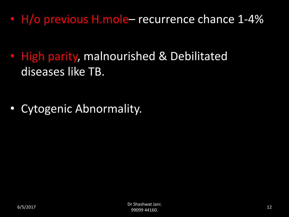
• H/o previous H.mole– recurrence chance 1-4%
• High parity, malnourished & Debilitated diseases like TB.
• Cytogenic Abnormality.
6/5/2017Dr Shashwat Jani.
99099 44160.12
Dr Shashwat Jani. 99099 44160.
Emptyovum
Emptyovum
46XX
46XX or 46XY
23X or Y23X
23XComplete Mole (46XX diploid)
Complete Mole (46XX or 46XY, diploid)
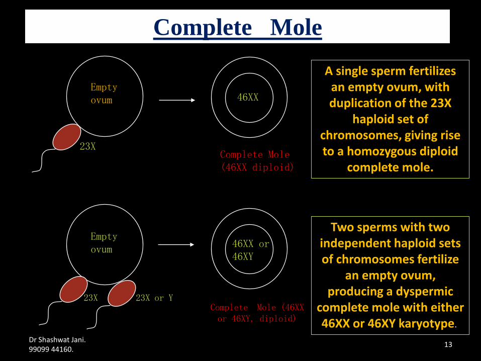
A single sperm fertilizes an empty ovum, with duplication of the 23X
haploid set of chromosomes, giving rise to a homozygous diploid
complete mole.
Two sperms with two independent haploid sets of chromosomes fertilize
an empty ovum, producing a dyspermic
complete mole with either 46XX or 46XY karyotype.
Complete Mole
13
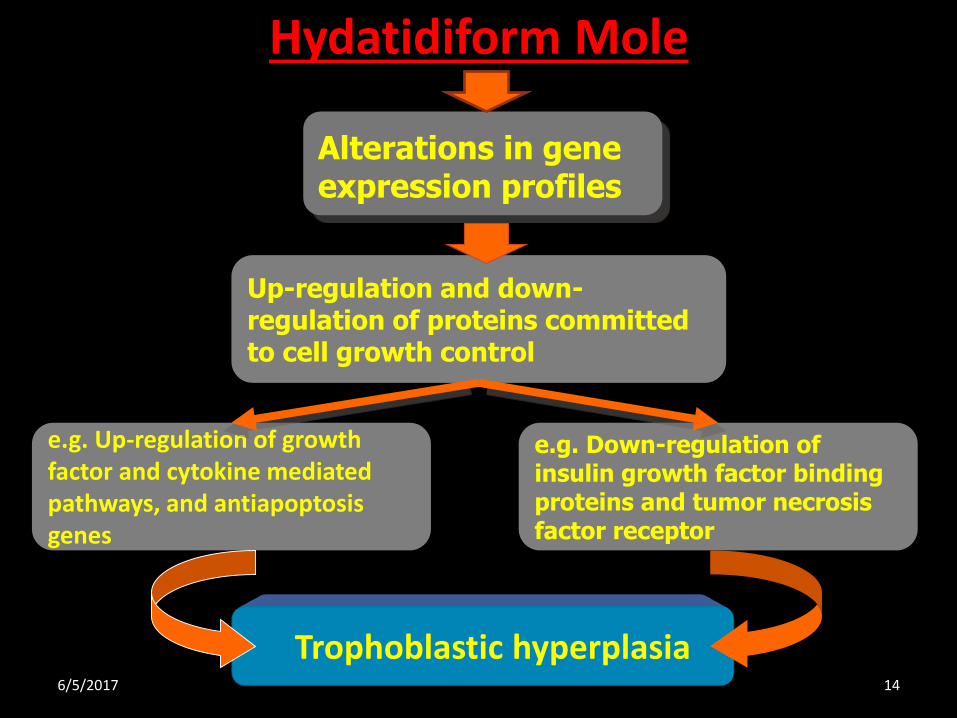
Hydatidiform Mole
Alterations in gene expression profiles
Up-regulation and down-regulation of proteins committed to cell growth control
e.g. Up-regulation of growth factor and cytokine mediated pathways, and antiapoptosisgenes
Trophoblastic hyperplasia
e.g. Down-regulation of insulin growth factor binding proteins and tumor necrosis factor receptor
6/5/2017 14
Hydatidiform Mole
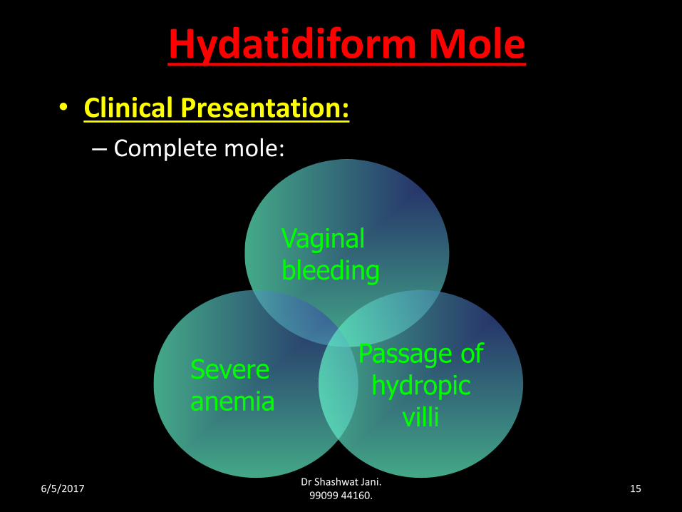
• Clinical Presentation:
– Complete mole:
Vaginal bleeding
Severe anemia
Passage of hydropic
villi
6/5/2017Dr Shashwat Jani.
99099 44160.15
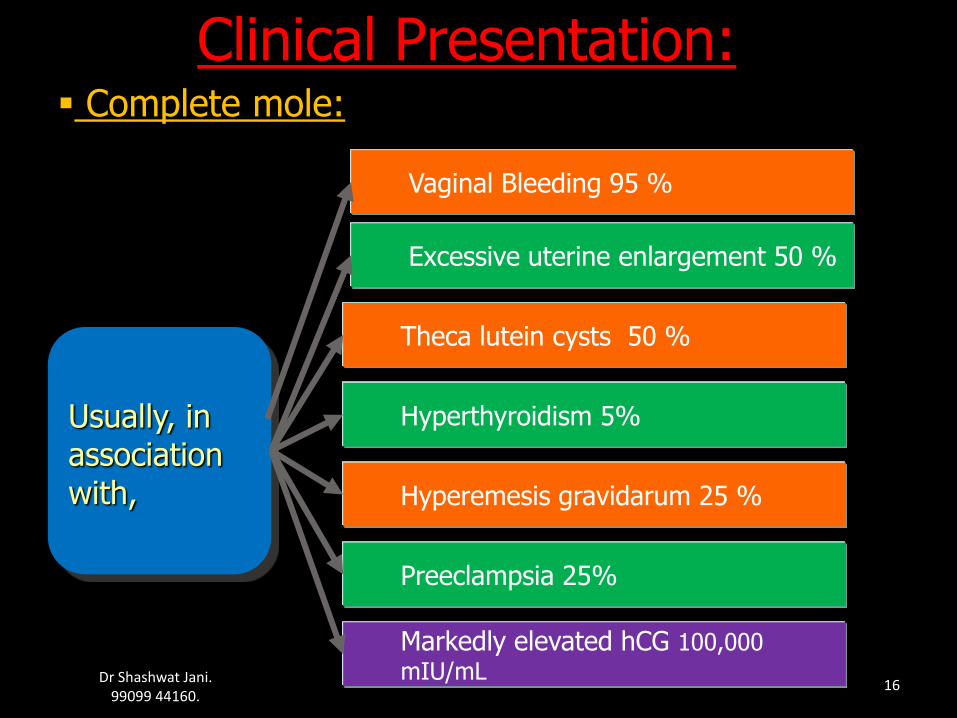
Usually, in association with,
Excessive uterine enlargement 50 %
Hyperemesis gravidarum 25 %
Preeclampsia 25%
Markedly elevated hCG 100,000
mIU/mL
Hyperthyroidism 5%
Theca lutein cysts 50 %
Clinical Presentation: Complete mole:
Vaginal Bleeding 95 %
Dr Shashwat Jani. 99099 44160.
16
6/5/2017Dr Shashwat Jani.
99099 44160.17
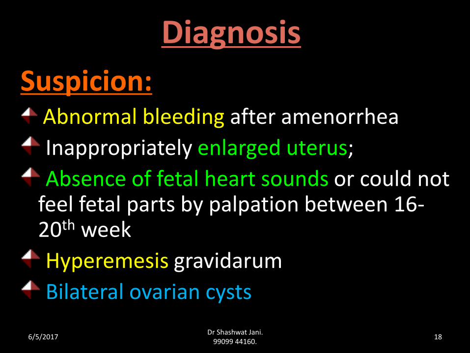
Diagnosis
Suspicion:Abnormal bleeding after amenorrhea
Inappropriately enlarged uterus;
Absence of fetal heart sounds or could not feel fetal parts by palpation between 16-20th week
Hyperemesis gravidarum
Bilateral ovarian cysts
6/5/2017Dr Shashwat Jani.
99099 44160.18
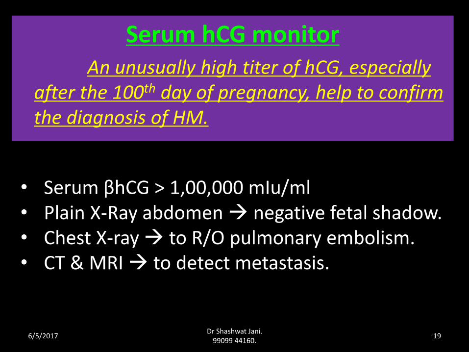
Serum hCG monitorAn unusually high titer of hCG, especially
after the 100th day of pregnancy, help to confirm the diagnosis of HM.
• Serum βhCG > 1,00,000 mIu/ml• Plain X-Ray abdomen negative fetal shadow.• Chest X-ray to R/O pulmonary embolism.• CT & MRI to detect metastasis.
6/5/2017Dr Shashwat Jani.
99099 44160.19
Ultrasonography:It is a reliable and sensitive technique
for the diagnosis of complete molar pregnancy.
Because the chorionic villi exhibit diffuse hydatidiform swelling.
A characteristic vesicular sonographic pattern,
usually referred to as a “Snowstorm” Pattern.
6/5/2017Dr Shashwat Jani.
99099 44160.20
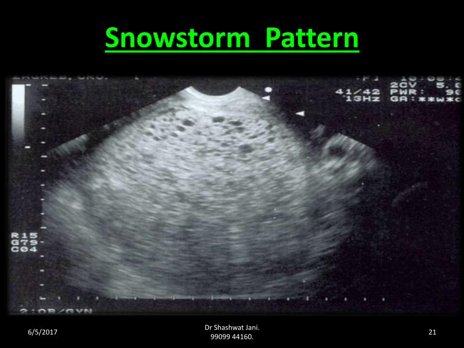
Snowstorm Pattern
6/5/2017Dr Shashwat Jani.
99099 44160.21
Differential Diagnosis
1. Threatened abortion
2. Fibroid uterus with pregnancy.
3. Ovarian tumour with pregnancy
4. Multiple pregnancy.
6/5/2017Dr Shashwat Jani.
99099 44160.22
MANAGEMENT
Principles in management :
• Suction evacuation of uterus
(safe upto 28 wks of gestation).
• Supportive therapy – correction of anemia & infection if any.
• Counseling for regular follow-up.
6/5/2017Dr Shashwat Jani.
99099 44160.23
Evacuation & Mx of Molar Pregnancies
Complete history & medical exam (anemia/ dehydration/ preeclampsia &/or thyrotoxicosis)
Appropriate lab & radiologic evaluation,….stabilize hemodynamically (preevacuation hCG, CBC, LFT, BUN, creatinine, TFT, pelvic USG, chest x-ray)
Based on these findings, perioperative complications shd be anticipated …preopn.ABG, postevacuation chest
x-ray, central monitoring in ICU setting
Suction evacuation gives the lowest incidence of sequelae
(metal canula, medical induction, prostaglandins …NO )
6/5/2017Dr Shashwat Jani.
99099 44160.24
Evacuation Technique• Stabilize hemodynamically, address all medical
complication (antihypertensive, β blocker)
• Large bore intravenous line… central venous monitoring
• Two units blood, laparotomy tray in O.R.
• Cx grasped with single tooth tenaculum,
• NO sounding,
• Cx dilated gently to accommodate 12-14mm cannula.
6/5/2017Dr Shashwat Jani.
99099 44160.25
• Cannula only up to lower portion of uterus,
• Start Oxytocin, massage fundus gently to assist involution, rotate cannula…advance only after involution
• Sharp curettage…both specimen separately for HPE
• Oxytocin to be cont. for 24hrs. evacuation, ...avoid fluid overload.
6/5/2017Dr Shashwat Jani.
99099 44160.26
Complications of Suction evacuation.
• Injury to uterus Perforation, infection.
• Hemorrhage.
• Shock
• Acute pulmonary insufficiency.
• Thyroid storm.
6/5/2017Dr Shashwat Jani.
99099 44160.27
Hysterectomy (↓ risk of GTN by 5%)
Indicated in • Patient with age > 35 yrs
• Completed family irrespective of age.
• Uncontrolled hemorrhage/perforation during suction evacuation.
6/5/2017Dr Shashwat Jani.
99099 44160.28
Hysterotomy• Rarely done.
Indicated in
• Profuse vaginal bleeding.
• Cervix unfavorable for immediate vaginal evacuation.
• Accidental perforation of uterus during evacuation.
6/5/2017Dr Shashwat Jani.
99099 44160.29
Theca lutein cysts
• They are hormone dependent.
• Disappear spontaneously after evacuation of the mole.
• So, they are not removed surgically unless complication occur as torsion or rupture.
6/5/2017Dr Shashwat Jani.
99099 44160.30
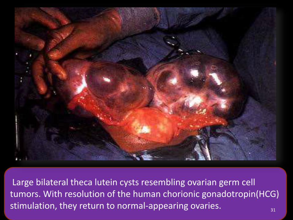
Large bilateral theca lutein cysts resembling ovarian germ cell tumors. With
resolution of the human chorionic gonadotropin(HCG) stimulation, they return to
normal-appearing ovaries.
Large bilateral theca lutein cysts resembling ovarian germ cell tumors. With resolution of the human chorionic gonadotropin(HCG) stimulation, they return to normal-appearing ovaries.
31
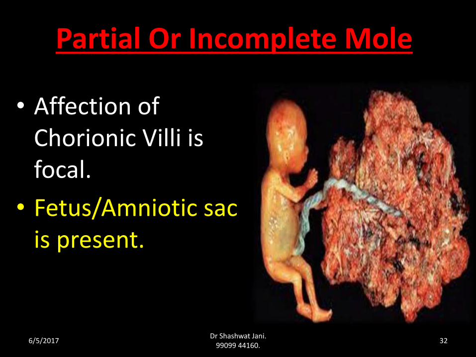
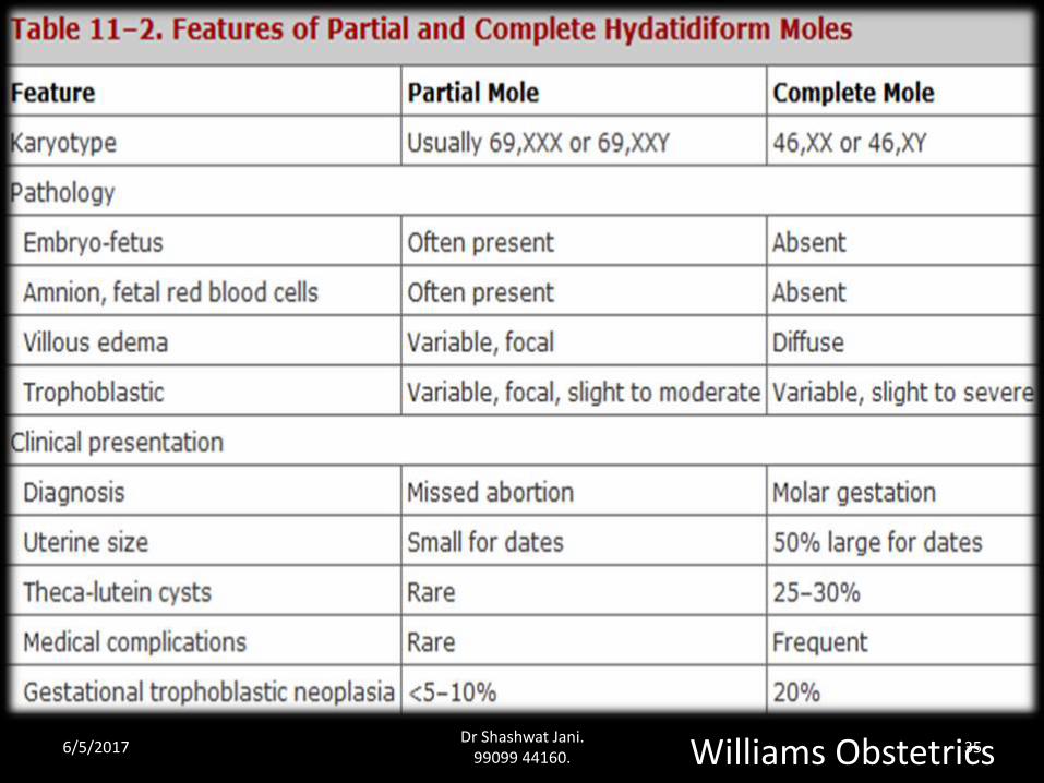
Partial Or Incomplete Mole
• Affection of Chorionic Villi is focal.
• Fetus/Amniotic sac is present.
6/5/2017Dr Shashwat Jani.
99099 44160.32
23X 23X
Dyspermy 23X/23Y or 23X/23X
23Y
Partial Mole (69XXY, or 69XXX, or 69XYY
triploid)
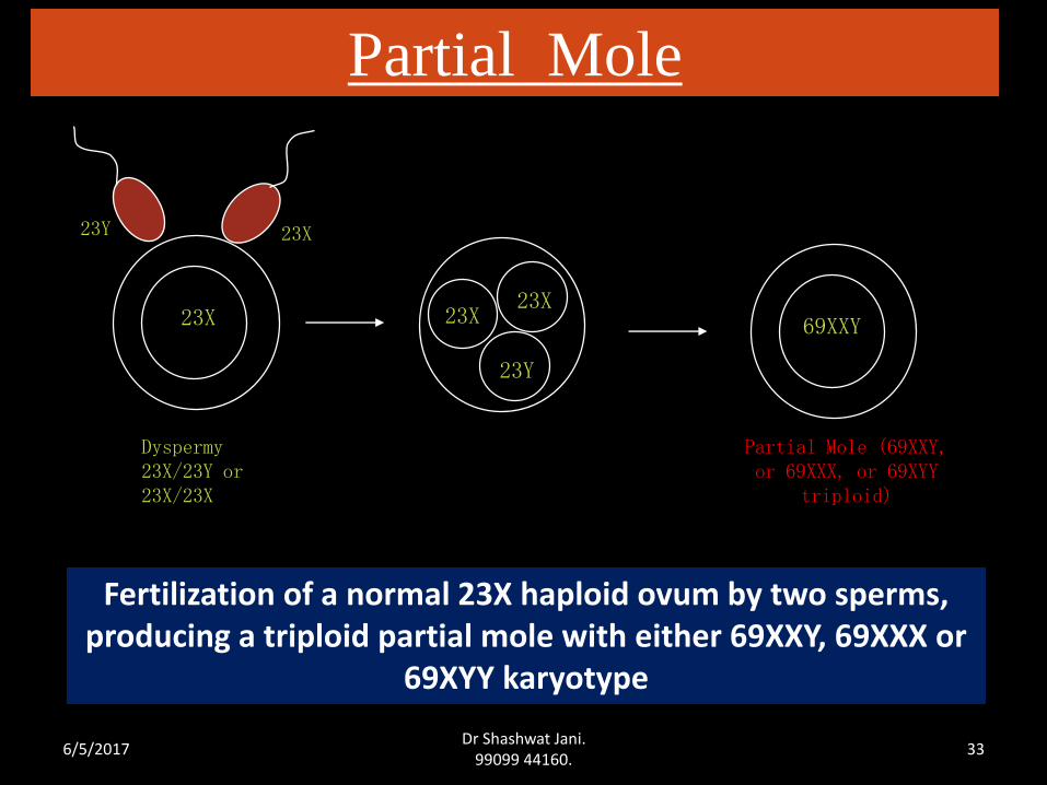
Partial Mole
23X
23X
23Y
69XXY
Fertilization of a normal 23X haploid ovum by two sperms, producing a triploid partial mole with either 69XXY, 69XXX or
69XYY karyotype
6/5/2017Dr Shashwat Jani.
99099 44160.33
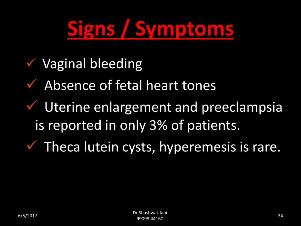
Signs / Symptoms
6/5/2017Dr Shashwat Jani.
99099 44160.34
Vaginal bleeding
Absence of fetal heart tones
Uterine enlargement and preeclampsia is reported in only 3% of patients.
Theca lutein cysts, hyperemesis is rare.
Williams Obstetrics6/5/2017Dr Shashwat Jani.
99099 44160.35
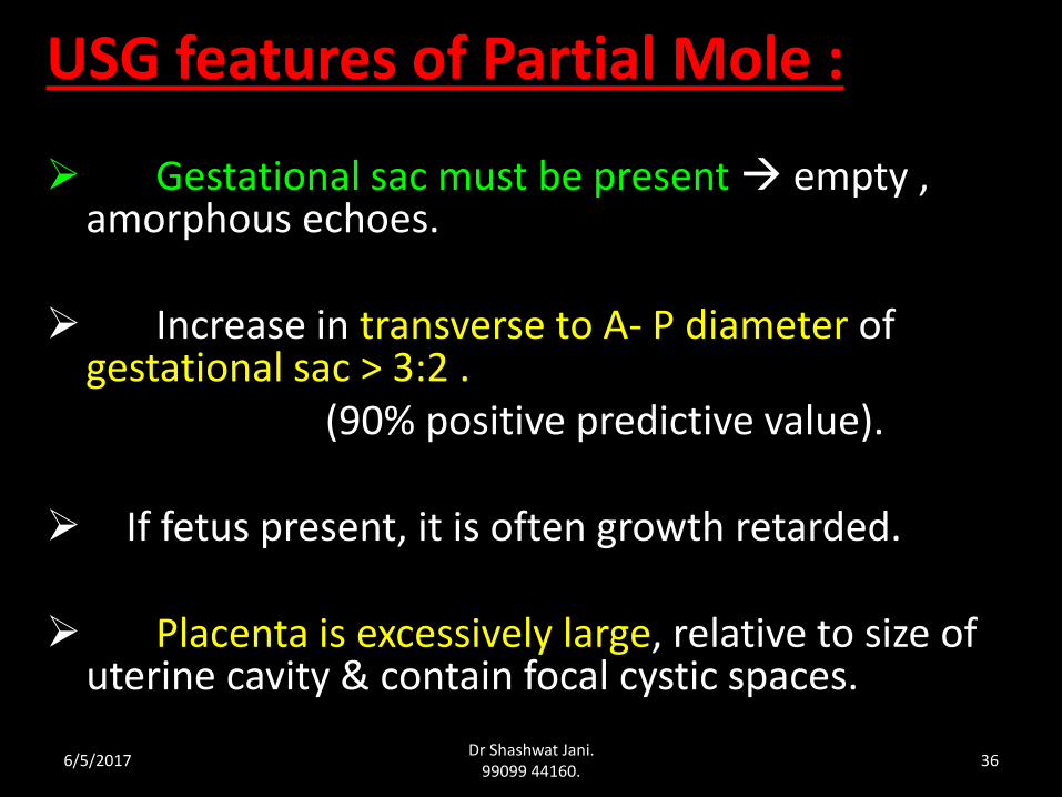
USG features of Partial Mole :
Gestational sac must be present empty , amorphous echoes.
Increase in transverse to A- P diameter of gestational sac > 3:2 .
(90% positive predictive value).
If fetus present, it is often growth retarded.
Placenta is excessively large, relative to size of uterine cavity & contain focal cystic spaces.
6/5/2017Dr Shashwat Jani.
99099 44160.36
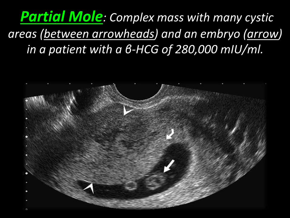
Partial Mole: Complex mass with many cystic
areas (between arrowheads) and an embryo (arrow) in a patient with a β-HCG of 280,000 mIU/ml.
Management • If fetus is not alive termination of
pregnancy.
• If fetus is alive woman counseled about ↑ed risk of perinatal morbidity & outcome of GTN. Terminate the pregnancy.
6/5/2017Dr Shashwat Jani.
99099 44160.38
Partial Mole In Twins
• Many cases have been reported of second normal live fetus up to age of viability.
• Can continue pregnancy after explaining all possible maternal & fetal complications.
6/5/2017Dr Shashwat Jani.
99099 44160.39
Contraception during follow up
• The combined pill is started when the beta-HCG becomes negative. Till this happens, the condom can be used.
• If the pill is used early the beta-HCG will take a longer time to become negative as oestrogen stimulates the growth of trophoplast.
6/5/2017Dr Shashwat Jani.
99099 44160.40
The intrauterine device is not used because it may lead to irregular uterine bleeding which confuses the follow up & also increases chances of perforation.
IUD during follow up
6/5/2017Dr Shashwat Jani.
99099 44160.41
Follow Up
• Objective to diagnose persistent GTT that is
considered malignant.
• If hCG Normal within 56 days follow up will be for 6 months from date of uterine evacuation.
• If hCG Not normal within 56 days then follow up will be for 6 months from normalization of hCG level.
• Woman with chemotherapy should follow up for 1 year after hCG has been normal.
6/5/2017Dr Shashwat Jani.
99099 44160.42
Follow up protocol
History
Physical examination
hCG assay
Chest X-ray
6/5/2017Dr Shashwat Jani.
99099 44160.43
• History :– h/o irregular P/V bleeding.
– Hemoptysis
– Breathlessness
– CNS disturbance like headache, blurring of vision, neurological deficit.
– Epigastric pain, hematuria, jaundice
6/5/2017Dr Shashwat Jani.
99099 44160.44
Physical examination
• General examination
• P/A
– Sub-involution of uterus,
– Palpation of mass
– Tenderness
– Hepatomegaly
• P/S Vaginal metastasis
• P/V Sub-involution of uterus,
Regression of theca lutein cyst .6/5/2017
Dr Shashwat Jani. 99099 44160.
45
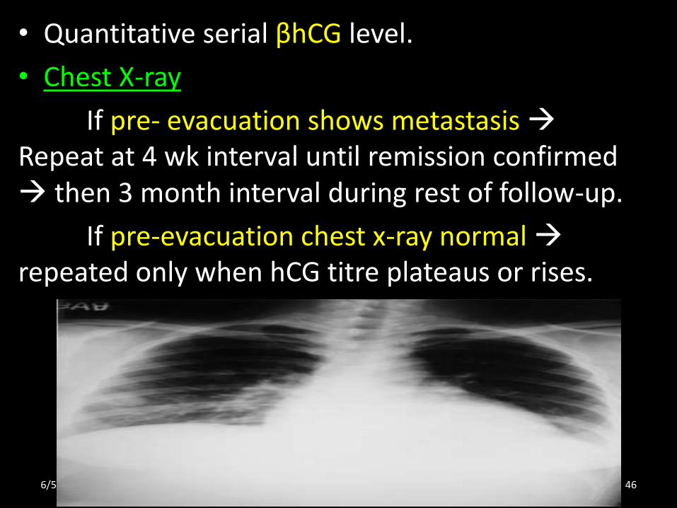
• Quantitative serial βhCG level.
• Chest X-ray
If pre- evacuation shows metastasis Repeat at 4 wk interval until remission confirmed then 3 month interval during rest of follow-up.
If pre-evacuation chest x-ray normalrepeated only when hCG titre plateaus or rises.
6/5/2017Dr Shashwat Jani.
99099 44160.46
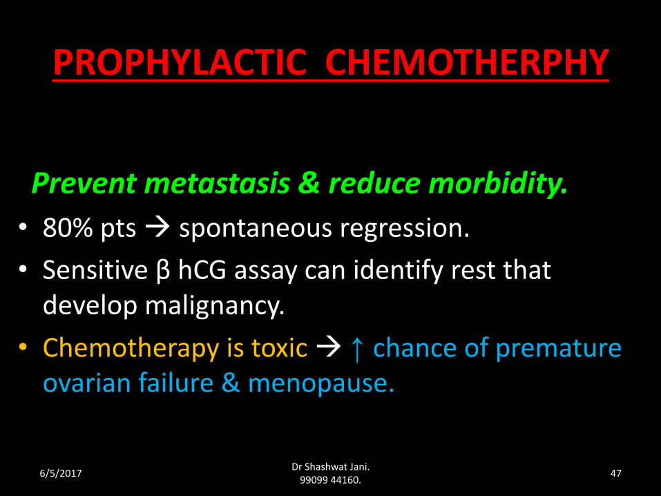
PROPHYLACTIC CHEMOTHERPHY
Prevent metastasis & reduce morbidity.
• 80% pts spontaneous regression.
• Sensitive β hCG assay can identify rest that develop malignancy.
• Chemotherapy is toxic ↑ chance of premature ovarian failure & menopause.
6/5/2017Dr Shashwat Jani.
99099 44160.47
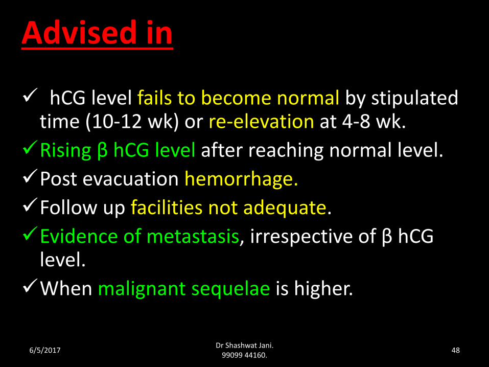
Advised in
hCG level fails to become normal by stipulated time (10-12 wk) or re-elevation at 4-8 wk.
Rising β hCG level after reaching normal level.
Post evacuation hemorrhage.
Follow up facilities not adequate.
Evidence of metastasis, irrespective of β hCG level.
When malignant sequelae is higher.
6/5/2017Dr Shashwat Jani.
99099 44160.48
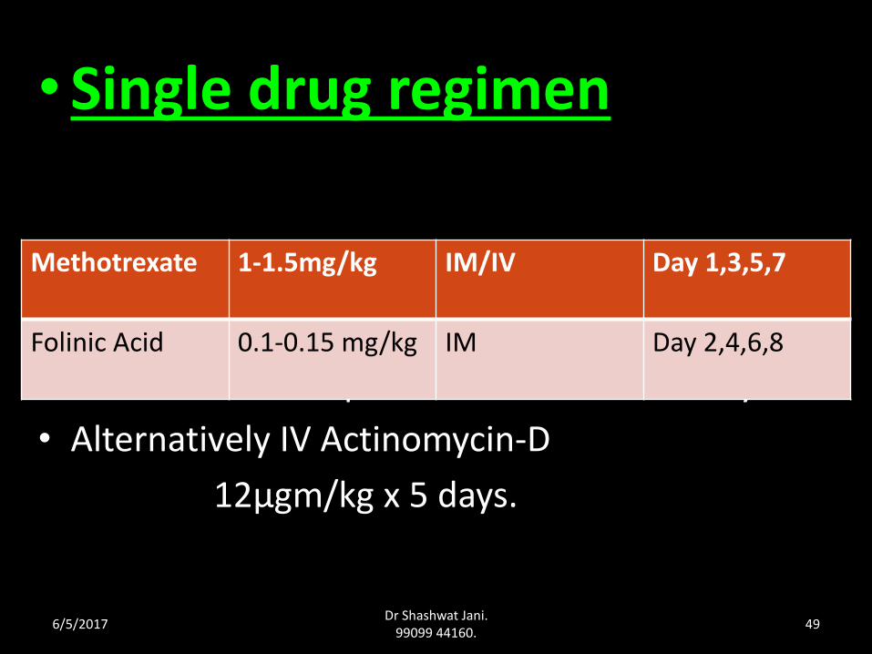
• Single drug regimen
• Course is to be repeated at interval of 7 days.
• Alternatively IV Actinomycin-D
12μgm/kg x 5 days.
Methotrexate 1-1.5mg/kg IM/IV Day 1,3,5,7
Folinic Acid 0.1-0.15 mg/kg IM Day 2,4,6,8
6/5/2017Dr Shashwat Jani.
99099 44160.49
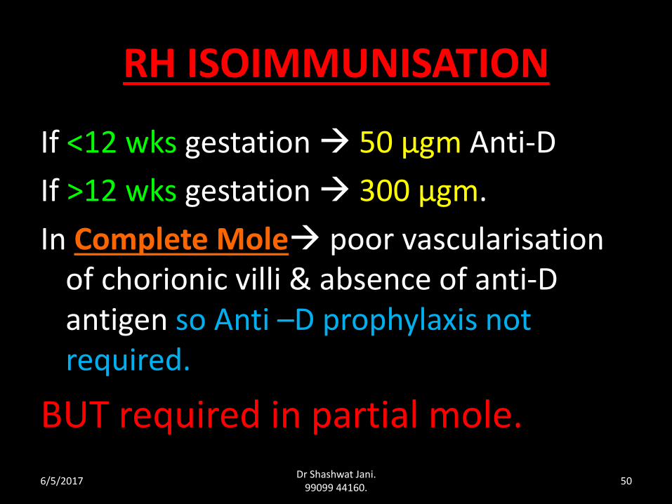
RH ISOIMMUNISATION
If <12 wks gestation 50 μgm Anti-D
If >12 wks gestation 300 μgm.
In Complete Mole poor vascularisation of chorionic villi & absence of anti-D antigen so Anti –D prophylaxis not required.
BUT required in partial mole.
6/5/2017Dr Shashwat Jani.
99099 44160.50
Placental Site Trophoblastic Tumour (PSTT)
• Rare
• Histological Diagnosis syncytotrophoblastic cells are generally absent persistent low level of serum or urinary hCG.
• Tumor from intermediate trophoblasts of placental bed composed mainly of cytotrophoblastic cells.
6/5/2017Dr Shashwat Jani.
99099 44160.51
• C/F Vaginal bleeding.
• Local invasion of Myometrium & Lymphatics.
• PSTT is not responsive to chemotherapy.
• Rx Hysterectomy.
6/5/2017Dr Shashwat Jani.
99099 44160.52
Invasive mole
6/5/2017Dr Shashwat Jani.
99099 44160.53
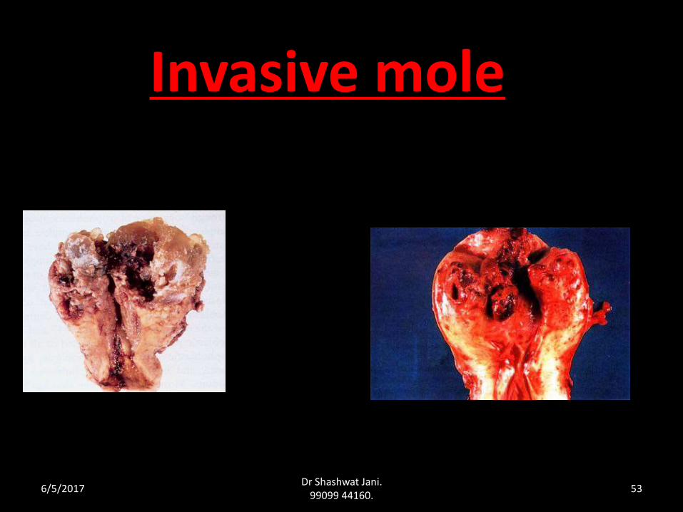
Invasive moleDefinition:
An invasive hydatidiform mole is one in which hydropic chorionic villi are within the myometrium or its vascular spaces or at distant sites, notably the vagina or lung.
“Mole that penetrates and even perforates the uterine wall”.
6/5/2017Dr Shashwat Jani.
99099 44160.54
Invasive mole
• Invasive hydatidiform mole is a sequela to hydatidiform mole, complete or partial.
• The pathologic diagnosis of invasive mole is made by establishing the presence of molar villi growing into the myometrium and broad ligament.
• The diagnosis of an invasive mole cannot be made on examination of curettage specimens except when curetted fragments of myometrium contain invasive molar villi.
6/5/2017Dr Shashwat Jani.
99099 44160.55

Clinically identified by:
Combination of Abnormal uterine USG
Persistent / rising hCG level after uterine evacuation for mole
Theca lutein cysts & uterine subinvolution
Histologic verification is rarely required
Repeat D&C contraindicated….risk of uterine perforation, infection, life threatening hemorrhage ..hysterectomy
6/5/2017Dr Shashwat Jani.
99099 44160.56
Persistent Gestational Trophoblastic Disease
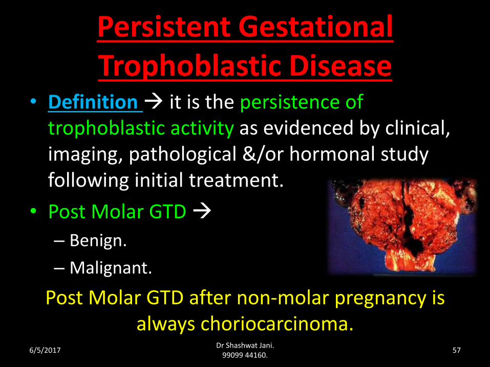
• Definition it is the persistence of trophoblastic activity as evidenced by clinical, imaging, pathological &/or hormonal study following initial treatment.
• Post Molar GTD
– Benign.
– Malignant.
Post Molar GTD after non-molar pregnancy is always choriocarcinoma.
6/5/2017Dr Shashwat Jani.
99099 44160.57
Incidence :• 50% following H.Mole
• 25% following abortion or ectopic pregnancy.
• 25% following normal delivery.
6/5/2017Dr Shashwat Jani.
99099 44160.58
DIAGNOSIS
During post-evacuation follow-up period
• Continued vaginal bleeding.
• Persistent Theca Lutein Cysts.
• Persistent soft & enlarged uterus.
• hCG titer either fail to become negative or plateau or re-elevation after initial fall by 8 wk post molar evacuation.
• Local, systemic metastasis ruled out by x-ray chest, CT, MRI of brain, Liver etc.
6/5/2017Dr Shashwat Jani.
99099 44160.59
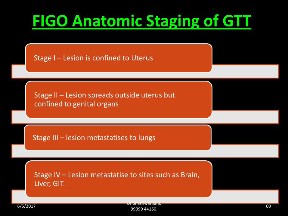
FIGO Anatomic Staging of GTT
Stage I – Lesion is confined to Uterus
Stage II – Lesion spreads outside uterus but confined to genital organs
Stage III – lesion metastatises to lungs
Stage IV – Lesion metastatise to sites such as Brain, Liver, GIT.
6/5/2017Dr Shashwat Jani.
99099 44160.60
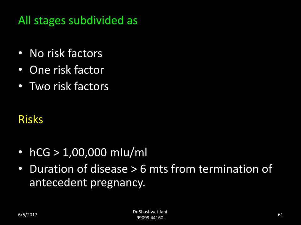
All stages subdivided as
• No risk factors
• One risk factor
• Two risk factors
Risks
• hCG > 1,00,000 mIu/ml
• Duration of disease > 6 mts from termination of antecedent pregnancy.
6/5/2017Dr Shashwat Jani.
99099 44160.61
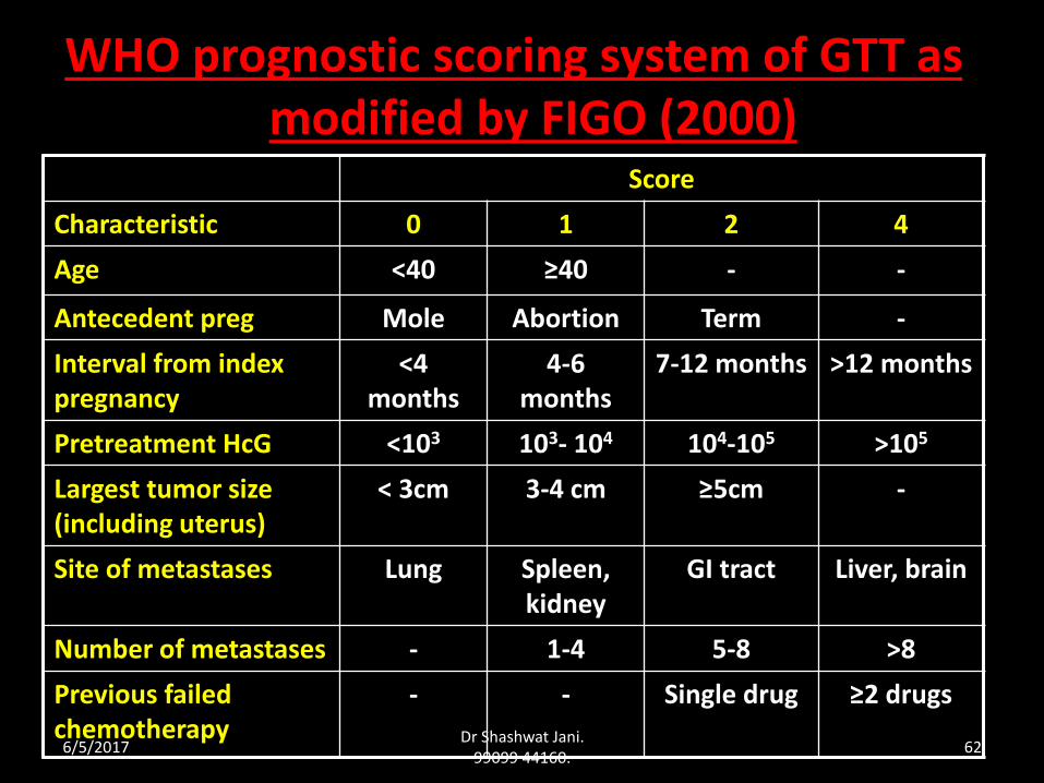
WHO prognostic scoring system of GTT as modified by FIGO (2000)
Score
Characteristic 0 1 2 4
Age <40 ≥40 - -
Antecedent preg Mole Abortion Term -
Interval from index pregnancy
<4 months
4-6 months
7-12 months >12 months
Pretreatment HcG <103 103- 104 104-105 >105
Largest tumor size (including uterus)
< 3cm 3-4 cm ≥5cm -
Site of metastases Lung Spleen, kidney
GI tract Liver, brain
Number of metastases - 1-4 5-8 >8
Previous failed chemotherapy
- - Single drug ≥2 drugs
6/5/2017Dr Shashwat Jani.
99099 44160.62
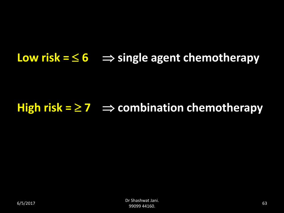
Low risk = 6 single agent chemotherapy
High risk = 7 combination chemotherapy
6/5/2017Dr Shashwat Jani.
99099 44160.63
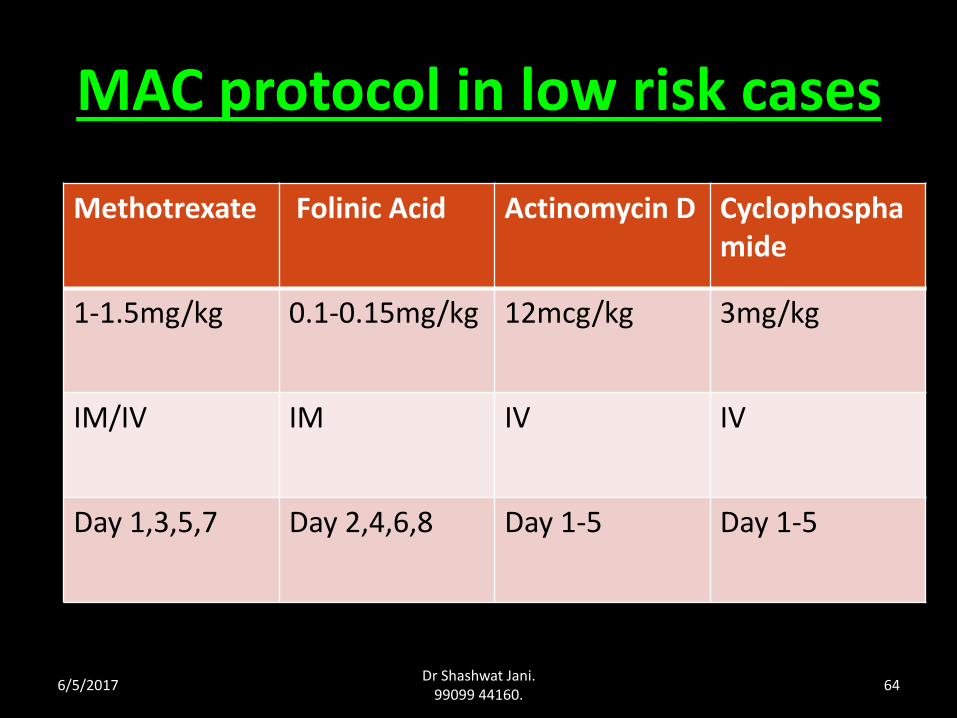
MAC protocol in low risk cases
Methotrexate Folinic Acid Actinomycin D Cyclophosphamide
1-1.5mg/kg 0.1-0.15mg/kg 12mcg/kg 3mg/kg
IM/IV IM IV IV
Day 1,3,5,7 Day 2,4,6,8 Day 1-5 Day 1-5
6/5/2017Dr Shashwat Jani.
99099 44160.64
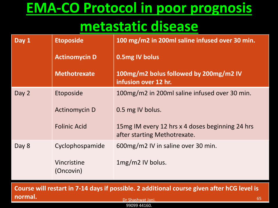
EMA-CO Protocol in poor prognosis metastatic disease
Day 1 Etoposide
Actinomycin D
Methotrexate
100 mg/m2 in 200ml saline infused over 30 min.
0.5mg IV bolus
100mg/m2 bolus followed by 200mg/m2 IV infusion over 12 hr.
Day 2 Etoposide
Actinomycin D
Folinic Acid
100mg/m2 in 200ml saline infused over 30 min.
0.5 mg IV bolus.
15mg IM every 12 hrs x 4 doses beginning 24 hrs after starting Methotrexate.
Day 8 Cyclophospamide
Vincristine(Oncovin)
600mg/m2 IV in saline over 30 min.
1mg/m2 IV bolus.
Course will restart in 7-14 days if possible. 2 additional course given after hCG level is normal.
Dr Shashwat Jani. 99099 44160.
65
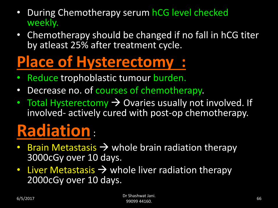
• During Chemotherapy serum hCG level checked weekly.
• Chemotherapy should be changed if no fall in hCG titer by atleast 25% after treatment cycle.
Place of Hysterectomy :• Reduce trophoblastic tumour burden.• Decrease no. of courses of chemotherapy.• Total Hysterectomy Ovaries usually not involved. If
involved- actively cured with post-op chemotherapy.
Radiation :
• Brain Metastasis whole brain radiation therapy 3000cGy over 10 days.
• Liver Metastasis whole liver radiation therapy 2000cGy over 10 days.
6/5/2017Dr Shashwat Jani.
99099 44160.66
Prognosis • Low risk almost 100%
• High risk 70%.
Recurrence • Non-metastatic GTN 2-3%
• Good prognosis metastatic disease 3-5%
• Poor prognosis disease 21%.
• Recurrence following 12 mts of normal hCG level < 1%.
6/5/2017Dr Shashwat Jani.
99099 44160.67
ROLE OF SURGERY
Surgery has been limited to the treatment of :-
- Resistant cases to chemotherapy,
- Uncontrollable haemorrhage from the uterus
- Tumour perforation of the uterus
- Infected uterine tumour not responding to antibiotics, thus delaying chemotherapy..
6/5/2017Dr Shashwat Jani.
99099 44160.68
Newer Regimen
• Newer chemotherapy agents
• Autologous bone marrow transplant
• Peripheral stem cell support
• colony stimulating factors
• Selective arterial embolisation.
FOLLOW-UP
• BhCG Titre wkly until 3 consecutive normal titres• Monthly for 12 months • 3 monthly for 1 additional year• 6 monthly indefinitely• Contraception for at least 1yr after remission(OCP,
Condom)
• Gynaecologic examination started 1 week post evacuation – assess Ut size, adnexal masses, check for metastases on the vulva, vagina, urethra, and cervix. If no complication repeat exam 4 wkly throughout period of surveillance.
6/5/2017Dr Shashwat Jani.
99099 44160.70