Embed Size (px)
Citation preview
Downloaded from http://aidsinfo.nih.gov/guidelines on 7/29/2015
Guidelines for Prevention and Treatment of OpportunisticInfections in HIV-Infected Adults and Adolescents
Downloaded from http://aidsinfo.nih.gov/guidelines on 7/29/2015
Visit the AIDSinfo website to access the most up-to-date guideline.
Register for e-mail notification of guideline updates at http://aidsinfo.nih.gov/e-news.
Downloaded from http://aidsinfo.nih.gov/guidelines on 7/29/2015
Guidelines for the Prevention and Treatment of Opportunistic Infections in HIV-Infected Adults
and Adolescents
Recommendations from the Centers for Disease Control and Prevention,the National Institutes of Health, and the HIV Medicine Association
of the Infectious Diseases Society of America
How to Cite the Adult and Adolescent Opportunistic Infection Guidelines:
Panel on Opportunistic Infections in HIV-Infected Adults and Adolescents. Guidelines for theprevention and treatment of opportunistic infections in HIV-infected adults and adolescents:recommendations from the Centers for Disease Control and Prevention, the National Institutesof Health, and the HIV Medicine Association of the Infectious Diseases Society of America.Available at http://aidsinfo.nih.gov/contentfiles/lvguidelines/adult_oi.pdf. Accessed (insert date)[include page numbers, table number, etc. if applicable]
It is emphasized that concepts relevant to HIV management evolve rapidly. The Panel has amechanism to update recommendations on a regular basis, and the most recent information isavailable on the AIDSinfo website (http://aidsinfo.nih.gov).
Access AIDSinfomobile site
Downloaded from http://aidsinfo.nih.gov/guidelines on 7/29/2015
Guidelines for the Prevention and Treatment of Opportunistic Infections in HIV-Infected Adults and Adolescents i
What’s New in the Guidelines
Updates to the Guidelines for the Prevention and Treatment of Opportunistic Infections
in HIV-Infected Adults and Adolescents
The Guidelines for the Prevention and Treatment of Opportunistic Infections in HIV Infected Adults andAdolescents document was published in an electronic format that could be easily updated as relevant changesin prevention and treatment recommendations occur.
The editors and subject matter experts are committed to timely changes in this document because so manyhealth care providers, patients, and policy experts rely on this source for vital clinical information.
All changes are developed by the subject matter groups listed in the document (changes in groupcomposition are also promptly posted). These changes are reviewed by the editors and by relevant outsidereviewers before the document is altered.
Major revisions within the last 6 months are as follows:
April 16, 2015:
1. Hepatitis B Virus: There is new information on techniques to evaluate the stage of liver fibrosis. Newdata on HBV immunization regimens is included in recommendations regarding thechoice of the optimalimmunization regimen.
Downloaded from http://aidsinfo.nih.gov/guidelines on 7/29/2015
Guidelines for the Prevention and Treatment of Opportunistic Infections in HIV-Infected Adults and Adolescents ii
Table of ContentsWhat’s New in the Guidelines ..........................................................................................................................i
Introduction ..................................................................................................................................................A-1
Pneumocystis Pneumonia .............................................................................................................................B-1
Toxoplasma gondii Encephalitis...................................................................................................................C-1
Cryptosporidiosis .........................................................................................................................................D-1
Microsporidiosis............................................................................................................................................E-1
Mycobacterium tuberculosis Infection and Disease ....................................................................................F-1
Disseminated Mycobacterium avium Complex Disease .............................................................................G-1
Bacterial Respiratory Disease .....................................................................................................................H-1
Bacterial Enteric Infections ..........................................................................................................................I-1
Bartonellosis ...................................................................................................................................................J-1
Syphilis ..........................................................................................................................................................K-1
Mucocutaneous Candidiasis ........................................................................................................................L-1
Endemic Mycoses Plus Aspergillosis..........................................................................................................M-1Cryptococcosis ..................................................................................................................................M-1Histoplasmosis ................................................................................................................................M-11Coccidioidomycosis ........................................................................................................................M-18Aspergillosis....................................................................................................................................M-25
Cytomegalovirus Disease .............................................................................................................................N-1
Non-CMV Herpes.........................................................................................................................................O-1Herpes Simplex Virus Disease...........................................................................................................O-1Varicella-Zoster Virus Diseases .........................................................................................................O-7Human Herpesvirus-8 Disease ........................................................................................................O-15
Human Papillomavirus Disease ...................................................................................................................P-1
Hepatitis B Virus Infection ..........................................................................................................................Q-1
Hepatitis C Virus Infection ..........................................................................................................................R-1
Progressive Multifocal Leukoencephalopathy/JC Virus Infection...........................................................S-1
Geographic Opportunistic Infections of Specific Consideration ..............................................................T-1Malaria................................................................................................................................................T-1Penicilliosis marneffei ........................................................................................................................T-9Leishmaniasis ...................................................................................................................................T-15Chagas Disease .................................................................................................................................T-26Isosporiasis (Cystoisosporiasis)........................................................................................................T-34
Downloaded from http://aidsinfo.nih.gov/guidelines on 7/29/2015
Guidelines for the Prevention and Treatment of Opportunistic Infections in HIV-Infected Adults and Adolescents iii
TablesTable 1. Prophylaxis to Prevent First Episode of Opportunistic Disease .........................................U-1
Table 2. Treatment of AIDS-Associated Opportunistic Infections (Includes Recommendations forAcute Treatment and Chronic Suppressive/Maintenance Therapy)...................................................U-6
Table 3. Recommended Doses of First-Line Drugs for Treatment of Tuberculosis in Adults and Adolescents ....................................................................................................................U-29
Table 4. Indications for Discontinuing and Restarting Opportunistic Infection Prophylaxis in HIV-Infected Adults and Adolescents...............................................................................................U-30
Table 5. Significant Pharmacokinetic Interactions for Drugs Used to Treat or Prevent Opportunistic Infections ..................................................................................................................U-33
Table 6. Common or Serious Adverse Reactions Associated With Drugs Used for Preventing or Treating Opportunistic Infections ....................................................................................................U-47
Table 7. Dosing Recommendations for Drugs Used in Treating or Preventing Opportunistic Infections Where Dosage Adjustment is Needed in Patients with Renal Insufficiency....................U-52
Table 8. Summary of Pre-Clinical and Human Data on, and Indications for, Opportunistic Infection Drugs During Pregnancy .................................................................................................U-59
Figure: Immunization Schedule for Human Immunodeficiency Virus (HIV)-Infected Adults.............V-1
Appendix A. Recommendations to Help HIV-Infected Patients Avoid Exposure to, orInfection from, Opportunistic Pathogens ..................................................................................................W-1
Appendix B. List of Abbreviations ..............................................................................................................X-1
Appendix C. Panel Roster and Financial Disclosures ...............................................................................Y-1
Appendix D. Contributors............................................................................................................................Z-1
Downloaded from http://aidsinfo.nih.gov/guidelines on 7/29/2015
Guidelines for the Prevention and Treatment of Opportunistic Infections in HIV-Infected Adults and Adolescents A-1
Introduction (Last updated June 17, 2013; last reviewed May 7, 2013)
Prior to the widespread use of potent combination antiretroviral therapy (ART), opportunistic infections(OIs), which have been defined as infections that are more frequent or more severe because ofimmunosuppression in HIV-infected persons,1,2 were the principal cause of morbidity and mortality in thispopulation. In the early 1990s, the use of chemoprophylaxis, immunization, and better strategies formanaging acute OIs contributed to improved quality of life and improved survival.3 Subsequently, thewidespread use of potent ART has had the most profound influence on reducing OI-related mortality in HIV-infected persons.3-10
Despite the availability of ART, OIs continue to cause considerable morbidity and mortality in the UnitedStates for three main reasons:
1. Approximately 20% of HIV-infected persons in the United States are unaware of their HIV infection,11,12
and many present with an OI as the initial indicator of their disease;13
2. Some individuals are aware of their HIV infection, but do not take ART due to psychosocial or economicfactors; and
3. Some patients are enrolled in HIV care and prescribed ART, but do not attain an adequate virologic andimmunologic response due to inconsistent retention in care, poor adherence, unfavorablepharmacokinetics, or unexplained biologic factors.6,14,15
Recent analyses suggest that while 77% of HIV-infected persons who are retained in care and prescribedART are virologically suppressed, only 20% to 28% of the total estimated HIV-infected population in theUnited States are virologically suppressed,11,16 with as few as 10% in some jurisdictions.17 Thus, whilehospitalizations and deaths have decreased dramatically due to ART, OIs continue to cause substantialmorbidity and mortality in HIV-infected persons.18-28 Clinicians must be knowledgeable about optimalstrategies for diagnosis, prevention, and treatment of OIs to provide comprehensive, high quality care forthese patients.
It is important to recognize that the relationship between OIs and HIV infection is bi-directional. HIV causesthe immunosuppression that allows opportunistic pathogens to cause disease in HIV-infected persons. OIs, aswell as other co-infections that may be common in HIV-infected persons, such as sexually transmittedinfections (STIs), can adversely affect the natural history of HIV infection by causing reversible increases incirculating viral load29-34 that could accelerate HIV progression and increase transmission of HIV.35 Thus,while chemoprophylaxis and vaccination directly prevent pathogen-specific morbidity and mortality, theymay also contribute to reduced rate of progression of HIV disease. For instance, randomized trials haveshown that chemoprophylaxis with trimethoprim-sulfamethoxazole can both decrease OI-related morbidityand improve survival. The survival benefit is likely to result, in part, from reduced progression of HIVinfection.36-40 In turn, the reduced progression of HIV infection would reduce the risk of subsequent OIs.
History of These Guidelines
In 1989, the Guidelines for Prophylaxis against Pneumocystis carinii Pneumonia for Persons Infected withthe Human Immunodeficiency Virus became the first HIV-related treatment guideline published by the U.S.Public Health Service.41 This publication was followed by a guideline on prevention of Mycobacteriumavium complex disease in 1993.42 In 1995 these guidelines were expanded to include the prevention of allHIV-related OIs and the Infectious Diseases Society of America (IDSA) joined as a co-sponsor.43 Theseprevention guidelines were revised in 1997, 1999, and 2002 and were published in Morbidity and MortalityWeekly Report (MMWR),44-46 Clinical Infectious Diseases,47-49 The Annals of Internal Medicine,50,51 AmericanFamily Physician,52,53 and Pediatrics;54 accompanying editorials appeared in the Journal of the AmericanMedical Association (JAMA)2,55 and in Topics in HIV Medicine.56
Downloaded from http://aidsinfo.nih.gov/guidelines on 7/29/2015
Guidelines for the Prevention and Treatment of Opportunistic Infections in HIV-Infected Adults and Adolescents A-2
In 2004 the Centers for Disease Control and Prevention (CDC), the National Institutes of Health (NIH), andthe HIV Medicine Association (HIVMA) of the IDSA published a new guideline including recommendationsfor treating OIs among HIV-infected adults and adolescents.57 Companion guidelines were published forHIV-infected children.58 Revised guidelines for both prevention and treatment of OIs in HIV-infected adultsand adolescents59 and HIV-exposed/infected children60 were published in 2009.
Responses to these guidelines (e.g., numbers of requests for reprints, website contacts) demonstrate that thesedocuments are valuable references for HIV health care providers. The inclusion of ratings that indicate boththe strength of each recommendation and the quality of supporting evidence allows readers to assess therelative importance of each recommendation. The present revision includes recommendations for preventionand treatment of OIs in HIV-infected adults and adolescents; a revision of recommendations for HIV-exposedand infected children can also be found in http://www.aidsinfo.nih.gov.
These guidelines are intended for clinicians, other health care providers, HIV-infected patients, and policymakers in the United States; guidelines pertinent to other regions of the world, especially resource-limitedcountries, may differ with respect to the spectrum of OIs of interest and diagnostic and therapeutic capacities.
Guidelines Development Process
These guidelines were prepared by the Opportunistic Infections Working Group under the auspices of theOffice of AIDS Research Advisory Council (OARAC) of the NIH. Briefly, six co-editors selected andappointed by their respective agencies (i.e., NIH, CDC, IDSA) convened working groups of clinicians andscientists with subject matter expertise in specific OIs. The co-editors appointed a leader for each workinggroup, which reviewed the literature since the last publication of these guidelines, conferred over a period ofseveral months, and produced draft revised recommendations. Issues requiring specific attention werereviewed and discussed by the co-editors and the leaders from each working group at the annual meeting ofthe IDSA in Vancouver, Canada, in October 2010. After further revision, the guidelines were reviewed bypatient care advocates and by primary care providers with extensive experience in the management of HIVinfection. The final document reflects further revision by the co-editors, the Office of AIDS Research (OAR),experts at CDC, and by the IDSA and affiliated HIV Medicine Association prior to final approval andpublication on the AIDSinfo website. The names and affiliations of all contributors as well as their financialdisclosures are provided in the Panel roster and Financial Disclosure section (Appendix C). The names of thepatient advocates and primary HIV care providers who reviewed the document are listed in Contributors(Appendix D).
Downloaded from http://aidsinfo.nih.gov/guidelines on 7/29/2015
Guidelines for the Prevention and Treatment of Opportunistic Infections in HIV-Infected Adults and Adolescents A-3
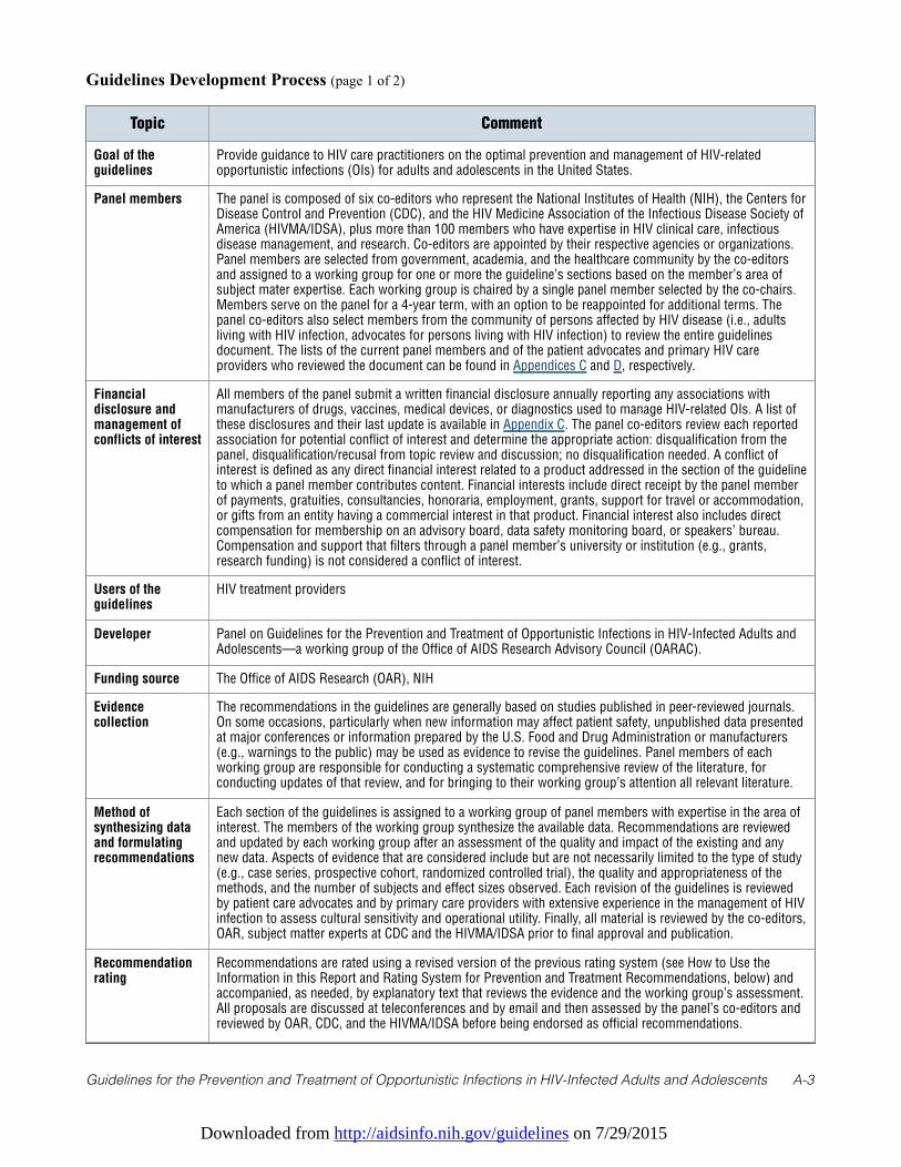
Guidelines Development Process (page 1 of 2)
Topic Comment
Goal of theguidelines
Provide guidance to HIV care practitioners on the optimal prevention and management of HIV-relatedopportunistic infections (OIs) for adults and adolescents in the United States.
Panel members The panel is composed of six co-editors who represent the National Institutes of Health (NIH), the Centers forDisease Control and Prevention (CDC), and the HIV Medicine Association of the Infectious Disease Society ofAmerica (HIVMA/IDSA), plus more than 100 members who have expertise in HIV clinical care, infectiousdisease management, and research. Co-editors are appointed by their respective agencies or organizations.Panel members are selected from government, academia, and the healthcare community by the co-editorsand assigned to a working group for one or more the guideline’s sections based on the member’s area ofsubject mater expertise. Each working group is chaired by a single panel member selected by the co-chairs.Members serve on the panel for a 4-year term, with an option to be reappointed for additional terms. Thepanel co-editors also select members from the community of persons affected by HIV disease (i.e., adultsliving with HIV infection, advocates for persons living with HIV infection) to review the entire guidelinesdocument. The lists of the current panel members and of the patient advocates and primary HIV careproviders who reviewed the document can be found in Appendices C and D, respectively.
Financialdisclosure andmanagement ofconflicts of interest
All members of the panel submit a written financial disclosure annually reporting any associations withmanufacturers of drugs, vaccines, medical devices, or diagnostics used to manage HIV-related OIs. A list ofthese disclosures and their last update is available in Appendix C. The panel co-editors review each reportedassociation for potential conflict of interest and determine the appropriate action: disqualification from thepanel, disqualification/recusal from topic review and discussion; no disqualification needed. A conflict ofinterest is defined as any direct financial interest related to a product addressed in the section of the guidelineto which a panel member contributes content. Financial interests include direct receipt by the panel memberof payments, gratuities, consultancies, honoraria, employment, grants, support for travel or accommodation,or gifts from an entity having a commercial interest in that product. Financial interest also includes directcompensation for membership on an advisory board, data safety monitoring board, or speakers’ bureau.Compensation and support that filters through a panel member’s university or institution (e.g., grants,research funding) is not considered a conflict of interest.
Users of theguidelines
HIV treatment providers
Developer Panel on Guidelines for the Prevention and Treatment of Opportunistic Infections in HIV-Infected Adults andAdolescents—a working group of the Office of AIDS Research Advisory Council (OARAC).
Funding source The Office of AIDS Research (OAR), NIH
Evidencecollection
The recommendations in the guidelines are generally based on studies published in peer-reviewed journals.On some occasions, particularly when new information may affect patient safety, unpublished data presentedat major conferences or information prepared by the U.S. Food and Drug Administration or manufacturers(e.g., warnings to the public) may be used as evidence to revise the guidelines. Panel members of eachworking group are responsible for conducting a systematic comprehensive review of the literature, forconducting updates of that review, and for bringing to their working group’s attention all relevant literature.
Method ofsynthesizing dataand formulatingrecommendations
Each section of the guidelines is assigned to a working group of panel members with expertise in the area ofinterest. The members of the working group synthesize the available data. Recommendations are reviewedand updated by each working group after an assessment of the quality and impact of the existing and anynew data. Aspects of evidence that are considered include but are not necessarily limited to the type of study(e.g., case series, prospective cohort, randomized controlled trial), the quality and appropriateness of themethods, and the number of subjects and effect sizes observed. Each revision of the guidelines is reviewedby patient care advocates and by primary care providers with extensive experience in the management of HIVinfection to assess cultural sensitivity and operational utility. Finally, all material is reviewed by the co-editors,OAR, subject matter experts at CDC and the HIVMA/IDSA prior to final approval and publication.
Recommendationrating
Recommendations are rated using a revised version of the previous rating system (see How to Use theInformation in this Report and Rating System for Prevention and Treatment Recommendations, below) andaccompanied, as needed, by explanatory text that reviews the evidence and the working group’s assessment.All proposals are discussed at teleconferences and by email and then assessed by the panel’s co-editors andreviewed by OAR, CDC, and the HIVMA/IDSA before being endorsed as official recommendations.
Downloaded from http://aidsinfo.nih.gov/guidelines on 7/29/2015
Guidelines for the Prevention and Treatment of Opportunistic Infections in HIV-Infected Adults and Adolescents A-4
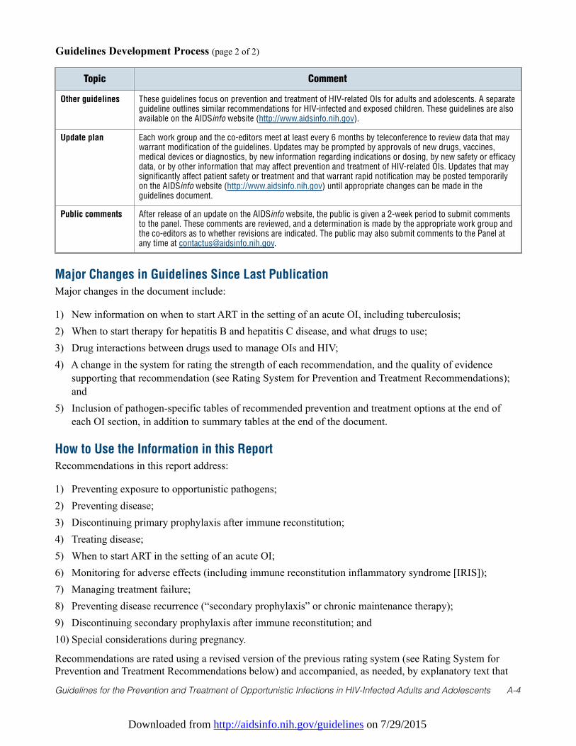
Guidelines Development Process (page 2 of 2)
Topic Comment
Other guidelines These guidelines focus on prevention and treatment of HIV-related OIs for adults and adolescents. A separateguideline outlines similar recommendations for HIV-infected and exposed children. These guidelines are alsoavailable on the AIDSinfo website (http://www.aidsinfo.nih.gov).
Update plan Each work group and the co-editors meet at least every 6 months by teleconference to review data that maywarrant modification of the guidelines. Updates may be prompted by approvals of new drugs, vaccines,medical devices or diagnostics, by new information regarding indications or dosing, by new safety or efficacydata, or by other information that may affect prevention and treatment of HIV-related OIs. Updates that maysignificantly affect patient safety or treatment and that warrant rapid notification may be posted temporarilyon the AIDSinfo website (http://www.aidsinfo.nih.gov) until appropriate changes can be made in theguidelines document.
Public comments After release of an update on the AIDSinfo website, the public is given a 2-week period to submit commentsto the panel. These comments are reviewed, and a determination is made by the appropriate work group andthe co-editors as to whether revisions are indicated. The public may also submit comments to the Panel atany time at [email protected].
Major Changes in Guidelines Since Last Publication
Major changes in the document include:
1) New information on when to start ART in the setting of an acute OI, including tuberculosis; 2) When to start therapy for hepatitis B and hepatitis C disease, and what drugs to use;3) Drug interactions between drugs used to manage OIs and HIV;4) A change in the system for rating the strength of each recommendation, and the quality of evidence
supporting that recommendation (see Rating System for Prevention and Treatment Recommendations);and
5) Inclusion of pathogen-specific tables of recommended prevention and treatment options at the end ofeach OI section, in addition to summary tables at the end of the document.
How to Use the Information in this Report
Recommendations in this report address:
1) Preventing exposure to opportunistic pathogens; 2) Preventing disease; 3) Discontinuing primary prophylaxis after immune reconstitution;4) Treating disease; 5) When to start ART in the setting of an acute OI; 6) Monitoring for adverse effects (including immune reconstitution inflammatory syndrome [IRIS]);7) Managing treatment failure;8) Preventing disease recurrence (“secondary prophylaxis” or chronic maintenance therapy);9) Discontinuing secondary prophylaxis after immune reconstitution; and 10) Special considerations during pregnancy.
Recommendations are rated using a revised version of the previous rating system (see Rating System forPrevention and Treatment Recommendations below) and accompanied, as needed, by explanatory text that
Downloaded from http://aidsinfo.nih.gov/guidelines on 7/29/2015
Guidelines for the Prevention and Treatment of Opportunistic Infections in HIV-Infected Adults and Adolescents A-5
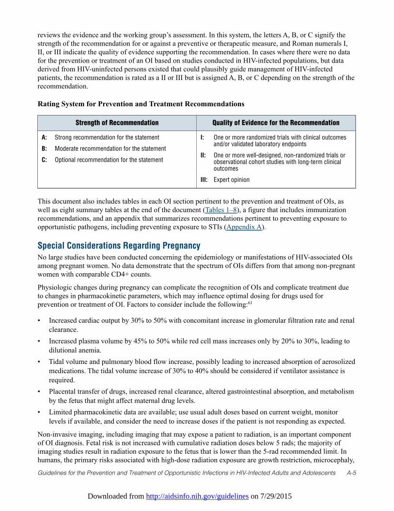
reviews the evidence and the working group’s assessment. In this system, the letters A, B, or C signify thestrength of the recommendation for or against a preventive or therapeutic measure, and Roman numerals I,II, or III indicate the quality of evidence supporting the recommendation. In cases where there were no datafor the prevention or treatment of an OI based on studies conducted in HIV-infected populations, but dataderived from HIV-uninfected persons existed that could plausibly guide management of HIV-infectedpatients, the recommendation is rated as a II or III but is assigned A, B, or C depending on the strength of therecommendation.
Rating System for Prevention and Treatment Recommendations
Strength of Recommendation Quality of Evidence for the Recommendation
A: Strong recommendation for the statement
B: Moderate recommendation for the statement
C: Optional recommendation for the statement
I: One or more randomized trials with clinical outcomesand/or validated laboratory endpoints
II: One or more well-designed, non-randomized trials orobservational cohort studies with long-term clinicaloutcomes
III: Expert opinion
This document also includes tables in each OI section pertinent to the prevention and treatment of OIs, aswell as eight summary tables at the end of the document (Tables 1–8), a figure that includes immunizationrecommendations, and an appendix that summarizes recommendations pertinent to preventing exposure toopportunistic pathogens, including preventing exposure to STIs (Appendix A).
Special Considerations Regarding Pregnancy
No large studies have been conducted concerning the epidemiology or manifestations of HIV-associated OIsamong pregnant women. No data demonstrate that the spectrum of OIs differs from that among non-pregnantwomen with comparable CD4+ counts.
Physiologic changes during pregnancy can complicate the recognition of OIs and complicate treatment dueto changes in pharmacokinetic parameters, which may influence optimal dosing for drugs used forprevention or treatment of OI. Factors to consider include the following:61
• Increased cardiac output by 30% to 50% with concomitant increase in glomerular filtration rate and renalclearance.
• Increased plasma volume by 45% to 50% while red cell mass increases only by 20% to 30%, leading todilutional anemia.
• Tidal volume and pulmonary blood flow increase, possibly leading to increased absorption of aerosolizedmedications. The tidal volume increase of 30% to 40% should be considered if ventilator assistance isrequired.
• Placental transfer of drugs, increased renal clearance, altered gastrointestinal absorption, and metabolismby the fetus that might affect maternal drug levels.
• Limited pharmacokinetic data are available; use usual adult doses based on current weight, monitorlevels if available, and consider the need to increase doses if the patient is not responding as expected.
Non-invasive imaging, including imaging that may expose a patient to radiation, is an important componentof OI diagnosis. Fetal risk is not increased with cumulative radiation doses below 5 rads; the majority ofimaging studies result in radiation exposure to the fetus that is lower than the 5-rad recommended limit. Inhumans, the primary risks associated with high-dose radiation exposure are growth restriction, microcephaly,
Downloaded from http://aidsinfo.nih.gov/guidelines on 7/29/2015
Guidelines for the Prevention and Treatment of Opportunistic Infections in HIV-Infected Adults and Adolescents A-6
and developmental disabilities. The most vulnerable period is 8 to 15 menstrual weeks of gestation, withminimal risk before 8 weeks and after 25 weeks. The apparent threshold for development of mentalretardation is 20 to 40 rads, with risk of more serious mental retardation increasing linearly with increasingexposure above this level. Among children, risk for carcinogenesis might be increased approximately 1 per1000 or less per rad of in utero radiation exposure.62 Therefore, pregnancy should not preclude usualdiagnostic evaluation when an OI is suspected.63 Abdominal shielding should be used when feasible tofurther limit radiation exposure to the fetus. Experience with use of magnetic resonance imaging (MRI) inpregnancy is limited, but no adverse fetal effects have been noted.64
Other procedures necessary for diagnosis of suspected OIs should be performed in pregnancy as indicated fornon-pregnant patients. A pregnant woman who is >20 weeks of gestation should not lie flat on her back butshould have her right hip elevated with a wedge to displace the uterus off the great vessels and preventsupine hypotension. Oxygenation should be monitored when pregnant patients are positioned such thatventilation or perfusion might be compromised.
In the United States, pregnancy is an indication to start antiretroviral therapy if the HIV-infected woman isnot already on therapy. A decision to defer therapy based on a current or recent OI should be made on thesame basis as for non-pregnant individuals supplemented by consultation with the obstetrician regardingfactors unique to each individual pregnancy.
After first-trimester exposure to agents of uncertain teratogenic potential, including many of the anti-infective agents described in this guideline, an ultrasound should be conducted every 4 to 6 weeks in thethird trimester to assess fetal growth and fluid volume, with antepartum testing if growth lag or decreasedfluid are noted.
References
1. Kaplan JE, Masur H, Holmes KK, et al. USPHS/IDSA guidelines for the prevention of opportunistic infections inpersons infected with human immunodeficiency virus: introduction. USPHS/IDSA Prevention of OpportunisticInfections Working Group. Clin Infect Dis. Aug 1995;21 Suppl 1:S1-11. Available athttp://www.ncbi.nlm.nih.gov/pubmed/8547495.
2. Kaplan JE, Masur H, Jaffe HW, Holmes KK. Reducing the impact of opportunistic infections in patients with HIVinfection. New guidelines. JAMA. Jul 26 1995;274(4):347-348. Available athttp://www.ncbi.nlm.nih.gov/pubmed/7609267.
3. Walensky RP, Paltiel AD, Losina E, et al. The survival benefits of AIDS treatment in the United States. J Infect Dis. Jul1 2006;194(1):11-19. Available at http://www.ncbi.nlm.nih.gov/pubmed/16741877.
4. Palella FJ, Jr., Delaney KM, Moorman AC, et al. Declining morbidity and mortality among patients with advancedhuman immunodeficiency virus infection. HIV Outpatient Study Investigators. N Engl J Med. Mar 261998;338(13):853-860. Available at http://www.ncbi.nlm.nih.gov/pubmed/9516219.
5. Detels R, Munoz A, McFarlane G, et al. Effectiveness of potent antiretroviral therapy on time to AIDS and death in menwith known HIV infection duration. Multicenter AIDS Cohort Study Investigators. JAMA. Nov 4 1998;280(17):1497-1503. Available at http://www.ncbi.nlm.nih.gov/pubmed/9809730.
6. Jones JL, Hanson DL, Dworkin MS, et al. Surveillance for AIDS-defining opportunistic illnesses, 1992-1997. MMWR.CDC surveillance summaries: Morbidity and mortality weekly report. CDC surveillance summaries / Centers forDisease Control. Apr 16 1999;48(2):1-22. Available at http://www.ncbi.nlm.nih.gov/pubmed/12412613.
7. Mocroft A, Vella S, Benfield TL, et al. Changing patterns of mortality across Europe in patients infected with HIV-1.EuroSIDA Study Group. Lancet. Nov 28 1998;352(9142):1725-1730. Available athttp://www.ncbi.nlm.nih.gov/pubmed/9848347.
8. McNaghten AD, Hanson DL, Jones JL, Dworkin MS, Ward JW. Effects of antiretroviral therapy and opportunisticillness primary chemoprophylaxis on survival after AIDS diagnosis. Adult/Adolescent Spectrum of Disease Group.AIDS. Sep 10 1999;13(13):1687-1695. Available at http://www.ncbi.nlm.nih.gov/pubmed/10509570.
9. Miller V, Mocroft A, Reiss P, et al. Relations among CD4 lymphocyte count nadir, antiretroviral therapy, and HIV-1
Downloaded from http://aidsinfo.nih.gov/guidelines on 7/29/2015
Guidelines for the Prevention and Treatment of Opportunistic Infections in HIV-Infected Adults and Adolescents A-7
disease progression: results from the EuroSIDA study. Ann Intern Med. Apr 6 1999;130(7):570-577. Available athttp://www.ncbi.nlm.nih.gov/pubmed/10189326.
10. Dore GJ, Li Y, McDonald A, Ree H, Kaldor JM, National HIVSC. Impact of highly active antiretroviral therapy onindividual AIDS-defining illness incidence and survival in Australia. J Acquir Immune Defic Syndr. Apr 12002;29(4):388-395. Available at http://www.ncbi.nlm.nih.gov/pubmed/11917244.
11. Centers for Disease C, Prevention. Vital signs: HIV prevention through care and treatment—United States. MMWRMorb Mortal Wkly Rep. Dec 2 2011;60(47):1618-1623. Available at http://www.ncbi.nlm.nih.gov/pubmed/22129997.
12. Campsmith ML, Rhodes PH, Hall HI, Green TA. Undiagnosed HIV prevalence among adults and adolescents in theUnited States at the end of 2006. J Acquir Immune Defic Syndr. Apr 2010;53(5):619-624. Available athttp://www.ncbi.nlm.nih.gov/pubmed/19838124.
13. Seal PS, Jackson DA, Chamot E, et al. Temporal trends in presentation for outpatient HIV medical care 2000-2010:implications for short-term mortality. J Gen Intern Med. Jul 2011;26(7):745-750. Available athttp://www.ncbi.nlm.nih.gov/pubmed/21465301.
14. Perbost I, Malafronte B, Pradier C, et al. In the era of highly active antiretroviral therapy, why are HIV-infected patientsstill admitted to hospital for an inaugural opportunistic infection? HIV Med. Jul 2005;6(4):232-239. Available athttp://www.ncbi.nlm.nih.gov/pubmed/16011527.
15. Palacios R, Hidalgo A, Reina C, de la Torre M, Marquez M, Santos J. Effect of antiretroviral therapy on admissions ofHIV-infected patients to an intensive care unit. HIV Med. Apr 2006;7(3):193-196. Available athttp://www.ncbi.nlm.nih.gov/pubmed/16494634.
16. Gardner EM, McLees MP, Steiner JF, Del Rio C, Burman WJ. The spectrum of engagement in HIV care and itsrelevance to test-and-treat strategies for prevention of HIV infection. Clin Infect Dis. Mar 15 2011;52(6):793-800.Available at http://www.ncbi.nlm.nih.gov/pubmed/21367734.
17. Greenberg AE, Hader SL, Masur H, Young AT, Skillicorn J, Dieffenbach CW. Fighting HIV/AIDS in Washington, D.C.Health affairs. Nov-Dec 2009;28(6):1677-1687. Available at http://www.ncbi.nlm.nih.gov/pubmed/19887408.
18. Gebo KA, Fleishman JA, Reilly ED, Moore RD, Network HIVR. High rates of primary Mycobacterium avium complexand Pneumocystis jiroveci prophylaxis in the United States. Medical care. Sep 2005;43(9 Suppl):III23-30. Available athttp://www.ncbi.nlm.nih.gov/pubmed/16116306.
19. Bonnet F, Lewden C, May T, et al. Opportunistic infections as causes of death in HIV-infected patients in the HAARTera in France. Scandinavian journal of infectious diseases. 2005;37(6-7):482-487. Available athttp://www.ncbi.nlm.nih.gov/pubmed/16089023.
20. Teshale EH, Hanson DL, Wolfe MI, et al. Reasons for lack of appropriate receipt of primary Pneumocystis jirovecipneumonia prophylaxis among HIV-infected persons receiving treatment in the United States: 1994-2003. Clin InfectDis. Mar 15 2007;44(6):879-883. Available at http://www.ncbi.nlm.nih.gov/pubmed/17304464.
21. Gebo KA, Fleishman JA, Moore RD. Hospitalizations for metabolic conditions, opportunistic infections, and injectiondrug use among HIV patients: trends between 1996 and 2000 in 12 states. J Acquir Immune Defic Syndr. Dec 152005;40(5):609-616. Available at http://www.ncbi.nlm.nih.gov/pubmed/16284539.
22. Betz ME, Gebo KA, Barber E, et al. Patterns of diagnoses in hospital admissions in a multistate cohort of HIV-positiveadults in 2001. Medical care. Sep 2005;43(9 Suppl):III3-14. Available athttp://www.ncbi.nlm.nih.gov/pubmed/16116304.
23. Moorman AC, Buchacz K, Richardson JT, al. e. Temporal trends in hospitalizations and hospital-associated diagnosesin the HIV Outpatient Study (HOPS) 1994-2002. In: XVI International AIDS Conference; August 13-18, 2006; Toronto,Canada. Abstract MOPE0071.
24. Louie JK, Hsu LC, Osmond DH, Katz MH, Schwarcz SK. Trends in causes of death among persons with acquiredimmunodeficiency syndrome in the era of highly active antiretroviral therapy, San Francisco, 1994-1998. J Infect Dis.Oct 1 2002;186(7):1023-1027. Available at http://www.ncbi.nlm.nih.gov/pubmed/12232845.
25. Palella FJ, Jr., Baker RK, Moorman AC, et al. Mortality in the highly active antiretroviral therapy era: changing causesof death and disease in the HIV outpatient study. J Acquir Immune Defic Syndr. Sep 2006;43(1):27-34. Available athttp://www.ncbi.nlm.nih.gov/pubmed/16878047.
26. Smit C, Geskus R, Walker S, et al. Effective therapy has altered the spectrum of cause-specific mortality following HIVseroconversion. AIDS. Mar 21 2006;20(5):741-749. Available at http://www.ncbi.nlm.nih.gov/pubmed/16514305.
Downloaded from http://aidsinfo.nih.gov/guidelines on 7/29/2015
Guidelines for the Prevention and Treatment of Opportunistic Infections in HIV-Infected Adults and Adolescents A-8
27. Buchacz K, Baker RK, Moorman AC, et al. Rates of hospitalizations and associated diagnoses in a large multisitecohort of HIV patients in the United States, 1994-2005. AIDS. Jul 11 2008;22(11):1345-1354. Available athttp://www.ncbi.nlm.nih.gov/pubmed/18580614.
28. Buchacz K, Baker RK, Palella FJ, Jr., et al. AIDS-defining opportunistic illnesses in US patients, 1994-2007: a cohortstudy. AIDS. Jun 19 2010;24(10):1549-1559. Available at http://www.ncbi.nlm.nih.gov/pubmed/20502317.
29. Lawn SD, Butera ST, Folks TM. Contribution of immune activation to the pathogenesis and transmission of humanimmunodeficiency virus type 1 infection. Clin Microbiol Rev. Oct 2001;14(4):753-777, table of contents. Available athttp://www.ncbi.nlm.nih.gov/pubmed/11585784.
30. Toossi Z, Mayanja-Kizza H, Hirsch CS, et al. Impact of tuberculosis (TB) on HIV-1 activity in dually infected patients.Clinical and experimental immunology. Feb 2001;123(2):233-238. Available athttp://www.ncbi.nlm.nih.gov/pubmed/11207653.
31. Sadiq ST, McSorley J, Copas AJ, et al. The effects of early syphilis on CD4 counts and HIV-1 RNA viral loads in bloodand semen. Sexually transmitted infections. Oct 2005;81(5):380-385. Available athttp://www.ncbi.nlm.nih.gov/pubmed/16199736.
32. Bentwich Z. Concurrent infections that rise the HIV viral load. Journal of HIV Therapy. Aug 2003;8(3):72-75.Available at http://www.ncbi.nlm.nih.gov/pubmed/12951545.
33. Kublin JG, Patnaik P, Jere CS, et al. Effect of Plasmodium falciparum malaria on concentration of HIV-1-RNA in theblood of adults in rural Malawi: a prospective cohort study. Lancet. Jan 15-21 2005;365(9455):233-240. Available athttp://www.ncbi.nlm.nih.gov/pubmed/15652606.
34. Abu-Raddad LJ, Patnaik P, Kublin JG. Dual infection with HIV and malaria fuels the spread of both diseases in sub-Saharan Africa. Science. Dec 8 2006;314(5805):1603-1606. Available at http://www.ncbi.nlm.nih.gov/pubmed/17158329.
35. Quinn TC, Wawer MJ, Sewankambo N, et al. Viral load and heterosexual transmission of human immunodeficiencyvirus type 1. Rakai Project Study Group. N Engl J Med. Mar 30 2000;342(13):921-929. Available athttp://www.ncbi.nlm.nih.gov/pubmed/10738050.
36. DiRienzo AG, van Der Horst C, Finkelstein DM, Frame P, Bozzette SA, Tashima KT. Efficacy of trimethoprim-sulfamethoxazole for the prevention of bacterial infections in a randomized prophylaxis trial of patients with advancedHIV infection. AIDS research and human retroviruses. Jan 20 2002;18(2):89-94. Available athttp://www.ncbi.nlm.nih.gov/pubmed/11839141.
37. Wiktor SZ, Sassan-Morokro M, Grant AD, et al. Efficacy of trimethoprim-sulphamethoxazole prophylaxis to decreasemorbidity and mortality in HIV-1-infected patients with tuberculosis in Abidjan, Cote d'Ivoire: a randomised controlledtrial. Lancet. May 1 1999;353(9163):1469-1475. Available at http://www.ncbi.nlm.nih.gov/pubmed/10232312.
38. Whalen CC, Johnson JL, Okwera A, et al. A trial of three regimens to prevent tuberculosis in Ugandan adults infectedwith the human immunodeficiency virus. Uganda-Case Western Reserve University Research Collaboration. N Engl JMed. Sep 18 1997;337(12):801-808. Available at http://www.ncbi.nlm.nih.gov/pubmed/9295239.
39. Anglaret X, Chene G, Attia A, et al. Early chemoprophylaxis with trimethoprim-sulphamethoxazole for HIV-1-infectedadults in Abidjan, Cote d'Ivoire: a randomised trial. Cotrimo-CI Study Group. Lancet. May 1 1999;353(9163):1463-1468. Available at http://www.ncbi.nlm.nih.gov/pubmed/10232311.
40. Chintu C, Bhat GJ, Walker AS, et al. Co-trimoxazole as prophylaxis against opportunistic infections in HIV-infectedZambian children (CHAP): a double-blind randomised placebo-controlled trial. Lancet. Nov 20-262004;364(9448):1865-1871. Available at http://www.ncbi.nlm.nih.gov/pubmed/15555666.
41. Centers for Disease C. Guidelines for prophylaxis against Pneumocystis carinii pneumonia for persons infected withhuman immunodeficiency virus. MMWR Morb Mortal Wkly Rep. Jun 16 1989;38 Suppl 5(Suppl 5):1-9. Available athttp://www.ncbi.nlm.nih.gov/pubmed/2524643.
42. Masur H. Recommendations on prophylaxis and therapy for disseminated Mycobacterium avium complex disease inpatients infected with the human immunodeficiency virus. Public Health Service Task Force on Prophylaxis andTherapy for Mycobacterium avium Complex. N Engl J Med. Sep 16 1993;329(12):898-904. Available athttp://www.ncbi.nlm.nih.gov/pubmed/8395019.
43. USPHS/IDSA guidelines for the prevention of opportunistic infections in persons infected with humanimmunodeficiency virus: a summary. MMWR Recomm Rep. Jul 14 1995;44(RR-8):1-34. Available athttp://www.ncbi.nlm.nih.gov/pubmed/7565547.
Downloaded from http://aidsinfo.nih.gov/guidelines on 7/29/2015
Guidelines for the Prevention and Treatment of Opportunistic Infections in HIV-Infected Adults and Adolescents A-9
44. 1997 USPHS/IDSA guidelines for the prevention of opportunistic infections in persons infected with humanimmunodeficiency virus. USPHS/IDSA Prevention of Opportunistic Infections Working Group. MMWR Recomm Rep.Jun 27 1997;46(RR-12):1-46. Available at http://www.ncbi.nlm.nih.gov/pubmed/9214702.
45. 1999 USPHS/IDSA guidelines for the prevention of opportunistic infections in persons infected with humanimmunodeficiency virus. U.S. Public Health Service (USPHS) and Infectious Diseases Society of America (IDSA). MMWRRecomm Rep. Aug 20 1999;48(RR-10):1-59, 61-56. Available at http://www.ncbi.nlm.nih.gov/pubmed/10499670.
46. Kaplan JE, Masur H, Holmes KK, Usphs, Infectious Disease Society of A. Guidelines for preventing opportunisticinfections among HIV-infected persons—2002. Recommendations of the U.S. Public Health Service and the InfectiousDiseases Society of America. MMWR Recomm Rep. Jun 14 2002;51(RR-8):1-52. Available athttp://www.ncbi.nlm.nih.gov/pubmed/12081007.
47. USPHS/IDSA guidelines for the prevention of opportunistic infections in persons infected with human immunodeficiencyvirus: disease-specific recommendations. USPHS/IDSA Prevention of Opportunistic Infections Working Group. Clin InfectDis. Aug 1995;21 Suppl 1:S32-43. Available at http://www.ncbi.nlm.nih.gov/pubmed/8547510.
48. 1997 USPHS/IDSA guidelines for the prevention of opportunistic infections in persons infected with humanimmunodeficiency virus: disease-specific recommendations. USPHS/IDSA Prevention of Opportunistic InfectionsWorking Group. US Public Health Services/Infectious Diseases Society of America. Clin Infect Dis. Oct 1997;25 Suppl3:S313-335. Available at http://www.ncbi.nlm.nih.gov/pubmed/9356832.
49. 1999 USPHS/IDSA guidelines for the prevention of opportunistic infections in persons infected with humanimmunodeficiency virus. Clin Infect Dis. Apr 2000;30 Suppl 1:S29-65. Available athttp://www.ncbi.nlm.nih.gov/pubmed/10770913.
50. USPHS/IDSA guidelines for the prevention of opportunistic infections in persons infected with humanimmunodeficiency virus: a summary. Ann Intern Med. Feb 1 1996;124(3):349-368. Available athttp://www.ncbi.nlm.nih.gov/pubmed/8554235.
51. 1997 USPHS/IDSA guidelines for the prevention of opportunistic infections in persons infected with humanimmunodeficiency virus. Ann Intern Med. Nov 15 1997;127(10):922-946. Available athttp://www.ncbi.nlm.nih.gov/pubmed/9382373.
52. 1997 USPHS/IDSA guidelines for the prevention of opportunistic infections in persons infected with HIV: Part I.Prevention of exposure. U.S. Department of Health and Human Services, Public Health Service, Centers for DiseaseControl and Prevention. American family physician. Sep 1 1997;56(3):823-834. Available athttp://www.ncbi.nlm.nih.gov/pubmed/9301575.
53. 1999 USPHS/IDSA guidelines for the prevention of opportunistic infections in persons infected with HIV: part I.Prevention of exposure. American family physician. Jan 1 2000;61(1):163-174. Available athttp://www.ncbi.nlm.nih.gov/pubmed/10643957.
54. Antiretroviral therapy and medical management of pediatric HIV infection and 1997 USPHS/IDSA report on theprevention of opportunistic infections in persons infected with human immunodeficiency virus. Pediatrics. Oct1998;102(4 Pt 2):999-1085. Available at http://www.ncbi.nlm.nih.gov/pubmed/9826994.
55. Kaplan JE, Masur H, Jaffe HW, Holmes KK. Preventing opportunistic infections in persons infected with HIV: 1997guidelines. JAMA. Jul 23-30 1997;278(4):337-338. Available at http://www.ncbi.nlm.nih.gov/pubmed/9228443.
56. Brooks JT, Kaplan JE, Masur H. What's new in the 2009 US guidelines for prevention and treatment of opportunisticinfections among adults and adolescents with HIV? Top HIV Med. Jul-Aug 2009;17(3):109-114. Available athttp://www.ncbi.nlm.nih.gov/pubmed/19675369.
57. Benson CA, Kaplan JE, Masur H, et al. Treating opportunistic infections among HIV-infected adults and adolescents:recommendations from CDC, the National Institutes of Health, and the HIV Medicine Association/Infectious DiseasesSociety of America. MMWR Recomm Rep. Dec 17 2004;53(RR-15):1-112. Available athttp://www.ncbi.nlm.nih.gov/pubmed/15841069.
58. Mofenson LM, Oleske J, Serchuck L, et al. Treating opportunistic infections among HIV-exposed and infected children:recommendations from CDC, the National Institutes of Health, and the Infectious Diseases Society of America. MMWRRecomm Rep. Dec 3 2004;53(RR-14):1-92. Available at http://www.ncbi.nlm.nih.gov/pubmed/15577752.
59. Kaplan JE, Benson C, Holmes KH, et al. Guidelines for prevention and treatment of opportunistic infections in HIV-infected adults and adolescents: recommendations from CDC, the National Institutes of Health, and the HIV Medicine
Downloaded from http://aidsinfo.nih.gov/guidelines on 7/29/2015
Guidelines for the Prevention and Treatment of Opportunistic Infections in HIV-Infected Adults and Adolescents A-10
Association of the Infectious Diseases Society of America. MMWR Recomm Rep. Apr 10 2009;58(RR-4):1-207; quizCE201-204. Available at http://www.ncbi.nlm.nih.gov/pubmed/19357635.
60. Mofenson LM, Brady MT, Danner SP, et al. Guidelines for the Prevention and Treatment of Opportunistic Infectionsamong HIV-exposed and HIV-infected children: recommendations from CDC, the National Institutes of Health, theHIV Medicine Association of the Infectious Diseases Society of America, the Pediatric Infectious Diseases Society, andthe American Academy of Pediatrics. MMWR Recomm Rep. Sep 4 2009;58(RR-11):1-166. Available athttp://www.ncbi.nlm.nih.gov/pubmed/19730409.
61. Cruickshank DP, Wigton TR, Hays PM. Maternal physiology in pregnancy. In: Gabbe SG, Neibyl JR, Simpson JL, eds.Obstetrics: Normal and Problem Pregnancies. New York, NY: Churchchill Livingstone, 1996.
62. ACOG Committee on Obstetric Practice. ACOG Committee Opinion. Number 299, September 2004 (replaces No. 158,September 1995). Guidelines for diagnostic imaging during pregnancy. Obstet Gynecol. Sep 2004;104(3):647-651.Available at http://www.ncbi.nlm.nih.gov/pubmed/15339791.
63. Toppenberg KS, Hill DA, Miller DP. Safety of radiographic imaging during pregnancy. American family physician. Apr1 1999;59(7):1813-1818, 1820. Available at http://www.ncbi.nlm.nih.gov/pubmed/10208701.
64. Adelstein SJ. Administered radionuclides in pregnancy. Teratology. Apr 1999;59(4):236-239. Available athttp://www.ncbi.nlm.nih.gov/pubmed/10331526.
Downloaded from http://aidsinfo.nih.gov/guidelines on 7/29/2015
Guidelines for Prevention and Treatment of Opportunistic Infections in HIV-Infected Adults and Adolescents B-1
Pneumocystis Pneumonia (Last updated October 28, 2014; last reviewedOctober 28, 2014)
Epidemiology
Pneumocystis pneumonia (PCP) is caused by Pneumocystis jirovecii, a ubiquitous organism that is classifiedas a fungus but also shares biologic characteristics with protozoa. The taxonomy of the organism has beenchanged; Pneumocystis carinii now refers only to the Pneumocystis that infects rats, and P. jirovecii refers tothe distinct species that infects humans. The abbreviation PCP is still used to designate Pneumocystispneumonia. Initial infection with P. jirovecii usually occurs in early childhood; two-thirds of healthy childrenhave antibodies to P. jirovecii by ages 2 to 4 years.1
Rodent studies and case clusters in immunosuppressed patients suggest that Pneumocystis spreads by theairborne route. Disease probably occurs by new acquisition of infection and by reactivation of latentinfection.2-11 Before the widespread use of PCP prophylaxis and antiretroviral therapy (ART), PCP occurredin 70% to 80% of patients with AIDS;12 the course of treated PCP was associated with a 20% to 40%mortality rate in individuals with profound immunosuppression. Approximately 90% of PCP cases occurredin patients with CD4 T-lymphocyte (CD4 cell) counts <200 cells/mm3. Other factors associated with a higherrisk of PCP included CD4 cell percentage <14%, previous episodes of PCP, oral thrush, recurrent bacterialpneumonia, unintentional weight loss, and higher plasma HIV RNA levels.13,14
The incidence of PCP has declined substantially with widespread use of PCP prophylaxis and ART; recentincidence among patients with AIDS in Western Europe and the United States is <1 case per 100 person-years.15 Most cases occur in patients who are unaware of their HIV infection or are not receiving ongoingcare for HIV,16 and in those with advanced immunosuppression (CD4 counts <100 cells/mm3).17
Clinical Manifestations
In HIV-infected patients, the most common manifestations of PCP are subacute onset of progressive dyspnea,fever, non-productive cough, and chest discomfort that worsens within days to weeks. The fulminantpneumonia observed in patients who are not infected with HIV is less common.18,19
In mild cases, pulmonary examination usually is normal at rest. With exertion, tachypnea, tachycardia, anddiffuse dry (cellophane) rales may be observed.19 Oral thrush is a common co infection. Fever is apparent inmost cases and may be the predominant symptom in some patients. Extrapulmonary disease is rare but canoccur in any organ and has been associated with use of aerosolized pentamidine prophylaxis.20
Hypoxemia, the most characteristic laboratory abnormality, can range from mild (room air arterial oxygen[pO2] ≥70 mm Hg or alveolar-arterial O2 difference, [A-a] DO2 <35 mm Hg) to moderate ([A-a] DO2 ≥35and <45 mm Hg) to severe ([A-a] DO2 ≥45 mm Hg). Oxygen desaturation with exercise is often abnormalbut is non-specific.21 Elevation of lactate dehydrogenase levels to >500 mg/dL is common but non-specific.22
Chest radiograph typically demonstrates diffuse, bilateral, symmetrical interstitial infiltrates emanating fromthe hila in a butterfly pattern;19 however, a chest radiograph may be normal in patients with early disease.23
Atypical radiographic presentations also occur, such as nodules, blebs and cysts, asymmetric disease, upperlobe localization, and pneumothorax. Spontaneous pneumothorax in a patient with HIV infection shouldraise the suspicion of PCP.24,25 Cavitation, intrathoracic adenopathy, and pleural effusion are uncommon inthe absence of other pulmonary pathogens or malignancy, and their presence may indicate an alternativediagnosis. Approximately 13% to 18% of patients with documented PCP have another concurrent cause ofpulmonary dysfunction, such as tuberculosis (TB), Kaposi sarcoma (KS), or bacterial pneumonia.26,27
Thin-section computed tomography (CT) demonstrating patchy ground-glass attenuation28,29 increases thelikelihood that a diagnostic study, such as bronchoscopy, will demonstrate PCP in patients with mild-to-moderate symptoms and normal chest radiograph and, therefore, may be useful as an adjunct.
Downloaded from http://aidsinfo.nih.gov/guidelines on 7/29/2015
Guidelines for the Prevention and Treatment of Opportunistic Infections in HIV-Infected Adults and Adolescents B-2
Diagnosis
Because clinical presentation, blood tests, and chest radiographs are not pathognomonic for PCP, andbecause the organism cannot be cultivated routinely, histopathologic or cytopathologic demonstration oforganisms in tissue, bronchoalveolar lavage (BAL) fluid, or induced sputum samples18,26,27,30 is required for adefinitive diagnosis. Spontaneously expectorated sputum has low sensitivity and should not be submitted tothe laboratory to diagnose PCP. Giemsa, Diff-Quik, and Wright stains detect both the cystic and trophicforms but do not stain the cyst wall; Gomori methenamine silver, Gram-Weigert, cresyl violet, and toluidineblue stain the cyst wall. Some laboratories prefer direct immunofluorescent staining. Previous studies ofstained respiratory tract samples obtained by various methods indicate the following relative diagnosticsensitivities: induced sputum <50% to >90% (the sensitivity depends on the pathogen load and specimenquality, while the specificity depends on the experience of the microbiologist or pathologist), bronchoscopywith BAL 90% to 99%, transbronchial biopsy 95% to 100%, and open lung biopsy 95% to 100%.
Polymerase chain reaction (PCR) is an emerging method for diagnosing PCP.31 The sensitivity of PCR forbronchoalveolar lavage appears to be high; the ability of PCR to distinguish colonization from disease is lessclear.31-34 1,3ß-D-glucan (a component of fungal cell walls) may be elevated in patients with PCP, but theassay’s sensitivity and specificity for establishing a PCP diagnosis are problematic,35,36 and other fungaldiseases can produce elevation.
Because certain processes produce similar clinical manifestations, a specific diagnosis of PCP should besought rather than relying on a presumptive diagnosis, especially in patients with moderate-to-severe disease.Treatment can be initiated before making a definitive diagnosis because organisms persist in clinicalspecimens for days or weeks after effective therapy is initiated.30
Preventing Exposure
Pneumocystis can be quantified in the air near patients with PCP,37 and multiple outbreaks, each caused by adistinct strain of Pneumocystis, have been documented among kidney transplant patients.5-11,38 Althoughthese strongly suggest that high-risk patients without PCP may benefit from isolation from other patientswith known PCP infection, data are insufficient to support isolation as standard practice (CIII).
Preventing Disease
Indication for Primary ProphylaxisHIV-infected adults and adolescents, including pregnant women and those on ART, should receivechemoprophylaxis against PCP if they have CD4 counts <200 cells/mm3 (AI) or a history of oropharyngealcandidiasis (AII).12,13,39 Persons who have a CD4 cell percentage of <14% or a history of an AIDS-definingillness, but who do not otherwise qualify, should be considered for prophylaxis (BII).12,13,39 Initiation ofchemoprophylaxis at CD4 counts between 200 and 250 cells/mm3 also should be considered when frequentmonitoring of CD4 counts, such as every 3 months, is impossible (BII).13 Patients receiving pyrimethamine-sulfadiazine for treatment or suppression of toxoplasmosis do not require additional prophylaxis for PCP (AII).40
Trimethoprim-sulfamethoxazole (TMP-SMX) is the recommended prophylactic agent (AI).39,41-43 Onedouble-strength tablet daily is the preferred regimen (AI), but one single-strength tablet daily43 also iseffective and may be better tolerated than the double-strength tablet (AI). One double-strength tablet threetimes weekly also is effective (BI).44 TMP-SMX at a dose of one double-strength tablet daily confers crossprotection against toxoplasmosis45 and many respiratory bacterial infections.41,46 Lower doses of TMP-SMXlikely also confer such protection. TMP-SMX chemoprophylaxis should be continued, if clinically feasible,in patients who have non life threatening adverse reactions. In those who discontinue TMP-SMX because ofa mild adverse reaction, re-institution should be considered after the reaction has resolved (AII). Therapyshould be permanently discontinued (with no rechallenge) in patients with life threatening adverse reactionsincluding possible or definite Stevens-Johnson syndrome or toxic epidermal necrolysis (TEN) (AIII).
Downloaded from http://aidsinfo.nih.gov/guidelines on 7/29/2015
Guidelines for the Prevention and Treatment of Opportunistic Infections in HIV-Infected Adults and Adolescents B-3
Patients who have experienced adverse events, including fever and rash, may better tolerate re-introductionof the drug if the dose is gradually increased (desensitization) according to published regimens (BI)47,48 or ifTMP-SMX is given at a reduced dose or frequency (CIII). As many as 70% of patients can tolerate such re-institution of therapy.46
For patients who cannot tolerate TMP-SMX, alternative prophylactic regimens include dapsone (BI),41 dapsoneplus pyrimethamine plus leucovorin (BI),49-51 aerosolized pentamidine administered with the Respirgard IInebulizer (manufactured by Marquest; Englewood, Colorado) (BI),42 and atovaquone (BI).52,53 Atovaquone isas effective as aerosolized pentamidine52 or dapsone53 but substantially more expensive than the otherregimens. For patients seropositive for Toxoplasma gondii who cannot tolerate TMP-SMX, recommendedalternatives for prophylaxis against both PCP and toxoplasmosis include dapsone plus pyrimethamine plusleucovorin (BI),49-51 or atovaquone with or without pyrimethamine plus leucovorin (CIII).
Oral pyrimethamine plus sulfadoxine also has activity against PCP.54-56 However, this combination isassociated with an increased risk of severe cutaneous reactions, including Stevens-Johnson syndrome,57 andthe long half-life of both pyrimethamine and sulfadoxine results in delayed clearance when the drug isstopped. Because TMP-SMX has superior safety, widespread availability, and is low cost, oralpyrimethamine plus sulfadoxine should not be used in the United States (AIII).
The following regimens cannot be recommended as alternatives because data regarding their efficacy forPCP prophylaxis are insufficient: • Aerosolized pentamidine administered by nebulization devices other than the Respirgard II nebulizer • Intermittently administered parenteral pentamidine • Oral clindamycin plus primaquine
Clinicians can consider using these agents, however, in situations in which the recommended agents cannotbe administered or are not tolerated (CIII).
Discontinuing Primary ProphylaxisPrimary Pneumocystis prophylaxis should be discontinued for adult and adolescent patients who haveresponded to ART with an increase in CD4 counts from <200 cells/mm3 to ≥200 cells/mm3 for >3 months(AI). In observational and randomized studies supporting this recommendation, most patients had CD4counts >200 cells/mm3 for more than 3 months before discontinuing PCP prophylaxis.58-67 The median CD4count at the time prophylaxis was discontinued was >300 cells/mm3, most patients had a CD4 cell percentage≥14%, and many had sustained suppression of HIV plasma RNA levels below detection limits for the assayemployed. Median follow-up was 6 to 19 months.
Discontinuing primary prophylaxis in these patients is recommended because its preventive benefits arelimited to PCP, toxoplasmosis, and bacterial infections;60,66 stopping the drugs reduces pill burden, cost, andthe potential for drug toxicity, drug interactions, and selection of drug-resistant pathogens. Prophylaxisshould be reintroduced if the CD4 count decreases to <200 cells/mm3 (AIII).
A combined analysis of 12 European cohorts68 and a case series69 found a low incidence of PCP in patients withCD4 counts between 100 and 200 cells/mm3, who were receiving ART and had HIV plasma viral loads <50 to400 copies/mL, and who had stopped or never received PCP prophylaxis, suggesting that primary PCPprophylaxis can be safely discontinued in selected patients with CD4 counts 100 to 200 cells/mm3 and HIVplasma RNA levels below limits of detection with commercial assays. Data on which to base recommendationsfor this approach are inadequate, but some experts believe it is reasonable and recommend it for their patients.
Treating Disease
TMP-SMX is the treatment of choice for PCP (AI).70,71 The dose must be adjusted for abnormal renalfunction. Multiple randomized clinical trials indicate that TMP-SMX is as effective as parenteral
Downloaded from http://aidsinfo.nih.gov/guidelines on 7/29/2015
Guidelines for the Prevention and Treatment of Opportunistic Infections in HIV-Infected Adults and Adolescents B-4
pentamidine and more effective than other regimens. Adding leucovorin to prevent myelosuppression duringacute treatment is not recommended because efficacy is questionable and some evidence exists for a higherfailure rate (AII).72 Oral outpatient therapy with TMP-SMX is highly effective in patients with mild-to-moderate disease (AI).71
Mutations associated with resistance to sulfa drugs have been documented, but their effect on clinicaloutcome is uncertain.73-76 Patients who have PCP despite TMP-SMX prophylaxis usually can be treatedeffectively with standard doses of TMP-SMX (BIII).
Patients with documented or suspected PCP and moderate-to-severe disease, defined by room air pO2 <70mm Hg or Alveolar-arterial O2 gradient ≥35 mm Hg, should receive adjunctive corticosteroids as early aspossible and certainly within 72 hours after starting specific PCP therapy (AI).77-82 The benefits of startingsteroids later are unclear, but most clinicians would use them in such circumstances for patients withmoderate-to-severe disease (BIII). Methylprednisolone at 75% of the respective prednisone dose can be usedif parenteral administration is necessary.
Alternative therapeutic regimens for mild-to-moderate disease include: dapsone and TMP (BI),71,83 whichmay have efficacy similar to TMP-SMX and fewer side effects, but is less convenient because of the numberof pills; primaquine plus clindamycin (BI)84-86 (the clindamycin component can be administeredintravenously [IV] for more severe cases, but primaquine is only available orally); and atovaquonesuspension (BI),53,58,70,87 which is less effective than TMP-SMX for mild-to-moderate disease but has fewerside effects. Whenever possible, patients should be tested for glucose-6-phosphate dehydrogenase (G6PD)deficiency before primaquine or dapsone is administered.
Alternative therapeutic regimens for patients with moderate-to-severe disease include clindamycin-primaquine or IV pentamidine (AI).86,88,89 Some clinicians prefer clindamycin-primaquine because of itshigher degree of efficacy and lesser toxicity compared with pentamidine.86,90-92
Aerosolized pentamidine should not be used to treat PCP because its efficacy is limited and it is associatedwith more frequent relapse (AI).88,93,94 Trimetrexate is no longer commercially available.
The recommended duration of therapy for PCP is 21 days (AII).18 The probability and rate of response totherapy depend on the agent used, number of previous PCP episodes, severity of pulmonary illness, degree ofimmunodeficiency, timing of initiation of therapy and comorbidities.
The overall prognosis remains poor for patients who have such severe hypoxemia that admission to anintensive care unit (ICU) is necessary. However, in recent years, such patients have had much better survivalthan in the past, perhaps because of better management of comorbidities and better supportive care.95-98
Because long-term survival is possible for patients in whom ART is effective, individuals with AIDS andsevere PCP should be offered ICU admission or mechanical ventilation if their functional status is such that itwould be appropriate, just as with HIV-uninfected patients (AII).
Special Consideration with Regards to Starting ARTIn patients not on ART, ART should be initiated, when possible, within 2 weeks of diagnosis of PCP (AI). Ina randomized controlled trial of 282 patients with opportunistic infections (OIs) other than TB, 63% ofwhom had PCP, a significantly lower incidence of AIDS progression or death (a secondary study endpoint)was seen in subjects randomized to early (median 12 days after initiation of therapy for OI) versus deferredinitiation of ART (median 45 days).99 Of note, no patients with PCP and respiratory failure requiringintubation were enrolled in the study.99 Paradoxical immune reconstitution inflammatory syndrome (IRIS)has been reported following PCP.100 Most cases have occurred within weeks of the episode of PCP;symptoms include fever and recurrence or exacerbation of pulmonary symptoms including cough andshortness of breath. Although IRIS in the setting of PCP has only rarely been life threatening,101 patientsshould be closely followed for recurrence of symptoms after initiation of ART. Management of PCP-associated IRIS is not well defined; some experts would consider corticosteroids in patients with respiratory
Downloaded from http://aidsinfo.nih.gov/guidelines on 7/29/2015
Guidelines for the Prevention and Treatment of Opportunistic Infections in HIV-Infected Adults and Adolescents B-5
deterioration if other causes are ruled out.
Monitoring of Response to Therapy and Adverse Events (Including IRIS)Careful monitoring during anti-PCP therapy is important to evaluate response to treatment and to detecttoxicity as soon as possible. Follow-up after therapy includes assessment for early relapse, especially whentherapy has been with an agent other than TMP-SMX or was shortened for toxicity. PCP prophylaxis shouldbe initiated immediately upon completion of therapy and maintained until the CD4 count is >200 cells/mm3
for at least 3 months.
In patients with AIDS, rates of adverse reaction to TMP-SMX are high (20%–85%).70,71,83,85,89,102-106 Commonadverse effects are rash (30%–55%) (including Stevens-Johnson syndrome), fever (30%–40%), leukopenia(30%–40%), thrombocytopenia (15%), azotemia (1%–5%), hepatitis (20%), and hyperkalemia. Supportivecare for common adverse effects should be attempted before TMP-SMX is discontinued (AIII). Rashes oftencan be “treated through” with antihistamines, nausea can be controlled with antiemetics, and fever can bemanaged with antipyretics.
The most common adverse effects of alternative therapies include methemoglobinemia and hemolysis withdapsone or primaquine (especially in those with G6PD deficiency); rash and fever with dapsone;71,83
azotemia, pancreatitis, hypo- or hyperglycemia, leukopenia, electrolyte abnormalities, and cardiacdysrhythmia with pentamidine;87-89,105 anemia, rash, fever, and diarrhea with primaquine andclindamycin;71,84,85 and headache, nausea, diarrhea, rash, and transaminase elevations with atovaquone.70,104
Managing Treatment FailureClinical failure is defined as lack of improvement or worsening of respiratory function documented byarterial blood gases (ABGs) after at least 4 to 8 days of anti-PCP treatment. Failure attributed to lack of drugefficacy occurs in approximately 10% of those with mild-to-moderate disease. No convincing clinical trialsexist on which to base recommendations for the management of treatment failure attributed to lack of drugefficacy. Clinicians should wait at least 4 to 8 days before switching therapy for lack of clinical improvement(BIII). In the absence of corticosteroid therapy, early and reversible deterioration within the first 3 to 5 daysof therapy is typical, probably because of the inflammatory response caused by antibiotic-induced lysis oforganisms in the lung. Other concomitant infections must be excluded as a cause of clinical failure;26,27
bronchoscopy with BAL should be strongly considered to evaluate for this possibility, even if the procedurewas conducted before initiating therapy.
Treatment failure attributed to treatment-limiting toxicities occurs in up to one-third of patients.71 Switchingto another regimen is the appropriate management for treatment-related toxicity (BII). When TMP-SMX isnot effective or cannot be used for moderate-to-severe disease because of toxicity, the common practice is touse parenteral pentamidine or oral primaquine combined with intravenous clindamycin (BII).85,89,106 For milddisease, atovaquone is a reasonable alternative (BII). Although a meta-analysis, systematic review, andcohort study concluded that the combination of clindamycin and primaquine might be the most effectiveregimen for salvage therapy,86,91,92 no prospective clinical trials have evaluated the optimal approach topatients who experience a therapy failure with TMP-SMX.
Preventing Recurrence
When to Start Secondary ProphylaxisPatients who have a history of PCP should be given chemoprophylaxis for life with TMP-SMX (i.e.,secondary prophylaxis or chronic maintenance therapy) unless immune reconstitution occurs as a result ofART (see below) (AI).107 For patients who are intolerant of TMP-SMX, the alternatives are dapsone, dapsonecombined with pyrimethamine, atovaquone, and aerosolized pentamidine.
Downloaded from http://aidsinfo.nih.gov/guidelines on 7/29/2015
Guidelines for the Prevention and Treatment of Opportunistic Infections in HIV-Infected Adults and Adolescents B-6
When to Stop Secondary ProphylaxisSecondary prophylaxis should be discontinued in adult and adolescent patients whose CD4 counts haveincreased from <200 to ≥200 cells mm3 for >3 months as a result of ART (AII). Reports from observationalstudies59,65,108,109 and from two randomized trials66,110 and a combined analysis of eight European cohortsbeing followed prospectively111 support this recommendation. In these studies, patients responded to ARTwith an increase in CD4 counts to ≥200 cells/mm3 for >3 months. At the time prophylaxis was discontinued,the median CD4 count was >300 cells/mm3 and most patients had a CD4 cell percentage >14%. Mostpatients had sustained suppression of plasma HIV RNA levels below the limits of detection for the assayemployed; the longest follow-up was 40 months. Prophylaxis should be reintroduced if the CD4 countdecreases to <200 cells/mm3 (AIII).
If an episode of PCP occurs at a CD4 count ≥200 cells/mm3, it would be prudent to continue PCPprophylaxis for life, regardless of how high the CD4 cell count rises as a consequence of ART (BIII).
Special Considerations During Pregnancy
PCP diagnostic considerations for pregnant women are the same as for women who are not pregnant.
Indications for therapy are the same as for non-pregnant women. Some data suggest an increased risk ofPCP-associated mortality in pregnancy compared with non-pregnant adults, although there are no large, well-controlled studies evaluating the impact of pregnancy on PCP outcomes.112
The preferred initial therapy during pregnancy is TMP-SMX, although alternate therapies can be used ifpatients are unable to tolerate or are unresponsive to TMP-SMX (AI).113 In case-control studies,trimethoprim has been associated with an increased risk of neural tube defects and cardiovascular, urinarytract, and multiple anomalies after first-trimester exposure.114-116 One small study reported an increased riskof birth defects in infants born to women receiving ARV drugs and folate antagonists, primarilytrimethoprim, whereas no increase was observed among those with exposure to either an ARV drug or afolate antagonist alone.117 Although a small increased risk of birth defects may be associated with first-trimester exposure to trimethoprim, women in their first trimester with PCP still should be treated withTMP-SMX because of its considerable benefit (AIII).
Although folic acid supplementation of 0.4 mg/day is routinely recommended for all pregnant women,118
there are no trials evaluating whether supplementation at higher levels (such as the 4 mg/day recommendedfor pregnant women with a previous infant with a neural tube defect) would reduce the risk of birth defectsassociated with first-trimester TMP-SMX use. Epidemiologic data do suggest, however, that folic acidsupplementation may reduce the risk of congenital anomalies.115,116 In a large, population-based, case-controlstudy, the increased odds of congenital cardiovascular anomalies associated with TMP-SMX use inpregnancy were not seen in women also receiving folic acid supplementation, most of whom received 6mg/day (odds ratio [OR] 1.24; 95% confidence interval [CI]: 0.94-1.62).119 Although the risk of multiplecongenital anomalies associated with TMP-SMX use persisted with supplemental folic acid, the ORdecreased from 6.4 (TMP-SMX, no folic acid) to 1.9 (TMP-SMX plus folic acid). As such, clinicians canconsider giving supplemental folic acid (>0.4 mg/day routinely recommended) to women in their firsttrimester who are on TMP-SMX (BIII). On the other hand, a randomized, controlled trial demonstrated thatadding folinic acid to TMP-SMX treatment for PCP was associated with an increased risk of therapeuticfailure and death.72 In addition, there are case reports of failure of TMP-SMX prophylaxis in the setting ofconcurrent folinic acid use.120 Therefore, if supplemental folic acid (>0.4 mg/day routinely recommended) isto be given, its use should be limited to the first trimester during the teratogenic window (AIII). Whether ornot a woman receives supplemental folic acid during the first trimester, a follow-up ultrasound isrecommended at 18 to 20 weeks to assess fetal anatomy (BIII).
A randomized, controlled trial published in 1956 found that premature infants receiving prophylacticpenicillin/sulfisoxazole were at significantly higher risk of kernicterus and mortality, specifically kernicterus,
Downloaded from http://aidsinfo.nih.gov/guidelines on 7/29/2015
Guidelines for the Prevention and Treatment of Opportunistic Infections in HIV-Infected Adults and Adolescents B-7
compared with infants who received oxytetracycline.121 Because of these findings, some clinicians areconcerned about the risk of neonatal kernicterus in the setting of maternal sulfonamide or dapsone use neardelivery, although no published studies to date link late third-trimester exposure to either drug with neonataldeath or kernicterus.
Adjunctive corticosteroid therapy should be used to improve the mother’s treatment outcome as indicated innonpregnant adults (AIII).122-125 Patients with documented or suspected PCP and moderate-to-severe disease,as defined by room air pO2 <70 mm Hg or arterial-alveolar O2 gradient ≥35 mm Hg, should receiveadjunctive corticosteroids as early as possible. A systematic review of case-control studies evaluating womenwith first-trimester exposure to corticosteroids found a 3.4 increase in odds of delivering a baby with a cleftpalate.126 On the other hand, other large population-based studies have not found an association betweenmaternal use of corticosteroids and congenital anomalies.127,128 Corticosteroid use in pregnancy may beassociated with an increased risk of maternal hypertension, glucose intolerance/gestational diabetes, andinfection.129 Maternal glucose levels should be monitored closely when corticosteroids are used in the thirdtrimester because the risk of glucose intolerance is increased (AIII). Moreover, women receiving 20 mg/dayof prednisone (or its dosing equivalent for other exogenous corticosteroids) for more than 3 weeks may havea suppressed hypothalamic-pituitary-adrenal (HPA) axis and consideration should be given to the use ofstress-dose steroids during delivery (BIII). HPA axis suppression is rarely seen among neonates born towomen on chronic corticosteroids during pregnancy.
Alternative therapeutic regimens for mild-to-moderate disease include dapsone and TMP, primaquine plusclindamycin, atovaquone suspension, and IV pentamidine.
Dapsone appears to cross the placenta.130,131 It has been used safely over the past several decades to treatleprosy, malaria, and various dermatologic conditions during pregnancy.131,132 Long-term therapy isassociated with a risk of mild maternal hemolysis, and exposed fetuses with G6PD deficiency are at potentialrisk (albeit extremely low) of hemolytic anemia.133
Clindamycin, which appears to cross the placenta, is a Food and Drug Administration (FDA) PregnancyCategory B medication and considered safe for use throughout pregnancy.
Primaquine generally is not used in pregnancy because of the risk of maternal hemolysis. As with dapsone,there is potential risk of hemolytic anemia in exposed fetuses with G6PD deficiency. The degree ofintravascular hemolysis appears to be associated with both dose of primaquine and severity of G6PDdeficiency.134
Data on atovaquone in humans are limited but preclinical studies have not demonstrated toxicity.134
Pentamidine is embryotoxic but not teratogenic in rats and rabbits.135
Pneumonia during pregnancy increases rates of preterm labor and delivery. Pregnant women with pneumoniaafter 20 weeks’ gestation should be monitored for evidence of contractions (BIII).
Chemoprophylaxis for PCP should be administered to pregnant women the same as for other adults andadolescents (AIII). TMP-SMX is the recommended prophylactic agent. Given theoretical concerns aboutpossible teratogenicity associated with first-trimester drug exposures, health care providers may considerusing alternative prophylactic regimens such as aerosolized pentamidine or oral atovaquone during thisperiod (CIII) rather than withholding chemoprophylaxis.
Preconception CareClinicians who are providing pre-conception care for HIV-infected women receiving PCP prophylaxis candiscuss with their patients the option of deferring pregnancy until PCP prophylaxis can be safelydiscontinued; that is, until the CD4 cell count is ≥200 cells/mm3 for 3 months (BIII).
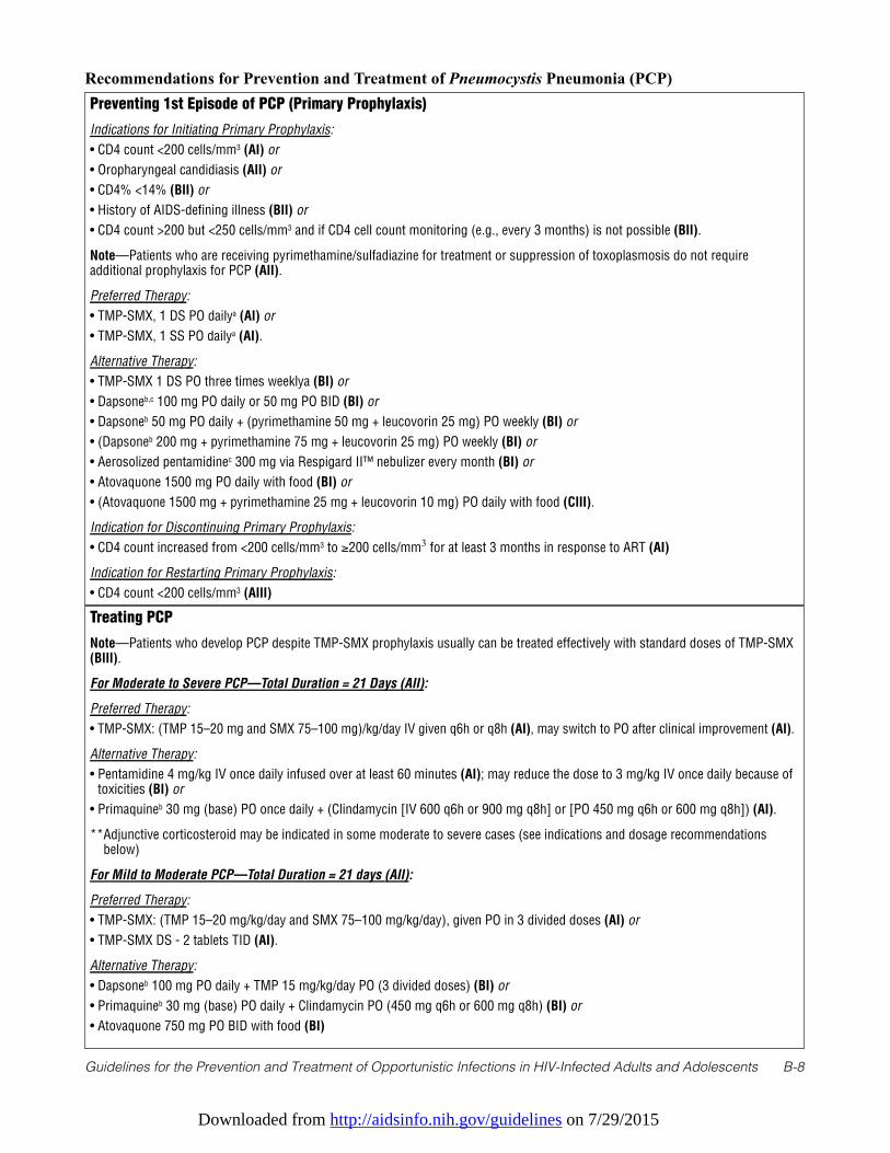
Downloaded from http://aidsinfo.nih.gov/guidelines on 7/29/2015
Guidelines for the Prevention and Treatment of Opportunistic Infections in HIV-Infected Adults and Adolescents B-8
Recommendations for Prevention and Treatment of Pneumocystis Pneumonia (PCP) Preventing 1st Episode of PCP (Primary Prophylaxis)
Indications for Initiating Primary Prophylaxis:
• CD4 count <200 cells/mm3 (AI) or
• Oropharyngeal candidiasis (AII) or
• CD4% <14% (BII) or
• History of AIDS-defining illness (BII) or
• CD4 count >200 but <250 cells/mm3 and if CD4 cell count monitoring (e.g., every 3 months) is not possible (BII).
Note—Patients who are receiving pyrimethamine/sulfadiazine for treatment or suppression of toxoplasmosis do not requireadditional prophylaxis for PCP (AII).
Preferred Therapy:
• TMP-SMX, 1 DS PO dailya (AI) or
• TMP-SMX, 1 SS PO dailya (AI).
Alternative Therapy:
• TMP-SMX 1 DS PO three times weeklya (BI) or
• Dapsoneb,c 100 mg PO daily or 50 mg PO BID (BI) or
• Dapsoneb 50 mg PO daily + (pyrimethamine 50 mg + leucovorin 25 mg) PO weekly (BI) or
• (Dapsoneb 200 mg + pyrimethamine 75 mg + leucovorin 25 mg) PO weekly (BI) or
• Aerosolized pentamidinec 300 mg via Respigard II™ nebulizer every month (BI) or
• Atovaquone 1500 mg PO daily with food (BI) or
• (Atovaquone 1500 mg + pyrimethamine 25 mg + leucovorin 10 mg) PO daily with food (CIII).
Indication for Discontinuing Primary Prophylaxis:
• CD4 count increased from <200 cells/mm3 to ≥200 cells/mm3 for at least 3 months in response to ART (AI)
Indication for Restarting Primary Prophylaxis:
• CD4 count <200 cells/mm3 (AIII)
Treating PCP
Note—Patients who develop PCP despite TMP-SMX prophylaxis usually can be treated effectively with standard doses of TMP-SMX(BIII).
For Moderate to Severe PCP—Total Duration = 21 Days (AII):
Preferred Therapy:
• TMP-SMX: (TMP 15–20 mg and SMX 75–100 mg)/kg/day IV given q6h or q8h (AI), may switch to PO after clinical improvement (AI).
Alternative Therapy:
• Pentamidine 4 mg/kg IV once daily infused over at least 60 minutes (AI); may reduce the dose to 3 mg/kg IV once daily because oftoxicities (BI) or
• Primaquineb 30 mg (base) PO once daily + (Clindamycin [IV 600 q6h or 900 mg q8h] or [PO 450 mg q6h or 600 mg q8h]) (AI).
**Adjunctive corticosteroid may be indicated in some moderate to severe cases (see indications and dosage recommendationsbelow)
For Mild to Moderate PCP—Total Duration = 21 days (AII):
Preferred Therapy:
• TMP-SMX: (TMP 15–20 mg/kg/day and SMX 75–100 mg/kg/day), given PO in 3 divided doses (AI) or
• TMP-SMX DS - 2 tablets TID (AI).
Alternative Therapy:
• Dapsoneb 100 mg PO daily + TMP 15 mg/kg/day PO (3 divided doses) (BI) or
• Primaquineb 30 mg (base) PO daily + Clindamycin PO (450 mg q6h or 600 mg q8h) (BI) or
• Atovaquone 750 mg PO BID with food (BI)
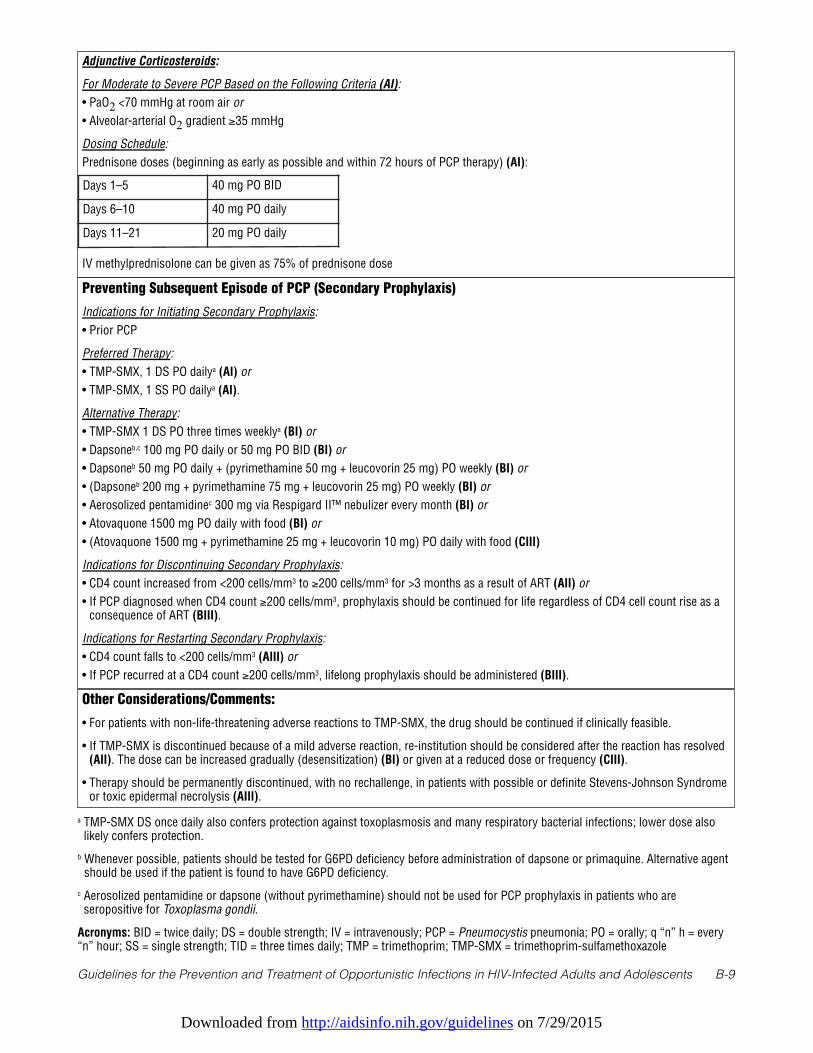
Downloaded from http://aidsinfo.nih.gov/guidelines on 7/29/2015
Guidelines for the Prevention and Treatment of Opportunistic Infections in HIV-Infected Adults and Adolescents B-9
Adjunctive Corticosteroids:
For Moderate to Severe PCP Based on the Following Criteria (AI):
• PaO2 <70 mmHg at room air or
• Alveolar-arterial O2 gradient ≥35 mmHg
Dosing Schedule:
Prednisone doses (beginning as early as possible and within 72 hours of PCP therapy) (AI):
IV methylprednisolone can be given as 75% of prednisone dose
Days 1–5 40 mg PO BID
Days 6–10 40 mg PO daily
Days 11–21 20 mg PO daily
Preventing Subsequent Episode of PCP (Secondary Prophylaxis)
Indications for Initiating Secondary Prophylaxis:
• Prior PCP
Preferred Therapy:
• TMP-SMX, 1 DS PO dailya (AI) or
• TMP-SMX, 1 SS PO dailya (AI).
Alternative Therapy:
• TMP-SMX 1 DS PO three times weeklya (BI) or
• Dapsoneb,c 100 mg PO daily or 50 mg PO BID (BI) or
• Dapsoneb 50 mg PO daily + (pyrimethamine 50 mg + leucovorin 25 mg) PO weekly (BI) or
• (Dapsoneb 200 mg + pyrimethamine 75 mg + leucovorin 25 mg) PO weekly (BI) or
• Aerosolized pentamidinec 300 mg via Respigard II™ nebulizer every month (BI) or
• Atovaquone 1500 mg PO daily with food (BI) or
• (Atovaquone 1500 mg + pyrimethamine 25 mg + leucovorin 10 mg) PO daily with food (CIII)
Indications for Discontinuing Secondary Prophylaxis:
• CD4 count increased from <200 cells/mm3 to ≥200 cells/mm3 for >3 months as a result of ART (AII) or
• If PCP diagnosed when CD4 count ≥200 cells/mm3, prophylaxis should be continued for life regardless of CD4 cell count rise as aconsequence of ART (BIII).
Indications for Restarting Secondary Prophylaxis:
• CD4 count falls to <200 cells/mm3 (AIII) or
• If PCP recurred at a CD4 count ≥200 cells/mm3, lifelong prophylaxis should be administered (BIII).
Other Considerations/Comments:
• For patients with non-life-threatening adverse reactions to TMP-SMX, the drug should be continued if clinically feasible.
• If TMP-SMX is discontinued because of a mild adverse reaction, re-institution should be considered after the reaction has resolved(AII). The dose can be increased gradually (desensitization) (BI) or given at a reduced dose or frequency (CIII).
• Therapy should be permanently discontinued, with no rechallenge, in patients with possible or definite Stevens-Johnson Syndromeor toxic epidermal necrolysis (AIII).
a TMP-SMX DS once daily also confers protection against toxoplasmosis and many respiratory bacterial infections; lower dose alsolikely confers protection.
b Whenever possible, patients should be tested for G6PD deficiency before administration of dapsone or primaquine. Alternative agentshould be used if the patient is found to have G6PD deficiency.
c Aerosolized pentamidine or dapsone (without pyrimethamine) should not be used for PCP prophylaxis in patients who areseropositive for Toxoplasma gondii.
Acronyms: BID = twice daily; DS = double strength; IV = intravenously; PCP = Pneumocystis pneumonia; PO = orally; q “n” h = every“n” hour; SS = single strength; TID = three times daily; TMP = trimethoprim; TMP-SMX = trimethoprim-sulfamethoxazole
Downloaded from http://aidsinfo.nih.gov/guidelines on 7/29/2015
Guidelines for the Prevention and Treatment of Opportunistic Infections in HIV-Infected Adults and Adolescents B-10
References
1. Pifer LL, Hughes WT, Stagno S, Woods D. Pneumocystis carinii infection: evidence for high prevalence in normal andimmunosuppressed children. Pediatrics. Jan 1978;61(1):35-41. Available at http://www.ncbi.nlm.nih.gov/pubmed/400818.
2. Keely SP, Stringer JR, Baughman RP, Linke MJ, Walzer PD, Smulian AG. Genetic variation among Pneumocystiscarinii hominis isolates in recurrent pneumocystosis. J Infect Dis. Aug 1995;172(2):595-598. Available athttp://www.ncbi.nlm.nih.gov/pubmed/7542688.
3. Helweg-Larsen J, Tsolaki AG, Miller RF, Lundgren B, Wakefield AE. Clusters of Pneumocystis carinii pneumonia:analysis of person-to-person transmission by genotyping. QJM. Dec 1998;91(12):813-820. Available athttp://www.ncbi.nlm.nih.gov/pubmed/10024946.
4. Huang L, Beard CB, Creasman J, et al. Sulfa or sulfone prophylaxis and geographic region predict mutations in thePneumocystis carinii dihydropteroate synthase gene. J Infect Dis. Oct 2000;182(4):1192-1198. Available athttp://www.ncbi.nlm.nih.gov/pubmed/10979917.
5. Sassi M, Ripamonti C, Mueller NJ, et al. Outbreaks of Pneumocystis pneumonia in 2 renal transplant centers linked to asingle strain of Pneumocystis: implications for transmission and virulence. Clin Infect Dis. May 2012;54(10):1437-1444. Available at http://www.ncbi.nlm.nih.gov/pubmed/22431811.
6. de Boer MG, Kroon FP, le Cessie S, de Fijter JW, van Dissel JT. Risk factors for Pneumocystis jirovecii pneumonia inkidney transplant recipients and appraisal of strategies for selective use of chemoprophylaxis. Transpl Infect Dis. Dec2011;13(6):559-569. Available at http://www.ncbi.nlm.nih.gov/pubmed/21689251.
7. Arichi N, Kishikawa H, Mitsui Y, et al. Cluster outbreak of Pneumocystis pneumonia among kidney transplant patientswithin a single center. Transplant Proc. Jan–Feb 2009;41(1):170-172. Available athttp://www.ncbi.nlm.nih.gov/pubmed/19249506.
8. Gianella S, Haeberli L, Joos B, et al. Molecular evidence of interhuman transmission in an outbreak of Pneumocystisjirovecii pneumonia among renal transplant recipients. Transpl Infect Dis. Feb 2010;12(1):1-10. Available athttp://www.ncbi.nlm.nih.gov/pubmed/19744285.
9. Mori S, Cho I, Sugimoto M. A cluster of Pneumocystis jirovecii infection among outpatients with rheumatoid arthritis. JRheumatol. Jul 2010;37(7):1547-1548. Available at http://www.ncbi.nlm.nih.gov/pubmed/20595296.
10. Schmoldt S, Schuhegger R, Wendler T, et al. Molecular evidence of nosocomial Pneumocystis jirovecii transmissionamong 16 patients after kidney transplantation. J Clin Microbiol. Mar 2008;46(3):966-971. Available athttp://www.ncbi.nlm.nih.gov/pubmed/18216217.
11. Yazaki H, Goto N, Uchida K, Kobayashi T, Gatanaga H, Oka S. Outbreak of Pneumocystis jiroveci pneumonia in renaltransplant recipients: P. jiroveci is contagious to the susceptible host. Transplantation. Aug 15 2009;88(3):380-385.Available at http://www.ncbi.nlm.nih.gov/pubmed/19667941.
12. Phair J, Munoz A, Detels R, Kaslow R, Rinaldo C, Saah A. The risk of Pneumocystis carinii pneumonia among meninfected with human immunodeficiency virus type 1. Multicenter AIDS Cohort Study Group. N Engl J Med. Jan 181990;322(3):161-165. Available at http://www.ncbi.nlm.nih.gov/pubmed/1967190.
13. Kaplan JE, Hanson DL, Navin TR, Jones JL. Risk factors for primary Pneumocystis carinii pneumonia in humanimmunodeficiency virus-infected adolescents and adults in the United States: reassessment of indications forchemoprophylaxis. J Infect Dis. Oct 1998;178(4):1126-1132. Available at http://www.ncbi.nlm.nih.gov/pubmed/9806044.
14. Kaplan JE, Hanson DL, Jones JL, Dworkin MS. Viral load as an independent risk factor for opportunistic infections inHIV-infected adults and adolescents. AIDS. Sep 28 2001;15(14):1831-1836. Available athttp://www.ncbi.nlm.nih.gov/entrez/query.fcgi?cmd=Retrieve&db=PubMed&dopt=Citation&list_uids=11579245.
15. Buchacz K, Baker RK, Palella FJ, Jr., et al. AIDS-defining opportunistic illnesses in US patients, 1994-2007: a cohortstudy. AIDS. Jun 19 2010;24(10):1549-1559. Available at http://www.ncbi.nlm.nih.gov/pubmed/20502317.
16. Lundberg BE, Davidson AJ, Burman WJ. Epidemiology of Pneumocystis carinii pneumonia in an era of effectiveprophylaxis: the relative contribution of non-adherence and drug failure. AIDS. Nov 10 2000;14(16):2559-2566.Available at http://www.ncbi.nlm.nih.gov/pubmed/11101068.
17. Wolff AJ, O'Donnell AE. Pulmonary manifestations of HIV infection in the era of highly active antiretroviral therapy.Chest. Dec 2001;120(6):1888-1893. Available at http://www.ncbi.nlm.nih.gov/pubmed/11742918.
18. Kovacs JA, Hiemenz JW, Macher AM, et al. Pneumocystis carinii pneumonia: a comparison between patients with the
Downloaded from http://aidsinfo.nih.gov/guidelines on 7/29/2015
Guidelines for the Prevention and Treatment of Opportunistic Infections in HIV-Infected Adults and Adolescents B-11
acquired immunodeficiency syndrome and patients with other immunodeficiencies. Ann Intern Med. May1984;100(5):663-671. Available at http://www.ncbi.nlm.nih.gov/pubmed/6231873.
19. Selwyn PA, Pumerantz AS, Durante A, et al. Clinical predictors of Pneumocystis carinii pneumonia, bacterialpneumonia and tuberculosis in HIV-infected patients. AIDS. May 28 1998;12(8):885-893. Available athttp://www.ncbi.nlm.nih.gov/pubmed/9631142.
20. Ng VL, Yajko DM, Hadley WK. Extrapulmonary pneumocystosis. Clin Microbiol Rev. Jul 1997;10(3):401-418.Available at http://www.ncbi.nlm.nih.gov/pubmed/9227859.
21. Smith DE, McLuckie A, Wyatt J, Gazzard B. Severe exercise hypoxaemia with normal or near normal X-rays: a featureof Pneumocystis carinii infection. Lancet. Nov 5 1988;2(8619):1049-1051. Available athttp://www.ncbi.nlm.nih.gov/pubmed/2903279.
22. Zaman MK, White DA. Serum lactate dehydrogenase levels and Pneumocystis carinii pneumonia. Diagnostic andprognostic significance. Am Rev Respir Dis. Apr 1988;137(4):796-800. Available athttp://www.ncbi.nlm.nih.gov/pubmed/3258483.
23. Opravil M, Marincek B, Fuchs WA, et al. Shortcomings of chest radiography in detecting Pneumocystis cariniipneumonia. J Acquir Immune Defic Syndr. Jan 1994;7(1):39-45. Available athttp://www.ncbi.nlm.nih.gov/pubmed/8263751.
24. Metersky ML, Colt HG, Olson LK, Shanks TG. AIDS-related spontaneous pneumothorax. Risk factors and treatment.Chest. Oct 1995;108(4):946-951. Available at http://www.ncbi.nlm.nih.gov/pubmed/7555166.
25. Sepkowitz KA, Telzak EE, Gold JW, et al. Pneumothorax in AIDS. Ann Intern Med. Mar 15 1991;114(6):455-459.Available at http://www.ncbi.nlm.nih.gov/pubmed/1994791.
26. Baughman RP, Dohn MN, Frame PT. The continuing utility of bronchoalveolar lavage to diagnose opportunisticinfection in AIDS patients. Am J Med. Dec 1994;97(6):515-522. Available athttp://www.ncbi.nlm.nih.gov/pubmed/7985710.
27. Stover DE, Zaman MB, Hajdu SI, Lange M, Gold J, Armstrong D. Bronchoalveolar lavage in the diagnosis of diffusepulmonary infiltrates in the immunosuppressed host. Ann Intern Med. Jul 1984;101(1):1-7. Available athttp://www.ncbi.nlm.nih.gov/pubmed/6375497.
28. Gruden JF, Huang L, Turner J, et al. High-resolution CT in the evaluation of clinically suspected Pneumocystis cariniipneumonia in AIDS patients with normal, equivocal, or nonspecific radiographic findings. AJR Am J Roentgenol. Oct1997;169(4):967-975. Available at http://www.ncbi.nlm.nih.gov/pubmed/9308446.
29. Hidalgo A, Falco V, Mauleon S, al. e. Accuracy of high-resolution CT in distinguishing between Pneumocystis cariniipneumonia and non-Pneumocystis carinii pneumonia in AIDS Patients. European Radiology. 2003;13:1179-1184.
30. Roger PM, Vandenbos F, Pugliese P, et al. Persistence of Pneumocystis carinii after effective treatment of P. cariniipneumonia is not related to relapse or survival among patients infected with human immunodeficiency virus. Clin InfectDis. Feb 1998;26(2):509-510. Available at http://www.ncbi.nlm.nih.gov/pubmed/9502487.
31. Harris JR, Marston BJ, Sangrujee N, DuPlessis D, Park B. Cost-effectiveness analysis of diagnostic options forpneumocystis pneumonia (PCP). PLoS One. 2011;6(8):e23158. Available athttp://www.ncbi.nlm.nih.gov/pubmed/21858013.
32. Torres J, Goldman M, Wheat LJ, et al. Diagnosis of Pneumocystis carinii pneumonia in human immunodeficiencyvirus-infected patients with polymerase chain reaction: a blinded comparison to standard methods. Clin Infect Dis. Jan2000;30(1):141-145. Available at http://www.ncbi.nlm.nih.gov/pubmed/10619742.
33. Larsen HH, Masur H, Kovacs JA, et al. Development and evaluation of a quantitative, touch-down, real-time PCRassay for diagnosing Pneumocystis carinii pneumonia. J Clin Microbiol. Feb 2002;40(2):490-494. Available athttp://www.ncbi.nlm.nih.gov/pubmed/11825961.
34. Larsen HH, Huang L, Kovacs JA, et al. A prospective, blinded study of quantitative touch-down polymerase chainreaction using oral-wash samples for diagnosis of Pneumocystis pneumonia in HIV-infected patients. J Infect Dis. May1 2004;189(9):1679-1683. Available at http://www.ncbi.nlm.nih.gov/pubmed/15116305.
35. Pisculli ML, Sax PE. Use of a serum beta-glucan assay for diagnosis of HIV-related Pneumocystis jiroveci pneumoniain patients with negative microscopic examination results. Clin Infect Dis. Jun 15 2008;46(12):1928-1930. Available athttp://www.ncbi.nlm.nih.gov/pubmed/18540807.
Downloaded from http://aidsinfo.nih.gov/guidelines on 7/29/2015
Guidelines for the Prevention and Treatment of Opportunistic Infections in HIV-Infected Adults and Adolescents B-12
36. Sax PE, Komarow L, Finkelman MA, et al. Blood (1->3)-beta-D-glucan as a diagnostic test for HIV-relatedPneumocystis jirovecii pneumonia. Clin Infect Dis. Jul 15 2011;53(2):197-202. Available athttp://www.ncbi.nlm.nih.gov/pubmed/21690628.
37. Choukri F, Menotti J, Sarfati C, et al. Quantification and spread of Pneumocystis jirovecii in the surrounding air ofpatients with Pneumocystis pneumonia. Clin Infect Dis. Aug 1 2010;51(3):259-265. Available athttp://www.ncbi.nlm.nih.gov/pubmed/20572759.
38. Pliquett RU, Asbe-Vollkopf A, Hauser PM, et al. A Pneumocystis jirovecii pneumonia outbreak in a single kidney-transplant center: role of cytomegalovirus co-infection. Eur J Clin Microbiol Infect Dis. Sep 2012;31(9):2429-2437.Available at http://www.ncbi.nlm.nih.gov/pubmed/22402816.
39. Centers for Disease Controm and Prevention. Guidelines for prophylaxis against Pneumocystis carinii pneumonia forpersons infected with human immunodeficiency virus. MMWR Morb Mortal Wkly Rep. Jun 16 1989;38 Suppl 5(Suppl5):1-9. Available at http://www.ncbi.nlm.nih.gov/pubmed/2524643.
40. Heald A, Flepp M, Chave JP, et al. Treatment for cerebral toxoplasmosis protects against Pneumocystis cariniipneumonia in patients with AIDS. The Swiss HIV Cohort Study. Ann Intern Med. Nov 15 1991;115(10):760-763.Available at http://www.ncbi.nlm.nih.gov/pubmed/1929023.
41. Bozzette SA, Finkelstein DM, Spector SA, et al; with the NIAID AIDS Clinical Trials Group. A randomized trial ofthree antipneumocystis agents in patients with advanced human immunodeficiency virus infection. N Engl J Med. Mar16 1995;332(11):693-699. Available at http://www.ncbi.nlm.nih.gov/pubmed/7854375.
42. Schneider MM, Hoepelman AI, Eeftinck Schattenkerk JK, et al; with the The Dutch AIDS Treatment Group. Acontrolled trial of aerosolized pentamidine or trimethoprim-sulfamethoxazole as primary prophylaxis againstPneumocystis carinii pneumonia in patients with human immunodeficiency virus infection. N Engl J Med. Dec 241992;327(26):1836-1841. Available at http://www.ncbi.nlm.nih.gov/pubmed/1360145.
43. Schneider MM, Nielsen TL, Nelsing S, et al; with Dutch AIDS Treatment Group. Efficacy and toxicity of two doses oftrimethoprim-sulfamethoxazole as primary prophylaxis against Pneumocystis carinii pneumonia in patients with humanimmunodeficiency virus. J Infect Dis. Jun 1995;171(6):1632-1636. Available athttp://www.ncbi.nlm.nih.gov/pubmed/7769306.
44. El-Sadr WM, Luskin-Hawk R, Yurik TM, et al; with Terry Beirn Community Programs for Clinical Research on AIDS(CPCRA). A randomized trial of daily and thrice-weekly trimethoprim-sulfamethoxazole for the prevention ofPneumocystis carinii pneumonia in human immunodeficiency virus-infected persons. Clin Infect Dis. Oct1999;29(4):775-783. Available at http://www.ncbi.nlm.nih.gov/pubmed/10589887.
45. Carr A, Tindall B, Brew BJ, et al. Low-dose trimethoprim-sulfamethoxazole prophylaxis for toxoplasmic encephalitis inpatients with AIDS. Ann Intern Med. Jul 15 1992;117(2):106-111. Available athttp://www.ncbi.nlm.nih.gov/pubmed/1351371.
46. Hardy WD, Feinberg J, Finkelstein DM, et al; with AIDS Clinical Trials Group Protocol 021. A controlled trial oftrimethoprim-sulfamethoxazole or aerosolized pentamidine for secondary prophylaxis of Pneumocystis cariniipneumonia in patients with the acquired immunodeficiency syndrome. N Engl J Med. Dec 24 1992;327(26):1842-1848.Available at http://www.ncbi.nlm.nih.gov/pubmed/1448121.
47. Para MF, Finkelstein D, Becker S, Dohn M, Walawander A, Black JR. Reduced toxicity with gradual initiation oftrimethoprim-sulfamethoxazole as primary prophylaxis for Pneumocystis carinii pneumonia: AIDS Clinical TrialsGroup 268. J Acquir Immune Defic Syndr. Aug 1 2000;24(4):337-343. Available athttp://www.ncbi.nlm.nih.gov/pubmed/11015150.
48. Leoung GS, Stanford JF, Giordano MF, et al. Trimethoprim-sulfamethoxazole (TMP-SMZ) dose escalation versusdirect rechallenge for Pneumocystis Carinii pneumonia prophylaxis in human immunodeficiency virus-infected patientswith previous adverse reaction to TMP-SMZ. J Infect Dis. Oct 15 2001;184(8):992-997. Available athttp://www.ncbi.nlm.nih.gov/pubmed/11574913.
49. Podzamczer D, Salazar A, Jimenez J, et al. Intermittent trimethoprim-sulfamethoxazole compared with dapsone-pyrimethamine for the simultaneous primary prophylaxis of Pneumocystis pneumonia and toxoplasmosis in patientsinfected with HIV. Ann Intern Med. May 15 1995;122(10):755-761. Available athttp://www.ncbi.nlm.nih.gov/pubmed/7717598.
50. Opravil M, Hirschel B, Lazzarin A, et al. Once-weekly administration of dapsone/pyrimethamine vs. aerosolizedpentamidine as combined prophylaxis for Pneumocystis carinii pneumonia and toxoplasmic encephalitis in human
Downloaded from http://aidsinfo.nih.gov/guidelines on 7/29/2015
Guidelines for the Prevention and Treatment of Opportunistic Infections in HIV-Infected Adults and Adolescents B-13
immunodeficiency virus-infected patients. Clin Infect Dis. Mar 1995;20(3):531-541. Available athttp://www.ncbi.nlm.nih.gov/pubmed/7756472.
51. Girard PM, Landman R, Gaudebout C, al. e. Dapsone-pyrimethamine compared with aerosolized pentamidine asprimary prophylaxis against Pneumocystis carinii pneumonia and toxoplasmosis in HIV infection. The PRIO StudyGroup. N Engl J Med. 1993;328(21):1514-1520. Available athttp://www.ncbi.nlm.nih.gov/entrez/query.fcgi?db=pubmed&cmd=Retrieve&dopt=AbstractPlus&list_uids=8479488&query_hl= 14&itool=pubmed_docsum.
52. Chan C, Montaner J, Lefebvre EA, et al. Atovaquone suspension compared with aerosolized pentamidine for preventionof Pneumocystis carinii pneumonia in human immunodeficiency virus-infected subjects intolerant of trimethoprim orsulfonamides. J Infect Dis. Aug 1999;180(2):369-376. Available at http://www.ncbi.nlm.nih.gov/pubmed/10395851.
53. El-Sadr WM, Murphy RL, Yurik TM, et al; with Community Program for Clinical Research on AIDS and the AIDSClinical Trials Group. Atovaquone compared with dapsone for the prevention of Pneumocystis carinii pneumonia inpatients with HIV infection who cannot tolerate trimethoprim, sulfonamides, or both. N Engl J Med. Dec 241998;339(26):1889-1895. Available at http://www.ncbi.nlm.nih.gov/pubmed/9862944.
54. Payen MC, De Wit S, Sommereijns B, Clumeck N. A controlled trial of dapsone versus pyrimethamine-sulfadoxine forprimary prophylaxis of Pneumocystis carinii pneumonia and toxoplasmosis in patients with AIDS. BiomedPharmacother. 1997;51(10):439-445. Available at http://www.ncbi.nlm.nih.gov/pubmed/9863502.
55. Schurmann D, Bergmann F, Albrecht H, et al. Twice-weekly pyrimethamine-sulfadoxine effectively preventsPneumocystis carinii pneumonia relapse and toxoplasmic encephalitis in patients with AIDS. J Infect. Jan 2001;42(1):8-15. Available at http://www.ncbi.nlm.nih.gov/pubmed/11243747.
56. Schurmann D, Bergmann F, Albrecht H, et al. Effectiveness of twice-weekly pyrimethamine-sulfadoxine as primaryprophylaxis of Pneumocystis carinii pneumonia and toxoplasmic encephalitis in patients with advanced HIV infection.Eur J Clin Microbiol Infect Dis. May 2002;21(5):353-361. Available at http://www.ncbi.nlm.nih.gov/pubmed/12072919.
57. Navin TR, Miller KD, Satriale RF, Lobel HO. Adverse reactions associated with pyrimethamine-sulfadoxineprophylaxis for Pneumocystis carinii infections in AIDS. Lancet. Jun 8 1985;1(8441):1332. Available athttp://www.ncbi.nlm.nih.gov/pubmed/2860516.
58. Furrer H, Egger M, Opravil M, et al; Swiss HIV Cohort Study. Discontinuation of primary prophylaxis againstPneumocystis carinii pneumonia in HIV-1-infected adults treated with combination antiretroviral therapy. N Engl JMed. Apr 29 1999;340(17):1301-1306. Available at http://www.ncbi.nlm.nih.gov/pubmed/10219064.
59. Dworkin MS, Hanson DL, Kaplan JE, Jones JL, Ward JW. Risk for preventable opportunistic infections in persons withAIDS after antiretroviral therapy increases CD4+ T lymphocyte counts above prophylaxis thresholds. J Infect Dis. Aug2000;182(2):611-615. Available at http://www.ncbi.nlm.nih.gov/pubmed/10915098.
60. Mussini C, Pezzotti P, Govoni A, et al. Discontinuation of primary prophylaxis for Pneumocystis carinii pneumonia andtoxoplasmic encephalitis in human immunodeficiency virus type I-infected patients: the changes in opportunisticprophylaxis study. J Infect Dis. May 2000;181(5):1635-1642. Available at http://www.ncbi.nlm.nih.gov/pubmed/10823763.
61. Schneider MM, Borleffs JC, Stolk RP, Jaspers CA, Hoepelman AI. Discontinuation of prophylaxis for Pneumocystiscarinii pneumonia in HIV-1-infected patients treated with highly active antiretroviral therapy. Lancet. Jan 161999;353(9148):201-203. Available at http://www.ncbi.nlm.nih.gov/pubmed/9923876.
62. Weverling GJ, Mocroft A, Ledergerber B, et al; with the EuroSIDA Study Group. Discontinuation of Pneumocystiscarinii pneumonia prophylaxis after start of highly active antiretroviral therapy in HIV-1 infection. Lancet. Apr 171999;353(9161):1293-1298. Available at http://www.ncbi.nlm.nih.gov/pubmed/10218526.
63. Yangco BG, Von Bargen JC, Moorman AC, Holmberg SD; with the HIV Outpatient Study (HOPS) Investigators.Discontinuation of chemoprophylaxis against Pneumocystis carinii pneumonia in patients with HIV infection. AnnIntern Med. Feb 1 2000;132(3):201-205. Available at http://www.ncbi.nlm.nih.gov/pubmed/10651600.
64. Furrer H, Opravil M, Rossi M, et al. Discontinuation of primary prophylaxis in HIV-infected patients at high risk ofPneumocystis carinii pneumonia: prospective multicentre study. AIDS. Mar 9 2001;15(4):501-507. Available athttp://www.ncbi.nlm.nih.gov/pubmed/11242147.
65. Kirk O, Lundgren JD, Pedersen C, Nielsen H, Gerstoft J. Can chemoprophylaxis against opportunistic infections bediscontinued after an increase in CD4 cells induced by highly active antiretroviral therapy? AIDS. Sep 101999;13(13):1647-1651. Available at http://www.ncbi.nlm.nih.gov/pubmed/10509565.
Downloaded from http://aidsinfo.nih.gov/guidelines on 7/29/2015
Guidelines for the Prevention and Treatment of Opportunistic Infections in HIV-Infected Adults and Adolescents B-14
66. Lopez Bernaldo de Quiros JC, Miro JM, Pena JM, et al; with the Grupo de Estudio del SIDA 04/98. A randomized trialof the discontinuation of primary and secondary prophylaxis against Pneumocystis carinii pneumonia after highly activeantiretroviral therapy in patients with HIV infection. N Engl J Med. Jan 18 2001;344(3):159-167. Available athttp://www.ncbi.nlm.nih.gov/pubmed/11172138.
67. Green H, Hay P, Dunn DT, McCormack S, Investigators S. A prospective multicentre study of discontinuingprophylaxis for opportunistic infections after effective antiretroviral therapy. HIV Med. Jul 2004;5(4):278-283.Available at http://www.ncbi.nlm.nih.gov/pubmed/15236617.
68. Opportunistic Infections Project Team of the Collaboration of Observational HIVERiE, Mocroft A, Reiss P, et al. Is itsafe to discontinue primary Pneumocystis jiroveci pneumonia prophylaxis in patients with virologically suppressed HIVinfection and a CD4 cell count <200 cells/microL? Clin Infect Dis. Sep 1 2010;51(5):611-619. Available athttp://www.ncbi.nlm.nih.gov/pubmed/20645862.
69. D'Egidio GE, Kravcik S, Cooper CL, Cameron DW, Fergusson DA, Angel JB. Pneumocystis jiroveci pneumoniaprophylaxis is not required with a CD4+ T-cell count < 200 cells/microl when viral replication is suppressed. AIDS. Aug20 2007;21(13):1711-1715. Available at http://www.ncbi.nlm.nih.gov/pubmed/17690568.
70. Hughes W, Leoung G, Kramer F, et al. Comparison of atovaquone (566C80) with trimethoprim-sulfamethoxazole totreat Pneumocystis carinii pneumonia in patients with AIDS. N Engl J Med. May 27 1993;328(21):1521-1527.Available at http://www.ncbi.nlm.nih.gov/pubmed/8479489.
71. Safrin S, Finkelstein DM, Feinberg J, et al. Comparison of three regimens for treatment of mild to moderatePneumocystis carinii pneumonia in patients with AIDS. A double-blind, randomized, trial of oral trimethoprim-sulfamethoxazole, dapsone-trimethoprim, and clindamycin-primaquine. ACTG 108 Study Group. Ann Intern Med. May1 1996;124(9):792-802. Available at http://www.ncbi.nlm.nih.gov/pubmed/8610948.
72. Safrin S, Lee BL, Sande MA. Adjunctive folinic acid with trimethoprim-sulfamethoxazole for Pneumocystis cariniipneumonia in AIDS patients is associated with an increased risk of therapeutic failure and death. J Infect Dis. Oct1994;170(4):912-917. Available at http://www.ncbi.nlm.nih.gov/pubmed/7930736.
73. Crothers K, Beard CB, Turner J, et al. Severity and outcome of HIV-associated Pneumocystis pneumonia containingPneumocystis jirovecii dihydropteroate synthase gene mutations. AIDS. May 20 2005;19(8):801-805. Available athttp://www.ncbi.nlm.nih.gov/pubmed/15867494.
74. Huang L, Crothers K, Atzori C, et al. Dihydropteroate synthase gene mutations in Pneumocystis and sulfa resistance.Emerg Infect Dis. Oct 2004;10(10):1721-1728. Available at http://www.ncbi.nlm.nih.gov/pubmed/15504256.
75. Stein CR, Poole C, Kazanjian P, Meshnick SR. Sulfa use, dihydropteroate synthase mutations, and Pneumocystisjirovecii pneumonia. Emerg Infect Dis. Oct 2004;10(10):1760-1765. Available athttp://www.ncbi.nlm.nih.gov/pubmed/15504261.
76. Alvarez-Martinez MJ, Miro JM, Valls ME, et al. Prevalence of dihydropteroate synthase genotypes before and after theintroduction of combined antiretroviral therapy and their influence on the outcome of Pneumocystis pneumonia in HIV-1-infected patients. Diagn Microbiol Infect Dis. Sep 2010;68(1):60-65. Available athttp://www.ncbi.nlm.nih.gov/pubmed/20727472.
77. Nielsen TL, Eeftinck Schattenkerk JK, Jensen BN, et al. Adjunctive corticosteroid therapy for Pneumocystis cariniipneumonia in AIDS: a randomized European multicenter open label study. J Acquir Immune Defic Syndr.1992;5(7):726-731. Available at http://www.ncbi.nlm.nih.gov/pubmed/1613673.
78. Bozzette SA, Sattler FR, Chiu J, et al. A controlled trial of early adjunctive treatment with corticosteroids forPneumocystis carinii pneumonia in the acquired immunodeficiency syndrome. California Collaborative TreatmentGroup. N Engl J Med. Nov 22 1990;323(21):1451-1457. Available at http://www.ncbi.nlm.nih.gov/pubmed/2233917.
79. The National Institutes of Health-University of California Expert Panel for Corticosteroids as Adjunctive Therapy forPneumocystis Pneumonia. Consensus statement on the use of corticosteroids as adjunctive therapy for pneumocystispneumonia in the acquired immunodeficiency syndrome. N Engl J Med. Nov 22 1990;323(21):1500-1504. Available athttp://www.ncbi.nlm.nih.gov/pubmed/2136587.
80. Montaner JS, Lawson LM, Levitt N, Belzberg A, Schechter MT, Ruedy J. Corticosteroids prevent early deterioration inpatients with moderately severe Pneumocystis carinii pneumonia and the acquired immunodeficiency syndrome(AIDS). Ann Intern Med. Jul 1 1990;113(1):14-20. Available at http://www.ncbi.nlm.nih.gov/pubmed/2190515.
81. Gallant JE, Chaisson RE, Moore RD. The effect of adjunctive corticosteroids for the treatment of Pneumocystis carinii
Downloaded from http://aidsinfo.nih.gov/guidelines on 7/29/2015
Guidelines for the Prevention and Treatment of Opportunistic Infections in HIV-Infected Adults and Adolescents B-15
pneumonia on mortality and subsequent complications. Chest. Nov 1998;114(5):1258-1263. Available athttp://www.ncbi.nlm.nih.gov/pubmed/9823998.
82. Briel M, Bucher HC, Boscacci R, Furrer H. Adjunctive corticosteroids for Pneumocystis jiroveci pneumonia in patientswith HIV-infection. Cochrane Database Syst Rev. 2006;3:CD006150(3):CD006150. Available athttp://www.ncbi.nlm.nih.gov/pubmed/16856118.
83. Medina I, Mills J, Leoung G, et al. Oral therapy for Pneumocystis carinii pneumonia in the acquired immunodeficiencysyndrome. A controlled trial of trimethoprim-sulfamethoxazole versus trimethoprim-dapsone. N Engl J Med. Sep 201990;323(12):776-782. Available at http://www.ncbi.nlm.nih.gov/pubmed/2392131.
84. Black JR, Feinberg J, Murphy RL, et al. Clindamycin and primaquine therapy for mild-to-moderate episodes ofPneumocystis carinii pneumonia in patients with AIDS: AIDS Clinical Trials Group 044. Clin Infect Dis. Jun1994;18(6):905-913. Available at http://www.ncbi.nlm.nih.gov/pubmed/8086551.
85. Toma E, Thorne A, Singer J, et al; with the CTN-PCP Study Group. Clindamycin with primaquine vs. Trimethoprim-sulfamethoxazole therapy for mild and moderately severe Pneumocystis carinii pneumonia in patients with AIDS: amulticenter, double-blind, randomized trial (CTN 004). Clin Infect Dis. Sep 1998;27(3):524-530. Available athttp://www.ncbi.nlm.nih.gov/pubmed/9770152.
86. Smego RA, Jr., Nagar S, Maloba B, Popara M. A meta-analysis of salvage therapy for Pneumocystis carinii pneumonia.Arch Intern Med. Jun 25 2001;161(12):1529-1533. Available at http://www.ncbi.nlm.nih.gov/pubmed/11427101.
87. Dohn MN, Weinberg WG, Torres RA, et al; with the Atovaquone Study Group. Oral atovaquone compared withintravenous pentamidine for Pneumocystis carinii pneumonia in patients with AIDS. Ann Intern Med. Aug 11994;121(3):174-180. Available at http://www.ncbi.nlm.nih.gov/pubmed/7880228.
88. Conte JE, Jr., Chernoff D, Feigal DW, Jr., Joseph P, McDonald C, Golden JA. Intravenous or inhaled pentamidine fortreating Pneumocystis carinii pneumonia in AIDS. A randomized trial. Ann Intern Med. Aug 1 1990;113(3):203-209.Available at http://www.ncbi.nlm.nih.gov/pubmed/2197911.
89. Wharton JM, Coleman DL, Wofsy CB, et al. Trimethoprim-sulfamethoxazole or pentamidine for Pneumocystis cariniipneumonia in the acquired immunodeficiency syndrome. A prospective randomized trial. Ann Intern Med. Jul1986;105(1):37-44. Available at http://www.ncbi.nlm.nih.gov/pubmed/3521428.
90. Kim T, Kim SH, Park KH, et al. Clindamycin-primaquine versus pentamidine for the second-line treatment ofpneumocystis pneumonia. J Infect Chemother. Oct 2009;15(5):343-346. Available athttp://www.ncbi.nlm.nih.gov/pubmed/19856077.
91. Helweg-Larsen J, Benfield T, Atzori C, Miller RF. Clinical efficacy of first- and second-line treatments for HIV-associated Pneumocystis jirovecii pneumonia: a tri-centre cohort study. J Antimicrob Chemother. Dec2009;64(6):1282-1290. Available at http://www.ncbi.nlm.nih.gov/pubmed/19858161.
92. Benfield T, Atzori C, Miller RF, Helweg-Larsen J. Second-line salvage treatment of AIDS-associated Pneumocystisjirovecii pneumonia: a case series and systematic review. J Acquir Immune Defic Syndr. May 1 2008;48(1):63-67.Available at http://www.ncbi.nlm.nih.gov/pubmed/18360286.
93. Soo Hoo GW, Mohsenifar Z, Meyer RD. Inhaled or intravenous pentamidine therapy for Pneumocystis cariniipneumonia in AIDS. A randomized trial. Ann Intern Med. Aug 1 1990;113(3):195-202. Available athttp://www.ncbi.nlm.nih.gov/pubmed/2197910.
94. Montgomery AB, Feigal DW, Jr., Sattler F, et al. Pentamidine aerosol versus trimethoprim-sulfamethoxazole forPneumocystis carinii in acquired immune deficiency syndrome. Am J Respir Crit Care Med. Apr 1995;151(4):1068-1074. Available at http://www.ncbi.nlm.nih.gov/pubmed/7697233.
95. Dworkin MS, Hanson DL, Navin TR. Survival of patients with AIDS, after diagnosis of Pneumocystis cariniipneumonia, in the United States. J Infect Dis. May 1 2001;183(9):1409-1412. Available athttp://www.ncbi.nlm.nih.gov/pubmed/11294675.
96. Morris A, Wachter RM, Luce J, Turner J, Huang L. Improved survival with highly active antiretroviral therapy in HIV-infected patients with severe Pneumocystis carinii pneumonia. AIDS. Jan 3 2003;17(1):73-80. Available athttp://www.ncbi.nlm.nih.gov/pubmed/12478071.
97. Miller RF, Allen E, Copas A, Singer M, Edwards SG. Improved survival for HIV infected patients with severePneumocystis jirovecii pneumonia is independent of highly active antiretroviral therapy. Thorax. Aug 2006;61(8):716-721. Available at http://www.ncbi.nlm.nih.gov/pubmed/16601092.
Downloaded from http://aidsinfo.nih.gov/guidelines on 7/29/2015
Guidelines for the Prevention and Treatment of Opportunistic Infections in HIV-Infected Adults and Adolescents B-16
98. Powell K, Davis JL, Morris AM, Chi A, Bensley MR, Huang L. Survival for patients With HIV admitted to the ICUcontinues to improve in the current era of combination antiretroviral therapy. Chest. Jan 2009;135(1):11-17. Available athttp://www.ncbi.nlm.nih.gov/pubmed/18719058.
99. Zolopa A, Andersen J, Powderly W, et al. Early antiretroviral therapy reduces AIDS progression/death in individualswith acute opportunistic infections: a multicenter randomized strategy trial. PLoS One. 2009;4(5):e5575. Available athttp://www.ncbi.nlm.nih.gov/pubmed/19440326.
100. Grant PM, Komarow L, Andersen J, et al. Risk factor analyses for immune reconstitution inflammatory syndrome in arandomized study of early vs. deferred ART during an opportunistic infection. PLoS One. 2010;5(7):e11416. Availableat http://www.ncbi.nlm.nih.gov/pubmed/20617176.
101. Jagannathan P, Davis E, Jacobson M, Huang L. Life-threatening immune reconstitution inflammatory syndrome afterPneumocystis pneumonia: a cautionary case series. AIDS. Aug 24 2009;23(13):1794-1796. Available athttp://www.ncbi.nlm.nih.gov/pubmed/19684486.
102. Eeftinck Schattenkerk JK, Lange JM, van Steenwijk RP, Danner SA. Can the course of high dose cotrimoxazole forPneumocystis carinii pneumonia in AIDS be shorter? A possible solution to the problem of cotrimoxazole toxicity. JIntern Med. May 1990;227(5):359-362. Available at http://www.ncbi.nlm.nih.gov/pubmed/2341830.
103. Gordin FM, Simon GL, Wofsy CB, Mills J. Adverse reactions to trimethoprim-sulfamethoxazole in patients with theacquired immunodeficiency syndrome. Ann Intern Med. Apr 1984;100(4):495-499. Available athttp://www.ncbi.nlm.nih.gov/pubmed/6230976.
104. Hughes WT, LaFon SW, Scott JD, Masur H. Adverse events associated with trimethoprim-sulfamethoxazole andatovaquone during the treatment of AIDS-related Pneumocystis carinii pneumonia. J Infect Dis. May1995;171(5):1295-1301. Available at http://www.ncbi.nlm.nih.gov/pubmed/7751706.
105. Klein NC, Duncanson FP, Lenox TH, et al. Trimethoprim-sulfamethoxazole versus pentamidine for Pneumocystiscarinii pneumonia in AIDS patients: results of a large prospective randomized treatment trial. AIDS. Mar1992;6(3):301-305. Available at http://www.ncbi.nlm.nih.gov/pubmed/1567574.
106. Sattler FR, Frame P, Davis R, et al. Trimetrexate with leucovorin versus trimethoprim-sulfamethoxazole for moderate tosevere episodes of Pneumocystis carinii pneumonia in patients with AIDS: a prospective, controlled multicenterinvestigation of the AIDS Clinical Trials Group Protocol 029/031. J Infect Dis. Jul 1994;170(1):165-172. Available athttp://www.ncbi.nlm.nih.gov/pubmed/8014493.
107. Masur H, Kaplan JE, Holmes KK, Service USPH, Infectious Diseases Society of A. Guidelines for preventingopportunistic infections among HIV-infected persons—2002. Recommendations of the U.S. Public Health Service andthe Infectious Diseases Society of America. Ann Intern Med. Sep 3 2002;137(5 Pt 2):435-478. Available athttp://www.ncbi.nlm.nih.gov/pubmed/12617574.
108. Soriano V, Dona C, Rodriguez-Rosado R, Barreiro P, Gonzalez-Lahoz J. Discontinuation of secondary prophylaxis foropportunistic infections in HIV-infected patients receiving highly active antiretroviral therapy. AIDS. Mar 102000;14(4):383-386. Available at http://www.ncbi.nlm.nih.gov/pubmed/10770540.
109. Zellweger C, Opravil M, Bernasconi E, et al. Long-term safety of discontinuation of secondary prophylaxis againstPneumocystis pneumonia: prospective multicentre study. AIDS. Oct 21 2004;18(15):2047-2053. Available athttp://www.ncbi.nlm.nih.gov/pubmed/15577626.
110. Mussini C, Pezzotti P, Antinori A, et al. Discontinuation of secondary prophylaxis for Pneumocystis carinii pneumoniain human immunodeficiency virus-infected patients: a randomized trial by the CIOP Study Group. Clin Infect Dis. Mar1 2003;36(5):645-651. Available at http://www.ncbi.nlm.nih.gov/pubmed/12594647.
111. Ledergerber B, Mocroft A, Reiss P, et al. Discontinuation of secondary prophylaxis against Pneumocystis cariniipneumonia in patients with HIV infection who have a response to antiretroviral therapy. Eight European Study Groups.N Engl J Med. Jan 18 2001;344(3):168-174. Available at http://www.ncbi.nlm.nih.gov/pubmed/11188837.
112. Ahmad H, Mehta NJ, Manikal VM, et al. Pneumocystis carinii pneumonia in pregnancy. Chest. Aug 2001;120(2):666-671. Available athttp://www.ncbi.nlm.nih.gov/entrez/query.fcgi?cmd=Retrieve&db=PubMed&dopt=Citation&list_uids=11502676.
113. Connelly RT, Lourwood DL. Pneumocystis carinii pneumonia prophylaxis during pregnancy. Pharmacotherapy. Jul–Aug 1994;14(4):424-429. Available at http://www.ncbi.nlm.nih.gov/pubmed/7937279.
114. Czeizel AE, Rockenbauer M, Sorensen HT, Olsen J. The teratogenic risk of trimethoprim-sulfonamides: a population
Downloaded from http://aidsinfo.nih.gov/guidelines on 7/29/2015
Guidelines for the Prevention and Treatment of Opportunistic Infections in HIV-Infected Adults and Adolescents B-17
based case-control study. Reprod Toxicol. Nov-Dec 2001;15(6):637-646. Available athttp://www.ncbi.nlm.nih.gov/pubmed/11738517.
115. Hernandez-Diaz S, Werler MM, Walker AM, Mitchell AA. Folic acid antagonists during pregnancy and the risk of birthdefects. N Engl J Med. Nov 30 2000;343(22):1608-1614. Available at http://www.ncbi.nlm.nih.gov/pubmed/11096168.
116. Hernandez-Diaz S, Werler MM, Walker AM, Mitchell AA. Neural tube defects in relation to use of folic acidantagonists during pregnancy. American Journal of Epidemiology. May 15 2001;153(10):961-968. Available athttp://www.ncbi.nlm.nih.gov/pubmed/11384952.
117. Jungmann EM, Mercey D, DeRuiter A, et al. Is first trimester exposure to the combination of antiretroviral therapy andfolate antagonists a risk factor for congenital abnormalities? Sexually Transmitted Infections. Dec 2001;77(6):441-443.Available at http://www.ncbi.nlm.nih.gov/pubmed/11714944.
118. Recommendations for the use of folic acid to reduce the number of cases of spina bifida and other neural tube defects.MMWR Recomm Rep. Sep 11 1992;41(RR-14):1-7. Available at http://www.ncbi.nlm.nih.gov/pubmed/1522835.
119. Czeizel AE, Rockenbauer M, Sorensen HT, Olsen J. The teratogenic risk of trimethoprim-sulfonamides: a populationbased case-control study. Reproductive Toxicology. Nov–Dec 2001;15(6):637-646. Available athttp://www.ncbi.nlm.nih.gov/pubmed/11738517.
120. Razavi B, Lund B, Allen BL, Schlesinger L. Failure of trimethoprim/sulfamethoxazole prophylaxis for Pneumocystiscarinii pneumonia with concurrent leucovorin use. Infection. Jan 2002;30(1):41-42. Available athttp://www.ncbi.nlm.nih.gov/entrez/query.fcgi?cmd=Retrieve&db=PubMed&dopt=Citation&list_uids=11876516.
121. Andersen DH, Blanc WA, Crozier DN, Silverman WA. A difference in mortality rate and incidence of kernicterusamong premature infants allotted to two prophylactic antibacterial regimens. Pediatrics. Oct 1956;18(4):614-625.Available at http://www.ncbi.nlm.nih.gov/pubmed/13370229.
122. Albino JA, Shapiro JM. Respiratory failure in pregnancy due to Pneumocystis carinii: report of a successful outcome.Obstet Gynecol. May 1994;83(5 Pt 2):823-824. Available at http://www.ncbi.nlm.nih.gov/pubmed/8159362.
123. Madinger NE, Greenspoon JS, Ellrodt AG. Pneumonia during pregnancy: has modern technology improved maternaland fetal outcome? Am J Obstet Gynecol. Sep 1989;161(3):657-662. Available athttp://www.ncbi.nlm.nih.gov/pubmed/2782348.
124. Koonin LM, Ellerbrock TV, Atrash HK, et al. Pregnancy-associated deaths due to AIDS in the United States. JAMA.Mar 3 1989;261(9):1306-1309. Available at http://www.ncbi.nlm.nih.gov/pubmed/2783746.
125. Benedetti TJ, Valle R, Ledger WJ. Antepartum pneumonia in pregnancy. Am J Obstet Gynecol. Oct 151982;144(4):413-417. Available at http://www.ncbi.nlm.nih.gov/pubmed/7124859.
126. Park-Wyllie L, Mazzotta P, Pastuszak A, et al. Birth defects after maternal exposure to corticosteroids: prospectivecohort study and meta-analysis of epidemiological studies. Teratology. Dec 2000;62(6):385-392. Available athttp://www.ncbi.nlm.nih.gov/entrez/query.fcgi?cmd=Retrieve&db=PubMed&dopt=Citation&list_uids=11091360.
127. Czeizel AE, Rockenbauer M. Population-based case-control study of teratogenic potential of corticosteroids.Teratology. Nov 1997;56(5):335-340. Available athttp://www.ncbi.nlm.nih.gov/entrez/query.fcgi?cmd=Retrieve&db=PubMed&dopt=Citation&list_uids=9451758.
128. Kallen B. Maternal drug use and infant cleft lip/palate with special reference to corticoids. Cleft Palate Craniofac J.Nov 2003;40(6):624-628. Available athttp://www.ncbi.nlm.nih.gov/entrez/query.fcgi?cmd=Retrieve&db=PubMed&dopt=Citation&list_uids=14577813.
129. Ostensen M, Khamashta M, Lockshin M, et al. Anti-inflammatory and immunosuppressive drugs and reproduction.Arthritis Res Ther. 2006;8(3):209. Available athttp://www.ncbi.nlm.nih.gov/entrez/query.fcgi?cmd=Retrieve&db=PubMed&dopt=Citation&list_uids=16712713.
130. Zuidema J, Hilbers-Modderman ES, Merkus FW. Clinical pharmacokinetics of dapsone. Clin Pharmacokinet. Jul-Aug1986;11(4):299-315. Available athttp://www.ncbi.nlm.nih.gov/entrez/query.fcgi?cmd=Retrieve&db=PubMed&dopt=Citation&list_uids=3530584.
131. Brabin BJ, Eggelte TA, Parise M, Verhoeff F. Dapsone therapy for malaria during pregnancy: maternal and fetaloutcomes. Drug Saf. 2004;27(9):633-648. Available athttp://www.ncbi.nlm.nih.gov/entrez/query.fcgi?cmd=Retrieve&db=PubMed&dopt=Citation&list_uids=15230645.
132. Newman RD, Parise ME, Slutsker L, Nahlen B, Steketee RW. Safety, efficacy and determinants of effectiveness of
Downloaded from http://aidsinfo.nih.gov/guidelines on 7/29/2015
Guidelines for the Prevention and Treatment of Opportunistic Infections in HIV-Infected Adults and Adolescents B-18
antimalarial drugs during pregnancy: implications for prevention programmes in Plasmodium falciparum-endemic sub-Saharan Africa. Trop Med Int Health. Jun 2003;8(6):488-506. Available athttp://www.ncbi.nlm.nih.gov/entrez/query.fcgi?cmd=Retrieve&db=PubMed&dopt=Citation&list_uids=12791054.
133. Thornton YS, Bowe ET. Neonatal hyperbilirubinemia after treatment of maternal leprosy. South Med J. May1989;82(5):668. Available athttp://www.ncbi.nlm.nih.gov/entrez/query.fcgi?cmd=Retrieve&db=PubMed&dopt=Citation&list_uids=2717998.
134. Nosten F, McGready R, d'Alessandro U, et al. Antimalarial drugs in pregnancy: a review. Curr Drug Saf. Jan2006;1(1):1-15. Available athttp://www.ncbi.nlm.nih.gov/entrez/query.fcgi?cmd=Retrieve&db=PubMed&dopt=Citation&list_uids=18690910.
135. Harstad TW, Little BB, Bawdon RE, Knoll K, Roe D, Gilstrap LC, 3rd. Embryofetal effects of pentamidine isethionateadministered to pregnant Sprague-Dawley rats. Am J Obstet Gynecol. Sep 1990;163(3):912-916. Available athttp://www.ncbi.nlm.nih.gov/pubmed/2403167.
Downloaded from http://aidsinfo.nih.gov/guidelines on 7/29/2015
Guidelines for the Prevention and Treatment of Opportunistic Infections in HIV-Infected Adults and Adolescents C-1
Toxoplasma gondii Encephalitis (Last updated May 7, 2013; last reviewedMay 7, 2013)
Toxoplasmic encephalitis (TE) is caused by the protozoan Toxoplasma gondii. Disease appears to occuralmost exclusively because of reactivation of latent tissue cysts.1-4 Primary infection occasionally is associatedwith acute cerebral or disseminated disease.
Epidemiology
Seroprevalence of anti-Toxoplasma antibody varies substantially among different geographic locales, with aprevalence of approximately 11% in the United States, versus 50% to 80% in certain European, LatinAmerican, and African countries.4-6 In the era before antiretroviral therapy (ART), the 12-month incidence ofTE was approximately 33% in patients with advanced immunosuppression who were seropositive for T.gondii and not receiving prophylaxis with drugs against the disease. A low incidence of toxoplasmosis is seenin patients who are seronegative for T. gondii. If patients are truly seronegative, their toxoplasmosispresumably represents one of three possible scenarios:
1) Primary infection,2) Re-activation of latent disease in individuals who cannot produce detectable antibodies, or3) Testing with insensitive assays.7,8
Clinical disease is rare among patients with CD4 T lymphocyte (CD4) cell counts >200 cells/µL. Patients withCD4 counts <50 cells/µL are at greatest risk.1,3,8,9 Primary infection occurs after eating undercooked meatcontaining tissue cysts or ingesting oocysts that have been shed in cat feces and sporulated in the environment,a process that takes at least 24 hours. In the United States, eating raw shellfish including oysters, clams, andmussels recently was identified as a novel risk factor for acute infection.10 Up to 50% of individuals withdocumented primary infection do not have an identifiable risk factor.11 The organism is not transmittedthrough person-to-person contact.
Clinical Manifestations
Among patients with AIDS, the most common clinical presentation of T. gondii infection is focal encephalitis withheadache, confusion, or motor weakness and fever.1,3,9 Patients may also present with non-focal manifestations,including only non-specific headache and psychiatric symptoms. Focal neurological abnormalities may be presenton physical examination, and in the absence of treatment, disease progression results in seizures, stupor, and coma.Retinochoroiditis, pneumonia, and evidence of other multifocal organ system involvement are rare in patients withAIDS. Computed tomography (CT) scan or magnetic resonance imaging (MRI) of the brain will typically showmultiple contrast-enhancing lesions in the grey matter of the cortex or basal ganglia, often with associatededema.1,9,12-14 Toxoplasmosis also can manifest as a single brain lesion or diffuse encephalitis without evidence offocal brain lesions on imaging studies.15 This latter presentation tends to be rapidly progressive and fatal.
Diagnosis
HIV-infected patients with TE are almost uniformly seropositive for anti-toxoplasma immunoglobulin G (IgG)antibodies.1,3,9,16 The absence of IgG antibody makes a diagnosis of toxoplasmosis unlikely but not impossible.Anti-toxoplasma immunoglobulin M (IgM) antibodies usually are absent. Quantitative antibody titers are notuseful for diagnosis.
Definitive diagnosis of TE requires a compatible clinical syndrome; identification of one or more mass lesionsby CT, MRI, or other radiographic testing; and detection of the organism in a clinical sample. On imagingstudies, lesions are usually ring-enhancing and have a predilection for the basal ganglia. MRI has sensitivitysuperior to that of CT studies for radiological diagnosis of TE. Positron emission tomography13 or single-photonemission computed tomography scanning14 may be helpful in distinguishing between TE and primary central
Downloaded from http://aidsinfo.nih.gov/guidelines on 7/29/2015
Guidelines for the Prevention and Treatment of Opportunistic Infections in HIV-Infected Adults and Adolescents C-2
nervous system (CNS) lymphoma, but no imaging technique is completely specific. For TE, detection of theorganism requires a brain biopsy, which is most commonly performed by a stereotactic CT-guided needle biopsy.Hematoxylin and eosin stains can be used for detection of T. gondii, but sensitivity is significantly increased ifimmunoperoxidase staining is used and if experienced laboratories process the specimens.17 If safe and feasible,a lumbar puncture should be performed for T. gondii polymerase chain reaction (PCR), as well as for cytology,culture, cryptococcal antigen and PCR for Mycobacterium tuberculosis, Epstein-Barr Virus (EBV) and JC Virus(JCV), either at initial presentation or subsequently, especially in patients in whom empiric therapy fails.Detection of T. gondii by PCR in CSF has high specificity (96%–100%), but low sensitivity (50%), especiallyonce specific anti-toxoplasma therapy has been started.18-20
The differential diagnosis of focal neurological disease in patients with AIDS most often includes primaryCNS lymphoma and progressive multifocal leucoencephalopathy (PML). In the absence of immunereconstitution inflammatory syndrome (IRIS), PML (but not lymphoma) can be distinguished on the basis ofimaging studies. PML lesions typically involve white matter rather than gray matter, are non-contrastenhancing, and produce no mass effect. Less common causes of focal neurologic disease in patients with AIDSinclude mycobacterial infection (especially tuberculosis [TB]); fungal infection, such as cryptococcosis;Chagas disease; and pyogenic brain abscess, particularly in IV drug abusers.
Most clinicians initially rely on an empiric diagnosis, which can be established as an objective response,documented by clinical and radiographic improvement, to specific anti-T. gondii therapy in the absence of alikely alternative diagnosis. Brain biopsy is reserved for patients who fail to respond to specific therapy,although earlier biopsy should be strongly considered if results from imaging, serology, or PCR suggest anetiology other than toxoplasmosis. In patients with contrast-enhancing mass lesions, detection of EBV andJCV by PCR in CSF is highly suggestive of CNS lymphoma21,22 or PML,23 respectively.
Preventing Exposure
HIV-infected individuals should be tested for IgG antibody to Toxoplasma soon after they are diagnosed withHIV to detect latent infection with T. gondii (BIII). They also should be counseled regarding sources ofToxoplasma infection, especially if they lack IgG antibody to Toxoplasma.
To minimize risk of acquiring toxoplasmosis, HIV-infected individuals should be advised not to eat raw orundercooked meat, including undercooked lamb, beef, pork, or venison, and not to eat raw shellfish includingoysters, clams, and mussels (BIII). Lamb, beef, venison, and pork should be cooked to an internal temperature of165ºF to 170ºF;24 meat cooked until it is no longer pink inside usually has an internal temperature of 165ºF to170ºF, and therefore, from a more practical perspective, satisfies this requirement. To minimize the risk foracquiring toxoplasmosis, HIV-infected individuals should wash their hands after contact with raw meat and aftergardening or other contact with soil; they should also wash fruits and vegetables well before eating them raw(BIII). Patients who are seronegative and who own cats should be advised to have someone who is HIV-negativeand not pregnant change the litter box daily. If they must change the litter box themselves, they should wash theirhands thoroughly after doing so (BIII). HIV-infected patients also should be encouraged to keep their cats insideand not to adopt or handle stray cats (BIII). Cats should be fed only canned or dried commercial food or well-cooked table food, not raw or undercooked meats (BIII). Patients do not need to be advised to part with theircats or to have their cats tested for toxoplasmosis (AII).
Preventing Disease
Indication for Primary ProphylaxisToxoplasma-seropositive patients who have CD4 counts <100 cells/µL should receive prophylaxis against TE(AII).25,26
The double-strength-tablet daily dose of trimethoprim-sulfamethoxazole (TMP-SMX), which is the preferredregimen for Pneumocystis jirovecii pneumonia (PCP) prophylaxis, is effective against TE and is recommended
Downloaded from http://aidsinfo.nih.gov/guidelines on 7/29/2015
Guidelines for the Prevention and Treatment of Opportunistic Infections in HIV-Infected Adults and Adolescents C-3
(AII). TMP-SMX, one double-strength tablet three times weekly, is an alternative (BIII). If patients cannottolerate TMP-SMX, the recommended alternative is dapsone-pyrimethamine plus leucovorin, which is alsoeffective against PCP (BI).27-29 Atovaquone with or without pyrimethamine/leucovorin is active against PCPand also can be considered (CIII). Aerosolized pentamidine does not protect against TE and is notrecommended for antitoxoplasma prophylaxis (AI).25,30
Toxoplasma-seronegative persons who are not taking a PCP prophylactic regimen known to be active againstTE, such as aerosolized pentamidine, should be retested for IgG antibody to Toxoplasma when their CD4counts decline to <100 cells/µL to determine whether they have seroconverted and therefore are at risk for TE.Patients who have seroconverted should be administered prophylaxis for TE as previously described (AII).
Discontinuing Primary ProphylaxisProphylaxis against TE should be discontinued in adult and adolescent patients receiving ART whose CD4counts increase to >200 cells/µL for more than 3 months (AI). Multiple observational studies31-33 and tworandomized trials34,35 have reported that primary prophylaxis can be discontinued, with minimal risk fordevelopment of TE, in patients receiving ART whose CD4 counts increase from <200 cells/µL to >200 cells/µLfor more than 3 months. In these studies, most patients were taking HIV protease inhibitor-containing regimensand the median CD4 count at the time prophylaxis was discontinued was >300 cells/µL. At the time prophylaxiswas discontinued, most patients had sustained suppression of plasma HIV RNA levels below the detection limitsof available assays; the median follow-up was 7 to 22 months. Patients with CD4 counts of <100 cells/µL are atgreatest risk for having TE, but the risk for TE with CD4 counts of 100 to 200 cells/µL has not been studied asrigorously as increases to >200 cells/µL. Thus, the recommendation specifies discontinuing prophylaxis after anincrease to >200 cells/µL. When CD4 counts are >200 cells/µL for at least 3 months, primary TE prophylaxisshould be discontinued because it adds little value in preventing toxoplasmosis and increases pill burden,potential for drug toxicity and interaction, likelihood of development of drug-resistant pathogens, and cost.Prophylaxis for TE should be reintroduced if the CD4 count decreases to <100 to 200 cells/µL (AIII). When adecision to stop PCP prophylaxis is contemplated in patients with CD4 counts of 100 to 200 cells/µL and plasmaHIV RNA viral loads below the limits of detection with commercial assays, toxoplasma sero-status should beconsidered, because seropositive patients may then be at risk for developing TE.
Treating Disease
The initial therapy of choice for TE consists of the combination of pyrimethamine plus sulfadiazine plusleucovorin (AI).2,36-38 Pyrimethamine penetrates the brain parenchyma efficiently even in the absence ofinflammation.39 Leucovorin reduces the likelihood of development of hematologic toxicities associated withpyrimethamine therapy.40 Pyrimethamine plus clindamycin plus leucovorin (AI)36,37 is the preferredalternative regimen for patients with TE who cannot tolerate sulfadiazine or do not respond to first-linetherapy. It does not prevent PCP, therefore additional PCP prophylaxis must be administered when it is used(AII) (see discussion under Preventing Recurrence).
In a small (77 patients) randomized trial, TMP-SMX was reported to be effective and better tolerated thanpyrimethamine-sulfadiazine.41 Others have reported similar efficacy in open-label observational studies.42
TMP-SMX has less in vitro activity and experience using this drug to treat toxoplasmosis in developedcountries is limited; however, it can be considered an option if there is a valid reason not to usepyrimethamine plus sulfadiazine. (BI)
No well-studied options exist for patients who cannot take an oral regimen. No parenteral formulation ofpyrimethamine exists and the only widely available parenteral sulfonamide is the sulfamethoxazolecomponent of TMP-SMX. Some specialists will use parenteral TMP-SMX (BI) or oral pyrimethamine plusparenteral clindamycin (CIII) as initial treatment in severely ill patients who require parenteral therapy.
The following regimens have been shown to be effective in treating TE in at least two nonrandomized,uncontrolled trials, although their relative efficacy compared with the previous regimens is unknown:
Downloaded from http://aidsinfo.nih.gov/guidelines on 7/29/2015
Guidelines for the Prevention and Treatment of Opportunistic Infections in HIV-Infected Adults and Adolescents C-4
atovaquone (with meals or oral nutritional supplements) plus either pyrimethamine plus leucovorin orsulfadiazine or, for patients intolerant of both pyrimethamine and sulfadiazine, as a single agent (BII)43,44,45 (ifatovaquone is used alone, clinicians should be aware that the absorption of the drug from patient to patient ishighly variable; plasma levels >18.5 µg/mL are associated with an improved response rate but measurementsare not routinely available);44-46 and azithromycin plus pyrimethamine plus leucovorin daily (CII).47,48
The following regimens have been reported to have activity in treatment of TE in small cohorts of patients orin case reports of one or several patients: clarithromycin plus pyrimethamine (CIII);49 5-fluorouracil plusclindamycin (CIII),50 dapsone plus pyrimethamine plus leucovorin (CIII);51 and minocycline or doxycyclinecombined with either pyrimethamine plus leucovorin, sulfadiazine, or clarithromycin (CIII).52,53 Althoughthe clarithromycin dose used in the only published study was 1g twice a day, doses >500 mg have beenassociated with increased mortality in HIV-infected patients treated for disseminated Mycobacterium aviumComplex. Doses >500 mg twice a day should not be used (BIII).
Clinical response to acute therapy occurs in 90% of patients with TE within 14 days of initiation ofappropriate anti-toxoplasma treatment.2 The reasons why some patients fail therapy are not clearly proven;whether such failures are due to poor adherence or to other host factors of antimicrobial resistance has notbeen well delineated. Acute therapy for TE should be continued for at least 6 weeks, if there is clinical andradiologic improvement (BII).1-4 Longer courses may be appropriate if clinical or radiologic disease isextensive or response is incomplete at 6 weeks. The radiologic goals for treatment include completeresolution of the lesion(s) in terms of size and contrast enhancement, although small scars may persistindefinitely. Adjunctive corticosteroids such as dexamethasone should only be administered to patients withTE when they are clinically indicated to treat a mass effect associated with focal lesions or associated edema(BIII). In those treated with corticosteroids, caution may be needed in diagnosing CNS toxoplasmosis on thebasis of treatment response, since primary CNS lymphoma may respond clinically and radiographically tocorticosteroids alone; these patients should be monitored carefully as corticosteroids are tapered. In addition,corticosteroids should be discontinued as soon as clinically feasible because of their potential to causeimmunosuppression. Patients receiving corticosteroids should be monitored closely for development of otheropportunistic infections (OIs), including cytomegalovirus retinitis and TB.
Anticonvulsants should be administered to patients with TE who have a history of seizures (AIII), butshould not be administered prophylactically to all patients (BIII). Anticonvulsants, if administered, shouldbe continued at least through the period of acute therapy.
Special Considerations with Regard to Starting ARTThere are no data on which to base a recommendation regarding when to start ART in a patient with TE.However, many physicians would initiate ART within 2 to 3 weeks after the diagnosis of toxoplasmosis(CIII), based on the significantly lower incidence of AIDS progression or death (a secondary study endpoint)seen in the ART arm of a controlled trial of 282 patients with OIs other than TB (only 5% of whom hadtoxoplasmosis) who were randomized to early (median 12 days after initiation of OI therapy) versus deferred(median 45 days) initiation of ART.54
Monitoring of Response to Therapy and Adverse Events (including IRIS)Changes in antibody titers are not useful for monitoring responses to therapy. Patients with TE should bemonitored routinely for adverse events and clinical and radiologic improvement (AIII). Common pyrimethaminetoxicities such as rash, nausea, and bone marrow suppression (neutropenia, anemia, and thrombocytopenia) oftencan be reversed by increasing the leucovorin dose to 10, 25, or 50 mg 4 times daily (CIII).
Common sulfadiazine toxicities include rash, fever, leukopenia, hepatitis, nausea, vomiting, diarrhea, renalinsufficiency, and crystalluria. Common clindamycin toxicities include fever, rash, nausea, diarrhea(including pseudomembranous colitis or diarrhea related to Clostridium difficile toxin), and hepatotoxicity.Common TMP-SMX toxicities include rash, fever, leukopenia, thrombocytopenia, and hepatotoxicity.

Downloaded from http://aidsinfo.nih.gov/guidelines on 7/29/2015
Guidelines for the Prevention and Treatment of Opportunistic Infections in HIV-Infected Adults and Adolescents C-5
Common atovaquone toxicities include nausea, vomiting, diarrhea, rash, headache, hyperglycemia, and fever.Drug interactions between anticonvulsants and antiretroviral agents should be evaluated carefully; ifnecessary, doses should be adjusted or alternative anticonvulsants should be used.
IRIS associated with TE has been reported but appears to be rare (~5% in one report).55-57 Given the rarity ofTE-associated IRIS, recommendations for management of such events are difficult to develop.
Managing Treatment FailureA brain biopsy should be strongly considered in patients who did not have an initial biopsy prior to therapyand who fail to respond to initial therapy for TE (BII) as defined by clinical or radiologic deteriorationduring the first week despite adequate therapy, or who do not show clinical improvement within 10 to 14days. A switch to an alternative regimen, as previously described, should be considered for those whoundergo brain biopsy and have confirmed histopathologic evidence of TE, or who have a CSF PCR positivefor T. gondii (BIII). In patients who adhere to their regimens, disease recurrence is unusual in the setting ofsecondary maintenance therapy after an initial clinical and radiographic response.
Preventing Recurrence
When to Start Secondary ProphylaxisPatients who have completed initial therapy for TE should be given suppressive therapy with secondaryprophylaxis or chronic maintenance therapy (AI)36,37 until immune reconstitution occurs as a consequence ofART, in which case treatment discontinuation is indicated. The combination of pyrimethamine plussulfadiazine plus leucovorin is highly effective as suppressive therapy for patients with TE (AI) and providesprotection against PCP (AII). Although sulfadiazine is routinely dosed as a four-times-a-day regimen, apharmacokinetic study suggests bioequivalence for the same total daily dose when given either twice or fourtimes a day,58 and limited clinical experience suggests that twice-daily dosing is effective.59 Pyrimethamineplus clindamycin is commonly used as suppressive therapy for patients with TE who cannot tolerate sulfadrugs (BI). Because of the high failure rate observed with lower doses,36 a dose of 600 mg clindamycin every8 hours is recommended (CIII). Because this regimen does not provide protection against PCP (AII), anadditional agent, such as aerosol pentamidine, must be used. Atovaquone with or without pyrimethamine orsulfadiazine is also active against both TE45,46 and PCP60 (BII), but is substantially more expensive. A small,uncontrolled study in patients who had been receiving ART for a median of 13 months suggested that TMP-SMX could be used as a suppressive regimen to reduce pill burden.61
Although there are no data on the long-term suppressive efficacy of the other alternative regimens notedabove, clinicians might consider using these agents in unusual situations in which the recommended agentscannot be administered (CIII).
When to Stop Secondary ProphylaxisAdult and adolescent patients receiving secondary prophylaxis or chronic maintenance therapy for TE are atlow risk for recurrence of TE when they have successfully completed initial therapy for TE, remainasymptomatic with regard to signs and symptoms of TE, and have an increase in their CD4 counts to >200cells/µL after ART that is sustained for more than 6 months.32,35,62,63 Discontinuing chronic maintenancetherapy in such patients is a reasonable consideration, although occasional recurrences have been reported.The recommendation is based on results in a limited number of patients from observational studies and onerandomized clinical trial and inference from more extensive cumulative data indicating the safety ofdiscontinuing secondary prophylaxis for other OIs during advanced disease (BI). As part of the evaluation todetermine whether discontinuation of therapy is appropriate, some specialists recommend obtaining an MRIof the brain to assess for resolution of brain lesions.
Secondary prophylaxis (chronic maintenance therapy) for TE should be reintroduced if the CD4 countdecreases to <200 cells/µL (AIII).
Downloaded from http://aidsinfo.nih.gov/guidelines on 7/29/2015
Guidelines for the Prevention and Treatment of Opportunistic Infections in HIV-Infected Adults and Adolescents C-6
Special Considerations During Pregnancy
Documentation of baseline maternal T. gondii serologic status (IgG) should be obtained in HIV-infected womenwho are pregnant. Primary T. gondii infection can typically be distinguished from chronic infection with the useof multiple serologic assays, including IgG, IgM, IgA, and IgE antibodies; IgG avidity; and the differentialagglutination tests.64,65 Pregnant HIV-infected women with suspected or confirmed primary T. gondii infectionduring pregnancy should be managed in consultation with a maternal-fetal medicine specialist or anotherappropriate specialist who can perform specialized laboratory testing (BIII)65,66 (e.g., the Palo Alto MedicalFoundation Toxoplasmosis Serology Laboratory; Palo Alto, CA; http://www.pamf.org/serology/ at 650-853-4828 and [email protected]; and the National Collaborative Chicago-based Congenital Toxoplasmosis Study;Chicago, IL; http://www.uchospitals.edu/specialties/infectious-diseases/toxoplasmosis/ at 773-834-4131 [email protected]).
Toxoplasmosis diagnostic considerations are the same in pregnant women as in non-pregnant women.
Indications for treatment of T. gondii during pregnancy should be based on confirmed or suspectedsymptomatic disease in the mother and the risk of transmission of the parasite from mother to fetus. Maternaltreatment of TE should be the same as in non-pregnant adults (BIII), including pyrimethamine plussulfadiazine plus leucovorin (AI).2,36-38 This regimen is also believed to prevent mother-to-child transmissionof T. gondii and it may be therapeutic for affected fetuses.65
Although pyrimethamine has been associated with birth defects in animals, human data have not suggestedan increased risk for defects, therefore, it can be administered to pregnant women after the first trimester.67-71
Similarly, sulfadiazine appears safe in pregnancy.72 A randomized, controlled trial published in 1956 foundthat premature infants receiving prophylactic penicillin/sulfisoxazole were at significantly higher risk ofmortality (specifically kernicterus), compared with infants who received oxytetracycline.73 Because of thesefindings, some clinicians are concerned about the risk of neonatal kernicterus in the setting of maternal use ofsulfa (including sulfadiazine) near delivery, although are no studies published to date link late third-trimestermaternal sulfa use and neonatal death or kernicterus.
The preferred alternative regimen for patients with TE who are unable to tolerate or who fail to respond tofirst-line therapy is pyrimethamine plus clindamycin plus leucovorin (AI).36,37 Clindamycin is Food and DrugAdministration Pregnancy Category B and considered safe throughout pregnancy. Atovaquone may be usedif indicated. While there are limited data on atovaquone safety in humans, preclinical studies have notdemonstrated toxicity.68
Although perinatal transmission of T. gondii normally occurs only with acute infection in theimmunocompetent host, case reports have documented transmission with reactivation of chronic infection inHIV-infected women with severe immunosuppression.70,74 Pregnant HIV-infected women who have evidenceof primary toxoplasmic infection or active toxoplasmosis, including TE, should be evaluated and managedduring pregnancy in consultation with appropriate specialists (BIII). Detailed ultrasound examination of thefetus specifically evaluating for hydrocephalus, cerebral calcifications, and growth restriction should be donefor HIV-infected women with suspected primary or symptomatic reactivation of T. gondii during pregnancy(AIII).65 Amniocentesis does not appear to increase the risk of perinatal HIV transmission, particularly inwomen receiving HAART.75 Therefore, PCR of amniotic fluid can be considered during gestation in pregnantwomen on ART with serologic evidence of acquired infection, and also for women with ultrasound findingssuggestive of fetal T. gondii infection (BIII).65 Because the risk for transmission with chronic infectionappears low, routine fetal evaluation for infection with amniocentesis is not indicated.
Pediatric-care providers should be informed about HIV-infected mothers who have suspected or confirmed T.gondii infection to allow evaluation of their neonates for evidence of congenital infection (AIII).
TMP-SMX can be administered for primary prophylaxis against TE as described for PCP (AIII). The risks ofTMP-SMX in the first trimester, as discussed for PCP, must be balanced against the risk of TE. Secondary
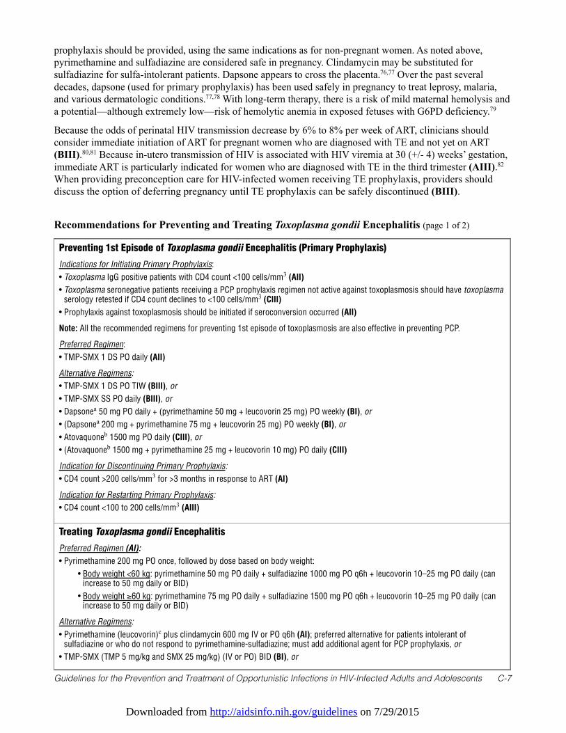
Downloaded from http://aidsinfo.nih.gov/guidelines on 7/29/2015
Guidelines for the Prevention and Treatment of Opportunistic Infections in HIV-Infected Adults and Adolescents C-7
prophylaxis should be provided, using the same indications as for non-pregnant women. As noted above,pyrimethamine and sulfadiazine are considered safe in pregnancy. Clindamycin may be substituted forsulfadiazine for sulfa-intolerant patients. Dapsone appears to cross the placenta.76,77 Over the past severaldecades, dapsone (used for primary prophylaxis) has been used safely in pregnancy to treat leprosy, malaria,and various dermatologic conditions.77,78 With long-term therapy, there is a risk of mild maternal hemolysis anda potential—although extremely low—risk of hemolytic anemia in exposed fetuses with G6PD deficiency.79
Because the odds of perinatal HIV transmission decrease by 6% to 8% per week of ART, clinicians shouldconsider immediate initiation of ART for pregnant women who are diagnosed with TE and not yet on ART(BIII).80,81 Because in-utero transmission of HIV is associated with HIV viremia at 30 (+/- 4) weeks’ gestation,immediate ART is particularly indicated for women who are diagnosed with TE in the third trimester (AIII).82
When providing preconception care for HIV-infected women receiving TE prophylaxis, providers shoulddiscuss the option of deferring pregnancy until TE prophylaxis can be safely discontinued (BIII).
Preventing 1st Episode of Toxoplasma gondii Encephalitis (Primary Prophylaxis)
Indications for Initiating Primary Prophylaxis:
• Toxoplasma IgG positive patients with CD4 count <100 cells/mm3 (AII)
• Toxoplasma seronegative patients receiving a PCP prophylaxis regimen not active against toxoplasmosis should have toxoplasmaserology retested if CD4 count declines to <100 cells/mm3 (CIII)
• Prophylaxis against toxoplasmosis should be initiated if seroconversion occurred (AII)
Note: All the recommended regimens for preventing 1st episode of toxoplasmosis are also effective in preventing PCP.
Preferred Regimen:
• TMP-SMX 1 DS PO daily (AII)
Alternative Regimens:
• TMP-SMX 1 DS PO TIW (BIII), or
• TMP-SMX SS PO daily (BIII), or
• Dapsonea 50 mg PO daily + (pyrimethamine 50 mg + leucovorin 25 mg) PO weekly (BI), or
• (Dapsonea 200 mg + pyrimethamine 75 mg + leucovorin 25 mg) PO weekly (BI), or
• Atovaquoneb 1500 mg PO daily (CIII), or
• (Atovaquoneb 1500 mg + pyrimethamine 25 mg + leucovorin 10 mg) PO daily (CIII)
Indication for Discontinuing Primary Prophylaxis:
• CD4 count >200 cells/mm3 for >3 months in response to ART (AI)
Indication for Restarting Primary Prophylaxis:
• CD4 count <100 to 200 cells/mm3 (AIII)
Treating Toxoplasma gondii Encephalitis
Preferred Regimen (AI):
• Pyrimethamine 200 mg PO once, followed by dose based on body weight:
• Body weight <60 kg: pyrimethamine 50 mg PO daily + sulfadiazine 1000 mg PO q6h + leucovorin 10–25 mg PO daily (canincrease to 50 mg daily or BID)
• Body weight ≥60 kg: pyrimethamine 75 mg PO daily + sulfadiazine 1500 mg PO q6h + leucovorin 10–25 mg PO daily (canincrease to 50 mg daily or BID)
Alternative Regimens:
• Pyrimethamine (leucovorin)c plus clindamycin 600 mg IV or PO q6h (AI); preferred alternative for patients intolerant ofsulfadiazine or who do not respond to pyrimethamine-sulfadiazine; must add additional agent for PCP prophylaxis, or
• TMP-SMX (TMP 5 mg/kg and SMX 25 mg/kg) (IV or PO) BID (BI), or
Recommendations for Preventing and Treating Toxoplasma gondii Encephalitis (page 1 of 2)
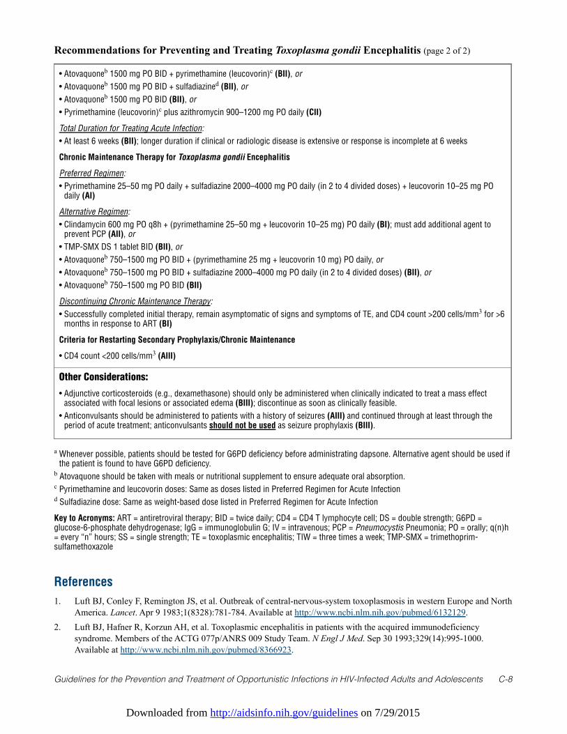
Downloaded from http://aidsinfo.nih.gov/guidelines on 7/29/2015
Guidelines for the Prevention and Treatment of Opportunistic Infections in HIV-Infected Adults and Adolescents C-8
• Atovaquoneb 1500 mg PO BID + pyrimethamine (leucovorin)c (BII), or
• Atovaquoneb 1500 mg PO BID + sulfadiazined (BII), or
• Atovaquoneb 1500 mg PO BID (BII), or
• Pyrimethamine (leucovorin)c plus azithromycin 900–1200 mg PO daily (CII)
Total Duration for Treating Acute Infection:
• At least 6 weeks (BII); longer duration if clinical or radiologic disease is extensive or response is incomplete at 6 weeks
Chronic Maintenance Therapy for Toxoplasma gondii Encephalitis
Preferred Regimen:
• Pyrimethamine 25–50 mg PO daily + sulfadiazine 2000–4000 mg PO daily (in 2 to 4 divided doses) + leucovorin 10–25 mg POdaily (AI)
Alternative Regimen:
• Clindamycin 600 mg PO q8h + (pyrimethamine 25–50 mg + leucovorin 10–25 mg) PO daily (BI); must add additional agent toprevent PCP (AII), or
• TMP-SMX DS 1 tablet BID (BII), or
• Atovaquoneb 750–1500 mg PO BID + (pyrimethamine 25 mg + leucovorin 10 mg) PO daily, or
• Atovaquoneb 750–1500 mg PO BID + sulfadiazine 2000–4000 mg PO daily (in 2 to 4 divided doses) (BII), or
• Atovaquoneb 750–1500 mg PO BID (BII)
Discontinuing Chronic Maintenance Therapy:
• Successfully completed initial therapy, remain asymptomatic of signs and symptoms of TE, and CD4 count >200 cells/mm3 for >6months in response to ART (BI)
Criteria for Restarting Secondary Prophylaxis/Chronic Maintenance
• CD4 count <200 cells/mm3 (AIII)
Other Considerations:
• Adjunctive corticosteroids (e.g., dexamethasone) should only be administered when clinically indicated to treat a mass effectassociated with focal lesions or associated edema (BIII); discontinue as soon as clinically feasible.
• Anticonvulsants should be administered to patients with a history of seizures (AIII) and continued through at least through theperiod of acute treatment; anticonvulsants should not be used as seizure prophylaxis (BIII).
Recommendations for Preventing and Treating Toxoplasma gondii Encephalitis (page 2 of 2)
a Whenever possible, patients should be tested for G6PD deficiency before administrating dapsone. Alternative agent should be used ifthe patient is found to have G6PD deficiency.
b Atovaquone should be taken with meals or nutritional supplement to ensure adequate oral absorption.c Pyrimethamine and leucovorin doses: Same as doses listed in Preferred Regimen for Acute Infectiond Sulfadiazine dose: Same as weight-based dose listed in Preferred Regimen for Acute Infection
Key to Acronyms: ART = antiretroviral therapy; BID = twice daily; CD4 = CD4 T lymphocyte cell; DS = double strength; G6PD =glucose-6-phosphate dehydrogenase; IgG = immunoglobulin G; IV = intravenous; PCP = Pneumocystis Pneumonia; PO = orally; q(n)h= every “n” hours; SS = single strength; TE = toxoplasmic encephalitis; TIW = three times a week; TMP-SMX = trimethoprim-sulfamethoxazole
References
1. Luft BJ, Conley F, Remington JS, et al. Outbreak of central-nervous-system toxoplasmosis in western Europe and NorthAmerica. Lancet. Apr 9 1983;1(8328):781-784. Available at http://www.ncbi.nlm.nih.gov/pubmed/6132129.
2. Luft BJ, Hafner R, Korzun AH, et al. Toxoplasmic encephalitis in patients with the acquired immunodeficiencysyndrome. Members of the ACTG 077p/ANRS 009 Study Team. N Engl J Med. Sep 30 1993;329(14):995-1000.Available at http://www.ncbi.nlm.nih.gov/pubmed/8366923.
Downloaded from http://aidsinfo.nih.gov/guidelines on 7/29/2015
Guidelines for the Prevention and Treatment of Opportunistic Infections in HIV-Infected Adults and Adolescents C-9
3. Wong B, Gold JW, Brown AE, et al. Central-nervous-system toxoplasmosis in homosexual men and parenteral drugabusers. Ann Intern Med. Jan 1984;100(1):36-42. Available at http://www.ncbi.nlm.nih.gov/pubmed/6691657.
4. Israelski DM, Chmiel JS, Poggensee L, Phair JP, Remington JS. Prevalence of Toxoplasma infection in a cohort ofhomosexual men at risk of AIDS and toxoplasmic encephalitis. J Acquir Immune Defic Syndr. Apr 1993;6(4):414-418.Available at http://www.ncbi.nlm.nih.gov/pubmed/8455146.
5. Mathews WC, Fullerton SC. Use of a clinical laboratory database to estimate Toxoplasma seroprevalence among humanimmunodeficiency virus-infected patients. Overcoming bias in secondary analysis of clinical records. Archives ofpathology & laboratory medicine. Aug 1994;118(8):807-810. Available at http://www.ncbi.nlm.nih.gov/pubmed/8060230.
6. Jones JL, Kruszon-Moran D, Sanders-Lewis K, Wilson M. Toxoplasma gondii infection in the United States, 19992004, decline from the prior decade. Am J Trop Med Hyg. Sep 2007;77(3):405-410. Available athttp://www.ncbi.nlm.nih.gov/pubmed/17827351.
7. Abgrall S, Rabaud C, Costagliola D, Clinical Epidemiology Group of the French Hospital Database on HIV. Incidenceand risk factors for toxoplasmic encephalitis in human immunodeficiency virus-infected patients before and during thehighly active antiretroviral therapy era. Clin Infect Dis. Nov 15 2001;33(10):1747-1755. Available athttp://www.ncbi.nlm.nih.gov/pubmed/11595976.
8. Leport C, Chene G, Morlat P, et al. Pyrimethamine for primary prophylaxis of toxoplasmic encephalitis in patients withhuman immunodeficiency virus infection: a double-blind, randomized trial. ANRS 005-ACTG 154 Group Members.Agence Nationale de Recherche sur le SIDA. AIDS Clinical Trial Group. J Infect Dis. Jan 1996;173(1):91-97. Availableat http://www.ncbi.nlm.nih.gov/pubmed/8537688.
9. Luft BJ, Brooks RG, Conley FK, McCabe RE, Remington JS. Toxoplasmic encephalitis in patients with acquired immunedeficiency syndrome. JAMA. Aug 17 1984;252(7):913-917. Available at http://www.ncbi.nlm.nih.gov/pubmed/6748191.
10. Jones JL, Dargelas V, Roberts J, Press C, Remington JS, Montoya JG. Risk factors for Toxoplasma gondii infection in theUnited States. Clin Infect Dis. Sep 15 2009;49(6):878-884. Available at http://www.ncbi.nlm.nih.gov/pubmed/19663709.
11. Boyer KM, Holfels E, Roizen N, et al. Risk factors for Toxoplasma gondii infection in mothers of infants withcongenital toxoplasmosis: Implications for prenatal management and screening. Am J Obstet Gynecol. Feb2005;192(2):564-571. Available at http://www.ncbi.nlm.nih.gov/pubmed/15696004.
12. Kupfer MC, Zee CS, Colletti PM, Boswell WD, Rhodes R. MRI evaluation of AIDS-related encephalopathy:toxoplasmosis vs. lymphoma. Magnetic resonance imaging. 1990;8(1):51-57. Available athttp://www.ncbi.nlm.nih.gov/pubmed/2325518.
13. Pierce MA, Johnson MD, Maciunas RJ, et al. Evaluating contrast-enhancing brain lesions in patients with AIDS byusing positron emission tomography. Ann Intern Med. Oct 15 1995;123(8):594-598. Available athttp://www.ncbi.nlm.nih.gov/pubmed/7677300.
14. Ruiz A, Ganz WI, Post MJ, et al. Use of thallium-201 brain SPECT to differentiate cerebral lymphoma fromtoxoplasma encephalitis in AIDS patients. AJNR. American journal of neuroradiology. Nov 1994;15(10):1885-1894.Available at http://www.ncbi.nlm.nih.gov/pubmed/7863938.
15. Gray F, Gherardi R, Wingate E, et al. Diffuse "encephalitic" cerebral toxoplasmosis in AIDS. Report of four cases. JNeurol. Jul 1989;236(5):273-277. Available at http://www.ncbi.nlm.nih.gov/pubmed/2760644.
16. Derouin F, Leport C, Pueyo S, et al. Predictive value of Toxoplasma gondii antibody titres on the occurrence oftoxoplasmic encephalitis in HIV-infected patients. ANRS 005/ACTG 154 Trial Group. AIDS. Nov 1996;10(13):1521-1527. Available at http://www.ncbi.nlm.nih.gov/pubmed/8931787.
17. Conley FK, Jenkins KA, Remington JS. Toxoplasma gondii infection of the central nervous system. Use of theperoxidase-antiperoxidase method to demonstrate toxoplasma in formalin fixed, paraffin embedded tissue sections.Hum Pathol. Aug 1981;12(8):690-698. Available at http://www.ncbi.nlm.nih.gov/pubmed/7026410.
18. Novati R, Castagna A, Morsica G, et al. Polymerase chain reaction for Toxoplasma gondii DNA in the cerebrospinalfluid of AIDS patients with focal brain lesions. AIDS. Dec 1994;8(12):1691-1694. Available athttp://www.ncbi.nlm.nih.gov/pubmed/7888118.
19. Cinque P, Scarpellini P, Vago L, Linde A, Lazzarin A. Diagnosis of central nervous system complications in HIV-infected patients: cerebrospinal fluid analysis by the polymerase chain reaction. AIDS. Jan 1997;11(1):1-17. Available athttp://www.ncbi.nlm.nih.gov/pubmed/9110070.
20. Mesquita RT, Ziegler AP, Hiramoto RM, Vidal JE, Pereira-Chioccola VL. Real-time quantitative PCR in cerebral
Downloaded from http://aidsinfo.nih.gov/guidelines on 7/29/2015
Guidelines for the Prevention and Treatment of Opportunistic Infections in HIV-Infected Adults and Adolescents C-10
toxoplasmosis diagnosis of Brazilian human immunodeficiency virus-infected patients. Journal of medicalmicrobiology. Jun 2010;59(Pt 6):641-647. Available at http://www.ncbi.nlm.nih.gov/pubmed/20150319.
21. Antinori A, Ammassari A, De Luca A, et al. Diagnosis of AIDS-related focal brain lesions: a decision-making analysisbased on clinical and neuroradiologic characteristics combined with polymerase chain reaction assays in CSF.Neurology. Mar 1997;48(3):687-694. Available at http://www.ncbi.nlm.nih.gov/pubmed/9065549.
22. Antinori A, De Rossi G, Ammassari A, et al. Value of combined approach with thallium-201 single-photon emissioncomputed tomography and Epstein-Barr virus DNA polymerase chain reaction in CSF for the diagnosis of AIDS-relatedprimary CNS lymphoma. J Clin Oncol. Feb 1999;17(2):554-560. Available athttp://www.ncbi.nlm.nih.gov/pubmed/10080599.
23. Koralnik IJ, Boden D, Mai VX, Lord CI, Letvin NL. JC virus DNA load in patients with and without progressivemultifocal leukoencephalopathy. Neurology. Jan 15 1999;52(2):253-260. Available athttp://www.ncbi.nlm.nih.gov/pubmed/9932940.
24. US Department of Health & Human Services. FoodSafety.gov: your gateway to federal food safety information.Available at http://www.foodsafety.gov. Accessed March 26, 2013.
25. Carr A, Tindall B, Brew BJ, et al. Low-dose trimethoprim-sulfamethoxazole prophylaxis for toxoplasmic encephalitis inpatients with AIDS. Ann Intern Med. Jul 15 1992;117(2):106-111. Available athttp://www.ncbi.nlm.nih.gov/pubmed/1351371.
26. Miro JM, Murray HW, Katlama C. Toxoplasmosis. In: Dolin R, H. M, M. S, eds. AIDS Therapy. Third ed. Philadelphia:Churchill Livingstone; 2008:659-681.
27. Podzamczer D, Salazar A, Jimenez J, et al. Intermittent trimethoprim-sulfamethoxazole compared with dapsone-pyrimethamine for the simultaneous primary prophylaxis of Pneumocystis pneumonia and toxoplasmosis in patientsinfected with HIV. Ann Intern Med. May 15 1995;122(10):755-761. Available athttp://www.ncbi.nlm.nih.gov/pubmed/7717598.
28. Opravil M, Hirschel B, Lazzarin A, et al. Once-weekly administration of dapsone/pyrimethamine vs. aerosolizedpentamidine as combined prophylaxis for Pneumocystis carinii pneumonia and toxoplasmic encephalitis in humanimmunodeficiency virus-infected patients. Clin Infect Dis. Mar 1995;20(3):531-541. Available athttp://www.ncbi.nlm.nih.gov/pubmed/7756472.
29. Girard PM, Landman R, Gaudebout C, et al. Dapsone-pyrimethamine compared with aerosolized pentamidine asprimary prophylaxis against Pneumocystis carinii pneumonia and toxoplasmosis in HIV infection. The PRIO StudyGroup. N Engl J Med. May 27 1993;328(21):1514-1520. Available at http://www.ncbi.nlm.nih.gov/pubmed/8479488.
30. Bozzette SA, Finkelstein DM, Spector SA, et al. A randomized trial of three antipneumocystis agents in patients withadvanced human immunodeficiency virus infection. NIAID AIDS Clinical Trials Group. N Engl J Med. Mar 161995;332(11):693-699. Available at http://www.ncbi.nlm.nih.gov/pubmed/7854375.
31. Dworkin MS, Hanson DL, Kaplan JE, Jones JL, Ward JW. Risk for preventable opportunistic infections in persons withAIDS after antiretroviral therapy increases CD4+ T lymphocyte counts above prophylaxis thresholds. J Infect Dis. Aug2000;182(2):611-615. Available at http://www.ncbi.nlm.nih.gov/pubmed/10915098.
32. Kirk O, Lundgren JD, Pedersen C, Nielsen H, Gerstoft J. Can chemoprophylaxis against opportunistic infections bediscontinued after an increase in CD4 cells induced by highly active antiretroviral therapy? AIDS. Sep 101999;13(13):1647-1651. Available at http://www.ncbi.nlm.nih.gov/pubmed/10509565.
33. Furrer H, Opravil M, Bernasconi E, Telenti A, Egger M. Stopping primary prophylaxis in HIV-1-infected patients athigh risk of toxoplasma encephalitis. Swiss HIV Cohort Study. Lancet. Jun 24 2000;355(9222):2217-2218. Available athttp://www.ncbi.nlm.nih.gov/pubmed/10881897.
34. Mussini C, Pezzotti P, Govoni A, et al. Discontinuation of primary prophylaxis for Pneumocystis carinii pneumonia andtoxoplasmic encephalitis in human immunodeficiency virus type I-infected patients: the changes in opportunisticprophylaxis study. J Infect Dis. May 2000;181(5):1635-1642. Available at http://www.ncbi.nlm.nih.gov/pubmed/10823763.
35. Miro JM, Lopez JC, Podzamczer D, et al. Discontinuation of primary and secondary Toxoplasma gondii prophylaxis issafe in HIV-infected patients after immunological restoration with highly active antiretroviral therapy: results of anopen, randomized, multicenter clinical trial. Clin Infect Dis. Jul 1 2006;43(1):79-89. Available athttp://www.ncbi.nlm.nih.gov/pubmed/16758422.
36. Katlama C, De Wit S, O'Doherty E, Van Glabeke M, Clumeck N. Pyrimethamine-clindamycin vs. pyrimethamine-
Downloaded from http://aidsinfo.nih.gov/guidelines on 7/29/2015
Guidelines for the Prevention and Treatment of Opportunistic Infections in HIV-Infected Adults and Adolescents C-11
sulfadiazine as acute and long-term therapy for toxoplasmic encephalitis in patients with AIDS. Clin Infect Dis. Feb1996;22(2):268-275. Available at http://www.ncbi.nlm.nih.gov/pubmed/8838183.
37. Dannemann B, McCutchan JA, Israelski D, et al. Treatment of toxoplasmic encephalitis in patients with AIDS. Arandomized trial comparing pyrimethamine plus clindamycin to pyrimethamine plus sulfadiazine. The CaliforniaCollaborative Treatment Group. Ann Intern Med. Jan 1 1992;116(1):33-43. Available athttp://www.ncbi.nlm.nih.gov/pubmed/1727093.
38. Leport C, Raffi F, Matheron S, et al. Treatment of central nervous system toxoplasmosis withpyrimethamine/sulfadiazine combination in 35 patients with the acquired immunodeficiency syndrome. Efficacy oflong-term continuous therapy. Am J Med. Jan 1988;84(1):94-100. Available athttp://www.ncbi.nlm.nih.gov/pubmed/3337134.
39. Leport C, Meulemans A, Robine D, Dameron G, Vilde JL. Levels of pyrimethamine in serum and penetration into braintissue in humans. AIDS. Sep 1992;6(9):1040-1041. Available at http://www.ncbi.nlm.nih.gov/pubmed/1388895.
40. Van Delden C, Hirschel B. Folinic acid supplements to pyrimethamine-sulfadiazine for Toxoplasma encephalitis areassociated with better outcome. J Infect Dis. May 1996;173(5):1294-1295. Available athttp://www.ncbi.nlm.nih.gov/pubmed/8627092.
41. Torre D, Casari S, Speranza F, et al. Randomized trial of trimethoprim-sulfamethoxazole versus pyrimethamine-sulfadiazine for therapy of toxoplasmic encephalitis in patients with AIDS. Italian Collaborative Study Group.Antimicrob Agents Chemother. Jun 1998;42(6):1346-1349. Available at http://www.ncbi.nlm.nih.gov/pubmed/9624473.
42. Beraud G, Pierre-Francois S, Foltzer A, et al. Cotrimoxazole for treatment of cerebral toxoplasmosis: an observationalcohort study during 1994-2006. Am J Trop Med Hyg. Apr 2009;80(4):583-587. Available athttp://www.ncbi.nlm.nih.gov/pubmed/19346380.
43. Chirgwin K, Hafner R, Leport C, et al. Randomized phase II trial of atovaquone with pyrimethamine or sulfadiazine fortreatment of toxoplasmic encephalitis in patients with acquired immunodeficiency syndrome: ACTG 237/ANRS 039Study. AIDS Clinical Trials Group 237/Agence Nationale de Recherche sur le SIDA, Essai 039. Clin Infect Dis. May 12002;34(9):1243-1250. Available at http://www.ncbi.nlm.nih.gov/pubmed/11941551.
44. Kovacs JA. Efficacy of atovaquone in treatment of toxoplasmosis in patients with AIDS. The NIAID-Clinical CenterIntramural AIDS Program. Lancet. Sep 12 1992;340(8820):637-638. Available athttp://www.ncbi.nlm.nih.gov/pubmed/1355212.
45. Torres RA, Weinberg W, Stansell J, et al. Atovaquone for salvage treatment and suppression of toxoplasmic encephalitisin patients with AIDS. Atovaquone/Toxoplasmic Encephalitis Study Group. Clin Infect Dis. Mar 1997;24(3):422-429.Available at http://www.ncbi.nlm.nih.gov/pubmed/9114194.
46. Katlama C, Mouthon B, Gourdon D, Lapierre D, Rousseau F. Atovaquone as long-term suppressive therapy fortoxoplasmic encephalitis in patients with AIDS and multiple drug intolerance. Atovaquone Expanded Access Group.AIDS. Sep 1996;10(10):1107-1112. Available at http://www.ncbi.nlm.nih.gov/pubmed/8874627.
47. Saba J, Morlat P, Raffi F, et al. Pyrimethamine plus azithromycin for treatment of acute toxoplasmic encephalitis inpatients with AIDS. Eur J Clin Microbiol Infect Dis. Nov 1993;12(11):853-856. Available athttp://www.ncbi.nlm.nih.gov/pubmed/8112357.
48. Jacobson JM, Hafner R, Remington J, et al. Dose-escalation, phase I/II study of azithromycin and pyrimethamine forthe treatment of toxoplasmic encephalitis in AIDS. AIDS. Mar 30 2001;15(5):583-589. Available athttp://www.ncbi.nlm.nih.gov/pubmed/11316995.
49. Fernandez-Martin J, Leport C, Morlat P, Meyohas MC, Chauvin JP, Vilde JL. Pyrimethamine-clarithromycincombination for therapy of acute Toxoplasma encephalitis in patients with AIDS. Antimicrob Agents Chemother. Oct1991;35(10):2049-2052. Available at http://www.ncbi.nlm.nih.gov/pubmed/1836943.
50. Dhiver C, Milandre C, Poizot-Martin I, Drogoul MP, Gastaut JL, Gastaut JA. 5-Fluoro-uracil-clindamycin for treatmentof cerebral toxoplasmosis. AIDS. Jan 1993;7(1):143-144. Available at http://www.ncbi.nlm.nih.gov/pubmed/8442914.
51. Derouin F, Piketty C, Chastang C, Chau F, Rouveix B, Pocidalo JJ. Anti-Toxoplasma effects of dapsone alone andcombined with pyrimethamine. Antimicrob Agents Chemother. Feb 1991;35(2):252-255. Available athttp://www.ncbi.nlm.nih.gov/pubmed/2024957.
52. Lacassin F, Schaffo D, Perronne C, Longuet P, Leport C, Vilde JL. Clarithromycin-minocycline combination as salvagetherapy for toxoplasmosis in patients infected with human immunodeficiency virus. Antimicrob Agents Chemother. Jan
Downloaded from http://aidsinfo.nih.gov/guidelines on 7/29/2015
Guidelines for the Prevention and Treatment of Opportunistic Infections in HIV-Infected Adults and Adolescents C-12
1995;39(1):276-277. Available at http://www.ncbi.nlm.nih.gov/pubmed/7695324.53. Hagberg L, Palmertz B, Lindberg J. Doxycycline and pyrimethamine for toxoplasmic encephalitis. Scandinavian
journal of infectious diseases. 1993;25(1):157-160. Available at http://www.ncbi.nlm.nih.gov/pubmed/8460343.54. Zolopa A, Andersen J, Powderly W, et al. Early antiretroviral therapy reduces AIDS progression/death in individuals
with acute opportunistic infections: a multicenter randomized strategy trial. PLoS One. 2009;4(5):e5575. Available athttp://www.ncbi.nlm.nih.gov/pubmed/19440326.
55. Pfeffer G, Prout A, Hooge J, Maguire J. Biopsy-proven immune reconstitution syndrome in a patient with AIDS and cerebraltoxoplasmosis. Neurology. Jul 28 2009;73(4):321-322. Available at http://www.ncbi.nlm.nih.gov/pubmed/19636053.
56. Tremont-Lukats IW, Garciarena P, Juarbe R, El-Abassi RN. The immune inflammatory reconstitution syndrome andcentral nervous system toxoplasmosis. Ann Intern Med. May 5 2009;150(9):656-657. Available athttp://www.ncbi.nlm.nih.gov/pubmed/19414855.
57. Martin-Blondel G, Alvarez M, Delobel P, et al. Toxoplasmic encephalitis IRIS in HIV-infected patients: a case seriesand review of the literature. J Neurol Neurosurg Psychiatry. Jun 2011;82(6):691-693. Available athttp://www.ncbi.nlm.nih.gov/pubmed/20660912.
58. Jordan MK, Burstein AH, Rock-Kress D, et al. Plasma pharmacokinetics of sulfadiazine administered twice dailyversus four times daily are similar in human immunodeficiency virus-infected patients. Antimicrob Agents Chemother.Feb 2004;48(2):635-637. Available at http://www.ncbi.nlm.nih.gov/pubmed/14742225.
59. Podzamczer D, Miro JM, Ferrer E, et al. Thrice-weekly sulfadiazine-pyrimethamine for maintenance therapy oftoxoplasmic encephalitis in HIV-infected patients. Spanish Toxoplasmosis Study Group. Eur J Clin Microbiol InfectDis. Feb 2000;19(2):89-95. Available at http://www.ncbi.nlm.nih.gov/pubmed/10746493.
60. El-Sadr WM, Murphy RL, Yurik TM, et al. Atovaquone compared with dapsone for the prevention of Pneumocystiscarinii pneumonia in patients with HIV infection who cannot tolerate trimethoprim, sulfonamides, or both. CommunityProgram for Clinical Research on AIDS and the AIDS Clinical Trials Group. N Engl J Med. Dec 24 1998;339(26):1889-1895. Available at http://www.ncbi.nlm.nih.gov/pubmed/9862944.
61. Duval X, Pajot O, Le Moing V, et al. Maintenance therapy with cotrimoxazole for toxoplasmic encephalitis in the era ofhighly active antiretroviral therapy. AIDS. Jun 18 2004;18(9):1342-1344. Available athttp://www.ncbi.nlm.nih.gov/pubmed/15362670.
62. Soriano V, Dona C, Rodriguez-Rosado R, Barreiro P, Gonzalez-Lahoz J. Discontinuation of secondary prophylaxis foropportunistic infections in HIV-infected patients receiving highly active antiretroviral therapy. AIDS. Mar 102000;14(4):383-386. Available at http://www.ncbi.nlm.nih.gov/pubmed/10770540.
63. Bertschy S, Opravil M, Cavassini M, et al. Discontinuation of maintenance therapy against toxoplasma encephalitis inAIDS patients with sustained response to anti-retroviral therapy. Clinical microbiology and infection: the officialpublication of the European Society of Clinical Microbiology and Infectious Diseases. Jul 2006;12(7):666-671.Available at http://www.ncbi.nlm.nih.gov/pubmed/16774564.
64. Montoya JG. Laboratory diagnosis of Toxoplasma gondii infection and toxoplasmosis. J Infect Dis. Feb 15 2002;185Suppl 1:S73-82. Available at http://www.ncbi.nlm.nih.gov/pubmed/11865443.
65. Montoya JG, Remington JS. Management of Toxoplasma gondii infection during pregnancy. Clin Infect Dis. Aug 152008;47(4):554-566. Available at http://www.ncbi.nlm.nih.gov/pubmed/18624630.
66. Mitchell CD, Erlich SS, Mastrucci MT, Hutto SC, Parks WP, Scott GB. Congenital toxoplasmosis occurring in infantsperinatally infected with human immunodeficiency virus 1. Pediatr Infect Dis J. Jul 1990;9(7):512-518. Available athttp://www.ncbi.nlm.nih.gov/pubmed/2371084.
67. Peters PJ, Thigpen MC, Parise ME, Newman RD. Safety and toxicity of sulfadoxine/pyrimethamine: implications formalaria prevention in pregnancy using intermittent preventive treatment. Drug Saf. 2007;30(6):481-501. Available athttp://www.ncbi.nlm.nih.gov/pubmed/17536875.
68. Nosten F, McGready R, d'Alessandro U, et al. Antimalarial drugs in pregnancy: a review. Current drug safety. Jan2006;1(1):1-15. Available at http://www.ncbi.nlm.nih.gov/pubmed/18690910.
69. Wong SY, Remington JS. Toxoplasmosis in pregnancy. Clin Infect Dis. Jun 1994;18(6):853-861; quiz 862. Available athttp://www.ncbi.nlm.nih.gov/pubmed/8086543.
70. Dunn CS, Beyer C, Kieny MP, et al. High viral load and CD4 lymphopenia in rhesus and cynomolgus macaques
Downloaded from http://aidsinfo.nih.gov/guidelines on 7/29/2015
Guidelines for the Prevention and Treatment of Opportunistic Infections in HIV-Infected Adults and Adolescents C-13
infected by a chimeric primate lentivirus constructed using the env, rev, tat, and vpu genes from HIV-1 Lai. Virology.Sep 15 1996;223(2):351-361. Available at http://www.ncbi.nlm.nih.gov/pubmed/8806570.
71. Deen JL, von Seidlein L, Pinder M, Walraven GE, Greenwood BM. The safety of the combination artesunate andpyrimethamine-sulfadoxine given during pregnancy. Trans R Soc Trop Med Hyg. Jul-Aug 2001;95(4):424-428.Available at http://www.ncbi.nlm.nih.gov/pubmed/11579889.
72. Baskin CG, Law S, Wenger NK. Sulfadiazine rheumatic fever prophylaxis during pregnancy: does it increase the risk ofkernicterus in the newborn? Cardiology. 1980;65(4):222-225. Available at http://www.ncbi.nlm.nih.gov/pubmed/7388849.
73. Andersen DH, Blanc WA, Crozier DN, Silverman WA. A difference in mortality rate and incidence of kernicterusamong premature infants allotted to two prophylactic antibacterial regimens. Pediatrics. Oct 1956;18(4):614-625.Available at http://www.ncbi.nlm.nih.gov/pubmed/13370229.
74. Low incidence of congenital toxoplasmosis in children born to women infected with human immunodeficiency virus.European Collaborative Study and Research Network on Congenital Toxoplasmosis. Eur J Obstet Gynecol Reprod Biol.Sep 1996;68(1-2):93-96. Available at http://www.ncbi.nlm.nih.gov/pubmed/8886688.
75. Mandelbrot L, Jasseron C, Ekoukou D, et al. Amniocentesis and mother-to-child human immunodeficiency virustransmission in the Agence Nationale de Recherches sur le SIDA et les Hepatites Virales French Perinatal Cohort. Am JObstet Gynecol. Feb 2009;200(2):160 e161-169. Available at http://www.ncbi.nlm.nih.gov/pubmed/18986640.
76. Zuidema J, Hilbers-Modderman ES, Merkus FW. Clinical pharmacokinetics of dapsone. Clinical pharmacokinetics. Jul-Aug 1986;11(4):299-315. Available at http://www.ncbi.nlm.nih.gov/pubmed/3530584.
77. Brabin BJ, Eggelte TA, Parise M, Verhoeff F. Dapsone therapy for malaria during pregnancy: maternal and fetaloutcomes. Drug Saf. 2004;27(9):633-648. Available at http://www.ncbi.nlm.nih.gov/pubmed/15230645.
78. Newman RD, Parise ME, Slutsker L, Nahlen B, Steketee RW. Safety, efficacy and determinants of effectiveness ofantimalarial drugs during pregnancy: implications for prevention programmes in Plasmodium falciparum-endemic sub-Saharan Africa. Tropical medicine & international health: TM & IH. Jun 2003;8(6):488-506. Available athttp://www.ncbi.nlm.nih.gov/pubmed/12791054.
79. Thornton YS, Bowe ET. Neonatal hyperbilirubinemia after treatment of maternal leprosy. Southern medical journal.May 1989;82(5):668. Available at http://www.ncbi.nlm.nih.gov/pubmed/2717998.
80. Hoffman RM, Black V, Technau K, et al. Effects of highly active antiretroviral therapy duration and regimen on risk formother-to-child transmission of HIV in Johannesburg, South Africa. J Acquir Immune Defic Syndr. May 12010;54(1):35-41. Available at http://www.ncbi.nlm.nih.gov/pubmed/20216425.
81. Warszawski J, Tubiana R, Le Chenadec J, et al. Mother-to-child HIV transmission despite antiretroviral therapy in theANRS French Perinatal Cohort. AIDS. Jan 11 2008;22(2):289-299. Available athttp://www.ncbi.nlm.nih.gov/pubmed/18097232.
82. Tubiana R, Le Chenadec J, Rouzioux C, et al. Factors associated with mother-to-child transmission of HIV-1 despite amaternal viral load <500 copies/ml at delivery: a case-control study nested in the French perinatal cohort (EPF-ANRSCO1). Clin Infect Dis. Feb 15 2010;50(4):585-596. Available at http://www.ncbi.nlm.nih.gov/pubmed/20070234.
Downloaded from http://aidsinfo.nih.gov/guidelines on 7/29/2015
Guidelines for the Prevention and Treatment of Opportunistic Infections in HIV-Infected Adults and Adolescents D-1
Cryptosporidiosis (Last updated June 17, 2013; last reviewed May 7, 2013)
Epidemiology
Cryptosporidiosis is caused by various species of the protozoan parasite Cryptosporidium, which infect thesmall bowel mucosa and, if symptomatic, typically cause diarrhea. Cryptosporidium can also infect othergastrointestinal and extraintestinal sites, especially in individuals whose immune systems are suppressed.Advanced immunosuppression—typically CD4 T lymphocyte cell (CD4) counts of <100 cells/µL1—isassociated with the greatest risk for prolonged, severe, or extraintestinal cryptosporidiosis. The three speciesthat most commonly infect humans are Cryptosporidium hominis, Cryptosporidium parvum, andCryptosporidium meleagridis. Infections are usually caused by one species, but a mixed infection ispossible.2
Cryptosporidiosis remains a common cause of chronic diarrhea in AIDS patients in developing countries,with up to 74% of diarrheal stools demonstrating the organism.3 In developed countries with low rates ofenvironmental contamination and where potent antiretroviral therapy (ART) is widely available,cryptosporidiosis has decreased and occurs at an incidence of <1 case per 1000 person-years in patients withAIDS.4 Infection occurs through ingestion of Cryptosporidium oocysts. Viable oocysts in feces can betransmitted directly through contact with infected humans or animals, particularly those with diarrhea.Oocysts can contaminate recreational water sources such as swimming pools and lakes, and public watersupplies and may persist despite standard chlorination (see Appendix: Food and Water-Related Exposures).Person-to-person transmission is common, especially among sexually active men who have sex with men.
Clinical Manifestations
Patients with cryptosporidiosis most commonly have acute or subacute onset of watery diarrhea, which maybe accompanied by nausea, vomiting, and lower abdominal cramping. Severity can range from asymptomaticto profuse, cholera-like diarrhea. More severe symptoms tend to occur in immune-suppressed patients,whereas transient diarrhea alone is typical in hosts with competent immune systems. Fever is present inapproximately one-third of patients and malabsorption is common. The epithelium of the biliary tract and thepancreatic duct can be infected with Cryptosporidium, leading to sclerosing cholangitis and to pancreatitissecondary to papillary stenosis, particularly among patients with prolonged disease and low CD4 cellcounts.5-8 Pulmonary infections also have been reported,9,10 and may be under-recognized.11
Diagnosis
Diagnosis of cryptosporidiosis can be made by microscopic identification of the oocysts in stool or tissuewith acid-fast staining or direct immunofluorescence, which offers better sensitivity.12 Immunofluorescenceis estimated to be 10 times more sensitive than acid-fast staining and is now the gold standard for stoolexamination. Concentration methods (i.e., formalin ether or formalin-ethyl acetate) and flotation methods(i.e., Sheather’s sucrose or sodium chloride) may facilitate diagnosis, but they are very labor intensive andnot routinely used in clinical laboratories. Antigen-detection by enzyme-linked immunosorbent assay orimmunochromatographic tests also are useful, with sensitivities reportedly ranging from 66% to 100%,depending on the specific test. Molecular methods such as polymerase chain reaction (PCR) are even moresensitive,13 detecting as few as five oocysts in spiked stool samples and nearly double the number of casesidentified by microscopic methods. Cryptosporidial enteritis also can be diagnosed from small sections fromintestinal biopsy.
A single stool specimen is usually adequate for diagnosis in individuals with profuse diarrheal illness,whereas repeat stool sampling is recommended for those with milder disease.
Downloaded from http://aidsinfo.nih.gov/guidelines on 7/29/2015
Guidelines for the Prevention and Treatment of Opportunistic Infections in HIV-Infected Adults and Adolescents D-2
Preventing Exposure
HIV-infected individuals should be educated and counseled about the different ways that Cryptosporidiumcan be transmitted (BIII). Modes of transmission include having direct contact with infected adults, diaper-aged children, and infected animals; coming into contact with contaminated water during recreationalactivities; drinking contaminated water; and eating contaminated food.
Detailed prevention recommendations related to food and water exposures (including methods for removingCryptosporidium from drinking water), pet exposures, and travel-related exposures can be found in Appendix A:Recommendations to Help HIV-infected Patients Avoid Exposure to, or Infection from, Opportunistic Pathogens.
Scrupulous handwashing can reduce the risk of diarrhea in HIV-infected individuals, including diarrheacaused by Cryptosporidium.14 HIV-infected patients should be advised to wash their hands after potentialcontact with human feces (including after diapering small children). Hand-washing also should berecommended in association with the following activities: after handling pets or other animals, gardening orhaving other contact with soil; before preparing food or eating; and before and after sex (BIII). HIV-infectedpatients should avoid unprotected sex, especially practices that could lead to direct (e.g., oral-anal) orindirect (e.g., penile-anal) contact with feces. They should be advised to use barriers such as condoms anddental dams during sex to reduce such exposures (BIII).
HIV-infected individuals—particularly those with CD4 counts <200 cells/µL—should avoid direct contactwith diarrhea or stool from pets (BIII). Gloves should be worn when handling feces or cleaning areas thatmight have been contaminated by feces from pets (BIII). They should also limit or avoid direct exposure tocalves and lambs (BII). Paying attention to hygiene and avoiding direct contact with stool are importantwhen visiting premises such as farms or petting zoos where these animals are housed or exhibited.
HIV-infected individuals should not drink water directly from lakes or rivers (AIII). Waterborne infectionalso can result from swallowing water during recreational activities. HIV-infected individuals should bemade aware that lakes, rivers, and salt water beaches and some swimming pools, recreational water parks,and ornamental water fountains may be contaminated with human or animal waste that containsCryptosporidium. They should avoid swimming in water that is likely contaminated and should avoidswallowing water while swimming or playing in recreational water (BIII).
Outbreaks of cryptosporidiosis have been linked to drinking water from municipal water supplies. Duringoutbreaks or in other situations that impose a community advisory to boil water, boiling water for at least 1minute will eliminate the risk for cryptosporidiosis (AIII). Using submicron personal-use water filters(home/office types) or bottled water also may reduce the risk of infection from municipal and well water (BII).
For persons with low CD4 cell counts, the magnitude of the risk of acquiring cryptosporidiosis from drinkingwater in a non-outbreak setting is uncertain, and available data are inadequate to recommend that all HIV-infected persons boil water or avoid drinking tap water in non-outbreak settings. However, HIV-infectedindividuals should consider drinking only filtered water (CIII), despite the complexities involved in selectingappropriate products, the lack of enforceable standards for removal of oocysts, the costs of the products, andthe logistic difficulty of using these products consistently. Note that ice made from contaminated tap wateralso can be a source of infection.
HIV-infected patients with low CD4 cell counts should be cautious about eating raw oysters becausecryptosporidial oocysts can survive in oysters for longer than 2 months and have been found in oysters takenfrom certain commercial oyster beds (CIII). In the hospital setting, standard precautions for use of gloves andfor hand-washing after removal of gloves should be sufficient to prevent transmission of cryptosporidiosis froman infected patient to a susceptible HIV-infected individual (BIII). Because of the potential for fomitetransmission, some specialists recommend that HIV-infected patients, especially individuals who are severelyimmunocompromised, not share a room with a patient with cryptosporidiosis (CIII).
HIV-infected individuals who travel to developing countries should be warned to avoid drinking tap water or
Downloaded from http://aidsinfo.nih.gov/guidelines on 7/29/2015
Guidelines for the Prevention and Treatment of Opportunistic Infections in HIV-Infected Adults and Adolescents D-3
using tap water to brush their teeth (BIII). Ice that is not made from bottled water and consumption of rawfruits or vegetables that could have been washed in tap water should also be avoided (BIII). HIV-infectedindividuals also should avoid other sources of Cryptosporidium oocysts as much as possible (BIII). Theseinclude working directly with people with diarrhea; with farm animals such as cattle and sheep; and withdomestic pets that are very young or have diarrhea. If exposure is unavoidable, gloves should be used andpractices for good hand hygiene observed.
Preventing Disease
Because chronic cryptosporidiosis occurs primarily in patients with advanced immunodeficiency, appropriateinitiation of combination ART before the patient becomes severely immunosuppressed should prevent thisdisease (AII). Rifabutin and possibly clarithromycin, when taken for Mycobacterium avium complexprophylaxis, have been found to protect against cryptosporidiosis.15,16 Data are insufficient, however, towarrant a recommendation for using rifabutin or clarithromycin as chemoprophylaxis for cryptosporidiosis.
Treating Disease
In the setting of severe immune suppression, ART with immune restoration to a CD4 count >100 cells/µLusually leads to resolution of clinical cryptosporidiosis17-21 and is the mainstay of treatment. Therefore,patients with cryptosporidiosis should be started on ART as part of the initial management of their infection(AII). HIV protease inhibitors (PIs) can inhibit Cryptosporidium in vitro and in animal models, and someexperts believe that PI-based ART is preferable in patients with documented cryptosporidiosis (CIII).22,23
Management should also include symptomatic treatment of diarrhea with anti-motility agents (AIII).Tincture of opium may be more effective than loperamide (CIII). Octreotide, a synthetic octapeptide analogof naturally occurring somatostatin that is approved to treat secreting tumor-induced diarrhea, is no moreeffective than other oral antidiarrheal agents and is usually not recommended (CII).24 Because diarrhea cancause lactase deficiency, patients should avoid milk products (CIII).
Rehydration and repletion of electrolyte losses by either the oral or intravenous route are important. Severediarrhea can exceed >10 L/day among patients with AIDS, often requiring intensive support. Oralrehydration should be pursued aggressively with oral rehydration solutions (AIII).
Patients with biliary tract involvement may require endoscopic retrograde choledocoduodenoscopy fordiagnosis. They may also benefit from sphincterotomy and/or stenting.25
Several agents have been investigated in small, randomized controlled clinical trials of HIV-infected adults,including nitazoxanide, paromomycin, spiramycin, bovine hyperimmune colostrum, and bovine dialyzableleukocyte extract. No pharmacologic or immunologic therapy directed specifically against Cryptosporidiumhas been shown to be consistently effective when used without ART.19
Nitazoxanide is an orally administered nitrothiazole benzamide with in vivo activity against a broad range ofhelminths, bacteria, and protozoa.26,27 It is approved by the U.S. Food and Drug Administration for treatmentof cryptosporidiosis in children and adults. When administered for 3 days at 500 mg twice daily to HIV-uninfected adults with cryptosporidiosis, nitazoxanide resulted in higher rates of diarrhea resolution andoocyst-free stools than placebo.26 In one study, HIV-infected adults with cryptosporidiosis with CD4 counts>50 cells/µL were treated with nitazoxanide 500 to 1000 mg twice daily for 14 days; they experiencedsubstantially higher rates of parasitological cure and resolution of diarrhea than those in the placebo group.27
This finding was not confirmed, however, in two randomized trials in children.28,29 Data from acompassionate use program before the advent of potent ART, which included primarily white male adultswith median CD4 counts less than 50 cells/µL, reported that a majority of patients experienced some degreeof clinical response (reduction in frequency of total stool and of liquid stools), usually within the first weekof treatment.30 Adverse events associated with nitazoxanide are limited and typically mild, and no importantdrug-drug interactions have been reported. Because of the clinical significance of cryptosporidiosis, a trial of
Downloaded from http://aidsinfo.nih.gov/guidelines on 7/29/2015
Guidelines for the Prevention and Treatment of Opportunistic Infections in HIV-Infected Adults and Adolescents D-4
nitazoxanide or other anti-parasitic drugs in conjunction with ART, but never instead of ART, can beconsidered (CIII).
Paromomycin is a non-absorbable aminoglycoside indicated for the treatment of intestinal amebiasis but notspecifically approved for cryptosporidiosis. It is effective in high doses for the treatment of cryptosporidiosisin animal models.31 A meta-analysis of 11 published studies of paromomycin in humans reported a responserate of 67%; however, relapses were common, with long-term success rates of only 33%.25 Two randomizedtrials comparing paromomycin with placebo among patients with AIDS and cryptosporidiosis showed thatthe drug had limited effectiveness in patients with AIDS,32,33 and a meta-analysis of the two trials found thedrug was not significantly more effective than placebo at reducing diarrheal frequency or parasite burden, butthat analysis was limited by the small sample size and methodologic problems.19 One case series suggested abetter response rate in patients receiving paromomycin along with ART.34 Paromomycin may be used insteadof nitazoxanide along with, but never instead of ART (CIII).
Special considerations with regard to starting ARTAs noted above, patients with cryptosporidiosis should be offered ART as part of the initial management oftheir infection (AII). PIs can inhibit Cryptosporidium in vitro and in animal models, thus some authoritiesfeel that PI-based ART is preferable in patients with documented cryptosporidiosis (CIII).22,23
Monitoring of response to therapy and adverse events (including IRIS)Patients should be monitored closely for signs and symptoms of volume depletion, electrolyte imbalance,weight loss, and malnutrition. Total parenteral nutrition may be indicated in certain patients (CIII). Immunereconstitution inflammatory syndrome (IRIS) has not been described in association with treatment ofcryptosporidiosis.
Managing treatment failureSupportive treatment and optimization of ART to achieve full virologic suppression are the only feasibleapproaches to managing treatment failure (AIII).
Preventing Recurrence
No pharmacologic interventions are known to be effective in preventing the recurrence of cryptosporidiosis.
Special Considerations During Pregnancy
Rehydration and initiation of ART are the mainstays of initial treatment of cryptosporidiosis duringpregnancy, as they are in non-pregnant women (AII). Pregnancy should not preclude the use of ART and infact is always an indication for ART.35 Nitazoxanide is not teratogenic in animals but no human data on usein pregnancy are available. Nitazoxanide can be used in pregnancy after the first trimester in women withsevere symptoms (CIII). Limited information is available about the teratogenic potential of paromomycin,but oral administration is associated with minimal systemic absorption, which may minimize potential risk.Paromomycin can be used in pregnancy after the first trimester in women with severe symptoms (CIII).Loperamide is poorly absorbed and has not been associated with birth defects in animal studies. However, arecent study identified an increased risk of congenital malformations, and specifically hypospadias, among683 women with exposure to loperamide early in pregnancy.36 Therefore, loperamide should be avoided inthe first trimester, unless benefits are felt to outweigh potential risks (CIII). Loperamide is the preferred anti-motility agent in late pregnancy (CIII). Opiate exposure in late pregnancy has been associated with neonatalrespiratory depression, and chronic exposure may result in neonatal withdrawal, therefore tincture of opiumis not recommended in late pregnancy (AIII).
Downloaded from http://aidsinfo.nih.gov/guidelines on 7/29/2015
Guidelines for the Prevention and Treatment of Opportunistic Infections in HIV-Infected Adults and Adolescents D-5
Recommendations for Preventing and Managing Cryptosporidiosis
Preventing Chronic Cryptosporidiosis
• Because chronic cryptosporidiosis occurs primarily in persons with advanced immunodeficiency, initiation of ART before thepatient becomes severely immunosuppressed should prevent the disease (AII).
Managing Cryptosporidiosis
Preferred Management Strategies:
• Initiate or optimize ART for immune restoration to CD4 count >100 cells/mm3 (AII).
• Aggressive oral and/or IV rehydration and replacement of electrolyte loss (AIII), and symptomatic treatment of diarrhea with anti-motility agent (AIII).
• Tincture of opium may be more effective than loperamide as an anti-diarrheal agent (CIII).
Alternative Management Strategies:
No therapy has been shown to be effective without ART. Trial of these agents may be used in conjunction with, but not instead of,ART:
• Nitazoxanide 500–1000 mg PO BID with food for 14 days (CIII) + optimized ART, symptomatic treatment, and rehydration andelectrolyte replacement, or alternatively
• Paromomycin 500 mg PO QID for 14 to 21 days (CIII) + optimized ART, symptomatic treatment and rehydration and electrolytereplacement
Other Considerations:
• Since diarrhea can cause lactase deficiency, patients should avoid milk products (CIII).
Key to Acronyms: ART = antiretroviral therapy; IV = intraveneously; PO = orally; BID = twice a day; QID = four times a day
References
1. Flanigan T, Whalen C, Turner J, et al. Cryptosporidium infection and CD4 counts. Ann Intern Med. May 151992;116(10):840-842. Available at http://www.ncbi.nlm.nih.gov/pubmed/1348918.
2. Cama V, Gilman RH, Vivar A, et al. Mixed Cryptosporidium infections and HIV. Emerg Infect Dis. Jun2006;12(6):1025-1028. Available at http://www.ncbi.nlm.nih.gov/pubmed/16707069.
3. Tumwine JK, Kekitiinwa A, Bakeera-Kitaka S, et al. Cryptosporidiosis and microsporidiosis in Ugandan children withpersistent diarrhea with and without concurrent infection with the human immunodeficiency virus. Am J Trop Med Hyg.Nov 2005;73(5):921-925. Available at http://www.ncbi.nlm.nih.gov/pubmed/16282304.
4. Buchacz K, Baker RK, Palella FJ, Jr., et al. AIDS-defining opportunistic illnesses in US patients, 1994-2007: a cohortstudy. AIDS. Jun 19 2010;24(10):1549-1559. Available at http://www.ncbi.nlm.nih.gov/pubmed/20502317.
5. Ducreux M, Buffet C, Lamy P, et al. Diagnosis and prognosis of AIDS-related cholangitis. AIDS. Aug 1995;9(8):875-880. Available at http://www.ncbi.nlm.nih.gov/pubmed/7576321.
6. Chen XM, LaRusso NF. Cryptosporidiosis and the pathogenesis of AIDS-cholangiopathy. Semin Liver Dis. Aug2002;22(3):277-289. Available at http://www.ncbi.nlm.nih.gov/pubmed/12360421.
7. Chen C, Gulati P, French SW. Pathologic quiz case: a patient with acquired immunodeficiency syndrome and anunusual biliary infection. Arch Pathol Lab Med. Feb 2003;127(2):243-244. Available athttp://www.ncbi.nlm.nih.gov/pubmed/12562247.
8. de Souza Ldo R, Rodrigues MA, Morceli J, Kemp R, Mendes RP. Cryptosporidiosis of the biliary tract mimickingpancreatic cancer in an AIDS patient. Rev Soc Bras Med Trop. Mar-Apr 2004;37(2):182-185. Available athttp://www.ncbi.nlm.nih.gov/pubmed/15094908.
9. Moore JA, Frenkel JK. Respiratory and enteric cryptosporidiosis in humans. Arch Pathol Lab Med. Nov1991;115(11):1160-1162. Available at http://www.ncbi.nlm.nih.gov/pubmed/1747035.
10. Mercado R, Buck GA, Manque PA, Ozaki LS. Cryptosporidium hominis infection of the human respiratory tract. EmergInfect Dis. Mar 2007;13(3):462-464. Available at http://www.ncbi.nlm.nih.gov/pubmed/17552101.
11. Mor SM, Tumwine JK, Ndeezi G, et al. Respiratory cryptosporidiosis in HIV-seronegative children in Uganda: potential
Downloaded from http://aidsinfo.nih.gov/guidelines on 7/29/2015
Guidelines for the Prevention and Treatment of Opportunistic Infections in HIV-Infected Adults and Adolescents D-6
for respiratory transmission. Clin Infect Dis. May 15 2010;50(10):1366-1372. Available athttp://www.ncbi.nlm.nih.gov/pubmed/20377408.
12. Weber R, Bryan RT, Bishop HS, Wahlquist SP, Sullivan JJ, Juranek DD. Threshold of detection of Cryptosporidiumoocysts in human stool specimens: evidence for low sensitivity of current diagnostic methods. J Clin Microbiol. Jul1991;29(7):1323-1327. Available at http://www.ncbi.nlm.nih.gov/pubmed/1715881.
13. Nair P, Mohamed JA, DuPont HL, et al. Epidemiology of cryptosporidiosis in North American travelers to Mexico. AmJ Trop Med Hyg. Aug 2008;79(2):210-214. Available at http://www.ncbi.nlm.nih.gov/pubmed/18689626.
14. Huang DB, Zhou J. Effect of intensive handwashing in the prevention of diarrhoeal illness among patients with AIDS: arandomized controlled study. J Med Microbiol. May 2007;56(Pt 5):659-663. Available athttp://www.ncbi.nlm.nih.gov/pubmed/17446290.
15. Holmberg SD, Moorman AC, Von Bargen JC, et al. Possible effectiveness of clarithromycin and rifabutin forcryptosporidiosis chemoprophylaxis in HIV disease. HIV Outpatient Study (HOPS) Investigators. JAMA. Feb 41998;279(5):384-386. Available at http://www.ncbi.nlm.nih.gov/pubmed/9459473.
16. Fichtenbaum CJ, Zackin R, Feinberg J, Benson C, Griffiths JK, Team ACTGNWCS. Rifabutin but not clarithromycinprevents cryptosporidiosis in persons with advanced HIV infection. AIDS. Dec 22 2000;14(18):2889-2893. Available athttp://www.ncbi.nlm.nih.gov/pubmed/11153670.
17. Carr A, Marriott D, Field A, Vasak E, Cooper DA. Treatment of HIV-1-associated microsporidiosis andcryptosporidiosis with combination antiretroviral therapy. Lancet. Jan 24 1998;351(9098):256-261. Available athttp://www.ncbi.nlm.nih.gov/pubmed/9457096.
18. Miao YM, Awad-El-Kariem FM, Franzen C, et al. Eradication of cryptosporidia and microsporidia following successfulantiretroviral therapy. J Acquir Immune Defic Syndr. Oct 1 2000;25(2):124-129. Available athttp://www.ncbi.nlm.nih.gov/pubmed/11103042.
19. Cabada MM, White AC, Jr. Treatment of cryptosporidiosis: do we know what we think we know? Curr Opin Infect Dis.Oct 2010;23(5):494-499. Available at http://www.ncbi.nlm.nih.gov/pubmed/20689422.
20. Dillingham RA, Pinkerton R, Leger P, et al. High early mortality in patients with chronic acquired immunodeficiencysyndrome diarrhea initiating antiretroviral therapy in Haiti: a case-control study. Am J Trop Med Hyg. Jun2009;80(6):1060-1064. Available at http://www.ncbi.nlm.nih.gov/pubmed/19478276.
21. Maggi P, Larocca AM, Quarto M, et al. Effect of antiretroviral therapy on cryptosporidiosis and microsporidiosis inpatients infected with human immunodeficiency virus type 1. Eur J Clin Microbiol Infect Dis. Mar 2000;19(3):213-217.Available at http://www.ncbi.nlm.nih.gov/pubmed/10795595.
22. Mele R, Gomez Morales MA, Tosini F, Pozio E. Indinavir reduces Cryptosporidium parvum infection in both in vitroand in vivo models. Int J Parasitol. Jul 2003;33(7):757-764. Available athttp://www.ncbi.nlm.nih.gov/pubmed/12814654.
23. Hommer V, Eichholz J, Petry F. Effect of antiretroviral protease inhibitors alone, and in combination withparomomycin, on the excystation, invasion and in vitro development of Cryptosporidium parvum. J AntimicrobChemother. Sep 2003;52(3):359-364. Available at http://www.ncbi.nlm.nih.gov/pubmed/12888587.
24. Simon DM, Cello JP, Valenzuela J, et al. Multicenter trial of octreotide in patients with refractory acquiredimmunodeficiency syndrome-associated diarrhea. Gastroenterology. Jun 1995;108(6):1753-1760. Available athttp://www.ncbi.nlm.nih.gov/pubmed/7768380.
25. Hashmey R, Smith NH, Cron S, Graviss EA, Chappell CL, White AC, Jr. Cryptosporidiosis in Houston, Texas. A report of95 cases. Medicine (Baltimore). Mar 1997;76(2):118-139. Available at http://www.ncbi.nlm.nih.gov/pubmed/9100739.
26. Rossignol JF, Ayoub A, Ayers MS. Treatment of diarrhea caused by Cryptosporidium parvum: a prospectiverandomized, double-blind, placebo-controlled study of Nitazoxanide. J Infect Dis. Jul 1 2001;184(1):103-106. Availableat http://www.ncbi.nlm.nih.gov/pubmed/11398117.
27. Rossignol JF, Hidalgo H, Feregrino M, et al. A double-'blind' placebo-controlled study of nitazoxanide in the treatmentof cryptosporidial diarrhoea in AIDS patients in Mexico. Trans R Soc Trop Med Hyg. Nov-Dec 1998;92(6):663-666.Available at http://www.ncbi.nlm.nih.gov/pubmed/10326116.
28. Amadi B, Mwiya M, Musuku J, et al. Effect of nitazoxanide on morbidity and mortality in Zambian children withcryptosporidiosis: a randomised controlled trial. Lancet. Nov 2 2002;360(9343):1375-1380. Available athttp://www.ncbi.nlm.nih.gov/pubmed/12423984.
Downloaded from http://aidsinfo.nih.gov/guidelines on 7/29/2015
Guidelines for the Prevention and Treatment of Opportunistic Infections in HIV-Infected Adults and Adolescents D-7
29. Amadi B, Mwiya M, Sianongo S, et al. High dose prolonged treatment with nitazoxanide is not effective forcryptosporidiosis in HIV positive Zambian children: a randomised controlled trial. BMC Infect Dis. 2009;9:195.Available at http://www.ncbi.nlm.nih.gov/pubmed/19954529.
30. Rossignol JF. Nitazoxanide in the treatment of acquired immune deficiency syndrome-related cryptosporidiosis: resultsof the United States compassionate use program in 365 patients. Aliment Pharmacol Ther. Sep 1 2006;24(5):887-894.Available at http://www.ncbi.nlm.nih.gov/pubmed/16918894.
31. Tzipori S, Rand W, Griffiths J, Widmer G, Crabb J. Evaluation of an animal model system for cryptosporidiosis:therapeutic efficacy of paromomycin and hyperimmune bovine colostrum-immunoglobulin. Clin Diagn Lab Immunol.Jul 1994;1(4):450-463. Available at http://www.ncbi.nlm.nih.gov/pubmed/8556484.
32. White AC, Jr., Chappell CL, Hayat CS, Kimball KT, Flanigan TP, Goodgame RW. Paromomycin for cryptosporidiosisin AIDS: a prospective, double-blind trial. J Infect Dis. Aug 1994;170(2):419-424. Available athttp://www.ncbi.nlm.nih.gov/pubmed/8035029.
33. Hewitt RG, Yiannoutsos CT, Higgs ES, et al. Paromomycin: no more effective than placebo for treatment ofcryptosporidiosis in patients with advanced human immunodeficiency virus infection. AIDS Clinical Trial Group. ClinInfect Dis. Oct 2000;31(4):1084-1092. Available at http://www.ncbi.nlm.nih.gov/pubmed/11049793.
34. Maggi P, Larocca AM, Ladisa N, et al. Opportunistic parasitic infections of the intestinal tract in the era of highly activeantiretroviral therapy: is the CD4(+) count so important? Clin Infect Dis. Nov 1 2001;33(9):1609-1611. Available athttp://www.ncbi.nlm.nih.gov/pubmed/11588705.
35. Panel on Antiretroviral Guidelines for Adults and Adolescents. Guidelines for the use of antiretroviral agents in HIV-1-infected adults and adolescents. Department of Health and Human Services. Available athttp://aidsinfo.nih.gov/contentfiles/lvguidelines/AdultandAdolescentGL.pdf. Accessed on March 4, 2013.
36. Kallen B, Nilsson E, Otterblad Olausson P. Maternal use of loperamide in early pregnancy and delivery outcome. ActaPaediatr. May 2008;97(5):541-545. Available at http://www.ncbi.nlm.nih.gov/pubmed/18394096.
Downloaded from http://aidsinfo.nih.gov/guidelines on 7/29/2015
Guidelines for the Prevention and Treatment of Opportunistic Infections in HIV-Infected Adults and Adolescents E-1
Microsporidiosis (Last updated May 7, 2013; last reviewed May 7, 2013)
Epidemiology
Microsporidia are protists related to fungi, defined by the presence of a unique invasive organelle consistingof a single polar tube that coils around the interior of the spore. They are ubiquitous organisms and are likelyzoonotic and/or waterborne in origin. The microsporidia reported as pathogens in humans includeEncephalitozoon cuniculi, Encephalitozoon hellem, Encephalitozoon (syn Septata) intestinalis,Enterocytozoon bieneusi, Trachipleistophora hominis, Trachipleistophora anthropophthera, Pleistophoraspecies, P. ronneafiei, Vittaforma (syn Nosema) corneae, Microsporidium sp, Nosema ocularum, Anncaliia(syns Brachiola/Nosema) connori, Anncaliia (syn Brachiola) vesicularum, and Anncaliia (synsBrachiola/Nosema) algerae.1-7 In the pre-antiretroviral (ART) era, reported prevalence rates ofmicrosporidiosis varied between 2% and 70% among HIV-infected patients with diarrhea, depending on thediagnostic techniques employed and the patient population described.2-4,7 The incidence of microsporidiosishas declined with the widespread use of effective ART, but continues to occur among HIV-infected patientswho are unable to obtain ART or to remain on it.8 Microsporidiosis is increasingly recognized among HIV-uninfected persons, including children, travelers, organ transplant recipients, contact lens wearers, and theelderly. In patients with immune suppression, clinical signs related to microsporidiosis are most commonlyobserved when CD4 T lymphocyte cell (CD4) counts are <100 cells/µL.2-4,7
Clinical Manifestations
The most common manifestation of microsporidiosis is gastrointestinal tract infection with diarrhea;however, encephalitis, ocular infection, sinusitis, myositis, and disseminated infection have also beendescribed.2-4,7
Clinical syndromes can vary by infecting species. E. bieneusi is associated with malabsorption, diarrhea, andcholangitis. E. cuniculi is associated with hepatitis, encephalitis, and disseminated disease. E. intestinalis isassociated with diarrhea, disseminated infection, and superficial keratoconjunctivitis. E. hellem is associatedwith superficial keratoconjunctivitis, sinusitis, respiratory disease, prostatic abscesses, and disseminatedinfection. Anncaliia and Trachipleistophora are associated with keratoconjunctivitis. Nosema, Vittaforma,and Microsporidium are associated with stromal keratitis following trauma in immunocompetent hosts.Pleistophora, Anncaliia, and Trachipleistophora are associated with myositis. Trachipleistophora isassociated with encephalitis and disseminated disease.
Diagnosis
Effective morphologic demonstration of microsporidia by light microscopy can be accomplished withstaining methods that produce differential contrast between the spores of the microsporidia and the cells anddebris in clinical samples such as stool. In addition, because of the small size of the spores (1–5 mm),magnification up to 1,000 times is required for visualization. Chromotrope 2R and the fluorescentbrighteners calcofluor white and Uvitex 2B are useful as selective stains for microsporidia in stool and otherbody fluids.6
In biopsy specimens, microsporidia can be visualized with Giemsa, tissue Gram stains (Brown-Hopps Gramstain), calcofluor white or Uvitex 2B (fluorescent brighteners) staining, Warthin-Starry silver staining, orChromotrope 2A.6 In gastrointestinal disease, examination of three stools with chromotrope andchemofluorescent stains is often sufficient for diagnosis. If stool examination is negative andmicrosporidiosis is suspected, a small bowel biopsy may be useful. If the etiologic agent is Encephalitozoonor Trachipleistophora sp., examination of urine often also reveals the organism. Determination of the speciesof microsporidia causing disease can be made by the morphology of the organism demonstrated bytransmission electron microscopy, by staining with species-specific antibodies, or by polymerase chain
Downloaded from http://aidsinfo.nih.gov/guidelines on 7/29/2015
Guidelines for the Prevention and Treatment of Opportunistic Infections in HIV-Infected Adults and Adolescents E-2
reaction using species- or genus-specific primers.6,9 Assistance of specialists familiar with the speciesdifferentiation of microsporidia should be sought.
Preventing Exposure
Patients with AIDS who have CD4 counts <200 cells/µL should avoid untreated water sources (AIII).Additional recommendations include general attention to hand washing and personal hygiene, avoidingeating undercooked meat or seafood, and limiting exposure to animals known to be infected withmicrosporidia (BIII).10 The precautions described in the section on cryptosporidiosis also are applicable tomicrosporidiosis (see also Appendix: Food and Water-Related Exposures).
Preventing Disease
Because chronic microsporidiosis occurs primarily in patients with advanced immunodeficiency, appropriateinitiation of combination ART before the patient becomes severely immunosuppressed should prevent thisdisease (AII). No specific chemoprophylactic regimens are known to be effective in preventingmicrosporidiosis.
Treating Disease
Data suggest that treatment with ART enables a patient’s own defenses to eradicate microsporidia,11,12 andadministration of ART with immune restoration (an increase in CD4 count to >100 cells/µL) is associatedwith resolution of symptoms of enteric microsporidiosis, including that caused by E. bieneusi.11-14 Allpatients therefore should be offered ART as part of the initial management of microsporidial infection (AII).They should be given fluid support if they have signs of diarrhea and dehydration (AII). Patients withmalnutrition and wasting should be treated with nutritional supplementation (AIII). Antimotility agents canbe used if required for diarrhea control (BIII).
No specific therapeutic agent is available for E. bieneusi infection. A controlled clinical trial suggested thatE. bieneusi infection may respond to oral fumagillin (60 mg/day), a water-insoluble antibiotic made byAspergillus fumigatus (BII),15,16 or to its synthetic analog, TNP-470 (BIII).17 However, fumagillin and TNP-470 are not available for systemic use in the United States. One report indicated that treatment withnitazoxanide might resolve chronic diarrhea caused by E. bieneusi in the absence of ART;18 however, theeffect appeared to be minimal among patients with low CD4 cell counts. Therefore, this drug cannot berecommended with confidence (CIII).
Albendazole, a benzimidazole that binds to β-tubulin, has activity against many species of microsporidia, butit is not effective against Enterocytozoon infections or V. corneae. The tubulin genes of both E. bieneusi19 andV corneae20 have amino acid residues associated with albendazole resistance. Albendazole is onlyrecommended for initial therapy of intestinal and disseminated microsporidiosis caused by microsporidiaother than E. bieneusi and V. corneae (AII).21-23
Itraconazole may be useful in disseminated disease when combined with albendazole, especially in infectionscaused by Trachipleistophora or Anncaliia (CIII). Treatment with furazolidone (an agent that is not currentlyavailable in the United States) combined with albendazole was reported to improve clinical signs in fourHIV-infected patients with persistent diarrhea and E. bieneusi infection (CIII);24 however, furazolidone hasnot been demonstrated to be active in other case reports. Metronidazole and atovaquone are not active invitro or in animal models and should not be used to treat microsporidiosis (AII).
Ocular infections caused by microsporidia should be treated with topical Fumidil B (fumagillinbicylohexylammonium) in saline (to achieve a concentration of 70 µg/mL of fumagillin) (BII).21 Topicalfumagillin is the only formulation available for treatment in the United States and is investigational.Although clearance of microsporidia from the eye can be demonstrated, the organism often is still presentsystemically and can be detected in urine or in nasal smears. Therefore, the use of albendazole as a
Downloaded from http://aidsinfo.nih.gov/guidelines on 7/29/2015
Guidelines for the Prevention and Treatment of Opportunistic Infections in HIV-Infected Adults and Adolescents E-3
companion systemic agent to fumagillin is recommended in ocular infections (BIII).
Special Considerations with Regard to Starting ARTAs noted above, all patients should be offered ART as part of the initial management of microsporidialinfection and also fluid support if they have signs of diarrhea and dehydration (AII). Data suggest thattreatment with ART, which results in immune reconstitution, enables a patient’s own defenses to eradicatemicrosporidia.11,12
Monitoring of Response to Therapy and Adverse Events (Including IRIS)Although side effects with albendazole are rare, hepatic enzymes should be monitored because elevationshave been reported. Albendazole is not known to be carcinogenic or mutagenic. Topical fumagillin has notbeen associated with substantial side effects. Oral fumagillin has been associated with thrombocytopenia,which is reversible on stopping the drug.
One report of immune reconstitution inflammatory syndrome (IRIS) has been described in an HIV-infectedpatient treated with ART in the setting of E. bieneusi infection;25 however, no IRIS reactions have beenreported with other species of microsporidia or with other cases of E. bieneusi. Concerns about IRIS shouldnot alter therapy or the institution of ART (AIII).
Managing Treatment FailureSupportive treatment and optimization of ART to attempt to achieve full virologic suppression are the onlycurrently feasible approaches to managing treatment failure (AIII).
Preventing Recurrence
In individuals with relatively competent immune systems (>200 CD4 cells/µL blood), treatment can probablybe discontinued after ocular infection resolves (CIII), but it should be continued indefinitely if CD4 countsfall below 200 cells/µL blood because recurrence or relapse may occur after treatment discontinuation (BIII).Whether it is safe to discontinue treatment for other manifestations after immune restoration with ART isunknown. Based on experience with discontinuation of secondary prophylaxis for other opportunisticinfections, it is reasonable to discontinue chronic maintenance therapy in patients who no longer have signsand symptoms of microsporidiosis and have a sustained increase in their CD4 counts to levels >200 cells/µLfor 6 months after ART (BIII).12
Special Considerations During Pregnancy
Rehydration and initiation of ART should be the mainstays of initial treatment of cryptosporidiosis duringpregnancy, as in nonpregnant women (AII). In rats and rabbits, albendazole is embryotoxic and teratogenicat exposure levels less than that estimated with therapeutic human dosing. There are no adequate and well-controlled studies of albendazole exposure in early human pregnancy. A recent randomized trial in whichalbendazole was used for second-trimester treatment of soil-transmitted helminth infections found noevidence of teratogenicity or other adverse pregnancy effects.26
Based on these data, albendazole is not recommended for use during the first trimester (BIII); use in laterpregnancy should be considered only if benefits are felt to outweigh potential risk (CIII). Systemicfumagillin has been associated with increased resorption and growth retardation in rats. No data on use inhuman pregnancy are available. However, because of the antiangiogenic effect of fumagillin, this drugshould not be used systemically in pregnant women (AIII). Topical fumagillin has not been associated withembryotoxic or teratogenic effects and can be considered when therapy with this agent is appropriate (CIII).Furazolidone is not teratogenic in animal studies, but human data are limited to a case series that found noassociation between first-trimester use of furazolidone and birth defects in 132 exposed pregnancies.27 Casereports exist of birth defects in infants exposed to itraconazole, but prospective cohort studies of more than
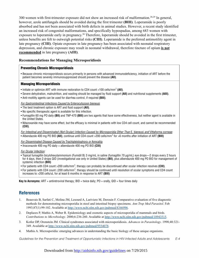
Downloaded from http://aidsinfo.nih.gov/guidelines on 7/29/2015
Guidelines for the Prevention and Treatment of Opportunistic Infections in HIV-Infected Adults and Adolescents E-4
300 women with first-trimester exposure did not show an increased risk of malformation.28,29 In general,however, azole antifungals should be avoided during the first trimester (BIII). Loperamide is poorlyabsorbed and has not been associated with birth defects in animal studies. However, a recent study identifiedan increased risk of congenital malformations, and specifically hypospadias, among 683 women withexposure to loperamide early in pregnancy.30 Therefore, loperamide should be avoided in the first trimester,unless benefits are felt to outweigh potential risks (CIII). Loperamide is the preferred antimotility agent inlate pregnancy (CIII). Opiate exposure in late pregnancy has been associated with neonatal respiratorydepression, and chronic exposure may result in neonatal withdrawal, therefore tincture of opium is notrecommended in late pregnancy (AIII).
Recommendations for Managing Microsporidiosis
Preventing Chronic Microsporidiosis
• Because chronic microsporidiosis occurs primarily in persons with advanced immunodeficiency, initiation of ART before thepatient becomes severely immunosuppressed should prevent the disease (AII).
Managing Microsporidiosis
• Initiate or optimize ART with immune restoration to CD4 count >100 cells/mm3 (AII).
• Severe dehydration, malnutrition, and wasting should be managed by fluid support (AII) and nutritional supplements (AIII).
• Anti-motility agents can be used for diarrhea control, if required (BIII).
For Gastrointestinal Infections Caused by Enterocytozoon bieneusi
• The best treatment option is ART and fluid support (AII).
• No specific therapeutic agent is available for this infection.
• Fumagillin 60 mg PO daily (BII) and TNP-470 (BIII) are two agents that have some effectiveness, but neither agent is available inthe United States.
• Nitazoxanide may have some effect, but the efficacy is minimal in patients with low CD4 cell count, and cannot be recommended(CIII).
For Intestinal and Disseminated (Not Ocular) Infection Caused by Microsporidia Other Than E. bieneusi and Vittaforma corneae:
• Albendazole 400 mg PO BID (AII), continue until CD4 count >200 cells/mm3 for >6 months after initiation of ART (BIII)
For Disseminated Disease Caused by Trachipleistophora or Anncaliia
• Itraconazole 400 mg PO daily + albendazole 400 mg PO BID (CIII)
For Ocular Infection:
• Topical fumagillin bicylohexylammonium (Fumidil B) 3 mg/mL in saline (fumagillin 70 µg/mL) eye drops—2 drops every 2 hoursfor 4 days, then 2 drops QID (investigational use only in United States) (BII), plus albendazole 400 mg PO BID for management ofsystemic infection (BIII)
• For patients with CD4 count >200 cells/mm3, therapy can probably be discontinued after ocular infection resolves (CIII).
• For patients with CD4 count ≤200 cells/mm3, therapy should be continued until resolution of ocular symptoms and CD4 countincreases to >200 cells/uL for at least 6 months in response to ART (BIII)
Key to Acronyms: ART = antiretroviral therapy; BID = twice daily; PO = orally, QID = four times daily
References
1. Beauvais B, Sarfati C, Molina JM, Lesourd A, Lariviere M, Derouin F. Comparative evaluation of five diagnosticmethods for demonstrating microsporidia in stool and intestinal biopsy specimens. Ann Trop Med Parasitol. Feb1993;87(1):99-102. Available at http://www.ncbi.nlm.nih.gov/pubmed/8346996.
2. Deplazes P, Mathis A, Weber R. Epidemiology and zoonotic aspects of microsporidia of mammals and birds.Contributions to Microbiology. 2000;6:236-260. Available at http://www.ncbi.nlm.nih.gov/pubmed/10943515.
3. Kotler DP, Orenstein JM. Clinical syndromes associated with microsporidiosis. Advances in Parasitology. 1998;40:321-349. Available at http://www.ncbi.nlm.nih.gov/pubmed/9554078.
4. Mathis A. Microsporidia: emerging advances in understanding the basic biology of these unique organisms.
Downloaded from http://aidsinfo.nih.gov/guidelines on 7/29/2015
Guidelines for the Prevention and Treatment of Opportunistic Infections in HIV-Infected Adults and Adolescents E-5
International Journal for Parasitology. Jun 2000;30(7):795-804. Available athttp://www.ncbi.nlm.nih.gov/pubmed/10899524.
5. Weber R, Bryan RT, Owen RL, Wilcox CM, Gorelkin L, Visvesvara GS. Improved light-microscopical detection ofmicrosporidia spores in stool and duodenal aspirates. The Enteric Opportunistic Infections Working Group. N Engl JMed. Jan 16 1992;326(3):161-166. Available at http://www.ncbi.nlm.nih.gov/pubmed/1370122.
6. Weiss LM, Vossbrinck CR. Microsporidiosis: molecular and diagnostic aspects. Advances in Parasitology.1998;40:351-395. Available at http://www.ncbi.nlm.nih.gov/pubmed/9554079.
7. Wittner M, Weiss L. The Microsporidia and Microsporidiosis. Washington DC: ASM Press; 1999.8. Stark D, Barratt JL, van Hal S, Marriott D, Harkness J, Ellis JT. Clinical significance of enteric protozoa in the
immunosuppressed human population. Clin Microbiol Rev. Oct 2009;22(4):634-650. Available athttp://www.ncbi.nlm.nih.gov/pubmed/19822892.
9. Sheoran AS, Feng X, Singh I, et al. Monoclonal antibodies against Enterocytozoon bieneusi of human origin. ClinDiagn Lab Immunol. Sep 2005;12(9):1109-1113. Available at http://www.ncbi.nlm.nih.gov/pubmed/16148179.
10. Didier ES, Weiss LM. Microsporidiosis: current status. Curr Opin Infect Dis. Oct 2006;19(5):485-492. Available athttp://www.ncbi.nlm.nih.gov/pubmed/16940873.
11. Goguel J, Katlama C, Sarfati C, Maslo C, Leport C, Molina JM. Remission of AIDS-associated intestinalmicrosporidiosis with highly active antiretroviral therapy. AIDS. Nov 1997;11(13):1658-1659. Available athttp://www.ncbi.nlm.nih.gov/pubmed/9365777.
12. Miao YM, Awad-El-Kariem FM, Franzen C, et al. Eradication of cryptosporidia and microsporidia following successfulantiretroviral therapy. J Acquir Immune Defic Syndr. Oct 1 2000;25(2):124-129. Available athttp://www.ncbi.nlm.nih.gov/pubmed/11103042.
13. Conteas CN, Berlin OG, Speck CE, Pandhumas SS, Lariviere MJ, Fu C. Modification of the clinical course of intestinalmicrosporidiosis in acquired immunodeficiency syndrome patients by immune status and anti-humanimmunodeficiency virus therapy. Am J Trop Med Hyg. May 1998;58(5):555-558. Available athttp://www.ncbi.nlm.nih.gov/pubmed/9598440.
14. Maggi P, Larocca AM, Quarto M, et al. Effect of antiretroviral therapy on cryptosporidiosis and microsporidiosis inpatients infected with human immunodeficiency virus type 1. Eur J Clin Microbiol Infect Dis. Mar 2000;19(3):213-217.Available at http://www.ncbi.nlm.nih.gov/pubmed/10795595.
15. Molina J, J G, Sarfati C. Trial of oral fumagillin for the treatment of intestinal microsporidiosis in patients with HIVinfection (Letter). AIDS. 2000;14:1341-1348.
16. Molina JM, Tourneur M, Sarfati C, et al. Fumagillin treatment of intestinal microsporidiosis. N Engl J Med. Jun 202002;346(25):1963-1969. Available at http://www.ncbi.nlm.nih.gov/pubmed/12075057.
17. Didier PJ, Phillips JN, Kuebler DJ, et al. Antimicrosporidial activities of fumagillin, TNP-470, ovalicin, and ovalicinderivatives in vitro and in vivo. Antimicrob Agents Chemother. Jun 2006;50(6):2146-2155. Available athttp://www.ncbi.nlm.nih.gov/pubmed/16723577.
18. Bicart-See A, Massip P, Linas MD, Datry A. Successful treatment with nitazoxanide of Enterocytozoon bieneusimicrosporidiosis in a patient with AIDS. Antimicrob Agents Chemother. Jan 2000;44(1):167-168. Available athttp://www.ncbi.nlm.nih.gov/pubmed/10602740.
19. Akiyoshi DE, Weiss LM, Feng X, et al. Analysis of the beta-tubulin genes from Enterocytozoon bieneusi isolates from ahuman and rhesus macaque. The Journal of Eukaryotic Microbiology. Jan-Feb 2007;54(1):38-41. Available athttp://www.ncbi.nlm.nih.gov/pubmed/17300517.
20. Franzen C, Salzberger B. Analysis of the beta-tubulin gene from Vittaforma corneae suggests benzimidazole resistance.Antimicrob Agents Chemother. Feb 2008;52(2):790-793. Available at http://www.ncbi.nlm.nih.gov/pubmed/18056284.
21. Diesenhouse MC, Wilson LA, Corrent GF, Visvesvara GS, Grossniklaus HE, Bryan RT. Treatment of microsporidialkeratoconjunctivitis with topical fumagillin. Am J Ophthalmol. Mar 15 1993;115(3):293-298. Available athttp://www.ncbi.nlm.nih.gov/pubmed/8117342.
22. Dieterich DT, Lew EA, Kotler DP, Poles MA, Orenstein JM. Treatment with albendazole for intestinal disease due toEnterocytozoon bieneusi in patients with AIDS. J Infect Dis. Jan 1994;169(1):178-183. Available at
Downloaded from http://aidsinfo.nih.gov/guidelines on 7/29/2015
Guidelines for the Prevention and Treatment of Opportunistic Infections in HIV-Infected Adults and Adolescents E-6
http://www.ncbi.nlm.nih.gov/pubmed/8277179.23. Molina JM, Chastang C, Goguel J, et al. Albendazole for treatment and prophylaxis of microsporidiosis due to
Encephalitozoon intestinalis in patients with AIDS: a randomized double-blind controlled trial. J Infect Dis. May1998;177(5):1373-1377. Available at http://www.ncbi.nlm.nih.gov/pubmed/9593027.
24. Dionisio D, Manneschi LI, Di Lollo S, et al. Persistent damage to Enterocytozoon bieneusi, with persistent symptomaticrelief, after combined furazolidone and albendazole in AIDS patients. J Clin Pathol. Oct 1998;51(10):731-736.Available at http://www.ncbi.nlm.nih.gov/pubmed/10023334.
25. Sriaroon C, Mayer CA, Chen L, Accurso C, Greene JN, Vincent AL. Diffuse intra-abdominal granulomatous seeding asa manifestation of immune reconstitution inflammatory syndrome associated with microsporidiosis in a patient withHIV. AIDS Patient Care STDS. Aug 2008;22(8):611-612. Available at http://www.ncbi.nlm.nih.gov/pubmed/18627278.
26. Ndyomugyenyi R, Kabatereine N, Olsen A, Magnussen P. Efficacy of ivermectin and albendazole alone and incombination for treatment of soil-transmitted helminths in pregnancy and adverse events: a randomized open labelcontrolled intervention trial in Masindi district, western Uganda. Am J Trop Med Hyg. Dec 2008;79(6):856-863.Available at http://www.ncbi.nlm.nih.gov/pubmed/19052293.
27. Heinonen OP, Slone D, Shapiro S. Birth Defects and Drugs in Pregnancy. Littleton: Publishing Sciences Group; 1977.28. De Santis M, Di Gianantonio E, Cesari E, Ambrosini G, Straface G, Clementi M. First-trimester itraconazole exposure
and pregnancy outcome: a prospective cohort study of women contacting teratology information services in Italy. DrugSaf. 2009;32(3):239-244. Available at http://www.ncbi.nlm.nih.gov/pubmed/19338381.
29. Bar-Oz B, Moretti ME, Bishai R, et al. Pregnancy outcome after in utero exposure to itraconazole: a prospective cohortstudy. Am J Obstet Gynecol. Sep 2000;183(3):617-620. Available at http://www.ncbi.nlm.nih.gov/pubmed/10992182.
30. Kallen B, Nilsson E, Otterblad Olausson P. Maternal use of loperamide in early pregnancy and delivery outcome. Actapaediatrica. May 2008;97(5):541-545. Available at http://www.ncbi.nlm.nih.gov/pubmed/18394096.
Downloaded from http://aidsinfo.nih.gov/guidelines on 7/29/2015
Guidelines for the Prevention and Treatment of Opportunistic Infections in HIV-Infected Adults and Adolescents F-1
Mycobacterium tuberculosis Infection and Disease (Last updatedMay 7, 2013; last reviewed May 7, 2013)
Epidemiology
Tuberculosis (TB) infection occurs when a susceptible person inhales droplet nuclei containingMycobacterium tuberculosis organisms. The immune response usually limits multiplication of tuberclebacilli within 2 to 12 weeks after infection. However, viable bacilli persist for years, a condition referred toas latent TB infection (LTBI). Individuals with LTBI are asymptomatic and are not infectious. TB disease(clinically active disease, often with positive cultures) can develop soon after exposure (primary disease) orafter reactivation of latent infection.
In individuals with LTBI, the risk of reactivation with TB disease increases very soon after HIV infection.1The estimated annual risk of reactivation with TB disease among those with untreated HIV infection andLTBI is 3% to 16% and approximates the lifetime risk for HIV-uninfected individuals with LTBI (~5%).2 TBdisease can occur at any CD4 T lymphocyte (CD4 cell) count, although the risk increases with progressiveimmunodeficiency.3
Antiretroviral therapy (ART) results in a prompt and marked decrease in the incidence of TB disease, aneffect that has been documented in settings with low4 and high case rates.5,6 Even with the beneficial effectsof ART, HIV-infected patients remain at higher risk of TB disease than the general population.7
Rates of TB in the United States are declining, with 3.6 new cases per 100,000 population reported in 20108
(a total of 11,182 cases). The prevalence of LTBI in the general population of the United States is 4%.9 Theincidence of HIV-related TB has declined more rapidly than the rate of active TB in the general population,10
which is probably related to the widespread use of ART. In recent years there have been fewer than 1000 newcases of HIV/TB co-infection identified per year in the United States.8,11,12
As with TB in the general U.S. population, HIV-related TB disease is increasingly seen in people bornoutside of the United States.10 Notably, TB disease has not decreased significantly in recent years amongforeign-born persons with HIV disease in the United States.10,13
Despite these favorable epidemiological trends, TB remains an important opportunistic illness in the UnitedStates. In the era of potent ART, TB disease remained the second most common initial opportunistic illness inNew York City.14 Unlike most opportunistic infections (OIs), TB is transmissible, particularly to others whoare HIV-infected. Therefore, clinicians caring for patients with HIV must remain vigilant in efforts to preventTB, knowledgeable about the clinical presentation of HIV-related TB, and cognizant of the complexities ofcotreatment of HIV and TB.
Preventing Exposure
The most common predisposing factor for TB is birth or residence outside of the United States. Therefore,patients with HIV infection who travel or work internationally in settings with a high prevalence of TBshould be counseled about the risk of TB acquisition and the advisability of testing for LTBI upon return(AIII). Although some health care and correctional settings in the United States present risks of TBexposure, HIV-infected individuals need to take no precautions beyond those recommended for anyone inthose environments (AIII).
Preventing Disease–Diagnosis and Treatment of Latent TB Infection
The estimated annual risk of active TB among HIV-infected patients with LTBI is 3 to 12 times higher thanfor the general population.15,16 Furthermore, development of HIV-related TB increases viral load17 and therisk of HIV disease progression17 and death18 compared with CD4-matched, HIV seropositive controls.
Downloaded from http://aidsinfo.nih.gov/guidelines on 7/29/2015
Guidelines for the Prevention and Treatment of Opportunistic Infections in HIV-Infected Adults and Adolescents F-2
Among HIV-infected individuals, treatment of LTBI decreases the risk of TB disease by 62% and the risk ofdeath by 26%.19 Therefore, prevention of TB disease by screening for and appropriately treating LTBI is akey component of HIV care.
Diagnosis of Latent Tuberculosis InfectionTesting for LTBI at the time of HIV diagnosis should be routine, regardless of an individual’s epidemiologicalrisk of TB exposure. Individuals with negative diagnostic tests for LTBI who have advanced HIV infection(CD4 cell count <200 cells/mm3) and no indications for initiating empiric LTBI treatment should be retested forLTBI once they start ART and attain a CD4 count ≥200 cells/mm3.20,21 Annual testing for LTBI is recommendedonly for HIV-infected patients who are at high risk of repeated or ongoing exposure to those with active TB.
Traditionally, LTBI has been defined by the presence of a positive tuberculin skin test (TST) (≥5 mm ofinduration at 48–72 hours) in individuals with no clinical or radiographic evidence of TB disease. Althoughexperience with the TST in HIV-infected individuals is extensive, it has several disadvantages: the requirementfor two visits to place and read the test, decreased specificity in those who received Bacillus Calmette-Guerin(BCG) vaccination, and decreased sensitivity in those with advanced immunodeficiency.22 Limitations of theTST have led to interest in interferon-gamma release assays (IGRAs) for detection of LTBI.
Current evidence suggests that IGRAs have higher specificity (92%–97%) than TST (56%–95%), bettercorrelation with surrogate measures of exposure to M. tuberculosis,23 and less cross reactivity because ofBCG vaccination or other non-tuberculous mycobacteria exposure.24,25 Three IGRAs are Food and DrugAdministration (FDA) approved and available in the United States. Progressive immunodeficiency isassociated with decreased sensitivity of IGRAs, although immunodeficiency may have less impact on thesensitivity of IGRAs than on the sensitivity of TST.26
In HIV-infected patients, the correlation between TST and IGRAs is poor to moderate.27,28 In prospectivestudies, positive results with either TST or IGRA were associated with an increased risk of developing TBdisease;29,30 in some studies, patients with a positive IGRA were at a higher risk of subsequently developingTB disease than were those with a positive TST.31,32 For all of its limitations, TST response remains stronglypredictive of response to isoniazid preventive therapy among those with HIV infection.19 Whether the sameis true of the IGRAs remains to be demonstrated.
In programmatic settings in the United States, TB screening based on the TST has been suboptimal, withonly 47% to 65% of patients completing screening.33-35 A higher proportion of patients may completescreening for TB if testing is done with IGRAs.
No definitive comparisons have been done of TST and IGRAs for screening HIV-infected individuals in low-burden settings such as the United States. Both TST and the FDA-approved IGRAs are appropriate for TBscreening in HIV-infected individuals.36 Some experts have suggested using both TST and IGRA to screenfor LTBI, but the predictive value of this approach is unclear, and it would be more expensive and moredifficult to implement. Routine use of both TST and IGRAs to screen for LTBI is not recommended in theUnited States.36
Patients with TB disease often demonstrate immune reactivity against M. tuberculosis in TST and IGRAtesting. Therefore, any positive result with TST or IGRA should trigger expeditious evaluation for thepossibility of active TB. Most, but not all, HIV-infected individuals with TB disease have symptoms; theabsence of any symptoms has a 97% negative predictive value for culture-positive TB.37 The addition of achest radiograph improves the sensitivity of symptom screening algorithms. Sputum culture is the goldstandard for diagnosing pulmonary TB disease but is not cost effective for screening HIV-infected patientswho are asymptomatic, particularly in the United States, where TB prevalence is very low. Therefore,screening for symptoms (asking for cough of any duration) coupled with chest radiography is recommendedto exclude TB disease in a patient with a positive TST or IGRA.
Downloaded from http://aidsinfo.nih.gov/guidelines on 7/29/2015
Guidelines for the Prevention and Treatment of Opportunistic Infections in HIV-Infected Adults and Adolescents F-3
When to Start Primary Prophylaxis (i.e., Treating Latent Tuberculosis Infection)HIV-infected individuals who test positive for LTBI but have no evidence of TB disease should receive LTBItreatment (AI). HIV-infected close contacts of anyone who has infectious TB also should receiveprophylaxis, regardless of results of screening tests for LTBI (AII). Notably, for HIV-infected individualswho are anergic and have not had recent contact with anyone with infectious TB, treatment of LTBI is notassociated with clinical benefit and is not recommended (AI).38-41
Preferred and Alternative Drugs for Treatment of Latent Tuberculosis Infection Isoniazid administered for 9 months remains the preferred therapy, with proven efficacy, good tolerability,and infrequent severe toxicity (AII). Isoniazid can potentiate the risk of peripheral neuropathy when usedwith some antiretroviral (ARV) drugs, most notably the dideoxynucleosides (didanosine, stavudine), whichare seldom used in clinical practice in the United States. Isoniazid, when used with efavirenz- or nevirapine-based regimens, does not significantly increase risk of hepatitis—the most important adverse effect.42,43
Isoniazid should be supplemented with pyridoxine at a dose of 25 mg/day to prevent peripheral neuropathy(AIII). A significant disadvantage of the 9-month regimen is that most patients in the United States andCanada do not complete all 9 months of therapy.44-46 Shorter regimens are more likely to be completed.44-46
Recent data from an open-label, randomized non-inferiority trial comparing a 3-month regimen of isoniazidplus rifapentine, given by directly observed therapy (DOT) once weekly, with a 9-month regimen of self-administered once daily isoniazid demonstrated that, after 33 months of follow-up, the 3-monthisoniazid-rifapentine regimen was as effective as the 9-month isoniazid regimen.47 The shorter courseregimen had the advantage of a higher completion rate. These results led to a recent Centers for DiseaseControl and Prevention (CDC) recommendation that 3-months of once weekly isoniazid-rifapentine given byDOT can be used as an equal alternative to the standard 9-month regimen.48 However, the 3-month regimenis not recommended for HIV-infected patients receiving ART because of potentially significant druginteractions between rifapentine and some ARV drugs (AIII).48 Other alternative therapies forchemoprophylaxis are shown in Table 1; the regimen of 2 months rifampin plus pyrazinamide is notrecommended because of the risk of severe and sometimes fatal hepatotoxicity (AII). Rifampin- orrifabutin-containing regimens may require dose adjustments of ARV or rifabutin (Table 5).
LTBI treatment and ART act independently to decrease the risk of TB disease.49-51 Therefore, use of bothinterventions is recommended for those who have LTBI and an indication for ART (AII).
Monitoring of Response to Treatment of Latent Tuberculosis InfectionPatients receiving daily LTBI treatment through self-administration should be seen by the prescribing clinicianon a monthly basis to assess adherence and evaluate for possible drug toxicity; generally, not more than 1month’s supply of drugs should be prescribed. Individuals taking a twice-weekly regimen should receive LTBItreatment by direct observation. Risk of hepatitis from isoniazid prophylaxis may not be higher in HIV-infectedindividuals than those who are uninfected, but baseline measurements of serum aspartate aminotransferase(AST) or alanine aminotransferase (ALT) and total bilirubin are recommended, and if results are abnormal,testing should be repeated.52 Individuals with concomitant chronic viral hepatitis may be at increased risk ofisoniazid-related hepatotoxicity, and they should be treated for LTBI and closely monitored. With isoniazid,liver enzymes typically increase in the first 3 months but then (through the process of hepatic adaptation) returnto normal despite continued therapy. LTBI treatment should be stopped in asymptomatic patients who have amore-than-fivefold increase in AST levels above the upper limit of normal (ULN), symptomatic patients whohave a more-than-threefold increase above ULN in AST levels, and patients regardless of symptoms withbaseline abnormal transaminases who have a more-than-twofold increase above their baseline AST levels.Patients should be reminded at each visit about potential adverse effects (i.e., unexplained anorexia, nausea,vomiting, dark urine, icterus, rash, persistent paresthesias of the hands and feet, persistent fatigue, weakness orfever lasting 3 days or more, abdominal tenderness, easy bruising or bleeding, and arthralgia) and told toimmediately stop isoniazid and return to the clinic for an assessment.
Downloaded from http://aidsinfo.nih.gov/guidelines on 7/29/2015
Guidelines for the Prevention and Treatment of Opportunistic Infections in HIV-Infected Adults and Adolescents F-4
The ultimate decision regarding resumption of therapy with the same or a different agent for LTBI treatmentshould be made after weighing the risk of additional hepatic injury against the benefit of preventingprogression to TB disease52 and in consultation with an expert in treating LTBI.
Clinical Manifestations of Tuberculosis Disease
Common clinical symptoms of TB disease include productive cough, fever, sweats, weight loss, and fatigue.Culture-positive TB disease can be sub-clinical or oligo-symptomatic.37 After initiation of ART, immunereconstitution can unmask subclinical active TB, resulting in pronounced inflammatory reactions at the sitesof infection.
In HIV-infected individuals, the presentation of active TB disease is influenced by the degree ofimmunodeficiency.53,54 In HIV-infected patients without pronounced immunodeficiency, (that is, CD4 cellcounts >350 cells/mm3), HIV-related TB clinically resembles the disease seen in HIV-uninfected patients.Most patients have disease limited to the lungs, and common chest radiographic manifestations include upperlobe fibronodular infiltrates with or without cavitation.55 Extrapulmonary disease is more common in HIV-infected individuals than in those who are uninfected, regardless of CD4 cell counts, although clinicalmanifestations are not substantially different from those described in HIV-uninfected individuals. TB must beconsidered in disease processes involving any site in the body,56 but especially those related to centralnervous system (CNS) or meningeal symptoms in which early TB treatment is essential to improveoutcomes.57,58
In patients with advanced HIV disease, the chest radiographic findings of pulmonary TB are markedlydifferent than those in patients with less severe immunosuppression. Lower lobe, middle lobe, interstitial,and miliary infiltrates are common and cavitation is less common.53,55,59 Intrathoracic lymphadenopathy iscommon, with mediastinal involvement seen more often than hilar adenopathy. Even with normal chestradiographs, patients with HIV infection and pulmonary TB may test positive on acid-fast bacilli (AFB)sputum smears and cultures, particularly if they have cervical node involvement.
The greater the degree of immunodeficiency, the higher the likelihood of extrapulmonary TB, such aslymphadenitis; pleuritis; pericarditis; and meningitis, all with or without pulmonary involvement, and it isfound in most TB patients with CD4 cell counts <200 cells/mm3.54 In these individuals, TB can be a severesystemic disease with high fevers, rapid progression, and sepsis syndrome.60
Histopathologic findings also are affected by the degree of immunodeficiency. Patients with relatively intactimmune function have typical granulomatous inflammation associated with TB disease. With progressiveimmunodeficiency, granulomas become poorly formed or may be completely absent.54
Diagnosis of Tuberculosis Disease
Initial diagnostic testing is directed at the anatomic site of symptoms or signs, such as the lungs, lymphnodes, and cerebrospinal fluid (CSF). Even in the absence of pulmonary symptoms or signs, the initialevaluation of a patient suspected of having HIV-related TB should always include a chest radiograph;pulmonary involvement is common whatever the CD4 cell count.37,61 However, chest radiography is animperfect screen for sputum culture-positive TB, particularly in patients with advanced immunodeficiency.Therefore, sputum smear and culture should be considered in symptomatic patients with normal chestradiographs who are being evaluated for possible TB disease.
Sputum smear-negative TB is common in HIV-infected patients, particularly those with advancedimmunodeficiency and noncavitary disease.62 However, the yield of sputum mycobacterial culture is notaffected by HIV or the degree of immunodeficiency. If a sensitive broth culture technique is used, thesensitivity of sputum culture is quite high. Smear and culture of three sputum specimens is recommended, inthat there was a 10% incremental yield for broth culture between the second and third specimens in a recentlarge study of patients with HIV.63
Downloaded from http://aidsinfo.nih.gov/guidelines on 7/29/2015
Guidelines for the Prevention and Treatment of Opportunistic Infections in HIV-Infected Adults and Adolescents F-5
Nodal involvement is common in HIV-related TB, and the combined yield of histopathology, smear, andculture from needle aspirates of enlarged lymph nodes is quite high.64 Pleural fluid, pericardial fluid, ascites,and CSF should be sampled if there is clinical evidence of involvement. The yield of mycobacterial urine andblood cultures depends upon the clinical setting; in patients with advanced immunodeficiency, the yield ofculture from these two readily available body fluids can be relatively high54,56 and may allow definitivediagnosis and a source for an isolate for drug-susceptibility testing.
Nucleic-acid amplification (NAA) tests provide rapid diagnosis of TB, in contrast to the prolonged timeneeded for detection of mycobacterial growth, and can be considered for patients with advancedimmunodeficiency who are at risk of rapid clinical progression of TB (some assays also provide rapiddetection of drug resistance as discussed below). NAA tests have at least two uses in patients with suspectedHIV-related TB. First, they are highly predictive of TB in specimens that are AFB smear-positive. Non-tuberculous mycobacterial infections are relatively common in patients with advanced immunodeficiency,and NAA tests can be used to direct therapy and make decisions about the need for respiratory isolation inpatients with a smear-positive specimen. Second, NAA tests are more sensitive than AFB smear, producingpositive results for 50% to 80% of smear-negative, culture-positive specimens.65,66 Therefore, the use of aNAA test is recommended on at least one specimen from all patients being evaluated for suspectedpulmonary TB.67 The NAA tests currently available are licensed only for evaluation of sputum samples;much less experience exists with samples from extrapulmonary sites.
Immunological screening for TB with TST and IGRA may be helpful in unusual circumstances that make itdifficult to obtain definitive culture evidence for active TB; evidence of prior infection increases thelikelihood that a clinical illness may be TB. A negative test, however, should never be interpreted as rulingout TB disease.
Drug-susceptibility testing should be performed on the initial isolates from all patients suspected of havingTB because resistance to isoniazid and/or rifampin is associated with an increased risk of treatment failure,recurrent TB, and amplification of resistance to additional TB medications.68 The presence of multidrug-resistant TB (MDR TB; defined as resistance to at least isoniazid and rifampin) or extensively drug-resistantTB (XDR TB; defined as MDR TB with additional resistance to a fluoroquinolone and either kanamycin,amikacin, or capreomycin) is associated with a markedly increased risk of death.69 Thus, early identificationof drug resistance, with appropriate adjustment of therapy based on results, is critical to successfully treatingTB disease and to curbing transmission of drug-resistant M. tuberculosis.
Drug-susceptibility testing to first-line TB drugs (i.e., isoniazid, rifampin, ethambutol, and pyrazinamide)should be performed on all patients with TB disease, regardless of the source of the specimen. These testsshould be repeated if sputum cultures remain positive for M. tuberculosis at or after 4 months of treatment orbecome positive 1 month or longer after culture conversion to negative. Drug susceptibility testing forsecond-line TB medications (e.g., fluoroquinolones, aminoglycosides, capreomycin, ethionamide) should beperformed only in reference laboratories that have substantial experience with these techniques and should belimited to specimens with resistance to first-line TB medications.
Conventional drug-susceptibility testing is widely used and has been well validated for first-line drugs. Thedisadvantage of this technique, however, is the combined turnaround time for culture and drug-susceptibilitytesting, which can be as long as 6 weeks70 because of the slow growth of M. tuberculosis in culture. Duringthis time, patients with drug-resistant TB may be receiving ineffective, empiric first-line TB therapy, whichcould allow for ongoing transmission, further clinical deterioration, and death—particularly in HIV-infectedindividuals.69
Genotypic testing, which identifies drug-resistance mutations, allows rapid detection of resistance. Therelationship between these mutations and drug resistance has been studied for a number of TB medications,71
and commercial tests have been developed and validated to identify genotypic resistance for rifampin65,72 andisoniazid.72 Development is under way of commercial tests to identify genotypic resistance to other TB
Downloaded from http://aidsinfo.nih.gov/guidelines on 7/29/2015
Guidelines for the Prevention and Treatment of Opportunistic Infections in HIV-Infected Adults and Adolescents F-6
medications.73 Genotypic assays can provide a result in 24 hours and can be performed directly on sputumspecimens.
Clinicians who suspect that an HIV-infected patient has drug-resistant TB should make every effort toexpedite diagnosis. In the United States, the CDC Division of TB Elimination has a Molecular Detection ofDrug Resistance service to make rapid molecular testing for first-and second-line TB medications availablefor patients who have or are suspected to have TB and do not have local access to such testing(http://www.cdc.gov/tb/topic/laboratory/default.htm).
Drug resistance should be considered in any patient with:
• known exposure to an individual with drug-resistant TB• residence in a setting with high rates of primary drug-resistant TB, such as a country or area with high
rates of drug-resistant TB in newly diagnosed patients74
• persistently positive smear or culture results at or after 4 months of treatment• previous TB treatment, particularly if it was not directly observed or was interrupted for any reason.
Treating Disease
In some settings in the United States, non-tuberculous mycobacterial infections are more common than TBamong HIV-infected patients. However, because TB is highly virulent and represents a greater risk oftransmission to others, treatment for it is more urgent than for non-tuberculous mycobacterial infections.Furthermore, first-line TB drugs are highly active against Mycobacterium kansasii, a relatively common non-tuberculous mycobacterial infection that presents clinically and radiographically like TB.75 Finally, withappropriate access to broth culture and molecular diagnostics (NAA and genotypic tests for resistance), thetime between finding a smear-positive specimen and identifying the species should be short.
TB in individuals with advanced immunodeficiency can be rapidly progressive and fatal if treatment isdelayed. Furthermore, such patients often have smear-negative sputum specimens. Therefore, after collectionof available specimens for culture and molecular diagnostic tests, empiric treatment for TB is warranted inpatients with clinical and radiographic presentation suggestive of HIV-related TB (AIII).
Treatment of suspected TB in HIV-infected individuals is the same as for those who are HIV uninfected andshould include an initial four-drug combination of isoniazid, rifampin, pyrazinamide, and ethambutol (AI).An expanded initial regimen—including at least moxifloxacin or levofloxacin and an aminoglycoside orcapreomycin—should be used if there is a significant concern about resistance to rifampin, with or withoutresistance to other drugs (BIII). A TB expert should be consulted if drug resistance is suspected. DOT isrecommended for all patients with suspected HIV-related TB (AII). The likelihood of treatment success isfurther enhanced with comprehensive case management, assistance with housing and other social support,and assistance in establishing or re-engaging with HIV care, if needed (i.e., enhanced DOT).
Drug-susceptible TB is treated with a 2-month intensive phase of the 4 drugs previously listed. Ethambutolcan be discontinued when susceptibility to isoniazid and rifampin has been confirmed. Pyrazinamide may bediscontinued after 2 months. Thereafter, isoniazid and a rifamycin are used in the continuation phase oftherapy.
Intermittent dosing (administration less often than daily) of anti-TB treatment facilitates DOT. However,regimens that included twice- or thrice-weekly dosing during the intensive phase have been associated with anincreased risk of treatment failure or relapse with acquired drug resistance to the rifamycin class.76-78 Therefore,daily therapy (5–7 days per week) given as DOT is recommended during the intensive phase (AII).
Daily (5–7 days per week) or thrice-weekly dosing is recommended during the continuation phase of therapy(AII). Regimens that included once- or twice-weekly dosing during the continuation phase of therapy were
Downloaded from http://aidsinfo.nih.gov/guidelines on 7/29/2015
Guidelines for the Prevention and Treatment of Opportunistic Infections in HIV-Infected Adults and Adolescents F-7
associated with increased risk of treatment failure or relapse with acquired rifamycin resistance.79,80 Whetherthere is a difference between daily and thrice-weekly dosing during the continuation phase of therapy has notbeen adequately studied in randomized trials; in observational studies and a meta-analysis, thrice-weeklytherapy during the continuation phase was not associated with an increased risk of adverse TB outcomes(i.e., treatment failure, recurrence, or acquired drug resistance).81
The optimal duration of TB treatment for patients with HIV infection and drug-susceptible TB disease isunknown. In general, the outcomes have been good with 6-month regimens (2 months of isoniazid, rifampin,pyrazinamide, and ethambutol, followed by 4 months of isoniazid and rifampin) given as DOT to patientswith HIV co-infection. A randomized trial in the United States showed excellent and comparable outcomesof TB therapy among patients assigned to 6 months or 9 months of therapy, but the trial was underpowered.82
Two trials in high-burden settings showed higher risks of recurrent TB among patients treated with 6 monthsof therapy compared with those assigned to 9-76 or 12-month regimens.83 However, the applicability of thesetwo trials is uncertain in low-burden settings in which ART is used, such as the United States.
Pending the outcome of further studies, 6 months of therapy for most patients with HIV-related, drug-susceptible TB disease is recommended (BII). Extension of therapy to 9 months is recommended for thosewith a positive 2-month sputum culture (BII). Extension of therapy to 9 to 12 months is also recommendedfor patients with CNS involvement (BII). Treatment for 6 to 9 months is recommended for patients withbone and joint TB (BII). The duration of therapy should be based on number of doses received, not oncalendar time (BIII) because there may be substantial differences between dose number and calendar time ifdoses were missed due to poor adherence or for management of problems with tolerability or toxicity.
Adjunctive corticosteroid therapy increases survival for patients with HIV-related TB involving the CNS84
and pericardium85 (AI). No trials to date have compared different doses and treatment durations of adjunctivecorticosteroids. Dexamethasone was used in trials of adjunctive corticosteroids for CNS disease (0.3–0.4mg/kg/day for 2–4 weeks, then taper 0.1 mg/kg per week until dose of 0.1 mg/kg, then 4 mg per day andtaper by 1 mg/week; total duration of 12 weeks); prednisone or prednisolone was used in trials of pericardialdisease (60 g/day and taper 10 mg per week; total duration of 6 weeks).
Special Considerations with Regard to Starting ART Optimal management of HIV-related TB requires that both infections be addressed; sequential treatment ofTB followed by HIV treatment is not recommended.86 Co-treatment of HIV and TB is complex because ofthe adherence demands of multidrug therapy for two infections, drug-drug interactions between therifamycins and many ARV drugs, overlapping side effect profiles of antituberculosis and ARV drugs, and thefrequency of immune reconstitution inflammatory syndrome (IRIS). Despite these substantial clinicalchallenges, co-treatment of HIV-related TB improves survival,86 particularly in patients with CD4 counts <50cells/mm3; decreases the risk of additional opportunistic illnesses including TB;87 can achieve high rates ofviral suppression;88 and may improve TB treatment outcomes.89
Starting ART early in the course of TB treatment can complicate clinical management because of increasedpill burden, drug toxicities, drug interactions, and IRIS events. However, recently completed randomizedclinical trials demonstrate that ART can be safely given during TB treatment without jeopardizing HIVtreatment responses and that ART reduces mortality and HIV-related illnesses.86-88
The SAPIT trial randomized 642 South African adults with CD4 cell counts <500/mm3 and AFB smear-positive TB to start ART according to one of three strategies; at TB treatment initiation; after the intensivephase of TB therapy but before TB treatment completion; or after TB treatment completion.86 The study wasstopped early when the mortality of the 2 integrated treatment arms was 56% lower than the sequentialtreatment arm, demonstrating that ART should be started before completion of TB treatment. Notably, therewas a survival benefit across the range of CD4 cell counts among patients enrolled, including within thestratum of baseline CD4 counts from 200 to 500 cells/mm3. Updated results of the SAPIT trial indicated thatthe benefit of early ART was greatest for those with CD4 counts of <50 cells/mm3 and that individuals with
Downloaded from http://aidsinfo.nih.gov/guidelines on 7/29/2015
Guidelines for the Prevention and Treatment of Opportunistic Infections in HIV-Infected Adults and Adolescents F-8
higher CD4 cell counts who started ART within the first 4 weeks of the continuation phase of TB treatmenthad a lower incidence of IRIS and adverse events.90
The CAMELIA and A5221 trials shed further light on the optimal timing of ART during the course of TBtreatment. In CAMELIA, 661 adults in Cambodia with confirmed pulmonary TB and a median CD4 count of25 cells/mm3 (interquartile range [IQR] 10,56) were randomized to receive ART at 2 or 8 weeks after startingTB treatment. The risk of death was decreased from 27% in the 8-week arm to 18% in the 2-week arm and,among those who survived, viral suppression rates were very high (>95%).88 The ACTG A5221 studyrandomized 809 patients from North America, South America, Africa, and Asia with confirmed or suspectedTB and a median CD4 count of 77 cells/mm3 (IQR 33,146) to immediate ART (within 2 weeks) or early ART(8–12 weeks).87 A new OI or death occurred among 12.9% of patients in the immediate arm and 16.1% in theearly arm by week 48 (P = 0.45). In patients with screening CD4 counts <50 cells/mm3, 15.5% of patients onthe immediate arm versus 26.6% on early ART experienced AIDS or death, (P = 0.02). Tuberculous-associated IRIS (TB-IRIS) was more common in the immediate ART arm (11%) compared with the earlyarm (5%) (P = 0.002). Viral suppression rates were similar between the arms.
Other recently completed smaller and non-randomized studies provide further support for early ARTinitiation. In the PART study, which included only patients with TB and HIV with CD4 cell counts >350cells/mm3, even a short 6-month course of ART started at TB diagnosis resulted in lower rates of AIDS ordeath compared with delaying ART until a CD4 threshold of 250 cells/mm3.91 A recent retrospective analysisof HIV-infected adults with XDR TB showed a 62% reduction in mortality in those who received ART.92
The optimal strategy in TB meningitis is less clear. A randomized trial conducted in Vietnam compared ARTinitiated immediately or 2 months after starting TB treatment in 253 patients with HIV-related TBmeningitis.93 This study did not show a survival benefit for early initiation of ART. On the contrary, earlyART was associated with significantly more severe adverse events (102) compared with the deferred ARTarm (87; P = 0.04). The overall mortality rates and severe adverse event rates in this study wereextraordinarily high (58% and 89–90%, respectively), in part reflecting the very ill AIDS population, andmay not be generalizable to other settings. Nonetheless, caution in early ART initiation is warranted inpatients with tuberculous meningitis.
When TB occurs in patients already on ART, treatment for TB must be started immediately (AIII), and ARTshould be modified to reduce the risk of drug interactions and maintain virologic suppression. When TBoccurs in the setting of virologic failure, ART drug-resistance testing should be performed and a new ARTregimen constructed, along with intensified adherence counseling to achieve virologic suppression andminimize drug interactions with the anti-TB regimen.
In summary, ART is recommended in all HIV-infected persons with TB (AI). For ART-naive patients, ARTshould be started within 2 weeks when the CD4 count is <50 cells/mm3 and by 8 to 12 weeks for all others(AI). Given the need for the initiation of five to seven new medications in a short time, adherence supportshould be offered. In patients with TB meningitis and low CD4 cell counts, early ART may pose a risk thatcalls for careful monitoring and consultation with experts. Early ART initiation requires close collaborationbetween HIV and TB care clinics, expertise in management of ART regimen selection, and support andadherence services for clients.
Drug-drug interactions in the treatment of HIV-related tuberculosisThe rifamycin class of antibiotics is the key to effective, short-course TB treatment. However, the rifamycinscurrently available (rifampin, rifabutin, and rifapentine) have clinically significant interactions with a numberof ARV drugs (Table 5). These drug-drug interactions are complex, but most result from the potent inductionby the rifamycin of genes involved in the metabolism and transport of ARV agents.
The preferred cotreatment regimen for HIV-related TB disease is rifampin-based TB therapy with an ARVregimen of efavirenz plus two nucleoside(tide) analogues (AII). Efavirenz-based ART is associated with
Downloaded from http://aidsinfo.nih.gov/guidelines on 7/29/2015
Guidelines for the Prevention and Treatment of Opportunistic Infections in HIV-Infected Adults and Adolescents F-9
excellent TB and HIV treatment outcomes and has low rates of serious toxicity.94 Data conflict on themagnitude of the change in efavirenz concentrations when co-administered with rifampin. Early studiesreported a 26% reduction in efavirenz exposure,95 but more recent and larger studies in HIV-infected patientswith TB (including patients with higher body weight) have not shown a significant effect of rifampin onefavirenz exposure.96,97 Previous recommendations to increase the dose of efavirenz, especially in patientswho weigh >60 kg, are thus not supported by good data and have several disadvantages; complexity ofdosing, inability to take advantage of the simplicity of the co-formulation of efavirenz, tenofovir, andemtricitabine, and possibility of increased neuropsychiatric side effects. Given the excellent treatmentoutcomes of co-treatment with standard-dose efavirenz,94,98 the 600-mg daily dose of efavirenz isrecommended (BII).
Rifampin has a more significant effect on the concentration of nevirapine, but clinical outcomes have beenreasonably good among patients on a co-treatment regimen of rifampin-based TB treatment with an ARVregimen of nevirapine plus two nucleoside analogues.94,99,100 However, a recent randomized controlled trialshowed that a once daily nevirapine regimen used with didanosine and lamivudine was inferior to a oncedaily efavirenz regimen used with the same NRTIs in HIV-associated TB treated with a rifampin regimen.101
For patients absolutely unable to take efavirenz due to intolerance or early pregnancy, nevirapine-based ARTcan be used, but the lead-in dose of nevirapine should be omitted for patients who are established onrifampin for at least 2 weeks and plasma HIV RNA levels should be monitored closely.94
For patients who have HIV strains resistant to non-nucleoside reverse transcriptase inhibitors (NNRTIs) orare unable to tolerate efavirenz and nevirapine, the preferred co-treatment regimen is rifabutin-based TBtherapy with an ARV regimen that includes a ritonavir-boosted protease inhibitor (PI) (BIII). The dramaticeffects of rifampin on serum concentrations of lopinavir can be overcome by high-dose ritonavir,102 but highrates of hepatotoxicity have been reported when adjusted ritonavir-boosted PIs were given with rifampin tohealthy volunteers.103-105 Rifabutin has little effect on ritonavir-boosted lopinavir106 or atazanavir,107 and itsco-administration results in moderate increases in darunavir108 and fosamprenavir concentrations.109
However, all PIs markedly increase serum concentrations of rifabutin (and one of its principal metabolites,desacetyl-rifabutin). Therefore, the dose of rifabutin must be decreased to avoid dose-related toxicity, such asuveitis.110 The magnitude of the dose reduction for rifabutin remains somewhat controversial. In studies ofhealthy volunteers, a 150-mg dose every other day together with a ritonavir-boosted PI achieved serumconcentrations of rifabutin comparable to or higher (with much higher concentrations of the desacetylmetabolite) than those achieved with 300 mg rifabutin daily in the absence of a PI.108,109,111 However, amongHIV-infected individuals with TB, there have been case reports of acquired rifamycin resistance with 150-mgthrice-weekly dosing in the presence of a boosted PI-based ARV regimen.112,113
Pending additional data, we recommend a dosage of 150 mg of rifabutin daily (at least during the first 2months of TB treatment) for patients who are on a PI-containing ARV regimen (BIII). Therapeutic drugmonitoring for rifabutin can be considered in this situation.113 Close monitoring of adherence to ART isimportant because these reduced doses of rifabutin would be inadequate if patients stopped taking the PI.
Clinical experience is minimal for use of rifamycins with raltegravir, CCR5 receptor antagonists, andsecond-generation NNRTIs. Raltegravir concentrations are decreased when coadministered with rifampin,and a raltegravir dose increase (to 800 mg twice daily [BID]) is recommended but has not been evaluated inclinical trials. Similarly, there is no published experience with rifampin or rifabutin and elvitegravir boostedwith cobicistat, although the drug interactions and required dose adjustments are expected to be similar tothose with boosted PIs. These ARV drugs should be used only when required for ARV potency and inconsultation with an expert in this field. As new antiretroviral drugs are approved, recommendations will bedeveloped about their use in conjunction with antituberculous regimens.
The breadth and magnitude of drug-drug interactions between the rifamycins and many ARV drugs can bedaunting. Nevertheless, every effort should be made to include a rifamycin in the TB treatment regimen; the
Downloaded from http://aidsinfo.nih.gov/guidelines on 7/29/2015
Guidelines for the Prevention and Treatment of Opportunistic Infections in HIV-Infected Adults and Adolescents F-10
drug-drug interactions between rifamycins and ARV drugs should be managed, not avoided. Rifamycinsremain the most potent drug class for TB treatment, and regimens that included just 2 months of rifampinwere associated with increased risks of treatment failure and recurrence among patients with HIV-relatedTB.114,115 Patients with rifamycin-susceptible M. tuberculosis isolates should only be treated with a regimenthat does not contain a rifamycin if they have had a serious event that is highly likely to be due to the drug.
Monitoring of Response to Therapy and Adverse Events (including IRIS)Patients with pulmonary TB should have monthly sputum smears and cultures to document cultureconversion on therapy (defined as two consecutive negative cultures). Sputum cultures typically convert tonegative in patients with susceptible TB within the first 2 months of first-line TB therapy; sputum cultureconversion may take longer in patients with a high burden of disease, such as cavitary TB disease.116 Patientswho have not had sputum culture conversion at or after 4 months of therapy should be evaluated for possibletreatment failure and acquired drug resistance.
Adverse events during the treatment of HIV-related TB are common.52,117-120 Because alternative drugs oftenhave less efficacy and more toxicities than first-line anti-TB drugs and diagnosing a drug reaction anddetermining the responsible agent can be difficult, the first-line drugs (especially isoniazid, rifampin, orrifabutin) should not be stopped permanently without strong evidence that a specific anti-TB drug was thecause of the reaction. In such situations, consultation with a specialist in treating TB disease in HIV-infectedindividuals is recommended.
Gastrointestinal (GI) reactions are common with many of the anti-TB medications.121 If GI symptoms occur,AST and bilirubin should be measured to determine if hepatic toxicity is the cause. Typically, GI symptomsnot related to hepatic toxicity should be managed without discontinuing TB medications; initial approachesshould include either changing the time of administration or administering drugs with food.
Skin rashes are common with all anti-TB drugs. If rash is minor, affects a limited area, or causes pruritus,antihistamines should be administered for symptomatic relief and all anti-TB medications continued. If therash is severe, all TB medications should be stopped until the rash is substantially improved, and TB drugsrestarted one by one at intervals of 2 to 3 days. Rifampin or rifabutin should be restarted first because theirrole in treatment is critical. If the rash recurs, the last drug that had been added should be stopped. If apetechial rash thought to be caused by thrombocytopenia occurs, rifampin or rifabutin should be stoppedpermanently.122 If a generalized rash associated with either fever or mucous membrane involvement occurs,all drugs should be stopped immediately, patients should be switched to alternative anti-TB agents, and LTBIor TB treatment should be managed in consultation with a specialist.
Fever in HIV-infected patients who have been receiving effective TB therapy for several weeks mayrepresent drug fever, another infection, or IRIS.123 If superinfection or worsening TB is excluded as apotential cause, all TB drugs should be stopped. Once the fever has resolved, the general guidelinesdescribed for restarting/stopping drugs in the presence of a rash should be followed.
An increase in AST occurs in approximately 20% of patients treated with the standard four-drug, anti-TBregimen.124 Drug-induced liver injury can be caused by isoniazid, rifamycins, pyrazinamide, or a number ofARV drugs. Drug-induced liver injury is defined as an AST elevation to ≥3 times the ULN or baseline(whichever is higher) in the presence of symptoms, or >5 times the ULN in the absence of symptoms.125 Inaddition to AST elevation, disproportionate increases in bilirubin and alkaline phosphatase occasionally occur.This latter pattern is more consistent with rifamycin hepatotoxicity than with isoniazid or pyrazinamidehepatotoxicity. In most patients, asymptomatic aminotransferase elevations spontaneously resolve.
In the absence of symptoms, elevations of AST <3 times ULN should not prompt changes of TB therapy, butthe frequency of clinical and laboratory monitoring should be increased. If AST levels are ≥5 times the ULNregardless of symptoms, >3 times the ULN with symptoms, or if a significant increase in bilirubin and/oralkaline phosphatase occurs, hepatotoxic drugs should be stopped and patients should be evaluated
Downloaded from http://aidsinfo.nih.gov/guidelines on 7/29/2015
Guidelines for the Prevention and Treatment of Opportunistic Infections in HIV-Infected Adults and Adolescents F-11
immediately. For any substantial new transaminase or bilirubin elevation, serologic testing for hepatitis A, B,and C should be performed, and patients should be questioned regarding symptoms suggestive of biliary tractdisease and exposures to alcohol and other hepatotoxins.
If anti-TB drugs must be stopped for hepatotoxicity, it may be prudent to substitute more than threenonhepatotoxic anti-TB drugs (depending on the stage of TB therapy, the degree of clinical illness, and theseverity of immunodeficiency) until the specific cause of hepatotoxicity can be determined and an alternativelonger-term regimen constructed. The anti-TB medications should be restarted one at a time after the ASTlevel returns to <2 times the ULN or to near baseline for patients with pre-existing abnormalities. Becausethe rifamycins are a critical part of the TB regimen and are less likely to cause hepatotoxicity than isoniazidor pyrazinamide,45,124 they should be restarted first. If no increase in AST occurs after 1 week, isoniazid maybe restarted. Pyrazinamide can be restarted 1 week after isoniazid if AST does not increase. If symptomsrecur or AST increases, the last drug added should be stopped. If rifampin and isoniazid are tolerated andhepatitis was severe, pyrazinamide should be presumed responsible and should be discontinued. In this lastcircumstance, therapy can be extended to 9 months with rifampin and isoniazid alone, depending on thenumber of doses of pyrazinamide taken, severity of disease, and bacteriological status.
In patients with recently diagnosed or undiagnosed active TB, TB-IRIS is a common early complication. Thecondition is thought to result from the recovering immune system driving inflammatory reactions directed atM. tuberculosis antigen present at sites of disease.126-128 TB-IRIS is characterized by excessive local orsystemic inflammation. Two forms of TB-IRIS are recognized: paradoxical TB-IRIS and unmasking TB-IRIS. Proposed case definitions for these syndromes have been published.129
Paradoxical TB-IRIS occurs in patients who are diagnosed with active TB before starting ART. Typically,these patients have had clinical improvement on TB treatment before starting ART. Within the first weeks ofART (though sometimes later) they develop new or recurrent symptoms and new, worsening, or recurrentclinical and radiologic features of TB. Common and important manifestations of paradoxical TB-IRISinclude hectic fevers, new or worsening lymphadenopathy, and new or worsening pulmonary infiltrates.Mortality from paradoxical TB-IRIS is uncommon,127,130 but life-threatening manifestations include enlargingcerebral tuberculomas, meningitis, enlargement of pericardial effusions causing cardiac tamponade,extensive pulmonary involvement with respiratory failure, nodal enlargement causing airway obstruction,and splenic rupture due to rapid enlargement.127,131,132 In patients with disseminated TB, hepatic TB-IRIS iscommon and manifests with tender hepatic enlargement, nausea and vomiting, cholestatic liver functionderangement, and occasionally jaundice.133,134 On liver biopsy, a granulomatous hepatitis is demonstrated.Hepatic TB-IRIS may be difficult to differentiate from drug-induced liver injury.
Paradoxical TB-IRIS is relatively common in patients starting ART while on TB treatment (8%–43%). Arecent meta-analysis provided a pooled estimate of incidence of IRIS of 15.7%, with a case fatality rate of3.2%.130 Onset of paradoxical TB-IRIS symptoms typically occurs 1 to 4 weeks after ART is initiated.135-140
On average, the syndrome lasts for 2 to 3 months,131,141 but some patients have symptoms for months and, inrare cases, local manifestations may persist or recur more than a year after onset.129,141,142
The most consistently identified risk factors for paradoxical TB-IRIS are low CD4 cell count at start of ART(especially CD4 cell counts <100 cells/mm3);133,143 disseminated or extrapulmonary TB;131,137,139,143 and a shortinterval between starting TB treatment and ART, particularly within the first 2 months of TB treatment.131,136,138
The diagnosis of paradoxical TB-IRIS can be challenging and there is no definitive confirmatory test. Thus,diagnosis relies upon a characteristic clinical presentation: improvement of TB symptoms prior to ART;deterioration with features of TB soon after starting ART; demonstration of a response to ART (CD4 riseand/or viral load reduction); and, most important, investigations to exclude alternative causes fordeterioration, particularly undetected TB drug resistance.
Most cases of paradoxical TB-IRIS are self-limiting. Many patients require symptomatic therapy (analgesia,antiemetics) and, if symptoms are significant, anti-inflammatory therapy should be considered. One
Downloaded from http://aidsinfo.nih.gov/guidelines on 7/29/2015
Guidelines for the Prevention and Treatment of Opportunistic Infections in HIV-Infected Adults and Adolescents F-12
randomized, placebo-controlled trial among patients with moderately severe paradoxical IRIS showed thattreatment with prednisone (1.5 mg/kg/day for 2 weeks followed by 0.75 mg/kg/day for 2 weeks) resulted in areduction of a combined endpoint of days hospitalized plus outpatient therapeutic procedures.144 Those onprednisone experienced more rapid symptom and radiographic improvement. No mortality benefit wasdemonstrated, but immediately life-threatening cases, such as those with neurological involvement, wereexcluded from this study. The above study,144 observational data,132 and clinical trials of patients treated withcorticosteroids at the time of TB meningitis presentation (in which corticosteroids reduced mortality)84
suggest that corticosteroids should be used for TB-IRIS involving the CNS. For a minority of patients, 4weeks of prednisone is insufficient, and they may require more gradual tapering of steroids over a fewmonths (BIII).144 Tapering of corticosteroids should be guided by repeated clinical assessment of symptomsand markers of inflammation, such as fever and tachycardia (BIII). Corticosteroids should be avoided inpatients with Kaposi sarcoma145 and where the diagnosis of paradoxical TB-IRIS is not certain.
Some clinicians use non-steroidal, anti-inflammatory drugs to provide symptomatic relief in patients withmild TB-IRIS (CIII). Needle aspiration of enlarging serous effusions, large tuberculous abscesses, orsuppurative lymphadenitis may provide symptomatic relief. Repeated aspirations may be required becausecollections and effusions often reaccumulate.131
Unmasking TB-IRIS can occur in patients who have unrecognized TB at the time they start ART (because itis sub-clinical, is oligo-symptomatic, or the diagnosis has been missed). These patients present with aparticularly accelerated and inflammatory presentation of TB in the first weeks of ART.129 A commonpresentation is pulmonary TB presenting with rapid symptom onset and clinical features similar to bacterialpneumonia, with high fever, respiratory distress, sepsis syndrome, and consolidation on chestradiograph.129,146-148 Focal inflammatory manifestations such as abscesses and lymphadenitis also maydevelop.149 The treatment is standard TB treatment and corticosteroids if the manifestations are lifethreatening, although there is no clinical trial evidence to support their use (BIII).
Managing Treatment FailureThe causes of treatment failure include undetected primary drug resistance, inadequate adherence to therapy,prescription of an incorrect or inadequate regimen, subtherapeutic drug levels due to malabsorption or druginteractions, superinfection with drug-resistant M. tuberculosis, and acquired drug resistance.
Patients with suspected treatment failure should be evaluated with a history, physical exam, and chestradiograph to determine whether they have clinically responded to therapy, even though their cultures have notconverted. The initial culture results and drug-resistance tests, treatment regimen, and adherence also should bereviewed. Samples from all available sites should be taken for repeat culture and drug-susceptibility testing,and strong consideration should be given to performing rapid resistance testing on direct specimens or positivecultures to identify acquired drug resistance or superinfection with a drug-resistant strain.
Pending results of repeat cultures and rapid resistance testing, empiric TB treatment should be broadenedusing second-line TB drugs, in consultation with an expert in the field (BIII).
Managing drug-resistant tuberculosisClinical trials are needed to determine the optimal management of patients with drug-resistant TB. The mostactive and effective TB drugs are those used in first-line TB treatment regimens (isoniazid and rifampin, inparticular). When resistance to these medications develops, alternative combinations of first- and second-lineTB medications must be used, but their optimal use has not been tested using rigorous clinical trials.
The standard first-line TB regimen initially was believed to be adequate for isoniazid mono-resistant TB.However, growing evidence demonstrates that there is an increased risk of treatment failure associated withbaseline isoniazid resistance,150 particularly in patients with HIV co-infection.76 Substitution of afluoroquinolone (levofloxacin or moxifloxacin) for isoniazid is suggested for at least the first 2 months of
Downloaded from http://aidsinfo.nih.gov/guidelines on 7/29/2015
Guidelines for the Prevention and Treatment of Opportunistic Infections in HIV-Infected Adults and Adolescents F-13
therapy (BIII) and perhaps for the continuation phase with rifampin and ethambutol as well (CIII), for atotal duration of treatment of 9 months (BII).
The complexity and duration of treatment are substantially increased for TB strains resistant to rifampinalone or to rifampin and other drugs. These patients require treatment with second-line, and perhaps third-line, TB medications that should be selected based on drug-susceptibility testing results, and that are lesseffective, more toxic, and require 12 to 24 months of treatment.151 Furthermore, therapy for MDR-TB israpidly evolving as novel drugs for TB treatment are introduced. Thus, treatment of MDR-TB should involvean expert with experience in treating drug-resistant TB. If a local expert is not available, one option is tocontact a CDC Regional Training and Medical Consultation Center athttp://www.cdc.gov/tb/education/rtmc/default.htm.
Preventing Recurrence
The risk of recurrent TB in patients with HIV co-infection appears to be somewhat higher than in those whoare HIV-uninfected and receiving the same TB treatment regimen in the same setting.152 In TB-endemicsettings, much of the increased risk of recurrent TB appears to be due to the higher risk of re-infection with anew strain of M. tuberculosis, with subsequent rapid progression to TB disease.153,154 In settings with lowrates of TB (e.g., the United States), recurrent TB due to re-infection is uncommon, even among HIV-infected patients.155
Several interventions have been suggested to decrease the risk of recurrent TB among patients with HIVcoinfection: longer TB treatment regimens, more frequent dosing of TB therapy, post-treatment isoniazidtherapy, and use of ART. None of these interventions has been adequately evaluated in randomized trials insettings with low TB burdens. Post-treatment isoniazid (6–9 months of daily isoniazid therapy after thecompletion of standard multidrug therapy) has been shown to be effective in high-burden settings in whichthe risk of re-exposure is high,156,157 suggesting that this intervention decreases the risk of re-infection.However, post-treatment isoniazid is not recommended in low-burden settings such as the United States.Given its beneficial effects on the risk of initially developing TB disease, it is very likely that ART decreasesthe risk of re-infection with TB.
Special Considerations During Pregnancy
HIV-infected pregnant women who do not have documentation of a prior negative TB screening test result orwho are at high risk for repeated or ongoing exposure to individuals with active TB should be tested duringpregnancy (AIII). The frequency of anergy is not increased during pregnancy, and routine anergy testing forHIV-infected pregnant women is not recommended.158-161 There are only limited data on the performance of theIGRAs for diagnosis of LTBI in pregnant women. In a study in HIV-infected pregnant women in Kenya, apositive IGRA result was associated with a 4.5-fold increased risk of developing active TB disease; in womenwith CD4 cell counts <250 cells/µL, a positive IGRA result was associated with a 5-fold increased risk ofmaternal mortality or active TB and a 3-fold increased risk of either active TB or mortality in infants.162
If LTBI is diagnosed during pregnancy and active TB has been ruled out, preventive treatment should beconsidered during pregnancy (BIII). The potential risk of isoniazid toxicity must be weighed against theconsequences of active TB developing during pregnancy and postpartum. Studies in HIV/TB co-infectedindividuals who are not receiving ART have found a high risk of progression from LTBI to active TB (10%per year) and there is a high risk of maternal and infant mortality in HIV-infected pregnant women withactive TB.163,164 However, the risk of progression from LTBI to active TB in individuals on ART issignificantly decreased;165 therefore, HIV-infected pregnant women should be receiving ART for preventionof mother-to-child transmission. Pregnant women receiving isoniazid should receive daily pyridoxinesupplementation as they are at risk of isoniazid-associated peripheral neuropathy.166
The diagnostic evaluation for TB disease in pregnant women is the same as for non-pregnant adults. Chest
Downloaded from http://aidsinfo.nih.gov/guidelines on 7/29/2015
Guidelines for the Prevention and Treatment of Opportunistic Infections in HIV-Infected Adults and Adolescents F-14
radiographs with abdominal shielding result in minimal fetal radiation exposure. An increase in pregnancycomplications and undesirable outcomes including preterm birth, low birthweight, and intrauterine growthretardation might be observed among pregnant women with either pulmonary or extrapulmonary TB notconfined to the lymph nodes, especially when treatment is not begun until late in pregnancy.158-161,167-170
Congenital TB infection of the infant has been reported, although it appears relatively uncommon.171
However, in 1 study of 107 women with active TB during pregnancy in South Africa, TB was detected in16% of neonates (12 by culture and 4 by smear microscopy) sampled within the first 3 weeks of life.172
Treatment of TB disease for pregnant women should be the same as for non-pregnant women, but withattention given to the following considerations (BIII):
• Although isoniazid is not teratogenic in animals or humans, hepatotoxicity caused by isoniazid mightoccur more frequently in pregnancy and the postpartum period.173 Monthly monitoring of livertransaminases during pregnancy and the postpartum period is recommended (CIII).
• Rifampin is not teratogenic in humans. • Pyrazinamide is not teratogenic among animals. Experience is limited with use in human pregnancy.
Although the World Health Organization and the International Union Against Tuberculosis and LungDiseases174,175 have made recommendations for the routine use of pyrazinamide in pregnant women, ithas not been recommended for general use during pregnancy in the United States because datacharacterizing its effects in this setting are limited.176 If pyrazinamide is not included in the initialtreatment regimen, the minimum duration of TB therapy should be 9 months (CIII). The decisionregarding whether to include pyrazinamide for treatment should be made after consultation amongobstetricians, TB specialists, and patients, taking into account gestational age and likely susceptibilitypattern of the infecting strain.
• Ethambutol is teratogenic among rodents and rabbits at doses that are much higher than those usedamong humans. No evidence of teratogenicity has been observed among humans. Ocular toxicity hasbeen reported among adults taking ethambutol, but changes in visual acuity have not been detected ininfants born after exposure in utero.
Experience with using the majority of the second-line drugs for TB during pregnancy is limited.177-180 MDR-TB in pregnancy should be managed in consultation with a specialist. Therapy should not be withheldbecause of pregnancy (AIII). The following concerns should be considered when selecting second-line anti-TB drugs for use among pregnant women:
• Streptomycin use has been associated with a 10% rate of eighth nerve toxicity in infants exposed inutero; its use during pregnancy should be avoided if possible (AIII).
• Hearing loss has been detected in approximately 2% of children exposed to long-term kanamycin therapyin utero; like streptomycin, this agent should typically be avoided if possible (AIII). The fetus is at atheoretical risk for ototoxicity with in utero exposure to amikacin and capreomycin, but this risk has notbeen documented and these drugs might be alternatives when an aminoglycoside is required fortreatment of MDR-TB (CIII).
• Because arthropathy has been noted in immature animals exposed in utero to quinolones, quinolones aretypically not recommended for pregnant women and among children aged <18 years (CIII). However,studies evaluating quinolone use in pregnant women did not find an increased risk of birth defects ormusculoskeletal abnormalities.181,182 Thus, fluoroquinolones can be used in pregnancy for drug-resistantTB if they are required on the basis of susceptibility testing (CIII).183
• Para-aminosalicylic acid is not teratogenic among rats or rabbits.176 In one study, a possible increase inlimb and ear anomalies was reported among 143 infants delivered by women who were exposed during
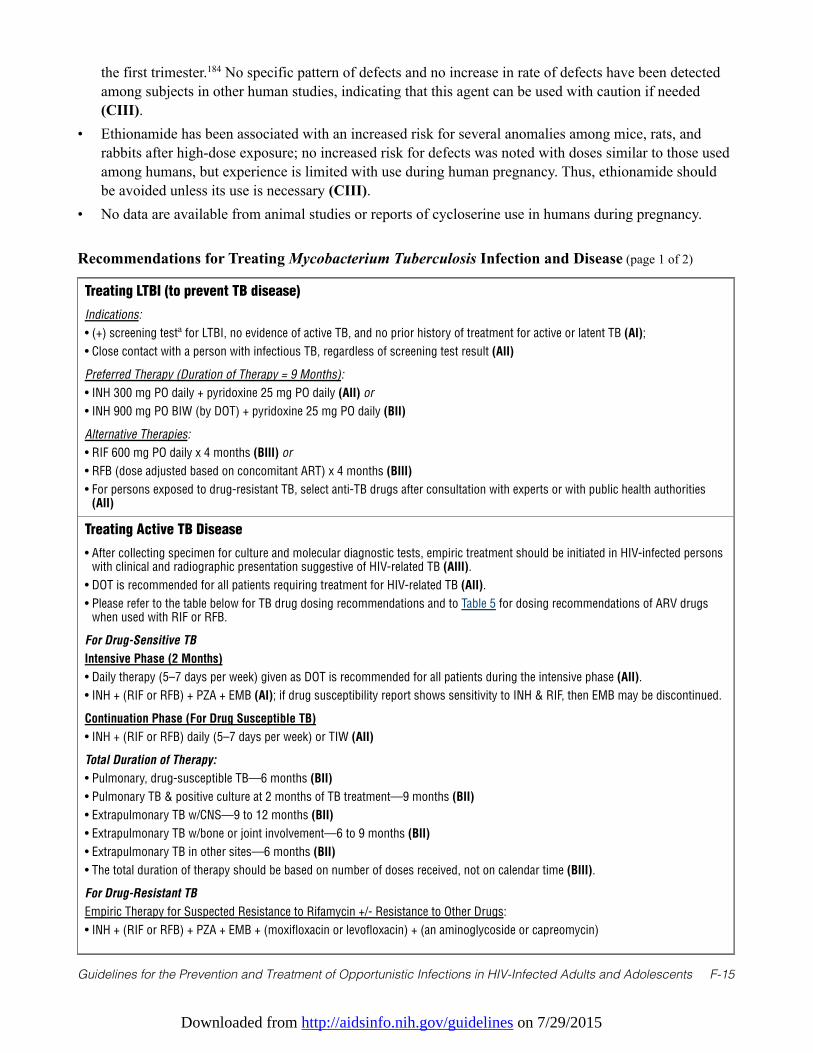
Downloaded from http://aidsinfo.nih.gov/guidelines on 7/29/2015
Guidelines for the Prevention and Treatment of Opportunistic Infections in HIV-Infected Adults and Adolescents F-15
the first trimester.184 No specific pattern of defects and no increase in rate of defects have been detectedamong subjects in other human studies, indicating that this agent can be used with caution if needed(CIII).
• Ethionamide has been associated with an increased risk for several anomalies among mice, rats, andrabbits after high-dose exposure; no increased risk for defects was noted with doses similar to those usedamong humans, but experience is limited with use during human pregnancy. Thus, ethionamide shouldbe avoided unless its use is necessary (CIII).
• No data are available from animal studies or reports of cycloserine use in humans during pregnancy.
Recommendations for Treating Mycobacterium Tuberculosis Infection and Disease (page 1 of 2)
Treating LTBI (to prevent TB disease)
Indications:
• (+) screening testa for LTBI, no evidence of active TB, and no prior history of treatment for active or latent TB (AI);
• Close contact with a person with infectious TB, regardless of screening test result (AII)
Preferred Therapy (Duration of Therapy = 9 Months):
• INH 300 mg PO daily + pyridoxine 25 mg PO daily (AII) or
• INH 900 mg PO BIW (by DOT) + pyridoxine 25 mg PO daily (BII)
Alternative Therapies:
• RIF 600 mg PO daily x 4 months (BIII) or
• RFB (dose adjusted based on concomitant ART) x 4 months (BIII)
• For persons exposed to drug-resistant TB, select anti-TB drugs after consultation with experts or with public health authorities(AII)
Treating Active TB Disease
• After collecting specimen for culture and molecular diagnostic tests, empiric treatment should be initiated in HIV-infected personswith clinical and radiographic presentation suggestive of HIV-related TB (AIII).
• DOT is recommended for all patients requiring treatment for HIV-related TB (AII).
• Please refer to the table below for TB drug dosing recommendations and to Table 5 for dosing recommendations of ARV drugswhen used with RIF or RFB.
For Drug-Sensitive TB
Intensive Phase (2 Months)
• Daily therapy (5–7 days per week) given as DOT is recommended for all patients during the intensive phase (AII).
• INH + (RIF or RFB) + PZA + EMB (AI); if drug susceptibility report shows sensitivity to INH & RIF, then EMB may be discontinued.
Continuation Phase (For Drug Susceptible TB)
• INH + (RIF or RFB) daily (5–7 days per week) or TIW (AII)
Total Duration of Therapy:
• Pulmonary, drug-susceptible TB—6 months (BII)
• Pulmonary TB & positive culture at 2 months of TB treatment—9 months (BII)
• Extrapulmonary TB w/CNS—9 to 12 months (BII)
• Extrapulmonary TB w/bone or joint involvement—6 to 9 months (BII)
• Extrapulmonary TB in other sites—6 months (BII)
• The total duration of therapy should be based on number of doses received, not on calendar time (BIII).
For Drug-Resistant TB
Empiric Therapy for Suspected Resistance to Rifamycin +/- Resistance to Other Drugs:
• INH + (RIF or RFB) + PZA + EMB + (moxifloxacin or levofloxacin) + (an aminoglycoside or capreomycin)
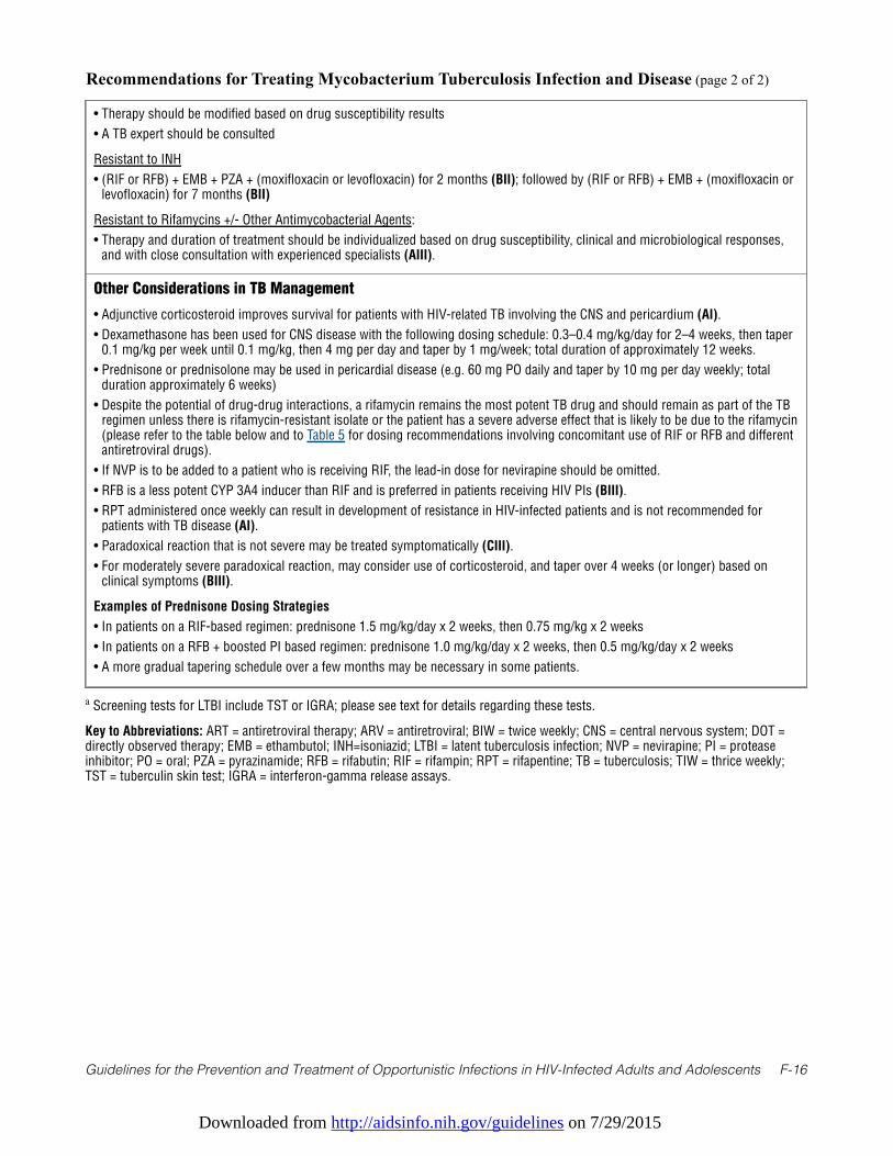
Downloaded from http://aidsinfo.nih.gov/guidelines on 7/29/2015
Guidelines for the Prevention and Treatment of Opportunistic Infections in HIV-Infected Adults and Adolescents F-16
Recommendations for Treating Mycobacterium Tuberculosis Infection and Disease (page 2 of 2)
• Therapy should be modified based on drug susceptibility results
• A TB expert should be consulted
Resistant to INH
• (RIF or RFB) + EMB + PZA + (moxifloxacin or levofloxacin) for 2 months (BII); followed by (RIF or RFB) + EMB + (moxifloxacin orlevofloxacin) for 7 months (BII)
Resistant to Rifamycins +/- Other Antimycobacterial Agents:
• Therapy and duration of treatment should be individualized based on drug susceptibility, clinical and microbiological responses,and with close consultation with experienced specialists (AIII).
Other Considerations in TB Management
• Adjunctive corticosteroid improves survival for patients with HIV-related TB involving the CNS and pericardium (AI).
• Dexamethasone has been used for CNS disease with the following dosing schedule: 0.3–0.4 mg/kg/day for 2–4 weeks, then taper0.1 mg/kg per week until 0.1 mg/kg, then 4 mg per day and taper by 1 mg/week; total duration of approximately 12 weeks.
• Prednisone or prednisolone may be used in pericardial disease (e.g. 60 mg PO daily and taper by 10 mg per day weekly; totalduration approximately 6 weeks)
• Despite the potential of drug-drug interactions, a rifamycin remains the most potent TB drug and should remain as part of the TBregimen unless there is rifamycin-resistant isolate or the patient has a severe adverse effect that is likely to be due to the rifamycin(please refer to the table below and to Table 5 for dosing recommendations involving concomitant use of RIF or RFB and differentantiretroviral drugs).
• If NVP is to be added to a patient who is receiving RIF, the lead-in dose for nevirapine should be omitted.
• RFB is a less potent CYP 3A4 inducer than RIF and is preferred in patients receiving HIV PIs (BIII).
• RPT administered once weekly can result in development of resistance in HIV-infected patients and is not recommended forpatients with TB disease (AI).
• Paradoxical reaction that is not severe may be treated symptomatically (CIII).
• For moderately severe paradoxical reaction, may consider use of corticosteroid, and taper over 4 weeks (or longer) based onclinical symptoms (BIII).
Examples of Prednisone Dosing Strategies
• In patients on a RIF-based regimen: prednisone 1.5 mg/kg/day x 2 weeks, then 0.75 mg/kg x 2 weeks
• In patients on a RFB + boosted PI based regimen: prednisone 1.0 mg/kg/day x 2 weeks, then 0.5 mg/kg/day x 2 weeks
• A more gradual tapering schedule over a few months may be necessary in some patients.
a Screening tests for LTBI include TST or IGRA; please see text for details regarding these tests.
Key to Abbreviations: ART = antiretroviral therapy; ARV = antiretroviral; BIW = twice weekly; CNS = central nervous system; DOT =directly observed therapy; EMB = ethambutol; INH=isoniazid; LTBI = latent tuberculosis infection; NVP = nevirapine; PI = proteaseinhibitor; PO = oral; PZA = pyrazinamide; RFB = rifabutin; RIF = rifampin; RPT = rifapentine; TB = tuberculosis; TIW = thrice weekly;TST = tuberculin skin test; IGRA = interferon-gamma release assays.
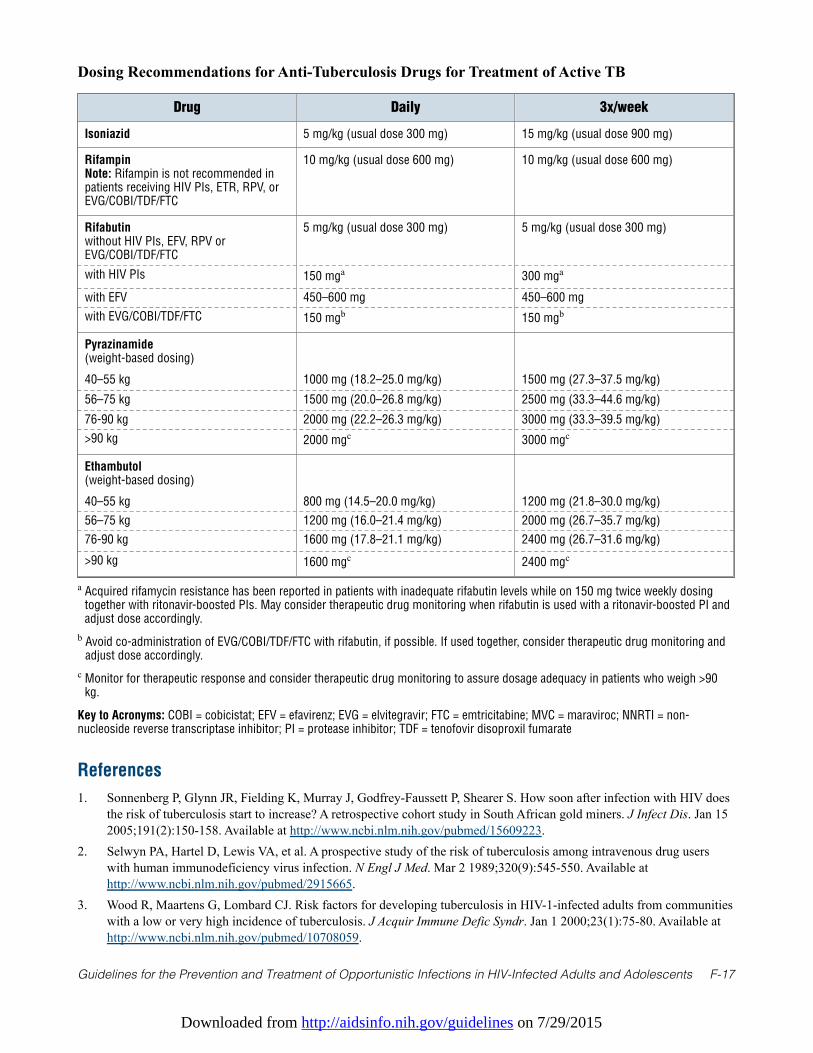
Downloaded from http://aidsinfo.nih.gov/guidelines on 7/29/2015
Guidelines for the Prevention and Treatment of Opportunistic Infections in HIV-Infected Adults and Adolescents F-17
Dosing Recommendations for Anti-Tuberculosis Drugs for Treatment of Active TB
Drug Daily 3x/week
Isoniazid 5 mg/kg (usual dose 300 mg) 15 mg/kg (usual dose 900 mg)
RifampinNote: Rifampin is not recommended inpatients receiving HIV PIs, ETR, RPV, orEVG/COBI/TDF/FTC
10 mg/kg (usual dose 600 mg) 10 mg/kg (usual dose 600 mg)
Rifabutinwithout HIV PIs, EFV, RPV orEVG/COBI/TDF/FTC
5 mg/kg (usual dose 300 mg) 5 mg/kg (usual dose 300 mg)
with HIV PIs 150 mga 300 mga
with EFV 450–600 mg 450–600 mg
with EVG/COBI/TDF/FTC 150 mgb 150 mgb
Pyrazinamide(weight-based dosing)
40–55 kg 1000 mg (18.2–25.0 mg/kg) 1500 mg (27.3–37.5 mg/kg)
56–75 kg 1500 mg (20.0–26.8 mg/kg) 2500 mg (33.3–44.6 mg/kg)
76-90 kg 2000 mg (22.2–26.3 mg/kg) 3000 mg (33.3–39.5 mg/kg)
>90 kg 2000 mgc 3000 mgc
Ethambutol(weight-based dosing)
40–55 kg 800 mg (14.5–20.0 mg/kg) 1200 mg (21.8–30.0 mg/kg)
56–75 kg 1200 mg (16.0–21.4 mg/kg) 2000 mg (26.7–35.7 mg/kg)
76-90 kg 1600 mg (17.8–21.1 mg/kg) 2400 mg (26.7–31.6 mg/kg)
>90 kg 1600 mgc 2400 mgc
a Acquired rifamycin resistance has been reported in patients with inadequate rifabutin levels while on 150 mg twice weekly dosingtogether with ritonavir-boosted PIs. May consider therapeutic drug monitoring when rifabutin is used with a ritonavir-boosted PI andadjust dose accordingly.
b Avoid co-administration of EVG/COBI/TDF/FTC with rifabutin, if possible. If used together, consider therapeutic drug monitoring andadjust dose accordingly.
c Monitor for therapeutic response and consider therapeutic drug monitoring to assure dosage adequacy in patients who weigh >90kg.
Key to Acronyms: COBI = cobicistat; EFV = efavirenz; EVG = elvitegravir; FTC = emtricitabine; MVC = maraviroc; NNRTI = non-nucleoside reverse transcriptase inhibitor; PI = protease inhibitor; TDF = tenofovir disoproxil fumarate
References
1. Sonnenberg P, Glynn JR, Fielding K, Murray J, Godfrey-Faussett P, Shearer S. How soon after infection with HIV doesthe risk of tuberculosis start to increase? A retrospective cohort study in South African gold miners. J Infect Dis. Jan 152005;191(2):150-158. Available at http://www.ncbi.nlm.nih.gov/pubmed/15609223.
2. Selwyn PA, Hartel D, Lewis VA, et al. A prospective study of the risk of tuberculosis among intravenous drug userswith human immunodeficiency virus infection. N Engl J Med. Mar 2 1989;320(9):545-550. Available athttp://www.ncbi.nlm.nih.gov/pubmed/2915665.
3. Wood R, Maartens G, Lombard CJ. Risk factors for developing tuberculosis in HIV-1-infected adults from communitieswith a low or very high incidence of tuberculosis. J Acquir Immune Defic Syndr. Jan 1 2000;23(1):75-80. Available athttp://www.ncbi.nlm.nih.gov/pubmed/10708059.
Downloaded from http://aidsinfo.nih.gov/guidelines on 7/29/2015
Guidelines for the Prevention and Treatment of Opportunistic Infections in HIV-Infected Adults and Adolescents F-18
4. Jones JL, Hanson DL, Dworkin MS, DeCock KM, Adult/Adolescent Spectrum of HIVDG. HIV-associated tuberculosisin the era of highly active antiretroviral therapy. The Adult/Adolescent Spectrum of HIV Disease Group. Int J TubercLung Dis. Nov 2000;4(11):1026-1031. Available at http://www.ncbi.nlm.nih.gov/pubmed/11092714.
5. Severe P, Juste MA, Ambroise A, et al. Early versus standard antiretroviral therapy for HIV-infected adults in Haiti. NEngl J Med. Jul 15 2010;363(3):257-265. Available at http://www.ncbi.nlm.nih.gov/pubmed/20647201.
6. Badri M, Wilson D, Wood R. Effect of highly active antiretroviral therapy on incidence of tuberculosis in South Africa: acohort study. Lancet. Jun 15 2002;359(9323):2059-2064. Available at http://www.ncbi.nlm.nih.gov/pubmed/12086758.
7. Seyler C, Toure S, Messou E, Bonard D, Gabillard D, Anglaret X. Risk factors for active tuberculosis after antiretroviraltreatment initiation in Abidjan. Am J Respir Crit Care Med. Jul 1 2005;172(1):123-127. Available athttp://www.ncbi.nlm.nih.gov/pubmed/15805184.
8. Centers for Disease Control and Prevention. Reported Tuberculosis in the United States, 2010. Available athttp://www.cdc.gov/tb/statistics/reports/2010/pdf/report2010.pdf. Accessed March 19, 2013.2011.
9. Bennett DE, Courval JM, Onorato I, et al. Prevalence of tuberculosis infection in the United States population: thenational health and nutrition examination survey, 1999-2000. Am J Respir Crit Care Med. Feb 1 2008;177(3):348-355.Available at http://www.ncbi.nlm.nih.gov/pubmed/17989346.
10. Albalak R, O'Brien RJ, Kammerer JS, et al. Trends in tuberculosis/human immunodeficiency virus comorbidity, United States,1993-2004. Arch Intern Med. Dec 10 2007;167(22):2443-2452. Available at http://www.ncbi.nlm.nih.gov/pubmed/18071166.
11. Centers for Disease Control and Prevention. Reported Tuberculosis in the United States, 2009. Available athttp://www.cdc.gov/tb/statistics/reports/2009/pdf/report2009.pdf. Accessed March 19, 2013.2010.
12. Centers for Disease Control and Prevention. Reported Tuberculosis in the United States, 2011. Available athttp://www.cdc.gov/tb/statistics/reports/2011/pdf/report2011.pdf. Accessed March 19, 2013. 2012.
13. Trieu L, Li J, Hanna DB, Harris TG. Tuberculosis rates among HIV-infected persons in New York City, 2001-2005. Am JPublic Health. Jun 2010;100(6):1031-1034. Available at http://www.ncbi.nlm.nih.gov/pubmed/20395574.
14. Hanna DB, Gupta LS, Jones LE, Thompson DM, Kellerman SE, Sackoff JE. AIDS-defining opportunistic illnesses in theHAART era in New York City. AIDS Care. Feb 2007;19(2):264-272. Available athttp://www.ncbi.nlm.nih.gov/pubmed/17364409.
15. Targeted tuberculin testing and treatment of latent tuberculosis infection. American Thoracic Society. MMWR RecommRep. Jun 9 2000;49(RR-6):1-51. Available at http://www.ncbi.nlm.nih.gov/pubmed/10881762.
16. Horsburgh CR, Jr. Priorities for the treatment of latent tuberculosis infection in the United States. N Engl J Med. May 132004;350(20):2060-2067. Available at http://www.ncbi.nlm.nih.gov/pubmed/15141044.
17. Day JH, Grant AD, Fielding KL, et al. Does tuberculosis increase HIV load? J Infect Dis. Nov 1 2004;190(9):1677-1684.Available at http://www.ncbi.nlm.nih.gov/pubmed/15478075.
18. Lopez-Gatell H, Cole SR, Margolick JB, et al. Effect of tuberculosis on the survival of HIV-infected men in a countrywith low tuberculosis incidence. AIDS. Sep 12 2008;22(14):1869-1873. Available athttp://www.ncbi.nlm.nih.gov/pubmed/18753866.
19. Akolo C, Adetifa I, Shepperd S, Volmink J. Treatment of latent tuberculosis infection in HIV infected persons. CochraneDatabase Syst Rev. 2010(1):CD000171. Available at http://www.ncbi.nlm.nih.gov/pubmed/20091503.
20. Fisk TL, Hon HM, Lennox JL, Fordham von Reyn C, Horsburgh CR, Jr. Detection of latent tuberculosis among HIV-infected patients after initiation of highly active antiretroviral therapy. AIDS. May 2 2003;17(7):1102-1104. Available athttp://www.ncbi.nlm.nih.gov/pubmed/12700468.
21. Girardi E, Palmieri F, Zaccarelli M, et al. High incidence of tuberculin skin test conversion among HIV-infectedindividuals who have a favourable immunological response to highly active antiretroviral therapy. AIDS. Sep 272002;16(14):1976-1979. Available at http://www.ncbi.nlm.nih.gov/pubmed/12351964.
22. Markowitz N, Hansen NI, Wilcosky TC, et al. Tuberculin and anergy testing in HIV-seropositive and HIV-seronegativepersons. Pulmonary Complications of HIV Infection Study Group. Ann Intern Med. Aug 1 1993;119(3):185-193.Available at http://www.ncbi.nlm.nih.gov/pubmed/8100692.
23. Ewer K, Deeks J, Alvarez L, et al. Comparison of T-cell-based assay with tuberculin skin test for diagnosis ofMycobacterium tuberculosis infection in a school tuberculosis outbreak. Lancet. Apr 5 2003;361(9364):1168-1173.Available at http://www.ncbi.nlm.nih.gov/pubmed/12686038.
Downloaded from http://aidsinfo.nih.gov/guidelines on 7/29/2015
Guidelines for the Prevention and Treatment of Opportunistic Infections in HIV-Infected Adults and Adolescents F-19
24. Nahid P, Pai M, Hopewell PC. Advances in the diagnosis and treatment of tuberculosis. Proceedings of the AmericanThoracic Society. 2006;3(1):103-110. Available at http://www.ncbi.nlm.nih.gov/pubmed/16493157.
25. Menzies D, Pai M, Comstock G. Meta-analysis: new tests for the diagnosis of latent tuberculosis infection: areas ofuncertainty and recommendations for research. Ann Intern Med. Mar 6 2007;146(5):340-354. Available athttp://www.ncbi.nlm.nih.gov/pubmed/17339619.
26. Raby E, Moyo M, Devendra A, et al. The effects of HIV on the sensitivity of a whole blood IFN-gamma release assayin Zambian adults with active tuberculosis. PLoS One. 2008;3(6):e2489. Available athttp://www.ncbi.nlm.nih.gov/pubmed/18560573.
27. Luetkemeyer AF, Charlebois ED, Flores LL, et al. Comparison of an interferon-gamma release assay with tuberculinskin testing in HIV-infected individuals. Am J Respir Crit Care Med. Apr 1 2007;175(7):737-742. Available athttp://www.ncbi.nlm.nih.gov/pubmed/17218620.
28. Talati NJ, Seybold U, Humphrey B, et al. Poor concordance between interferon-gamma release assays and tuberculinskin tests in diagnosis of latent tuberculosis infection among HIV-infected individuals. BMC Infect Dis. 2009;9:15.Available at http://www.ncbi.nlm.nih.gov/pubmed/19208218.
29. Hill PC, Jackson-Sillah DJ, Fox A, et al. Incidence of tuberculosis and the predictive value of ELISPOT and Mantoux testsin Gambian case contacts. PLoS One. 2008;3(1):e1379. Available at http://www.ncbi.nlm.nih.gov/pubmed/18167540.
30. Aichelburg MC, Rieger A, Breitenecker F, et al. Detection and prediction of active tuberculosis disease by a whole-blood interferon-gamma release assay in HIV-1-infected individuals. Clin Infect Dis. Apr 1 2009;48(7):954-962.Available at http://www.ncbi.nlm.nih.gov/pubmed/19245343.
31. Diel R, Loddenkemper R, Meywald-Walter K, Niemann S, Nienhaus A. Predictive value of a whole blood IFN-gammaassay for the development of active tuberculosis disease after recent infection with Mycobacterium tuberculosis. Am JRespir Crit Care Med. May 15 2008;177(10):1164-1170. Available at http://www.ncbi.nlm.nih.gov/pubmed/18276940.
32. Leung CC, Yam WC, Yew WW, et al. T-Spot.TB outperforms tuberculin skin test in predicting tuberculosis disease. AmJ Respir Crit Care Med. Sep 15 2010;182(6):834-840. Available at http://www.ncbi.nlm.nih.gov/pubmed/20508217.
33. Wilson IB, Landon BE, Hirschhorn LR, et al. Quality of HIV care provided by nurse practitioners, physician assistants, andphysicians. Ann Intern Med. Nov 15 2005;143(10):729-736. Available at http://www.ncbi.nlm.nih.gov/pubmed/16287794.
34. Backus LI, Boothroyd DB, Phillips BR, et al. National quality forum performance measures for HIV/AIDS care: theDepartment of Veterans Affairs' experience. Arch Intern Med. Jul 26 2010;170(14):1239-1246. Available athttp://www.ncbi.nlm.nih.gov/pubmed/20660844.
35. Lee LM, Lobato MN, Buskin SE, Morse A, Costa OS. Low adherence to guidelines for preventing TB among personswith newly diagnosed HIV infection, United States. Int J Tuberc Lung Dis. Feb 2006;10(2):209-214. Available athttp://www.ncbi.nlm.nih.gov/pubmed/16499263.
36. Mazurek GH, Jereb J, Vernon A, et al. Updated guidelines for using Interferon Gamma Release Assays to detectMycobacterium tuberculosis infection - United States, 2010. MMWR Recomm Rep. Jun 25 2010;59(RR-5):1-25.Available at http://www.ncbi.nlm.nih.gov/pubmed/20577159.
37. Cain KP, McCarthy KD, Heilig CM, et al. An algorithm for tuberculosis screening and diagnosis in people with HIV. NEngl J Med. Feb 25 2010;362(8):707-716. Available at http://www.ncbi.nlm.nih.gov/pubmed/20181972.
38. Gordin FM, Matts JP, Miller C, et al. A controlled trial of isoniazid in persons with anergy and human immunodeficiencyvirus infection who are at high risk for tuberculosis. Terry Beirn Community Programs for Clinical Research on AIDS. NEngl J Med. Jul 31 1997;337(5):315-320. Available at http://www.ncbi.nlm.nih.gov/pubmed/9233868.
39. Whalen CC, Johnson JL, Okwera A, et al. A trial of three regimens to prevent tuberculosis in Ugandan adults infectedwith the human immunodeficiency virus. Uganda-Case Western Reserve University Research Collaboration. N Engl JMed. Sep 18 1997;337(12):801-808. Available at http://www.ncbi.nlm.nih.gov/pubmed/9295239.
40. Mwinga A, Hosp M, Godfrey-Faussett P, et al. Twice weekly tuberculosis preventive therapy in HIV infection inZambia. AIDS. Dec 24 1998;12(18):2447-2457. Available at http://www.ncbi.nlm.nih.gov/pubmed/9875583.
41. Mohammed A, Myer L, Ehrlich R, Wood R, Cilliers F, Maartens G. Randomised controlled trial of isoniazid preventivetherapy in South African adults with advanced HIV disease. Int J Tuberc Lung Dis. Oct 2007;11(10):1114-1120.Available at http://www.ncbi.nlm.nih.gov/pubmed/17945069.
42. Tedla Z, Nyirenda S, Peeler C, et al. Isoniazid-associated hepatitis and antiretroviral drugs during tuberculosisprophylaxis in hiv-infected adults in Botswana. Am J Respir Crit Care Med. Jul 15 2010;182(2):278-285. Available at
Downloaded from http://aidsinfo.nih.gov/guidelines on 7/29/2015
Guidelines for the Prevention and Treatment of Opportunistic Infections in HIV-Infected Adults and Adolescents F-20
http://www.ncbi.nlm.nih.gov/pubmed/20378730.43. Hoffmann CJ, Charalambous S, Thio CL, et al. Hepatotoxicity in an African antiretroviral therapy cohort: the effect of
tuberculosis and hepatitis B. AIDS. Jun 19 2007;21(10):1301-1308. Available athttp://www.ncbi.nlm.nih.gov/pubmed/17545706.
44. Gordin F, Chaisson RE, Matts JP, et al. Rifampin and pyrazinamide vs isoniazid for prevention of tuberculosis in HIV-infectedpersons: an international randomized trial. Terry Beirn Community Programs for Clinical Research on AIDS, the Adult AIDSClinical Trials Group, the Pan American Health Organization, and the Centers for Disease Control and Prevention StudyGroup. JAMA. Mar 15 2000;283(11):1445-1450. Available at http://www.ncbi.nlm.nih.gov/pubmed/10732934.
45. Menzies D, Long R, Trajman A, et al. Adverse events with 4 months of rifampin therapy or 9 months of isoniazid therapyfor latent tuberculosis infection: a randomized trial. Ann Intern Med. Nov 18 2008;149(10):689-697. Available athttp://www.ncbi.nlm.nih.gov/pubmed/19017587.
46. Li J, Munsiff SS, Tarantino T, Dorsinville M. Adherence to treatment of latent tuberculosis infection in a clinicalpopulation in New York City. Int J Infect Dis. Apr 2010;14(4):e292-297. Available athttp://www.ncbi.nlm.nih.gov/pubmed/19656705.
47. Sterling TR, Villarino ME, Borisov AS, et al. Three months of rifapentine and isoniazid for latent tuberculosis infection. NEngl J Med. Dec 8 2011;365(23):2155-2166. Available at http://www.ncbi.nlm.nih.gov/pubmed/22150035.
48. Centers for Disease C, Prevention. Recommendations for use of an isoniazid-rifapentine regimen with direct observation totreat latent Mycobacterium tuberculosis infection. MMWR Morb Mortal Wkly Rep. Dec 9 2011;60(48):1650-1653.Available at http://www.ncbi.nlm.nih.gov/pubmed/22157884.
49. Golub JE, Saraceni V, Cavalcante SC, et al. The impact of antiretroviral therapy and isoniazid preventive therapy ontuberculosis incidence in HIV-infected patients in Rio de Janeiro, Brazil. AIDS. Jul 11 2007;21(11):1441-1448. Available athttp://www.ncbi.nlm.nih.gov/pubmed/17589190.
50. Golub JE, Pronyk P, Mohapi L, et al. Isoniazid preventive therapy, HAART and tuberculosis risk in HIV-infected adults in SouthAfrica: a prospective cohort. AIDS. Mar 13 2009;23(5):631-636. Available at http://www.ncbi.nlm.nih.gov/pubmed/19525621.
51. Samandari T, Agizew TB, Nyirenda S, et al. 6-month versus 36-month isoniazid preventive treatment for tuberculosis inadults with HIV infection in Botswana: a randomised, double-blind, placebo-controlled trial. Lancet. May 72011;377(9777):1588-1598. Available at http://www.ncbi.nlm.nih.gov/pubmed/21492926.
52. Saukkonen JJ, Cohn DL, Jasmer RM, et al. An official ATS statement: hepatotoxicity of antituberculosis therapy. Am JRespir Crit Care Med. Oct 15 2006;174(8):935-952. Available at http://www.ncbi.nlm.nih.gov/pubmed/17021358.
53. Batungwanayo J, Taelman H, Dhote R, Bogaerts J, Allen S, Van de Perre P. Pulmonary tuberculosis in Kigali, Rwanda.Impact of human immunodeficiency virus infection on clinical and radiographic presentation. The American review ofrespiratory disease. Jul 1992;146(1):53-56. Available at http://www.ncbi.nlm.nih.gov/pubmed/1626814.
54. Jones BE, Young SM, Antoniskis D, Davidson PT, Kramer F, Barnes PF. Relationship of the manifestations of tuberculosisto CD4 cell counts in patients with human immunodeficiency virus infection. The American review of respiratory disease.Nov 1993;148(5):1292-1297. Available at http://www.ncbi.nlm.nih.gov/pubmed/7902049.
55. Perlman DC, el-Sadr WM, Nelson ET, et al. Variation of chest radiographic patterns in pulmonary tuberculosis by degreeof human immunodeficiency virus-related immunosuppression. The Terry Beirn Community Programs for ClinicalResearch on AIDS (CPCRA). The AIDS Clinical Trials Group (ACTG). Clin Infect Dis. Aug 1997;25(2):242-246.Available at http://www.ncbi.nlm.nih.gov/pubmed/9332519.
56. Shafer RW, Kim DS, Weiss JP, Quale JM. Extrapulmonary tuberculosis in patients with human immunodeficiency virusinfection. Medicine (Baltimore). Nov 1991;70(6):384-397. Available at http://www.ncbi.nlm.nih.gov/pubmed/1956280.
57. Whalen C, Horsburgh CR, Jr., Hom D, Lahart C, Simberkoff M, Ellner J. Site of disease and opportunistic infection predictsurvival in HIV-associated tuberculosis. AIDS. Mar 15 1997;11(4):455-460. Available athttp://www.ncbi.nlm.nih.gov/pubmed/9084792.
58. Kourbatova EV, Leonard MK, Jr., Romero J, Kraft C, del Rio C, Blumberg HM. Risk factors for mortality among patientswith extrapulmonary tuberculosis at an academic inner-city hospital in the US. European journal of epidemiology.2006;21(9):715-721. Available at http://www.ncbi.nlm.nih.gov/pubmed/17072539.
59. Post FA, Wood R, Pillay GP. Pulmonary tuberculosis in HIV infection: radiographic appearance is related to CD4+ T-lymphocyte count. Tubercle and lung disease: the official journal of the International Union against Tuberculosis andLung Disease. Dec 1995;76(6):518-521. Available at http://www.ncbi.nlm.nih.gov/pubmed/8593372.
Downloaded from http://aidsinfo.nih.gov/guidelines on 7/29/2015
Guidelines for the Prevention and Treatment of Opportunistic Infections in HIV-Infected Adults and Adolescents F-21
60. Ahuja SS, Ahuja SK, Phelps KR, Thelmo W, Hill AR. Hemodynamic confirmation of septic shock in disseminatedtuberculosis. Critical care medicine. Jun 1992;20(6):901-903. Available at http://www.ncbi.nlm.nih.gov/pubmed/1597048.
61. Lewis JJ, Charalambous S, Day JH, et al. HIV infection does not affect active case finding of tuberculosis in SouthAfrican gold miners. Am J Respir Crit Care Med. Dec 15 2009;180(12):1271-1278. Available athttp://www.ncbi.nlm.nih.gov/pubmed/19745207.
62. Elliott AM, Halwiindi B, Hayes RJ, et al. The impact of human immunodeficiency virus on presentation and diagnosisof tuberculosis in a cohort study in Zambia. The Journal of tropical medicine and hygiene. Feb 1993;96(1):1-11.Available at http://www.ncbi.nlm.nih.gov/pubmed/8429569.
63. Monkongdee P, McCarthy KD, Cain KP, et al. Yield of acid-fast smear and mycobacterial culture for tuberculosisdiagnosis in people with human immunodeficiency virus. Am J Respir Crit Care Med. Nov 1 2009;180(9):903-908.Available at http://www.ncbi.nlm.nih.gov/pubmed/19628775.
64. Shriner KA, Mathisen GE, Goetz MB. Comparison of mycobacterial lymphadenitis among persons infected withhuman immunodeficiency virus and seronegative controls. Clin Infect Dis. Oct 1992;15(4):601-605. Available athttp://www.ncbi.nlm.nih.gov/pubmed/1420673.
65. Boehme CC, Nabeta P, Hillemann D, et al. Rapid molecular detection of tuberculosis and rifampin resistance. N Engl JMed. Sep 9 2010;363(11):1005-1015. Available at http://www.ncbi.nlm.nih.gov/pubmed/20825313.
66. Dinnes J, Deeks J, Kunst H, et al. A systematic review of rapid diagnostic tests for the detection of tuberculosis infection.Health technology assessment. Jan 2007;11(3):1-196. Available at http://www.ncbi.nlm.nih.gov/pubmed/17266837.
67. Centers for Disease C, Prevention. Updated guidelines for the use of nucleic acid amplification tests in the diagnosis oftuberculosis. MMWR Morb Mortal Wkly Rep. Jan 16 2009;58(1):7-10. Available athttp://www.ncbi.nlm.nih.gov/pubmed/19145221.
68. Lew W, Pai M, Oxlade O, Martin D, Menzies D. Initial drug resistance and tuberculosis treatment outcomes: systematicreview and meta-analysis. Ann Intern Med. Jul 15 2008;149(2):123-134. Available athttp://www.ncbi.nlm.nih.gov/pubmed/18626051.
69. Gandhi NR, Shah NS, Andrews JR, et al. HIV coinfection in multidrug- and extensively drug-resistant tuberculosisresults in high early mortality. Am J Respir Crit Care Med. Jan 1 2010;181(1):80-86. Available athttp://www.ncbi.nlm.nih.gov/pubmed/19833824.
70. Moore DA, Evans CA, Gilman RH, et al. Microscopic-observation drug-susceptibility assay for the diagnosis of TB. NEngl J Med. Oct 12 2006;355(15):1539-1550. Available at http://www.ncbi.nlm.nih.gov/pubmed/17035648.
71. Mathema B, Kurepina NE, Bifani PJ, Kreiswirth BN. Molecular epidemiology of tuberculosis: current insights. ClinMicrobiol Rev. Oct 2006;19(4):658-685. Available at http://www.ncbi.nlm.nih.gov/pubmed/17041139.
72. Barnard M, Albert H, Coetzee G, O'Brien R, Bosman ME. Rapid molecular screening for multidrug-resistanttuberculosis in a high-volume public health laboratory in South Africa. Am J Respir Crit Care Med. Apr 12008;177(7):787-792. Available at http://www.ncbi.nlm.nih.gov/pubmed/18202343.
73. Hillemann D, Rusch-Gerdes S, Richter E. Feasibility of the GenoType MTBDRsl assay for fluoroquinolone, amikacin-capreomycin, and ethambutol resistance testing of Mycobacterium tuberculosis strains and clinical specimens. J ClinMicrobiol. Jun 2009;47(6):1767-1772. Available at http://www.ncbi.nlm.nih.gov/pubmed/19386845.
74. World Health Organization. Global tuberculosis control 2011. Accessed on April 19, 2012, athttp://www.who.int/tb/publications/global_report/en/. 2011.
75. Canueto-Quintero J, Caballero-Granado FJ, Herrero-Romero M, et al. Epidemiological, clinical, and prognosticdifferences between the diseases caused by Mycobacterium kansasii and Mycobacterium tuberculosis in patientsinfected with human immunodeficiency virus: a multicenter study. Clin Infect Dis. Aug 15 2003;37(4):584-590.Available at http://www.ncbi.nlm.nih.gov/pubmed/12905144.
76. Swaminathan S, Narendran G, Venkatesan P, et al. Efficacy of a 6-month versus 9-month intermittent treatment regimenin HIV-infected patients with tuberculosis: a randomized clinical trial. Am J Respir Crit Care Med. Apr 12010;181(7):743-751. Available at http://www.ncbi.nlm.nih.gov/pubmed/19965813.
77. Nettles RE, Mazo D, Alwood K, et al. Risk factors for relapse and acquired rifamycin resistance after directly observedtuberculosis treatment: a comparison by HIV serostatus and rifamycin use. Clin Infect Dis. Mar 1 2004;38(5):731-736.Available at http://www.ncbi.nlm.nih.gov/pubmed/14986259.
Downloaded from http://aidsinfo.nih.gov/guidelines on 7/29/2015
Guidelines for the Prevention and Treatment of Opportunistic Infections in HIV-Infected Adults and Adolescents F-22
78. Li J, Munsiff SS, Driver CR, Sackoff J. Relapse and acquired rifampin resistance in HIV-infected patients withtuberculosis treated with rifampin- or rifabutin-based regimens in New York City, 1997-2000. Clin Infect Dis. Jul 12005;41(1):83-91. Available at http://www.ncbi.nlm.nih.gov/pubmed/15937767.
79. Vernon A, Burman W, Benator D, Khan A, Bozeman L. Acquired rifamycin monoresistance in patients with HIV-relatedtuberculosis treated with once-weekly rifapentine and isoniazid. Tuberculosis Trials Consortium. Lancet. May 291999;353(9167):1843-1847. Available at http://www.ncbi.nlm.nih.gov/pubmed/10359410.
80. Burman W, Benator D, Vernon A, et al. Acquired rifamycin resistance with twice-weekly treatment of HIV-relatedtuberculosis. Am J Respir Crit Care Med. Feb 1 2006;173(3):350-356. Available athttp://www.ncbi.nlm.nih.gov/pubmed/16109981.
81. Khan FA, Minion J, Pai M, et al. Treatment of active tuberculosis in HIV-coinfected patients: a systematic review and meta-analysis. Clin Infect Dis. May 1 2010;50(9):1288-1299. Available at http://www.ncbi.nlm.nih.gov/pubmed/20353364.
82. el-Sadr WM, Perlman DC, Matts JP, et al. Evaluation of an intensive intermittent-induction regimen and duration ofshort-course treatment for human immunodeficiency virus-related pulmonary tuberculosis. Terry Beirn CommunityPrograms for Clinical Research on AIDS (CPCRA) and the AIDS Clinical Trials Group (ACTG). Clin Infect Dis. May1998;26(5):1148-1158. Available at http://www.ncbi.nlm.nih.gov/pubmed/9597244.
83. Perriens JH, St Louis ME, Mukadi YB, et al. Pulmonary tuberculosis in HIV-infected patients in Zaire. A controlledtrial of treatment for either 6 or 12 months. N Engl J Med. Mar 23 1995;332(12):779-784. Available athttp://www.ncbi.nlm.nih.gov/pubmed/7862181.
84. Thwaites GE, Nguyen DB, Nguyen HD, et al. Dexamethasone for the treatment of tuberculous meningitis inadolescents and adults. N Engl J Med. Oct 21 2004;351(17):1741-1751. Available athttp://www.ncbi.nlm.nih.gov/pubmed/15496623.
85. Hakim JG, Ternouth I, Mushangi E, Siziya S, Robertson V, Malin A. Double blind randomised placebo controlled trialof adjunctive prednisolone in the treatment of effusive tuberculous pericarditis in HIV seropositive patients. Heart. Aug2000;84(2):183-188. Available at http://www.ncbi.nlm.nih.gov/pubmed/10908256.
86. Abdool Karim SS, Naidoo K, Grobler A, et al. Timing of initiation of antiretroviral drugs during tuberculosis therapy. NEngl J Med. Feb 25 2010;362(8):697-706. Available at http://www.ncbi.nlm.nih.gov/pubmed/20181971.
87. Havlir DV, Kendall MA, Ive P, et al. Timing of antiretroviral therapy for HIV-1 infection and tuberculosis. N Engl JMed. Oct 20 2011;365(16):1482-1491. Available at http://www.ncbi.nlm.nih.gov/pubmed/22010914.
88. Blanc FX, Sok T, Laureillard D, et al. Earlier versus later start of antiretroviral therapy in HIV-infected adults withtuberculosis. N Engl J Med. Oct 20 2011;365(16):1471-1481. Available at http://www.ncbi.nlm.nih.gov/pubmed/22010913.
89. Nahid P, Gonzalez LC, Rudoy I, et al. Treatment outcomes of patients with HIV and tuberculosis. Am J Respir CritCare Med. Jun 1 2007;175(11):1199-1206. Available at http://www.ncbi.nlm.nih.gov/pubmed/17290042.
90. Abdool Karim SS, Naidoo K, Grobler A, et al. Integration of antiretroviral therapy with tuberculosis treatment. N Engl JMed. Oct 20 2011;365(16):1492-1501. Available at http://www.ncbi.nlm.nih.gov/pubmed/22010915.
91. Nanteza MW, Mayanja-Kizza H, Charlebois E, et al. A randomized trial of punctuated antiretroviral therapy in UgandanHIV-seropositive adults with pulmonary tuberculosis and CD4(+) T-cell counts of >/= 350 cells/muL. J Infect Dis. Sep15 2011;204(6):884-892. Available at http://www.ncbi.nlm.nih.gov/pubmed/21849285.
92. Dheda K, Shean K, Zumla A, et al. Early treatment outcomes and HIV status of patients with extensively drug-resistanttuberculosis in South Africa: a retrospective cohort study. Lancet. May 22 2010;375(9728):1798-1807. Available athttp://www.ncbi.nlm.nih.gov/pubmed/20488525.
93. Torok ME, Yen NT, Chau TT, et al. Timing of initiation of antiretroviral therapy in human immunodeficiency virus(HIV)--associated tuberculous meningitis. Clin Infect Dis. Jun 2011;52(11):1374-1383. Available athttp://www.ncbi.nlm.nih.gov/pubmed/21596680.
94. Boulle A, Van Cutsem G, Cohen K, et al. Outcomes of nevirapine- and efavirenz-based antiretroviral therapy whencoadministered with rifampicin-based antitubercular therapy. JAMA. Aug 6 2008;300(5):530-539. Available athttp://www.ncbi.nlm.nih.gov/pubmed/18677025.
95. Lopez-Cortes LF, Ruiz-Valderas R, Viciana P, et al. Pharmacokinetic interactions between efavirenz and rifampicin inHIV-infected patients with tuberculosis. Clinical pharmacokinetics. 2002;41(9):681-690. Available athttp://www.ncbi.nlm.nih.gov/pubmed/12126459.
Downloaded from http://aidsinfo.nih.gov/guidelines on 7/29/2015
Guidelines for the Prevention and Treatment of Opportunistic Infections in HIV-Infected Adults and Adolescents F-23
96. Cohen K, Grant A, Dandara C, et al. Effect of rifampicin-based antitubercular therapy and the cytochrome P450 2B6516G>T polymorphism on efavirenz concentrations in adults in South Africa. Antivir Ther. 2009;14(5):687-695. Availableat http://www.ncbi.nlm.nih.gov/pubmed/19704172.
97. Ramachandran G, Hemanth Kumar AK, Rajasekaran S, et al. CYP2B6 G516T polymorphism but not rifampincoadministration influences steady-state pharmacokinetics of efavirenz in human immunodeficiency virus-infectedpatients in South India. Antimicrob Agents Chemother. Mar 2009;53(3):863-868. Available athttp://www.ncbi.nlm.nih.gov/pubmed/19124658.
98. Manosuthi W, Kiertiburanakul S, Sungkanuparph S, et al. Efavirenz 600 mg/day versus efavirenz 800 mg/day in HIV-infected patients with tuberculosis receiving rifampicin: 48 weeks results. AIDS. Jan 2 2006;20(1):131-132. Available athttp://www.ncbi.nlm.nih.gov/pubmed/16327334.
99. Manosuthi W, Sungkanuparph S, Thakkinstian A, et al. Plasma nevirapine levels and 24-week efficacy in HIV-infectedpatients receiving nevirapine-based highly active antiretroviral therapy with or without rifampicin. Clin Infect Dis. Jul 152006;43(2):253-255. Available at http://www.ncbi.nlm.nih.gov/pubmed/16779754.
100. Shipton LK, Wester CW, Stock S, et al. Safety and efficacy of nevirapine- and efavirenz-based antiretroviral treatment inadults treated for TB-HIV co-infection in Botswana. Int J Tuberc Lung Dis. Mar 2009;13(3):360-366. Available athttp://www.ncbi.nlm.nih.gov/pubmed/19275797.
101. Swaminathan S, Padmapriyadarsini C, Venkatesan P, et al. Efficacy and safety of once-daily nevirapine- or efavirenz-based antiretroviral therapy in HIV-associated tuberculosis: a randomized clinical trial. Clin Infect Dis. Oct2011;53(7):716-724. Available at http://www.ncbi.nlm.nih.gov/pubmed/21890776.
102. la Porte CJ, Colbers EP, Bertz R, et al. Pharmacokinetics of adjusted-dose lopinavir-ritonavir combined with rifampin inhealthy volunteers. Antimicrob Agents Chemother. May 2004;48(5):1553-1560. Available athttp://www.ncbi.nlm.nih.gov/pubmed/15105105.
103. Nijland HM, L'Homme R F, Rongen GA, et al. High incidence of adverse events in healthy volunteers receivingrifampicin and adjusted doses of lopinavir/ritonavir tablets. AIDS. May 11 2008;22(8):931-935. Available athttp://www.ncbi.nlm.nih.gov/pubmed/18453852.
104. Haas DW, Koletar SL, Laughlin L, et al. Hepatotoxicity and gastrointestinal intolerance when healthy volunteers takingrifampin add twice-daily atazanavir and ritonavir. J Acquir Immune Defic Syndr. Mar 1 2009;50(3):290-293. Available athttp://www.ncbi.nlm.nih.gov/pubmed/19194314.
105. Schmitt C, Riek M, Winters K, Schutz M, Grange S. Unexpected Hepatotoxicity of Rifampin and Saquinavir/Ritonavir inHealthy Male Volunteers. Archives of drug information. Mar 2009;2(1):8-16. Available athttp://www.ncbi.nlm.nih.gov/pubmed/19381336.
106. Abbott. 2001. Lopinavir/ritonavir package insert. http://www.rxabbott.com/pdf/kaletratabpi.pdf.107. Bristol-Myers Squibb. 2001. Atazanavir package insert. http://packageinserts.bms.com/pi/pi_reyataz.pdf. 108. Sekar V, Lavreys L, Van de Casteele T, et al. Pharmacokinetics of darunavir/ritonavir and rifabutin coadministered in HIV-
negative healthy volunteers. Antimicrob Agents Chemother. Oct 2010;54(10):4440-4445. Available athttp://www.ncbi.nlm.nih.gov/pubmed/20660678.
109. Ford SL, Chen YC, Lou Y, et al. Pharmacokinetic interaction between fosamprenavir-ritonavir and rifabutin in healthy subjects.Antimicrob Agents Chemother. Feb 2008;52(2):534-538. Available at http://www.ncbi.nlm.nih.gov/pubmed/18056271.
110. Lin HC, Lu PL, Chang CH. Uveitis associated with concurrent administration of rifabutin and lopinavir/ritonavir(Kaletra). Eye. Dec 2007;21(12):1540-1541. Available at http://www.ncbi.nlm.nih.gov/pubmed/17962822.
111. Gallicano K, Khaliq Y, Carignan G, Tseng A, Walmsley S, Cameron DW. A pharmacokinetic study of intermittentrifabutin dosing with a combination of ritonavir and saquinavir in patients infected with human immunodeficiency virus.Clinical pharmacology and therapeutics. Aug 2001;70(2):149-158. Available athttp://www.ncbi.nlm.nih.gov/pubmed/11503009.
112. Jenny-Avital ER, Joseph K. Rifamycin-resistant Mycobacterium tuberculosis in the highly active antiretroviral therapyera: a report of 3 relapses with acquired rifampin resistance following alternate-day rifabutin and boosted proteaseinhibitor therapy. Clin Infect Dis. May 15 2009;48(10):1471-1474. Available athttp://www.ncbi.nlm.nih.gov/pubmed/19368504.
113. Boulanger C, Hollender E, Farrell K, et al. Pharmacokinetic evaluation of rifabutin in combination with lopinavir-ritonavirin patients with HIV infection and active tuberculosis. Clin Infect Dis. Nov 1 2009;49(9):1305-1311. Available at
Downloaded from http://aidsinfo.nih.gov/guidelines on 7/29/2015
Guidelines for the Prevention and Treatment of Opportunistic Infections in HIV-Infected Adults and Adolescents F-24
http://www.ncbi.nlm.nih.gov/pubmed/19807276.114. Johnson JL, Okwera A, Nsubuga P, et al. Efficacy of an unsupervised 8-month rifampicin-containing regimen for the treatment
of pulmonary tuberculosis in HIV-infected adults. Uganda-Case Western Reserve University Research Collaboration. Int JTuberc Lung Dis. Nov 2000;4(11):1032-1040. Available at http://www.ncbi.nlm.nih.gov/pubmed/11092715.
115. Jindani A, Nunn AJ, Enarson DA. Two 8-month regimens of chemotherapy for treatment of newly diagnosed pulmonarytuberculosis: international multicentre randomised trial. Lancet. Oct 2-8 2004;364(9441):1244-1251. Available athttp://www.ncbi.nlm.nih.gov/pubmed/15464185.
116. Benator D, Bhattacharya M, Bozeman L, et al. Rifapentine and isoniazid once a week versus rifampicin and isoniazidtwice a week for treatment of drug-susceptible pulmonary tuberculosis in HIV-negative patients: a randomised clinicaltrial. Lancet. Aug 17 2002;360(9332):528-534. Available at http://www.ncbi.nlm.nih.gov/pubmed/12241657.
117. Yew WW, Leung CC. Antituberculosis drugs and hepatotoxicity. Am J Respir Crit Care Med. Apr 15 2007;175(8):858;author reply 858-859. Available at http://www.ncbi.nlm.nih.gov/pubmed/17405943.
118. Breen RA, Miller RF, Gorsuch T, al e. Adverse events and treatment interruption in tuberculosis patients with and withoutHIV co-infection. Thorax. 2006;61(9):791-794. Available at http://www.ncbi.nlm.nih.gov/entrez/query.fcgi?db=pubmed&cmd=Retrieve&dopt=AbstractPlus&list_uids=16844730&query_hl=49&itool=pubmed_docsum.
119. Yee D, Valiquette C, Pelletier M, Parisien I, Rocher I, Menzies D. Incidence of serious side effects from first-lineantituberculosis drugs among patients treated for active tuberculosis. Am J Respir Crit Care Med. Jun 12003;167(11):1472-1477. Available at http://www.ncbi.nlm.nih.gov/pubmed/12569078.
120. Dean GL, Edwards SG, Ives NJ, et al. Treatment of tuberculosis in HIV-infected persons in the era of highly activeantiretroviral therapy. AIDS. Jan 4 2002;16(1):75-83. Available at http://www.ncbi.nlm.nih.gov/pubmed/11741165.
121. CDC. Core curriculum on tuberculosis: what the clinician should know, 4th edition. Atlanta, GA: US Department ofHealth and Human Services. Available at http://www.cdc.gov/nchstp/tb. 2000.
122. Mehta YS, Jijina FF, Badakere SS, Pathare AV, Mohanty D. Rifampicin-induced immune thrombocytopenia. Tubercle andlung disease: the official journal of the International Union against Tuberculosis and Lung Disease. Dec 1996;77(6):558-562. Available at http://www.ncbi.nlm.nih.gov/pubmed/9039451.
123. Chien JW, Johnson JL. Paradoxical reactions in HIV and pulmonary TB. Chest. Sep 1998;114(3):933-936. Available athttp://www.ncbi.nlm.nih.gov/pubmed/9743188.
124. Steele MA, Burk RF, DesPrez RM. Toxic hepatitis with isoniazid and rifampin. A meta-analysis. Chest. Feb1991;99(2):465-471. Available at http://www.ncbi.nlm.nih.gov/pubmed/1824929.
125. Ormerod LP, Skinner C, Wales J. Hepatotoxicity of antituberculosis drugs. Thorax. Feb 1996;51(2):111-113. Available athttp://www.ncbi.nlm.nih.gov/pubmed/8711637.
126. French MA, Price P, Stone SF. Immune restoration disease after antiretroviral therapy. AIDS. Aug 20 2004;18(12):1615-1627. Available at http://www.ncbi.nlm.nih.gov/pubmed/15280772.
127. Lawn SD, Bekker LG, Miller RF. Immune reconstitution disease associated with mycobacterial infections in HIV-infectedindividuals receiving antiretrovirals. Lancet Infect Dis. Jun 2005;5(6):361-373. Available athttp://www.ncbi.nlm.nih.gov/pubmed/15919622.
128. Meintjes G, Rabie H, Wilkinson RJ, Cotton MF. Tuberculosis-associated immune reconstitution inflammatory syndromeand unmasking of tuberculosis by antiretroviral therapy. Clin Chest Med. Dec 2009;30(4):797-810, x. Available athttp://www.ncbi.nlm.nih.gov/pubmed/19925968.
129. Meintjes G, Lawn SD, Scano F, et al. Tuberculosis-associated immune reconstitution inflammatory syndrome: casedefinitions for use in resource-limited settings. Lancet Infect Dis. Aug 2008;8(8):516-523. Available athttp://www.ncbi.nlm.nih.gov/pubmed/18652998.
130. Muller M, Wandel S, Colebunders R, et al. Immune reconstitution inflammatory syndrome in patients startingantiretroviral therapy for HIV infection: a systematic review and meta-analysis. Lancet Infect Dis. Apr 2010;10(4):251-261. Available at http://www.ncbi.nlm.nih.gov/pubmed/20334848.
131. Burman W, Weis S, Vernon A, et al. Frequency, severity and duration of immune reconstitution events in HIV-relatedtuberculosis. Int J Tuberc Lung Dis. Dec 2007;11(12):1282-1289. Available at http://www.ncbi.nlm.nih.gov/pubmed/18229435.
132. Pepper DJ, Marais S, Maartens G, et al. Neurologic manifestations of paradoxical tuberculosis-associated immunereconstitution inflammatory syndrome: a case series. Clin Infect Dis. Jun 1 2009;48(11):e96-107. Available at
Downloaded from http://aidsinfo.nih.gov/guidelines on 7/29/2015
Guidelines for the Prevention and Treatment of Opportunistic Infections in HIV-Infected Adults and Adolescents F-25
http://www.ncbi.nlm.nih.gov/pubmed/19405867.133. Lawn SD, Wood R. Hepatic involvement with tuberculosis-associated immune reconstitution disease. AIDS. Nov 12
2007;21(17):2362-2363. Available at http://www.ncbi.nlm.nih.gov/pubmed/18090294.134. Meintjes G, Rangaka MX, Maartens G, et al. Novel relationship between tuberculosis immune reconstitution
inflammatory syndrome and antitubercular drug resistance. Clin Infect Dis. Mar 1 2009;48(5):667-676. Available athttp://www.ncbi.nlm.nih.gov/pubmed/19191655.
135. Narita M, Ashkin D, Hollender ES, Pitchenik AE. Paradoxical worsening of tuberculosis following antiretroviral therapyin patients with AIDS. Am J Respir Crit Care Med. Jul 1998;158(1):157-161. Available athttp://www.ncbi.nlm.nih.gov/pubmed/9655723.
136. Breen RA, Smith CJ, Bettinson H, et al. Paradoxical reactions during tuberculosis treatment in patients with and without HIVco-infection. Thorax. Aug 2004;59(8):704-707. Available at http://www.ncbi.nlm.nih.gov/pubmed/15282393.
137. Breton G, Duval X, Estellat C, et al. Determinants of immune reconstitution inflammatory syndrome in HIV type 1-infected patients with tuberculosis after initiation of antiretroviral therapy. Clin Infect Dis. Dec 1 2004;39(11):1709-1712.Available at http://www.ncbi.nlm.nih.gov/pubmed/15578375.
138. Lawn SD, Myer L, Bekker LG, Wood R. Tuberculosis-associated immune reconstitution disease: incidence, risk factorsand impact in an antiretroviral treatment service in South Africa. AIDS. Jan 30 2007;21(3):335-341. Available athttp://www.ncbi.nlm.nih.gov/pubmed/17255740.
139. Manosuthi W, Kiertiburanakul S, Phoorisri T, Sungkanuparph S. Immune reconstitution inflammatory syndrome oftuberculosis among HIV-infected patients receiving antituberculous and antiretroviral therapy. J Infect. Dec2006;53(6):357-363. Available at http://www.ncbi.nlm.nih.gov/pubmed/16487593.
140. Serra FC, Hadad D, Orofino RL, et al. Immune reconstitution syndrome in patients treated for HIV and tuberculosis inRio de Janeiro. The Brazilian journal of infectious diseases: an official publication of the Brazilian Society of InfectiousDiseases. Oct 2007;11(5):462-465. Available at http://www.ncbi.nlm.nih.gov/pubmed/17962870.
141. Olalla J, Pulido F, Rubio R, et al. Paradoxical responses in a cohort of HIV-1-infected patients with mycobacterialdisease. Int J Tuberc Lung Dis. Jan 2002;6(1):71-75. Available at http://www.ncbi.nlm.nih.gov/pubmed/11931404.
142. Huyst V, Lynen L, Bottieau E, Zolfo M, Kestens L, Colebunders R. Immune reconstitution inflammatory syndrome in anHIV/TB co-infected patient four years after starting antiretroviral therapy. Acta clinica Belgica. Mar-Apr 2007;62(2):126-129. Available at http://www.ncbi.nlm.nih.gov/pubmed/17547295.
143. Michailidis C, Pozniak AL, Mandalia S, Basnayake S, Nelson MR, Gazzard BG. Clinical characteristics of IRISsyndrome in patients with HIV and tuberculosis. Antivir Ther. 2005;10(3):417-422. Available athttp://www.ncbi.nlm.nih.gov/pubmed/15918332.
144. Meintjes G, Wilkinson RJ, Morroni C, et al. Randomized placebo-controlled trial of prednisone for paradoxicaltuberculosis-associated immune reconstitution inflammatory syndrome. AIDS. Sep 24 2010;24(15):2381-2390. Availableat http://www.ncbi.nlm.nih.gov/pubmed/20808204.
145. Volkow PF, Cornejo P, Zinser JW, Ormsby CE, Reyes-Teran G. Life-threatening exacerbation of Kaposi's sarcoma afterprednisone treatment for immune reconstitution inflammatory syndrome. AIDS. Mar 12 2008;22(5):663-665. Available athttp://www.ncbi.nlm.nih.gov/pubmed/18317012.
146. John L, Baalwa J, Kalimugogo P, et al. Response to 'Does immune reconstitution promote active tuberculosis in patientsreceiving highly active antiretroviral therapy?' AIDS, 22 July 2005. AIDS. Nov 18 2005;19(17):2049-2050. Available athttp://www.ncbi.nlm.nih.gov/pubmed/16260919.
147. Goldsack NR, Allen S, Lipman MC. Adult respiratory distress syndrome as a severe immune reconstitution diseasefollowing the commencement of highly active antiretroviral therapy. Sexually transmitted infections. Aug 2003;79(4):337-338. Available at http://www.ncbi.nlm.nih.gov/pubmed/12902592.
148. Lawn SD, Wainwright H, Orrell C. Fatal unmasking tuberculosis immune reconstitution disease with bronchiolitisobliterans organizing pneumonia: the role of macrophages. AIDS. Jan 2 2009;23(1):143-145. Available athttp://www.ncbi.nlm.nih.gov/pubmed/19050399.
149. Chen WL, Lin YF, Tsai WC, Tsao YT. Unveiling tuberculous pyomyositis: an emerging role of immune reconstitutioninflammatory syndrome. The American journal of emergency medicine. Feb 2009;27(2):251 e251-252. Available athttp://www.ncbi.nlm.nih.gov/pubmed/19371548.
Downloaded from http://aidsinfo.nih.gov/guidelines on 7/29/2015
Guidelines for the Prevention and Treatment of Opportunistic Infections in HIV-Infected Adults and Adolescents F-26
150. Menzies D, Benedetti A, Paydar A, et al. Standardized treatment of active tuberculosis in patients with previous treatmentand/or with mono-resistance to isoniazid: a systematic review and meta-analysis. PLoS Med. Sep 2009;6(9):e1000150.Available at http://www.ncbi.nlm.nih.gov/pubmed/20101802.
151. Blumberg HM, Burman WJ, Chaisson RE, et al. American Thoracic Society/Centers for Disease Control andPrevention/Infectious Diseases Society of America: treatment of tuberculosis. Am J Respir Crit Care Med. Feb 152003;167(4):603-662. Available at http://www.ncbi.nlm.nih.gov/pubmed/12588714.
152. Korenromp EL, Scano F, Williams BG, Dye C, Nunn P. Effects of human immunodeficiency virus infection onrecurrence of tuberculosis after rifampin-based treatment: an analytical review. Clin Infect Dis. Jul 1 2003;37(1):101-112.Available at http://www.ncbi.nlm.nih.gov/pubmed/12830415.
153. Sonnenberg P, Murray J, Glynn JR, Shearer S, Kambashi B, Godfrey-Faussett P. HIV-1 and recurrence, relapse, andreinfection of tuberculosis after cure: a cohort study in South African mineworkers. Lancet. Nov 172001;358(9294):1687-1693. Available at http://www.ncbi.nlm.nih.gov/pubmed/11728545.
154. Narayanan S, Swaminathan S, Supply P, et al. Impact of HIV infection on the recurrence of tuberculosis in South India. JInfect Dis. Mar 2010;201(5):691-703. Available at http://www.ncbi.nlm.nih.gov/pubmed/20121433.
155. Jasmer RM, Bozeman L, Schwartzman K, et al. Recurrent tuberculosis in the United States and Canada: relapse orreinfection? Am J Respir Crit Care Med. Dec 15 2004;170(12):1360-1366. Available athttp://www.ncbi.nlm.nih.gov/pubmed/15477492.
156. Fitzgerald DW, Desvarieux M, Severe P, Joseph P, Johnson WD, Jr., Pape JW. Effect of post-treatment isoniazid onprevention of recurrent tuberculosis in HIV-1-infected individuals: a randomised trial. Lancet. Oct 282000;356(9240):1470-1474. Available at http://www.ncbi.nlm.nih.gov/pubmed/11081529.
157. Haller L, Sossouhounto R, Coulibaly IM, et al. Isoniazid plus sulphadoxine-pyrimethamine can reduce morbidity of HIV-positive patients treated for tuberculosis in Africa: a controlled clinical trial. Chemotherapy. Nov-Dec1999;45(6):452-465. Available at http://www.ncbi.nlm.nih.gov/pubmed/10567776.
158. Mofenson LM, Rodriguez EM, Hershow R, et al. Mycobacterium tuberculosis infection in pregnant and nonpregnantwomen infected with HIV in the Women and Infants Transmission Study. Arch Intern Med. May 22 1995;155(10):1066-1072. Available at http://www.ncbi.nlm.nih.gov/pubmed/7748050.
159. Eriksen NL, Helfgott AW. Cutaneous anergy in pregnant and nonpregnant women with human immunodeficiency virus.Infectious diseases in obstetrics and gynecology. 1998;6(1):13-17. Available at http://www.ncbi.nlm.nih.gov/pubmed/9678142.
160. Jana N, Vasishta K, Jindal SK, Khunnu B, Ghosh K. Perinatal outcome in pregnancies complicated by pulmonarytuberculosis. Int J Gynaecol Obstet. Feb 1994;44(2):119-124. Available at http://www.ncbi.nlm.nih.gov/pubmed/7911094.
161. Jana N, Vasishta K, Saha SC, Ghosh K. Obstetrical outcomes among women with extrapulmonary tuberculosis. N Engl JMed. Aug 26 1999;341(9):645-649. Available at http://www.ncbi.nlm.nih.gov/pubmed/10460815.
162. Jonnalagadda S, Lohman Payne B, Brown E, et al. Latent tuberculosis detection by interferon gamma release assay duringpregnancy predicts active tuberculosis and mortality in human immunodeficiency virus type 1-infected women and theirchildren. J Infect Dis. Dec 15 2010;202(12):1826-1835. Available at http://www.ncbi.nlm.nih.gov/pubmed/21067370.
163. Lawn SD, Wood R, De Cock KM, Kranzer K, Lewis JJ, Churchyard GJ. Antiretrovirals and isoniazid preventive therapyin the prevention of HIV-associated tuberculosis in settings with limited health-care resources. Lancet Infect Dis. Jul2010;10(7):489-498. Available at http://www.ncbi.nlm.nih.gov/pubmed/20610331.
164. Gupta A, Nayak U, Ram M, et al. Postpartum tuberculosis incidence and mortality among HIV-infected women and theirinfants in Pune, India, 2002-2005. Clin Infect Dis. Jul 15 2007;45(2):241-249. Available athttp://www.ncbi.nlm.nih.gov/pubmed/17578786.
165. Middelkoop K, Bekker LG, Myer L, et al. Antiretroviral program associated with reduction in untreated prevalenttuberculosis in a South African township. Am J Respir Crit Care Med. Oct 15 2010;182(8):1080-1085. Available athttp://www.ncbi.nlm.nih.gov/pubmed/20558626.
166. Mnyani CN, McIntyre JA. Tuberculosis in pregnancy. BJOG. Jan 2011;118(2):226-231. Available athttp://www.ncbi.nlm.nih.gov/pubmed/21083862.
167. Brost BC, Newman RB. The maternal and fetal effects of tuberculosis therapy. Obstetrics and gynecology clinics ofNorth America. Sep 1997;24(3):659-673. Available at http://www.ncbi.nlm.nih.gov/pubmed/9266585.
168. Bothamley G. Drug treatment for tuberculosis during pregnancy: safety considerations. Drug Saf. 2001;24(7):553-565.Available at http://www.ncbi.nlm.nih.gov/pubmed/11444726.
Downloaded from http://aidsinfo.nih.gov/guidelines on 7/29/2015
Guidelines for the Prevention and Treatment of Opportunistic Infections in HIV-Infected Adults and Adolescents F-27
169. Czeizel AE, Rockenbauer M, Olsen J, Sorensen HT. A population-based case-control study of the safety of oral anti-tuberculosis drug treatment during pregnancy. Int J Tuberc Lung Dis. Jun 2001;5(6):564-568. Available athttp://www.ncbi.nlm.nih.gov/pubmed/11409585.
170. Efferen LS. Tuberculosis and pregnancy. Current opinion in pulmonary medicine. May 2007;13(3):205-211. Availableat http://www.ncbi.nlm.nih.gov/pubmed/17414128.
171. Vilarinho LC. Congenital tuberculosis: a case report. The Brazilian journal of infectious diseases: an officialpublication of the Brazilian Society of Infectious Diseases. Oct 2006;10(5):368-370. Available athttp://www.ncbi.nlm.nih.gov/pubmed/17293929.
172. Pillay T, Sturm AW, Khan M, et al. Vertical transmission of Mycobacterium tuberculosis in KwaZulu Natal: impact ofHIV-1 co-infection. Int J Tuberc Lung Dis. Jan 2004;8(1):59-69. Available athttp://www.ncbi.nlm.nih.gov/pubmed/14974747.
173. Franks AL, Binkin NJ, Snider DE, Jr., Rokaw WM, Becker S. Isoniazid hepatitis among pregnant and postpartumHispanic patients. Public Health Rep. Mar-Apr 1989;104(2):151-155. Available athttp://www.ncbi.nlm.nih.gov/pubmed/2495549.
174. World Health Organizations. Treatment of tuberculosis:guidelines for national programs. Paper presented at:WHO/TB/97.2201997; Geneva, Switzerland.
175. Enarson D, Rieder H, Arnodottir T, Trebucq A. Management of tuberculosis: a guide for low income countries. 4th ed.Paris, France: International Union Against Tuberculosis and Lung Disease; 1996.
176. Dluzniewski A, Gastol-Lewinska L. The search for teratogenic activity of some tuberlostatic drugs. Diss PharmPharmacol. 1971;23:383-392.
177. Shin S, Guerra D, Rich M, et al. Treatment of multidrug-resistant tuberculosis during pregnancy: a report of 7 cases.Clin Infect Dis. Apr 15 2003;36(8):996-1003. Available at http://www.ncbi.nlm.nih.gov/pubmed/12684912.
178. Lessnau KD, Qarah S. Multidrug-resistant tuberculosis in pregnancy: case report and review of the literature. Chest.Mar 2003;123(3):953-956. Available at http://www.ncbi.nlm.nih.gov/pubmed/12628902.
179. Drobac PC, del Castillo H, Sweetland A, et al. Treatment of multidrug-resistant tuberculosis during pregnancy: long-term follow-up of 6 children with intrauterine exposure to second-line agents. Clin Infect Dis. Jun 12005;40(11):1689-1692. Available at http://www.ncbi.nlm.nih.gov/pubmed/15889370.
180. Palacios E, Dallman R, Munoz M, et al. Drug-resistant tuberculosis and pregnancy: treatment outcomes of 38 cases inLima, Peru. Clin Infect Dis. May 15 2009;48(10):1413-1419. Available athttp://www.ncbi.nlm.nih.gov/pubmed/19361302.
181. Schaefer C, Amoura-Elefant E, Vial T, et al. Pregnancy outcome after prenatal quinolone exposure. Evaluation of a caseregistry of the European Network of Teratology Information Services (ENTIS). Eur J Obstet Gynecol Reprod Biol. Nov1996;69(2):83-89. Available at http://www.ncbi.nlm.nih.gov/pubmed/8902438.
182. Loebstein R, Addis A, Ho E, et al. Pregnancy outcome following gestational exposure to fluoroquinolones: amulticenter prospective controlled study. Antimicrob Agents Chemother. Jun 1998;42(6):1336-1339. Available athttp://www.ncbi.nlm.nih.gov/pubmed/9624471.
183. Nahum GG, Uhl K, Kennedy DL. Antibiotic use in pregnancy and lactation: what is and is not known about teratogenic andtoxic risks. Obstet Gynecol. May 2006;107(5):1120-1138. Available at http://www.ncbi.nlm.nih.gov/pubmed/16648419.
184. Varpela E. On the Effect Exerted by First-Line Tuberculosis Medicines on the Foetus. Acta tuberculosea etpneumologica Scandinavica. 1964;45:53-69. Available at http://www.ncbi.nlm.nih.gov/pubmed/14209270.
Downloaded from http://aidsinfo.nih.gov/guidelines on 7/29/2015
Guidelines for the Prevention and Treatment of Opportunistic Infections in HIV-Infected Adults and Adolescents G-1
Disseminated Mycobacterium avium Complex Disease (Last updatedMay 7, 2013; last reviewed May 7, 2013)
Epidemiology
Organisms of the Mycobacterium avium complex (MAC) are ubiquitous in the environment.1-3 M. avium is theetiologic agent in >95% of patients with AIDS who acquire disseminated MAC disease.1,4-9 An estimated 7% to12% of adults have been previously infected with MAC, although rates of disease vary in different geographiclocations.1,5,8,9 Although epidemiologic associations have been identified, no environmental exposure orbehavior has been consistently linked to subsequent risk of developing MAC disease.
The mode of transmission is thought to be through inhalation, ingestion, or inoculation via the respiratory orgastrointestinal tract. Household or close contacts of those with MAC disease do not appear to be atincreased risk of disease, and person-to-person transmission is unlikely.
MAC disease typically occurs in patients with CD4 T lymphocyte (CD4) cell counts <50 cells/mm3. Theincidence of disseminated MAC disease is 20% to 40% in patients with severe AIDS-associatedimmunosuppression, in the absence of effective antiretroviral therapy (ART) or chemoprophylaxis.10,11 Theoverall incidence of disseminated MAC disease among HIV-infected patients has fallen more than 10-foldsince the introduction of effective ART, to a current level of 2.5 cases of MAC as the first opportunisticinfection (OI), per 1,000 person-years, for individuals in care.12 Factors other than a CD4 count <50cells/mm3 that are associated with increased susceptibility to MAC disease are high plasma HIV RNA levels(>100,000 copies/mL), previous OIs, previous colonization of the respiratory or gastrointestinal tract withMAC, and reduced in vitro lymphoproliferative immune responses to M. avium antigens, possibly reflectingdefects in T-cell repertoire.
Clinical Manifestations
In patients with AIDS who are not on ART, MAC disease typically is a disseminated, multi-organinfection.13-17 Early symptoms may be minimal and can precede detectable mycobacteremia by severalweeks. Symptoms include fever, night sweats, weight loss, fatigue, diarrhea, and abdominal pain.5
Laboratory abnormalities particularly associated with disseminated MAC disease include anemia (often outof proportion to that expected for the stage of HIV disease) and elevated liver alkaline phosphataselevels.1,2,4-11,18,19 Hepatomegaly, splenomegaly, or lymphadenopathy (paratracheal, retroperitoneal, para-aortic, or less commonly peripheral) may be identified on physical examination or by radiographic or otherimaging studies. Other focal physical findings or laboratory abnormalities may occur with localized disease.
Localized manifestations of MAC disease have been reported most often in patients who are receiving andhave responded to ART with an increase in CD4 T-cell counts, suggesting improved immune function.Localized syndromes include cervical or mesenteric lymphadenitis, pneumonitis, pericarditis, osteomyelitis,skin or soft-tissue abscesses, genital ulcers, or central nervous system infection. Localized syndromes mayalso be manifestations of immune reconstitution inflammatory syndrome (IRIS), described below.
Initially characterized by focal lymphadenitis with fever, IRIS subsequently has been recognized as asystemic inflammatory syndrome with signs and symptoms that are clinically indistinguishable from activeMAC infection. Its occurrence with MAC disease is similar to IRIS or paradoxical reactions observed withtuberculosis (TB) disease.20-23 Bacteremia is absent. The syndrome has been described in patients withsubclinical (unmasking IRIS) or established MAC disease and advanced immunosuppression who begin ARTand have a rapid and marked increase in CD4 cell count (≥100 cells/mm3). As with TB, the syndrome may bebenign and self-limited or may result in severe, unremitting symptoms that improve with the use of systemicanti-inflammatory therapy or corticosteroids in doses similar to those described for TB-associated IRIS.
Downloaded from http://aidsinfo.nih.gov/guidelines on 7/29/2015
Guidelines for the Prevention and Treatment of Opportunistic Infections in HIV-Infected Adults and Adolescents G-2
Diagnosis
A confirmed diagnosis of disseminated MAC disease is based on compatible clinical signs and symptomscoupled with the isolation of MAC from cultures of blood, lymph node, bone marrow, or other normallysterile tissue or body fluids.11,16,17,24,25 Species identification should be performed using specific DNA probes,high-performance liquid chromatography, or biochemical tests.
Other ancillary studies provide supportive diagnostic information, including acid-fast bacilli smear andculture of stool or tissue biopsy material, radiographic imaging, or other studies aimed at isolating organismsfrom focal infection sites.
Preventing Exposure
MAC organisms commonly contaminate environmental sources, such as food and water. Availableinformation does not support specific recommendations regarding avoidance of exposure.
Preventing Disease
Indication for Primary ProphylaxisHIV-infected adults and adolescents should receive chemoprophylaxis against disseminated MAC disease ifthey have CD4 counts <50 cells/mm3 (AI).
Preferred and Alternative Drugs for ProphylaxisAzithromycin26 and clarithromycin2,27 are the preferred prophylactic agents (AI). The combination ofclarithromycin and rifabutin is no more effective than clarithromycin alone for chemoprophylaxis, associatedwith a higher rate of adverse effects than either drug alone, and should not be used (AI).2 The combinationof azithromycin with rifabutin is more effective than azithromycin alone in preventing MAC disease.26
However, based on the additional cost, increased occurrence of adverse effects, potential for druginteractions, and absence of a survival difference compared with azithromycin alone, this regimen is notrecommended (AI). Azithromycin and clarithromycin also each confer protection against respiratorybacterial infections. In patients who cannot tolerate azithromycin or clarithromycin, rifabutin is an alternativeprophylactic agent for MAC disease (BI), although drug interactions may complicate use of this agent.Before prophylaxis is initiated, disseminated MAC disease should be ruled out by clinical assessment, whichfor some patients may include obtaining a blood culture for MAC. TB also should be excluded beforerifabutin is used for MAC prophylaxis because treatment with the drug could result in acquired resistance toM. tuberculosis in patients who have active TB.
Detection of MAC organisms in the respiratory or GI tract may predict disseminated MAC infection, but nodata are available regarding efficacy of prophylaxis with clarithromycin, azithromycin, rifabutin, or otherdrugs among asymptomatic patients harboring MAC organisms at these sites in the presence of a negativeblood culture. Therefore, routine screening of respiratory or GI specimens for MAC is not recommended.
Discontinuing Primary ProphylaxisPrimary MAC prophylaxis should be discontinued in adults and adolescents who have responded to ARTwith an increase in CD4 count to >100 cells/mm3 for ≥3 months (AI). Two randomized, placebo-controlledtrials and observational data have demonstrated that such patients can discontinue primary prophylaxis withminimal risk of acquiring MAC disease.28-32 Discontinuing primary prophylaxis in patients who meet thesecriteria is recommended to reduce pill burden, potential for drug toxicity, drug interactions, selection of drug-resistant pathogens, and cost. Primary prophylaxis should be reintroduced if the CD4 count decreases to <50cells/mm3 (AIII).
Downloaded from http://aidsinfo.nih.gov/guidelines on 7/29/2015
Guidelines for the Prevention and Treatment of Opportunistic Infections in HIV-Infected Adults and Adolescents G-3
Treating Disease
Initial treatment of MAC disease should consist of two or more antimycobacterial drugs to prevent or delaythe emergence of resistance (AI).3,8,9,33-40 Clarithromycin is the preferred first agent (AI); it has been studiedmore extensively than azithromycin in patients with AIDS and appears to be associated with more rapidclearance of MAC from the blood.3,33,35,39-41 However, azithromycin can be substituted for clarithromycinwhen drug interactions or intolerance to clarithromycin preclude its use (AII). Testing MAC isolates forsusceptibility to clarithromycin or azithromycin is recommended for all patients.42,43
Ethambutol is the recommended second drug (AI). Some clinicians add rifabutin as a third drug (CI). Onerandomized clinical trial demonstrated that adding rifabutin to the combination of clarithromycin andethambutol improved survival, and in two randomized clinical trials, this approach reduced emergence ofdrug resistance3,35 in individuals with AIDS and disseminated MAC disease. These studies were completedbefore the availability of effective ART. Whether similar results would be observed for patients receivingeffective ART has not been established. The addition of a third or fourth drug should be considered inpatients with advanced immunosuppression (CD4 count <50 cells/mm3), high mycobacterial loads (>2 log10colony-forming units/mL of blood), or in the absence of effective ART, settings in which mortality isincreased and emergence of drug resistance is most likely (CIII). On the basis of data in patients not infectedwith HIV, the third or fourth drug can include an injectable agent such as amikacin or streptomycin (CIII), orpossibly a fluoroquinolone such as levofloxacin or moxifloxacin (CIII), both of which appear to have invitro activity against MAC, although no randomized clinical trials have evaluated their singular efficacy inthe setting of clarithromycin or azithromycin treatment or effective ART.42
Special Considerations with Regard to Starting ARTART generally should be started as soon as possible after the first 2 weeks of initiating antimycobacterialtherapy in patients with disseminated MAC disease who have not been treated previously with or are notreceiving effective ART (CIII). The rationale for starting antimycobacterial therapy first is to lower the initialpill burden and to reduce the risk of drug interactions and complications associated with IRIS that might occurshould both therapies be started simultaneously (CIII). The rationale for starting ART as soon as possible afterthe first 2 weeks of antimycobacterial therapy is to reduce the risk of further AIDS-defining OIs and to furtherimprove the response to antimycobacterial therapy in the setting of advanced immunosuppression (CIII). IfART has already been instituted, it should be continued and optimized unless drug interactions preclude safeconcomitant use of antiretroviral and antimycobacterial drugs (CIII). Patients will need continuousantimycobacterial treatment unless they achieve immune reconstitution via antiretroviral drugs.
Monitoring of Response to Therapy and Adverse Events (including IRIS) A repeat blood culture for MAC should be obtained 4 to 8 weeks after initiating antimycobacterial therapyonly in patients who fail to have a clinical response to their initial treatment regimens. Improvement in feverand a decline in quantity of mycobacteria in blood or tissue can be expected within 2 to 4 weeks afterinitiation of appropriate therapy; clinical response may be delayed, however, in those with more extensivedisease or advanced immunosuppression.
Adverse effects with clarithromycin and azithromycin include nausea, vomiting, abdominal pain, abnormaltaste, and elevations in liver transaminase levels or hypersensitivity reactions. Doses of clarithromycin >1g/day for treatment of disseminated MAC disease have been associated with increased mortality and shouldnot be used (AI).44 Rifabutin doses of ≥450 mg/day have been associated with higher risk of adverse druginteractions when used with clarithromycin or other drugs that inhibit cytochrome P450 (CYP450)isoenzyme 3A4 and may be associated with a higher risk of experiencing uveitis, arthralgias, neutropenia, orother adverse drug reactions.45,46
Patients who develop moderate-to-severe symptoms typical of IRIS during ART should receive initialtreatment with non-steroidal, anti-inflammatory drugs (CIII). If IRIS symptoms do not improve, short-term
Downloaded from http://aidsinfo.nih.gov/guidelines on 7/29/2015
Guidelines for the Prevention and Treatment of Opportunistic Infections in HIV-Infected Adults and Adolescents G-4
(4–8 weeks) systemic corticosteroid therapy, in doses equivalent to 20 to 40 mg of oral prednisone daily, hasbeen successful in reducing symptoms and morbidity (CII).21,47
Dosage adjustment with rifabutin is necessary in patients receiving protease inhibitors (PIs) and non-nucleoside reverse transcriptase inhibitors (NNRTIs) because of complex drug interactions.48,49 PIs canincrease clarithromycin levels, but no recommendation to adjust the dose of either clarithromycin or PIs canbe made on the basis of existing data. The ability of efavirenz to induce metabolism of clarithromycin canresult in reduced serum concentration of clarithromycin but increased concentration of the 14-OH activemetabolite of clarithromycin. Although the clinical significance of this interaction is unknown, the efficacyof clarithromycin for MAC prophylaxis could be reduced because of this interaction. Azithromycinmetabolism is not affected by the CYP450 system; azithromycin can be used safely in the presence of PIs orNNRTIs without concerns about drug interactions.
Managing Treatment FailureTreatment failure is defined by the absence of a clinical response and the persistence of mycobacteremiaafter 4 to 8 weeks of treatment. Repeat testing of MAC isolates for susceptibility to clarithromycin orazithromycin is recommended for patients whose disease relapses after an initial response. Most patients whoexperience failure of clarithromycin or azithromycin primary prophylaxis in clinical trials had isolatessusceptible to these drugs at the time MAC disease was detected.3,8,9,33,50,51
Because the number of drugs with demonstrated clinical activity against MAC is limited, results ofsusceptibility testing should be used to construct a new multidrug regimen. The regimen should consist of atleast two new drugs not used previously, to which the isolate is susceptible. Drugs from which to choose areethambutol, rifabutin, amikacin, or a fluoroquinolone (moxifloxacin, ciprofloxacin, or levofloxacin), althoughdata supporting a survival or microbiologic benefit when these agents are added have not been compelling(CII).8,9,34-38,41,52-56 Data in patients being treated for MAC who are HIV-uninfected indicate that an injectableagent such as amikacin or streptomycin should be considered (CIII).42 Whether continuing clarithromycin orazithromycin despite resistance provides additional benefit is unknown. Clofazimine should not be usedbecause randomized trials have demonstrated lack of efficacy and an association with increased mortality(AI).34,36,54 Anecdotal evidence exists for use of other second-line agents, such as ethionamide, thiacetazone(which is not available in the United States) and cycloserine in combination with clarithromycin andazithromycin as salvage therapy, but their role in this setting is not well defined. Optimization of ART is animportant adjunct to second-line or salvage therapy for MAC disease in patients for whom initial treatment isunsuccessful or who have disease that is resistant to antimycobacterial drugs (AIII).
Adjunctive treatment of MAC disease with immunomodulators has not been thoroughly studied, and data areinsufficient to support a recommendation for routine use.
Preventing Recurrence
When to Start Secondary ProphylaxisAdult and adolescent patients with disseminated MAC disease should continue secondary prophylaxis(chronic maintenance therapy) (AII) unless immune reconstitution occurs as a result of ART.29,30
When to Stop Secondary ProphylaxisPatients are at low risk of recurrence of MAC when they have completed a course of ≥12 months of treatmentfor MAC, remain asymptomatic with respect to MAC signs and symptoms, and have an increase in their CD4counts to >100 cells/mm3 that is sustained for >6 months after ART. It is reasonable to discontinue maintenancetherapy in these patients, given experience with patients who have been evaluated and inferences from moreextensive data that indicate the safety of discontinuing secondary prophylaxis for other OIs (AI).30,38,57,58
Secondary prophylaxis should be reintroduced if the CD4 count decreases to <100 cells/mm3 (AIII).
Downloaded from http://aidsinfo.nih.gov/guidelines on 7/29/2015
Guidelines for the Prevention and Treatment of Opportunistic Infections in HIV-Infected Adults and Adolescents G-5
Special Considerations During Pregnancy
Chemoprophylaxis for MAC disease in pregnant women and adolescents is the same as for those who are notpregnant (AIII). Because clarithromycin is associated with an increased risk of birth defects evident incertain animal studies, it is not recommended as the first-line agent for prophylaxis or treatment of MAC inpregnancy (BIII). Two studies, each with slightly more than 100 women with first-trimester exposure toclarithromycin, did not demonstrate an increase in or specific pattern of defects, although an increased risk ofspontaneous abortion was noted in one study.59,60 Azithromycin did not produce defects in animal studies, butexperience is limited with use in humans during the first trimester. Azithromycin is recommended forprimary prophylaxis in pregnancy (BIII). For secondary prophylaxis (chronic maintenance therapy),azithromycin plus ethambutol is the preferred drug combination (BIII).
Diagnostic considerations and indications for treatment of pregnant women are the same as for women whoare not pregnant. On the basis of animal data discussed previously, azithromycin is preferred overclarithromycin as the second agent to be combined with ethambutol for treatment of MAC disease (BIII).Use of ethambutol should minimize concerns regarding drug interactions, allowing initiation of ART as soonas possible during pregnancy to decrease the risk of perinatal transmission of HIV. Pregnant women whosedisease fails to respond to a primary regimen should be managed in consultation with infectious disease andobstetrical specialists.
Recommendations for Preventing and Treating Disseminated Mycobacterium avium Complex(MAC) Disease (page 1 of 2)
Preventing 1st Episode of Disseminated MAC Disease (Primary Prophylaxis)
Indications for Initiating Primary Prophylaxis:
• CD4 count <50 cells/mm3 after ruling out disseminated MAC disease based on clinical assessment (which may includemycobacterial blood culture for some patients) (AI)
Preferred Therapy:
• Azithromycin 1200 mg PO once weekly (AI), or
• Clarithromycin 500 mg PO BID (AI), or
• Azithromycin 600 mg PO twice weekly (BIII)
Alternative Therapy:
• Rifabutin 300 mg PO daily (BI) (dosage adjusted may be necessary based on drug-drug interactions, please refer to Table 5 fordosing recommendation when used with ARV drugs).
Note: Active TB should be ruled out before starting rifabutin.
Indication for Discontinuing Primary Prophylaxis:
• CD4 count >100 cells/mm3 for ≥3 months in response to ART (AI)
Indication for Restarting Primary Prophylaxis:
• CD4 count <50 cells/mm3 (AIII)
Treating Disseminated MAC Disease
Preferred Therapy:
At least 2 drugs as initial therapy to prevent or delay emergence of resistance (AI)
• Clarithromycin 500 mg PO twice daily (AI) + ethambutol 15 mg/kg PO daily (AI), or
• Azithromycin 500–600 mg (AII) + ethambutol 15 mg/kg PO daily (AI) when drug interactions or intolerance precludes the use ofclarithromycin
Note: Testing of susceptibility to clarithromycin or azithromycin is recommended.
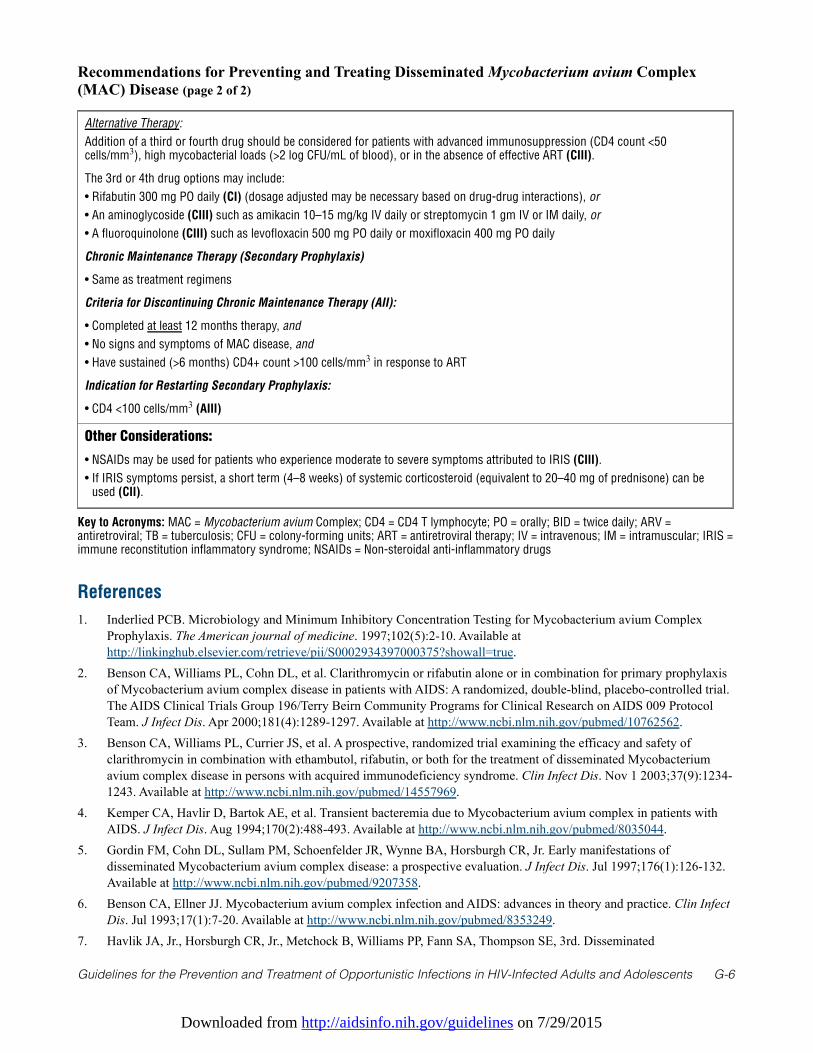
Downloaded from http://aidsinfo.nih.gov/guidelines on 7/29/2015
Guidelines for the Prevention and Treatment of Opportunistic Infections in HIV-Infected Adults and Adolescents G-6
References
1. Inderlied PCB. Microbiology and Minimum Inhibitory Concentration Testing for Mycobacterium avium ComplexProphylaxis. The American journal of medicine. 1997;102(5):2-10. Available athttp://linkinghub.elsevier.com/retrieve/pii/S0002934397000375?showall=true.
2. Benson CA, Williams PL, Cohn DL, et al. Clarithromycin or rifabutin alone or in combination for primary prophylaxisof Mycobacterium avium complex disease in patients with AIDS: A randomized, double-blind, placebo-controlled trial.The AIDS Clinical Trials Group 196/Terry Beirn Community Programs for Clinical Research on AIDS 009 ProtocolTeam. J Infect Dis. Apr 2000;181(4):1289-1297. Available at http://www.ncbi.nlm.nih.gov/pubmed/10762562.
3. Benson CA, Williams PL, Currier JS, et al. A prospective, randomized trial examining the efficacy and safety ofclarithromycin in combination with ethambutol, rifabutin, or both for the treatment of disseminated Mycobacteriumavium complex disease in persons with acquired immunodeficiency syndrome. Clin Infect Dis. Nov 1 2003;37(9):1234-1243. Available at http://www.ncbi.nlm.nih.gov/pubmed/14557969.
4. Kemper CA, Havlir D, Bartok AE, et al. Transient bacteremia due to Mycobacterium avium complex in patients withAIDS. J Infect Dis. Aug 1994;170(2):488-493. Available at http://www.ncbi.nlm.nih.gov/pubmed/8035044.
5. Gordin FM, Cohn DL, Sullam PM, Schoenfelder JR, Wynne BA, Horsburgh CR, Jr. Early manifestations ofdisseminated Mycobacterium avium complex disease: a prospective evaluation. J Infect Dis. Jul 1997;176(1):126-132.Available at http://www.ncbi.nlm.nih.gov/pubmed/9207358.
6. Benson CA, Ellner JJ. Mycobacterium avium complex infection and AIDS: advances in theory and practice. Clin InfectDis. Jul 1993;17(1):7-20. Available at http://www.ncbi.nlm.nih.gov/pubmed/8353249.
7. Havlik JA, Jr., Horsburgh CR, Jr., Metchock B, Williams PP, Fann SA, Thompson SE, 3rd. Disseminated
Recommendations for Preventing and Treating Disseminated Mycobacterium avium Complex(MAC) Disease (page 2 of 2)
Alternative Therapy:
Addition of a third or fourth drug should be considered for patients with advanced immunosuppression (CD4 count <50cells/mm3), high mycobacterial loads (>2 log CFU/mL of blood), or in the absence of effective ART (CIII).
The 3rd or 4th drug options may include:
• Rifabutin 300 mg PO daily (CI) (dosage adjusted may be necessary based on drug-drug interactions), or
• An aminoglycoside (CIII) such as amikacin 10–15 mg/kg IV daily or streptomycin 1 gm IV or IM daily, or
• A fluoroquinolone (CIII) such as levofloxacin 500 mg PO daily or moxifloxacin 400 mg PO daily
Chronic Maintenance Therapy (Secondary Prophylaxis)
• Same as treatment regimens
Criteria for Discontinuing Chronic Maintenance Therapy (AII):
• Completed at least 12 months therapy, and
• No signs and symptoms of MAC disease, and
• Have sustained (>6 months) CD4+ count >100 cells/mm3 in response to ART
Indication for Restarting Secondary Prophylaxis:
• CD4 <100 cells/mm3 (AIII)
Other Considerations:
• NSAIDs may be used for patients who experience moderate to severe symptoms attributed to IRIS (CIII).
• If IRIS symptoms persist, a short term (4–8 weeks) of systemic corticosteroid (equivalent to 20–40 mg of prednisone) can beused (CII).
Key to Acronyms: MAC = Mycobacterium avium Complex; CD4 = CD4 T lymphocyte; PO = orally; BID = twice daily; ARV =antiretroviral; TB = tuberculosis; CFU = colony-forming units; ART = antiretroviral therapy; IV = intravenous; IM = intramuscular; IRIS =immune reconstitution inflammatory syndrome; NSAIDs = Non-steroidal anti-inflammatory drugs
Downloaded from http://aidsinfo.nih.gov/guidelines on 7/29/2015
Guidelines for the Prevention and Treatment of Opportunistic Infections in HIV-Infected Adults and Adolescents G-7
Mycobacterium avium complex infection: clinical identification and epidemiologic trends. J Infect Dis. Mar1992;165(3):577-580. Available at http://www.ncbi.nlm.nih.gov/pubmed/1347060.
8. Benson CA. Treatment of disseminated disease due to the Mycobacterium avium complex in patients with AIDS. ClinInfect Dis. Apr 1994;18 Suppl 3:S237-242. Available at http://www.ncbi.nlm.nih.gov/pubmed/8204776.
9. Benson CA. Disease due to the Mycobacterium avium complex in patients with AIDS: epidemiology and clinicalsyndrome. Clin Infect Dis. Apr 1994;18 Suppl 3:S218-222. Available at http://www.ncbi.nlm.nih.gov/pubmed/8204773.
10. Nightingale SD, Byrd LT, Southern PM, Jockusch JD, Cal SX, Wynne BA. Incidence of Mycobacterium avium-intracellulare complex bacteremia in human immunodeficiency virus-positive patients. J Infect Dis. Jun1992;165(6):1082-1085. Available at http://www.ncbi.nlm.nih.gov/pubmed/1349906.
11. Chaisson RE, Moore RD, Richman DD, Keruly J, Creagh T. Incidence and natural history of Mycobacterium avium-complex infections in patients with advanced human immunodeficiency virus disease treated with zidovudine. TheZidovudine Epidemiology Study Group. The American review of respiratory disease. Aug 1992;146(2):285-289.Available at http://www.ncbi.nlm.nih.gov/pubmed/1362634.
12. Buchacz K, Baker RK, Palella FJ, Jr., et al. AIDS-defining opportunistic illnesses in US patients, 1994-2007: a cohortstudy. AIDS. Jun 19 2010;24(10):1549-1559. Available at http://www.ncbi.nlm.nih.gov/pubmed/20502317.
13. Barbaro DJ, Orcutt VL, Coldiron BM. Mycobacterium avium-Mycobacterium intracellulare infection limited to the skinand lymph nodes in patients with AIDS. Reviews of infectious diseases. Jul-Aug 1989;11(4):625-628. Available athttp://www.ncbi.nlm.nih.gov/pubmed/2772468.
14. Hellyer TJ, Brown IN, Taylor MB, Allen BW, Easmon CS. Gastro-intestinal involvement in Mycobacterium avium-intracellulare infection of patients with HIV. J Infect. Jan 1993;26(1):55-66. Available athttp://www.ncbi.nlm.nih.gov/pubmed/8454889.
15. Torriani FJ, McCutchan JA, Bozzette SA, Grafe MR, Havlir DV. Autopsy findings in AIDS patients withMycobacterium avium complex bacteremia. J Infect Dis. Dec 1994;170(6):1601-1605. Available athttp://www.ncbi.nlm.nih.gov/pubmed/7996004.
16. Roth RI, Owen RL, Keren DF, Volberding PA. Intestinal infection with Mycobacterium avium in acquired immunedeficiency syndrome (AIDS). Histological and clinical comparison with Whipple's disease. Digestive diseases andsciences. May 1985;30(5):497-504. Available at http://www.ncbi.nlm.nih.gov/pubmed/2580679.
17. Gillin JS, Urmacher C, West R, Shike M. Disseminated Mycobacterium avium-intracellulare infection in acquiredimmunodeficiency syndrome mimicking Whipple's disease. Gastroenterology. Nov 1983;85(5):1187-1191. Available athttp://www.ncbi.nlm.nih.gov/pubmed/6194041.
18. Inderlied CB, Kemper CA, Bermudez LE. The Mycobacterium avium complex. Clin Microbiol Rev. Jul 1993;6(3):266-310. Available at http://www.ncbi.nlm.nih.gov/pubmed/8358707.
19. Packer SJ, Cesario T, Williams JH, Jr. Mycobacterium avium complex infection presenting as endobronchial lesions inimmunosuppressed patients. Ann Intern Med. Sep 1 1988;109(5):389-393. Available athttp://www.ncbi.nlm.nih.gov/pubmed/3165608.
20. Phillips P, Kwiatkowski MB, Copland M, Craib K, Montaner J. Mycobacterial lymphadenitis associated with theinitiation of combination antiretroviral therapy. J Acquir Immune Defic Syndr Hum Retrovirol. Feb 1 1999;20(2):122-128. Available at http://www.ncbi.nlm.nih.gov/pubmed/10048898.
21. Phillips P, Bonner S, Gataric N, et al. Nontuberculous mycobacterial immune reconstitution syndrome in HIV-infectedpatients: spectrum of disease and long-term follow-up. Clin Infect Dis. Nov 15 2005;41(10):1483-1497. Available athttp://www.ncbi.nlm.nih.gov/pubmed/16231262.
22. Race EM, Adelson-Mitty J, Kriegel GR, et al. Focal mycobacterial lymphadenitis following initiation of protease-inhibitor therapy in patients with advanced HIV-1 disease. Lancet. Jan 24 1998;351(9098):252-255. Available athttp://www.ncbi.nlm.nih.gov/pubmed/9457095.
23. Cabie A, Abel S, Brebion A, Desbois N, Sobesky G. Mycobacterial lymphadenitis after initiation of highly activeantiretroviral therapy. Eur J Clin Microbiol Infect Dis. Nov 1998;17(11):812-813. Available athttp://www.ncbi.nlm.nih.gov/pubmed/9923530.
24. Shanson DC, Dryden MS. Comparison of methods for isolating Mycobacterium avium-intracellulare from blood ofpatients with AIDS. J Clin Pathol. Jun 1988;41(6):687-690. Available at http://www.ncbi.nlm.nih.gov/pubmed/3385000.
Downloaded from http://aidsinfo.nih.gov/guidelines on 7/29/2015
Guidelines for the Prevention and Treatment of Opportunistic Infections in HIV-Infected Adults and Adolescents G-8
25. Hafner R, Inderlied CB, Peterson DM, et al. Correlation of quantitative bone marrow and blood cultures in AIDSpatients with disseminated Mycobacterium avium complex infection. J Infect Dis. Aug 1999;180(2):438-447. Availableat http://www.ncbi.nlm.nih.gov/pubmed/10395860.
26. Havlir DV, Dube MP, Sattler FR, et al. Prophylaxis against disseminated Mycobacterium avium complex with weeklyazithromycin, daily rifabutin, or both. California Collaborative Treatment Group. N Engl J Med. Aug 81996;335(6):392-398. Available at http://www.ncbi.nlm.nih.gov/pubmed/8676932.
27. Pierce M, Crampton S, Henry D, et al. A randomized trial of clarithromycin as prophylaxis against disseminatedMycobacterium avium complex infection in patients with advanced acquired immunodeficiency syndrome. N Engl JMed. Aug 8 1996;335(6):384-391. Available at http://www.ncbi.nlm.nih.gov/pubmed/8663871.
28. Dworkin MS, Hanson DL, Kaplan JE, Jones JL, Ward JW. Risk for preventable opportunistic infections in persons withAIDS after antiretroviral therapy increases CD4+ T lymphocyte counts above prophylaxis thresholds. J Infect Dis. Aug2000;182(2):611-615. Available at http://www.ncbi.nlm.nih.gov/pubmed/10915098.
29. El-Sadr WM, Burman WJ, Grant LB, et al. Discontinuation of prophylaxis for Mycobacterium avium complex disease inHIV-infected patients who have a response to antiretroviral therapy. Terry Beirn Community Programs for Clinical Researchon AIDS. N Engl J Med. Apr 13 2000;342(15):1085-1092. Available at http://www.ncbi.nlm.nih.gov/pubmed/10766581.
30. Currier JS, Williams PL, Koletar SL, et al. Discontinuation of Mycobacterium avium complex prophylaxis in patientswith antiretroviral therapy-induced increases in CD4+ cell count. A randomized, double-blind, placebo-controlled trial.AIDS Clinical Trials Group 362 Study Team. Ann Intern Med. Oct 3 2000;133(7):493-503. Available athttp://www.ncbi.nlm.nih.gov/pubmed/11015162.
31. Furrer H, Telenti A, Rossi M, Ledergerber B. Discontinuing or withholding primary prophylaxis againstMycobacterium avium in patients on successful antiretroviral combination therapy. The Swiss HIV Cohort Study. AIDS.Jul 7 2000;14(10):1409-1412. Available at http://www.ncbi.nlm.nih.gov/pubmed/10930156.
32. Brooks JT, Song R, Hanson DL, et al. Discontinuation of primary prophylaxis against Mycobacterium avium complexinfection in HIV-infected persons receiving antiretroviral therapy: observations from a large national cohort in theUnited States, 1992-2002. Clin Infect Dis. Aug 15 2005;41(4):549-553. Available athttp://www.ncbi.nlm.nih.gov/pubmed/16028167.
33. Chaisson RE, Benson CA, Dube MP, et al. Clarithromycin therapy for bacteremic Mycobacterium avium complex disease.A randomized, double-blind, dose-ranging study in patients with AIDS. AIDS Clinical Trials Group Protocol 157 StudyTeam. Ann Intern Med. Dec 15 1994;121(12):905-911. Available at http://www.ncbi.nlm.nih.gov/pubmed/7978715.
34. May T, Brel F, Beuscart C, et al. Comparison of combination therapy regimens for treatment of humanimmunodeficiency virus-infected patients with disseminated bacteremia due to Mycobacterium avium. ANRS Trial 033Curavium Group. Agence Nationale de Recherche sur le Sida. Clin Infect Dis. Sep 1997;25(3):621-629. Available athttp://www.ncbi.nlm.nih.gov/pubmed/9314450.
35. Gordin FM, Sullam PM, Shafran SD, et al. A randomized, placebo-controlled study of rifabutin added to a regimen ofclarithromycin and ethambutol for treatment of disseminated infection with Mycobacterium avium complex. Clin InfectDis. May 1999;28(5):1080-1085. Available at http://www.ncbi.nlm.nih.gov/pubmed/10452638.
36. Dube MP, Sattler FR, Torriani FJ, et al. A randomized evaluation of ethambutol for prevention of relapse and drugresistance during treatment of Mycobacterium avium complex bacteremia with clarithromycin-based combinationtherapy. California Collaborative Treatment Group. J Infect Dis. Nov 1997;176(5):1225-1232. Available athttp://www.ncbi.nlm.nih.gov/pubmed/9359722.
37. Cohn DL, Fisher EJ, Peng GT, et al. A prospective randomized trial of four three-drug regimens in the treatment ofdisseminated Mycobacterium avium complex disease in AIDS patients: excess mortality associated with high-doseclarithromycin. Terry Beirn Community Programs for Clinical Research on AIDS. Clin Infect Dis. Jul 1999;29(1):125-133. Available at http://www.ncbi.nlm.nih.gov/pubmed/10433575.
38. Aberg JA, Yajko DM, Jacobson MA. Eradication of AIDS-related disseminated mycobacterium avium complexinfection after 12 months of antimycobacterial therapy combined with highly active antiretroviral therapy. J Infect Dis.Nov 1998;178(5):1446-1449. Available at http://www.ncbi.nlm.nih.gov/pubmed/9780266.
39. Ward TT, Rimland D, Kauffman C, Huycke M, Evans TG, Heifets L. Randomized, open-label trial of azithromycin plusethambutol vs. clarithromycin plus ethambutol as therapy for Mycobacterium avium complex bacteremia in patientswith human immunodeficiency virus infection. Veterans Affairs HIV Research Consortium. Clin Infect Dis. Nov1998;27(5):1278-1285. Available at http://www.ncbi.nlm.nih.gov/pubmed/9827282.
Downloaded from http://aidsinfo.nih.gov/guidelines on 7/29/2015
Guidelines for the Prevention and Treatment of Opportunistic Infections in HIV-Infected Adults and Adolescents G-9
40. Dunne M, Fessel J, Kumar P, et al. A randomized, double-blind trial comparing azithromycin and clarithromycin in thetreatment of disseminated Mycobacterium avium infection in patients with human immunodeficiency virus. Clin InfectDis. Nov 2000;31(5):1245-1252. Available at http://www.ncbi.nlm.nih.gov/pubmed/11073759.
41. Shafran SD, Singer J, Zarowny DP, et al. A comparison of two regimens for the treatment of Mycobacterium aviumcomplex bacteremia in AIDS: rifabutin, ethambutol, and clarithromycin versus rifampin, ethambutol, clofazimine, andciprofloxacin. Canadian HIV Trials Network Protocol 010 Study Group. N Engl J Med. Aug 8 1996;335(6):377-383.Available at http://www.ncbi.nlm.nih.gov/pubmed/8676931.
42. Griffith DE, Aksamit T, Brown-Elliott BA, et al. An official ATS/IDSA statement: diagnosis, treatment, and preventionof nontuberculous mycobacterial diseases. Am J Respir Crit Care Med. Feb 15 2007;175(4):367-416. Available athttp://www.ncbi.nlm.nih.gov/pubmed/17277290.
43. Gardner EM, Burman WJ, DeGroote MA, Hildred G, Pace NR. Conventional and molecular epidemiology of macrolideresistance among new Mycobacterium avium complex isolates recovered from HIV-infected patients. Clin Infect Dis.Oct 1 2005;41(7):1041-1044. Available at http://www.ncbi.nlm.nih.gov/pubmed/16142672.
44. Abbot Laboratories. clarithromycin (biaxin). Abbot Park, IL: Abbot Laboratories; 1995.45. Shafran SD, Deschenes J, Miller M, Phillips P, Toma E. Uveitis and pseudojaundice during a regimen of
clarithromycin, rifabutin, and ethambutol. MAC Study Group of the Canadian HIV Trials Network. N Engl J Med. Feb10 1994;330(6):438-439. Available at http://www.ncbi.nlm.nih.gov/pubmed/8284019.
46. Hafner R, Bethel J, Power M, et al. Tolerance and pharmacokinetic interactions of rifabutin and clarithromycin inhuman immunodeficiency virus-infected volunteers. Antimicrob Agents Chemother. Mar 1998;42(3):631-639. Availableat http://www.ncbi.nlm.nih.gov/pubmed/9517944.
47. Wormser GP, Horowitz H, Dworkin B. Low-dose dexamethasone as adjunctive therapy for disseminatedMycobacterium avium complex infections in AIDS patients. Antimicrob Agents Chemother. Sep 1994;38(9):2215-2217.Available at http://www.ncbi.nlm.nih.gov/pubmed/7811052.
48. TB/HIV Drug Interactions. www.cdc.gov/tb_HIV_Drugs/Rifabutin.htm. 49. Panel on Antiretroviral Guidelines for Adults and Adolescents. Guidelines for the use of antiretroviral agents in HIV-1-
infected adults and adolescents. Department of Health and Human Services. Available athttp://aidsinfo.nih.gov/contentfiles/lvguidelines/AdultandAdolescentGL.pdf. Accessed on March 4, 2013.
50. Heifets L, Lindholm LP, Libonati J. Radiometric broth macrodilution method for determination of minimal inhibitoryconcentrations(MIC) with Mycobacterium avium complex isolates: proposed guidelines. Paper presented at: nationalJewish Center for Immunology and Respiratory Medicine. 1993.
51. Heifets L, Mor N, Vanderkolk J. Mycobacterium avium strains resistant to clarithromycin and azithromycin. AntimicrobAgents Chemother. Nov 1993;37(11):2364-2370. Available at http://www.ncbi.nlm.nih.gov/pubmed/8031351.
52. Masur H. Recommendations on prophylaxis and therapy for disseminated Mycobacterium avium complex disease inpatients infected with the human immunodeficiency virus. Public Health Service Task Force on Prophylaxis andTherapy for Mycobacterium avium Complex. N Engl J Med. Sep 16 1993;329(12):898-904. Available athttp://www.ncbi.nlm.nih.gov/pubmed/8395019.
53. Kemper CA, Meng TC, Nussbaum J, et al. Treatment of Mycobacterium avium complex bacteremia in AIDS with afour-drug oral regimen. Rifampin, ethambutol, clofazimine, and ciprofloxacin. The California Collaborative TreatmentGroup. Ann Intern Med. Mar 15 1992;116(6):466-472. Available at http://www.ncbi.nlm.nih.gov/pubmed/1739237.
54. Chaisson RE, Keiser P, Pierce M, et al. Clarithromycin and ethambutol with or without clofazimine for the treatment ofbacteremic Mycobacterium avium complex disease in patients with HIV infection. AIDS. Mar 1997;11(3):311-317.Available at http://www.ncbi.nlm.nih.gov/pubmed/9147422.
55. Chiu J, Nussbaum J, Bozzette S, et al. Treatment of disseminated Mycobacterium avium complex infection in AIDSwith amikacin, ethambutol, rifampin, and ciprofloxacin. California Collaborative Treatment Group. Ann Intern Med.Sep 1 1990;113(5):358-361. Available at http://www.ncbi.nlm.nih.gov/pubmed/2382918.
56. Rodriguez Diaz JC, Lopez M, Ruiz M, Royo G. In vitro activity of new fluoroquinolones and linezolid against non-tuberculous mycobacteria. International journal of antimicrobial agents. Jun 2003;21(6):585-588. Available athttp://www.ncbi.nlm.nih.gov/pubmed/12791475.
57. El-Sadr WM, Murphy RL, Yurik TM, et al. Atovaquone compared with dapsone for the prevention of Pneumocystiscarinii pneumonia in patients with HIV infection who cannot tolerate trimethoprim, sulfonamides, or both. Community
Downloaded from http://aidsinfo.nih.gov/guidelines on 7/29/2015
Guidelines for the Prevention and Treatment of Opportunistic Infections in HIV-Infected Adults and Adolescents G-10
Program for Clinical Research on AIDS and the AIDS Clinical Trials Group. N Engl J Med. Dec 24 1998;339(26):1889-1895. Available at http://www.ncbi.nlm.nih.gov/pubmed/9862944.
58. Aberg JA, Williams PL, Liu T, et al. A study of discontinuing maintenance therapy in human immunodeficiency virus-infected subjects with disseminated Mycobacterium avium complex: AIDS Clinical Trial Group 393 Study Team. JInfect Dis. Apr 1 2003;187(7):1046-1052. Available at http://www.ncbi.nlm.nih.gov/pubmed/12660918.
59. Einarson A, Phillips E, Mawji F, et al. A prospective controlled multicentre study of clarithromycin in pregnancy.American journal of perinatology. 1998;15(9):523-525. Available at http://www.ncbi.nlm.nih.gov/pubmed/9890248.
60. Drinkard CR, Shatin D, Clouse J. Postmarketing surveillance of medications and pregnancy outcomes: clarithromycinand birth malformations. Pharmacoepidemiology and drug safety. Dec 2000;9(7):549-556. Available athttp://www.ncbi.nlm.nih.gov/pubmed/11338912.
Downloaded from http://aidsinfo.nih.gov/guidelines on 7/29/2015
Guidelines for the Prevention and Treatment of Opportunistic Infections in HIV-Infected Adults and Adolescents H-1
Bacterial Respiratory Disease (Last updated May 7, 2013; last reviewed May7, 2013)
Epidemiology
Bacterial respiratory diseases; including sinusitis, bronchitis, otitis, and pneumonia; are among the mostcommon infectious complications in patients with HIV infection, occurring with increased frequency at allCD4 T lymphocyte cell (CD4) counts,1 and some data suggest that bacterial pneumonia may occur withincreased severity in this population. This chapter will focus on the diagnosis, prevention, and managementof bacterial pneumonia in HIV-infected patients.
Bacterial pneumonia is a common cause of HIV-associated morbidity and recurrent pneumonia (2 or moreepisodes within a 1-year period) is an AIDS-defining condition. The incidence of bacterial pneumonia ishigher in HIV-infected individuals than in those who are not HIV infected.2 More recently, the incidence ofbacterial pneumonia in HIV-infected individuals has declined. In one study, the incidence of bacterialpneumonia declined from 22.7 episodes per 100 person-years in the era before combination antiretroviraltherapy (ART) to 9.1 episodes per 100 person-years by 1997.3-5
Bacterial pneumonia may be the first manifestation of underlying HIV infection and can occur at any stageof HIV disease and at any CD4 count. The high rates of bacterial pneumonia in HIV-infected individualsprobably result from multiple factors, including qualitative B-cell defects that impair ability to producepathogen-specific antibody; impaired neutrophil function or numbers, or both; and factors, such as injectiondrug use, that are associated with underlying HIV infection. Risk factors associated with an increased risk ofbacterial pneumonia include low CD4 count (< 200 cells/mm3), no or intermittent use of ART, cigarettesmoking, injection drug use, and chronic viral hepatitis.
In HIV-infected individuals, as in those who are not HIV infected, Streptococcus pneumoniae and Haemophilusspecies are the most frequently identified causes of community-acquired bacterial pneumonia.6-12 Atypicalbacterial pathogens such as Legionella pneumophila, Mycoplasma pneumoniae, and Chlamydophila specieshave been reported as infrequent causes of community-acquired bacterial pneumonia in HIV-infectedindividuals.9,13
The frequency of Pseudomonas aeruginosa and Staphylococcus aureus as community-acquired pathogens ishigher in HIV-infected individuals than in those not HIV infected.10,14 Methicillin-resistant Staphylococcusaureus (MRSA) infection, in particular, should be considered as a potential etiology for pneumonia, giventhat community outbreaks of MRSA have been seen in men who have sex with men and nasal carriage ofMRSA is more common in HIV-infected individuals, particularly at lower CD4 cell counts.15 Also,community-acquired MRSA pneumonia may not invariably be associated with preceding influenza illness.16
In HIV-infected patients, particularly those infected with S. pneumoniae, incidence of bacteremiaaccompanying pneumonia is increased compared with that in individuals who are not HIV infected. In onestudy, the estimated rate of pneumococcal bacteremia in patients with AIDS (1,094 cases per 100,000) was~55 times that in HIV-uninfected individuals (20 cases per 100,000). This disparity narrowed but was noteliminated after the introduction of ART.17 Other studies have highlighted the declining incidence ofpneumococcal bacteremia in the era of ART.18
Bacterial pneumonia is associated with increased mortality in HIV-infected individuals.10,19,20 In HIV-infectedindividuals with community-acquired bacterial pneumonia, a prospective, multicenter study documentedCD4 count <100 cells/mm3, radiographic progression of disease, and presence of shock as independentpredictors of increased mortality.21 In that study, multilobar infiltrates, cavitary infiltrates, and pleuraleffusion on baseline radiograph all were independent predictors of radiographic progression of disease.
Downloaded from http://aidsinfo.nih.gov/guidelines on 7/29/2015
Guidelines for the Prevention and Treatment of Opportunistic Infections in HIV-Infected Adults and Adolescents H-2
Clinical Manifestations
Clinical and radiographic presentation of bacterial pneumonia in HIV-infected individuals is similar to that inthose who are not HIV infected. Patients with pneumonias caused by bacteria such as S. pneumoniae orHaemophilus species characteristically have acute onset (3–5 days) of symptoms, including fevers, chills,rigors, chest pain or pleurisy, cough productive of purulent sputum, and dyspnea.22 They are often febrile andthe presence of fever, tachycardia, or hypotension can be an indicator of sepsis. Tachypnea and decreasedarterial oxygen saturation indicate moderate-to-severe pneumonia and clinicians should strongly considerhospitalizing such patients.
Patients with bacterial pneumonia typically have signs of focal consolidation, such as egophony, and/orpleural effusion on lung examination. In contrast, lung examination often is normal in those withPneumocystis pneumonia (PCP), and if abnormal, reveals inspiratory crackles. In patients with bacterialpneumonia, the white blood cell (WBC) count usually is elevated. The elevation may be relative to baselineWBC in those with advanced HIV. A left shift in WBC differential may be present.
Individuals with bacterial pneumonia characteristically exhibit unilateral, focal, segmental, or lobarconsolidation on chest radiograph. The frequency of these typical radiographic findings, however, maydepend on the underlying bacterial pathogen. Those with pneumonia due to S. pneumoniae or Haemophilustypically present with consolidation, whereas presence of cavitation may be a feature more suggestive of P.aeruginosa or S. aureus.
Disease severity and arterial oxygenation should be assessed in all patients with pneumonia. Noninvasivemeasurement of arterial oxygen saturation via pulse oximetry is an appropriate screening test. Arterial bloodgas analysis is indicated for those with evidence of hypoxemia suggested by noninvasive assessment and forpatients who have tachypnea and/or respiratory distress. Criteria developed to assess disease severity in HIV-uninfected persons, such as the Pneumonia Severity Index (PSI) (http://pda.ahrq.gov/clinic/psi/psicalc.asp)appear to be valid for HIV-infected patients, especially when used in combination with CD4 count21,23
(discussed in further detail in Treating Disease).
Diagnosis
Guidelines for diagnosing and managing community-acquired pneumonia (CAP) in individuals who are notHIV infected also apply to those who are infected.24 Patients with clinical symptoms and signs suggestive ofCAP should have posteroanterior and lateral chest radiographs, if possible. If previous radiographs areavailable, they should be reviewed to assess for presence of new findings. The clinical diagnosis of bacterialpneumonia requires a demonstrable infiltrate.
Given the increased incidence of Mycobacterium tuberculosis in HIV-infected individuals, a tuberculosis(TB) diagnosis should always be considered in HIV-infected patients who have pneumonia. Those withclinical and radiographic findings suggestive of TB should be managed as potentially having TB (that is,with respiratory isolation if hospitalized), and two to three sputum specimens should be obtained for acid fastbacilli evaluation. In settings where the prevalence of TB is high, initiation of empiric therapy for bothbacterial pneumonia and TB may be appropriate for patients in whom both diagnoses are strongconsiderations and after diagnostic studies are undertaken.
Often, the differential diagnosis of pneumonia in HIV-infected individuals is broad and a confirmedmicrobiologic diagnosis allows clinicians to target the specific pathogen and discontinue broad spectrumantibiotic therapy and/or empiric therapy (such as empiric PCP therapy) that targets non-bacterial pathogens.
HIV-infected patients with suspected CAP should undergo investigation for specific pathogens that wouldsignificantly alter standard (empirical) management decisions when presence of such pathogens is suspectedbased on epidemiologic, clinical, or radiologic clues. P. aeruginosa should be considered in HIV-infectedpatients with advanced HIV disease (that is, CD4 count ≤50 cells/mm3), pre-existing lung disease such as
Downloaded from http://aidsinfo.nih.gov/guidelines on 7/29/2015
Guidelines for the Prevention and Treatment of Opportunistic Infections in HIV-Infected Adults and Adolescents H-3
bronchiectasis, or underlying neutropenia. It is also a consideration for HIV-infected patients who usecorticosteroids, are severely malnourished, have been hospitalized in the past 90 days or reside in a healthcare facility or nursing home, or are on chronic hemodialysis. Because cavitary infiltrates are common inpatients with P. aeruginosa, that radiographic finding also should prompt an investigation for this pathogen.S. aureus should be considered in patients with recent viral (or influenza) infection; a history of injectiondrug use; or severe, bilateral, necrotizing pneumonia.
Routine diagnostic tests to identify an etiologic diagnosis are optional for HIV-infected patients withsuspected CAP who are well enough to be treated as outpatients, especially if the microbiologic studiescannot be performed promptly.
In contrast, a pre-treatment expectorated sputum specimen for Gram stain and culture and two blood culturesshould be obtained from HIV-infected patients hospitalized for suspected CAP, particularly those whorequire intensive care.
Gram stain and culture of expectorated sputum should be performed only if a good-quality specimen can beobtained and quality performance measures can be met for collection, transport, and processing of samples.Correlation of sputum culture with Gram stain can help in interpretation of sputum culture data. Forintubated patients, an endotracheal aspirate sample should be obtained. Bronchoscopy with bronchoalveolarlavage should be considered, especially if the differential diagnosis is broad and includes pathogens such asPneumocystis jirovecii.
The increased incidence of bacteremia in HIV-infected patients, especially those with low CD4 cell counts,and the high specificity of blood cultures argue for their collection in such individuals. Low sensitivity ofblood cultures in persons with higher CD4 counts argues against routine collection. However, patients withHIV infection are at increased risk of infection with drug-resistant pneumococci.25,26 Because identificationof this organism could lead to changes in management, collection of blood specimens in HIV-infectedpatients with CAP should always be considered.
In addition to the above tests, urinary antigen tests for L. pneumophila and S. pneumoniae should beconsidered.
Diagnostic thoracentesis should be considered in all patients with pleural effusion, especially if concernexists for accompanying empyema, and therapeutic thoracentesis should be performed to relieve respiratorydistress secondary to a moderate-to-large-sized pleural effusion.
Preventing Exposure
No effective means exist to reduce exposure to S. pneumoniae and Haemophilus influenzae, which arecommon in the community.
Preventing Disease
Vaccination against S. pneumoniae and influenza, use of combination ART, and lifestyle modifications are allimportant measures in preventing bacterial pneumonia. Multiple observational studies of pneumococcalpolysaccharide vaccine (PPV) in the United States have reported benefits from such vaccination in HIV-infected persons.27-32 Several studies also have documented an association between vaccination and a reducedrisk of pneumococcal bacteremia.18,32 One randomized placebo-controlled trial of PPV in Africaparadoxically found that vaccination was associated with an increased risk of pneumonia.33 Follow-up of thiscohort confirmed the increase in pneumonia in vaccinated subjects but also showed a decrease in all-causemortality.34
A 13-valent pneumococcal conjugate vaccine (PCV13) has recently been recommended by the AdvisoryCommittee on Immunization Practices for use in adults with immunocompromising conditions, includingHIV infection.35 A randomized, double-blind, placebo-controlled trial of 7-valent PCV among HIV-infected
Downloaded from http://aidsinfo.nih.gov/guidelines on 7/29/2015
Guidelines for the Prevention and Treatment of Opportunistic Infections in HIV-Infected Adults and Adolescents H-4
adults in Malawi demonstrated 74% efficacy against vaccine-type invasive pneumococcal disease, with clearevidence of efficacy in those with CD4 counts <200 cells/mm3.36
HIV-infected adults and adolescents who have never received any pneumococcal vaccine should receive asingle dose of PCV13 regardless of CD4 count (AI).35 Patients with CD4 counts ≥200 cells/mm3 should thenreceive a dose of 23-valent PPV (PPV23) at least 8 weeks later (AII).27-32,37-39 HIV-infected patients withCD4 counts <200 cells/mm3 can be offered PPV23 at least 8 weeks after receiving PCV13 (CIII); however,it may be preferable to defer PPV23 until after the CD4 count increases to >200 cells/mm3 on ART (BIII).Clinical evidence supporting use of PPV23 in persons with CD4 counts <200 cells/mm3 appears strongest inpatients who also have HIV RNA <100,000 copies/mL;37,39 evidence also suggests benefit for those who startART before receiving PPV.32
The duration of the protective effect of PPV23 is unknown; a single revaccination with PPV is recommendedif ≥5 years have elapsed since the first dose of PPV23 was given (BIII).31 A third dose of PPV23 should begiven at age 65 years or later, as long as 5 years have elapsed since the most recent dose and it was givenbefore age 65 years (BIII).
PCV13 should also be given in HIV-infected patients who have already received PPV23 (AII). However,such patients should wait at least 1 year after their most recent dose of PPV23 before receiving a single doseof PCV13 (BIII).35 Subsequent doses of PPV23 should be given according to the schedule outlined above(i.e., at least 5 years between doses of PPV23 with no more than 3 lifetime doses).
Inactivated influenza vaccine should be administered annually during influenza season to all HIV-infectedindividuals (AIII).40 This recommendation is pertinent to prevention of bacterial pneumonia, which canoccur as a complication of influenza. Use of live attenuated influenza vaccine is contraindicated and is notrecommended in HIV-infected individuals (AIII).
The incidence of H. influenzae type b infection in HIV-infected adults is low. Therefore, H. influenzae typevaccine is not usually recommended for adult use (BIII) unless a patient also has anatomic or functionalasplenia.
Several factors are associated with a decreased risk of bacterial pneumonia, including use of ART and oftrimethoprim-sulfamethoxazole (TMP-SMX) for PCP prophylaxis.20 In many studies, daily administration ofTMP-SMX for PCP prophylaxis also reduced the frequency of bacterial respiratory infections.2,41,42 Thispoint should be considered when selecting an agent for PCP prophylaxis; however, indiscriminate use of thisdrug (when not indicated for PCP prophylaxis or other specific reasons) may promote development of TMP-SMX-resistant organisms. Thus, TMP-SMX should not be prescribed solely to prevent bacterial respiratoryinfection (BIII). Similarly, clarithromycin administered daily and azithromycin administered weekly are thedrugs of choice for Mycobacterium avium complex (MAC) prophylaxis and may be effective in preventingbacterial respiratory infections.43,44 However, these drugs also should not be prescribed solely for preventingbacterial respiratory infection (BIII).
A decreased absolute neutrophil count (e.g., <500 cells/mm3) is associated with an increased risk of bacterialinfections, including pneumonia, although this risk has been demonstrated primarily in persons withmalignancies. To reduce the risk of such bacterial infections, clinicians can consider taking steps to reverseneutropenia, either by stopping myelosuppressive drugs (CIII) or by administering granulocyte-colonystimulating factor (CIII), although these interventions have not been demonstrated to be effective in HIV-infected persons.
Modifiable factors associated with an increased risk of bacterial pneumonia include smoking cigarettes andusing injection drugs and alcohol.2,38,45-47 Clinicians should encourage cessation of these behaviors, and datasuggest that smoking cessation can decrease the risk of bacterial pneumonia.48
Downloaded from http://aidsinfo.nih.gov/guidelines on 7/29/2015
Guidelines for the Prevention and Treatment of Opportunistic Infections in HIV-Infected Adults and Adolescents H-5
Treating Disease
Whether patients should be treated on an outpatient basis or admitted to the hospital depends on severalfactors. One study suggested that the site of care decision be dictated by considering the PSI and CD4 counttogether.23 Mortality was increased in patients with higher PSI class, but even in those without an increasedmortality risk by PSI, the presence of a CD4 count <200 cells/mm3 was associated with an increased risk ofdeath.23 This led to the suggestion to always offer hospitalization to CAP patients with CD4 counts <200cells/mm3 and to use the PSI to help guide the decision in those with higher CD4 counts.49 In fact, in oneseries of 118 HIV-infected patients with CAP who were hospitalized, 62% fell into PSI Classes I and II,groups that are rarely hospitalized if not HIV infected.50 In another study, 40% of hospitalized HIV-infectedpatients in low-risk PSI classes had CD4 counts <200 cells/mm3.23
The basic principles of treatment of community-acquired bacterial pneumonia are the same for HIV-infectedpatients as for those who are not HIV infected.24 As discussed in the Diagnosis section, if specimens are to becollected for diagnosis, they should be taken before antibiotic therapy is initiated. Antibiotic therapy shouldbe administered promptly, however, without waiting for the results of diagnostic testing.
Empiric Antibiotic Therapy by Treatment Setting and Severity of DiseasesOutpatient TreatmentHIV-infected individuals who are being treated as outpatients should receive an oral beta-lactam plus an oralmacrolide (AII) or an oral respiratory fluoroquinolone (AII). Preferred beta-lactams are high-doseamoxicillin or amoxicillin-clavulanate; alternatives are cefpodoxime or cefuroxime. Preferred macrolides areazithromycin or clarithromycin. Doxycycline is an alternative to the macrolide (CIII). Preferred oralrespiratory fluoroquinolones are moxifloxacin or levofloxacin.
An oral respiratory fluoroquinolone (moxifloxacin or levofloxacin) should be used in patients who areallergic to penicillin (AII).
Respiratory fluoroquinolones also are active against M. tuberculosis. Thus, patients with TB who are treatedwith fluoroquinolone monotherapy may have an initial but misleading response that could delay diagnosis ofTB and initiation of appropriate multidrug TB therapy and increase risk of drug-resistant TB and TBtransmission. Fluoroquinolones, therefore, should be used with caution in patients in whom TB is suspected butwho are not being treated with concurrent standard four-drug TB therapy. Increasing rates of pneumococcalresistance suggest that empirical therapy with a macrolide alone cannot be routinely recommended (BIII).Patients who are receiving a macrolide for MAC prophylaxis should never receive macrolide monotherapy forempiric treatment of bacterial pneumonia, but macrolides can be used as part of a combination regimen.
Non-Intensive Care Unit Inpatient TreatmentHIV-infected individuals who are being treated as inpatients should receive an intravenous (IV) beta-lactamplus a macrolide (AII) or an IV respiratory fluoroquinolone (AII). Preferred beta-lactams are ceftriaxone,cefotaxime, or ampicillin-sulbactam. Preferred macrolides are azithromycin and clarithromycin. Doxycycline isan alternative to the macrolide (CIII). Preferred respiratory fluoroquinolones are moxifloxacin or levofloxacin.Clinical and Laboratory Standards Institute and U.S. Food and Drug Administration changes in the penicillinbreakpoints for treatment of non-meningitis pneumococcal disease imply that clinicians can consider treatmentwith IV penicillin in HIV-infected patients confirmed to have pneumococcal pneumonia (BIII).51
In patients who are allergic to penicillin, an IV respiratory fluoroquinolone (moxifloxacin or levofloxacin[750 mg/day]) should be used (AII).
Because of the activity of fluoroquinolones against M. tuberculosis and the dangers of monotherapy in thosewith TB, as previously discussed, fluoroquinolones should be used with caution in patients in whom TB issuspected but who are not being treated with concurrent standard four-drug TB therapy.
Downloaded from http://aidsinfo.nih.gov/guidelines on 7/29/2015
Guidelines for the Prevention and Treatment of Opportunistic Infections in HIV-Infected Adults and Adolescents H-6
Increasing rates of pneumococcal resistance suggest that empirical therapy with a macrolide alone cannot berecommended routinely (BIII). Patients who are receiving a macrolide for MAC prophylaxis should neverreceive macrolide monotherapy for empiric treatment of bacterial pneumonia, but macrolides can be used aspart of a combination regimen.
Intensive Care Unit TreatmentIntensive care unit patients should not receive empiric monotherapy, even with a fluoroquinolone, becausethe efficacy of this approach has not been established. In one study, the use of dual therapy (usually with abeta-lactam plus a macrolide) was associated with reduced mortality in patients with bacteremicpneumococcal pneumonia, including those admitted to the intensive care unit.52 Patients with severepneumonia who require intensive care should be treated with an IV beta-lactam plus either IV azithromycin(AII) or an IV respiratory fluoroquinolone (moxifloxacin or levofloxacin [750 mg/day]) (AII). Preferredbeta-lactams are ceftriaxone, cefotaxime, or ampicillin-sulbactam.
In patients who are allergic to penicillin, aztreonam plus an IV respiratory fluoroquinolone (moxifloxacin orlevofloxacin [750 mg/day]) should be used (BIII).
The majority of CAP pathogens can be treated adequately with recommended empiric regimens. Theincreased incidence of P. aeruginosa and S. aureus (including community-acquired MRSA) as causes ofCAP are exceptions. Both of these pathogens occur in specific epidemiologic patterns with distinct clinicalpresentations, for which empiric antibiotic coverage may be warranted. Diagnostic tests (sputum Gram stainand culture) are likely to be of high yield for these pathogens, allowing early discontinuation of empirictreatment if results are negative.
Empiric Pseudomonas aeruginosa TreatmentIf risk factors for Pseudomonas infection are present, an antipneumococcal, antipseudomonal beta-lactamplus either ciprofloxacin or levofloxacin (750-mg dose) should be used (BIII). Preferred beta-lactams arepiperacillin-tazobactam, cefepime, imipenem, or meropenem. Alternatives are an antipneumococcal,antipseudomonal beta-lactam plus an aminoglycoside and azithromycin (BIII) or an antipneumococcal,antipseudomonal beta-lactam plus an aminoglycoside and an antipneumococcal fluoroquinolone (BIII). Inpatients who are allergic to penicillin, aztreonam can be used in place of the beta-lactam (BIII).
Empiric Staphylococcus aureus TreatmentIn patients who have risk factors for S. aureus infection, including community-acquired MRSA, vancomycinor linezolid should be added to the antibiotic regimen (BIII). Although not routinely recommended, theaddition of clindamycin (to vancomycin, but not to linezolid) may be considered if severe necrotizingpneumonia is present to minimize bacterial toxin production (CIII).
Pathogen-Directed TherapyWhen the etiology of the pneumonia has been identified on the basis of reliable microbiological methods,antimicrobial therapy should be modified and directed at that pathogen.
Switch from Intravenous to Oral TherapyA switch to oral therapy should be considered in patients with CAP on IV antibiotic therapy who haveimproved clinically, can swallow and tolerate oral medications, and have intact gastrointestinal function.Suggested criteria for clinical stability include oral temperature <37.8°C, heart rate <100 beats/minute,respiratory rate <24 breaths/minute, systolic blood pressure ≥90 mm Hg, and room air oxygen saturation>90% or partial pressure of oxygen in arterial blood (PaO2) >60 mm Hg.24
Special Considerations Regarding When to Start Antiretroviral TherapyThe presence of acute opportunistic infection (OI), including bacterial pneumonia, increases the urgency of
Downloaded from http://aidsinfo.nih.gov/guidelines on 7/29/2015
Guidelines for the Prevention and Treatment of Opportunistic Infections in HIV-Infected Adults and Adolescents H-7
starting ART. In one randomized, controlled trial, use of ART early in the course of OIs, including bacterialinfections, led to less AIDS progression and death compared with later onset of therapy.53 Therefore, inpatients not already on ART, ART should be initiated early in the course of bacterial pneumonia (AI).
Monitoring of Response to Therapy and Adverse Events (Including IRIS) The clinical response to appropriate antimicrobial therapy is similar in HIV-infected patients and individualswho are not HIV infected.54 A clinical response (i.e., reduction in fever and improvement in respiratorysymptoms, physical findings, and laboratory studies) typically is observed within 48 to 72 hours afterinitiation of appropriate antimicrobial therapy. The presence of advanced HIV infection, CD4 count <100cells/mm3, and S. pneumoniae etiology were predictors of needing >7 days to reach clinical stability, whereasthose patients receiving ART tended to become clinically stable sooner.49 Usually, radiographic improvementlags behind clinical improvement.
Immune reconstitution inflammatory syndrome (IRIS) has not been described in association with bacterialrespiratory disease and treatment with ART in HIV-infected patients.
Managing Treatment FailurePatients who fail to respond to appropriate antimicrobial therapy should undergo further evaluation to searchfor other infectious and noninfectious causes of pulmonary dysfunction. The possibility of TB should alwaysbe considered in HIV-infected patients with pulmonary disease.
Preventing Recurrence
HIV-infected patients should receive pneumococcal and influenza vaccine as recommended. Antibioticchemoprophylaxis generally is not recommended specifically to prevent recurrences of bacterial respiratoryinfections because of the potential for development of drug-resistant microorganisms and drug toxicity.
Special Considerations During Pregnancy
The diagnosis of bacterial respiratory tract infections in pregnant women is the same as in those who are notpregnant, with appropriate shielding of the abdomen during radiographic procedures. Bacterial respiratorytract infections should be managed as in women who are not pregnant, with certain exceptions.Clarithromycin is not recommended as the first-line agent among macrolides because of an increased risk ofbirth defects seen in some animal studies. Two studies, each involving at least 100 women with first-trimester exposure to clarithromycin, did not document a clear increase in or specific pattern of birth defects,although an increased risk of spontaneous abortion was noted in one study.55,56 Azithromycin did not producebirth defects in animal studies, but experience with human use in the first trimester is limited. Azithromycinis recommended when a macrolide is indicated in pregnancy (BIII). Arthropathy has been noted in immatureanimals with in utero exposure to quinolones. However, studies evaluating quinolone use in pregnant womendid not find an increased risk of birth defects or musculoskeletal abnormalities.57,58 Thus, when indicated,quinolones can be used in pregnancy for serious respiratory infections (CIII).59
Doxycycline is not recommended for use during pregnancy because of increased hepatotoxicity and stainingof fetal teeth and bones. Beta-lactam antibiotics have not been associated with teratogenicity or increasedtoxicity in pregnancy. Aminoglycosides can be used as needed. A theoretical risk of fetal renal or eighthnerve damage exists with exposure during pregnancy, but this finding has not been documented in humans,except with streptomycin (10% risk) and kanamycin (2% risk). Experience with linezolid in humanpregnancy has been limited, but it was not teratogenic in mice, rats, and rabbits.
Pneumonia during pregnancy is associated with increased rates of preterm labor and delivery. Pregnantwomen with pneumonia after 20 weeks’ gestation should be monitored for evidence of contractions (BII).
Pneumococcal vaccine can be administered during pregnancy (AIII). Although its safety during the first
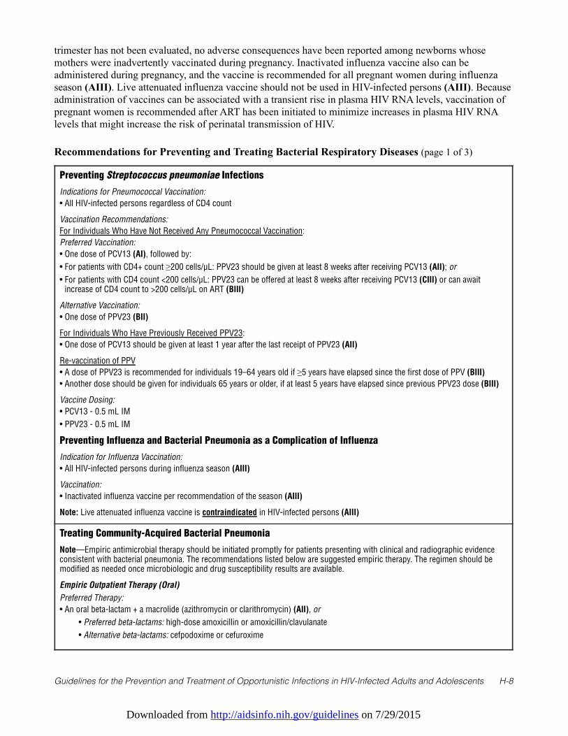
Downloaded from http://aidsinfo.nih.gov/guidelines on 7/29/2015
Guidelines for the Prevention and Treatment of Opportunistic Infections in HIV-Infected Adults and Adolescents H-8
trimester has not been evaluated, no adverse consequences have been reported among newborns whosemothers were inadvertently vaccinated during pregnancy. Inactivated influenza vaccine also can beadministered during pregnancy, and the vaccine is recommended for all pregnant women during influenzaseason (AIII). Live attenuated influenza vaccine should not be used in HIV-infected persons (AIII). Becauseadministration of vaccines can be associated with a transient rise in plasma HIV RNA levels, vaccination ofpregnant women is recommended after ART has been initiated to minimize increases in plasma HIV RNAlevels that might increase the risk of perinatal transmission of HIV.
Recommendations for Preventing and Treating Bacterial Respiratory Diseases (page 1 of 3)
Preventing Streptococcus pneumoniae Infections
Indications for Pneumococcal Vaccination:
• All HIV-infected persons regardless of CD4 count
Vaccination Recommendations:
For Individuals Who Have Not Received Any Pneumococcal Vaccination:
Preferred Vaccination:
• One dose of PCV13 (AI), followed by:
• For patients with CD4+ count ≥200 cells/µL: PPV23 should be given at least 8 weeks after receiving PCV13 (AII); or
• For patients with CD4 count <200 cells/µL: PPV23 can be offered at least 8 weeks after receiving PCV13 (CIII) or can awaitincrease of CD4 count to >200 cells/µL on ART (BIII)
Alternative Vaccination:
• One dose of PPV23 (BII)
For Individuals Who Have Previously Received PPV23:
• One dose of PCV13 should be given at least 1 year after the last receipt of PPV23 (AII)
Re-vaccination of PPV
• A dose of PPV23 is recommended for individuals 19–64 years old if ≥5 years have elapsed since the first dose of PPV (BIII)
• Another dose should be given for individuals 65 years or older, if at least 5 years have elapsed since previous PPV23 dose (BIII)
Vaccine Dosing:
• PCV13 - 0.5 mL IM
• PPV23 - 0.5 mL IM
Preventing Influenza and Bacterial Pneumonia as a Complication of Influenza
Indication for Influenza Vaccination:
• All HIV-infected persons during influenza season (AIII)
Vaccination:
• Inactivated influenza vaccine per recommendation of the season (AIII)
Note: Live attenuated influenza vaccine is contraindicated in HIV-infected persons (AIII)
Treating Community-Acquired Bacterial Pneumonia
Note—Empiric antimicrobial therapy should be initiated promptly for patients presenting with clinical and radiographic evidenceconsistent with bacterial pneumonia. The recommendations listed below are suggested empiric therapy. The regimen should bemodified as needed once microbiologic and drug susceptibility results are available.
Empiric Outpatient Therapy (Oral)
Preferred Therapy:
• An oral beta-lactam + a macrolide (azithromycin or clarithromycin) (AII), or
• Preferred beta-lactams: high-dose amoxicillin or amoxicillin/clavulanate
• Alternative beta-lactams: cefpodoxime or cefuroxime
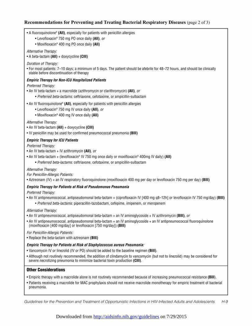
Downloaded from http://aidsinfo.nih.gov/guidelines on 7/29/2015
Guidelines for the Prevention and Treatment of Opportunistic Infections in HIV-Infected Adults and Adolescents H-9
Recommendations for Preventing and Treating Bacterial Respiratory Diseases (page 2 of 3)
• A fluoroquinolonea (AII), especially for patients with penicillin allergies
• Levofloxacina 750 mg PO once daily (AII), or
• Moxifloxacina 400 mg PO once daily (AII)
Alternative Therapy:
• A beta-lactam (AII) + doxycycline (CIII)
Duration of Therapy:
• For most patients: 7–10 days; a minimum of 5 days. The patient should be afebrile for 48–72 hours, and should be clinicallystable before discontinuation of therapy
Empiric Therapy for Non-ICU Hospitalized Patients
Preferred Therapy:
• An IV beta-lactam + a macrolide (azithromycin or clarithromycin) (AII), or
• Preferred beta-lactams: ceftriaxone, cefotaxime, or ampicillin-sulbactam
• An IV fluoroquinolonea (AII), especially for patients with penicillin allergies
• Levofloxacina 750 mg IV once daily (AII), or
• Moxifloxacina 400 mg IV once daily (AII)
Alternative Therapy:
• An IV beta-lactam (AII) + doxycycline (CIII)
• IV penicillin may be used for confirmed pneumococcal pneumonia (BIII)
Empiric Therapy for ICU Patients
Preferred Therapy:
• An IV beta-lactam + IV azithromycin (AII), or
• An IV beta-lactam + (levofloxacina IV 750 mg once daily or moxifloxacina 400mg IV daily) (AII)
• Preferred beta-lactams: ceftriaxone, cefotaxime, or ampicillin-sulbactam
Alternative Therapy:
For Penicillin-Allergic Patients:
• Aztreonam (IV) + an IV respiratory fluoroquinolone (moxifloxacin 400 mg per day or levofloxacin 750 mg per day) (BIII)
Empiric Therapy for Patients at Risk of Pseudomonas Pneumonia
Preferred Therapy:
• An IV antipneumococcal, antipseudomonal beta-lactam + (ciprofloxacin IV [400 mg q8–12h] or levofloxacin IV 750 mg/day) (BIII)
• Preferred beta-lactams: piperacillin-tazobactam, cefepime, imipenem, or meropenem
Alternative Therapy:
• An IV antipneumococcal, antipseudomonal beta-lactam + an IV aminoglycoside + IV azithromycin (BIII), or
• An IV antipneumococcal, antipseudomonal beta-lactam + an IV aminoglycoside + an IV antipneumococcal fluoroquinolone(moxifloxacin [400 mg/day] or levofloxacin [750 mg/day]) (BIII)
For Penicillin-Allergic Patients:
• Replace the beta-lactam with aztreonam (BIII)
Empiric Therapy for Patients at Risk of Staphylococcus aureus Pneumonia:
• Vancomycin IV or linezolid (IV or PO) should be added to the baseline regimen (BIII).
• Although not routinely recommended, the addition of clindamycin to vancomycin (but not to linezolid) may be considered forsevere necrotizing pneumonia to minimize bacterial toxin production (CIII).
Other Considerations
• Empiric therapy with a macrolide alone is not routinely recommended because of increasing pneumococcal resistance (BIII).
• Patients receiving a macrolide for MAC prophylaxis should not receive macrolide monotherapy for empiric treatment of bacterialpneumonia.
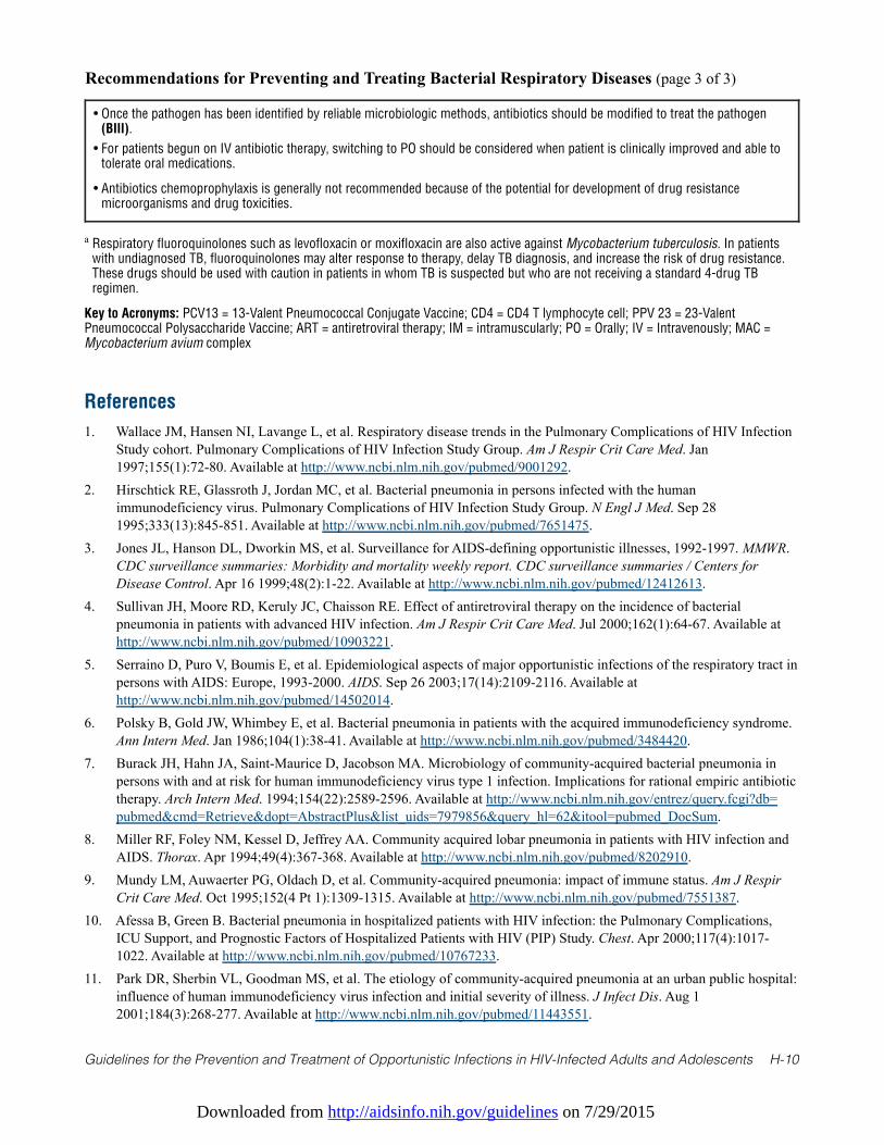
Downloaded from http://aidsinfo.nih.gov/guidelines on 7/29/2015
Guidelines for the Prevention and Treatment of Opportunistic Infections in HIV-Infected Adults and Adolescents H-10
Recommendations for Preventing and Treating Bacterial Respiratory Diseases (page 3 of 3)
• Once the pathogen has been identified by reliable microbiologic methods, antibiotics should be modified to treat the pathogen(BIII).
• For patients begun on IV antibiotic therapy, switching to PO should be considered when patient is clinically improved and able totolerate oral medications.
• Antibiotics chemoprophylaxis is generally not recommended because of the potential for development of drug resistancemicroorganisms and drug toxicities.
a Respiratory fluoroquinolones such as levofloxacin or moxifloxacin are also active against Mycobacterium tuberculosis. In patientswith undiagnosed TB, fluoroquinolones may alter response to therapy, delay TB diagnosis, and increase the risk of drug resistance.These drugs should be used with caution in patients in whom TB is suspected but who are not receiving a standard 4-drug TBregimen.
Key to Acronyms: PCV13 = 13-Valent Pneumococcal Conjugate Vaccine; CD4 = CD4 T lymphocyte cell; PPV 23 = 23-ValentPneumococcal Polysaccharide Vaccine; ART = antiretroviral therapy; IM = intramuscularly; PO = Orally; IV = Intravenously; MAC =Mycobacterium avium complex
References
1. Wallace JM, Hansen NI, Lavange L, et al. Respiratory disease trends in the Pulmonary Complications of HIV InfectionStudy cohort. Pulmonary Complications of HIV Infection Study Group. Am J Respir Crit Care Med. Jan1997;155(1):72-80. Available at http://www.ncbi.nlm.nih.gov/pubmed/9001292.
2. Hirschtick RE, Glassroth J, Jordan MC, et al. Bacterial pneumonia in persons infected with the humanimmunodeficiency virus. Pulmonary Complications of HIV Infection Study Group. N Engl J Med. Sep 281995;333(13):845-851. Available at http://www.ncbi.nlm.nih.gov/pubmed/7651475.
3. Jones JL, Hanson DL, Dworkin MS, et al. Surveillance for AIDS-defining opportunistic illnesses, 1992-1997. MMWR.CDC surveillance summaries: Morbidity and mortality weekly report. CDC surveillance summaries / Centers forDisease Control. Apr 16 1999;48(2):1-22. Available at http://www.ncbi.nlm.nih.gov/pubmed/12412613.
4. Sullivan JH, Moore RD, Keruly JC, Chaisson RE. Effect of antiretroviral therapy on the incidence of bacterialpneumonia in patients with advanced HIV infection. Am J Respir Crit Care Med. Jul 2000;162(1):64-67. Available athttp://www.ncbi.nlm.nih.gov/pubmed/10903221.
5. Serraino D, Puro V, Boumis E, et al. Epidemiological aspects of major opportunistic infections of the respiratory tract inpersons with AIDS: Europe, 1993-2000. AIDS. Sep 26 2003;17(14):2109-2116. Available athttp://www.ncbi.nlm.nih.gov/pubmed/14502014.
6. Polsky B, Gold JW, Whimbey E, et al. Bacterial pneumonia in patients with the acquired immunodeficiency syndrome.Ann Intern Med. Jan 1986;104(1):38-41. Available at http://www.ncbi.nlm.nih.gov/pubmed/3484420.
7. Burack JH, Hahn JA, Saint-Maurice D, Jacobson MA. Microbiology of community-acquired bacterial pneumonia inpersons with and at risk for human immunodeficiency virus type 1 infection. Implications for rational empiric antibiotictherapy. Arch Intern Med. 1994;154(22):2589-2596. Available at http://www.ncbi.nlm.nih.gov/entrez/query.fcgi?db=pubmed&cmd=Retrieve&dopt=AbstractPlus&list_uids=7979856&query_hl=62&itool=pubmed_DocSum.
8. Miller RF, Foley NM, Kessel D, Jeffrey AA. Community acquired lobar pneumonia in patients with HIV infection andAIDS. Thorax. Apr 1994;49(4):367-368. Available at http://www.ncbi.nlm.nih.gov/pubmed/8202910.
9. Mundy LM, Auwaerter PG, Oldach D, et al. Community-acquired pneumonia: impact of immune status. Am J RespirCrit Care Med. Oct 1995;152(4 Pt 1):1309-1315. Available at http://www.ncbi.nlm.nih.gov/pubmed/7551387.
10. Afessa B, Green B. Bacterial pneumonia in hospitalized patients with HIV infection: the Pulmonary Complications,ICU Support, and Prognostic Factors of Hospitalized Patients with HIV (PIP) Study. Chest. Apr 2000;117(4):1017-1022. Available at http://www.ncbi.nlm.nih.gov/pubmed/10767233.
11. Park DR, Sherbin VL, Goodman MS, et al. The etiology of community-acquired pneumonia at an urban public hospital:influence of human immunodeficiency virus infection and initial severity of illness. J Infect Dis. Aug 12001;184(3):268-277. Available at http://www.ncbi.nlm.nih.gov/pubmed/11443551.
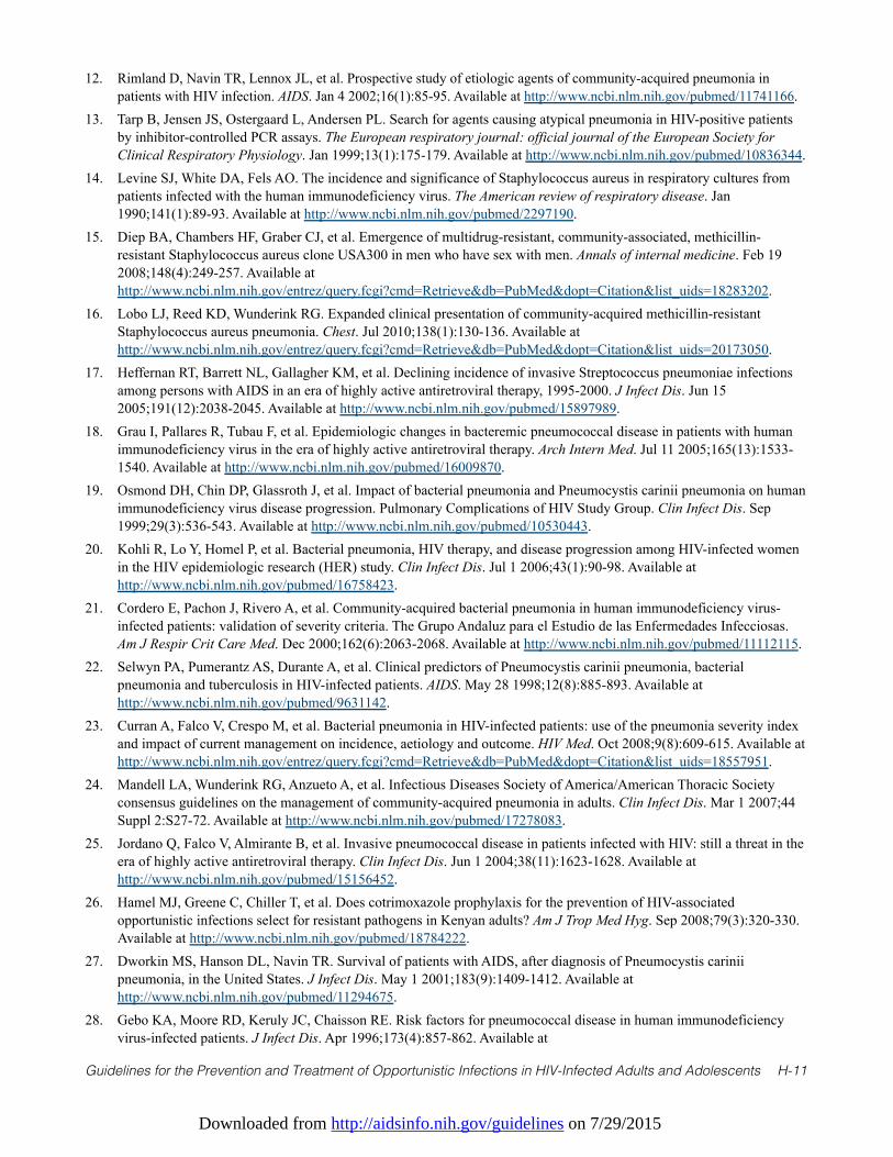
Downloaded from http://aidsinfo.nih.gov/guidelines on 7/29/2015
Guidelines for the Prevention and Treatment of Opportunistic Infections in HIV-Infected Adults and Adolescents H-11
12. Rimland D, Navin TR, Lennox JL, et al. Prospective study of etiologic agents of community-acquired pneumonia inpatients with HIV infection. AIDS. Jan 4 2002;16(1):85-95. Available at http://www.ncbi.nlm.nih.gov/pubmed/11741166.
13. Tarp B, Jensen JS, Ostergaard L, Andersen PL. Search for agents causing atypical pneumonia in HIV-positive patientsby inhibitor-controlled PCR assays. The European respiratory journal: official journal of the European Society forClinical Respiratory Physiology. Jan 1999;13(1):175-179. Available at http://www.ncbi.nlm.nih.gov/pubmed/10836344.
14. Levine SJ, White DA, Fels AO. The incidence and significance of Staphylococcus aureus in respiratory cultures frompatients infected with the human immunodeficiency virus. The American review of respiratory disease. Jan1990;141(1):89-93. Available at http://www.ncbi.nlm.nih.gov/pubmed/2297190.
15. Diep BA, Chambers HF, Graber CJ, et al. Emergence of multidrug-resistant, community-associated, methicillin-resistant Staphylococcus aureus clone USA300 in men who have sex with men. Annals of internal medicine. Feb 192008;148(4):249-257. Available athttp://www.ncbi.nlm.nih.gov/entrez/query.fcgi?cmd=Retrieve&db=PubMed&dopt=Citation&list_uids=18283202.
16. Lobo LJ, Reed KD, Wunderink RG. Expanded clinical presentation of community-acquired methicillin-resistantStaphylococcus aureus pneumonia. Chest. Jul 2010;138(1):130-136. Available athttp://www.ncbi.nlm.nih.gov/entrez/query.fcgi?cmd=Retrieve&db=PubMed&dopt=Citation&list_uids=20173050.
17. Heffernan RT, Barrett NL, Gallagher KM, et al. Declining incidence of invasive Streptococcus pneumoniae infectionsamong persons with AIDS in an era of highly active antiretroviral therapy, 1995-2000. J Infect Dis. Jun 152005;191(12):2038-2045. Available at http://www.ncbi.nlm.nih.gov/pubmed/15897989.
18. Grau I, Pallares R, Tubau F, et al. Epidemiologic changes in bacteremic pneumococcal disease in patients with humanimmunodeficiency virus in the era of highly active antiretroviral therapy. Arch Intern Med. Jul 11 2005;165(13):1533-1540. Available at http://www.ncbi.nlm.nih.gov/pubmed/16009870.
19. Osmond DH, Chin DP, Glassroth J, et al. Impact of bacterial pneumonia and Pneumocystis carinii pneumonia on humanimmunodeficiency virus disease progression. Pulmonary Complications of HIV Study Group. Clin Infect Dis. Sep1999;29(3):536-543. Available at http://www.ncbi.nlm.nih.gov/pubmed/10530443.
20. Kohli R, Lo Y, Homel P, et al. Bacterial pneumonia, HIV therapy, and disease progression among HIV-infected womenin the HIV epidemiologic research (HER) study. Clin Infect Dis. Jul 1 2006;43(1):90-98. Available athttp://www.ncbi.nlm.nih.gov/pubmed/16758423.
21. Cordero E, Pachon J, Rivero A, et al. Community-acquired bacterial pneumonia in human immunodeficiency virus-infected patients: validation of severity criteria. The Grupo Andaluz para el Estudio de las Enfermedades Infecciosas.Am J Respir Crit Care Med. Dec 2000;162(6):2063-2068. Available at http://www.ncbi.nlm.nih.gov/pubmed/11112115.
22. Selwyn PA, Pumerantz AS, Durante A, et al. Clinical predictors of Pneumocystis carinii pneumonia, bacterialpneumonia and tuberculosis in HIV-infected patients. AIDS. May 28 1998;12(8):885-893. Available athttp://www.ncbi.nlm.nih.gov/pubmed/9631142.
23. Curran A, Falco V, Crespo M, et al. Bacterial pneumonia in HIV-infected patients: use of the pneumonia severity indexand impact of current management on incidence, aetiology and outcome. HIV Med. Oct 2008;9(8):609-615. Available athttp://www.ncbi.nlm.nih.gov/entrez/query.fcgi?cmd=Retrieve&db=PubMed&dopt=Citation&list_uids=18557951.
24. Mandell LA, Wunderink RG, Anzueto A, et al. Infectious Diseases Society of America/American Thoracic Societyconsensus guidelines on the management of community-acquired pneumonia in adults. Clin Infect Dis. Mar 1 2007;44Suppl 2:S27-72. Available at http://www.ncbi.nlm.nih.gov/pubmed/17278083.
25. Jordano Q, Falco V, Almirante B, et al. Invasive pneumococcal disease in patients infected with HIV: still a threat in theera of highly active antiretroviral therapy. Clin Infect Dis. Jun 1 2004;38(11):1623-1628. Available athttp://www.ncbi.nlm.nih.gov/pubmed/15156452.
26. Hamel MJ, Greene C, Chiller T, et al. Does cotrimoxazole prophylaxis for the prevention of HIV-associatedopportunistic infections select for resistant pathogens in Kenyan adults? Am J Trop Med Hyg. Sep 2008;79(3):320-330.Available at http://www.ncbi.nlm.nih.gov/pubmed/18784222.
27. Dworkin MS, Hanson DL, Navin TR. Survival of patients with AIDS, after diagnosis of Pneumocystis cariniipneumonia, in the United States. J Infect Dis. May 1 2001;183(9):1409-1412. Available athttp://www.ncbi.nlm.nih.gov/pubmed/11294675.
28. Gebo KA, Moore RD, Keruly JC, Chaisson RE. Risk factors for pneumococcal disease in human immunodeficiencyvirus-infected patients. J Infect Dis. Apr 1996;173(4):857-862. Available at
Downloaded from http://aidsinfo.nih.gov/guidelines on 7/29/2015
Guidelines for the Prevention and Treatment of Opportunistic Infections in HIV-Infected Adults and Adolescents H-12
http://www.ncbi.nlm.nih.gov/pubmed/8603963.29. Guerrero M, Kruger S, Saitoh A, et al. Pneumonia in HIV-infected patients: a case-control survey of factors involved in
risk and prevention. AIDS. Oct 1 1999;13(14):1971-1975. Available at http://www.ncbi.nlm.nih.gov/pubmed/10513657.30. Breiman RF, Keller DW, Phelan MA, et al. Evaluation of effectiveness of the 23-valent pneumococcal capsular
polysaccharide vaccine for HIV-infected patients. Arch Intern Med. Sep 25 2000;160(17):2633-2638. Available athttp://www.ncbi.nlm.nih.gov/pubmed/10999977.
31. Advisory Committee on Immunization P. Recommended adult immunization schedule: United States, October 2007-September 2008. Ann Intern Med. Nov 20 2007;147(10):725-729. Available athttp://www.ncbi.nlm.nih.gov/pubmed/17947396.
32. Hung CC, Chen MY, Hsieh SM, Hsiao CF, Sheng WH, Chang SC. Clinical experience of the 23-valent capsularpolysaccharide pneumococcal vaccination in HIV-1-infected patients receiving highly active antiretroviral therapy: aprospective observational study. Vaccine. May 7 2004;22(15-16):2006-2012. Available athttp://www.ncbi.nlm.nih.gov/pubmed/15121313.
33. French N, Nakiyingi J, Carpenter LM, et al. 23-valent pneumococcal polysaccharide vaccine in HIV-1-infectedUgandan adults: double-blind, randomised and placebo controlled trial. Lancet. Jun 17 2000;355(9221):2106-2111.Available at http://www.ncbi.nlm.nih.gov/pubmed/10902624.
34. Watera C, Nakiyingi J, Miiro G, et al. 23-Valent pneumococcal polysaccharide vaccine in HIV-infected Ugandan adults:6-year follow-up of a clinical trial cohort. AIDS. May 21 2004;18(8):1210-1213. Available athttp://www.ncbi.nlm.nih.gov/pubmed/15166540.
35. Centers for Disease C, Prevention. Use of 13-valent pneumococcal conjugate vaccine and 23-valent pneumococcalpolysaccharide vaccine for adults with immunocompromising conditions: recommendations of the Advisory Committeeon Immunization Practices (ACIP). MMWR Morb Mortal Wkly Rep. Oct 12 2012;61(40):816-819. Available athttp://www.ncbi.nlm.nih.gov/pubmed/23051612.
36. French N, Gordon SB, Mwalukomo T, et al. A trial of a 7-valent pneumococcal conjugate vaccine in HIV-infectedadults. N Engl J Med. Mar 4 2010;362(9):812-822. Available athttp://www.ncbi.nlm.nih.gov/entrez/query.fcgi?cmd=Retrieve&db=PubMed&dopt=Citation&list_uids=20200385.
37. Penaranda M, Falco V, Payeras A, et al. Effectiveness of polysaccharide pneumococcal vaccine in HIV-infectedpatients: a case-control study. Clin Infect Dis. Oct 1 2007;45(7):e82-87. Available athttp://www.ncbi.nlm.nih.gov/entrez/query.fcgi?cmd=Retrieve&db=PubMed&dopt=Citation&list_uids=17806042.
38. Rodriguez-Barradas MC, Goulet J, Brown S, et al. Impact of pneumococcal vaccination on the incidence of pneumoniaby HIV infection status among patients enrolled in the Veterans Aging Cohort 5-Site Study. Clin Infect Dis. Apr 12008;46(7):1093-1100. Available athttp://www.ncbi.nlm.nih.gov/entrez/query.fcgi?cmd=Retrieve&db=PubMed&dopt=Citation&list_uids=18444830.
39. Teshale EH, Hanson D, Flannery B, et al. Effectiveness of 23-valent polysaccharide pneumococcal vaccine onpneumonia in HIV-infected adults in the United States, 1998—2003. Vaccine. Oct 29 2008;26(46):5830-5834. Availableat http://www.ncbi.nlm.nih.gov/entrez/query.fcgi?cmd=Retrieve&db=PubMed&dopt=Citation&list_uids=18786586.
40. Fiore AE, Uyeki TM, Broder K, et al. Prevention and control of influenza with vaccines: recommendations of theAdvisory Committee on Immunization Practices (ACIP), 2010. MMWR Recomm Rep. Aug 6 2010;59(RR-8):1-62.Available athttp://www.ncbi.nlm.nih.gov/entrez/query.fcgi?cmd=Retrieve&db=PubMed&dopt=Citation&list_uids=20689501.
41. Anglaret X, Chene G, Attia A, et al. Early chemoprophylaxis with trimethoprim-sulphamethoxazole for HIV-1-infectedadults in Abidjan, Cote d'Ivoire: a randomised trial. Cotrimo-CI Study Group. Lancet. May 1 1999;353(9163):1463-1468. Available at http://www.ncbi.nlm.nih.gov/pubmed/10232311.
42. Hardy WD, Feinberg J, Finkelstein DM, et al. A controlled trial of trimethoprim-sulfamethoxazole or aerosolizedpentamidine for secondary prophylaxis of Pneumocystis carinii pneumonia in patients with the acquiredimmunodeficiency syndrome. AIDS Clinical Trials Group Protocol 021. N Engl J Med. Dec 24 1992;327(26):1842-1848. Available at http://www.ncbi.nlm.nih.gov/pubmed/1448121.
43. Havlir DV, Dube MP, Sattler FR, et al. Prophylaxis against disseminated Mycobacterium avium complex with weeklyazithromycin, daily rifabutin, or both. California Collaborative Treatment Group. N Engl J Med. Aug 81996;335(6):392-398. Available at http://www.ncbi.nlm.nih.gov/pubmed/8676932.
Downloaded from http://aidsinfo.nih.gov/guidelines on 7/29/2015
Guidelines for the Prevention and Treatment of Opportunistic Infections in HIV-Infected Adults and Adolescents H-13
44. Oldfield EC, 3rd, Fessel WJ, Dunne MW, et al. Once weekly azithromycin therapy for prevention of Mycobacteriumavium complex infection in patients with AIDS: a randomized, double-blind, placebo-controlled multicenter trial. ClinInfect Dis. Mar 1998;26(3):611-619. Available at http://www.ncbi.nlm.nih.gov/pubmed/9524832.
45. Crothers K, Griffith TA, McGinnis KA, et al. The impact of cigarette smoking on mortality, quality of life, andcomorbid illness among HIV-positive veterans. J Gen Intern Med. 2005;20(12):1142-1145. Available athttp://www.ncbi.nlm.nih.gov/entrez/query.fcgi?db=pubmed&cmd=Retrieve&dopt=AbstractPlus&list_uids=16423106&query_hl=100&itool=pubmed_docsum.
46. Navin TR, Rimland D, Lennox JL, et al. Risk factors for community-acquired pneumonia among persons infected withhuman immunodeficiency virus. J Infect Dis. Jan 2000;181(1):158-164. Available athttp://www.ncbi.nlm.nih.gov/pubmed/10608762.
47. Justice AC, Lasky E, McGinnis KA, et al. Medical disease and alcohol use among veterans with humanimmunodeficiency infection: A comparison of disease measurement strategies. Medical care. Aug 2006;44(8 Suppl2):S52-60. Available at http://www.ncbi.nlm.nih.gov/pubmed/16849969.
48. Benard A, Mercie P, Alioum A, et al. Bacterial pneumonia among HIV-infected patients: decreased risk after tobaccosmoking cessation. ANRS CO3 Aquitaine Cohort, 2000-2007. PLoS One. 2010;5(1):e8896. Available athttp://www.ncbi.nlm.nih.gov/pubmed/20126646.
49. Madeddu G, Laura Fiori M, Stella Mura M. Bacterial community-acquired pneumonia in HIV-infected patients. CurrOpin Pulm Med. May 2010;16(3):201-207. Available athttp://www.ncbi.nlm.nih.gov/entrez/query.fcgi?cmd=Retrieve&db=PubMed&dopt=Citation&list_uids=20154625.
50. Malinis M, Myers J, Bordon J, et al. Clinical outcomes of HIV-infected patients hospitalized with bacterial community-acquired pneumonia. Int J Infect Dis. Jan 2010;14(1):e22-27. Available athttp://www.ncbi.nlm.nih.gov/entrez/query.fcgi?cmd=Retrieve&db=PubMed&dopt=Citation&list_uids=19586789.
51. Weinstein MP, Klugman KP, Jones RN. Rationale for revised penicillin susceptibility breakpoints versus Streptococcuspneumoniae: coping with antimicrobial susceptibility in an era of resistance. Clin Infect Dis. Jun 1 2009;48(11):1596-1600. Available athttp://www.ncbi.nlm.nih.gov/entrez/query.fcgi?cmd=Retrieve&db=PubMed&dopt=Citation&list_uids=19400744.
52. Baddour LM, Yu VL, Klugman KP, et al. Combination antibiotic therapy lowers mortality among severely ill patientswith pneumococcal bacteremia. Am J Respir Crit Care Med. Aug 15 2004;170(4):440-444. Available athttp://www.ncbi.nlm.nih.gov/entrez/query.fcgi?cmd=Retrieve&db=PubMed&dopt=Citation&list_uids=15184200.
53. Zolopa A, Andersen J, Powderly W, et al. Early antiretroviral therapy reduces AIDS progression/death in individualswith acute opportunistic infections: a multicenter randomized strategy trial. PLoS One. 2009;4(5):e5575. Available athttp://www.ncbi.nlm.nih.gov/entrez/query.fcgi?cmd=Retrieve&db=PubMed&dopt=Citation&list_uids=19440326.
54. Christensen D, Feldman C, Rossi P, et al. HIV infection does not influence clinical outcomes in hospitalized patientswith bacterial community-acquired pneumonia: results from the CAPO international cohort study. Clin Infect Dis.2005;41(4):554-556. Available at http://www.ncbi.nlm.nih.gov/entrez/query.fcgi?db=pubmed&cmd=Retrieve&dopt=AbstractPlus&list_uids=16028168&query_hl=88&itool=pubmed_docsum.
55. Einarson A, Phillips E, Mawji F, et al. A prospective controlled multicentre study of clarithromycin in pregnancy.American journal of perinatology. 1998;15(9):523-525. Available at http://www.ncbi.nlm.nih.gov/pubmed/9890248.
56. Drinkard CR, Shatin D, Clouse J. Postmarketing surveillance of medications and pregnancy outcomes: clarithromycinand birth malformations. Pharmacoepidemiology and drug safety. Dec 2000;9(7):549-556. Available athttp://www.ncbi.nlm.nih.gov/pubmed/11338912.
57. Schaefer C, Amoura-Elefant E, Vial T, et al. Pregnancy outcome after prenatal quinolone exposure. Evaluation of a caseregistry of the European Network of Teratology Information Services (ENTIS). Eur J Obstet Gynecol Reprod Biol. Nov1996;69(2):83-89. Available at http://www.ncbi.nlm.nih.gov/pubmed/8902438.
58. Loebstein R, Addis A, Ho E, et al. Pregnancy outcome following gestational exposure to fluoroquinolones: amulticenter prospective controlled study. Antimicrob Agents Chemother. Jun 1998;42(6):1336-1339. Available athttp://www.ncbi.nlm.nih.gov/pubmed/9624471.
59. Nahum GG, Uhl K, Kennedy DL. Antibiotic use in pregnancy and lactation: what is and is not known about teratogenicand toxic risks. Obstet Gynecol. May 2006;107(5):1120-1138. Available athttp://www.ncbi.nlm.nih.gov/pubmed/16648419.
Downloaded from http://aidsinfo.nih.gov/guidelines on 7/29/2015
Guidelines for the Prevention and Treatment of Opportunistic Infections in HIV-Infected Adults and Adolescents I-1
Bacterial Enteric Infections (Last updated October 28, 2014; last reviewedOctober 28, 2014)
Epidemiology
Rates of Gram-negative bacterial enteric infections are at least 10-fold higher among HIV-infected adultsthan in the general population but decline when patients are on antiretroviral therapy (ART).1-7 The risk ofbacterial diarrhea varies according to CD4 T-lymphocyte (CD4) count and is greatest in individuals withclinical AIDS and/or <200 CD4 cells/mm3.5 The most common routinely cultured enteric bacteria amongHIV-infected adults in the United States are Salmonella (particularly Salmonella enterica serotypesTyphimurium and Enteritidis), Shigella, and Campylobacter. Diarrheagenic Escherichia coli, particularlyenteroaggregative E. coli, may contribute to the burden of diarrheal disease8 but their role is poorlyunderstood because diagnosis requires specialized laboratory capacity. Clostridium difficile-associatedinfection (CDI) is common in HIV-infected patients; recent data9 suggest that low CD4 count (<50cells/mm3) is an independent disease risk factor in addition to the traditional risk factors such as exposure toa healthcare facility or to antibiotics. Increased recognition of community-associated CDI in HIV-uninfectedindividuals suggests that the healthcare provider should consider CDI in the evaluation of outpatientdiarrheal illnesses. Data on Helicobacter pylori infection in HIV infection are limited and do not suggestexcess risk in HIV-infected individuals. Other enteric infections that may cause diarrhea, such asMycobacterium avium complex (MAC) and cytomegalovirus are discussed elsewhere in these guidelines.
As with bacterial enteric infections in HIV-uninfected persons, the probable source for most enteric infections inHIV-infected patients is ingestion of contaminated food or water.3 Sexual activity with the potential for direct orindirect fecal-oral exposure also increases risk of infections, especially with Shigella10 and Campylobacter11 (seeAppendix for further details.). HIV-associated alterations in mucosal immunity or intestinal integrity andtreatment with acid-suppressive agents may facilitate acquisition of enteric bacterial infections.
Clinical Manifestations
The three major clinical syndromes of infection with Gram-negative enteric bacteria among HIV-infectedpatients are:
• Self-limited gastroenteritis; • More severe and prolonged diarrheal disease, potentially associated with fever, bloody diarrhea, and
weight loss; and • Bacteremia associated with extra-intestinal involvement, with or without concurrent or preceding
gastrointestinal (GI) illness.12-15
Severe community-associated diarrhea is often defined as ≥6 loose stools (loose stool is defined as defecatedmaterial that takes the shape of a container) per day with or without other signs of disease such as fecal blood,orthostatic hypotension, or fever. In HIV-infected patients, the risk of more profound illness increases with thedegree of immunosuppression.1,3,4,16 Relapses in infection with Salmonella and other Gram-negative bacterialenteric pathogens after appropriate treatment have been well documented in HIV-infected patients.17-19
Diagnosis
Assessment of patients with diarrhea should include a complete exposure history (see below); medicationreview, because diarrhea is a common side effect of some ART and antibiotics; quantification of the diarrhealillness by stool frequency, volume, duration, and presence of blood; and associated signs and symptoms,such as presence and duration of fever. Physical examination should include measurement of temperatureand assessment of volume and nutritional status.
Downloaded from http://aidsinfo.nih.gov/guidelines on 7/29/2015
Guidelines for the Prevention and Treatment of Opportunistic Infections in HIV-Infected Adults and Adolescents I-2
The diagnosis of Gram-negative bacterial enteric infection is established through cultures of stool and blood.Because incidence of bacteremia associated with Salmonella gastroenteritis is high in HIV-infectedindividuals, particularly those with advanced disease, blood cultures should be obtained from any patientwith diarrhea and fever. For shigellosis, blood cultures may be helpful but are less likely to be positive thanin salmonellosis.
Other infections for which HIV-infected patients are at risk, albeit at a lower rate, are non-jejuni non-coliCampylobacter species, such as Campylobacter fetus, Campylobacter upsaliensis, and Campylobacter lari,and the enterohepatic Helicobacter spp. (Helicobacter cineadi and Helicobacter fennelliae), which wereoriginally described as Campylobacter spp. Blood culture systems typically will grow these bacteria, but theyare unlikely to be identified on routine stool cultures performed by most laboratories because special stoolculture conditions are required for growth of these fastidious organisms.
A stool sample for C. difficile toxin or polymerase chain reaction (PCR) assay should be routinely performedfor patients with diarrhea who have recently or are currently receiving antibiotics (including antimicrobialprophylaxis) or cancer chemotherapy, those who have been hospitalized in the past 4 to 6 weeks (or arecurrently hospitalized), those who reside in a long-term care facility, those with CD4 counts <200 cells/mm3,those taking acid-suppressive medications, and those with moderate-to-severe community-acquireddiarrhea.20 The most commonly used toxin tests are enzyme immunoassays that suffer from low sensitivity.PCR assays or glutamate dehydrogenase antigen enzyme immunoassays (which must be combined with asecond confirmatory test) are recommended for testing.21 However, only diarrheal stool samples should betested for C. difficile to limit detection of asymptomatic colonization. Regardless of the test used, thediagnosis of CDI can only be made through careful selection of the correct population to test and acorrelation of clinical and laboratory findings.
Endoscopy generally should be reserved for patients in whom stool culture, microscopy, C. difficile toxinassay, and blood culture fail to reveal an etiology or in whom treatment for an established diagnosis fails.Endoscopy with biopsy may be required for diagnosing etiologies other than bacterial enteric infections,including cryptosporidiosis, microsporidiosis, cytomegalovirus or MAC gastroenteritis, and noninfectiouscauses of GI symptoms.
Clinicians should remain alert to the possibility of sexually transmitted disease (STD). Some sexuallytransmitted rectal infections (such as proctitis due to lymphogranuloma venereum or Neisseria gonorrhoeae)can produce symptoms similar to those seen with colitis due to Salmonella, Shigella, and Campylobacterspp. In patients with symptoms of proctitis or colitis, if stool cultures fail to yield enteric bacterial pathogens,diagnostic evaluation for STDs with anoscopy, culture, and biopsy should be considered.
Preventing Exposure
Multiple epidemiologic exposures can place patients at risk of enteric illnesses. The most common areingestion of contaminated food or water and fecal-oral exposures (detailed prevention recommendationsrelated to food and water exposures, pet exposures, and travel-related exposures can be found in theAppendix). Providing advice and education about such exposures is the responsibility of the healthcareprovider. A patient’s clinical condition and CD4 count can help the provider determine what preventionrecommendations are most appropriate. Patients with CD4 counts <200 cells/mm3 or a history of AIDS-defining illness22 are at the greatest risk of enteric illnesses;5 however, excess risk of undeterminedmagnitude or duration may persist in those with lesser degrees of immune impairment, including individualstreated with ART.
Patients should be advised to regularly wash their hands with soap and water or alcohol-based cleansers toreduce the risk of enteric infection (AIII). With regard to preventing enteric infection, soap and water arepreferred over alcohol-based cleansers, which do not kill C. difficile spores and are only partially activeagainst norovirus and Cryptosporidium (AIII). HIV-infected patients should be advised to wash their hands
Downloaded from http://aidsinfo.nih.gov/guidelines on 7/29/2015
Guidelines for the Prevention and Treatment of Opportunistic Infections in HIV-Infected Adults and Adolescents I-3
after potential contact with human feces, such as through defecation, cleaning feces from infants, or contactwith a person who has diarrhea; after handling pets or other animals; after gardening or other contact withsoil; before preparing food and eating; and before and after sex (AIII). HIV-infected patients should avoidunprotected sex practices, such as anal sex and oral-anal contact that could result in oral exposure to fecesand, in addition to handwashing, they should be advised to use barriers such as dental dams during sex toreduce exposures when possible (AIII).
Preventing Disease
Antimicrobial prophylaxis to prevent bacterial enteric illness usually is not recommended, including fortravelers (AIII). Prophylactic antimicrobial treatment can elicit adverse reactions, promote the emergence ofresistant organisms, and increase risk of CDI. In rare cases, however, antimicrobial prophylaxis withfluoroquinolones or rifaximin can be considered, such as for immunosuppressed travelers, depending on theirlevel of immunosuppression, the region of travel, and the trip’s duration (CIII). For pregnant women andpatients already taking trimethoprim-sulfamethoxazole (TMP-SMX) (such as for Pneumocystis jiroveciipneumonia prophylaxis), TMP-SMX may offer limited protection against travelers’ diarrhea as an alternativeto fluoroquinolones or rifaximin (BIII). Risk of toxicity should be considered before prophylaxis with TMP-SMX is initiated solely because of travel.
Treating Disease
Empiric TherapyIn most situations, treatment of diarrheal disease in HIV-infected patients does not differ significantly fromthat in immunocompetent individuals. Decisions on therapy depend on an assessment of diarrhea severityand hydration status. Patients should be informed of the importance of maintaining hydration and given oralor intravenous (IV) rehydration if indicated (AIII). Because diarrheal disease can produce temporarymalabsorption or lactose intolerance, consuming a bland diet and avoiding fat, dairy, and complexcarbohydrates also are likely to be useful (BIII). The effectiveness and safety of probiotics or antimotilityagents have not been adequately studied in HIV-infected patients with diarrheal illnesses.23 Antimotilityagents should be avoided if there is concern about inflammatory diarrhea including CDI (BIII).
After obtaining stool samples for diagnostic evaluation, initiation and duration of empiric antimicrobialtherapy depend upon the patient’s CD4 count and clinical appearance. If stool samples are obtained,antibiotic susceptibility testing should be performed to confirm and inform antibiotic choice. No furtherwork-up may be necessary and no treatment other than oral rehydration required, for example, in patientswith CD4 counts >500 cells/mm3 who have had 1 to 2 days of loose stools without fever or blood. However,a short course of antibiotics may be indicated in HIV-infected patients with CD4 counts of 200 to 500cells/mm3 who have diarrhea severe enough to compromise quality of life or ability to work. Patients withadvanced HIV disease, that is, CD4 counts <200 cells/mm3 or concomitant AIDS-defining illness, withclinically severe diarrhea (i.e., ≥6 stools per day or bloody stools and/or accompanied by fever or chills)should undergo diagnostic evaluation to determine the etiology of the diarrheal illness and receiveantimicrobial treatment. Empiric therapy with ciprofloxacin is reasonable (AIII). IV ceftriaxone or IVcefotaxime are reasonable alternatives (BIII). Therapy should be adjusted subsequently based on the resultsof the diagnostic work-up. Diarrhea that is persistent (i.e., lasting >14 days) in the absence of other clinicalsigns of severity, such as bloody stool or dehydration, should be evaluated and directed therapy should bestarted once a diagnosis is confirmed.
Diarrhea is one of the most common illnesses affecting international travelers. Antimicrobial resistance amongenteric bacterial pathogens outside the United States is an important public health problem. For example, in2007, 85% of Campylobacter jejuni isolates in Southeast Asia were reported as fluoroquinolone resistant.24
Clinicians should consider the possibility of a resistant infection when prescribing empiric therapy for HIV-infected travelers who experience diarrhea while traveling or upon returning to the United States.
Downloaded from http://aidsinfo.nih.gov/guidelines on 7/29/2015
Guidelines for the Prevention and Treatment of Opportunistic Infections in HIV-Infected Adults and Adolescents I-4
Pathogen-Specific TherapySalmonella spp.Immunocompetent hosts who are not HIV-infected often do not require treatment for Salmonellagastroenteritis, as the condition is usually self-limited and treatment may prolong the carrier state. In contrast,all HIV-infected patients with salmonellosis should be treated (AIII), although no clinical trials have comparedantimicrobial therapy with placebo. Notably, HIV infection increases the risk of Salmonella bacteremia 20- to100-fold and mortality as much as 7-fold compared with that in patients who are not HIV-infected.1,25
The initial treatment of choice for Salmonella infection is a fluoroquinolone (AIII). Ciprofloxacin is thepreferred agent (AIII).26 Other fluoroquinolones, such as levofloxacin and moxifloxacin, likely would beeffective in treating salmonellosis in HIV-infected patients but they have not been well evaluated in clinicalstudies (BIII). Depending on antibiotic susceptibility, alternatives to the fluoroquinolones might includeTMP-SMX or expanded-spectrum cephalosporins such as ceftriaxone or cefotaxime (BIII).
The optimal duration of therapy for HIV-related Salmonella infection has not been defined. For patients withCD4 counts ≥200 cells/mm3 who have mild gastroenteritis without bacteremia, 7 to 14 days of treatment isreasonable. For the same patients with bacteremia, 14 days is appropriate, provided clearance of bacteremiais documented. Longer treatment is suggested if bacteremia persists or if the infection is complicated, that is,if metastatic foci are present (BIII). For patients with advanced HIV disease (CD4 count <200 cells/mm3), 2to 6 weeks of antibiotics often is recommended (CIII).27 Some patients with Salmonella bacteremia mayremain febrile for 5 to 7 days despite effective therapy.
HIV-infected patients with Salmonella bacteremia, which typically occurs in those with advanced HIVdisease, should be monitored clinically for recurrence after treatment (BIII). Recurrence may present asbacteremia or as an anatomically localized infection, including intra-abdominal, endothelial, urinary tract,soft tissue, bone and joint, lung, or meningeal foci. Secondary prophylaxis should be considered for patientswith recurrent Salmonella bacteremia (CIII) and it might also be considered for patients with recurrentgastroenteritis (with or without bacteremia) and in those with CD4 counts <200 cell/mm3 with severediarrhea (CIII). The value of this secondary prophylaxis has not been established and must be weighedagainst the risks of long-term antibiotic exposure. Recurrent Salmonella bacteremia constitutes an AIDS-defining illness28 and suppression of HIV replication with ART is expected to decrease the risk of recurrentillnesses. In patients whose Salmonella infection is resolved and who have responded to ART with sustainedviral suppression and CD4 counts >200 cells/mm3, secondary prophylaxis for salmonellosis can probably bestopped (CII).7 Clinicians also should be aware that recurrence may represent development of antimicrobialresistance during therapy.
Shigella spp. Therapy for Shigella infections is recommended both to shorten the duration of illness and to possiblyprevent spread of the infection to others (AIII).26 The recommended treatment for shigellosis is with afluoroquinolone, preferably ciprofloxacin, for 7 to 10 days (AIII). Depending on antibiotic susceptibilities,alternative agents might include TMP-SMX (7–10 days) or azithromycin (5 days) (BIII). Azithromycin hasnot been evaluated in HIV-infected patients with shigellosis, and the therapy suggested is extrapolated fromlimited data in immunocompetent hosts.29 Recently, Shigella spp. with reduced susceptibility to azithromycinin HIV-infected MSM have been reported.30,31 Treatment for patients with Shigella bacteremia is less welldefined, but extending treatment to at least 14 days is reasonable (BIII). Azithromycin is not recommendedfor treatment of Shigella spp. bacteremia (AIII). Chronic suppressive or maintenance therapy is notrecommended for first-time Shigella infections (BIII). Recurrent infections can occur, particularly inindividuals with CD4 counts <200 cells/mm3, in which case extending antimicrobial therapy for up to 6weeks is reasonable (BIII). As with Salmonella infections, suppression of HIV replication with ART isexpected to decrease the risk of recurrent shigellosis.
Downloaded from http://aidsinfo.nih.gov/guidelines on 7/29/2015
Guidelines for the Prevention and Treatment of Opportunistic Infections in HIV-Infected Adults and Adolescents I-5
Campylobacter spp. The optimal treatment of campylobacteriosis in HIV-infected patients is poorly defined. Culture andsusceptibility of Campylobacter isolates is recommended (BIII); in 2011, 24% of Campylobacter isolates inthe United States were fluoroquinolone resistant (http://www.cdc.gov/NARMS). For patients with milddisease and CD4 counts >200 cells/mm3, some clinicians opt to withhold therapy unless symptoms persist formore than several days (CIII). For mild-to-moderate campylobacteriosis, initiating therapy with afluoroquinolone such as ciprofloxacin for 7 to 10 days (if the organism is sensitive) or azithromycin for 5days is a reasonable approach (BIII). Azithromycin has not been evaluated in HIV-infected patients withcampylobacteriosis and the therapy suggested is extrapolated from limited data in immunocompetent hosts.32
Patients with Campylobacter bacteremia should be treated for at least 14 days using a fluoroquinolone if theisolate is sensitive (BIII). Azithromycin is not recommended for treatment of Campylobacter bacteremia(AIII). Adding a second active agent, such as an aminoglycoside, may be prudent in these patients to limitthe emergence of antibiotic resistance (BIII). Antibiotic choice should be guided by antibiotic susceptibilitytests. Chronic suppressive or maintenance therapy is not recommended for first-time Campylobacterinfections in HIV-infected patients (BIII). However, recurrent infections can occur, particularly in patientswith CD4 counts <200 cells/mm3. In recurrent disease, extending the length of antimicrobial therapy for 2 to6 weeks is reasonable (BIII). As with Salmonella infections, suppression of HIV replication with ART isexpected to decrease the risk of recurrent Campylobacter spp. infections.
Clostridium difficileTreatment of CDI in HIV-infected patients is the same as in patients who are not HIV infected. Guidelinesfor the treatment of CDI have been published33,34 and can be consulted for further information.
Special Considerations with Regard to Starting ART ART initiation should follow standard guidelines. The presence of a diarrheal illness is relevant only in termsof a patient’s ability to ingest and absorb ART. If recurrent enteric infections are documented and/orSalmonella bacteremia occurs, prompt initiation of ART should be considered regardless of CD4 count; i.e.,the presence of an enteric infection should not delay ART initiation (BIII).
Monitoring of Response to Therapy and Adverse Events (Including IRIS) Patients should be monitored closely for response to treatment, defined clinically by improvement insystemic signs and symptoms, resolution of diarrhea, and sterilization of infected tissues or body fluids suchas blood. A follow-up stool culture to demonstrate clearance of the organism is not required if clinicalsymptoms and diarrhea resolve. Follow-up stool culture may be required when public health considerationsand state law dictate the need to ensure microbiologic cure, such as in healthcare or food service workers.
Immune reconstitution inflammatory syndrome has not been described in association with treatment forbacterial enteric pathogens.
Managing Treatment FailureFollow-up stool culture should be considered for patients who fail to respond clinically to appropriateantimicrobial therapy. In patients with persistent or recurrent diarrhea despite therapy, clinicians shouldconsider other enteric infections in the context of the patient’s immune status and, in all cases, the possibilityof C. difficile or the development of antimicrobial resistance.
Observational studies suggest that plasma drug concentrations (e.g., of ciprofloxacin) in HIV-infected patientsmay be decreased as a result of diarrhea or malabsorption.35,36 Coadministration of quinolones withmagnesium- or aluminum-containing antacids or with calcium, zinc, or iron should be avoided because theseinterfere with drug absorption. Although larger prospective studies are needed to determine the impact ofsevere diarrhea on antibiotic absorption, it is prudent to use IV antibiotics in clinically unstable patients (AIII).
Downloaded from http://aidsinfo.nih.gov/guidelines on 7/29/2015
Guidelines for the Prevention and Treatment of Opportunistic Infections in HIV-Infected Adults and Adolescents I-6
Preventing Recurrence
The pharmacologic approach to recurrent enteric infections is covered in the section on directed therapy foreach bacterial species. As noted above, secondary prophylaxis should be considered for patients with recurrentSalmonella bacteremia (CIII) and those with recurrent shigellosis (BIII) or campylobacteriosis (BIII).
Special Considerations During Pregnancy
The diagnosis of bacterial enteric infection in pregnant women is the same as in women who are notpregnant. Bacterial enteric infections in pregnant women should be managed the same as in women who arenot pregnant, with several considerations. Based on the safety profile, expanded-spectrum cephalosporins orazithromycin should be the first-line therapy for bacterial enteric infections during pregnancy ifantimicrobials are required, depending on the organism and the results of susceptibility testing (BIII).Arthropathy has been noted in the offspring of animals treated with quinolones during pregnancy. However,studies evaluating quinolone use in pregnant women did not find an increased risk of birth defects ormusculoskeletal abnormalities.37,38 Thus, quinolones can be used in pregnancy for bacterial enteric infectionsin HIV-infected pregnant women if indicated by susceptibility testing or failure of first-line therapy, as listedabove (BIII). TMP-SMX use in the first trimester should be avoided, if possible, because of an associationwith an increased risk of birth defects, specifically neural tube, cardiovascular, and urinary tract defects(BIII).39,40,41 Neonatal care providers should be informed if maternal sulfa therapy was used near deliverybecause of the theoretical increased risk to the newborn of hyperbilirubinemia and kernicterus.
Preventing Bacterial Enteric Illness
• Antimicrobial prophylaxis to prevent bacterial enteric illness usually is not recommended, including for travelers (AIII).
• In rare cases, such as for immunosuppressed travelers, depending on their level of immunosuppression, the region of travel, andthe trip’s duration, antimicrobial prophylaxis with fluoroquinolones or rifaximin can be considered (CIII).
• For pregnant women and patients already on trimethoprim-sulfamethoxazole (TMP-SMX) for prophylaxis against Pneuomcystisjirovecii, TMP-SMX may offer limited protection against travelers’ diarrhea as an alternative to fluoroquinolone or rifaximin (BIII).
General Considerations when Managing Patients with Bacterial Enteric Infections
• Oral or IV hydration therapy (if indicated) should be given to patients with diarrhea (AIII).
• Anti-motility agents should be avoided if there is concern about inflammatory diarrhea including Clostridium difficile infection(BIII).
• Diagnostic fecal specimens should be obtained prior to initiation of empiric antimicrobial therapy.
• If stool sample is obtained, antibiotic susceptibilities should be performed to confirm and inform antibiotic choice.
• Risk of a bacterial enteric infection increases as CD4 count declines with greatest risk with CD4 count <200 cells/mm3. Risk ofbacteremia also increases with decreasing CD4 count. If no clinical response after 5 to 7 days, consider follow-up stool culturewith antibiotic susceptibility testing and other methods to detect enteric pathogens (e.g., toxin assays, molecular methods),alternative diagnosis, antibiotic resistance, or drug-drug interactions.
• Effective ART may reduce the frequency, severity, and recurrence of bacterial enteric infections.
Recommendations for Preventing and Treating Bacterial Enteric Infections (page 1 of 3)
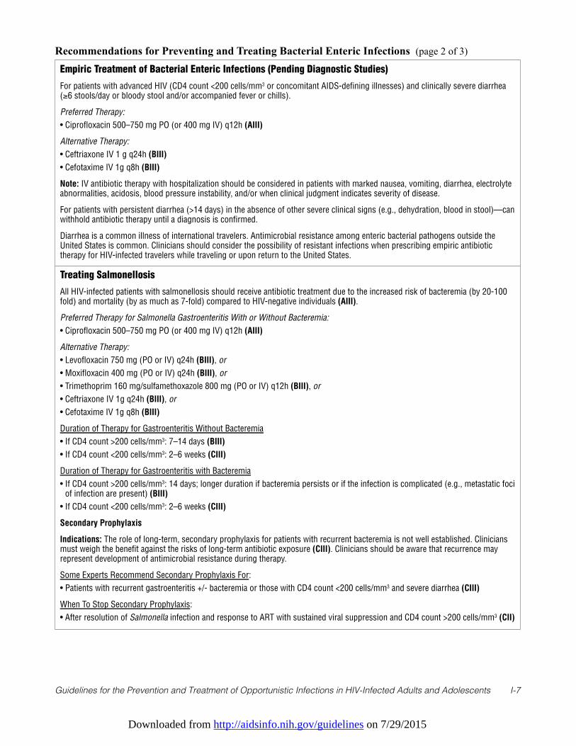
Downloaded from http://aidsinfo.nih.gov/guidelines on 7/29/2015
Guidelines for the Prevention and Treatment of Opportunistic Infections in HIV-Infected Adults and Adolescents I-7
Empiric Treatment of Bacterial Enteric Infections (Pending Diagnostic Studies)
For patients with advanced HIV (CD4 count <200 cells/mm3 or concomitant AIDS-defining illnesses) and clinically severe diarrhea(≥6 stools/day or bloody stool and/or accompanied fever or chills).
Preferred Therapy:
• Ciprofloxacin 500–750 mg PO (or 400 mg IV) q12h (AIII)
Alternative Therapy:
• Ceftriaxone IV 1 g q24h (BIII)
• Cefotaxime IV 1g q8h (BIII)
Note: IV antibiotic therapy with hospitalization should be considered in patients with marked nausea, vomiting, diarrhea, electrolyteabnormalities, acidosis, blood pressure instability, and/or when clinical judgment indicates severity of disease.
For patients with persistent diarrhea (>14 days) in the absence of other severe clinical signs (e.g., dehydration, blood in stool)—canwithhold antibiotic therapy until a diagnosis is confirmed.
Diarrhea is a common illness of international travelers. Antimicrobial resistance among enteric bacterial pathogens outside theUnited States is common. Clinicians should consider the possibility of resistant infections when prescribing empiric antibiotictherapy for HIV-infected travelers while traveling or upon return to the United States.
Treating Salmonellosis
All HIV-infected patients with salmonellosis should receive antibiotic treatment due to the increased risk of bacteremia (by 20-100fold) and mortality (by as much as 7-fold) compared to HIV-negative individuals (AIII).
Preferred Therapy for Salmonella Gastroenteritis With or Without Bacteremia:
• Ciprofloxacin 500–750 mg PO (or 400 mg IV) q12h (AIII)
Alternative Therapy:
• Levofloxacin 750 mg (PO or IV) q24h (BIII), or
• Moxifloxacin 400 mg (PO or IV) q24h (BIII), or
• Trimethoprim 160 mg/sulfamethoxazole 800 mg (PO or IV) q12h (BIII), or
• Ceftriaxone IV 1g q24h (BIII), or
• Cefotaxime IV 1g q8h (BIII)
Duration of Therapy for Gastroenteritis Without Bacteremia
• If CD4 count >200 cells/mm3: 7–14 days (BIII)
• If CD4 count <200 cells/mm3: 2–6 weeks (CIII)
Duration of Therapy for Gastroenteritis with Bacteremia
• If CD4 count >200 cells/mm3: 14 days; longer duration if bacteremia persists or if the infection is complicated (e.g., metastatic fociof infection are present) (BIII)
• If CD4 count <200 cells/mm3: 2–6 weeks (CIII)
Secondary Prophylaxis
Indications: The role of long-term, secondary prophylaxis for patients with recurrent bacteremia is not well established. Cliniciansmust weigh the benefit against the risks of long-term antibiotic exposure (CIII). Clinicians should be aware that recurrence mayrepresent development of antimicrobial resistance during therapy.
Some Experts Recommend Secondary Prophylaxis For:
• Patients with recurrent gastroenteritis +/- bacteremia or those with CD4 count <200 cells/mm3 and severe diarrhea (CIII)
When To Stop Secondary Prophylaxis:
• After resolution of Salmonella infection and response to ART with sustained viral suppression and CD4 count >200 cells/mm3 (CII)
Recommendations for Preventing and Treating Bacterial Enteric Infections (page 2 of 3)
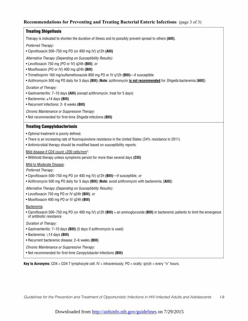
Downloaded from http://aidsinfo.nih.gov/guidelines on 7/29/2015
Guidelines for the Prevention and Treatment of Opportunistic Infections in HIV-Infected Adults and Adolescents I-8
Treating Shigellosis
Therapy is indicated to shorten the duration of illness and to possibly prevent spread to others (AIII).
Preferred Therapy:
• Ciprofloxacin 500–750 mg PO (or 400 mg IV) q12h (AIII)
Alternative Therapy (Depending on Susceptibility Results):
• Levofloxacin 750 mg (PO or IV) q24h (BIII); or
• Moxifloxacin (PO or IV) 400 mg q24h (BIII)
• Trimethoprim 160 mg/sulfamethoxazole 800 mg PO or IV q12h (BIII)—if susceptible
• Azithromycin 500 mg PO daily for 5 days (BIII) (Note: azithromycin is not recommended for Shigella bacteremia [AIII])
Duration of Therapy:
• Gastroenteritis: 7–10 days (AIII) (except azithromycin, treat for 5 days)
• Bacteremia: ≥14 days (BIII)
• Recurrent Infections: 2- 6 weeks (BIII)
Chronic Maintenance or Suppressive Therapy:
• Not recommended for first-time Shigella infections (BIII)
Treating Campylobacteriosis
• Optimal treatment is poorly defined.
• There is an increasing rate of fluoroquinolone resistance in the United States (24% resistance in 2011)
• Antimicrobial therapy should be modified based on susceptibility reports.
Mild disease if CD4 count >200 cells/mm3:
• Withhold therapy unless symptoms persist for more than several days (CIII)
Mild to Moderate Disease:
Preferred Therapy:
• Ciprofloxacin 500–750 mg PO (or 400 mg IV) q12h (BIII)—if susceptible, or
• Azithromycin 500 mg PO daily for 5 days (BIII) (Note: avoid azithromycin with bacteremia, [AIII])
Alternative Therapy (Depending on Susceptibility Results):
• Levofloxacin 750 mg PO or IV q24h (BIII); or
• Moxifloxacin 400 mg PO or IV q24h (BIII)
Bacteremia:
• Ciprofloxacin 500–750 mg PO (or 400 mg IV) q12h (BIII) + an aminoglycoside (BIII) in bacteremic patients to limit the emergenceof antibiotic resistance
Duration of Therapy:
• Gastroenteritis: 7–10 days (BIII) (5 days if azithromycin is used)
• Bacteremia: ≥14 days (BIII)
• Recurrent bacteremic disease: 2–6 weeks (BIII)
Chronic Maintenance or Suppressive Therapy:
• Not recommended for first-time Campylobacter infections (BIII)
Key to Acronyms: CD4 = CD4 T lymphocyte cell; IV = intravenously; PO = orally; q(n)h = every “n” hours.
Recommendations for Preventing and Treating Bacterial Enteric Infections (page 3 of 3)
Downloaded from http://aidsinfo.nih.gov/guidelines on 7/29/2015
Guidelines for the Prevention and Treatment of Opportunistic Infections in HIV-Infected Adults and Adolescents I-9
References
1. Celum CL, Chaisson RE, Rutherford GW, Barnhart JL, Echenberg DF. Incidence of salmonellosis in patients withAIDS. J Infect Dis. 1987;156(6):998-1002. Available at http://www.ncbi.nlm.nih.gov/pubmed/3680999.
2. Sorvillo FJ, Lieb LE, Waterman SH. Incidence of campylobacteriosis among patients with AIDS in Los AngelesCounty. J Acquir Immune Defic Syndr. 1991;4(6):598-602. Available at http://www.ncbi.nlm.nih.gov/pubmed/2023099.
3. Angulo FJ, Swerdlow DL. Bacterial enteric infections in persons infected with human immunodeficiency virus. Clinicalinfectious diseases: an official publication of the Infectious Diseases Society of America. 1995;21 Suppl 1:S84-93.Available at http://www.ncbi.nlm.nih.gov/pubmed/8547518.
4. Nelson MR, Shanson DC, Hawkins DA, Gazzard BG. Salmonella, Campylobacter and Shigella in HIV-seropositivepatients. AIDS. 1992;6(12):1495-1498. Available at http://www.ncbi.nlm.nih.gov/pubmed/1362879.
5. Sanchez TH, Brooks JT, Sullivan PS, et al. Bacterial diarrhea in persons with HIV infection, United States, 1992-2002.Clinical infectious diseases: an official publication of the Infectious Diseases Society of America. 2005;41(11):1621-1627. Available at http://www.ncbi.nlm.nih.gov/pubmed/16267735.
6. Wilcox CM, Saag MS. Gastrointestinal complications of HIV infection: changing priorities in the HAART era. Gut.2008;57(6):861-870. Available at http://www.ncbi.nlm.nih.gov/pubmed/18203808.
7. Hung CC, Hung MN, Hsueh PR, et al. Risk of recurrent nontyphoid Salmonella bacteremia in HIV-infected patients inthe era of highly active antiretroviral therapy and an increasing trend of fluoroquinolone resistance. Clin Infect Dis.2007;45(5):e60-67. Available athttp://www.ncbi.nlm.nih.gov/entrez/query.fcgi?cmd=Retrieve&db=PubMed&dopt=Citation&list_uids=17682981.
8. Huang DB, Mohanty A, DuPont HL, Okhuysen PC, Chiang T. A review of an emerging enteric pathogen:enteroaggregative Escherichia coli. J Med Microbiol. 2006;55(Pt 10):1303-1311. Available athttp://www.ncbi.nlm.nih.gov/pubmed/17005776.
9. Haines CF, Moore RD, Bartlett JG, et al. Clostridium difficile in a HIV-infected cohort: incidence, risk factors, andclinical outcomes. AIDS. 2013;27(17):2799-2807. Available at http://www.ncbi.nlm.nih.gov/pubmed/23842125.
10. Aragon TJ, Vugia DJ, Shallow S, et al. Case-control study of shigellosis in San Francisco: the role of sexualtransmission and HIV infection. Clinical infectious diseases: an official publication of the Infectious Diseases Society ofAmerica. 2007;44(3):327-334. Available at http://www.ncbi.nlm.nih.gov/pubmed/17205436.
11. Quinn TC, Goodell SE, Fennell C, et al. Infections with Campylobacter jejuni and Campylobacter-like organisms inhomosexual men. Ann Intern Med. 1984;101(2):187-192. Available at http://www.ncbi.nlm.nih.gov/pubmed/6547580.
12. Snijders F, Kuijper EJ, de Wever B, van der Hoek L, Danner SA, Dankert J. Prevalence of Campylobacter-associateddiarrhea among patients infected with human immunodeficiency virus. Clinical infectious diseases: an officialpublication of the Infectious Diseases Society of America. 1997;24(6):1107-1113. Available athttp://www.ncbi.nlm.nih.gov/pubmed/9195065.
13. Tee W, Mijch A. Campylobacter jejuni bacteremia in human immunodeficiency virus (HIV)-infected and non-HIV-infected patients: comparison of clinical features and review. Clinical infectious diseases: an official publication of theInfectious Diseases Society of America. 1998;26(1):91-96. Available at http://www.ncbi.nlm.nih.gov/pubmed/9455515.
14. Tee W, Mijch A, Wright E, Yung A. Emergence of multidrug resistance in Campylobacter jejuni isolates from threepatients infected with human immunodeficiency virus. Clinical infectious diseases: an official publication of theInfectious Diseases Society of America. 1995;21(3):634-638. Available at http://www.ncbi.nlm.nih.gov/pubmed/8527556.
15. Meier PA, Dooley DP, Jorgensen JH, Sanders CC, Huang WM, Patterson JE. Development of quinolone-resistantCampylobacter fetus bacteremia in human immunodeficiency virus-infected patients. J Infect Dis. 1998;177(4):951-954. Available at http://www.ncbi.nlm.nih.gov/pubmed/9534967.
16. Casado JL, Valdezate S, Calderon C, et al. Zidovudine therapy protects against Salmonella bacteremia recurrence inhuman immunodeficiency virus-infected patients. J Infect Dis. 1999;179(6):1553-1556. Available athttp://www.ncbi.nlm.nih.gov/entrez/query.fcgi?cmd=Retrieve&db=PubMed&dopt=Citation&list_uids=10228081.
17. Kristjansson M, Viner B, Maslow JN. Polymicrobial and recurrent bacteremia with Shigella in a patient with AIDS.Scand J Infect Dis. 1994;26(4):411-416. Available at http://www.ncbi.nlm.nih.gov/pubmed/7984973.
18. Mayer KH, Hanson E. Recurrent salmonella infection with a single strain in the acquired immunodeficiency syndrome.Confirmation by plasmid fingerprinting. Diagn Microbiol Infect Dis. 1986;4(1):71-76. Available at

Downloaded from http://aidsinfo.nih.gov/guidelines on 7/29/2015
Guidelines for the Prevention and Treatment of Opportunistic Infections in HIV-Infected Adults and Adolescents I-10
http://www.ncbi.nlm.nih.gov/pubmed/3510806.19. Rubino S, Spanu L, Mannazzu M, et al. Molecular typing of non-typhoid Salmonella strains isolated from HIV-infected
patients with recurrent salmonellosis. AIDS. 1999;13(1):137-139. Available at http://www.ncbi.nlm.nih.gov/pubmed/10207558.20. Pulvirenti JJ, Mehra T, Hafiz I, et al. Epidemiology and outcome of Clostridium difficile infection and diarrhea in HIV
infected inpatients. Diagn Microbiol Infect Dis. 2002;44(4):325-330. Available athttp://www.ncbi.nlm.nih.gov/entrez/query.fcgi?cmd=Retrieve&db=PubMed&dopt=Citation&list_uids=12543536.
21. Brecher SM, Novak-Weekley SM, Nagy E. Laboratory diagnosis of Clostridium difficile infections: there is light at the endof the colon. Clinical infectious diseases: an official publication of the Infectious Diseases Society of America.2013;57(8):1175-1181. Available at http://www.ncbi.nlm.nih.gov/pubmed/23788237.
22. Schneider E, Whitmore S, Glynn KM, et al. Revised surveillance case definitions for HIV infection among adults, adolescents,and children aged <18 months and for HIV infection and AIDS among children aged 18 months to <13 years—United States,2008. MMWR Recomm Rep. 2008;57(RR-10):1-12. Available at http://www.ncbi.nlm.nih.gov/pubmed/19052530.
23. Nwachukwu CE, Okebe JU. Antimotility agents for chronic diarrhoea in people with HIV/AIDS. Cochrane Database SystRev. 2008(4):CD005644. Available at http://www.ncbi.nlm.nih.gov/pubmed/18843696.
24. Tribble DR, Sanders JW, Pang LW, et al. Traveler's diarrhea in Thailand: randomized, double-blind trial comparing single-dose and 3-day azithromycin-based regimens with a 3-day levofloxacin regimen. Clinical infectious diseases: an officialpublication of the Infectious Diseases Society of America. 2007;44(3):338-346. Available athttp://www.ncbi.nlm.nih.gov/pubmed/17205438.
25. Cummings PL, Sorvillo F, Kuo T. Salmonellosis-related mortality in the United States, 1990-2006. Foodborne Pathog Dis.2010;7(11):1393-1399. Available at http://www.ncbi.nlm.nih.gov/pubmed/20617938.
26. Guerrant RL, Van Gilder T, Steiner TS, et al. Practice guidelines for the management of infectious diarrhea. Clinicalinfectious diseases: an official publication of the Infectious Diseases Society of America. 2001;32(3):331-351. Available athttp://www.ncbi.nlm.nih.gov/pubmed/11170940.
27. Gordon MA, Banda HT, Gondwe M, et al. Non-typhoidal salmonella bacteraemia among HIV-infected Malawian adults:high mortality and frequent recrudescence. AIDS. 2002;16(12):1633-1641. Available athttp://www.ncbi.nlm.nih.gov/pubmed/12172085.
28. 1993 revised classification system for HIV infection and expanded surveillance case definition for AIDS amongadolescents and adults. MMWR Recomm Rep. 1992;41(RR-17):1-19. Available athttp://www.ncbi.nlm.nih.gov/pubmed/1361652.
29. Khan WA, Seas C, Dhar U, Salam MA, Bennish ML. Treatment of shigellosis: V. Comparison of azithromycin andciprofloxacin. A double-blind, randomized, controlled trial. Ann Intern Med. 1997;126(9):697-703. Available athttp://www.ncbi.nlm.nih.gov/pubmed/9139555.
30. Heiman KE, Karlsson M, Grass J, et al. Notes from the field: Shigella with decreased susceptibility to azithromycin amongmen who have sex with men - United States, 2002-2013. MMWR. Morbidity and mortality weekly report. 2014;63(6):132-133. Available at http://www.ncbi.nlm.nih.gov/pubmed/24522098.
31. Hassing RJ, Melles DC, Goessens WH, Rijnders BJ. Case of Shigella flexneri infection with treatment failure due toazithromycin resistance in an HIV-positive patient. Infection. 2014. Available athttp://www.ncbi.nlm.nih.gov/pubmed/24488332.
32. Kuschner RA, Trofa AF, Thomas RJ, et al. Use of azithromycin for the treatment of Campylobacter enteritis in travelers toThailand, an area where ciprofloxacin resistance is prevalent. Clinical infectious diseases: an official publication of theInfectious Diseases Society of America. 1995;21(3):536-541. Available at http://www.ncbi.nlm.nih.gov/pubmed/8527539.
33. Cohen SH, Gerding DN, Johnson S, et al. Clinical practice guidelines for Clostridium difficile infection in adults: 2010update by the society for healthcare epidemiology of America (SHEA) and the infectious diseases society of America(IDSA). Infect Control Hosp Epidemiol. 2010;31(5):431-455. Available at http://www.ncbi.nlm.nih.gov/pubmed/20307191.
34. Surawicz CM, Brandt LJ, Binion DG, et al. Guidelines for diagnosis, treatment, and prevention of Clostridium difficileinfections. The American Journal of Gastroenterology. 2013;108(4):478-498; quiz 499. Available athttp://www.ncbi.nlm.nih.gov/pubmed/23439232.
35. Gurumurthy P, Ramachandran G, Hemanth Kumar AK, et al. Malabsorption of rifampin and isoniazid in HIV-infectedpatients with and without tuberculosis. Clinical infectious diseases : an official publication of the Infectious DiseasesSociety of America. 2004;38(2):280-283. Available at http://www.ncbi.nlm.nih.gov/pubmed/14699462.
Downloaded from http://aidsinfo.nih.gov/guidelines on 7/29/2015
Guidelines for the Prevention and Treatment of Opportunistic Infections in HIV-Infected Adults and Adolescents I-11
36. Peloquin CA, MacPhee AA, Berning SE. Malabsorption of antimycobacterial medications. N Engl J Med.1993;329(15):1122-1123. Available at http://www.ncbi.nlm.nih.gov/pubmed/8371737.
37. Schaefer C, Amoura-Elefant E, Vial T, et al. Pregnancy outcome after prenatal quinolone exposure. Evaluation of a caseregistry of the European Network of Teratology Information Services (ENTIS). Eur J Obstet Gynecol Reprod Biol.1996;69(2):83-89. Available at http://www.ncbi.nlm.nih.gov/pubmed/8902438.
38. Loebstein R, Addis A, Ho E, et al. Pregnancy outcome following gestational exposure to fluoroquinolones: amulticenter prospective controlled study. Antimicrob Agents Chemother. 1998;42(6):1336-1339. Available athttp://www.ncbi.nlm.nih.gov/pubmed/9624471.
39. Czeizel AE, Rockenbauer M, Sorensen HT, Olsen J. The teratogenic risk of trimethoprim-sulfonamides: a populationbased case-control study. Reprod Toxicol. 2001;15(6):637-646. Available athttp://www.ncbi.nlm.nih.gov/pubmed/11738517.
40. Hernandez-Diaz S, Werler MM, Walker AM, Mitchell AA. Folic acid antagonists during pregnancy and the risk of birthdefects. N Engl J Med. 2000;343(22):1608-1614. Available at http://www.ncbi.nlm.nih.gov/pubmed/11096168.
41. Hernandez-Diaz S, Werler MM, Walker AM, Mitchell AA. Neural tube defects in relation to use of folic acidantagonists during pregnancy. American Journal of Epidemiology. 2001;153(10):961-968. Available athttp://www.ncbi.nlm.nih.gov/pubmed/11384952.
Downloaded from http://aidsinfo.nih.gov/guidelines on 7/29/2015
Guidelines for the Prevention and Treatment of Opportunistic Infections in HIV-Infected Adults and Adolescents J-1
Bartonellosis (Last updated May 7, 2013; last reviewed May 7, 2013)
Epidemiology
Bartonella species cause infections that include cat scratch disease, retinitis, trench fever, relapsingbacteremia, endocarditis, bacillary angiomatosis (BA), and bacillary peliosis hepatis.1 The latter twomanifestations occur only in individuals who are immunocompromised. BA is caused by either Bartonellaquintana or Bartonella henselae.1,2 Twenty-four species and three subspecies of Bartonella have beenisolated and are officially recognized (http://www.bacterio.cict.fr/b/bartonella.html), and eight have beenisolated from humans. However, only B. henselae and B. quintana infections have been identified in HIV-infected patients.2 BA most often occurs late in HIV infection, in patients with median CD4 T lymphocyte(CD4 cell) counts <50 cells/mm3.2 In HIV-infected patients, bartonellosis is often a chronic illness, lasting formonths to years, with BA lesions and intermittent bacteremia.
Development of BA lesions caused by B. henselae is statistically linked to cat exposure in patients with HIVinfection.2 In contrast, BA caused by B. quintana is associated with body louse infestation andhomelessness.2 The body louse serves as the vector of B. quintana in humans. To avoid exposure to B.quintana, HIV-infected patients should avoid body lice and, if infected, treat the infestation. The cat flea isthe vector of B. henselae in cats. Cats are the most common vector (via a scratch) responsible fortransmitting B. henselae to humans, most likely when their claws become contaminated with feces from B.henselae-infected fleas. In some areas of the United States, the prevalence of B. henselae bacteremia in petcats approaches 50%.3 Control of cat flea infestation and avoidance of cat scratches are therefore criticalstrategies for preventing B. henselae infections in patients who are HIV infected.
Clinical Manifestations
BA lesions have been associated with nearly every organ system, but cutaneous lesions are the most readilyidentified. These lesions can be clinically indistinguishable from Kaposi sarcoma, pyogenic granuloma, andother skin conditions. BA also can cause subcutaneous nodules. Osteomyelitis is usually caused by B.quintana, and only B. henselae can cause bacillary peliosis hepatis. Although isolated organs can appear tobe the principal focus of disease, BA represents a hematogenously disseminated infection, and systemicsymptoms of fever, night sweats, and weight loss often accompany BA. Bartonella infection is a major causeof unexplained fever in patients with late-stage AIDS and should be considered in the differential diagnosisof patients with fever and CD4 counts <100 cells/mm3.4 Bartonella is a relatively common cause of culture-negative endocarditis in immunocompetent and immunocompromised humans and is most commonly causedby B. quintana and, less frequently, B. henselae.5
Diagnosis
Diagnosis can be confirmed by histopathologic examination of biopsied tissue.6 BA lesions are characterizedby vascular proliferation, and a modified silver stain (such as Warthin-Starry stain) usually demonstratesnumerous bacilli. Tissue Gram staining and acid-fast staining are negative.
A well-characterized serologic test was developed at Centers for Disease Control and Prevention7 and is alsoavailable at some state health labs. In addition, several private laboratories offer serological testing, but noneof these private laboratory tests has been evaluated for sensitivity or specificity with sera from HIV-infectedpatients with culture-documented Bartonella infection. In immunocompetent patients, anti-Bartonellaantibodies might not be detectable for 6 weeks after acute infection; in contrast, by the time Bartonellainfection is suspected in patients with late-stage HIV infection, they usually have been infected for months oreven >1 year. Note that as many as 25% of Bartonella culture-positive patients never develop antibodies inthe setting of advanced HIV infection.4 In those patients who do develop anti-Bartonella antibodies,monitoring of antibody levels can correlate with resolution and recrudescence of Bartonella infection.
Downloaded from http://aidsinfo.nih.gov/guidelines on 7/29/2015
Guidelines for the Prevention and Treatment of Opportunistic Infections in HIV-Infected Adults and Adolescents J-2
Bartonella species can be isolated (with difficulty) from blood, using ethylenediaminetetraacetic acid(EDTA) tubes. The organisms have been isolated from tissue in only a few laboratories because of thefastidious nature of Bartonella.2 Polymerase chain reaction methods have been developed for identificationand speciation of Bartonella but are not widely available.
Preventing Exposure
HIV-infected patients, specifically those who are severely immunocompromised (CD4 counts <100cells/mm3), are at high risk of severe disease when infected by B. quintana and B. henselae. The major riskfactors for acquisition of B. henselae are contact with cats infested with fleas and receiving cat scratches.Immunocompromised individuals should consider the potential risks of cat ownership (AIII). Patients whowant cats should acquire animals that are older than age 1 year and in good health (BII). Cats should beacquired from a known environment, have a documented health history, and be free of fleas. Stray cats andcats with flea infestation should be avoided. Declawing is not advised, but HIV-infected individuals shouldavoid rough play with cats and situations in which scratches are likely (AII). Patients should avoid contactwith flea feces (i.e., flea dirt), and any cat-associated wound should be washed promptly with soap and water(BIII). Care of cats should include a comprehensive, ongoing flea-control program under the supervision ofa veterinarian (BIII). No evidence indicates any benefits to cats or their owners from routine culture orserologic testing of the pet for Bartonella infection or from antibiotic treatment of healthy, serologicallypositive cats (BII). The major risk factor for B. quintana infection is body lice infestation. Patients who arehomeless or in marginal housing should be informed that body louse infestation can be associated withserious illness and provided with appropriate measures to eradicate body lice, if present (AII).
Preventing Disease
Primary chemoprophylaxis for Bartonella-associated disease is not recommended (BIII). However, note thatin a retrospective case-control study, Mycobacterium avium complex prophylaxis using a macrolide orrifamycin was protective against developing Bartonella infection.2
Treating Disease
All HIV-infected patients with Bartonella infection should receive antibiotic treatment (AII). Guidelines fortreatment of Bartonella infections have been published.8 No randomized, controlled clinical trials haveevaluated antimicrobial treatment of bartonellosis in HIV-infected patients. Erythromycin and doxycyclinehave been used successfully to treat BA, peliosis hepatis, bacteremia, and osteomyelitis and are consideredfirst-line treatment for bartonellosis on the basis of reported experience in case series (AII).1,2 Therapyshould be administered for ≥3 months (AII). Doxycycline, with or without a rifamycin, is the treatment ofchoice for bartonellosis infection involving the central nervous system (CNS) (AIII). For severe Bartonellainfections, combination therapy using erythromycin or doxycycline with a rifamycin is recommended (BIII);intravenous therapy may be needed initially (AIII). Treatment of confirmed Bartonella endocarditis shouldinclude doxycycline with the addition of gentamicin for 2 weeks (if tolerated); a rifamycin can be substitutedfor gentamicin in the setting of renal insufficiency (BII).8
Clarithromycin or azithromycin treatment has been associated with clinical response and either of these canbe an alternative therapy Bartonella infections (except for endocarditis or CNS infections) (BIII).Azithromycin is recommended for patients who are less likely to comply with the more frequent dosingschedule for doxycycline or erythromycin. A third-generation cephalosporin, ceftizoxime,9 was usedsuccessfully to treat Bartonella in a pregnant HIV-infected woman, but because there are no other data, amacrolide is the drug of first choice. Penicillins and first-generation cephalosporins have no in vivo activityand should not be used for treatment of bartonellosis (BII). Quinolones and trimethoprim-sulfamethoxazole(TMP-SMX) have variable in vitro activity and an inconsistent clinical response in case reports and are notrecommended (BIII).
Downloaded from http://aidsinfo.nih.gov/guidelines on 7/29/2015
Guidelines for the Prevention and Treatment of Opportunistic Infections in HIV-Infected Adults and Adolescents J-3
Special Consideration with Regard to Starting ARTAntiretroviral-naive patients with Bartonella CNS or ophthalmic lesions should probably be treated withdoxycycline and a rifamycin for 2 to 4 weeks before instituting antiretroviral therapy (CIII).
Monitoring of Response to Therapy and Adverse Effects (Including IRIS) Patients should have anti-Bartonella IgG antibody titers checked at the time of diagnosis and, if positive,should be followed with sequential titers every 6 to 8 weeks until a four-fold decrease is documented. Thistest is available at the Centers for Disease Control and Prevention and several large commercial labs. Patientstreated with oral doxycycline should be cautioned about pill-associated ulcerative esophagitis that occursmost often when a dose is taken with only a small amount of liquid or at night just before retiring.10
Photosensitivity also can occur during doxycycline treatment. Adverse effects associated with macrolidesinclude nausea, vomiting, abdominal pain, and elevations of liver transaminase levels. Serious side effectscan occur during treatment with rifamycins, including hypersensitivity reactions (includingthrombocytopenia, interstitial nephritis, and hemolytic anemia), and hepatitis. Administration of rifamycinsstrongly induces the cytochrome P450 enzyme system, which is an important consideration when othermedications, including many ARV drugs, are taken simultaneously.
Immune reconstitution inflammatory syndrome (IRIS) has not been described in association withBartonellosis and treatment with ART in HIV-infected persons.
Managing Treatment FailureAmong patients who fail to respond to initial treatment, 1 or more of the second-line alternative regimensshould be considered (AIII), again with treatment duration of ≥3 months. For patients with positive orincreasing antibody titers, treatment should continue until a fourfold decrease is documented.
Preventing Recurrence
If a relapse occurs after a minimum 3-month course of primary treatment, long-term suppression of infectionwith doxycycline or a macrolide is recommended, as long as the CD4 count remains <200 cells/mm3 (AIII).
Long-term suppression can be discontinued after the patient has received at least 3 to 4 months of therapyand when the CD4 count remains >200 cells/mm3 for ≥6 months (CIII). Some specialists would discontinuetherapy only if the Bartonella titers have also decreased by four-fold (CIII).
Special Considerations During Pregnancy
Infection with Bartonella bacilliformis in immunocompetent patients during pregnancy has been associatedwith increased complications and risk of death.11 No data are available on the effect of B. henselae or B.quintana infections in pregnant women with concomitant HIV infection.
The approach to diagnosis of Bartonella infections in pregnant women is the same as in non-pregnantwomen. Erythromycin treatment should be used (AIII) rather than tetracyclines during pregnancy because ofthe increased risk of hepatotoxicity and the accumulation of tetracycline in fetal teeth and bones, resulting indark, permanent staining of fetal teeth. Third-generation cephalosporins such as ceftizoxime9 or ceftriaxonemay have efficacy against Bartonella in pregnant women who are HIV infected, but it should be consideredsecond-line therapy after a macrolide. First- and second-generation cephalosporins are not recommendedbecause of their lack of efficacy against Bartonella (AII).
Downloaded from http://aidsinfo.nih.gov/guidelines on 7/29/2015
Guidelines for the Prevention and Treatment of Opportunistic Infections in HIV-Infected Adults and Adolescents J-4
Recommendations for Treating Bartonella Infections
Preferred Therapy
For Bacillary Angiomatosis, Peliosis Hepatis, Bacteremia, and Osteomyelitis:
• Doxycycline 100 mg PO or IV q12h (AII), or
• Erythromycin 500 mg PO or IV q6h (AII)
For Infections Involving the CNS:
• Doxycycline 100 mg PO or IV q12h +/- rifampin 300 mg PO or IV q12h (AIII)
For Confirmed Bartonella Endocarditis:
• (Doxycycline 100 mg IV q12h + gentamicin 1 mg/kg IV q8h) x 2 weeks, then continue with doxycycline 100 mg IV or PO q12h(BII), or
• For patients with renal insufficiency: (doxycycline 100 mg IV q12h + rifampin 300 mg IV or PO q12h) x 2 weeks, then continuewith doxycycline 100 mg IV or PO q12h (BII)
For Other Severe Infections
• Doxycycline 100 mg PO or IV q12h + rifampin 300 mg PO or IV q12h (BIII), or
• Erythromycin 500 mg PO or IV q6h + rifampin 300 mg PO or IV q12h (BIII)
Alternative Therapy for Bartonella Infections (Not for Endocarditis or CNS Infections):
• Azithromycin 500 mg PO daily (BIII), or
• Clarithromycin 500 mg PO BID (BIII)
Duration of Therapy:
• At least 3 months
Indication for Long-Term Suppressive Therapy
If a relapse occurs after a ≥3 month course of primary treatment:
• A macrolide or doxycycline as long as the CD4 count remains <200 cells/mm3 (AIII)
Indications for Discontinuing Long-Term Suppressive Therapy (CIII):
• Received at least 3 to 4 months of treatment; and
• CD4 count >200 cells/mm3 for at least 6 months
• Some specialists would only discontinue therapy if Bartonella titers have also decreased by four-fold
Other Considerations
• Rifampin is a potent hepatic enzyme inducer and may lead to significant interaction with many drugs; including ARV agents (seeTable 5 for dosing recommendations)
Key to Abbreviations: ARV = antiretroviral; BID = twice daily; CD4 = CD4 T lymphocyte cell; CNS = central nervous system, IV =intravenously, PO = orally; q(n)h = every “n” hours
References
1. Spach DH, Koehler JE. Bartonella-associated infections. Infect Dis Clin North Am. Mar 1998;12(1):137-155. Availableat http://www.ncbi.nlm.nih.gov/pubmed/9494835.
2. Koehler JE, Sanchez MA, Garrido CS, et al. Molecular epidemiology of bartonella infections in patients with bacillaryangiomatosis-peliosis. N Engl J Med. Dec 25 1997;337(26):1876-1883. Available athttp://www.ncbi.nlm.nih.gov/pubmed/9407154.
3. Koehler JE, Glaser CA, Tappero JW. Rochalimaea henselae infection. A new zoonosis with the domestic cat asreservoir. JAMA. Feb 16 1994;271(7):531-535. Available at http://www.ncbi.nlm.nih.gov/pubmed/8301768.
4. Koehler JE, Sanchez MA, Tye S, et al. Prevalence of Bartonella infection among human immunodeficiency virus-infected patients with fever. Clin Infect Dis. Aug 15 2003;37(4):559-566. Available athttp://www.ncbi.nlm.nih.gov/pubmed/12905141.
5. Houpikian P, Raoult D. Blood culture-negative endocarditis in a reference center: etiologic diagnosis of 348 cases.Medicine (Baltimore). May 2005;84(3):162-173. Available at http://www.ncbi.nlm.nih.gov/pubmed/15879906.
Downloaded from http://aidsinfo.nih.gov/guidelines on 7/29/2015
Guidelines for the Prevention and Treatment of Opportunistic Infections in HIV-Infected Adults and Adolescents J-5
6. LeBoit PE, Berger TG, Egbert BM, Beckstead JH, Yen TS, Stoler MH. Bacillary angiomatosis. The histopathology anddifferential diagnosis of a pseudoneoplastic infection in patients with human immunodeficiency virus disease. TheAmerican journal of surgical pathology. Nov 1989;13(11):909-920. Available athttp://www.ncbi.nlm.nih.gov/pubmed/2802010.
7. Regnery RL, Olson JG, Perkins BA, Bibb W. Serological response to "Rochalimaea henselae" antigen in suspected cat-scratch disease. Lancet. Jun 13 1992;339(8807):1443-1445. Available at http://www.ncbi.nlm.nih.gov/pubmed/1351130.
8. Rolain JM, Brouqui P, Koehler JE, Maguina C, Dolan MJ, Raoult D. Recommendations for treatment of humaninfections caused by Bartonella species. Antimicrob Agents Chemother. Jun 2004;48(6):1921-1933. Available athttp://www.ncbi.nlm.nih.gov/pubmed/15155180.
9. Riley LE, Tuomala RE. Bacillary angiomatosis in a pregnant patient with acquired immunodeficiency syndrome. ObstetGynecol. May 1992;79(5 ( Pt 2)):818-819. Available at http://www.ncbi.nlm.nih.gov/pubmed/1565376.
10. Kikendall JW, Friedman AC, Oyewole MA, Fleischer D, Johnson LF. Pill-induced esophageal injury. Case reports andreview of the medical literature. Digestive diseases and sciences. Feb 1983;28(2):174-182. Available athttp://www.ncbi.nlm.nih.gov/pubmed/6825537.
11. Maguina C, Garcia PJ, Gotuzzo E, Cordero L, Spach DH. Bartonellosis (Carrion's disease) in the modern era. ClinInfect Dis. Sep 15 2001;33(6):772-779. Available at http://www.ncbi.nlm.nih.gov/pubmed/11512081.
Downloaded from http://aidsinfo.nih.gov/guidelines on 7/29/2015
Guidelines for the Prevention and Treatment of Opportunistic Infections in HIV-Infected Adults and Adolescents K-1
Syphilis (Last updated May 7, 2013; last reviewed May 7, 2013)
Epidemiology
Syphilis is associated with increased risk of sexual acquisition and transmission of HIV.1,2 In recent years,there has been a resurgence of the disease in men in several U.S. cities and in Western Europe(http://www.cdc.gov/std/stats).3-8 Although coexistent HIV infection, particularly in the advanced stages, maymodify the diagnosis, natural history, or management of Treponema pallidum infection, the principles ofsyphilis management are the same for persons with and without coexistent HIV infection.9-13
Clinical Manifestations
The effect of coexistent HIV on the protean manifestations of syphilis have been documented in multiplecase reports and small case series, but in only a limited number of large studies. Some studies suggest thatHIV infection may shift the clinical manifestations of syphilis, making clinical lesions more apparent, andmay accelerate progression of syphilitic disease.10,11,14,15 Early syphilis in HIV-infected patients also maycause a transient decrease in CD4 T-lymphocyte (CD4) count and increase in HIV viral load that improveswith recommended syphilis treatment regimens.16-20
Primary syphilis commonly manifests as a single painless nodule at the site of contact that rapidly ulceratesto form a classic chancre; in HIV-infected patients, however, multiple or atypical chancres occur and primarylesions may be absent or missed.10,21
Progression to secondary syphilis typically follows 2 to 8 weeks after primary inoculation. Although more rapidprogression or severe disease can occur in HIV-infected patients with advanced immunosuppression, the clinicalmanifestations are similar to those in HIV-uninfected individuals. The manifestations of secondary syphilisinvolve virtually all organ systems. The most common manifestations—macular, maculopapular,papulosquamous, or pustular skin lesions—can involve the palms and soles and be accompanied by generalizedlymphadenopathy, fever, malaise, anorexia, arthralgias, and headache.11,12,19 Condyloma lata (moist, flat, papularlesions in warm intertrigenous regions) can occur and may resemble human papillomavirus infection. Luesmaligna is a rare manifestation of secondary syphilis, characterized by papulopustular skin lesions that evolveinto ulcerative lesions with sharp borders and a dark central crust.22 Secondary syphilis, especially whenassociated with symptomatic early neurosyphilis, can resemble acute primary HIV infection. Constitutionalsymptoms, along with nonfocal central nervous system (CNS) symptoms and cerebrospinal fluid (CSF)abnormalities such as lymphocytic pleocytosis with a mildly elevated CSF protein, are common to bothsecondary syphilis and acute primary HIV infection.14,15,21,23-26 Signs and symptoms of secondary syphilis canpersist from a few days to several weeks before resolving and evolving to latent or later stages.
Latent syphilis lacks overt clinical signs and symptoms, but relapse of manifestations of secondary syphilis canoccur, most commonly during the first year after infection. Manifestations of tertiary syphilis generally includecardiovascular syphilis and gummatous syphilis or a slowly progressive disease that can affect any organsystem. Neurosyphilis can occur at any stage of syphilis and manifest in varied clinical presentations, such ascranial nerve dysfunction, stroke, meningitis, acute or chronic change in mental status, loss of vibration sense,and auditory or ophthalmic abnormalities. Manifestations of symptomatic neurosyphilis in HIV-infected patientsare similar to those in individuals who are not HIV infected. However, clinical manifestations of neurosyphilis,such as concomitant uveitis and meningitis, may be more common in HIV-infected persons.14,15,26-28
Diagnosis
Darkfield microscopy and tests to detect T. pallidum in lesion exudates or tissue (biopsy with silver stain) aredefinitive for diagnosing early syphilis, although no T. pallidum direct detection tests are commerciallyavailable. A presumptive serologic diagnosis of syphilis is possible based upon non-treponemal tests (i.e.,Venereal Disease Research Laboratory [VDRL] and rapid plasma reagin [RPR]) and treponemal tests (i.e.,
Downloaded from http://aidsinfo.nih.gov/guidelines on 7/29/2015
Guidelines for the Prevention and Treatment of Opportunistic Infections in HIV-Infected Adults and Adolescents K-2
fluorescent treponemal antibody absorbed [FTA-ABS], T. pallidum particle agglutination [TP-PA], enzymeimmunoassays [EIAs], and chemiluminescence immunoassays [CIA]).
Serologic diagnosis of syphilis traditionally has involved screening for non-treponemal antibodies withconfirmation of reactive tests by treponemal-based assays.19,29 Recently, some laboratories have initiated atesting algorithm using EIA or CIA as a screening test, followed by a reflex-quantitative, non-treponemal testif the EIA or CIA is positive. This latter strategy may identify those with previously treated syphilis infectionmore often than those with untreated infection.30
In persons with a positive treponemal screening test and a negative reflex-quantitative, non-treponemal test,the laboratory should perform a second treponemal test (based on different antigens from the initial test) toconfirm the results of the positive initial treponemal test. If a second treponemal test is positive, anassessment is needed of current sexual risk factors and prior syphilis treatment. Physical examination shouldbe performed to assess for evidence of syphilis, especially primary disease. Patients with suspected primarysyphilis should be empirically treated and retested with a non-treponemal test in several weeks (if initial non-treponemal test was non-reactive) to confirm the diagnosis. Persons with discordant sera (reactive EIA/CIAand non-reactive, non-treponemal test) and a reactive TP-PA assay should be treated for late-latent syphilis ifpast treatment cannot be confirmed. If the second treponemal test is negative, no treatment is indicated.19,31 Inthe absence of neurologic signs or symptoms, risk of neurosyphilis is low in patients with a reactivetreponemal test and a non-reactive, non-treponemal test;32 examination of CSF is not recommended.
Early-stage disease (i.e., primary, secondary, and early-latent syphilis) in HIV-infected patients is confirmedwith the same diagnostic tests used in those who are not infected with HIV: darkfield microscopy of amucocutaneous lesion and standard serologic tests. Results with VDRL and RPR may be higher, lower, ordelayed in HIV-infected versus HIV-uninfected patients with early-stage syphilis.33-37 No data indicate thattreponemal tests perform differently among HIV-infected patients compared with HIV-uninfected patients,38
although uncommon, false-negative serologic tests for syphilis can occur in both HIV-uninfected and HIV-infected patients with documented T. pallidum infection.36,37 Therefore, if serologic tests do not confirm thediagnosis of suspected syphilis, other diagnostic procedures, such as repeat serology in 2 to 4 weeks,exclusion of prozone phenomenon, biopsy, or darkfield examination, should be pursued. By definition,persons with latent syphilis have serological evidence of syphilis in the absence of clinical manifestations.Early-latent syphilis is defined as evidence of infection <1 year; late-latent syphilis is evidence of infectionfor >1 year after acquisition of syphilis or latent infection of unknown duration. Diagnostic testingrecommended for detection of late-stage syphilis (i.e., cardiovascular and gummatous syphilis) in HIV-infected patients is the same as in patients who are not infected with HIV.19
All persons with syphilis and signs or symptoms suggesting neurologic disease (e.g., cranial nervedysfunction, meningitis, stroke, alteration in mental status, auditory or ophthalmic abnormalities) warrantevaluation for neurosyphilis and for ocular or otic syphilis if ophthalmic or auditory symptoms are present.CSF abnormalities (i.e., elevated protein and mononuclear pleocytosis) are common in early syphilis and inpatients with HIV infection, even those with no neurologic symptoms. There is no evidence that the clinicaland prognostic significance of such CSF abnormalities differs between HIV-infected and -uninfected patientswith primary, secondary, or early-latent syphilis.
CSF examination should be performed in patients who have neurologic, auditory, or ophthalmic signs (e.g.,iritis, uveitis) or symptoms, active tertiary syphilis, or serologic treatment failure. Several studies havedemonstrated that in HIV-infected patients with syphilis, clinical and CSF abnormalities consistent withneurosyphilis are associated with CD4 counts ≤350 cells/mm3 alone or in combination with RPR titers≥1:32.25,26,39,40 Unless neurologic symptoms are present, however, CSF examination in this setting has notbeen associated with improved clinical outcomes. The risk of later developing clinical neurosyphilis and thebenefits of a CSF examination in this circumstance are unknown.
Laboratory testing is useful in supporting the diagnosis of neurosyphilis, but no single test can be used to
Downloaded from http://aidsinfo.nih.gov/guidelines on 7/29/2015
Guidelines for the Prevention and Treatment of Opportunistic Infections in HIV-Infected Adults and Adolescents K-3
diagnose it. In patients who are not HIV infected, CSF examination supports diagnosis of neurosyphilis,which may indicate mild mononuclear pleocytosis (6–200 cells/mm3), normal or mildly elevated proteinconcentration, or a reactive (CSF-VDRL).19,25,26 CSF-VDRL is specific but not sensitive, and a reactive testestablishes the diagnosis of neurosyphilis, but a non-reactive test does not exclude it. In comparison, CSFFTA-ABS is less specific than CSF-VDRL but highly sensitive. Calculated indices (T. pallidumhemaglutination assay index) are of limited value in establishing the diagnosis of neurosyphilis. Polymerase-chain-reaction-based diagnostic methods are not currently recommended as diagnostic tests forneurosyphilis. A reactive CSF-VDRL and a CSF white blood cell (WBC) count >10 cells/mm3 support thediagnosis of neurosyphilis; in the absence of other abnormalities, elevation in CSF protein concentrationsshould not be used as the sole diagnostic criterion. Therefore, the laboratory tests used to support thediagnosis of neurosyphilis depend on various combinations of reactive serologic tests, CSF cell count andprotein, and a reactive CSF-VDRL with or without clinical manifestations.
Establishing the diagnosis of neurosyphilis can be more difficult in patients with HIV infection because HIVinfection itself may be associated with mild mononuclear CSF pleocytosis (6–15 cells/mm3). Using a higherCSF WBC cutoff of >20 WBC/mm3 may improve the specificity of neurosyphilis diagnosis in HIV-infectedpatients.41 CSF FTA-ABS testing in HIV-uninfected persons suggests that the CSF FTA-ABS test is lessspecific for neurosyphilis than the CSF-VDRL but is highly sensitive.19,42 Thus, the use of this test may beconsidered in HIV-infected patients.
Preventing Exposure
The resurgence of syphilis in patients with HIV infection in the United States underscores the importance ofprimary prevention of syphilis in this population, which should begin with routine discussion of sexualbehaviors. Health care providers should discuss client-centered risk reduction messages and provide specificactions that can reduce the risk of acquiring sexually transmitted diseases and of transmitting HIVinfection.19,43-47 Routine serologic screening for syphilis is recommended at least annually for all HIV-infected patients who are sexually active, with more frequent screening (every 3–6 months) for those whohave multiple partners, unprotected intercourse, sex in conjunction with illicit drug use, or usemethamphetamines (or whose partners participate in such activities).19,48-50 The occurrence of syphilis in anHIV-infected individual is an indication of high-risk behavior and should prompt intensified counselingmessages and strong consideration of referral for behavioral intervention. Patients undergoing screening ortreatment for syphilis also should be evaluated for all common sexually transmitted diseases such aschlamydia and gonorrhea at anatomic sites of exposure.19,51
Preventing Disease
The same measures that apply to preventing exposure apply to preventing disease. Studies in the pre-HIV erademonstrated that approximately one-third of the sex partners of patients who have infectious syphilis willdevelop syphilis within 30 days of exposure, and empiric treatment of incubating syphilis will prevent thedevelopment of disease in those who are exposed.52-55 Those exposed sexually to a patient with syphilis inany stage should be evaluated clinically and serologically and treated presumptively with regimens outlinedin current recommendations.19 Specifically, individuals who were exposed within the 90 days precedingdiagnosis of primary, secondary, or early-latent syphilis in a sex partner may be infected even if they areseronegative. Therefore, they should be treated presumptively (AII). Individuals exposed >90 days beforediagnosis of primary, secondary, or early-latent syphilis in a sex partner should be treated presumptively ifserologic test results are not available immediately and the opportunity for follow-up is uncertain (AIII).
Treating Disease
Management of syphilis in HIV-infected patients is similar to that in individuals who are HIV-uninfected.13,19,34 Most HIV-infected patients respond appropriately to standard treatment. Closer follow-up is
Downloaded from http://aidsinfo.nih.gov/guidelines on 7/29/2015
Guidelines for the Prevention and Treatment of Opportunistic Infections in HIV-Infected Adults and Adolescents K-4
recommended, however, because rates of serologic treatment failure may be higher in those who are HIVinfected and they may be at increased risk of neurologic complications.15,56,57
Penicillin remains the treatment of choice for syphilis regardless of a patient’s HIV status. HIV-infectedpatients with early-stage (primary, secondary, or early-latent) syphilis should receive a single intramuscular(IM) injection of 2.4 million units of benzathine penicillin G (AII).19 The available data demonstrate thathigh-dose amoxicillin given with probenecid in addition to benzathine penicillin G in early syphilis is notassociated with improved clinical outcomes.34 Patients with a penicillin allergy whose compliance or follow-up cannot be ensured should be desensitized and treated with benzathine penicillin (AIII). The efficacy ofalternative non-penicillin regimens in HIV-infected patients with early syphilis has not been evaluatedsufficiently to warrant their use as first-line treatment.
Regardless of HIV infection status, use of any alternative penicillin treatment regimen should be undertakenonly with close clinical and serologic monitoring. Several retrospective studies support use of doxycycline,100 mg twice daily, to treat early syphilis (BII); however, the majority of the patients were HIVuninfected.58,59 Limited clinical studies suggest that ceftriaxone, 1 g daily either IM or intravenously (IV) for10 to 14 days, is effective for treating early syphilis (BII), but the optimal dose and duration of therapy havenot been defined.60 A single 2-g oral dose of azithromycin is effective for treating early syphilis;61-63 howeverT. pallidum chromosomal mutations associated with azithromycin resistance and treatment failures have beenreported and are more common in men who have sex with men (MSM).64-69 Azithromycin treatment has notbeen well studied in HIV-infected patients with early syphilis and it should be used with caution in instanceswhen treatment with penicillin or doxycycline is not feasible (BII). Azithromycin should not be used inMSM or in pregnant women (AII).
In HIV-infected patients with late-latent syphilis and no signs or symptoms of neurosyphilis, treatment with 3weekly IM injections of 2.4 million units benzathine penicillin G is recommended (AII). Alternative therapywith doxycycline, 100 mg by mouth twice a day for 28 days, has not been sufficiently evaluated in HIV-infected patients to warrant use as first-line treatment (BIII). Limited clinical studies and biologic andpharmacologic evidence suggest that ceftriaxone may be effective; however, the optimal dose and duration oftherapy have not been determined.70,71 If the clinical situation requires use of an alternative to penicillin,treatment should be undertaken with close clinical and serologic monitoring.
HIV-infected patients with clinical evidence of late-stage (tertiary) syphilis (cardiovascular or gummatousdisease) should have CSF examination to rule out neurosyphilis before therapy is initiated. Recommendedtreatment of late-stage syphilis is 3 weekly IM injections of 2.4 million units benzathine penicillin G (AII).19
However, the complexity of tertiary syphilis management is beyond the scope of these guidelines and healthcare providers are advised to consult an infectious disease specialist.
HIV-infected patients diagnosed with neurosyphilis or ocular or otic syphilis should receive IV aqueouscrystalline penicillin G, 18 to 24 million units daily, administered 3 to 4 million units IV every 4 hours or bycontinuous infusion for 10 to 14 days (AII) or procaine penicillin, 2.4 million units IM once daily plusprobenecid 500 mg orally 4 times a day for 10 to 14 days (BII).19,25,26 HIV-infected patients who are allergicto sulfa-containing medications should not be given probenecid because of potential allergic reaction (AIII).
Because neurosyphilis treatment regimens are of shorter duration than those used in late-latent syphilis, 2.4million units benzathine penicillin IM once per week for up to 3 weeks after completion of neurosyphilistreatment can be considered to provide a comparable duration of therapy (CIII).19 Desensitization topenicillin is the preferred approach to treating neurosyphilis in patients who are allergic to penicillin.However, limited data indicate that ceftriaxone (2 g daily IV for 10–14 days) may be an acceptablealternative regimen (BII).71 Other alternative regimens for neurosyphilis have not been evaluated adequately.Syphilis treatment recommendations are also available in the 2010 Centers for Disease Control andPrevention STD Treatment Guidelines.19
Downloaded from http://aidsinfo.nih.gov/guidelines on 7/29/2015
Guidelines for the Prevention and Treatment of Opportunistic Infections in HIV-Infected Adults and Adolescents K-5
Special Considerations with Regard to Starting ARTThere are no special considerations regarding the initiation of antiretroviral therapy (ART) in patients withsyphilis. Specifically, there is currently no evidence that treatment with ART needs to be delayed untiltreatment for syphilis has been completed. Immune reconstitution inflammatory syndrome (IRIS) inassociation with syphilis and treatment with ART in HIV-infected persons is uncommon.72
Monitoring and Adverse Events (Including IRIS) Clinical and serologic responses (four-fold decrease from the titer at the time of treatment) to treatment ofearly-stage (primary, secondary, and early-latent) disease should be monitored at 3, 6, 9, 12, and 24 monthsafter therapy. Serologic responses to treatment are similar in patients who are HIV infected and HIVuninfected; subtle variations can occur, however, including the temporal pattern of response.13,19,34,73 Ifclinical signs and symptoms persist or recur or there is a sustained four-fold increase in non-treponemaltiters, treatment failure should be considered and managed per recommendations below.
After successful treatment for early syphilis (HIV-infected and -uninfected persons), 15% to 20% of patientsmay remain “serofast,” meaning that serum non-treponemal test titers remain reactive at a stable level,usually <1:8, for prolonged periods.19,34 This serofast state probably does not represent treatment failure.Serologic detection of potential re-infection should be based on at least a sustained four-fold increase in titerabove the established serofast baseline and syphilis risk assessment.
Response to therapy for late-latent syphilis should be monitored using non-treponemal serologic tests at 6,12, 18, and 24 months to ensure at least a four-fold decline in titer, if initially high (≥1:32), within 12 to 24months of therapy. If clinical symptoms develop or a four-fold increase in non-treponemal titers is sustained,then treatment failure should be considered and managed per recommendations.19 The earliest CSF indicatorof response to neurosyphilis treatment is a decline in CSF lymphocytosis. The CSF-VDRL may respondmore slowly. If CSF pleocytosis was present initially, a CSF examination should be repeated at 6 months.Limited data suggest that changes in CSF parameters may occur more slowly in HIV-infected patients,especially those with advanced immunosuppression.14,25 If the cell count has not decreased after 6 months orif the CSF WBC is not normal after 2 years, re-treatment should be considered.
Use of ART in HIV-infected patients with syphilis has been associated with a reduced risk of serologicfailure of syphilis treatment,14 a lower risk of developing neurosyphilis,14 and normalization of CSFparameters associated with decline in serum RPR titers after treatment.74
The Jarisch-Herxheimer reaction is an acute febrile reaction frequently accompanied by headache andmyalgia that can occur within the first 24 hours after initiation of treatment for syphilis. Antipyretics can beused to manage symptoms but have not been proven to prevent this reaction. The Jarisch-Herxheimerreaction occurs most frequently in patients with early syphilis, high non-treponemal antibody titers, and priorpenicillin treatment.75
Managing Treatment FailureRe-treatment should be considered for patients with early-stage syphilis who
1) Do not have at least a four-fold decrease in serum non-treponemal titers 6 to 12 months after treatment 2) Have a sustained four-fold increase in serum non-treponemal titers after an initial four-fold decrease
following treatment, or3) Have persistent or recurring clinical signs or symptoms of disease, whether as a result of treatment
failure or of re-infection.
HIV-infected persons in whom treatment fails should be managed in the same manner as those who are HIVnegative. Because re-infection is difficult to document and treatment failure is difficult to rule out, CSF
Downloaded from http://aidsinfo.nih.gov/guidelines on 7/29/2015
Guidelines for the Prevention and Treatment of Opportunistic Infections in HIV-Infected Adults and Adolescents K-6
examination and re-treatment should be considered in those who meet the previously described criteria. IfCSF examination does not confirm the diagnosis of neurosyphilis, benzathine penicillin G, 2.4 million unitsat 1-week intervals for 3 weeks, should be administered (BIII). Failure of non-treponemal tests to declinefour-fold within 6 to 12 months after therapy for early syphilis may be indicative of treatment failure, butclinical trial data have demonstrated that regardless of HIV infection, >15% of persons with early syphilistreated with recommended therapy will not achieve the four-fold decline in non-treponemal titer used todefine treatment response at 1 year.34 If titers do not respond appropriately after CSF examination and re-treatment, the value of repeated CSF examination or additional therapy is unclear, but it is generally notrecommended. Person with HIV infection may be at increased risk of treatment failure, but the magnitude ofthese risks is not precisely defined and is likely low.19,24,57 Treatment with benzathine penicillin, 2.4 millionunits IM, and close clinical follow-up can be considered in patients with a four-fold increase in non-treponemal titers within the past year who are at high risk of syphilis re-infection (CIII).
Patients treated for late-latent syphilis should have a CSF examination and be retreated if they developclinical signs or symptoms of syphilis, have a sustained four-fold increase in serum non-treponemal test titer,or experience an inadequate serologic response (less than four-fold decline in an initially high [≥1:32] non-treponemal test titer) within 12 to 24 months of therapy. If CSF examination is consistent with CNSinvolvement, re-treatment should follow the neurosyphilis recommendations. Patients with late-latentsyphilis and a normal CSF examination should be treated with benzathine penicillin 2.4 million units IMweekly for 3 doses (BIII). As with early-stage syphilis, treatment with benzathine penicillin, 2.4 millionunits IM, and close clinical follow-up can be considered in patients with a four-fold increase in non-treponemal titers within the past year who are at high risk of re-infection (CIII). Re-treatment forneurosyphilis should be considered if the CSF WBC count has not decreased 6 months after completion oftreatment. Limited data suggest that changes in CSF parameters may occur more slowly in HIV-infectedpatients, especially those with advanced immunosuppression.25 If the cell count has not decreased after 6months or if the CSF WBC count is not normal after 2 years, re-treatment should be considered.19
Preventing Recurrence
No recommendations indicate the need for secondary prophylaxis or prolonged chronic maintenanceantimicrobial therapy for syphilis in HIV-infected patients. Targeted mass treatment of high-risk populationshas not been demonstrated to be effective and is not recommended.76 Azithromycin is not recommended assecondary prevention because of azithromycin treatment failures reported in HIV-infected patients andreports of chromosomal mutations associated with macrolide-resistant T. pallidum.64-66,68,69
Special Considerations During Pregnancy
Pregnant women should be screened for syphilis at the first prenatal visit. Syphilis screening should beperformed again early in the third trimester and at delivery in areas where syphilis prevalence is high and inwomen at high risk of infection and those who were previously untested.19 Syphilis screening also should beoffered at sites providing episodic care to pregnant women at high risk, including emergency departments, jails,and prisons. Antepartum screening with non-treponemal testing is typical but treponemal screening is being usedin some settings. Pregnant women with reactive treponemal screening tests should have reflex confirmatorytesting with non-treponemal tests (see Diagnosis section above). No infant should leave the hospital withoutdocumentation of maternal syphilis-serology status determined at least once during pregnancy.77 All women whodeliver stillborn infants after 20 weeks of gestation also should be tested for syphilis.
Rates of transmission to the fetus and adverse pregnancy outcomes for untreated syphilis are highest withprimary, secondary, and early-latent syphilis and decrease with increasing duration of infection. Pregnancydoes not appear to alter the clinical course, manifestations, or diagnostic test results for syphilis infection inadults. Concurrent syphilis infection has been associated with increased risk of perinatal transmission of HIVto the infant.78-83
Downloaded from http://aidsinfo.nih.gov/guidelines on 7/29/2015
Guidelines for the Prevention and Treatment of Opportunistic Infections in HIV-Infected Adults and Adolescents K-7
Treatment of syphilis during pregnancy should consist of the same regimen recommended for HIV-infectedadults who are not pregnant. Penicillin is effective for preventing maternal transmission to the fetus and fortreatment of fetal infection, but current evidence is insufficient to determine the optimal penicillin regimen.84
There is some evidence to suggest that additional therapy should be considered in HIV-uninfected pregnantwomen with early syphilis: a second dose of benzathine penicillin G, 2.4 million units IM administered 1week after the initial dose in women who have primary, secondary, and early-latent syphilis.19,85,86 Because ofconcerns about the efficacy of standard therapy in pregnant women who are not HIV infected, a secondinjection in 1 week should be considered for HIV-infected pregnant women (BIII).
No alternatives to penicillin have been proven effective and safe for treatment of syphilis during pregnancyor for prevention of fetal infection. Pregnant women who have a history of penicillin allergy should undergodesensitization and treatment with penicillin (AIII).19 Erythromycin and azithromycin do not reliably curematernal or fetal infection (AII); tetracyclines should not be used during pregnancy because of concernsabout hepatotoxicity and staining of fetal bones and teeth (AII).81,87 Data are insufficient on use ofceftriaxone88 for treatment of maternal infection and prevention of congenital syphilis (BIII).
Treatment of syphilis during the second half of pregnancy may precipitate preterm labor or fetal distress if itis associated with a Jarisch-Herxheimer reaction.89 Pregnant women should be advised to seek obstetricattention after treatment if they notice contractions or a decrease in fetal movement. During the second halfof pregnancy, syphilis management can be facilitated with sonographic fetal evaluation for congenitalsyphilis, but this evaluation should not delay therapy. Sonographic signs of fetal or placental syphilis indicatea greater risk of fetal treatment failure.90 Such cases should be managed in consultation with obstetricspecialists. Evidence is insufficient to recommend specific regimens for these situations. After >20 weeks ofgestation, fetal and contraction monitoring for 24 hours after initiation of treatment for early syphilis shouldbe considered when sonographic findings indicate fetal infection.
Repeat serologic titers should be performed in the third trimester and at delivery for women treated forsyphilis during pregnancy. Data are insufficient on the non-treponemal serologic response to syphilis afterstage-appropriate therapy in HIV-infected pregnant women. Non-treponemal titers can be assessed monthlyin women at high risk of re-infection. Clinical and non-treponemal antibody titers should be appropriate forthe stage of disease, although most women will deliver before their serologic response can be definitivelyassessed. Maternal treatment is likely to be inadequate if delivery occurs within 30 days of therapy, if awoman has clinical signs of infection at delivery, or if the maternal antibody titer is four-fold higher than thepre-treatment titer.19
Recommendations for Treating Treponema pallidum Infections (Syphilis) Preventing Infection(page 1 of 2)
Empiric treatment of incubating syphilis is recommended to prevent the development of disease in those who are sexually exposed.
Indication for Treatment:
• An individual who was exposed sexually within 90 days preceding the diagnosis of primary, secondary, or early-latent syphilis in asex partner (AII)
• Individuals exposed >90 days before syphilis diagnosis in a sex partner, if serologic test results are not available immediately andthe opportunity for follow-up is uncertain (AIII).
Treatment:
• Same as for early stage syphilis listed below
General Considerations for Treating Syphilis:
• The efficacy of non-penicillin alternatives has not been well evaluated in HIV-infected persons and should be undertaken only withclose clinical and serologic monitoring.
• The Jarisch-Herxheimer reaction is an acute febrile reaction accompanied by headache and myalgias that can occur within thefirst 24 hours after therapy for early syphilis.
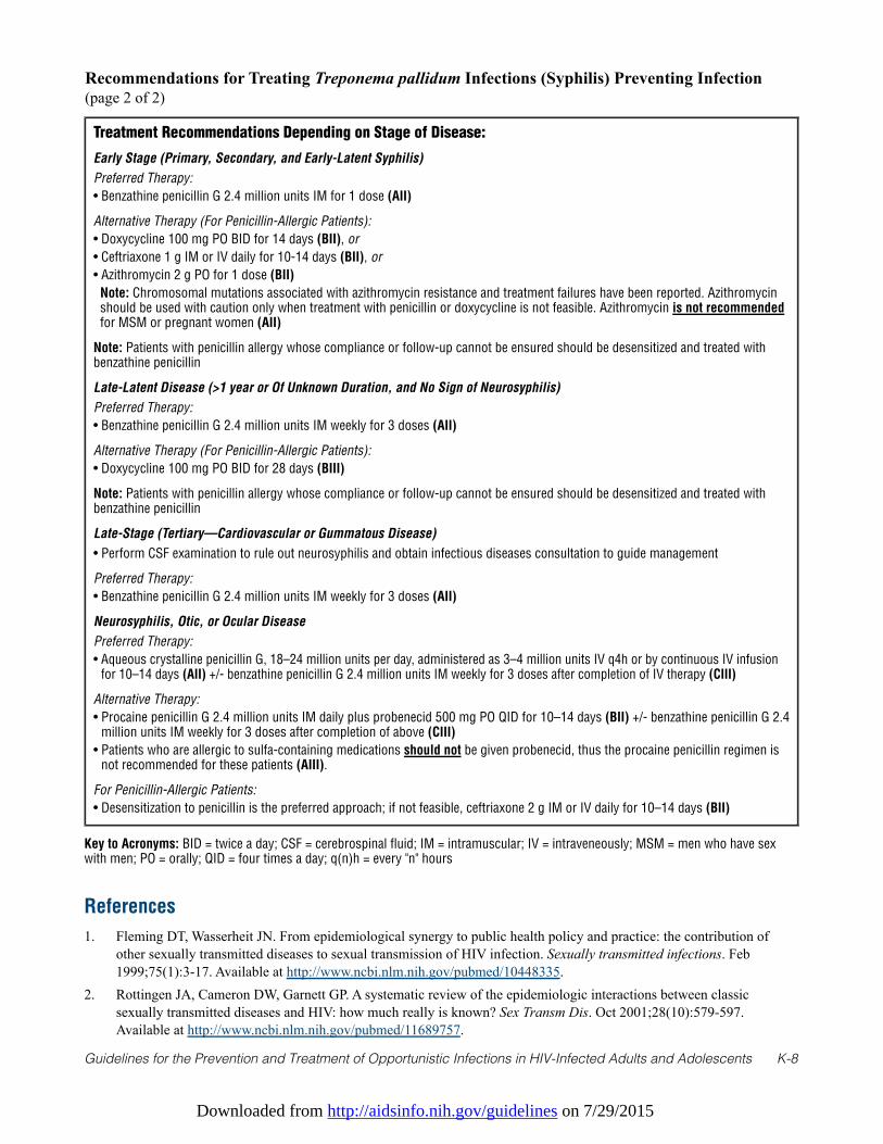
Downloaded from http://aidsinfo.nih.gov/guidelines on 7/29/2015
Guidelines for the Prevention and Treatment of Opportunistic Infections in HIV-Infected Adults and Adolescents K-8
Recommendations for Treating Treponema pallidum Infections (Syphilis) Preventing Infection(page 2 of 2)
Treatment Recommendations Depending on Stage of Disease:
Early Stage (Primary, Secondary, and Early-Latent Syphilis)
Preferred Therapy:
• Benzathine penicillin G 2.4 million units IM for 1 dose (AII)
Alternative Therapy (For Penicillin-Allergic Patients):
• Doxycycline 100 mg PO BID for 14 days (BII), or
• Ceftriaxone 1 g IM or IV daily for 10-14 days (BII), or
• Azithromycin 2 g PO for 1 dose (BII)
Note: Chromosomal mutations associated with azithromycin resistance and treatment failures have been reported. Azithromycinshould be used with caution only when treatment with penicillin or doxycycline is not feasible. Azithromycin is not recommendedfor MSM or pregnant women (AII)
Note: Patients with penicillin allergy whose compliance or follow-up cannot be ensured should be desensitized and treated withbenzathine penicillin
Late-Latent Disease (>1 year or Of Unknown Duration, and No Sign of Neurosyphilis)
Preferred Therapy:
• Benzathine penicillin G 2.4 million units IM weekly for 3 doses (AII)
Alternative Therapy (For Penicillin-Allergic Patients):
• Doxycycline 100 mg PO BID for 28 days (BIII)
Note: Patients with penicillin allergy whose compliance or follow-up cannot be ensured should be desensitized and treated withbenzathine penicillin
Late-Stage (Tertiary—Cardiovascular or Gummatous Disease)
• Perform CSF examination to rule out neurosyphilis and obtain infectious diseases consultation to guide management
Preferred Therapy:
• Benzathine penicillin G 2.4 million units IM weekly for 3 doses (AII)
Neurosyphilis, Otic, or Ocular Disease
Preferred Therapy:
• Aqueous crystalline penicillin G, 18–24 million units per day, administered as 3–4 million units IV q4h or by continuous IV infusionfor 10–14 days (AII) +/- benzathine penicillin G 2.4 million units IM weekly for 3 doses after completion of IV therapy (CIII)
Alternative Therapy:
• Procaine penicillin G 2.4 million units IM daily plus probenecid 500 mg PO QID for 10–14 days (BII) +/- benzathine penicillin G 2.4million units IM weekly for 3 doses after completion of above (CIII)
• Patients who are allergic to sulfa-containing medications should not be given probenecid, thus the procaine penicillin regimen isnot recommended for these patients (AIII).
For Penicillin-Allergic Patients:
• Desensitization to penicillin is the preferred approach; if not feasible, ceftriaxone 2 g IM or IV daily for 10–14 days (BII)
Key to Acronyms: BID = twice a day; CSF = cerebrospinal fluid; IM = intramuscular; IV = intraveneously; MSM = men who have sexwith men; PO = orally; QID = four times a day; q(n)h = every "n" hours
References
1. Fleming DT, Wasserheit JN. From epidemiological synergy to public health policy and practice: the contribution ofother sexually transmitted diseases to sexual transmission of HIV infection. Sexually transmitted infections. Feb1999;75(1):3-17. Available at http://www.ncbi.nlm.nih.gov/pubmed/10448335.
2. Rottingen JA, Cameron DW, Garnett GP. A systematic review of the epidemiologic interactions between classicsexually transmitted diseases and HIV: how much really is known? Sex Transm Dis. Oct 2001;28(10):579-597.Available at http://www.ncbi.nlm.nih.gov/pubmed/11689757.
Downloaded from http://aidsinfo.nih.gov/guidelines on 7/29/2015
Guidelines for the Prevention and Treatment of Opportunistic Infections in HIV-Infected Adults and Adolescents K-9
3. Blocker ME, Levine WC, St Louis ME. HIV prevalence in patients with syphilis, United States. Sex Transm Dis. Jan2000;27(1):53-59. Available at http://www.ncbi.nlm.nih.gov/pubmed/10654870.
4. Torian LV, Makki HA, Menzies IB, Murrill CS, Weisfuse IB. HIV infection in men who have sex with men, New YorkCity Department of Health sexually transmitted disease clinics, 1990-1999: a decade of serosurveillance finds thatracial disparities and associations between HIV and gonorrhea persist. Sex Transm Dis. Feb 2002;29(2):73-78. Availableat http://www.ncbi.nlm.nih.gov/pubmed/11818891.
5. Stolte IG, Dukers NH, de Wit JB, Fennema JS, Coutinho RA. Increase in sexually transmitted infections amonghomosexual men in Amsterdam in relation to HAART. Sexually transmitted infections. Jun 2001;77(3):184-186.Available at http://www.ncbi.nlm.nih.gov/pubmed/11402225.
6. Peterman TA, Heffelfinger JD, Swint EB, Groseclose SL. The changing epidemiology of syphilis. Sex Transm Dis. Oct2005;32(10 Suppl):S4-10. Available at http://www.ncbi.nlm.nih.gov/pubmed/16205291.
7. Paz-Bailey G, Meyers A, Blank S, et al. A case-control study of syphilis among men who have sex with men in NewYork City: association With HIV infection. Sex Transm Dis. Oct 2004;31(10):581-587. Available athttp://www.ncbi.nlm.nih.gov/pubmed/15388994.
8. Centers for Disease C, Prevention. Outbreak of syphilis among men who have sex with men--Southern California,2000. MMWR Morb Mortal Wkly Rep. Feb 23 2001;50(7):117-120. Available athttp://www.ncbi.nlm.nih.gov/pubmed/11393490.
9. Calza L, Manfredi R, Marinacci G, Tadolini M, Fortunato L, Chiodo F. Efficacy of penicillin G benzathine asantimicrobial treatment of cutaneous secondary syphilis in patients with HIV infection. Journal of chemotherapy. Oct2002;14(5):533-534. Available at http://www.ncbi.nlm.nih.gov/pubmed/12462435.
10. Rompalo AM, Lawlor J, Seaman P, Quinn TC, Zenilman JM, Hook EW, 3rd. Modification of syphilitic genital ulcermanifestations by coexistent HIV infection. Sex Transm Dis. Aug 2001;28(8):448-454. Available athttp://www.ncbi.nlm.nih.gov/pubmed/11473216.
11. Musher DM, Hamill RJ, Baughn RE. Effect of human immunodeficiency virus (HIV) infection on the course of syphilisand on the response to treatment. Ann Intern Med. Dec 1 1990;113(11):872-881. Available athttp://www.ncbi.nlm.nih.gov/pubmed/2240901.
12. Radolf JD, Kaplan RP. Unusual manifestations of secondary syphilis and abnormal humoral immune response toTreponema pallidum antigens in a homosexual man with asymptomatic human immunodeficiency virus infection.Journal of the American Academy of Dermatology. Feb 1988;18(2 Pt 2):423-428. Available athttp://www.ncbi.nlm.nih.gov/pubmed/2963840.
13. Ghanem KG, Workowski KA. Management of adult syphilis. Clin Infect Dis. Dec 2011;53 Suppl 3:S110-128. Availableat http://www.ncbi.nlm.nih.gov/pubmed/22080265.
14. Ghanem KG, Moore RD, Rompalo AM, Erbelding EJ, Zenilman JM, Gebo KA. Neurosyphilis in a clinical cohort ofHIV-1-infected patients. AIDS. Jun 19 2008;22(10):1145-1151. Available athttp://www.ncbi.nlm.nih.gov/pubmed/18525260.
15. Centers for Disease C, Prevention. Symptomatic early neurosyphilis among HIV-positive men who have sex with men--four cities, United States, January 2002-June 2004. MMWR Morb Mortal Wkly Rep. Jun 29 2007;56(25):625-628.Available at http://www.ncbi.nlm.nih.gov/pubmed/17597693.
16. Buchacz K, Patel P, Taylor M, et al. Syphilis increases HIV viral load and decreases CD4 cell counts in HIV-infectedpatients with new syphilis infections. AIDS. Oct 21 2004;18(15):2075-2079. Available athttp://www.ncbi.nlm.nih.gov/pubmed/15577629.
17. Modjarrad K, Vermund SH. Effect of treating co-infections on HIV-1 viral load: a systematic review. Lancet Infect Dis.Jul 2010;10(7):455-463. Available at http://www.ncbi.nlm.nih.gov/pubmed/20610327.
18. Palacios R, Jimenez-Onate F, Aguilar M, et al. Impact of syphilis infection on HIV viral load and CD4 cell counts inHIV-infected patients. J Acquir Immune Defic Syndr. Mar 1 2007;44(3):356-359. Available athttp://www.ncbi.nlm.nih.gov/pubmed/17159654.
19. Workowski KA, Berman S, Centers for Disease C, Prevention. Sexually transmitted diseases treatment guidelines,2010. MMWR Recomm Rep. Dec 17 2010;59(RR-12):1-110. Available athttp://www.ncbi.nlm.nih.gov/pubmed/21160459.
Downloaded from http://aidsinfo.nih.gov/guidelines on 7/29/2015
Guidelines for the Prevention and Treatment of Opportunistic Infections in HIV-Infected Adults and Adolescents K-10
20. Kofoed K, Gerstoft J, Mathiesen LR, Benfield T. Syphilis and human immunodeficiency virus (HIV)-1 coinfection:influence on CD4 T-cell count, HIV-1 viral load, and treatment response. Sex Transm Dis. Mar 2006;33(3):143-148.Available at http://www.ncbi.nlm.nih.gov/pubmed/16505739.
21. Rompalo AM, Joesoef MR, O'Donnell JA, et al. Clinical manifestations of early syphilis by HIV status and gender:results of the syphilis and HIV study. Sex Transm Dis. Mar 2001;28(3):158-165. Available athttp://www.ncbi.nlm.nih.gov/pubmed/11289198.
22. Dourmishev LA, Popov JM, Rusinova D. Paraneoplastic dermatomyositis associated with testicular cancer: a casereport and literature review. Acta Dermatovenerol Alp Panonica Adriat. 2010;19(1):39-43. Available athttp://www.ncbi.nlm.nih.gov/pubmed/20372774.
23. Bayne LL, Schmidley JW, Goodin DS. Acute syphilitic meningitis. Its occurrence after clinical and serologic cure ofsecondary syphilis with penicillin G. Arch Neurol. Feb 1986;43(2):137-138. Available athttp://www.ncbi.nlm.nih.gov/pubmed/3947251.
24. Berry CD, Hooton TM, Collier AC, Lukehart SA. Neurologic relapse after benzathine penicillin therapy for secondarysyphilis in a patient with HIV infection. N Engl J Med. Jun 18 1987;316(25):1587-1589. Available athttp://www.ncbi.nlm.nih.gov/pubmed/3587291.
25. Marra CM, Maxwell CL, Smith SL, et al. Cerebrospinal fluid abnormalities in patients with syphilis: association withclinical and laboratory features. J Infect Dis. Feb 1 2004;189(3):369-376. Available athttp://www.ncbi.nlm.nih.gov/pubmed/14745693.
26. Marra CM, Maxwell CL, Tantalo L, et al. Normalization of cerebrospinal fluid abnormalities after neurosyphilistherapy: does HIV status matter? Clin Infect Dis. Apr 1 2004;38(7):1001-1006. Available athttp://www.ncbi.nlm.nih.gov/pubmed/15034833.
27. Biotti D, Bidot S, Mahy S, et al. Ocular syphilis and HIV infection. Sex Transm Dis. Jan 2010;37(1):41-43. Available athttp://www.ncbi.nlm.nih.gov/pubmed/20118676.
28. Tucker JD, Li JZ, Robbins GK, et al. Ocular syphilis among HIV-infected patients: a systematic analysis of theliterature. Sexually transmitted infections. Feb 2011;87(1):4-8. Available athttp://www.ncbi.nlm.nih.gov/pubmed/20798396.
29. Wicher K, Horowitz HW, Wicher V. Laboratory methods of diagnosis of syphilis for the beginning of the thirdmillennium. Microbes and infection / Institut Pasteur. Oct 1999;1(12):1035-1049. Available athttp://www.ncbi.nlm.nih.gov/pubmed/10617935.
30. Centers for Disease C, Prevention. Discordant results from reverse sequence syphilis screening--five laboratories,United States, 2006-2010. MMWR Morb Mortal Wkly Rep. Feb 11 2011;60(5):133-137. Available athttp://www.ncbi.nlm.nih.gov/pubmed/21307823.
31. Centers for Disease C, Prevention. Syphilis testing algorithms using treponemal tests for initial screening--fourlaboratories, New York City, 2005-2006. MMWR Morb Mortal Wkly Rep. Aug 15 2008;57(32):872-875. Available athttp://www.ncbi.nlm.nih.gov/pubmed/18701877.
32. Wohrl S, Geusau A. Neurosyphilis is unlikely in patients with late latent syphilis and a negative blood VDRL-test. ActaDerm Venereol. 2006;86(4):335-339. Available at http://www.ncbi.nlm.nih.gov/pubmed/16874420.
33. Rompalo AM, Cannon RO, Quinn TC, Hook EW, 3rd. Association of biologic false-positive reactions for syphilis withhuman immunodeficiency virus infection. J Infect Dis. Jun 1992;165(6):1124-1126. Available athttp://www.ncbi.nlm.nih.gov/pubmed/1583332.
34. Rolfs RT, Joesoef MR, Hendershot EF, et al. A randomized trial of enhanced therapy for early syphilis in patients withand without human immunodeficiency virus infection. The Syphilis and HIV Study Group. N Engl J Med. Jul 311997;337(5):307-314. Available at http://www.ncbi.nlm.nih.gov/pubmed/9235493.
35. Augenbraun MH, DeHovitz JA, Feldman J, Clarke L, Landesman S, Minkoff HM. Biological false-positive syphilis testresults for women infected with human immunodeficiency virus. Clin Infect Dis. Dec 1994;19(6):1040-1044. Availableat http://www.ncbi.nlm.nih.gov/pubmed/7888531.
36. Hicks CB, Benson PM, Lupton GP, Tramont EC. Seronegative secondary syphilis in a patient infected with the humanimmunodeficiency virus (HIV) with Kaposi sarcoma. A diagnostic dilemma. Ann Intern Med. Oct 1987;107(4):492-495. Available at http://www.ncbi.nlm.nih.gov/pubmed/3307583.
37. Kingston AA, Vujevich J, Shapiro M, et al. Seronegative secondary syphilis in 2 patients coinfected with human
Downloaded from http://aidsinfo.nih.gov/guidelines on 7/29/2015
Guidelines for the Prevention and Treatment of Opportunistic Infections in HIV-Infected Adults and Adolescents K-11
immunodeficiency virus. Archives of dermatology. Apr 2005;141(4):431-433. Available athttp://www.ncbi.nlm.nih.gov/pubmed/15837859.
38. Augenbraun M, Rolfs R, Johnson R, Joesoef R, Pope V. Treponemal specific tests for the serodiagnosis of syphilis.Syphilis and HIV Study Group. Sex Transm Dis. Nov 1998;25(10):549-552. Available athttp://www.ncbi.nlm.nih.gov/pubmed/9858352.
39. Libois A, De Wit S, Poll B, et al. HIV and syphilis: when to perform a lumbar puncture. Sex Transm Dis. Mar2007;34(3):141-144. Available at http://www.ncbi.nlm.nih.gov/pubmed/16865051.
40. Ghanem KG. Sensitivity and specificity of lumbar puncture in HIV-infected patients with syphilis and no neurologicsymptoms reply. Clin Infect Dis. 2009;49:162-163.
41. Marra CM. Deja vu all over again: when to perform a lumbar puncture in HIV-infected patients with syphilis. SexTransm Dis. Mar 2007;34(3):145-146. Available at http://www.ncbi.nlm.nih.gov/pubmed/17325601.
42. Jaffe HW, Larsen SA, Peters M, Jove DF, Lopez B, Schroeter AL. Tests for treponemal antibody in CSF. Arch InternMed. Feb 1978;138(2):252-255. Available at http://www.ncbi.nlm.nih.gov/pubmed/343742.
43. Rietmeijer CA. Risk reduction counselling for prevention of sexually transmitted infections: how it works and how tomake it work. Sexually transmitted infections. Feb 2007;83(1):2-9. Available athttp://www.ncbi.nlm.nih.gov/pubmed/17283359.
44. Force USPST. Behavioral counseling to prevent sexually transmitted infections: U.S. Preventive Services Task Forcerecommendation statement. Ann Intern Med. Oct 7 2008;149(7):491-496, W495. Available athttp://www.ncbi.nlm.nih.gov/pubmed/18838729.
45. Kamb ML, Fishbein M, Douglas JM, Jr., et al. Efficacy of risk-reduction counseling to prevent humanimmunodeficiency virus and sexually transmitted diseases: a randomized controlled trial. Project RESPECT StudyGroup. JAMA. Oct 7 1998;280(13):1161-1167. Available at http://www.ncbi.nlm.nih.gov/pubmed/9777816.
46. Richardson JL, Milam J, Stoyanoff S, et al. Using patient risk indicators to plan prevention strategies in the clinical caresetting. J Acquir Immune Defic Syndr. Oct 1 2004;37 Suppl 2:S88-94. Available athttp://www.ncbi.nlm.nih.gov/pubmed/15385904.
47. Fisher JD, Cornman DH, Osborn CY, Amico KR, Fisher WA, Friedland GA. Clinician-initiated HIV risk reductionintervention for HIV-positive persons: Formative Research, Acceptability, and Fidelity of the Options Project. J AcquirImmune Defic Syndr. Oct 1 2004;37 Suppl 2:S78-87. Available at http://www.ncbi.nlm.nih.gov/pubmed/15385903.
48. Branger J, van der Meer JT, van Ketel RJ, Jurriaans S, Prins JM. High incidence of asymptomatic syphilis in HIV-infected MSM justifies routine screening. Sex Transm Dis. Feb 2009;36(2):84-85. Available athttp://www.ncbi.nlm.nih.gov/pubmed/18971797.
49. Aberg JA, Kaplan JE, Libman H, et al. Primary care guidelines for the management of persons infected with humanimmunodeficiency virus: 2009 update by the HIV medicine Association of the Infectious Diseases Society of America.Clin Infect Dis. Sep 1 2009;49(5):651-681. Available at http://www.ncbi.nlm.nih.gov/pubmed/19640227.
50. CDC, HRSA NIH, HIVMA/IDSA, and the HIV Prevention in Clinical Care Working Group. Recommendations forincorporating human immunodeficiency virus (HIV) prevention into the medical care of persons living with HIV. ClinInfect Dis. Jan 1 2004;38(1):104-121. Available at http://www.ncbi.nlm.nih.gov/pubmed/14679456.
51. Centers for Disease Control and Prevention. Recommendations for partner services programs for HIV infection,syphilis, gonorrhea, and chlamydial infection. MMWR Recomm Rep. Nov 7 2008;57(RR-9):1-83; quiz CE81-84.Available at http://www.ncbi.nlm.nih.gov/pubmed/18987617.
52. Moore MB, Jr., Price EV, Knox JM, Elgin LW. Epidemiologic Treatment of Contacts to Infectious Syphilis. PublicHealth Rep. Nov 1963;78:966-970. Available at http://www.ncbi.nlm.nih.gov/pubmed/14084872.
53. Schroeter AL, Turner RH, Lucas JB, Brown WJ. Therapy for incubating syphilis. Effectiveness of gonorrhea treatment.JAMA. Nov 1 1971;218(5):711-713. Available at http://www.ncbi.nlm.nih.gov/pubmed/5171497.
54. Schober PC, Gabriel G, White P, Felton WF, Thin RN. How infectious is syphilis? Br J Vener Dis. Aug 1983;59(4):217-219. Available at http://www.ncbi.nlm.nih.gov/pubmed/6871650.
55. Hook EW, 3rd, Marra CM. Acquired syphilis in adults. N Engl J Med. Apr 16 1992;326(16):1060-1069. Available athttp://www.ncbi.nlm.nih.gov/pubmed/1549153.
56. Malone JL, Wallace MR, Hendrick BB, et al. Syphilis and neurosyphilis in a human immunodeficiency virus type-1
Downloaded from http://aidsinfo.nih.gov/guidelines on 7/29/2015
Guidelines for the Prevention and Treatment of Opportunistic Infections in HIV-Infected Adults and Adolescents K-12
seropositive population: evidence for frequent serologic relapse after therapy. Am J Med. Jul 1995;99(1):55-63.Available at http://www.ncbi.nlm.nih.gov/pubmed/7598143.
57. Walter T, Lebouche B, Miailhes P, et al. Symptomatic relapse of neurologic syphilis after benzathine penicillin Gtherapy for primary or secondary syphilis in HIV-infected patients. Clin Infect Dis. Sep 15 2006;43(6):787-790.Available at http://www.ncbi.nlm.nih.gov/pubmed/16912958.
58. Ghanem KG, Erbelding EJ, Cheng WW, Rompalo AM. Doxycycline compared with benzathine penicillin for thetreatment of early syphilis. Clin Infect Dis. Mar 15 2006;42(6):e45-49. Available athttp://www.ncbi.nlm.nih.gov/pubmed/16477545.
59. Wong T, Singh AE, De P. Primary syphilis: serological treatment response to doxycycline/tetracycline versus benzathinepenicillin. Am J Med. Oct 2008;121(10):903-908. Available at http://www.ncbi.nlm.nih.gov/pubmed/18823862.
60. Hook EW, 3rd, Roddy RE, Handsfield HH. Ceftriaxone therapy for incubating and early syphilis. J Infect Dis. Oct1988;158(4):881-884. Available at http://www.ncbi.nlm.nih.gov/pubmed/3171231.
61. Kiddugavu MG, Kiwanuka N, Wawer MJ, et al. Effectiveness of syphilis treatment using azithromycin and/orbenzathine penicillin in Rakai, Uganda. Sex Transm Dis. Jan 2005;32(1):1-6. Available athttp://www.ncbi.nlm.nih.gov/pubmed/15614114.
62. Riedner G, Rusizoka M, Todd J, et al. Single-dose azithromycin versus penicillin G benzathine for the treatment ofearly syphilis. N Engl J Med. Sep 22 2005;353(12):1236-1244. Available athttp://www.ncbi.nlm.nih.gov/pubmed/16177249.
63. Hook EW, 3rd, Behets F, Van Damme K, et al. A phase III equivalence trial of azithromycin versus benzathine penicillinfor treatment of early syphilis. J Infect Dis. Jun 1 2010;201(11):1729-1735. Available athttp://www.ncbi.nlm.nih.gov/pubmed/20402591.
64. Centers for Disease Control and Prevention. Azithromycin treatment failures in syphilis infections--San Francisco,California, 2002-2003. MMWR Morb Mortal Wkly Rep. Mar 12 2004;53(9):197-198. Available athttp://www.ncbi.nlm.nih.gov/pubmed/15017376.
65. Lukehart SA, Godornes C, Molini BJ, et al. Macrolide resistance in Treponema pallidum in the United States andIreland. N Engl J Med. Jul 8 2004;351(2):154-158. Available at http://www.ncbi.nlm.nih.gov/pubmed/15247355.
66. Mitchell SJ, Engelman J, Kent CK, Lukehart SA, Godornes C, Klausner JD. Azithromycin-resistant syphilis infection:San Francisco, California, 2000-2004. Clin Infect Dis. Feb 1 2006;42(3):337-345. Available athttp://www.ncbi.nlm.nih.gov/pubmed/16392078.
67. Martin IE, Tsang RS, Sutherland K, et al. Molecular characterization of syphilis in patients in Canada: azithromycinresistance and detection of Treponema pallidum DNA in whole-blood samples versus ulcerative swabs. J ClinMicrobiol. Jun 2009;47(6):1668-1673. Available at http://www.ncbi.nlm.nih.gov/pubmed/19339468.
68. Wu H, Chang SY, Lee NY, et al. Evaluation of macrolide resistance and enhanced molecular typing of Treponemapallidum in patients with syphilis in Taiwan: a prospective multicenter study. J Clin Microbiol. Jul 2012;50(7):2299-2304. Available at http://www.ncbi.nlm.nih.gov/pubmed/22518868.
69. Chen CY, Chi KH, Pillay A, Nachamkin E, Su JR, Ballard RC. Detection of the A2058G and A2059G 23S rRNA GenePoint Mutations Associated with Azithromycin Resistance in Treponema pallidum by Use of a TaqMan Real-TimeMultiplex PCR Assay. J Clin Microbiol. Mar 2013;51(3):908-913. Available athttp://www.ncbi.nlm.nih.gov/pubmed/23284026.
70. Dowell ME, Ross PG, Musher DM, Cate TR, Baughn RE. Response of latent syphilis or neurosyphilis to ceftriaxonetherapy in persons infected with human immunodeficiency virus. Am J Med. Nov 1992;93(5):481-488. Available athttp://www.ncbi.nlm.nih.gov/pubmed/1442850.
71. Smith NH, Musher DM, Huang DB, et al. Response of HIV-infected patients with asymptomatic syphilis to intensiveintramuscular therapy with ceftriaxone or procaine penicillin. Int J STD AIDS. May 2004;15(5):328-332. Available athttp://www.ncbi.nlm.nih.gov/pubmed/15117503.
72. Bernal E, Munoz A, Ortiz Mdel M, Cano A. [Syphilitic panuveitis in an HIV-infected patient after immune restoration].Enferm Infecc Microbiol Clin. Oct 2009;27(8):487-489. Available at http://www.ncbi.nlm.nih.gov/pubmed/19406524.
73. Long CM, Klausner JD, Leon S, et al. Syphilis treatment and HIV infection in a population-based study of persons athigh risk for sexually transmitted disease/HIV infection in Lima, Peru. Sex Transm Dis. Mar 2006;33(3):151-155.
Downloaded from http://aidsinfo.nih.gov/guidelines on 7/29/2015
Guidelines for the Prevention and Treatment of Opportunistic Infections in HIV-Infected Adults and Adolescents K-13
Available at http://www.ncbi.nlm.nih.gov/pubmed/16508525.74. Marra CM, Maxwell CL, Tantalo LC, Sahi SK, Lukehart SA. Normalization of serum rapid plasma reagin titer predicts
normalization of cerebrospinal fluid and clinical abnormalities after treatment of neurosyphilis. Clin Infect Dis. Oct 12008;47(7):893-899. Available at http://www.ncbi.nlm.nih.gov/pubmed/18715154.
75. Yang CJ, Lee NY, Lin YH, et al. Jarisch-Herxheimer reaction after penicillin therapy among patients with syphilis in theera of the hiv infection epidemic: incidence and risk factors. Clin Infect Dis. Oct 15 2010;51(8):976-979. Available athttp://www.ncbi.nlm.nih.gov/pubmed/20825309.
76. Rekart ML, Patrick DM, Chakraborty B, et al. Targeted mass treatment for syphilis with oral azithromycin. Lancet. Jan25 2003;361(9354):313-314. Available at http://www.ncbi.nlm.nih.gov/pubmed/12559870.
77. Genc M, Ledger WJ. Syphilis in pregnancy. Sexually transmitted infections. Apr 2000;76(2):73-79. Available athttp://www.ncbi.nlm.nih.gov/pubmed/10858706.
78. Berman SM. Maternal syphilis: pathophysiology and treatment. Bulletin of the World Health Organization. Jun2004;82(6):433-438. Available at http://www.ncbi.nlm.nih.gov/pubmed/15356936.
79. Tess BH, Rodrigues LC, Newell ML, Dunn DT, Lago TD. Breastfeeding, genetic, obstetric and other risk factorsassociated with mother-to-child transmission of HIV-1 in Sao Paulo State, Brazil. Sao Paulo Collaborative Study forVertical Transmission of HIV-1. AIDS. Mar 26 1998;12(5):513-520. Available athttp://www.ncbi.nlm.nih.gov/pubmed/9543450.
80. Lee MJ, Hallmark RJ, Frenkel LM, Del Priore G. Maternal syphilis and vertical perinatal transmission of humanimmunodeficiency virus type-1 infection. Int J Gynaecol Obstet. Dec 1998;63(3):247-252. Available athttp://www.ncbi.nlm.nih.gov/pubmed/9989893.
81. Wendel GD, Jr., Sheffield JS, Hollier LM, Hill JB, Ramsey PS, Sanchez PJ. Treatment of syphilis in pregnancy andprevention of congenital syphilis. Clin Infect Dis. Oct 15 2002;35(Suppl 2):S200-209. Available athttp://www.ncbi.nlm.nih.gov/pubmed/12353207.
82. Kreitchmann R, Fuchs SC, Suffert T, Preussler G. Perinatal HIV-1 transmission among low income women participantsin the HIV/AIDS Control Program in Southern Brazil: a cohort study. BJOG. Jun 2004;111(6):579-584. Available athttp://www.ncbi.nlm.nih.gov/pubmed/15198786.
83. Mwapasa V, Rogerson SJ, Kwiek JJ, et al. Maternal syphilis infection is associated with increased risk of mother-to-child transmission of HIV in Malawi. AIDS. Sep 11 2006;20(14):1869-1877. Available athttp://www.ncbi.nlm.nih.gov/pubmed/16954728.
84. Walker GJ. Antibiotics for syphilis diagnosed during pregnancy. Cochrane Database Syst Rev. 2001(3):CD001143.Available at http://www.ncbi.nlm.nih.gov/pubmed/11686978.
85. Donders GG, Desmyter J, Hooft P, Dewet GH. Apparent failure of one injection of benzathine penicillin G for syphilisduring pregnancy in human immunodeficiency virus-seronegative African women. Sex Transm Dis. Feb 1997;24(2):94-101. Available at http://www.ncbi.nlm.nih.gov/pubmed/9111755.
86. Sheffield JS, Sanchez PJ, Morris G, et al. Congenital syphilis after maternal treatment for syphilis during pregnancy.Am J Obstet Gynecol. Mar 2002;186(3):569-573. Available at http://www.ncbi.nlm.nih.gov/pubmed/11904625.
87. Ramsey PS, Vaules MB, Vasdev GM, Andrews WW, Ramin KD. Maternal and transplacental pharmacokinetics ofazithromycin. Am J Obstet Gynecol. Mar 2003;188(3):714-718. Available athttp://www.ncbi.nlm.nih.gov/pubmed/12634646.
88. Zhou P, Gu Z, Xu J, Wang X, Liao K. A study evaluating ceftriaxone as a treatment agent for primary and secondarysyphilis in pregnancy. Sex Transm Dis. 2005; 32(8):495-498. Available athttp://www.ncbi.nlm.nih.gov/entrez/query.fcgi?cmd=Retrieve&db=PubMed&dopt=Citation&list_uids=16041252.
89. Klein VR, Cox SM, Mitchell MD, Wendel GD, Jr. The Jarisch-Herxheimer reaction complicating syphilotherapy inpregnancy. Obstet Gynecol. Mar 1990;75(3 Pt 1):375-380. Available at http://www.ncbi.nlm.nih.gov/pubmed/2304710.
90. Hollier LM, Harstad TW, Sanchez PJ, Twickler DM, Wendel GD, Jr. Fetal syphilis: clinical and laboratorycharacteristics. Obstet Gynecol. Jun 2001;97(6):947-953. Available at http://www.ncbi.nlm.nih.gov/pubmed/11384701.
Downloaded from http://aidsinfo.nih.gov/guidelines on 7/29/2015
Guidelines for the Prevention and Treatment of Opportunistic Infections in HIV-Infected Adults and Adolescents L-1
Mucocutaneous Candidiasis (Last updated October 28, 2014; last reviewed October 28, 2014)
Epidemiology
Oropharyngeal and esophageal candidiasis are common in HIV-infected patients.1,2 Most such infections arecaused by Candida albicans. The occurrence of oropharyngeal or esophageal candidiasis is recognized as anindicator of immune suppression and is most often observed in patients with CD4 T lymphocyte (CD4) cellcounts <200 cells/mm3, with esophageal disease typically occurring at lower CD4 counts than oropharyngealdisease.1,2 In contrast, vulvovaginal candidiasis—whether a single episode or recurrent—is common inhealthy, adult women and does not suggest HIV infection. The advent of antiretroviral therapy (ART) has ledto a dramatic decline in the prevalence of oropharyngeal and esophageal candidiasis and a markeddiminution in cases of refractory disease.
Fluconazole (or azole) resistance is predominantly the consequence of previous exposure to fluconazole (orother azoles), particularly repeated and long-term exposure.3-5 In this setting, C. albicans resistance has beenassociated with a gradual emergence of non-albicans Candida species, particularly Candida glabrata, as acause of refractory mucosal candidiasis in patients with advanced immunosuppression and low CD4 counts.3,6
Clinical Manifestations
Oropharyngeal candidiasis is characterized by painless, creamy white, plaque-like lesions that can occur onthe buccal surface, hard or soft palate, oropharyngeal mucosa, or tongue surface. Lesions can be easilyscraped off with a tongue depressor or other instrument. Less commonly, erythematous patches withoutwhite plaques can be seen on the anterior or posterior upper palate or diffusely on the tongue. Angularcheilosis also can be caused by Candida.
Patients with esophageal candidiasis generally present with retrosternal burning pain or discomfort alongwith odynophagia; occasionally esophageal candidiasis can be asymptomatic. Endoscopic examinationreveals whitish plaques similar to those observed with oropharyngeal disease. On occasion, the plaques mayprogress to superficial ulcerations of the esophageal mucosa with central or peripheral whitish exudates.
In HIV-infected women with early-stage disease, Candida vulvovaginitis usually presents as it does in HIV-uninfected women, with white adherent vaginal discharge associated with mucosal burning and itching ofmild-to-moderate severity and sporadic recurrences. In women with advanced immunosuppression, episodesmay be more severe and recur more frequently. In contrast to oropharyngeal candidiasis, vulvovaginalcandidiasis is less common and rarely refractory to azole therapy.
Diagnosis
Oropharyngeal candidiasis is usually diagnosed clinically based on the characteristic appearance of lesions.In contrast to oral hairy leukoplakia, the white plaques of oropharyngeal candidiasis can be scraped off themucosa. If laboratory confirmation is required, scrapings can be examined microscopically for characteristicyeast or hyphal forms, using a potassium hydroxide preparation. Cultures of clinical exudative material yieldthe species of Candida present.
The definitive diagnosis of esophageal candidiasis requires direct endoscopic visualization of lesions withhistopathologic demonstration of characteristic Candida yeast forms in tissue and confirmation by fungalculture and speciation. The diagnosis is often made empirically based on symptoms plus response to therapy,or visualization of lesions plus fungal smear or brushings without histopathologic examination.
Vulvovaginal candidiasis usually is diagnosed based on the clinical presentation coupled with thedemonstration of characteristic blastosphere and hyphal yeast forms in vaginal secretions when examinedmicroscopically after potassium hydroxide preparation. Culture confirmation is rarely required but may
Downloaded from http://aidsinfo.nih.gov/guidelines on 7/29/2015
Guidelines for the Prevention and Treatment of Opportunistic Infections in HIV-Infected Adults and Adolescents L-2
provide supportive information. Self-diagnosis of vulvovaginitis is unreliable; microscopic and cultureconfirmation is required to avoid unnecessary exposure to treatment.
Preventing Exposure
Candida organisms are common commensals on mucosal surfaces in healthy individuals. No measures areavailable to reduce exposure to these fungi.
Preventing Disease
Data from prospective controlled trials indicate that fluconazole can reduce the risk of mucosal disease (i.e.,oropharyngeal, esophageal, and vulvovaginal) in patients with advanced HIV.7-10 However, routine primaryprophylaxis is not recommended because mucosal disease is associated with very low attributable morbidity andmortality and, moreover, acute therapy is highly effective. Primary antifungal prophylaxis can lead to infectionscaused by drug-resistant Candida species and introduce significant drug-drug interactions. In addition long-termoral prophylaxis is expensive. Therefore, routine primary prophylaxis is not recommended (AIII).
Treating Disease
Oral fluconazole is as effective as and, in certain studies, superior to topical therapy for oropharyngealcandidiasis. In addition, oral therapy is more convenient than topical therapy and usually better tolerated.Therefore, oral fluconazole is considered the drug of choice to treat oropharyngeal candidiasis (AI).11 Usingtopical rather than systemic oral therapy reduces systemic drug exposure, diminishes risk of drug-druginteractions and systemic adverse events, and possibly decreases the development of secondary antifungalresistance. Mild-to-moderate episodes of oropharyngeal candidiasis can be adequately treated with topicaltherapy, including once-daily miconazole in 50-mg mucoadhesive buccal tablets (BI) or clotrimazole troches 5times daily (BI). In a multicenter, randomized study among HIV-infected individuals, 50-mg mucoadhesivebuccal tablets of miconazole applied once daily to the mucosal surface over the canine fossa were as effectiveas 10-mg clotrimazole troches used 5 times daily.12 Nystatin suspension or pastilles four times daily remain anadditional alternative (BII).13
Itraconazole oral solution for 7 to 14 days is as effective as oral fluconazole for oropharyngeal candidiasis butless well tolerated (BI).13 Posaconazole oral suspension14 also is as effective as fluconazole and generallybetter tolerated than itraconazole solution (BI). Both antifungals are alternatives to oral fluconazole, althoughfew situations require that these drugs be used in preference to fluconazole solely to treat mucosal candidiasis.In a multicenter, randomized study, posaconazole was proven more effective than fluconazole in sustainingclinical success after antifungal therapy was discontinued.14 A new solid oral delayed-release tabletformulation of posaconazole has been developed.15 Whether it offers any advantage for the treatment oforopharyngeal candidiasis is unknown and it currently is indicated only for prophylaxis of invasiveAspergillus and Candida infections in certain highly susceptible, non-HIV patients populations with persistentneutropenia or allogeneic hematopoeitic stem cell transplantation.16 Itraconazole capsules are less effectivethan fluconazole because of their more variable absorption and they are associated with more drug-druginteractions than fluconazole.
Systemic antifungals are required for effective treatment of esophageal candidiasis (AI). A 14- to 21-daycourse of either fluconazole (oral or intravenous [IV]) or oral itraconazole solution is highly effective (AI).However, patients with severe symptoms initially may have difficulty swallowing oral drugs. As withoropharyngeal candidiasis, itraconazole capsules for esophageal candidiasis are less effective than fluconazolebecause of variable absorption (CII). Voriconazole, amphotericin B (either deoxycholate or lipidformulations) and the echinocandins caspofungin, micafungin, and anidulafungin all are effective in treatingesophageal candidiasis (BI). However, esophageal candidiasis appears to have a higher relapse rate aftertreatment with the echinocandins.17,18 Therefore, oral or IV fluconazole remains the preferred therapy foresophageal candidiasis (AI). Although other pathogens (e.g., cytomegalovirus, herpes simplex virus
Downloaded from http://aidsinfo.nih.gov/guidelines on 7/29/2015
Guidelines for the Prevention and Treatment of Opportunistic Infections in HIV-Infected Adults and Adolescents L-3
esophagitis) can mimic the symptoms of esophageal candidiasis, a diagnostic and therapeutic trial ofantifungal therapy is usually warranted before endoscopy. In those who do not respond to antifungal therapy,endoscopy is recommended to identify different causes of esophagitis or drug-resistant Candida (AII).
In most HIV-infected women, vulvovaginal candidiasis is uncomplicated and responds readily to short-courseoral or topical treatment with any of several therapies, including: • Oral fluconazole (AII) • Topical azoles (clotrimazole, butoconazole, miconazole, tioconazole, or terconazole) (AII) • Itraconazole oral solution (BII)
Severe or recurrent episodes of vaginitis should be treated with oral fluconazole or topical antifungal therapyfor ≥7 days (AII).
Special Considerations with Regard to Starting ARTThere are no special considerations regarding initiation of ART in patients with mucocutaneous candidiasis.Specifically, there is as yet no evidence that treatment with ART needs to be delayed until treatment forcandidiasis has been completed.
Monitoring of Response to Therapy and Adverse Events (Including IRIS) For most patients with mucocutaneous candidiasis, response to antifungal therapy is rapid; signs andsymptoms improve within 48 to 72 hours. Short courses of topical therapy rarely result in adverse effects,although patients may experience cutaneous hypersensitivity reactions characterized by rash and pruritus. Oralazole therapy can be associated with nausea, vomiting, diarrhea, abdominal pain, or transaminase elevations.Periodic monitoring of liver function studies should be considered if azole therapy is anticipated for >21 days,especially in patients with other hepatic comorbidities (AII). The echinocandins appear to be associated withvery few adverse reactions: histamine-related infusion toxicity, transaminase elevations, and rash have beenattributed to these drugs. No dose adjustments are required in renal failure.
Immune reconstitution inflammatory syndrome with ART has not yet been reported for mucocutaneouscandidiasis in HIV-infected patients. Indeed, ART is associated with a markedly reduced incidence of candidiasis.
Managing Treatment FailureAntifungal treatment failure is typically defined as the persistence of signs or symptoms of oropharyngeal oresophageal candidiasis after 7 to 14 days of appropriate antifungal therapy. Refractory disease occurs inapproximately 4% to 5% of HIV-infected patients with oral or esophageal candidiasis, typically those withCD4 cell counts <50 cells/mm3 and who have received multiple courses of azole antifungals.4 Confirmatoryculture and, in the case of esophageal candidiasis, endoscopy are necessary to confirm treatment failure due toazole resistance or other causes of esophagitis, especially if these procedures were not initially performed.
Posaconazole immediate-release oral suspension (400 mg twice daily for 28 days) is effective in 75% ofpatients with azole-refractory oropharyngeal or esophageal candidiasis (AI).19 Again, although the new soliddelayed-release tablet formulation has been recently made available, it is not known if it offers an advantageover the suspension for treating this particular disease. Alternatively, oral itraconazole solution is effective, atleast transiently, in approximately two-thirds of patients with fluconazole-refractory mucosal candidiasis(BII).13 If necessary, azole-refractory esophageal candidiasis also can be treated with anidulafungin (BII),caspofungin (BII), micafungin (BII), or voriconazole (BII).
IV amphotericin B is usually effective for treating refractory disease (BII). Both amphotericin B deoxycholateand the lipid preparations of amphotericin B have been used successfully (BII). Amphotericin B oralsuspension (1 mL of the 100-mg/mL suspension 4 times daily) is sometimes effective in patients whoseoropharyngeal candidiasis does not respond to itraconazole (BII), but this product is not commerciallyavailable in the United States.
Downloaded from http://aidsinfo.nih.gov/guidelines on 7/29/2015
Guidelines for the Prevention and Treatment of Opportunistic Infections in HIV-Infected Adults and Adolescents L-4
Preventing Recurrence
When to Start Chronic Suppressive TherapyA randomized clinical trial10 in HIV-infected patients with CD4 counts <150 cells/mm3 documented asignificantly lower number of episodes of oropharyngeal candidiasis and other invasive fungal infectionswith continuous fluconazole therapy (3 times a week) compared with episodic fluconazole treatment forrecurrences. This clinical trial also demonstrated no difference in the risk of developing clinically significantfluconazole resistance between the two groups among those receiving ART.
However, chronic suppressive therapy is not recommended by most HIV specialists for recurrentoropharyngeal or vulvovaginal candidiasis unless patients have frequent or severe recurrences (BIII) becausetherapy for acute disease is effective, mortality associated with mucocutaneous disease is low, potentialexists for drug interactions and for the development of antifungal-resistant Candida, and chronic suppressivetherapy is costly.
If recurrences are frequent or severe, oral fluconazole can be used as suppressive therapy for eitheroropharyngeal (BI), esophageal (BI), or vulvovaginal (BII) candidiasis.7-9 Oral posaconazole twice daily isalso effective for esophageal candidiasis (BII).20 The potential for development of secondary azole resistanceshould be considered when contemplating chronic maintenance therapy using azoles in HIV-infected patientswho are severely immunocompromised. Several important factors should be taken into account when makingthe decision to use chronic suppressive therapy. These include the effect of recurrences on the patient’s well-being and quality of life, the need for prophylaxis against other fungal infections, cost, adverse events and,most importantly, drug-drug interactions.21
Rates of relapse are high in patients with azole-refractory oropharyngeal or esophageal candidiasis who haveinitially responded to echinocandins, voriconazole, or posaconazole therapy. In such patients, chronicsuppressive therapy should be instituted until ART produces immune reconstitution (AIII).
When to Stop Chronic Suppressive TherapyIn situations where chronic suppressive therapy has been instituted, no data exist to guide recommendationsregarding its discontinuation. On the basis of experience with other opportunistic infections, it would bereasonable to discontinue chronic suppressive therapy when the CD4 count has risen to >200 cells/mm3
following initiation of ART (AIII).
Special Considerations During Pregnancy
Pregnancy increases the risk of vaginal colonization with Candida species. Diagnosis of oropharyngeal,esophageal, and vulvovaginal candidiasis is the same in pregnant women as in those who are not pregnant.
Topical therapy is preferable for treatment of oral or vaginal candidiasis in pregnancy, when possible (AIII).Although single-dose, episodic treatment with oral fluconazole has not been associated with birth defects inhumans, its use has not been widely endorsed.22 Five cases of a syndrome consisting of craniosynostosis,characteristic facies, digital synostosis, and limb contractures (fluconazole embryopathy) have been reportedin women chronically prescribed fluconazole at doses of 400 mg daily or higher in pregnancy.23 On the basisof these data, substitution of amphotericin B for high-dose fluconazole in the first trimester is recommendedfor invasive or refractory esophageal candidal infections (AIII). Neonates born to women receiving chronicamphotericin B at delivery should be evaluated for renal dysfunction and hypokalemia. Itraconazole has beenshown to be teratogenic in animals at high doses, but the metabolic mechanism accounting for these defectsis not present in humans, so these data are not applicable. Case series in humans do not suggest an increasedrisk of birth defects with itraconazole,24 but experience is limited. Human data are not available forposaconazole; however, the drug was associated with skeletal abnormalities in rats and was embryotoxic inrabbits when given at doses that produced plasma levels equivalent to those seen in humans. Voriconazole isconsidered an FDA Category D drug because of its association with cleft palate and renal defects seen in rats
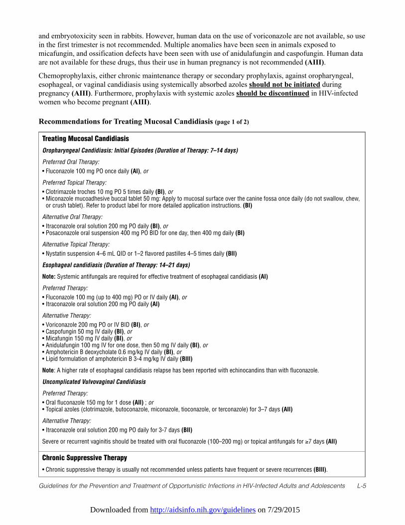
Downloaded from http://aidsinfo.nih.gov/guidelines on 7/29/2015
Guidelines for the Prevention and Treatment of Opportunistic Infections in HIV-Infected Adults and Adolescents L-5
and embryotoxicity seen in rabbits. However, human data on the use of voriconazole are not available, so usein the first trimester is not recommended. Multiple anomalies have been seen in animals exposed tomicafungin, and ossification defects have been seen with use of anidulafungin and caspofungin. Human dataare not available for these drugs, thus their use in human pregnancy is not recommended (AIII).
Chemoprophylaxis, either chronic maintenance therapy or secondary prophylaxis, against oropharyngeal,esophageal, or vaginal candidiasis using systemically absorbed azoles should not be initiated duringpregnancy (AIII). Furthermore, prophylaxis with systemic azoles should be discontinued in HIV-infectedwomen who become pregnant (AIII).
Recommendations for Treating Mucosal Candidiasis (page 1 of 2)
Treating Mucosal Candidiasis
Oropharyngeal Candidiasis: Initial Episodes (Duration of Therapy: 7–14 days)
Preferred Oral Therapy:
• Fluconazole 100 mg PO once daily (AI), or
Preferred Topical Therapy:
• Clotrimazole troches 10 mg PO 5 times daily (BI), or• Miconazole mucoadhesive buccal tablet 50 mg: Apply to mucosal surface over the canine fossa once daily (do not swallow, chew,
or crush tablet). Refer to product label for more detailed application instructions. (BI)
Alternative Oral Therapy:
• Itraconazole oral solution 200 mg PO daily (BI), or• Posaconazole oral suspension 400 mg PO BID for one day, then 400 mg daily (BI)
Alternative Topical Therapy:
• Nystatin suspension 4–6 mL QID or 1–2 flavored pastilles 4–5 times daily (BII)
Esophageal candidiasis (Duration of Therapy: 14–21 days)
Note: Systemic antifungals are required for effective treatment of esophageal candidiasis (AI)
Preferred Therapy:
• Fluconazole 100 mg (up to 400 mg) PO or IV daily (AI), or• Itraconazole oral solution 200 mg PO daily (AI)
Alternative Therapy:
• Voriconazole 200 mg PO or IV BID (BI), or• Caspofungin 50 mg IV daily (BI), or• Micafungin 150 mg IV daily (BI), or• Anidulafungin 100 mg IV for one dose, then 50 mg IV daily (BI), or• Amphotericin B deoxycholate 0.6 mg/kg IV daily (BI), or• Lipid formulation of amphotericin B 3-4 mg/kg IV daily (BIII)
Note: A higher rate of esophageal candidiasis relapse has been reported with echinocandins than with fluconazole.
Uncomplicated Vulvovaginal Candidiasis
Preferred Therapy:
• Oral fluconazole 150 mg for 1 dose (AII) ; or• Topical azoles (clotrimazole, butoconazole, miconazole, tioconazole, or terconazole) for 3–7 days (AII)
Alternative Therapy:
• Itraconazole oral solution 200 mg PO daily for 3-7 days (BII)
Severe or recurrent vaginitis should be treated with oral fluconazole (100–200 mg) or topical antifungals for ≥7 days (AII)
Chronic Suppressive Therapy
• Chronic suppressive therapy is usually not recommended unless patients have frequent or severe recurrences (BIII).
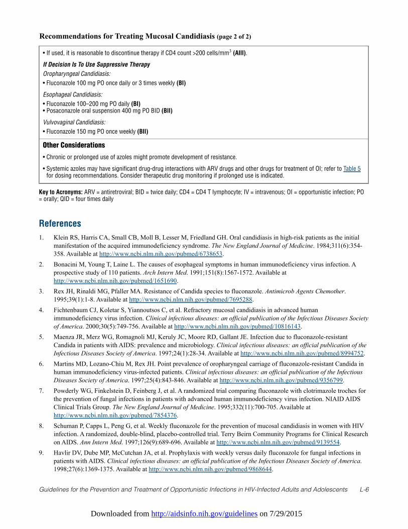
Downloaded from http://aidsinfo.nih.gov/guidelines on 7/29/2015
Guidelines for the Prevention and Treatment of Opportunistic Infections in HIV-Infected Adults and Adolescents L-6
References
1. Klein RS, Harris CA, Small CB, Moll B, Lesser M, Friedland GH. Oral candidiasis in high-risk patients as the initialmanifestation of the acquired immunodeficiency syndrome. The New England Journal of Medicine. 1984;311(6):354-358. Available at http://www.ncbi.nlm.nih.gov/pubmed/6738653.
2. Bonacini M, Young T, Laine L. The causes of esophageal symptoms in human immunodeficiency virus infection. Aprospective study of 110 patients. Arch Intern Med. 1991;151(8):1567-1572. Available athttp://www.ncbi.nlm.nih.gov/pubmed/1651690.
3. Rex JH, Rinaldi MG, Pfaller MA. Resistance of Candida species to fluconazole. Antimicrob Agents Chemother.1995;39(1):1-8. Available at http://www.ncbi.nlm.nih.gov/pubmed/7695288.
4. Fichtenbaum CJ, Koletar S, Yiannoutsos C, et al. Refractory mucosal candidiasis in advanced humanimmunodeficiency virus infection. Clinical infectious diseases: an official publication of the Infectious Diseases Societyof America. 2000;30(5):749-756. Available at http://www.ncbi.nlm.nih.gov/pubmed/10816143.
5. Maenza JR, Merz WG, Romagnoli MJ, Keruly JC, Moore RD, Gallant JE. Infection due to fluconazole-resistantCandida in patients with AIDS: prevalence and microbiology. Clinical infectious diseases: an official publication of theInfectious Diseases Society of America. 1997;24(1):28-34. Available at http://www.ncbi.nlm.nih.gov/pubmed/8994752.
6. Martins MD, Lozano-Chiu M, Rex JH. Point prevalence of oropharyngeal carriage of fluconazole-resistant Candida inhuman immunodeficiency virus-infected patients. Clinical infectious diseases: an official publication of the InfectiousDiseases Society of America. 1997;25(4):843-846. Available at http://www.ncbi.nlm.nih.gov/pubmed/9356799.
7. Powderly WG, Finkelstein D, Feinberg J, et al. A randomized trial comparing fluconazole with clotrimazole troches forthe prevention of fungal infections in patients with advanced human immunodeficiency virus infection. NIAID AIDSClinical Trials Group. The New England Journal of Medicine. 1995;332(11):700-705. Available athttp://www.ncbi.nlm.nih.gov/pubmed/7854376.
8. Schuman P, Capps L, Peng G, et al. Weekly fluconazole for the prevention of mucosal candidiasis in women with HIVinfection. A randomized, double-blind, placebo-controlled trial. Terry Beirn Community Programs for Clinical Researchon AIDS. Ann Intern Med. 1997;126(9):689-696. Available at http://www.ncbi.nlm.nih.gov/pubmed/9139554.
9. Havlir DV, Dube MP, McCutchan JA, et al. Prophylaxis with weekly versus daily fluconazole for fungal infections inpatients with AIDS. Clinical infectious diseases: an official publication of the Infectious Diseases Society of America.1998;27(6):1369-1375. Available at http://www.ncbi.nlm.nih.gov/pubmed/9868644.
Recommendations for Treating Mucosal Candidiasis (page 2 of 2)
• If used, it is reasonable to discontinue therapy if CD4 count >200 cells/mm3 (AIII).
If Decision Is To Use Suppressive Therapy
Oropharyngeal Candidiasis:
• Fluconazole 100 mg PO once daily or 3 times weekly (BI)
Esophageal Candidiasis:
• Fluconazole 100–200 mg PO daily (BI)• Posaconazole oral suspension 400 mg PO BID (BII)
Vulvovaginal Candidiasis:
• Fluconazole 150 mg PO once weekly (BII)
Other Considerations
• Chronic or prolonged use of azoles might promote development of resistance.
• Systemic azoles may have significant drug-drug interactions with ARV drugs and other drugs for treatment of OI; refer to Table 5for dosing recommendations. Consider therapeutic drug monitoring if prolonged use is indicated.
Key to Acronyms: ARV = antiretroviral; BID = twice daily; CD4 = CD4 T lymphocyte; IV = intravenous; OI = opportunistic infection; PO= orally; QID = four times daily
Downloaded from http://aidsinfo.nih.gov/guidelines on 7/29/2015
Guidelines for the Prevention and Treatment of Opportunistic Infections in HIV-Infected Adults and Adolescents L-7
10. Goldman M, Cloud GA, Wade KD, et al. A randomized study of the use of fluconazole in continuous versus episodictherapy in patients with advanced HIV infection and a history of oropharyngeal candidiasis: AIDS Clinical Trials GroupStudy 323/Mycoses Study Group Study 40. Clinical infectious diseases: an official publication of the InfectiousDiseases Society of America. 2005;41(10):1473-1480. Available at http://www.ncbi.nlm.nih.gov/pubmed/16231260.
11. Pappas PG, Rex JH, Sobel JD, et al. Guidelines for treatment of candidiasis. Clinical infectious diseases: an officialpublication of the Infectious Diseases Society of America. 2004;38(2):161-189. Available athttp://www.ncbi.nlm.nih.gov/pubmed/14699449.
12. Vazquez JA, Patton LL, Epstein JB, et al. Randomized, comparative, double-blind, double-dummy, multicenter trial ofmiconazole buccal tablet and clotrimazole troches for the treatment of oropharyngeal candidiasis: study of miconazoleLauriad(R) efficacy and safety (SMiLES). HIV Clin Trials. 2010;11(4):186-196. Available athttp://www.ncbi.nlm.nih.gov/pubmed/20974574.
13. Vazquez JA. Optimal management of oropharyngeal and esophageal candidiasis in patients living with HIV infection.HIV AIDS (Auckl). 2010;2(1):89-101. Available at http://www.ncbi.nlm.nih.gov/pubmed/22096388.
14. Vazquez JA, Skiest DJ, Nieto L, et al. A multicenter randomized trial evaluating posaconazole versus fluconazole forthe treatment of oropharyngeal candidiasis in subjects with HIV/AIDS. Clinical infectious diseases: an officialpublication of the Infectious Diseases Society of America. 2006;42(8):1179-1186. Available athttp://www.ncbi.nlm.nih.gov/pubmed/16575739.
15. Krishna G, Ma L, Martinho M, Preston RA, O'Mara E. A new solid oral tablet formulation of posaconazole: arandomized clinical trial to investigate rising single- and multiple-dose pharmacokinetics and safety in healthyvolunteers. The Journal of Antimicrobial Chemotherapy. 2012;67(11):2725-2730. Available athttp://www.ncbi.nlm.nih.gov/pubmed/22833639.
16. Merck Sharp & Dohme Corporation. Posaconazole package insert. 2014. Available athttps://www.merckconnect.com/noxafil/overview.html?hcpUser=yes. Accessed March 15, 2014.
17. de Wet N, Llanos-Cuentas A, Suleiman J, et al. A randomized, double-blind, parallel-group, dose-response study ofmicafungin compared with fluconazole for the treatment of esophageal candidiasis in HIV-positive patients. Clinicalinfectious diseases: an official publication of the Infectious Diseases Society of America. 2004;39(6):842-849. Availableat http://www.ncbi.nlm.nih.gov/pubmed/15472817.
18. Krause DS, Simjee AE, van Rensburg C, et al. A randomized, double-blind trial of anidulafungin versus fluconazole forthe treatment of esophageal candidiasis. Clinical infectious diseases: an official publication of the Infectious DiseasesSociety of America. 2004;39(6):770-775. Available at http://www.ncbi.nlm.nih.gov/pubmed/15472806.
19. Skiest DJ, Vazquez JA, Anstead GM, et al. Posaconazole for the treatment of azole-refractory oropharyngeal andesophageal candidiasis in subjects with HIV infection. Clinical infectious diseases: an official publication of theInfectious Diseases Society of America. 2007;44(4):607-614. Available athttp://www.ncbi.nlm.nih.gov/pubmed/17243069.
20. Vazquez JA, Skiest DJ, Tissot-Dupont H, Lennox JL, Boparai N, Isaacs R. Safety and efficacy of posaconazole in thelong-term treatment of azole-refractory oropharyngeal and esophageal candidiasis in patients with HIV infection. HIVClin Trials. 2007;8(2):86-97. Available at http://www.ncbi.nlm.nih.gov/pubmed/17507324.
21. Marty F, Mylonakis E. Antifungal use in HIV infection. Expert Opin Pharmacother. 2002;3(2):91-102. Available at 22. Molgaard-Nielsen D, Pasternak B, Hviid A. Use of oral fluconazole during pregnancy and the risk of birth defects. The
New England Journal of Medicine. 2013;369(9):830-839. Available at http://www.ncbi.nlm.nih.gov/pubmed/23984730.23. Lopez-Rangel E, Van Allen MI. Prenatal exposure to fluconazole: an identifiable dysmorphic phenotype. Birth Defects
Res A Clin Mol Teratol. 2005;73(11):919-923. Available at http://www.ncbi.nlm.nih.gov/pubmed/16265639.24. De Santis M, Di Gianantonio E, Cesari E, Ambrosini G, Straface G, Clementi M. First-trimester itraconazole exposure
and pregnancy outcome: a prospective cohort study of women contacting teratology information services in Italy. DrugSaf. 2009;32(3):239-244. Available at http://www.ncbi.nlm.nih.gov/pubmed/19338381.
Downloaded from http://aidsinfo.nih.gov/guidelines on 7/29/2015
Guidelines for the Prevention and Treatment of Opportunistic Infections in HIV-Infected Adults and Adolescents M-1
Cryptococcosis (Last updated May 7, 2013; last reviewed May 7, 2013)
Epidemiology
Most HIV-associated cryptococcal infections are caused by Cryptococcus neoformans, but occasionallyCryptococcus gattii is recognized. C. neoformans is found worldwide, whereas C. gattii most often is foundin Australia and similar subtropical regions and in the Pacific Northwest. Before the era of effectiveantiretroviral therapy (ART), approximately 5% to 8% of HIV-infected patients in developed countries werediagnosed with disseminated cryptococcosis.1 Current estimates indicate that every year, nearly 1 millioncases of cryptococcal meningitis are diagnosed worldwide and the disease accounts for more than 600,000deaths.2 In the last decade, incidence has declined substantially in areas with access to effective ART, andmost new infections are being recognized in patients recently diagnosed with HIV infection.3 Most cases areobserved in patients who have CD4 T-lymphocyte (CD4) cell counts <100 cells/mm3.
Clinical Manifestations
In HIV-infected patients, cryptococcosis commonly presents as a subacute meningitis or meningoencephalitiswith fever, malaise, and headache.1 Classic meningeal symptoms and signs, such as neck stiffness andphotophobia, occur in only one-quarter to one-third of patients. Some patients experience encephalopathicsymptoms, such as lethargy, altered mentation, personality changes, and memory loss that are usually a resultof increased intracranial pressure, thought to result from impaired cerebrospinal fluid (CSF) absorption, oryeast infection of the brain.
Cryptococcosis usually is disseminated when diagnosed in an HIV-infected patient. Any organ of the bodycan be involved, and skin lesions may be myriad, including umbilicated skin lesions mimicking molluscumcontagiosum. Isolated pulmonary infection is also possible; symptoms and signs include cough and dyspneain association with an abnormal chest radiograph, which typically demonstrates lobar consolidation, althoughlobar and nodular infiltrates have been reported. Pulmonary cryptococcosis may present as acute respiratorydistress syndrome and mimic Pneumocystis pneumonia.
Diagnosis
Analysis of CSF generally demonstrates mildly elevated levels of serum protein, low-to-normal glucoseconcentrations, and pleocytosis consisting mostly of lymphocytes. Some HIV-infected patients will havevery few CSF inflammatory cells, but an India ink or Gram’s stain preparation often will demonstratenumerous yeast forms. The opening pressure in the CSF may be elevated, with pressures ≥25 cm H2Ooccurring in 60 to 80% of patients.4,5
Cryptococcal disease can be diagnosed through culture of blood or CSF, CSF microscopy with India inkstaining, or cryptococcal antigen (CrAg) detection. In patients with HIV-related cryptococcal meningitis,55% of blood cultures and 95% of CSF cultures are positive and visible colonies can be detected within 7days. India ink staining of CSF demonstrates encapsulated yeast in 60% to 80% of cases, but manylaboratories in the United States no longer perform this test. CSF CrAg is usually positive in patients withmeningoencephalitis. Serum CrAg is usually positive in both meningeal and non-meningeal infection andmay be present weeks to months before symptom onset.6 A positive serum CrAg should prompt a lumbarpuncture to rule out meningeal disease. Three methods exist for antigen detection: latex agglutination,enzyme immunoassays, and lateral flow assay (a newly developed dipstick test). Testing for the antigen is auseful initial screening tool in diagnosing cryptococcosis in HIV-infected patients,7 and it may be particularlyuseful when lumbar punctures need to be delayed or are refused.
Downloaded from http://aidsinfo.nih.gov/guidelines on 7/29/2015
Guidelines for the Prevention and Treatment of Opportunistic Infections in HIV-Infected Adults and Adolescents M-2
Preventing Exposure
Cryptococcus is ubiquitous in the environment; it is found in soils. HIV-infected patients cannot completelyavoid exposure to C. neoformans or C. gattii. Limited epidemiological evidence suggests that exposure toaged bird droppings may increase risk of infection.
Preventing Disease
Because the incidence of cryptococcal disease is low among HIV-infected patients in the United States,routine testing of asymptomatic persons for serum cryptococcal polysaccharide antigen is not recommendedfor patients residing there.
Prospective, controlled trials indicate that prophylactic fluconazole or itraconazole can reduce the frequencyof primary cryptococcal disease in patients who have CD4 cell counts <100 cells/mm3.8,9 However, in theUnited States, primary prophylaxis or screening for serum CrAg in asymptomatic patients is notrecommended because of the relative infrequency of cryptococcal disease, lack of survival benefit associatedwith prophylaxis, possibility of drug interactions, potential antifungal drug resistance, and cost (BII).
Treating Disease
Treating cryptococcosis consists of three phases: induction, consolidation, and maintenance therapy. Thepreferred induction treatment for cryptococcal meningitis and other forms of extrapulmonary cryptococcosisis a lipid formulation of amphotericin B in combination with flucytosine (AI). Historically, amphotericin Bdeoxycholate was the preferred formulation at a dose of 0.7 to 1.0 mg/kg daily (AI). However, based on thegrowing body of evidence that lipid formulations of amphotericin B are effective for disseminatedcryptococcosis and should be used as the preferred formulation (AI), particularly in patients who experienceclinically significant renal dysfunction during therapy or who are likely to develop it. The non-comparativeCLEAR study demonstrated a 58% response rate in HIV-infected patients treated with amphotericin B lipidcomplex at mean dose of 4.4 mg/kg daily.10 In a Dutch and Australian study, a 3-week course of liposomalamphotericin B (4 mg/kg daily) resulted in more rapid sterilization of CSF than amphotericin B deoxycholate(0.7 mg/kg daily).11 A recently published comparison of amphotericin B deoxycholate (0.7 mg/kg daily), andliposomal amphotericin B (AmBisome®) (3 mg/kg or 6 mg/kg daily) showed similar efficacy for the threeregimens, but nephrotoxicity was lower with 3 mg/kg daily liposomal amphotericin B.12
Therefore, liposomal amphotericin B, in a dose of 3 to 4 mg/kg/daily, is recommended as the preferredamphotericin B formulation for primary induction therapy (AI), based on clinical experience and reducedrenal toxicity compared to amphotericin B deoxycholate. Amphotericin B lipid complex in a dose of 5 mg/kgdaily is an alternative (BII).
Amphotericin B formulations should be combined with flucytosine at a dose of 100 mg/kg daily in 4 divideddoses for ≥2 weeks in patients with normal renal function and is the preferred regimen for primary inductiontherapy (AI). Renal function should be monitored closely and the flucytosine dose adjusted accordingly forpatients with renal impairment. The addition of flucytosine to amphotericin B during acute treatment isassociated with more rapid sterilization of CSF.13-16 A recent randomized clinical trial also showed that thecombination of amphotericin B deoxycholate in a dose of 1.0 mg/kg/d combined with flucytosine wasassociated with improved survival compared to the same dose of amphotericin B without flucytosine.17
Amphotericin B deoxycholate in combination with fluconazole 400 mg daily was inferior to amphotericin Bin combination with flucytosine for clearing Cryptococcus from CSF.18 However, in two randomized trials,amphotericin B plus fluconazole 800 mg daily compared favorably with amphotericin B alone.17,19 Therefore,amphotericin B deoxycholate or lipid-formulated amphotericin B alone or combined with fluconazole at 800mg daily may be viable options in some circumstances but are less preferable alternatives than lipid-formulated amphotericin B combined with flucytosine (BI).
Downloaded from http://aidsinfo.nih.gov/guidelines on 7/29/2015
Guidelines for the Prevention and Treatment of Opportunistic Infections in HIV-Infected Adults and Adolescents M-3
Fluconazole (400–800 mg daily) combined with flucytosine is also a potential alternative to amphotericin Bregimens (BII).20 Fluconazole alone, based on early fungicidal activity, is inferior to amphotericin B21 forinduction therapy and is recommended only for patients who cannot tolerate or do not respond to standardtreatment. If it is used for primary induction therapy, the starting daily dose should be 1200 mg (CII).22
After at least 2 weeks of successful induction therapy—defined as substantial clinical improvement and anegative CSF culture after repeat lumbar puncture—amphotericin B and flucytosine can be discontinued andfollow-up or consolidation therapy initiated with fluconazole 400 mg daily (AI). This therapy shouldcontinue for at least 8 weeks (AI).13,14,23 Subsequently, the fluconazole should be reduced to 200 mg dailyand continued as chronic maintenance therapy to complete at least one year of azole therapy (see PreventingRecurrence section below).24 Limited data are available for the newer triazoles, voriconazole andposaconazole, as either primary or maintenance therapy for patients with cryptococcosis. Most of the data onuse of these extended-spectrum triazole antifungals have been reported for treatment of refractory cases, withsuccess rates of approximately 50%.25,26 At this time, the role of posaconazole and voriconazole in themanagement of cryptococcosis is not established. Voriconazole should be used cautiously with HIV proteaseinhibitors and efavirenz.
Non-central-nervous-system (CNS), extrapulmonary cryptococcosis and diffuse pulmonary disease should betreated similarly to CNS disease (BIII). For mild-to-moderate symptoms and focal pulmonary infiltrates,treatment with fluconazole (400 mg daily for 12 months) combined with effective ART is appropriate (BIII).All patients should have their CSF sampled to rule out CNS disease.
Special Considerations with Regard to Starting ARTOptimal timing for initiation of ART in patients with acute cryptococcal meningitis is controversial. Onerandomized, controlled trial that included 35 patients with cryptococcal meningitis suggested that ART wassafe when started within the first 14 days of diagnosis.27 A subsequent study from Africa demonstratedsignificantly worse outcomes in 54 patients started on ART within 72 hours of cryptococcal meningitisdiagnosis compared with those in which ART was delayed for at least 10 weeks.28 However, in the latter study,cryptococcal meningitis was managed with fluconazole alone, and ART consisted of nevirapine, stavudine, andlamivudine. Neither fluconazole alone nor the latter ART regimen are recommended as preferred initialtreatment in the United States. Lastly, another randomized clinical trial conducted in Africa in hospitalizedpatients with acute cryptococcal meningitis was recently halted by a Data and Safety Monitoring Board due tohigher mortality in the early ART arm (defined as ART started during the hospitalization) compared to the armin which patients waited to start ART until after their discharge from the hospital. In contrast to the otherAfrican study, this study used amphotericin B plus fluconazole during the induction phase of antifungaltreatment (http://www.niaid.nih.gov/news/newsreleases/2012/Pages/COAT.aspx). Such data must be viewedwith caution until fully reported and analyzed.
In patients with severe cryptococcosis (particularly those with elevated increased intracranial pressure[ICP]), it may be prudent to delay initiation of ART until induction (the first 2 weeks) or the totalinduction/consolidation phase (10 weeks) has been completed. However, for patients with advancedimmunosuppression (CD4 count <50 cells/mm3) earlier initiation of ART may be necessary (BIII). If thetreating physician elects to begin effective ART earlier, preparations should be made to aggressively addresscomplications of immune reconstitution inflammatory syndrome (IRIS) such as elevated ICP (BIII).
All the triazole antifungals have the potential for complex, and possibly bidirectional, interactions withcertain antiretroviral agents. Table 5 lists these interactions and recommendations for dosage adjustments,where feasible.
Monitoring of Response to Therapy and Adverse Events (including IRIS) ICP can cause clinical deterioration despite a microbiologic response and is more likely if the CSF openinglumbar pressure is ≥25cm H2O4,13 when obtained in the lateral decubitus position with good manometrics
Downloaded from http://aidsinfo.nih.gov/guidelines on 7/29/2015
Guidelines for the Prevention and Treatment of Opportunistic Infections in HIV-Infected Adults and Adolescents M-4
assured. In one large clinical trial, increased ICP was associated with 93% of deaths during the first 2 weeks oftherapy and 40% of deaths during weeks 3 to 10.4 Although it is uncertain which patients with high openinglumbar pressures will deteriorate, those with symptoms and signs of ICP require immediate clinical intervention.
Lumbar opening pressure should be measured in all patients with cryptococcal meningitis at the time ofdiagnosis. Measures to decrease ICP should be used for all patients with confusion, blurred vision,papilledema, lower extremity clonus, or other neurologic signs of increased pressure. Lumbar puncturesusually are recommended for initial management. One approach is to remove a volume of CSF (typically 20–30 mL) that at least halves the opening pressure29 and repeat daily until symptoms and signs consistentlyimprove. CSF shunting through a lumbar drain or ventriculostomy should be considered for patients whocannot tolerate lumbar puncture or in whom signs and symptoms of cerebral edema persist after multiplelumbar taps (BIII). Corticosteroids and mannitol have been shown to be ineffective in managing ICP and arenot recommended (CIII). Acetazolamide is hazardous as therapy for increased ICP management in thosewithout signs IRIS and is not recommended (BII).
After the first 2 weeks of treatment, many experts would advocate a repeat lumbar puncture to ensure thatviable organisms have been cleared from the CSF. Even in patients who have clinical improvement, positiveCSF cultures after 2 weeks of therapy are predictive of future relapse and less favorable outcome. In suchcases, some experts would continue amphotericin B plus flucytosine until the CSF cultures are negative (BIII).Monitoring titers of cryptococcal polysaccharide antigen in serum or CSF is of no value in determiningresponse to therapy and is not recommended. If new symptoms or clinical findings occur later, a repeatlumbar puncture, with measurement of opening lumbar pressure and CSF culture, should be performed.
Patients treated with amphotericin B formulations should be monitored for dose-dependent nephrotoxicityand electrolyte disturbances. Pre-infusion administration of 500 to 1000 mL of normal saline appears toreduce the risk of nephrotoxicity during amphotericin B treatment. Thirty minutes before infusion,acetaminophen (650 mg) and diphenhydramine (25–50 mg) or hydrocortisone (50–100 mg) typically areadministered in an attempt to ameliorate infusion-related adverse reactions (BIII), but data supporting thesepractices are scant. Meperidine (25–50 mg titrated during infusion) is effective for preventing and treatingamphotericin B-associated rigors (BII).
In patients receiving flucytosine, dosage should be adjusted based on changes in creatinine clearance andmight be guided by flucytosine levels. Peak serum flucytosine levels should be obtained 2 hours after an oraldose and the therapeutic range is between 30 and 80 µg/mL. Alternatively, frequent (i.e., at least bi-weekly)blood counts can be performed to detect development of cytopenia. Patients treated with flucytosine alsoshould be monitored for hepatotoxicity and gastrointestinal toxicities.
An estimated 30% of HIV-infected patients with cryptococcal meningitis experience IRIS after initiation orreinitiation of effective ART.30,31 Patients who have cryptococcal IRIS are more likely to be antiretroviralnaive, have higher HIV RNA levels, and have less CSF inflammation on initial presentation.32 DistinguishingIRIS from treatment failure may be difficult. In general, cryptococcal IRIS presents with worsening clinicaldisease despite microbiological evidence of effective antifungal therapy,32,33 whereas treatment failure isassociated with continued positive cultures. Appropriate management of IRIS is to continue both ART andantifungal therapy and reduce elevated ICP, if present (AII). In patients with severe symptoms of IRIS, somespecialists recommend a brief course of glucocorticosteroids (CIII), but data based management strategieshave not been developed.
Managing Treatment FailureTreatment failure is defined as lack of clinical improvement after 2 weeks of appropriate therapy, includingmanagement of increased ICP, with continued positive cultures; or relapse after an initial clinical response,defined as recurrence of symptoms with a positive CSF culture after ≥4 weeks of treatment. Direct primaryfluconazole resistance with C. neoformans has been reported in the United States but is uncommon.34
Therefore, susceptibility testing is not routinely recommended for intial management of cryptococcosis.
Downloaded from http://aidsinfo.nih.gov/guidelines on 7/29/2015
Guidelines for the Prevention and Treatment of Opportunistic Infections in HIV-Infected Adults and Adolescents M-5
Isolates collected to evaluate for persistence or relapse should, however, be checked for susceptibility andcompared with the original isolate. Strains with fluconazole minimum inhibitory concentrations ≥16 µg/mLare considered fluconazole resistant.35
Optimal therapy for patients with treatment failure has not been established. Patients who fail to respond toinduction with fluconazole monotherapy should be switched to amphotericin B, with or without flucytosine,and remain on it until a clinical response occurs. Liposomal amphotericin B (4–6 mg/kg/day) or amphotericinB lipid complex (5 mg/kg/day) is better tolerated and has greater efficacy than deoxycholate formulation in thissetting11,12,36 and should be considered when initial treatment with other regimens fails (AII).
Higher doses of fluconazole in combination with flucytosine also may be useful (BIII). Echinocandins haveno activity against Cryptococcus spp. and are not recommended for clinical management of cryptococcosis(AII). The newer triazoles—posaconazole and voriconazole—have activity against Cryptococcus spp. invitro and may have a role in salvage therapy, but probably offer no specific advantages over fluconazoleunless in vitro susceptibility testing indicates fluconazole resistance. Most clinical failures are a result ofinadequate induction therapy, drug interactions that interfere with treatment, or development of IRIS and arenot due to drug resistance.
Preventing Recurrence
When to Start Secondary ProphylaxisPatients who have completed the first 10 weeks of induction and consolidation therapy for acutecryptococcosis should be given chronic maintenance or suppressive therapy with fluconazole 200 mg daily(AI). Itraconazole is inferior to fluconazole for preventing relapse of cryptococcal disease and should not beused (CI).23
When to Stop Secondary ProphylaxisOnly a small number of patients have been evaluated for relapse after successful antifungal therapy forcryptococcosis and discontinuation of secondary prophylaxis while on ART. In a European study, recurrenceof cryptococcosis was seen in none of 39 subjects on potent ART whose antifungal therapy was discontinued.In this cohort, when maintenance therapy was stopped, the median CD4 cell count was 297 cells/mm3, themedian HIV RNA concentration was <500 copies/mL, and the median time on potent ART was 25 months.37
A prospective, randomized study of 60 patients in Thailand documented no recurrences of cryptococcosisduring 48 weeks of follow-up among 22 patients whose antifungal therapy was discontinued after havingachieved a CD4 count >100 cells/mm3 with a sustained undetectable HIV RNA level for 3 months on potentART.38 Given these data and inference from data on discontinuation of secondary prophylaxis for other HIV-associated opportunistic infections, it is reasonable to discontinue chronic antifungal maintenance therapy forcryptococcosis in patients whose CD4 cell counts are ≥100 cells/mm3, who have undetectable viral loads onART for >3 months, and who have received a minimum of 1 year of azole antifungal chronic maintenancetherapy after successful treatment of cryptococcosis (BII).39 Secondary prophylaxis should be reinitiated ifthe CD4 count decreases again to <100 cells/mm3 (AlII).
Special Considerations During Pregnancy
The diagnosis of cryptococcal infections during pregnancy is similar to that in non-pregnant adults.Treatment should be initiated promptly after a diagnosis is confirmed. It should be emphasized that thepostpartum period may be a high-risk period for the development of IRIS.
Lipid formulations of amphotericin B are the preferred initial regimen for the treatment of cryptococcalmeningoencephalitis, disseminated disease, or severe pulmonary cryptococcosis in pregnant patients.Extensive clinical experience with amphotericin has not documented teratogenicity. Neonates born to womenon chronic amphotericin B at delivery should be evaluated for renal dysfunction and hypokalemia.
Downloaded from http://aidsinfo.nih.gov/guidelines on 7/29/2015
Guidelines for the Prevention and Treatment of Opportunistic Infections in HIV-Infected Adults and Adolescents M-6
Flucytosine was teratogenic in animal studies, and human experience is limited to case reports and smallseries. Therefore, its use should be considered only when the benefits outweigh its risks to the fetus (CIII).
Congenital malformations similar to those observed in animals, including craniofacial and limbabnormalities, have been reported in infants born to mothers who received fluconazole at doses of ≥400mg/day or more through or beyond the first trimester of pregnancy.40 Although several cohort studies haveshown no increased risk of birth defects with early pregnancy exposure, most of these involved low dosesand short term exposure to fluconazole.41,42 Based on the reported birth defects, the FDA has changed thepregnancy category for fluconazole from C to D for any use other than a single, low dose for treatment ofvaginal candidiasis, (http://www.fda.gov/Drugs/DrugSafety/ucm266030.htm) and use of fluconazole in thefirst trimester should be considered only if the benefits clearly outweigh risks. For pregnant women,amphotericin should be continued throughout the first trimester with consideration of switching to oralfluconazole, if clinically appropriate, after the first trimester.
Although there are case reports of birth defects in infants exposed to itraconazole, prospective cohort studiesof over 300 women with first trimester exposure did not show an increased risk of malformation.43,44
However, in general azole antifungals should be avoided during the first trimester of pregnancy (BIII).Voriconazole and posaconazole are teratogenic and embryotoxic in animal studies, voriconazole at doseslower than recommended human doses; there are no adequate controlled studies in humans. These drugsshould be avoided in pregnancy, especially in the first trimester (AIII).
Recommendations for Preventing and Treating Cryptococcosis (page 1 of 2)
Treating Cryptococcal Meningitis
Induction Therapy (For At Least 2 Weeks, Followed by Consolidation Therapy)
Preferred Regimens:
• Liposomal amphotericin B 3–4 mg/kg IV daily + flucytosine 25 mg/kg PO QID (AI)
Note: Flucytosine dose should be adjusted in renal impairment
Alternative Regimens:
• Amphotericin B lipid complex 5 mg/kg IV daily + flucytosine 25 mg/kg PO QID (BII)
• Amphotericin B (deoxycholate 0.7-1.0 mg/kg IV daily + flucytosine 25 mg/kg PO QID (AI)
• Liposomal amphotericin B 3–4 mg/kg IV daily + fluconazole 800 mg PO or IV daily (BIII)
• Amphotericin B (deoxycholate 0.7-1.0 mg/kg IV daily) + fluconazole 800 mg PO or IV daily (BI)
• Liposomal amphotericin B 3-4 mg/kg IV daily alone (BII)
• Fluconazole 400–800 mg PO or IV daily + flucytosine 25 mg/kg PO QID (BII)
• Fluconazole 1200 mg PO or IV daily alone (CII)
Consolidation Therapy (For At Least 8 Weeks, Followed by Maintenance Therapy)
• To begin after at least 2 weeks of successful induction therapy (defined as substantial clinical improvement and a negative CSFculture after repeat LP)
Preferred Regimen:
• Fluconazole 400 mg PO or IV once daily (AI)
Alternative Regimen:
• Itraconazole 200 mg PO BID (CI)
Maintenance Therapy
Preferred Regimen:
• Fluconazole 200 mg PO for at least 1 year (AI)
Downloaded from http://aidsinfo.nih.gov/guidelines on 7/29/2015
Guidelines for the Prevention and Treatment of Opportunistic Infections in HIV-Infected Adults and Adolescents M-7
References
1. Aberg J, WG. P. Cryptococcosis. In: Dolin R MH, Saag MS, ed. AIDS Therapy. New York, NY: Churcill Livingstone;2002:498-510.
2. Park BJ, Wannemuehler KA, Marston BJ, Govender N, Pappas PG, Chiller TM. Estimation of the current global burdenof cryptococcal meningitis among persons living with HIV/AIDS. AIDS. Feb 20 2009;23(4):525-530. Available athttp://www.ncbi.nlm.nih.gov/pubmed/19182676.
3. Mirza SA, Phelan M, Rimland D, et al. The changing epidemiology of cryptococcosis: an update from population-basedactive surveillance in 2 large metropolitan areas, 1992-2000. Clin Infect Dis. Mar 15 2003;36(6):789-794. Available athttp://www.ncbi.nlm.nih.gov/pubmed/12627365.
4. Graybill JR, Sobel J, Saag M, et al. Diagnosis and management of increased intracranial pressure in patients with AIDSand cryptococcal meningitis. The NIAID Mycoses Study Group and AIDS Cooperative Treatment Groups. Clin InfectDis. Jan 2000;30(1):47-54. Available at http://www.ncbi.nlm.nih.gov/pubmed/10619732.
5. Bicanic T, Brouwer AE, Meintjes G, et al. Relationship of cerebrospinal fluid pressure, fungal burden and outcome inpatients with cryptococcal meningitis undergoing serial lumbar punctures. AIDS. Mar 27 2009;23(6):701-706. Availableat http://www.ncbi.nlm.nih.gov/pubmed/19279443.
6. French N, Gray K, Watera C, et al. Cryptococcal infection in a cohort of HIV-1-infected Ugandan adults. AIDS. May 32002;16(7):1031-1038. Available at http://www.ncbi.nlm.nih.gov/pubmed/11953469.
7. Powderly WG, Cloud GA, Dismukes WE, Saag MS. Measurement of cryptococcal antigen in serum and cerebrospinalfluid: value in the management of AIDS-associated cryptococcal meningitis. Clin Infect Dis. May 1994;18(5):789-792.
Recommendations for Preventing and Treating Cryptococcosis (page 2 of 2)
Stopping Maintenance Therapy
If the following criteria are fulfilled (BII):
• Completed initial (induction, consolidation) therapy, and at least 1 year on maintenance therapy, and
• Remains asymptomatic from cryptococcal infection, and
• CD4 count ≥100 cells/µL for ≥3 months and suppressed HIV RNA in response to effective ART
Restarting Maintenance Therapy:
• If CD4 count decline to ≤100 cells/µL (AIII)
Treating Non-CNS, Extrapulmonary Cryptococcosis and for Diffuse Pulmonary Disease:
• Same treatment as for CNS disease (BIII)
Treating Non-CNS Cryptocococcosis with Mild-to-Moderate Symptoms and Focal Pulmonary Infiltrates:
• Fluconazole 400 mg PO daily for 12 months (BIII)
Other Considerations
• Addition of flucytosine to amphotericin B has been associated with more rapid sterilization of CSF, decreased risk for subsequentrelapse, and improved survival.
• Patients receiving flucytosine should have either blood levels monitored (peak level 2 hours after dose should be between 30–80µg/mL) or close following of complete blood counts to identify developing cytopenias. Dosage should be adjusted in patients withrenal insufficiency (BII).
• Opening pressure should always be measured when a LP is performed. Repeated LPs or CSF shunting are essential to effectivelymanage symptomatic increased ICP.
• Corticosteroids and mannitol are ineffective in reducing ICP and are NOT recommended (BII).
• Infection due to C. gattii should be treated similarly to C. neoformans (BIII).
• All the triazole antifungals have the potential to interact with certain antiretroviral agents and other anti-infective agents. Theseinteractions are complex and can be bidirectional. Table 5 lists these interactions and recommends dosage adjustments wherefeasible.
Key to Acronyms: BID = twice daily; CD4 = CD4 T lymphocyte cell; CNS = central nervous system; CSF = cerebrospinal fluid; ICP =intracranial pressure; IV = intravenous; LP = lumbar puncture; PO = orally; QID = four times a day
Downloaded from http://aidsinfo.nih.gov/guidelines on 7/29/2015
Guidelines for the Prevention and Treatment of Opportunistic Infections in HIV-Infected Adults and Adolescents M-8
Available at http://www.ncbi.nlm.nih.gov/pubmed/8075272.8. Powderly WG, Finkelstein D, Feinberg J, et al. A randomized trial comparing fluconazole with clotrimazole troches for the
prevention of fungal infections in patients with advanced human immunodeficiency virus infection. NIAID AIDS ClinicalTrials Group. N Engl J Med. Mar 16 1995;332(11):700-705. Available at http://www.ncbi.nlm.nih.gov/pubmed/7854376.
9. McKinsey DS, Wheat LJ, Cloud GA, et al. Itraconazole prophylaxis for fungal infections in patients with advancedhuman immunodeficiency virus infection: randomized, placebo-controlled, double-blind study. National Institute ofAllergy and Infectious Diseases Mycoses Study Group. Clin Infect Dis. May 1999;28(5):1049-1056. Available athttp://www.ncbi.nlm.nih.gov/pubmed/10452633.
10. Baddour LM, Perfect JR, Ostrosky-Zeichner L. Successful use of amphotericin B lipid complex in the treatment ofcryptococcosis. Clin Infect Dis. May 1 2005;40 Suppl 6:S409-413. Available athttp://www.ncbi.nlm.nih.gov/pubmed/15809927.
11. Leenders AC, Reiss P, Portegies P, et al. Liposomal amphotericin B (AmBisome) compared with amphotericin B bothfollowed by oral fluconazole in the treatment of AIDS-associated cryptococcal meningitis. AIDS. Oct1997;11(12):1463-1471. Available at http://www.ncbi.nlm.nih.gov/pubmed/9342068.
12. Hamill RJ, Sobel JD, El-Sadr W, et al. Comparison of 2 doses of liposomal amphotericin B and conventionalamphotericin B deoxycholate for treatment of AIDS-associated acute cryptococcal meningitis: a randomized, double-blind clinical trial of efficacy and safety. Clin Infect Dis. Jul 15 2010;51(2):225-232. Available athttp://www.ncbi.nlm.nih.gov/pubmed/20536366.
13. van der Horst CM, Saag MS, Cloud GA, et al. Treatment of cryptococcal meningitis associated with the acquiredimmunodeficiency syndrome. National Institute of Allergy and Infectious Diseases Mycoses Study Group and AIDSClinical Trials Group. N Engl J Med. Jul 3 1997;337(1):15-21. Available at http://www.ncbi.nlm.nih.gov/pubmed/9203426.
14. Saag MS, Graybill RJ, Larsen RA, et al. Practice guidelines for the management of cryptococcal disease. InfectiousDiseases Society of America. Clin Infect Dis. Apr 2000;30(4):710-718. Available athttp://www.ncbi.nlm.nih.gov/pubmed/10770733.
15. Dromer F, Mathoulin-Pelissier S, Launay O, Lortholary O, French Cryptococcosis Study G. Determinants of diseasepresentation and outcome during cryptococcosis: the CryptoA/D study. PLoS Med. Feb 2007;4(2):e21. Available athttp://www.ncbi.nlm.nih.gov/pubmed/17284154.
16. Dromer F, Bernede-Bauduin C, Guillemot D, Lortholary O, French Cryptococcosis Study G. Major role foramphotericin B-flucytosine combination in severe cryptococcosis. PLoS One. 2008;3(8):e2870. Available athttp://www.ncbi.nlm.nih.gov/pubmed/18682846.
17. Day JN, Chau TT, Wolbers M, et al. Combination antifungal therapy for cryptococcal meningitis. N Engl J Med. Apr 42013;368(14):1291-1302. Available at http://www.ncbi.nlm.nih.gov/pubmed/23550668.
18. Brouwer AE, Rajanuwong A, Chierakul W, et al. Combination antifungal therapies for HIV-associated cryptococcalmeningitis: a randomised trial. Lancet. May 29 2004;363(9423):1764-1767. Available athttp://www.ncbi.nlm.nih.gov/pubmed/15172774.
19. Pappas PG, Chetchotisakd P, Larsen RA, et al. A phase II randomized trial of amphotericin B alone or combined withfluconazole in the treatment of HIV-associated cryptococcal meningitis. Clin Infect Dis. Jun 15 2009;48(12):1775-1783.Available at http://www.ncbi.nlm.nih.gov/pubmed/19441980.
20. Larsen RA, Bozzette SA, Jones BE, et al. Fluconazole combined with flucytosine for treatment of cryptococcalmeningitis in patients with AIDS. Clin Infect Dis. Oct 1994;19(4):741-745. Available athttp://www.ncbi.nlm.nih.gov/pubmed/7803641.
21. Bicanic T, Meintjes G, Wood R, et al. Fungal burden, early fungicidal activity, and outcome in cryptococcal meningitisin antiretroviral-naive or antiretroviral-experienced patients treated with amphotericin B or fluconazole. Clin Infect Dis.Jul 1 2007;45(1):76-80. Available at http://www.ncbi.nlm.nih.gov/pubmed/17554704.
22. Nussbaum JC, Jackson A, Namarika D, et al. Combination flucytosine and high-dose fluconazole compared withfluconazole monotherapy for the treatment of cryptococcal meningitis: a randomized trial in Malawi. Clin Infect Dis.Feb 1 2010;50(3):338-344. Available at http://www.ncbi.nlm.nih.gov/pubmed/20038244.
23. Saag MS, Cloud GA, Graybill JR, et al. A comparison of itraconazole versus fluconazole as maintenance therapy forAIDS-associated cryptococcal meningitis. National Institute of Allergy and Infectious Diseases Mycoses Study Group.Clin Infect Dis. Feb 1999;28(2):291-296. Available at http://www.ncbi.nlm.nih.gov/pubmed/10064246.
Downloaded from http://aidsinfo.nih.gov/guidelines on 7/29/2015
Guidelines for the Prevention and Treatment of Opportunistic Infections in HIV-Infected Adults and Adolescents M-9
24. Powderly WG, Saag MS, Cloud GA, et al. A controlled trial of fluconazole or amphotericin B to prevent relapse ofcryptococcal meningitis in patients with the acquired immunodeficiency syndrome. The NIAID AIDS Clinical TrialsGroup and Mycoses Study Group. N Engl J Med. Mar 19 1992;326(12):793-798. Available athttp://www.ncbi.nlm.nih.gov/pubmed/1538722.
25. Perfect JR, Marr KA, Walsh TJ, et al. Voriconazole treatment for less-common, emerging, or refractory fungalinfections. Clin Infect Dis. May 1 2003;36(9):1122-1131. Available at http://www.ncbi.nlm.nih.gov/pubmed/12715306.
26. Pitisuttithum P, Negroni R, Graybill JR, et al. Activity of posaconazole in the treatment of central nervous system fungalinfections. J Antimicrob Chemother. Oct 2005;56(4):745-755. Available at http://www.ncbi.nlm.nih.gov/pubmed/16135526.
27. Zolopa A, Andersen J, Powderly W, et al. Early antiretroviral therapy reduces AIDS progression/death in individualswith acute opportunistic infections: a multicenter randomized strategy trial. PLoS One. 2009;4(5):e5575. Available athttp://www.ncbi.nlm.nih.gov/pubmed/19440326.
28. Makadzange AT, Ndhlovu CE, Takarinda K, et al. Early versus delayed initiation of antiretroviral therapy for concurrentHIV infection and cryptococcal meningitis in sub-saharan Africa. Clin Infect Dis. Jun 1 2010;50(11):1532-1538.Available at http://www.ncbi.nlm.nih.gov/pubmed/20415574.
29. Fessler RD, Sobel J, Guyot L, et al. Management of elevated intracranial pressure in patients with Cryptococcalmeningitis. J Acquir Immune Defic Syndr Hum Retrovirol. Feb 1 1998;17(2):137-142. Available athttp://www.ncbi.nlm.nih.gov/pubmed/9473014.
30. Shelburne SA, 3rd, Darcourt J, White AC, Jr., et al. The role of immune reconstitution inflammatory syndrome inAIDS-related Cryptococcus neoformans disease in the era of highly active antiretroviral therapy. Clin Infect Dis. Apr 12005;40(7):1049-1052. Available at http://www.ncbi.nlm.nih.gov/pubmed/15825000.
31. Muller M, Wandel S, Colebunders R, et al. Immune reconstitution inflammatory syndrome in patients startingantiretroviral therapy for HIV infection: a systematic review and meta-analysis. Lancet Infect Dis. Apr 2010;10(4):251-261. Available at http://www.ncbi.nlm.nih.gov/pubmed/20334848.
32. Boulware DR, Bonham SC, Meya DB, et al. Paucity of initial cerebrospinal fluid inflammation in cryptococcalmeningitis is associated with subsequent immune reconstitution inflammatory syndrome. J Infect Dis. Sep 152010;202(6):962-970. Available at http://www.ncbi.nlm.nih.gov/pubmed/20677939.
33. Haddow LJ, Colebunders R, Meintjes G, et al. Cryptococcal immune reconstitution inflammatory syndrome in HIV-1-infected individuals: proposed clinical case definitions. Lancet Infect Dis. Nov 2010;10(11):791-802. Available athttp://www.ncbi.nlm.nih.gov/pubmed/21029993.
34. Brandt ME, Pfaller MA, Hajjeh RA, et al. Trends in antifungal drug susceptibility of Cryptococcus neoformans isolatesin the United States: 1992 to 1994 and 1996 to 1998. Antimicrob Agents Chemother. Nov 2001;45(11):3065-3069.Available at http://www.ncbi.nlm.nih.gov/pubmed/11600357.
35. Witt MD, Lewis RJ, Larsen RA, et al. Identification of patients with acute AIDS-associated cryptococcal meningitiswho can be effectively treated with fluconazole: the role of antifungal susceptibility testing. Clin Infect Dis. Feb1996;22(2):322-328. Available at http://www.ncbi.nlm.nih.gov/pubmed/8838190.
36. Chen SC, Australasian Society for Infectious Diseases Mycoses Iterest G. Cryptococcosis in Australasia and thetreatment of cryptococcal and other fungal infections with liposomal amphotericin B. J Antimicrob Chemother. Feb2002;49 Suppl 1(Suppl 1):57-61. Available at http://www.ncbi.nlm.nih.gov/pubmed/11801583.
37. Kirk O, Reiss P, Uberti-Foppa C, et al. Safe interruption of maintenance therapy against previous infection with fourcommon HIV-associated opportunistic pathogens during potent antiretroviral therapy. Ann Intern Med. Aug 202002;137(4):239-250. Available at http://www.ncbi.nlm.nih.gov/pubmed/12186514.
38. Vibhagool A, Sungkanuparph S, Mootsikapun P, et al. Discontinuation of secondary prophylaxis for cryptococcalmeningitis in human immunodeficiency virus-infected patients treated with highly active antiretroviral therapy: aprospective, multicenter, randomized study. Clin Infect Dis. May 15 2003;36(10):1329-1331. Available athttp://www.ncbi.nlm.nih.gov/pubmed/12746781.
39. Mussini C, Pezzotti P, Miro JM, et al. Discontinuation of maintenance therapy for cryptococcal meningitis in patientswith AIDS treated with highly active antiretroviral therapy: an international observational study. Clin Infect Dis. Feb 152004;38(4):565-571. Available at http://www.ncbi.nlm.nih.gov/pubmed/14765351.
40. Pursley TJ, Blomquist IK, Abraham J, Andersen HF, Bartley JA. Fluconazole-induced congenital anomalies in threeinfants. Clin Infect Dis. Feb 1996;22(2):336-340. Available at http://www.ncbi.nlm.nih.gov/pubmed/8838193.
Downloaded from http://aidsinfo.nih.gov/guidelines on 7/29/2015
Guidelines for the Prevention and Treatment of Opportunistic Infections in HIV-Infected Adults and Adolescents M-10
41. Norgaard M, Pedersen L, Gislum M, et al. Maternal use of fluconazole and risk of congenital malformations: a Danishpopulation-based cohort study. J Antimicrob Chemother. Jul 2008;62(1):172-176. Available athttp://www.ncbi.nlm.nih.gov/pubmed/18400803.
42. Mastroiacovo P, Mazzone T, Botto LD, et al. Prospective assessment of pregnancy outcomes after first-trimesterexposure to fluconazole. Am J Obstet Gynecol. Dec 1996;175(6):1645-1650. Available athttp://www.ncbi.nlm.nih.gov/pubmed/8987954.
43. De Santis M, Di Gianantonio E, Cesari E, Ambrosini G, Straface G, Clementi M. First-trimester itraconazole exposureand pregnancy outcome: a prospective cohort study of women contacting teratology information services in Italy. DrugSaf. 2009;32(3):239-244. Available at http://www.ncbi.nlm.nih.gov/pubmed/19338381.
44. Bar-Oz B, Moretti ME, Bishai R, et al. Pregnancy outcome after in utero exposure to itraconazole: a prospective cohortstudy. Am J Obstet Gynecol. Sep 2000;183(3):617-620. Available at http://www.ncbi.nlm.nih.gov/pubmed/10992182.
Downloaded from http://aidsinfo.nih.gov/guidelines on 7/29/2015
Guidelines for the Prevention and Treatment of Opportunistic Infections in HIV-Infected Adults and Adolescents M-11
Histoplasmosis (Last updated May 7, 2013; last reviewed May 7, 2013)
Epidemiology
Histoplasmosis is caused by the dimorphic fungus Histoplasma capsulatum. Infection is endemic to thecentral and south-central United States and is especially common in the Ohio and Mississippi River Valleys.It is also endemic in Latin America, including Puerto Rico. In endemic areas, annual incidence approaches5% in HIV-infected individuals. A CD4 T lymphocyte (CD4) count <150 cells/mm3 is associated with anincreased risk of symptomatic illness.1,2
Virtually all cases of primary histoplasmosis are acquired by inhalation of microconidia that form in themycelial phase. Asymptomatic dissemination of infection beyond the lungs is common, and cellularimmunity is critical in controlling infection. When cellular immunity wanes, reactivation of a silent focus ofinfection that was acquired years earlier can occur, and it is the presumed mechanism for disease occurrencein nonendemic areas. Incidence of symptomatic histoplasmosis in HIV-infected patients appears to havedeclined with the advent of effective antiretroviral therapy (ART). When histoplasmosis does occur,however, it is reported as the AIDS-defining illness in 25% to 61% of patients.3,4
Clinical Manifestations
In HIV-infected patients, common clinical manifestations of progressive disseminated histoplasmosis includefever, fatigue, weight loss, and hepatosplenomegaly. Cough, chest pain, and dyspnea occur in approximately50% of patients.1,4 Central nervous system (CNS), gastrointestinal, and cutaneous manifestations occur in asmaller percentage, although in a series from Panama, diarrhea occurred in 50% of patients.5 Approximately10% of patients experience shock and multi-organ failure. Patients with CNS histoplasmosis typicallyexperience fever and headache, and also (if brain involvement is present) seizures, focal neurologicaldeficits, and changes in mental status.6 Gastrointestinal disease usually manifests as diarrhea, fever,abdominal pain, and weight loss.7 For patients whose CD4 counts are >300 cells/mm3, histoplasmosis isoften limited to the respiratory tract and usually presents with cough, pleuritic chest pain, and fever.
Diagnosis
Detection of Histoplasma antigen in blood or urine is a sensitive method for rapid diagnosis of disseminatedhistoplasmosis and acute pulmonary histoplasmosis8 but is insensitive for chronic forms of pulmonaryinfection. Using a newer quantitative assay, antigen was detected in the urine of 100% and in the serum of92% of AIDS patients with disseminated histoplasmosis.9 Antigen detection in bronchoalveolar lavage fluidappears to be a useful method for diagnosis of pulmonary histoplasmosis.10 In patients with severedisseminated histoplasmosis, peripheral blood smears can show the organisms engulfed by white blood cells.Histopathological examination of biopsy material from involved tissues demonstrates the characteristic 2 to 4µm budding yeast and can provide a rapid diagnosis.
H. capsulatum can be cultured from blood, bone marrow, respiratory secretions, or other involved sites in>85% of patients with AIDS and disseminated histoplasmosis, but the organism requires several weeks togrow.11 Serologic tests are less useful than antigen assays in AIDS patients with disseminated histoplasmosisbut may be helpful in patients who have reasonably intact immune responses with pulmonary disease.11,12
The diagnosis of meningitis is often difficult. The usual cerebrospinal fluid (CSF) findings are a lymphocyticpleocytosis, elevated protein, and low glucose. Fungal stains are usually negative, and CSF cultures arepositive in a minority of cases.6 However, Histoplasma antigen or antibodies against H. capsulatum can bedetected in CSF in up to 70% of cases, and a positive result for either test is diagnostic. For some patients,none of these specific tests is positive, and a presumptive diagnosis of Histoplasma meningitis is appropriateif the patient has disseminated histoplasmosis and findings of CNS infection not explained by another cause.
Downloaded from http://aidsinfo.nih.gov/guidelines on 7/29/2015
Guidelines for the Prevention and Treatment of Opportunistic Infections in HIV-Infected Adults and Adolescents M-12
Preventing Exposure
HIV-infected individuals who live in or visit areas in which histoplasmosis is endemic cannot completelyavoid exposure to it, but those with CD4 counts <150 cells/mm3 should avoid activities known to beassociated with increased risk (BIII). These include creating dust when working with surface soil; cleaningchicken coops that are contaminated with droppings; disturbing areas contaminated with bird or batdroppings; cleaning, remodeling, or demolishing old buildings; and exploring caves.
Preventing Disease
When to Start Primary ProphylaxisData from a prospective, randomized, controlled trial indicate that itraconazole can reduce the frequency ofhistoplasmosis, although not mortality, in patients who have advanced HIV infection and who live in areaswhere histoplasmosis is highly endemic.13 Prophylaxis with itraconazole at a dose of 200 mg daily can beconsidered for patients with CD4 counts <150 cells/mm3 who are at high risk because of occupational exposureor who live in a community with a hyperendemic rate of histoplasmosis (>10 cases/100 patient-years) (BI).
When to Stop Primary ProphylaxisIf used, primary prophylaxis can be discontinued in patients on potent ART once CD4 counts are ≥150cells/mm3 for 6 months (BIII). Prophylaxis should be restarted if the CD4 count falls to <150 cells/mm3 (BIII).
Treating Disease
In a randomized clinical trial, intravenous (IV) liposomal amphotericin B (3 mg/kg daily) was more effectivethan standard IV amphotericin B deoxycholate (0.7 mg/kg daily), induced a more rapid and completeresponse, lowered mortality, and reduced toxicity.14 Based on these findings, patients with moderately severeto severe disseminated histoplasmosis should be treated with IV liposomal amphotericin B (3 mg/kg daily)for at least 2 weeks or until they clinically improve (AI). Another lipid formulation of amphotericin B can beused at the same dosage if cost is a concern or in patients who cannot tolerate liposomal amphotericin B(AIII). Step-down therapy to oral itraconazole, 200 mg 3 times daily for 3 days, and then 200 mg twicedaily, should be given for a total of at least 12 months (AII).15 Because of potential drug interactions betweenitraconazole and both protease inhibitors and efavirenz, it is advisable to obtain serum levels of itraconazoleafter 2 weeks of therapy. A randomly obtained serum level of at least 1.0 µg/mL is recommended and levels>10 µg/mL are unnecessary.
In patients with less severe disseminated histoplasmosis, oral itraconazole, 200 mg 3 times daily for 3 daysfollowed by 200 mg twice daily, is appropriate initial therapy (All).15,16 The liquid formulation ofitraconazole, which should be given on an empty stomach, is preferable because it is better absorbed anddoes not require gastric acid for absorption, but it is less well tolerated than the capsule formulation, whichshould be given with food.
Acute pulmonary histoplasmosis in an HIV-infected patient with intact immunity, as indicated by a CD4count >300 cells/mm3, should be managed in a manner similar to that used for a nonimmunocompromisedhost (AIII).15
In patients with confirmed meningitis, liposomal amphotericin B should be administered as initial therapy ata dosage of 5 mg/kg daily for 4 to 6 weeks (AIII). This should be followed by maintenance therapy withitraconazole at a dose of 200 mg 2 or 3 times daily for at least 1 year and until resolution of abnormal CSFfindings (AIII).15
Oral posaconazole and voriconazole have been reported to be effective for histoplasmosis in a small numberof patients who had AIDS or other immunosuppressive conditions17-20 and may be reasonable alternatives forpatients intolerant of itraconazole who are only moderately ill (BIII). Fluconazole is less effective than
Downloaded from http://aidsinfo.nih.gov/guidelines on 7/29/2015
Guidelines for the Prevention and Treatment of Opportunistic Infections in HIV-Infected Adults and Adolescents M-13
itraconazole for histoplasmosis but has been shown to be moderately effective at a dose of 800 mg daily andmay also be a reasonable alternative at this dose for those intolerant of itraconazole (CII).21 Theechinocandins are not active against H. capsulatum and should not be used to treat patients withhistoplasmosis (AIII).
Special Considerations with Regard to Starting ARTHIV-infected individuals diagnosed with histoplasmosis should be started on ART as soon as possible afterinitiating antifungal therapy (AIII). Immune reconstitution inflammatory syndrome (IRIS) is reportedlyuncommon in HIV-infected patients with histoplasmosis.22,23 ART should, therefore, not be withheld becauseof concern for the possible development of IRIS (AIII).
All of the triazole antifungals have the potential for complex, and possibly bidirectional, interactions withcertain ARV agents and other anti-infective agents. Table 5 lists these interactions and recommendations fordosage adjustments, where feasible.
Monitoring of Response to Therapy and Adverse Events (including IRIS)Serial monitoring of serum or urine for Histoplasma antigen is useful for determining response to therapy. Arise in antigen level suggests relapse. Because absorption of itraconazole can be erratic, a random serumitraconazole level should be obtained after 2 weeks of therapy if there is concern about adherence or ifmedications with potentially adverse interactions are added to the drug regimen. The serum concentrationshould be >1 µg/mL.
As previously indicated, IRIS is uncommon in HIV-infected individuals with histoplasmosis.22,23
Managing Treatment FailureMortality rates remain high for patients with AIDS who develop disseminated histoplasmosis, many of whomhad never received ART before diagnosis with histoplasmosis.3-5,12 Liposomal amphotericin B should be used inpatients who are severely ill or who have failed to respond to initial azole antifungal therapy (AIII). Oralposaconazole and voriconazole are reasonable alternatives for patients intolerant of itraconazole who are onlymoderately ill (BIII);17-20 fluconazole also can be used at a dose of 800 mg daily (CII).21 Drug interactions maylimit the use of voriconazole in patients who are taking non-nucleoside reverse transcriptase inhibitors orritonavir (Table 5). Posaconazole has fewer known drug interactions with ARV medications than voriconazole.
Preventing Recurrence
When to Start Secondary ProphylaxisLong-term suppressive therapy with itraconazole (200 mg daily) should be administered to patients with severedisseminated or CNS infection (AIII) and after re-induction therapy in those whose disease relapses despiteinitial receipt of appropriate therapy (BIII). Fluconazole is less effective than itraconazole for this purpose buthas some efficacy at 400 mg daily.21,24 The role of voriconazole or posaconazole has not been evaluated.
When to Stop Secondary ProphylaxisAn AIDS Clinical Treatment Group (ACTG)-sponsored study reported that discontinuing itraconazole wassafe for patients treated for histoplasmosis who have a good immunologic response to ART.25 Subjects in thattrial had received >1 year of itraconazole therapy; had negative fungal blood cultures, a Histoplasma serumantigen <2 units, and CD4 counts ≥150 cells/mm3; and had been on effective ART for 6 months. No relapseswere evident in 32 subjects who were followed for a median of 24 months.25 Thus, discontinuing suppressiveazole antifungal therapy appears to be safe for patients who meet the previously described criteria, notingthat the detectable antigen level is now designated as 2 ng/mL (AI). Suppressive therapy should be resumedif the CD4 count decreases to <150 cells/mm3 (BIII).
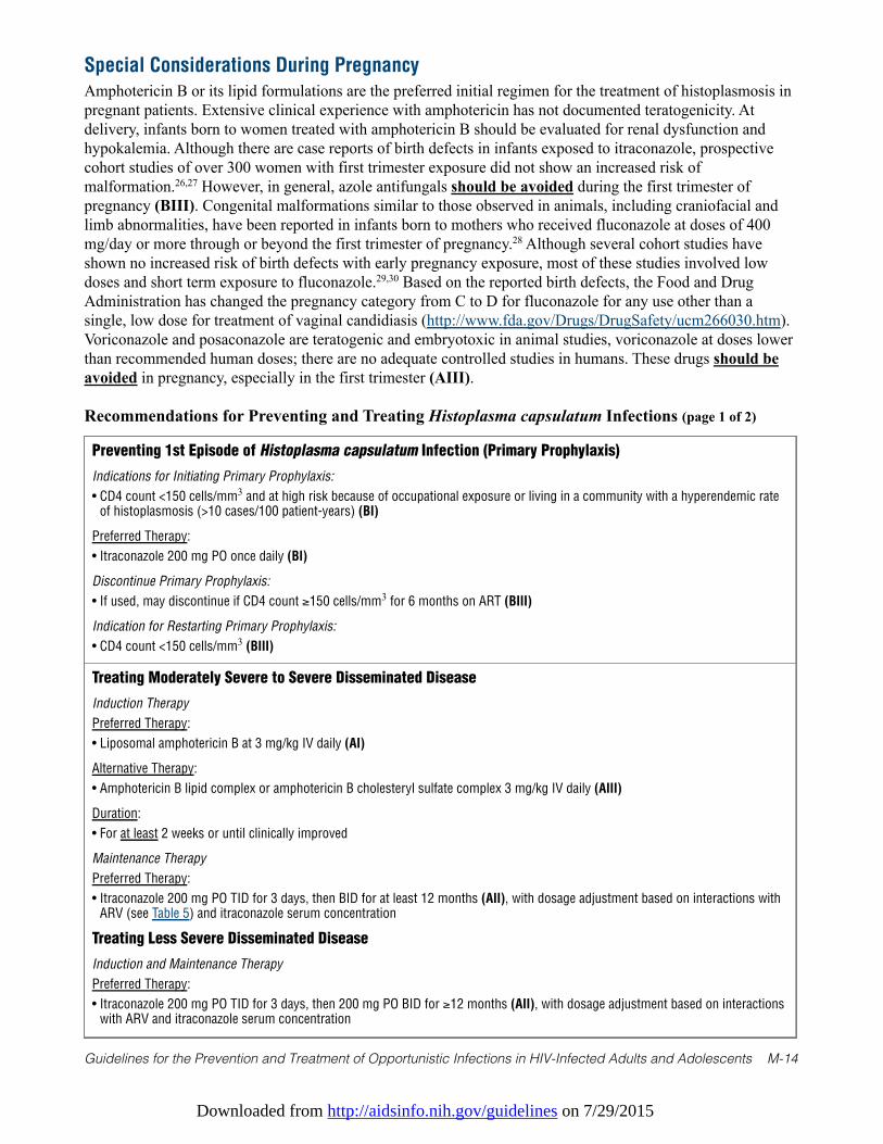
Downloaded from http://aidsinfo.nih.gov/guidelines on 7/29/2015
Guidelines for the Prevention and Treatment of Opportunistic Infections in HIV-Infected Adults and Adolescents M-14
Special Considerations During Pregnancy
Amphotericin B or its lipid formulations are the preferred initial regimen for the treatment of histoplasmosis inpregnant patients. Extensive clinical experience with amphotericin has not documented teratogenicity. Atdelivery, infants born to women treated with amphotericin B should be evaluated for renal dysfunction andhypokalemia. Although there are case reports of birth defects in infants exposed to itraconazole, prospectivecohort studies of over 300 women with first trimester exposure did not show an increased risk ofmalformation.26,27 However, in general, azole antifungals should be avoided during the first trimester ofpregnancy (BIII). Congenital malformations similar to those observed in animals, including craniofacial andlimb abnormalities, have been reported in infants born to mothers who received fluconazole at doses of 400mg/day or more through or beyond the first trimester of pregnancy.28 Although several cohort studies haveshown no increased risk of birth defects with early pregnancy exposure, most of these studies involved lowdoses and short term exposure to fluconazole.29,30 Based on the reported birth defects, the Food and DrugAdministration has changed the pregnancy category from C to D for fluconazole for any use other than asingle, low dose for treatment of vaginal candidiasis (http://www.fda.gov/Drugs/DrugSafety/ucm266030.htm).Voriconazole and posaconazole are teratogenic and embryotoxic in animal studies, voriconazole at doses lowerthan recommended human doses; there are no adequate controlled studies in humans. These drugs should beavoided in pregnancy, especially in the first trimester (AIII).
Recommendations for Preventing and Treating Histoplasma capsulatum Infections (page 1 of 2)
Preventing 1st Episode of Histoplasma capsulatum Infection (Primary Prophylaxis)
Indications for Initiating Primary Prophylaxis:
• CD4 count <150 cells/mm3 and at high risk because of occupational exposure or living in a community with a hyperendemic rateof histoplasmosis (>10 cases/100 patient-years) (BI)
Preferred Therapy:
• Itraconazole 200 mg PO once daily (BI)
Discontinue Primary Prophylaxis:
• If used, may discontinue if CD4 count ≥150 cells/mm3 for 6 months on ART (BIII)
Indication for Restarting Primary Prophylaxis:
• CD4 count <150 cells/mm3 (BIII)
Treating Moderately Severe to Severe Disseminated Disease
Induction Therapy
Preferred Therapy:
• Liposomal amphotericin B at 3 mg/kg IV daily (AI)
Alternative Therapy:
• Amphotericin B lipid complex or amphotericin B cholesteryl sulfate complex 3 mg/kg IV daily (AIII)
Duration:
• For at least 2 weeks or until clinically improved
Maintenance Therapy
Preferred Therapy:
• Itraconazole 200 mg PO TID for 3 days, then BID for at least 12 months (AII), with dosage adjustment based on interactions withARV (see Table 5) and itraconazole serum concentration
Treating Less Severe Disseminated Disease
Induction and Maintenance Therapy
Preferred Therapy:
• Itraconazole 200 mg PO TID for 3 days, then 200 mg PO BID for ≥12 months (AII), with dosage adjustment based on interactionswith ARV and itraconazole serum concentration
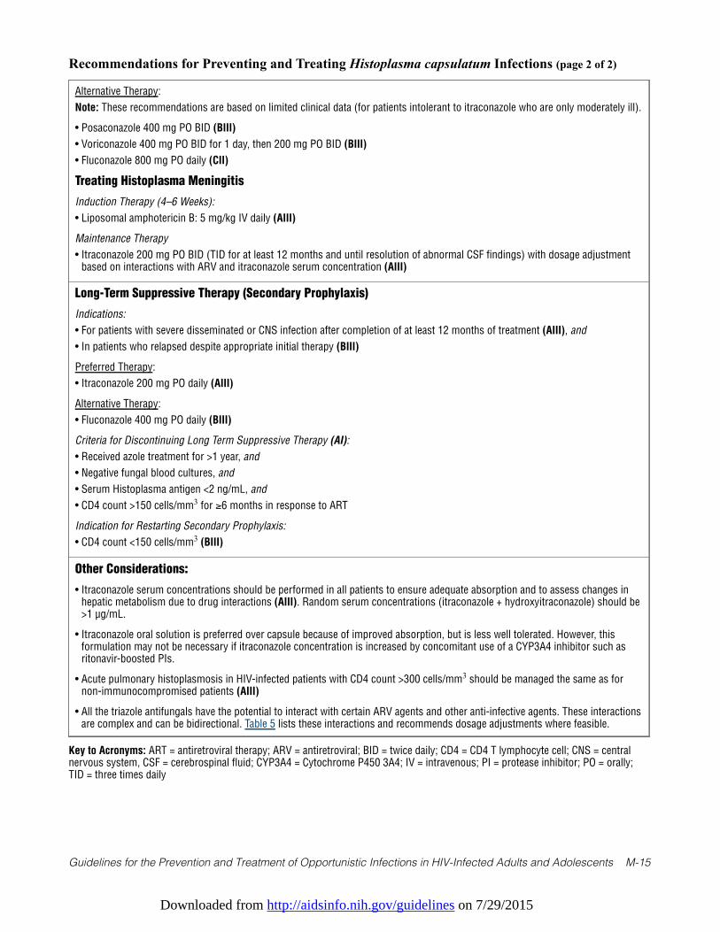
Downloaded from http://aidsinfo.nih.gov/guidelines on 7/29/2015
Guidelines for the Prevention and Treatment of Opportunistic Infections in HIV-Infected Adults and Adolescents M-15
Recommendations for Preventing and Treating Histoplasma capsulatum Infections (page 2 of 2)
Alternative Therapy:
Note: These recommendations are based on limited clinical data (for patients intolerant to itraconazole who are only moderately ill).
• Posaconazole 400 mg PO BID (BIII)
• Voriconazole 400 mg PO BID for 1 day, then 200 mg PO BID (BIII)
• Fluconazole 800 mg PO daily (CII)
Treating Histoplasma Meningitis
Induction Therapy (4–6 Weeks):
• Liposomal amphotericin B: 5 mg/kg IV daily (AIII)
Maintenance Therapy
• Itraconazole 200 mg PO BID (TID for at least 12 months and until resolution of abnormal CSF findings) with dosage adjustmentbased on interactions with ARV and itraconazole serum concentration (AIII)
Long-Term Suppressive Therapy (Secondary Prophylaxis)
Indications:
• For patients with severe disseminated or CNS infection after completion of at least 12 months of treatment (AIII), and
• In patients who relapsed despite appropriate initial therapy (BIII)
Preferred Therapy:
• Itraconazole 200 mg PO daily (AIII)
Alternative Therapy:
• Fluconazole 400 mg PO daily (BIII)
Criteria for Discontinuing Long Term Suppressive Therapy (AI):
• Received azole treatment for >1 year, and
• Negative fungal blood cultures, and
• Serum Histoplasma antigen <2 ng/mL, and
• CD4 count >150 cells/mm3 for ≥6 months in response to ART
Indication for Restarting Secondary Prophylaxis:
• CD4 count <150 cells/mm3 (BIII)
Other Considerations:
• Itraconazole serum concentrations should be performed in all patients to ensure adequate absorption and to assess changes inhepatic metabolism due to drug interactions (AIII). Random serum concentrations (itraconazole + hydroxyitraconazole) should be>1 µg/mL.
• Itraconazole oral solution is preferred over capsule because of improved absorption, but is less well tolerated. However, thisformulation may not be necessary if itraconazole concentration is increased by concomitant use of a CYP3A4 inhibitor such asritonavir-boosted PIs.
• Acute pulmonary histoplasmosis in HIV-infected patients with CD4 count >300 cells/mm3 should be managed the same as fornon-immunocompromised patients (AIII)
• All the triazole antifungals have the potential to interact with certain ARV agents and other anti-infective agents. These interactionsare complex and can be bidirectional. Table 5 lists these interactions and recommends dosage adjustments where feasible.
Key to Acronyms: ART = antiretroviral therapy; ARV = antiretroviral; BID = twice daily; CD4 = CD4 T lymphocyte cell; CNS = centralnervous system, CSF = cerebrospinal fluid; CYP3A4 = Cytochrome P450 3A4; IV = intravenous; PI = protease inhibitor; PO = orally;TID = three times daily
Downloaded from http://aidsinfo.nih.gov/guidelines on 7/29/2015
Guidelines for the Prevention and Treatment of Opportunistic Infections in HIV-Infected Adults and Adolescents M-16
References
1. Wheat LJ, Connolly-Stringfield PA, Baker RL, et al. Disseminated histoplasmosis in the acquired immune deficiencysyndrome: clinical findings, diagnosis and treatment, and review of the literature. Medicine (Baltimore). Nov1990;69(6):361-374. Available at http://www.ncbi.nlm.nih.gov/pubmed/2233233.
2. McKinsey DS, Spiegel RA, Hutwagner L, et al. Prospective study of histoplasmosis in patients infected with humanimmunodeficiency virus: incidence, risk factors, and pathophysiology. Clin Infect Dis. Jun 1997;24(6):1195-1203.Available at http://www.ncbi.nlm.nih.gov/pubmed/9195082.
3. Antinori S, Magni C, Nebuloni M, et al. Histoplasmosis among human immunodeficiency virus-infected people inEurope: report of 4 cases and review of the literature. Medicine (Baltimore). Jan 2006;85(1):22-36. Available athttp://www.ncbi.nlm.nih.gov/pubmed/16523050.
4. Baddley JW, Sankara IR, Rodriquez JM, Pappas PG, Many WJ, Jr. Histoplasmosis in HIV-infected patients in asouthern regional medical center: poor prognosis in the era of highly active antiretroviral therapy. Diagn MicrobiolInfect Dis. Oct 2008;62(2):151-156. Available at http://www.ncbi.nlm.nih.gov/pubmed/18597967.
5. Gutierrez ME, Canton A, Sosa N, Puga E, Talavera L. Disseminated histoplasmosis in patients with AIDS in Panama: areview of 104 cases. Clin Infect Dis. Apr 15 2005;40(8):1199-1202. Available athttp://www.ncbi.nlm.nih.gov/pubmed/15791523.
6. Wheat LJ, Musial CE, Jenny-Avital E. Diagnosis and management of central nervous system histoplasmosis. Clin InfectDis. Mar 15 2005;40(6):844-852. Available at http://www.ncbi.nlm.nih.gov/pubmed/15736018.
7. Assi M, McKinsey DS, Driks MR, et al. Gastrointestinal histoplasmosis in the acquired immunodeficiency syndrome:report of 18 cases and literature review. Diagn Microbiol Infect Dis. Jul 2006;55(3):195-201. Available athttp://www.ncbi.nlm.nih.gov/pubmed/16545932.
8. Swartzentruber S, Rhodes L, Kurkjian K, et al. Diagnosis of acute pulmonary histoplasmosis by antigen detection. ClinInfect Dis. Dec 15 2009;49(12):1878-1882. Available at http://www.ncbi.nlm.nih.gov/pubmed/19911965.
9. Connolly PA, Durkin MM, Lemonte AM, Hackett EJ, Wheat LJ. Detection of histoplasma antigen by a quantitativeenzyme immunoassay. Clin Vaccine Immunol. Dec 2007;14(12):1587-1591. Available athttp://www.ncbi.nlm.nih.gov/pubmed/17913863.
10. Hage CA, Davis TE, Fuller D, et al. Diagnosis of histoplasmosis by antigen detection in BAL fluid. Chest. Mar2010;137(3):623-628. Available at http://www.ncbi.nlm.nih.gov/pubmed/19837826.
11. Wheat LJ. Approach to the diagnosis of the endemic mycoses. Clin Chest Med. Jun 2009;30(2):379-389, viii. Availableat http://www.ncbi.nlm.nih.gov/pubmed/19375642.
12. Tobon AM, Agudelo CA, Rosero DS, et al. Disseminated histoplasmosis: a comparative study between patients withacquired immunodeficiency syndrome and non-human immunodeficiency virus-infected individuals. Am J Trop MedHyg. Sep 2005;73(3):576-582. Available at http://www.ncbi.nlm.nih.gov/pubmed/16172484.
13. McKinsey DS, Wheat LJ, Cloud GA, et al. Itraconazole prophylaxis for fungal infections in patients with advancedhuman immunodeficiency virus infection: randomized, placebo-controlled, double-blind study. National Institute ofAllergy and Infectious Diseases Mycoses Study Group. Clin Infect Dis. May 1999;28(5):1049-1056. Available athttp://www.ncbi.nlm.nih.gov/pubmed/10452633.
14. Johnson PC, Wheat LJ, Cloud GA, et al. Safety and efficacy of liposomal amphotericin B compared with conventionalamphotericin B for induction therapy of histoplasmosis in patients with AIDS. Ann Intern Med. Jul 16 2002;137(2):105-109. Available at http://www.ncbi.nlm.nih.gov/pubmed/12118965.
15. Wheat LJ, Freifeld AG, Kleiman MB, et al. Clinical practice guidelines for the management of patients withhistoplasmosis: 2007 update by the Infectious Diseases Society of America. Clin Infect Dis. Oct 1 2007;45(7):807-825.Available at http://www.ncbi.nlm.nih.gov/pubmed/17806045.
16. Wheat J, Hafner R, Korzun AH, et al. Itraconazole treatment of disseminated histoplasmosis in patients with theacquired immunodeficiency syndrome. AIDS Clinical Trial Group. Am J Med. Apr 1995;98(4):336-342. Available athttp://www.ncbi.nlm.nih.gov/pubmed/7709945.
17. Freifeld AG, Iwen PC, Lesiak BL, Gilroy RK, Stevens RB, Kalil AC. Histoplasmosis in solid organ transplantrecipients at a large Midwestern university transplant center. Transplant infectious disease: an official journal of theTransplantation Society. Sep-Dec 2005;7(3-4):109-115. Available at http://www.ncbi.nlm.nih.gov/pubmed/16390398.
Downloaded from http://aidsinfo.nih.gov/guidelines on 7/29/2015
Guidelines for the Prevention and Treatment of Opportunistic Infections in HIV-Infected Adults and Adolescents M-17
18. Al-Agha OM, Mooty M, Salarieh A. A 43-year-old woman with acquired immunodeficiency syndrome and fever ofundetermined origin. Disseminated histoplasmosis. Archives of pathology & laboratory medicine. Jan 2006;130(1):120-123. Available at http://www.ncbi.nlm.nih.gov/pubmed/16390228.
19. Restrepo A, Tobon A, Clark B, et al. Salvage treatment of histoplasmosis with posaconazole. J Infect. Apr2007;54(4):319-327. Available at http://www.ncbi.nlm.nih.gov/pubmed/16824608.
20. Freifeld A, Proia L, Andes D, et al. Voriconazole use for endemic fungal infections. Antimicrob Agents Chemother. Apr2009;53(4):1648-1651. Available at http://www.ncbi.nlm.nih.gov/pubmed/19139290.
21. Wheat J, MaWhinney S, Hafner R, et al. Treatment of histoplasmosis with fluconazole in patients with acquiredimmunodeficiency syndrome. National Institute of Allergy and Infectious Diseases Acquired ImmunodeficiencySyndrome Clinical Trials Group and Mycoses Study Group. Am J Med. Sep 1997;103(3):223-232. Available athttp://www.ncbi.nlm.nih.gov/pubmed/9316555.
22. Shelburne SA, 3rd, Darcourt J, White AC, Jr., et al. The role of immune reconstitution inflammatory syndrome inAIDS-related Cryptococcus neoformans disease in the era of highly active antiretroviral therapy. Clin Infect Dis. Apr 12005;40(7):1049-1052. Available at http://www.ncbi.nlm.nih.gov/pubmed/15825000.
23. Nacher M, Sarazin F, El Guedj M, et al. Increased incidence of disseminated histoplasmosis following highly activeantiretroviral therapy initiation. J Acquir Immune Defic Syndr. Apr 1 2006;41(4):468-470. Available athttp://www.ncbi.nlm.nih.gov/pubmed/16652055.
24. Hecht FM, Wheat J, Korzun AH, et al. Itraconazole maintenance treatment for histoplasmosis in AIDS: a prospective,multicenter trial. J Acquir Immune Defic Syndr Hum Retrovirol. Oct 1 1997;16(2):100-107. Available athttp://www.ncbi.nlm.nih.gov/pubmed/9358104.
25. Goldman M, Zackin R, Fichtenbaum CJ, et al. Safety of discontinuation of maintenance therapy for disseminatedhistoplasmosis after immunologic response to antiretroviral therapy. Clin Infect Dis. May 15 2004;38(10):1485-1489.Available at http://www.ncbi.nlm.nih.gov/pubmed/15156489.
26. De Santis M, Di Gianantonio E, Cesari E, Ambrosini G, Straface G, Clementi M. First-trimester itraconazole exposureand pregnancy outcome: a prospective cohort study of women contacting teratology information services in Italy. DrugSaf. 2009;32(3):239-244. Available at http://www.ncbi.nlm.nih.gov/pubmed/19338381.
27. Bar-Oz B, Moretti ME, Bishai R, et al. Pregnancy outcome after in utero exposure to itraconazole: a prospective cohortstudy. Am J Obstet Gynecol. Sep 2000;183(3):617-620. Available at http://www.ncbi.nlm.nih.gov/pubmed/10992182.
28. Pursley TJ, Blomquist IK, Abraham J, Andersen HF, Bartley JA. Fluconazole-induced congenital anomalies in threeinfants. Clin Infect Dis. Feb 1996;22(2):336-340. Available at http://www.ncbi.nlm.nih.gov/pubmed/8838193.
29. Norgaard M, Pedersen L, Gislum M, et al. Maternal use of fluconazole and risk of congenital malformations: a Danishpopulation-based cohort study. J Antimicrob Chemother. Jul 2008;62(1):172-176. Available athttp://www.ncbi.nlm.nih.gov/pubmed/18400803.
30. Mastroiacovo P, Mazzone T, Botto LD, et al. Prospective assessment of pregnancy outcomes after first-trimesterexposure to fluconazole. Am J Obstet Gynecol. Dec 1996;175(6):1645-1650. Available athttp://www.ncbi.nlm.nih.gov/pubmed/8987954.
Downloaded from http://aidsinfo.nih.gov/guidelines on 7/29/2015
Guidelines for the Prevention and Treatment of Opportunistic Infections in HIV-Infected Adults and Adolescents M-18
Coccidioidomycosis (Last updated May 7, 2013; last reviewed May 7, 2013)
Epidemiology
Coccidioidomycosis is caused by a soil-dwelling fungus that consists of two species, Coccidioides immitisand Coccidioides posadasii. Most cases of coccidioidomycosis in HIV-infected individuals have beenreported in the areas in which the disease is highly endemic.1 In the United States, these areas include thelower San Joaquin Valley in California; much of Arizona; the southern regions of Utah, Nevada, and NewMexico; and western Texas.2 Cases have been diagnosed outside those areas, presumably as a result ofreactivation of an infection previously acquired in an endemic region.
Risk of developing symptomatic disease is increased in HIV-infected patients living in an endemic area whohave CD4 T lymphocyte (CD4) cell counts <250 cells/mm3 or who have been diagnosed with AIDS.3Incidence and severity of HIV-associated coccidioidomycosis have declined since the introduction ofeffective antiretroviral therapy (ART).4,5
Clinical Manifestations
Lack of suppression of HIV replication and lower CD4 cell counts are significantly associated with theseverity of the presentation of coccidioidomycosis.5 Six common syndromes of coccidioidomycosis havebeen described in HIV-infected patients: focal pneumonia, diffuse pneumonia, cutaneous disease, meningitis,liver or lymph node involvement, and positive coccidioidal serology tests without evidence of localizedinfection.6
Focal pneumonia is most common in patients with CD4 counts ≥250 cells/mm3. This diagnosis can bedifficult to distinguish from a bacterial community-acquired pneumonia; patients present with symptoms thatinclude cough, fever, and pleuritic chest pain.7,8 The other syndromes usually occur in moreimmunosuppressed patients. Diffuse pulmonary disease presents with fever and dyspnea and can be difficultto clinically distinguish from Pneumocystis pneumonia.9 Meningitis presents with a persistent headache andprogressive lethargy. The cerebrospinal fluid (CSF) profile demonstrates a low glucose level with elevatedprotein and a lymphocytic pleocytosis.
Diagnosis
The diagnosis of coccidioidomycosis is confirmed by culture of the organism from clinical specimens or bydemonstration of the typical spherule on histopathological examination of involved tissue. Blood cultures arepositive in a minority of patients, usually those with diffuse pulmonary disease. Coccidioidalimmunoglobulin M (IgM) and immunoglobulin G (IgG) serology, performed by enzyme immunoassay,immunodiffusion, or classical tube precipitin or complement fixation methodology, is useful in diagnosis butmay be positive less often in patients with low CD4 cell counts than in those who are immunocompetent.10
Complement fixation IgG antibody often is detected in the CSF in coccidioidal meningitis and is useful inestablishing this diagnosis. Culture of the CSF is positive in less than one-third of patients with meningitis. Acoccidioidomycosis-specific antigen assay recently has become commercially available. It has been shown todetect antigen in urine11 and serum12 samples from HIV-infected individuals with active coccidioidomycosisand appears to be useful in diagnosing coccidioidomycosis in such patients.
Preventing Exposure
HIV-infected individuals cannot completely avoid activities involving exposure to infection while living in orvisiting areas in which Coccidioides spp. are endemic. They should, however, avoid extensive exposure todisturbed native soil, such as at building excavation sites, and stay inside during dust storms (BIII).
Downloaded from http://aidsinfo.nih.gov/guidelines on 7/29/2015
Guidelines for the Prevention and Treatment of Opportunistic Infections in HIV-Infected Adults and Adolescents M-19
Preventing Disease
Primary antifungal prophylaxis is of little benefit to patients with low CD4 cell counts who live in regionswhere Coccidioides spp. are endemic4 and it is not recommended (AIII).
Yearly serologic testing for coccidioidomycosis is reasonable for HIV-infected individuals who live inregions endemic for coccidioidomycosis. In such settings, a new positive test suggests imminent activedisease in patients with low CD4 cell counts13 and pre-emptive antifungal therapy with fluconazole 400 mgdaily is recommended for those with CD4 counts <250/mm3 (BIII). Outside endemic regions, routine testingdoes not appear to be useful and should not be performed.
Treating Disease
Initial therapy with a triazole antifungal is appropriate for patients who have clinically mild infection, such asfocal pneumonia (BII). Fluconazole or itraconazole at doses of 400 mg daily is recommended.14,15 Data arelimited on the newer triazoles (posaconazole16,17 and voriconazole), but these agents may be useful forpatients who fail to respond to fluconazole or itraconazole.
Amphotericin B is the preferred initial therapy for patients who have diffuse pulmonary involvement or areseverely ill with extrathoracic disseminated disease (AII).15 Most experience has been with the deoxycholateformulation, using an initial dose of 0.7 to 1.0 mg/kg intravenously (IV) daily. No data exist about use oflipid formulations of amphotericin B, but they are likely to be as effective as the deoxycholate formulationand may be considered as an alternative initial therapy (AIII).
Therapy with amphotericin B should continue until clinical improvement is observed. Some specialistsrecommend combining amphotericin B with a triazole (either fluconazole or itraconazole, with itraconazolepreferred for bone disease) at 400 mg daily at initiation of therapy, and then continue the triazole onceamphotericin B is stopped (BIII).15
Treatment of patients with coccidioidal meningitis requires consultation with a specialist. Therapy shouldbegin with a triazole antifungal. IV or oral fluconazole at a dose of 400 to 800 mg daily is preferred (AII),18
but itraconazole also has been used successfully (BII).19 Successful therapy with posaconazole (CIII)17,20 andvoriconazole (BIII)21-23 has been described in individual cases. Despite successful antifungal therapy, somepatients may develop hydrocephalus and require CSF shunting. In some instances, triazole antifungals areineffective and intrathecal amphotericin B is recommended (AIII). Intrathecal amphotericin B should beadministered by someone with experience in this technique.
Special Considerations with Regard to Starting ARTHIV-infected individuals diagnosed with coccidioidomycosis should be started on ART as soon as possibleafter initiating antifungal therapy (AIII). Immune reconstitution inflammatory syndrome (IRIS) has beenreported once24 but concern for the syndrome should not delay initiation of ART (AIII).
Monitoring of Response to Therapy and Adverse Events (including IRIS) Monitoring the titer of the complement-fixing antibody is useful in assessing response to therapy, and itshould be measured every 12 weeks. A rise suggests recurrence or worsening of clinical disease and shouldprompt reassessment of management. As indicated in previous sections, all of the triazole antifungals havethe potential for complex, and possibly bidirectional, interactions with certain ARV agents and other anti-infective agents. Table 5 lists such interactions and recommendations for dosage adjustments, where feasible.
Managing Treatment FailurePatients with severe coccidioidomycosis who fail treatment with fluconazole or itraconazole should have theirtreatment changed to IV amphotericin B, either deoxycholate or lipid formulation (AIII). For patients who arenot severely ill, posaconazole (BII) and voriconazole (BIII)—both given in doses of 200 mg orally twice
Downloaded from http://aidsinfo.nih.gov/guidelines on 7/29/2015
Guidelines for the Prevention and Treatment of Opportunistic Infections in HIV-Infected Adults and Adolescents M-20
daily—can be considered, although data are limited regarding their efficacy. Drug interactions may limit the useof voriconazole in patients who are taking non-nucleoside reverse transcriptase inhibitors or ritonavir (see Table5). Posaconazole has fewer known drug interactions with ARV medications than does voriconazole.
Preventing Recurrence
When To Start Secondary ProphylaxisPatients who complete initial therapy for coccidioidomycosis should be considered for lifelong suppressivetherapy using either fluconazole 400 mg daily or itraconazole 200 mg twice daily if their CD4 counts remain<250 cells/mm3 (AII). Posaconazole 200 mg twice daily (BII) or voriconazole 200 mg twice daily (BIII) arealternatives if the patient did not initially respond to either fluconazole or itraconazole.
When To Stop Secondary ProphylaxisPatients with focal coccidioidal pneumonia who have clinically responded to antifungal therapy appear to beat low risk of recurrence of coccidioidomycosis if their CD4 cell counts are ≥250 cells/mm3 and they arereceiving effective ART. A reasonable plan for treating these individuals is to discontinue secondaryprophylaxis after 12 months of therapy (AII) and continue monitoring for recurrence with serial chestradiographs and coccidioidal serology.
Relapse occurs in 25% to 33% of HIV-uninfected patients who have diffuse pulmonary disease ornonmeningeal disseminated coccidioidomycosis25,26 and can occur in HIV-infected patients with CD4 counts≥250 cells/mm3 on potent ART;27 therefore, some clinicians would continue antifungal therapy indefinitely(BIII), although this decision should be made in conjunction with expert consultation. Because relapses havebeen reported in 80% of patients with meningitis in whom triazoles have been discontinued,28 therapy forcoccidioidal meningitis should be lifelong (AII).
Special Considerations During PregnancyCoccidioidomycosis is more likely to disseminate if acquired during the second or third trimester ofpregnancy.29 Amphotericin B or its lipid formulations are the preferred initial regimen for the treatment ofcoccidioidomycosis in pregnant patients. Extensive clinical use of amphotericin has not been associated withteratogenicity. At delivery, infants born to women treated with amphotericin B should be evaluated for renaldysfunction and hypokalemia.
Congenital malformations similar to those observed in animals, including craniofacial and limbabnormalities, have been reported in infants born to mothers who received fluconazole through or beyond thefirst trimester of pregnancy.30 Although several cohort studies have shown no increased risk of birth defectswith early pregnancy exposure to fluconazole, most of these involved low doses and short term exposure.31,32
Based on the reported birth defects, the Food and Drug Administration has changed the pregnancy categoryfrom C to D for fluconazole for any use other than a single, 150 mg dose to treat vaginal candidiasis(http://www.fda.gov/Drugs/DrugSafety/ucm266030.htm). Although there are case reports of birth defects ininfants exposed to itraconazole, prospective cohort studies of over 300 women with first trimester exposuredid not show an increased risk of malformation.33,34 However, in general, azole antifungals should beavoided during the first trimester of pregnancy (BIII). One problematic area is coccidioidal meningitis, inwhich the only alternative treatment to triazole antifungals is intrathecal amphotericin B. For such situations,the decision regarding choice of treatment should be based on considerations of benefit versus potential riskand made in consultation with the mother, the infectious diseases consultant, and the obstetrician.35
Voriconazole and posaconazole are teratogenic and embryotoxic in animal studies, voriconazole at doseslower than recommended human doses; there are no adequate controlled studies in humans. These drugsshould be avoided in pregnancy, especially in the first trimester (AIII).
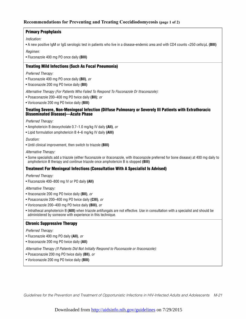
Downloaded from http://aidsinfo.nih.gov/guidelines on 7/29/2015
Guidelines for the Prevention and Treatment of Opportunistic Infections in HIV-Infected Adults and Adolescents M-21
Recommendations for Preventing and Treating Coccidiodomycosis (page 1 of 2)
Primary Prophylaxis
Indication:
• A new positive IgM or IgG serologic test in patients who live in a disease-endemic area and with CD4 counts <250 cells/µL (BIII)
Regimen:
• Fluconazole 400 mg PO once daily (BIII)
Treating Mild Infections (Such As Focal Pneumonia)
Preferred Therapy:
• Fluconazole 400 mg PO once daily (BII), or
• Itraconazole 200 mg PO twice daily (BII)
Alternative Therapy (For Patients Who Failed To Respond To Fluconazole Or Itraconazole):
• Posaconazole 200–400 mg PO twice daily (BII); or
• Voriconazole 200 mg PO twice daily (BIII)
Treating Severe, Non-Meningeal Infection (Diffuse Pulmonary or Severely Ill Patients with ExtrathoracicDisseminated Disease)—Acute Phase
Preferred Therapy:
• Amphotericin B deoxycholate 0.7–1.0 mg/kg IV daily (AII), or
• Lipid formulation amphotericin B 4–6 mg/kg IV daily (AIII)
Duration:
• Until clinical improvement, then switch to triazole (BIII)
Alternative Therapy:
• Some specialists add a triazole (either fluconazole or itraconazole, with itraconazole preferred for bone disease) at 400 mg daily toamphotericin B therapy and continue triazole once amphotericin B is stopped (BIII)
Treatment For Meningeal Infections (Consultation With A Specialist Is Advised)
Preferred Therapy:
• Fluconazole 400–800 mg IV or PO daily (AII)
Alternative Therapy:
• Itraconazole 200 mg PO twice daily (BII), or
• Posaconazole 200–400 mg PO twice daily (CIII), or
• Voriconazole 200–400 mg PO twice daily (BIII), or
• Intrathecal amphotericin B (AIII) when triazole antifungals are not effective. Use in consultation with a specialist and should beadministered by someone with experience in this technique.
Chronic Suppressive Therapy
Preferred Therapy:
• Fluconazole 400 mg PO daily (AII), or
• Itraconazole 200 mg PO twice daily (AII)
Alternative Therapy (If Patients Did Not Initially Respond to Fluconazole or Itraconazole):
• Posaconazole 200 mg PO twice daily (BII), or
• Voriconazole 200 mg PO twice daily (BIII)
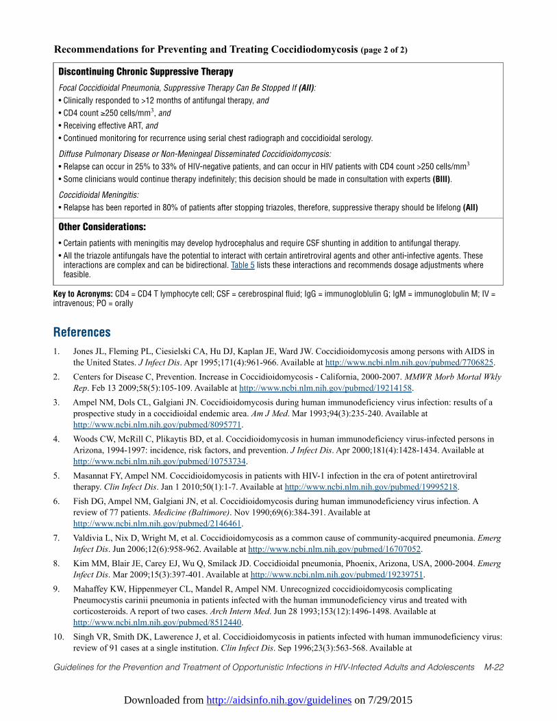
Downloaded from http://aidsinfo.nih.gov/guidelines on 7/29/2015
Guidelines for the Prevention and Treatment of Opportunistic Infections in HIV-Infected Adults and Adolescents M-22
Recommendations for Preventing and Treating Coccidiodomycosis (page 2 of 2)
Discontinuing Chronic Suppressive Therapy
Focal Coccidioidal Pneumonia, Suppressive Therapy Can Be Stopped If (AII):
• Clinically responded to >12 months of antifungal therapy, and
• CD4 count ≥250 cells/mm3, and
• Receiving effective ART, and
• Continued monitoring for recurrence using serial chest radiograph and coccidioidal serology.
Diffuse Pulmonary Disease or Non-Meningeal Disseminated Coccidioidomycosis:
• Relapse can occur in 25% to 33% of HIV-negative patients, and can occur in HIV patients with CD4 count >250 cells/mm3
• Some clinicians would continue therapy indefinitely; this decision should be made in consultation with experts (BIII).
Coccidioidal Meningitis:
• Relapse has been reported in 80% of patients after stopping triazoles, therefore, suppressive therapy should be lifelong (AII)
Other Considerations:
• Certain patients with meningitis may develop hydrocephalus and require CSF shunting in addition to antifungal therapy.
• All the triazole antifungals have the potential to interact with certain antiretroviral agents and other anti-infective agents. Theseinteractions are complex and can be bidirectional. Table 5 lists these interactions and recommends dosage adjustments wherefeasible.
Key to Acronyms: CD4 = CD4 T lymphocyte cell; CSF = cerebrospinal fluid; IgG = immunogloblulin G; IgM = immunoglobulin M; IV =intravenous; PO = orally
References
1. Jones JL, Fleming PL, Ciesielski CA, Hu DJ, Kaplan JE, Ward JW. Coccidioidomycosis among persons with AIDS inthe United States. J Infect Dis. Apr 1995;171(4):961-966. Available at http://www.ncbi.nlm.nih.gov/pubmed/7706825.
2. Centers for Disease C, Prevention. Increase in Coccidioidomycosis - California, 2000-2007. MMWR Morb Mortal WklyRep. Feb 13 2009;58(5):105-109. Available at http://www.ncbi.nlm.nih.gov/pubmed/19214158.
3. Ampel NM, Dols CL, Galgiani JN. Coccidioidomycosis during human immunodeficiency virus infection: results of aprospective study in a coccidioidal endemic area. Am J Med. Mar 1993;94(3):235-240. Available athttp://www.ncbi.nlm.nih.gov/pubmed/8095771.
4. Woods CW, McRill C, Plikaytis BD, et al. Coccidioidomycosis in human immunodeficiency virus-infected persons inArizona, 1994-1997: incidence, risk factors, and prevention. J Infect Dis. Apr 2000;181(4):1428-1434. Available athttp://www.ncbi.nlm.nih.gov/pubmed/10753734.
5. Masannat FY, Ampel NM. Coccidioidomycosis in patients with HIV-1 infection in the era of potent antiretroviraltherapy. Clin Infect Dis. Jan 1 2010;50(1):1-7. Available at http://www.ncbi.nlm.nih.gov/pubmed/19995218.
6. Fish DG, Ampel NM, Galgiani JN, et al. Coccidioidomycosis during human immunodeficiency virus infection. Areview of 77 patients. Medicine (Baltimore). Nov 1990;69(6):384-391. Available athttp://www.ncbi.nlm.nih.gov/pubmed/2146461.
7. Valdivia L, Nix D, Wright M, et al. Coccidioidomycosis as a common cause of community-acquired pneumonia. EmergInfect Dis. Jun 2006;12(6):958-962. Available at http://www.ncbi.nlm.nih.gov/pubmed/16707052.
8. Kim MM, Blair JE, Carey EJ, Wu Q, Smilack JD. Coccidioidal pneumonia, Phoenix, Arizona, USA, 2000-2004. EmergInfect Dis. Mar 2009;15(3):397-401. Available at http://www.ncbi.nlm.nih.gov/pubmed/19239751.
9. Mahaffey KW, Hippenmeyer CL, Mandel R, Ampel NM. Unrecognized coccidioidomycosis complicatingPneumocystis carinii pneumonia in patients infected with the human immunodeficiency virus and treated withcorticosteroids. A report of two cases. Arch Intern Med. Jun 28 1993;153(12):1496-1498. Available athttp://www.ncbi.nlm.nih.gov/pubmed/8512440.
10. Singh VR, Smith DK, Lawerence J, et al. Coccidioidomycosis in patients infected with human immunodeficiency virus:review of 91 cases at a single institution. Clin Infect Dis. Sep 1996;23(3):563-568. Available at
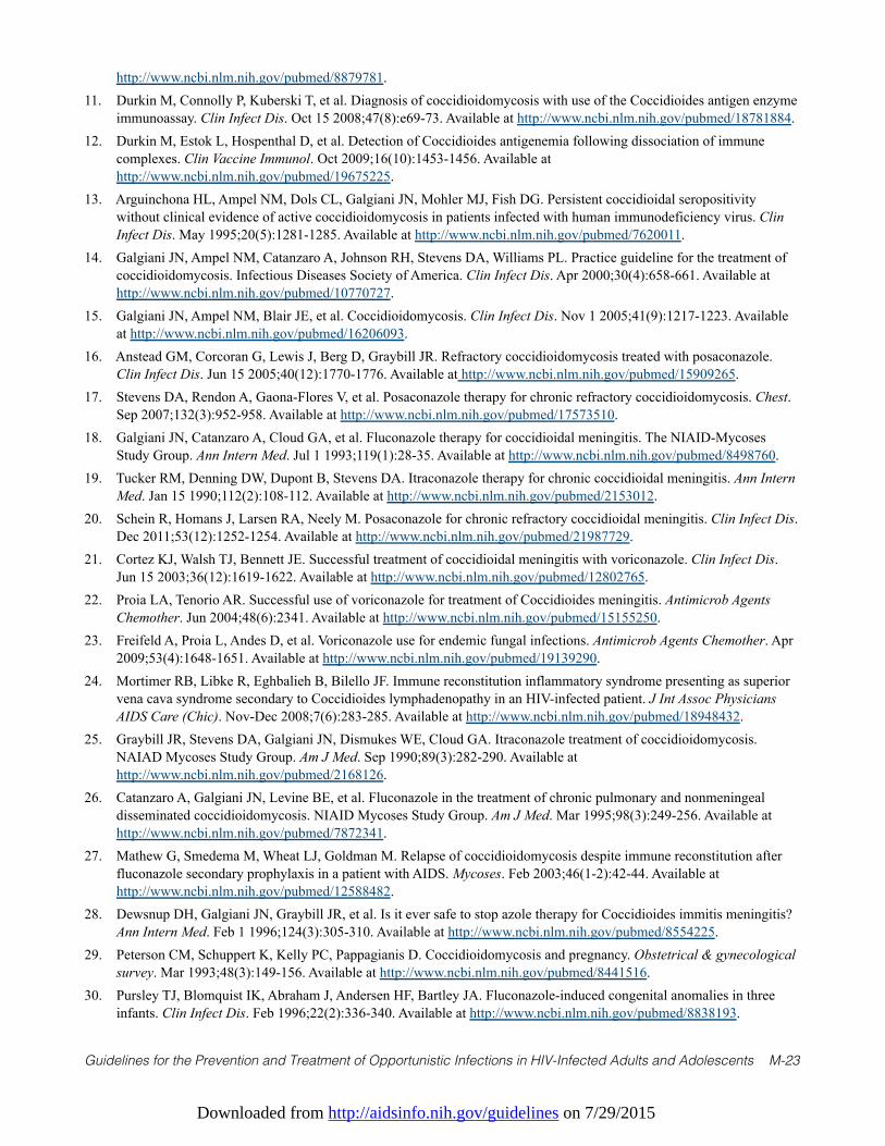
Downloaded from http://aidsinfo.nih.gov/guidelines on 7/29/2015
Guidelines for the Prevention and Treatment of Opportunistic Infections in HIV-Infected Adults and Adolescents M-23
http://www.ncbi.nlm.nih.gov/pubmed/8879781.11. Durkin M, Connolly P, Kuberski T, et al. Diagnosis of coccidioidomycosis with use of the Coccidioides antigen enzyme
immunoassay. Clin Infect Dis. Oct 15 2008;47(8):e69-73. Available at http://www.ncbi.nlm.nih.gov/pubmed/18781884.12. Durkin M, Estok L, Hospenthal D, et al. Detection of Coccidioides antigenemia following dissociation of immune
complexes. Clin Vaccine Immunol. Oct 2009;16(10):1453-1456. Available athttp://www.ncbi.nlm.nih.gov/pubmed/19675225.
13. Arguinchona HL, Ampel NM, Dols CL, Galgiani JN, Mohler MJ, Fish DG. Persistent coccidioidal seropositivitywithout clinical evidence of active coccidioidomycosis in patients infected with human immunodeficiency virus. ClinInfect Dis. May 1995;20(5):1281-1285. Available at http://www.ncbi.nlm.nih.gov/pubmed/7620011.
14. Galgiani JN, Ampel NM, Catanzaro A, Johnson RH, Stevens DA, Williams PL. Practice guideline for the treatment ofcoccidioidomycosis. Infectious Diseases Society of America. Clin Infect Dis. Apr 2000;30(4):658-661. Available athttp://www.ncbi.nlm.nih.gov/pubmed/10770727.
15. Galgiani JN, Ampel NM, Blair JE, et al. Coccidioidomycosis. Clin Infect Dis. Nov 1 2005;41(9):1217-1223. Availableat http://www.ncbi.nlm.nih.gov/pubmed/16206093.
16. Anstead GM, Corcoran G, Lewis J, Berg D, Graybill JR. Refractory coccidioidomycosis treated with posaconazole.Clin Infect Dis. Jun 15 2005;40(12):1770-1776. Available at http://www.ncbi.nlm.nih.gov/pubmed/15909265.
17. Stevens DA, Rendon A, Gaona-Flores V, et al. Posaconazole therapy for chronic refractory coccidioidomycosis. Chest.Sep 2007;132(3):952-958. Available at http://www.ncbi.nlm.nih.gov/pubmed/17573510.
18. Galgiani JN, Catanzaro A, Cloud GA, et al. Fluconazole therapy for coccidioidal meningitis. The NIAID-MycosesStudy Group. Ann Intern Med. Jul 1 1993;119(1):28-35. Available at http://www.ncbi.nlm.nih.gov/pubmed/8498760.
19. Tucker RM, Denning DW, Dupont B, Stevens DA. Itraconazole therapy for chronic coccidioidal meningitis. Ann InternMed. Jan 15 1990;112(2):108-112. Available at http://www.ncbi.nlm.nih.gov/pubmed/2153012.
20. Schein R, Homans J, Larsen RA, Neely M. Posaconazole for chronic refractory coccidioidal meningitis. Clin Infect Dis.Dec 2011;53(12):1252-1254. Available at http://www.ncbi.nlm.nih.gov/pubmed/21987729.
21. Cortez KJ, Walsh TJ, Bennett JE. Successful treatment of coccidioidal meningitis with voriconazole. Clin Infect Dis.Jun 15 2003;36(12):1619-1622. Available at http://www.ncbi.nlm.nih.gov/pubmed/12802765.
22. Proia LA, Tenorio AR. Successful use of voriconazole for treatment of Coccidioides meningitis. Antimicrob AgentsChemother. Jun 2004;48(6):2341. Available at http://www.ncbi.nlm.nih.gov/pubmed/15155250.
23. Freifeld A, Proia L, Andes D, et al. Voriconazole use for endemic fungal infections. Antimicrob Agents Chemother. Apr2009;53(4):1648-1651. Available at http://www.ncbi.nlm.nih.gov/pubmed/19139290.
24. Mortimer RB, Libke R, Eghbalieh B, Bilello JF. Immune reconstitution inflammatory syndrome presenting as superiorvena cava syndrome secondary to Coccidioides lymphadenopathy in an HIV-infected patient. J Int Assoc PhysiciansAIDS Care (Chic). Nov-Dec 2008;7(6):283-285. Available at http://www.ncbi.nlm.nih.gov/pubmed/18948432.
25. Graybill JR, Stevens DA, Galgiani JN, Dismukes WE, Cloud GA. Itraconazole treatment of coccidioidomycosis.NAIAD Mycoses Study Group. Am J Med. Sep 1990;89(3):282-290. Available athttp://www.ncbi.nlm.nih.gov/pubmed/2168126.
26. Catanzaro A, Galgiani JN, Levine BE, et al. Fluconazole in the treatment of chronic pulmonary and nonmeningealdisseminated coccidioidomycosis. NIAID Mycoses Study Group. Am J Med. Mar 1995;98(3):249-256. Available athttp://www.ncbi.nlm.nih.gov/pubmed/7872341.
27. Mathew G, Smedema M, Wheat LJ, Goldman M. Relapse of coccidioidomycosis despite immune reconstitution afterfluconazole secondary prophylaxis in a patient with AIDS. Mycoses. Feb 2003;46(1-2):42-44. Available athttp://www.ncbi.nlm.nih.gov/pubmed/12588482.
28. Dewsnup DH, Galgiani JN, Graybill JR, et al. Is it ever safe to stop azole therapy for Coccidioides immitis meningitis?Ann Intern Med. Feb 1 1996;124(3):305-310. Available at http://www.ncbi.nlm.nih.gov/pubmed/8554225.
29. Peterson CM, Schuppert K, Kelly PC, Pappagianis D. Coccidioidomycosis and pregnancy. Obstetrical & gynecologicalsurvey. Mar 1993;48(3):149-156. Available at http://www.ncbi.nlm.nih.gov/pubmed/8441516.
30. Pursley TJ, Blomquist IK, Abraham J, Andersen HF, Bartley JA. Fluconazole-induced congenital anomalies in threeinfants. Clin Infect Dis. Feb 1996;22(2):336-340. Available at http://www.ncbi.nlm.nih.gov/pubmed/8838193.
Downloaded from http://aidsinfo.nih.gov/guidelines on 7/29/2015
Guidelines for the Prevention and Treatment of Opportunistic Infections in HIV-Infected Adults and Adolescents M-24
31. Norgaard M, Pedersen L, Gislum M, et al. Maternal use of fluconazole and risk of congenital malformations: a Danishpopulation-based cohort study. J Antimicrob Chemother. Jul 2008;62(1):172-176. Available athttp://www.ncbi.nlm.nih.gov/pubmed/18400803.
32. Mastroiacovo P, Mazzone T, Botto LD, et al. Prospective assessment of pregnancy outcomes after first-trimesterexposure to fluconazole. Am J Obstet Gynecol. Dec 1996;175(6):1645-1650. Available athttp://www.ncbi.nlm.nih.gov/pubmed/8987954.
33. De Santis M, Di Gianantonio E, Cesari E, Ambrosini G, Straface G, Clementi M. First-trimester itraconazole exposureand pregnancy outcome: a prospective cohort study of women contacting teratology information services in Italy. DrugSaf. 2009;32(3):239-244. Available at http://www.ncbi.nlm.nih.gov/pubmed/19338381.
34. Bar-Oz B, Moretti ME, Bishai R, et al. Pregnancy outcome after in utero exposure to itraconazole: a prospective cohortstudy. Am J Obstet Gynecol. Sep 2000;183(3):617-620. Available at http://www.ncbi.nlm.nih.gov/pubmed/10992182.
35. Bercovitch RS, Catanzaro A, Schwartz BS, Pappagianis D, Watts DH, Ampel NM. Coccidioidomycosis duringpregnancy: a review and recommendations for management. Clin Infect Dis. Aug 2011;53(4):363-368. Available athttp://www.ncbi.nlm.nih.gov/pubmed/21810749.
Downloaded from http://aidsinfo.nih.gov/guidelines on 7/29/2015
Guidelines for the Prevention and Treatment of Opportunistic Infections in HIV-Infected Adults and Adolescents M-25
Aspergillosis (Last updated May 7, 2013; last reviewed May 7, 2013)
Epidemiology
Invasive aspergillosis is rare in HIV-infected individuals but often overlooked antemortem. In a recent autopsyseries of HIV-infected patients from Italy, invasive aspergillosis was the second most frequently identifiedinvasive mycosis in fatal cases, 88% of which were diagnosed only postmortem.1 Illness most often is causedby Aspergillus fumigatus, but Aspergillus flavus, Aspergillus niger, and Aspergillus terreus have been noted tocause disease. Invasive aspergillosis occurs in patients with advanced HIV infection and was more commonbefore the advent of effective antiretroviral therapy (ART).1-3 Specific risk factors include neutropenia, use ofcorticosteroids, exposure to broad-spectrum antibacterial therapy, and underlying lung disease. Patients whohave had HIV-associated aspergillosis typically have CD4 T lymphocyte (CD4) cell counts <100 cells/mm3, ahistory of other AIDS-defining opportunistic infections, and are not receiving potent ART.4
Clinical Manifestations
In HIV-infected patients, invasive aspergillosis most commonly presents as a respiratory illness that can be anecrotizing pneumonia or a tracheobronchitis.5 Symptoms of pneumonia include fever, cough, dyspnea, chestpain, hemoptysis, and hypoxemia; chest radiograph may demonstrate a diffuse, focal, or cavitary infiltrate. Ahalo of low attenuation surrounding a pulmonary nodule or a cavity on a computed tomography (CT) scan ofthe lung is suggestive of pulmonary aspergillosis. Tracheobronchitis is associated with fever, cough, dyspnea,stridor, and wheezing. Bronchoscopic examination demonstrates ulcerative or plaque-like lesions adherent tothe tracheal wall.6 Extrapulmonary forms of invasive aspergillosis include sinusitis, cutaneous disease,osteomyelitis, and brain abscess.7
Diagnosis
The diagnosis of probable invasive pulmonary aspergillosis is based on isolation of Aspergillus spp. fromrespiratory secretions or the finding of septate hyphae consistent with Aspergillus spp. in respiratory samplesin association with typical CT findings. Histological evidence of tissue invasion by septate hyphae with apositive culture for Aspergillus spp. establishes a definitive diagnosis.8
Detection of Aspergillus cell wall galactomannan by enzyme-linked immunosorbent assay (ELISA)performed on serum or bronchoalveolar lavage fluid has not been formally evaluated in HIV-infectedpatients. It has proven useful, however, in other immunosuppressed patients, especially recipients of stemcell transplants,9 and is listed by the European Organisation for Research and Treatment of Cancer/U.S.Mycosis Study Group Consensus Group as one of the criteria for establishing a diagnosis of probableinvasive aspergillosis.8 Bronchoalveolar lavage galactomannan is probably more sensitive than serumgalactomannan for diagnosis. The test is highly specific.
Preventing Exposure
Aspergillus spp. are ubiquitous in the environment, and exposure is unavoidable. Avoiding particularly dustyenvironments, especially areas of construction, is prudent because spore counts likely are higher in such settings.
Preventing Disease
No data exist about the prevention of primary aspergillosis in HIV-infected patients, although posaconazole hasbeen reported to be effective in patients with certain hematological malignancies and neutropenia.10 At this time,antifungal therapy is not recommended for prevention of aspergillosis in HIV-infected individuals (AIII).
Treating Disease
Treatment of aspergillosis in HIV-infected patients has not been systematically examined. Voriconazole is the
Downloaded from http://aidsinfo.nih.gov/guidelines on 7/29/2015
Guidelines for the Prevention and Treatment of Opportunistic Infections in HIV-Infected Adults and Adolescents M-26
recommended treatment for invasive aspergillosis in HIV-uninfected patients (AI).11 Because of drug-druginteractions, however, voriconazole should be used cautiously with protease inhibitors (PIs) and efavirenz(see Table 5). Alternatively, lipid-formulation amphotericin B or amphotericin B deoxycholate can be used(AII). Second-line agents include echinocandins (such as caspofungin, anidulafungin, or micafungin) orposaconazole (BIII). The role of combination antifungal therapy for primary treatment of invasiveaspergillosis is being evaluated in a large, randomized trial comparing voriconazole alone with voriconazoleplus anidulafungin in recipients of stem cell transplants. The length of therapy has not been established, buttreatment should continue at least until the peripheral blood CD4 count is >200 cells/mm3 and the infectionappears to be resolved (BIII).
Special Considerations with Regard to Starting ARTHIV-infected individuals diagnosed with aspergillosis should be started on ART as soon as possible afterinitiating antifungal therapy (AIII). Immune reconstitution inflammatory syndrome (IRIS) has rarely beenreported in HIV-infected patients with invasive aspergillosis12 and concern for the syndrome should not delayinitiation of ART (AIII).
All of the triazole antifungals have the potential for complex, and possibly bidirectional, interactions withcertain antiretroviral agents and other anti-infective agents. Table 5 lists such interactions andrecommendations for dosage adjustments, where feasible.
Monitoring of Response to Therapy and Adverse Events (including IRIS) Data are limited with regard to monitoring of Aspergillus galactomannan levels in response to therapy. Aspreviously stated, IRIS rarely has been reported in HIV-infected patients with invasive aspergillosis12 andnew or recurrent signs and symptoms should prompt evaluation for relapse or recurrence of aspergillosis.
Managing Treatment FailureThe overall prognosis for invasive aspergillosis is poor in patients with advanced immunosuppression and inthe absence of effective ART. No data are available to guide recommendations for management of treatmentfailure. If voriconazole was used initially, substitution can be considered with an amphotericin B formulationor with echinocandins in combination with voriconazole or amphotericin B (BIII).
Preventing Recurrence
No data are available on which to base a recommendation for or against chronic maintenance or suppressivetherapy in patients who have successfully completed an initial course of treatment.
Special Considerations During Pregnancy
Amphotericin B or its lipid formulations are the preferred initial regimen for the treatment of aspergillosis inpregnant patients. Extensive clinical experience with amphotericin has not documented teratogenicity. Atdelivery, infants born to women treated with amphotericin B should be evaluated for renal dysfunction andhypokalemia.
Voriconazole and posaconazole are teratogenic and embryotoxic in animal studies, voriconazole at doseslower than recommended human doses; here are no adequate controlled studies in humans. These drugsshould generally be avoided in pregnancy, especially in the first trimester (AIII). The echinocandins areassociated with bony and visceral abnormalities in animal studies, but no human experience is documented.These agents should be avoided in the first trimester of pregnancy; use in later pregnancy should be based onconsideration of benefit versus potential risk.
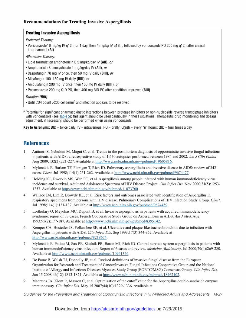
Downloaded from http://aidsinfo.nih.gov/guidelines on 7/29/2015
Guidelines for the Prevention and Treatment of Opportunistic Infections in HIV-Infected Adults and Adolescents M-27
References
1. Antinori S, Nebuloni M, Magni C, et al. Trends in the postmortem diagnosis of opportunistic invasive fungal infectionsin patients with AIDS: a retrospective study of 1,630 autopsies performed between 1984 and 2002. Am J Clin Pathol.Aug 2009;132(2):221-227. Available at http://www.ncbi.nlm.nih.gov/pubmed/19605816.
2. Mylonakis E, Barlam TF, Flanigan T, Rich JD. Pulmonary aspergillosis and invasive disease in AIDS: review of 342cases. Chest. Jul 1998;114(1):251-262. Available at http://www.ncbi.nlm.nih.gov/pubmed/9674477.
3. Holding KJ, Dworkin MS, Wan PC, et al. Aspergillosis among people infected with human immunodeficiency virus:incidence and survival. Adult and Adolescent Spectrum of HIV Disease Project. Clin Infect Dis. Nov 2000;31(5):1253-1257. Available at http://www.ncbi.nlm.nih.gov/pubmed/11073760.
4. Wallace JM, Lim R, Browdy BL, et al. Risk factors and outcomes associated with identification of Aspergillus inrespiratory specimens from persons with HIV disease. Pulmonary Complications of HIV Infection Study Group. Chest.Jul 1998;114(1):131-137. Available at http://www.ncbi.nlm.nih.gov/pubmed/9674459.
5. Lortholary O, Meyohas MC, Dupont B, et al. Invasive aspergillosis in patients with acquired immunodeficiencysyndrome: report of 33 cases. French Cooperative Study Group on Aspergillosis in AIDS. Am J Med. Aug1993;95(2):177-187. Available at http://www.ncbi.nlm.nih.gov/pubmed/8395142.
6. Kemper CA, Hostetler JS, Follansbee SE, et al. Ulcerative and plaque-like tracheobronchitis due to infection withAspergillus in patients with AIDS. Clin Infect Dis. Sep 1993;17(3):344-352. Available athttp://www.ncbi.nlm.nih.gov/pubmed/8218674.
7. Mylonakis E, Paliou M, Sax PE, Skolnik PR, Baron MJ, Rich JD. Central nervous system aspergillosis in patients withhuman immunodeficiency virus infection. Report of 6 cases and review. Medicine (Baltimore). Jul 2000;79(4):269-280.Available at http://www.ncbi.nlm.nih.gov/pubmed/10941356.
8. De Pauw B, Walsh TJ, Donnelly JP, et al. Revised definitions of invasive fungal disease from the EuropeanOrganization for Research and Treatment of Cancer/Invasive Fungal Infections Cooperative Group and the NationalInstitute of Allergy and Infectious Diseases Mycoses Study Group (EORTC/MSG) Consensus Group. Clin Infect Dis.Jun 15 2008;46(12):1813-1821. Available at http://www.ncbi.nlm.nih.gov/pubmed/18462102.
9. Maertens JA, Klont R, Masson C, et al. Optimization of the cutoff value for the Aspergillus double-sandwich enzymeimmunoassay. Clin Infect Dis. May 15 2007;44(10):1329-1336. Available at
Recommendations for Treating Invasive Aspergillosis
Treating Invasive Aspergillosis
Preferred Therapy:
• Voriconazolea 6 mg/kg IV q12h for 1 day, then 4 mg/kg IV q12h , followed by voriconazole PO 200 mg q12h after clinicalimprovement (AI)
Alternative Therapy:
• Lipid formulation amphotericin B 5 mg/kg/day IV (AII), or
• Amphotericin B deoxycholate 1 mg/kg/day IV (AII), or
• Caspofungin 70 mg IV once, then 50 mg IV daily (BIII), or
• Micafungin 100–150 mg IV daily (BIII), or
• Anidulafungin 200 mg IV once, then 100 mg IV daily (BIII), or
• Posaconazole 200 mg QID PO, then 400 mg BID PO after condition improved (BIII)
Duration (BIII):
• Until CD4 count >200 cells/mm3 and infection appears to be resolved.
a Potential for significant pharmacokinetic interactions between protease inhibitors or non-nucleoside reverse transciptase inhibitorswith voriconazole (see Table 5); this agent should be used cautiously in these situations. Therapeutic drug monitoring and dosageadjustment, if necessary, should be performed when using voriconazole.
Key to Acronyms: BID = twice daily; IV = intravenous; PO = orally; Q(n)h = every “n” hours; QID = four times a day
Downloaded from http://aidsinfo.nih.gov/guidelines on 7/29/2015
Guidelines for the Prevention and Treatment of Opportunistic Infections in HIV-Infected Adults and Adolescents M-28
http://www.ncbi.nlm.nih.gov/pubmed/17443470.10. Cornely OA, Maertens J, Winston DJ, et al. Posaconazole vs. fluconazole or itraconazole prophylaxis in patients with
neutropenia. N Engl J Med. Jan 25 2007;356(4):348-359. Available at http://www.ncbi.nlm.nih.gov/pubmed/17251531.11. Walsh TJ, Anaissie EJ, Denning DW, et al. Treatment of aspergillosis: clinical practice guidelines of the Infectious
Diseases Society of America. Clin Infect Dis. Feb 1 2008;46(3):327-360. Available athttp://www.ncbi.nlm.nih.gov/pubmed/18177225.
12. Sambatakou H, Denning DW. Invasive pulmonary aspergillosis transformed into fatal mucous impaction by immunereconstitution in an AIDS patient. Eur J Clin Microbiol Infect Dis. Sep 2005;24(9):628-633. Available athttp://www.ncbi.nlm.nih.gov/pubmed/16177885.
Downloaded from http://aidsinfo.nih.gov/guidelines on 7/29/2015
Guidelines for the Prevention and Treatment of Opportunistic Infections in HIV-Infected Adults and Adolescents N-1
Cytomegalovirus Disease (Last updated October 28, 2014; last reviewed July 8,2013)
Epidemiology
Cytomegalovirus (CMV) is a double-stranded DNA virus in the herpes virus family that can causedisseminated or localized end-organ disease in patients with advanced immunosuppression. Most clinicaldisease occurs in previously infected (seropositive) individuals and therefore represents either re-activationof latent infection or re-infection with a novel strain.
End-organ disease caused by CMV occurs in patients with advanced immunosuppression, typically thosewith CD4 T-lymphocyte cell (CD4) counts <50 cells/mm3, who are either not receiving or have failed torespond to antiretroviral therapy (ART).1-3 Other risk factors include previous opportunistic infections (OIs),a high level of CMV viremia (most often measured by polymerase chain reaction [PCR]), and high plasmaHIV RNA levels (>100,000 copies/mL).
Before potent ART, an estimated 30% of patients with AIDS experienced CMV retinitis sometime between thediagnosis of AIDS and death.1-3 The incidence of new cases of CMV end-organ disease has declined by 75% to80% with the advent of ART.4 For those with established CMV retinitis, recurrence of active lesions occurs at arate substantially lower than that seen in the pre-ART era. However, even for those with immune recoverysufficient to discontinue anti-CMV therapy, that is, CD4+ counts >100 cells/mm3, relapse of the retinitis occursat a rate of 0.03/person-year and occasionally can occur at CD4 counts as high as 1,250 cells/mm3.5 Therefore,whether anti-CMV therapy is continued or not, regular ophthalmologic follow-up is needed.
Clinical Manifestations
Retinitis is the most common clinical manifestation of CMV end-organ disease. It occurs as unilateraldisease in two-thirds of patients at presentation, but disease ultimately is bilateral in most patients in theabsence of therapy or immune recovery.5 In patients with unilateral CMV retinitis and CD4 count <50cells/mm3, rates of contralateral disease approach those of the pre-ART era.5
Peripheral retinitis may be asymptomatic or present with floaters, scotomata, or peripheral visual fielddefects. Central retinal lesions or lesions impinging on the macula or optic nerve are associated withdecreased visual acuity or central field defects. CMV retinitis is a full-thickness necrotizing retinitis, and thecharacteristic ophthalmologic appearance is that of fluffy yellow-white retinal lesions, with or withoutintraretinal hemorrhage, with little inflammation of the vitreous unless immune recovery with ARTintervenes.1 Blood vessels near the lesions may appear to be sheathed. Occasionally, CMV retinitis lesions,particularly peripheral lesions, may have a more granular appearance.
In the absence of ART or specific anti-CMV therapy, retinitis invariably progresses, usually within 10 to 21days after presentation. Progression of retinitis occurs in fits and starts and causes a characteristic brushfirepattern, with a granular, white leading edge advancing before an atrophic gliotic scar.6
Colitis occurs in 5% to 10% of patients with AIDS and CMV end-organ disease.2 The most frequent clinicalmanifestations are weight loss, anorexia, abdominal pain, debilitating diarrhea, and malaise. In the colon, andespecially in the cecum, CMV can produce perforation and present as an acute abdomen. If CMV colitis ispresent, computed tomography may show colonic thickening. Hemorrhage and perforation can be life-threatening complications.
Esophagitis occurs in a small percentage of patients with AIDS who experience CMV end-organ disease andcauses odynophagia, nausea, and occasionally midepigastric or retrosternal discomfort. Colitis andesophagitis may cause fever.
Downloaded from http://aidsinfo.nih.gov/guidelines on 7/29/2015
Guidelines for the Prevention and Treatment of Opportunistic Infections in HIV-Infected Adults and Adolescents N-2
CMV pneumonitis is extremely uncommon. CMV is detected frequently in the bronchoalveolar lavage but isa bystander most of the time and should trigger a search for a more likely causative agent.
CMV neurologic disease includes dementia, ventriculoencephalitis, and polyradiculomyelopathies.7 Patientswith dementia caused by CMV encephalitis typically have lethargy, confusion, and fever. Cerebrospinal fluid(CSF) typically demonstrates lymphocytic pleocytosis (although a mixture of neutrophils and lymphocytesmight be evident), low-to-normal glucose levels, and normal-to-elevated protein levels. Patients withventriculoencephalitis have a more acute course, with focal neurologic signs, often including cranial nervepalsies or nystagmus, and rapid progression to death. Periventricular enhancement of computed tomographyor magnetic resonance images is highly suggestive of CMV ventriculoencephalitis rather than HIV-relatedneurologic disease. CMV polyradiculomyelopathy causes a Guillian-Barre–like syndrome characterized byurinary retention and progressive bilateral leg weakness. Clinical symptoms usually progress over severalweeks to include loss of bowel and bladder control and flaccid paraplegia. A spastic myelopathy has beenreported and sacral paresthesia can occur. The CSF in CMV polyradiculopathy usually demonstratesneutrophilic pleocytosis (usually 100–200 neutrophils/µL and some erythrocytes) accompanied byhypoglycorrhachia and elevated protein levels.
Diagnosis
CMV viremia can be detected by PCR, antigen assays, or culture and is usually, but not invariably, present inend-organ disease. Viremia as detected by one of these assays can be present in disease-free patients with lowCD4 cell counts—that is, in the absence of end-organ disease.7-12 Blood tests to detect CMV by antigendetection, culture, or PCR are not recommended for diagnosis of CMV end-organ disease because of their poorpositive predictive value. A negative serum or plasma PCR assay also does not rule out CMV end-organ disease.
Of note, patients with CMV retinitis have CMV DNA detected in the vitreous in ~80% of cases, but in only70% in the blood, with the remaining cases diagnosed by clinical criteria plus response to therapy.13,14 CMVPCR can be particularly useful in assessing CSF or vitreous or aqueous humor specimens; a positive result ishighly suggestive that CMV is the cause of end-organ disease. However, PCR assays are not standardized;therefore, sensitivity, specificity, and interassay comparability are not clearly delineated.
Presence of serum antibodies to CMV is not diagnostically useful, although a negative immunoglobulin Gantibody level indicates that CMV is unlikely to be the cause of the disease process.
CMV retinitis usually is diagnosed based on recognition of characteristic retinal changes observed through adilated pupil during an ophthalmoscopic examination performed by an experienced ophthalmologist.Diagnosis in that setting has a 95% positive predictive value. In rare cases, diagnosis may be difficult andPCR of aqueous or vitreous specimens for CMV and other pathogens—especially herpes simplex virus,varicella zoster virus, and toxoplasmosis—can be useful for establishing the diagnosis.
CMV colitis is usually diagnosed based on demonstration of mucosal ulcerations on endoscopic examination,combined with with histopathologic demonstration of characteristic intranuclear and intracytoplasmicinclusions.2 CMV esophagitis is diagnosed by presence of ulcers of the distal esophagus and biopsy evidence ofintranuclear inclusion bodies in the endothelial cells with an inflammatory reaction at the edge of the ulcer.2,15
Specimens may contain many inclusion bodies or rare, isolated inclusion bodies. The significance of suchinclusion bodies is determined by clinical judgment plus the presence or absence of other plausible etiologies.
Culturing CMV from a biopsy or cells brushed from the colon or the esophagus is insufficient to establish thediagnosis of CMV colitis or esophagitis in the absence of histopathologic changes because a substantialnumber of patients with low CD4 cell counts may have positive cultures in the absence of clinical disease.12,15
The diagnosis of CMV pneumonitis is difficult and requires consistent clinical and radiological findings (i.e.,diffuse pulmonary interstitial infiltrates, fever, and cough or dyspnea), identification of multiple CMVinclusion bodies in lung tissue or cytology, and the absence of any other pathogens that are more commonly
Downloaded from http://aidsinfo.nih.gov/guidelines on 7/29/2015
Guidelines for the Prevention and Treatment of Opportunistic Infections in HIV-Infected Adults and Adolescents N-3
associated with pneumonitis.10
CMV neurologic disease is diagnosed on the basis of a compatible clinical syndrome and the presence ofCMV in CSF or brain tissue, most often evaluated with PCR.3,8,11
Preventing Exposure
HIV-infected patients who belong to groups with relatively low seroprevalence rates for CMV and, therefore,cannot be presumed to be seropositive may be tested for antibody to CMV (BIII). That includes individualswho have not had contact with men who have sex with men or used injection drugs, and patients withoutextensive exposure to children in day care centers. HIV-infected adolescents and adults should be advised thatCMV is shed in semen, cervical secretions, and saliva and that latex condoms must always be used duringsexual contact to reduce the risk of exposure to CMV as well as other sexually transmitted pathogens (AII).
HIV-infected adults and adolescents who are CMV-seronegative and provide child care (or are parents ofchildren in day care facilities) should be informed that they are at increased risk of acquiring CMV infection(BI). Risk of acquiring CMV infection can be diminished with optimal hygienic practices, such ashandwashing and use of latex gloves (AIII). HIV-infected adolescents, and adults who are seronegative forCMV and who require blood transfusion should be given only CMV antibody-negative or leukocyte-reducedcellular blood products in nonemergency situations (BIII).
Preventing Disease
CMV end-organ disease is best prevented using ART to maintain the CD4 count >100 cells/mm3. BeforeART was widely available, daily use of oral ganciclovir (no longer marketed in the United States) forprimary prophylaxis significantly reduced incidence of CMV disease in a randomized, placebo-controlledtrial.16 However, such prophylactic therapy never became standard of care because of the cost, toxicity, andnumber-needed-to-treat to reduce disease. More recently, another randomized, placebo-controlled trialaddressed whether valganciclovir (the current standard oral agent for treatment of CMV disease) mightreduce CMV end-organ disease in AIDS patients at high risk (CD4 count <100 cells/mm3 and CMV viremiadetected by plasma CMV DNA PCR assay) in the era of modern ART.17 This study failed to show a benefitfor such preventive therapy; therefore, valganciclovir primary prophylaxis is not recommended either inpatients who will be receiving ART, or in patients who will not be receiving ART (AI).
The primary method for preventing severe CMV disease is recognizing the early manifestations of thedisease and instituting proper therapy. Patients should be made aware of the implications of increasedfloaters in the eye and should be advised to assess their visual acuity regularly using simple techniques, suchas reading newsprint (BIII). Some specialists recommend yearly funduscopic examinations performed by anophthalmologist for patients with CD4 counts <50 cells/mm3 (CIII).
Treating Disease
CMV retinitis should ideally be treated with the active participation of an opthalmologist who is familiarwith the diagnosis and management of retinal disease.
Oral valganciclovir (AI), intravenous (IV) ganciclovir (AI), IV ganciclovir followed by oral valganciclovir(AI), IV foscarnet (AI), and IV cidofovir (BI) are all effective treatments for CMV retinitis.6,18-25 Theganciclovir implant, a surgically-implanted reservoir of ganciclovir, which lasts ~ 6 months, also is veryeffective but it no longer is being manufactured. In its absence, some clinicians will use intravitreal injectionsof ganciclovir or foscarnet in conjunction with oral valganciclovir, at least initially, to provide immediatehigh intraocular levels of drug and presumably faster control of the retinitis (AIII). The choice of initialtherapy for CMV retinitis should be individualized based on the location and severity of the lesion(s), thelevel of underlying immune suppression, and other factors such as concomitant medications and ability toadhere to treatment (AIII). Systemic therapy has been documented to reduce CMV involvement of the
Downloaded from http://aidsinfo.nih.gov/guidelines on 7/29/2015
Guidelines for the Prevention and Treatment of Opportunistic Infections in HIV-Infected Adults and Adolescents N-4
contralateral eye18 and to improve survival.19 Potential for prevention of contralateral involvement should beconsidered when choosing among oral, IV, and local options. There have been few comparative trialscomparing regimen efficacy during the past 15 years. None of the listed regimens has been proven, in aclinical trial, to have superior efficacy related to protecting vision. Thus, clinical judgment must be usedwhen choosing a regimen.20-24 Early clinical trials were conducted with oral ganciclovir, a preparation withpoor bioavailability that is no longer marketed in the United States. In these guidelines, valganciclovir hasreplaced oral ganciclovir in recommendations even though the best data in some situations come from earlytrials with oral ganciclovir.
In studies conducted in the pre-ART era,18,20,21,22 ganciclovir intraocular implant plus oral ganciclovir wassuperior to once-daily IV ganciclovir for treatment of CMV retinitis; however, the implant is no longermanufactured. Assuming that this observation can be extended to other combinations of systemically andlocally administered drugs, HIV specialists often recommend intravitreal ganciclovir or foscarnet injectionsplus oral valganciclovir as the preferred initial therapy for patients with immediate sight-threatening lesions(within 1500 microns of the fovea) (AIII). Intravitreal injections deliver high concentrations of the drug tothe target organ immediately while steady-state concentrations in the eye are achieved with systemicallydelivered medications.18 For patients with small peripheral lesions, oral valganciclovir alone often isadequate (AI).
Because ART can control CMV retinitis without anti-CMV therapy in patients who develop substantialimmune recovery, some clinicians may consider not treating small peripheral CMV lesions with anti-CMVtherapy in ART-naive patients who are initiating ART. However, this strategy has multiple potentialdrawbacks: ART can take 3 to 6 months to fully control HIV replication and stimulate sufficient immunerecovery to control the retinitis. Ocular complications, such as immune recovery ueitis and retinal detachment,are related to lesion size, so minimizing lesion size with anti-CMV therapy until there is sufficient immunerecovery to control the retinitis is logical. Furthermore, evidence from the pre-ART era demonstrated thatspecific anti-CMV therapy decreases mortality among patients with CMV retinitis and immunecompromise.12,19,25,26 Whether ART alone would have a similar effect is unknown. Moreover, some reports inthe current era indicate that only 50% of some patient populations with CMV retinitis will experience immunerecovery sufficient to meet criteria for discontinuation of anti-CMV therapy.27 Therefore, even in ART-naivepatients with small peripheral lesions, treatment with systemic anti-CMV therapy, such as oral valganciclovirfor the first 3 to 6 months until ART has induced immune recovery, likely will be beneficial (BII).
For patients who have colitis or esophagitis, many HIV specialists recommend anti-CMV therapy for 21 to42 days (CII) or until signs and symptoms have resolved. Some HIV specialists would withhold therapy formild disease if ART is to be initiated soon or can be optimized (CIII). IV ganciclovir generally is the therapyof choice, therapy can be switched to oral valganciclovir once the patient can tolerate oral medications (BI);foscarnet can be used as an alternative if ganciclovir-related toxicity is treatment limiting or in unusual casesof ganciclovir-resistant virus (BIII). Oral valganciclovir can be used in patients with mild disease (BIII).
Experience treating well-documented CMV pneumonia in patients with HIV infection is limited andanecdotal. Treatment with IV ganciclovir, or alternatively, with foscarnet, is logical (CIII). The optimalduration of therapy and the role of oral valganciclovir have not been established.
Therapy for well-documented neurologic disease also has not been extensively studied. Given the pooroutcomes in many patients with CMV-related neurologic disease, some experts would initiate therapy withboth IV ganciclovir and IV foscarnet, despite the substantial toxicities associated with such an approach(CIII). Optimizing ART is important, as in all types of CMV disease (BIII). The optimal duration of therapyand the role of oral valganciclovir have not been established.
Special Considerations with Regard to Starting ARTPermanent damage to the retina can be caused by immune reconstitution inflammatory syndrome (IRIS) inpatients who have active CMV retinitis and those who have had CMV retinitis in the recent or distant past.
Downloaded from http://aidsinfo.nih.gov/guidelines on 7/29/2015
Guidelines for the Prevention and Treatment of Opportunistic Infections in HIV-Infected Adults and Adolescents N-5
One historical controlled study suggested a substantial increase in immune reconstitution uveitis (IRU,described below) in association with immediate as opposed to deferred initiation of ART (71% vs. 31%),28
suggesting that a delay in therapy until retinitis was controlled might be beneficial in reducing the likelihoodor severity of IRU. However, this strategy must be weighed against the potential for occurrence of other OIsif ART initiation is delayed.
CMV replication usually is controlled within 1 to 2 weeks after anti-CMV therapy is initiated, and in thecurrent era, the rate of clinically significant IRU following initiation of ART appears to be low (~0.04 perperson-year).27 Most experts would not delay ART for more than 2 weeks after starting anti-CMV therapy forretinitis or for other end-organ diseases caused by CMV (CIII). IRIS is a particular concern with anyneurologic disease, including CMV encephalitis, ventriculitis, and radiculitis. In these cases, however, mostexperts would not defer initiation of ART for more than 2 weeks, although clinical judgment based onindividual cases is needed (CIII).
Monitoring of Response to Therapy and Adverse Events (Including IRIS) Indirect ophthalmoscopy through a dilated pupil should be performed at the time of diagnosis of CMVretinitis, 2 weeks after initiating therapy, and monthly thereafter while the patient is on anti-CMV treatment.The purpose of such examinations is to evaluate efficacy of treatment and to detect complications such asretinal detachment. Monthly fundus photographs, using a standardized technique that documents theappearance of the retina, provide the optimum method for following patients and detecting early relapse. Forpatients who have experienced immune recovery, the frequency of ophthalmologic follow-up can bedecreased to every 3 months, but clinicians should be aware that relapses and other retinal complications stilloccasionally occur in patients with immune reconstitution.
Adverse effects of ganciclovir/valganciclovir include anemia, neutropenia, thrombocytopenia, nausea,diarrhea, and renal dysfunction. Ganciclovir-related neutropenia often can be reversed with hematopoieticgrowth factors.29,30 Adverse effects of foscarnet include nephrotoxicity, and electrolyte abnormalities;seizures occur, characteristically in the context of renal insufficiency, and anemia.
In patients receiving ganciclovir or foscarnet, complete blood counts, serum electrolytes (includingpotassium, magnesium, calcium, and phosphorus), and renal function should be monitored twice weeklyduring induction and at least once weekly during maintenance therapy (AIII). Cidofovir is associated withdose-related nephrotoxicity, neutropenia, uveitis, and hypotony. In patients receiving IV cidofovir, blood ureanitrogen and creatinine levels should be tested and urinalysis performed before each infusion; drugadministration is contraindicated if renal dysfunction or significant proteinuria is detected. IV cidofovirrequires prehydration and oral probenecid before administration. Periodic ophthalmologic examinations areneeded to monitor for cidofovir-associated uveitis or hypotony even when organ dysfunction does not appearto include retinitis. Intraocular injections can be associated with bacterial or fungal infections, hemorrhage,or retinal detachment.
As noted previously, patients with CMV retinitis must have careful ophthalmologic monitoring to detect andmanage the wide range of complications related to CMV, the drugs used to treat CMV, and IRIS. IRU, theocular form of IRIS caused by an immunologic reaction to CMV, is characterized by inflammation in theanterior chamber or vitreous in the setting of immune recovery after initiation of ART. IRU usually isobserved in patients with a substantial rise in CD4 cell count in the first 4 to 12 weeks after initiation ofART.31-35
Ocular complications of IRU include macular edema and development of epiretinal membranes, which cancause loss of vision.
Treatment of IRU usually consists of some type of corticosteroid therapy.31-36 The benefit of anti-CMVtherapy is unclear.31,37 Many experts would use both corticosteroids and anti-CMV therapy (CIII). Data areinsufficient on which to base a recommendation regarding the preferred route of corticosteroid
Downloaded from http://aidsinfo.nih.gov/guidelines on 7/29/2015
Guidelines for the Prevention and Treatment of Opportunistic Infections in HIV-Infected Adults and Adolescents N-6
administration; periocular, intravitreal, and oral administration all have been reported to be potentiallysuccessful. When oral steroids are used, a short course rather than chronic therapy usually is recommended(BIII). IRU can occur even months or years after successful treatment of CMV retinitis in patients with ahistory of CMV retinitis who subsequently start taking ART or have such therapy optimized.
Managing Treatment Failure Failure of therapy for CMV retinitis or relapse is most likely in patients who do not have substantial immunereconstitution after initiation or optimization of ART.37 Treatment failure also may be a result of inadequateanti-CMV drug levels in the eye or CMV drug resistance. Many experts believe that early relapse is mostoften caused by the limited intraocular penetration of systemically administered drugs.38-40
When relapse occurs in patients receiving maintenance therapy, retinitis usually can be controlled with re-induction with the same drug as used for maintenance followed by re-institution of maintenance therapy,although results are likely to be seen for progressively shorter periods with each relapse (BIII).41 Ganciclovirand foscarnet in combination appear to be superior in efficacy to either agent alone and should be consideredfor patients whose disease does not respond to single-drug therapy, and for patients with multiple relapses ofretinitis (CIII).41 That drug combination, however, is associated with substantial toxicity.
Drug resistance occurs in patients receiving long-term anti-CMV therapy.42-45 Rates of approximately 25%per person-year were reported in the pre-ART era42,46,47 and reported rates are similar for ganciclovir,foscarnet, and cidofovir.42,43 In the ART era, the rate of resistance appears to be lower (approximately 5% perperson-year).48 Low-level resistance to ganciclovir occurs through mutations in the CMV UL97(phosphotransferase) gene, and high-level resistance to ganciclovir typically occurs because of mutations inboth the CMV UL97 and UL54 (DNA polymerase) genes.44,49-53 Resistance to foscarnet or cidofovir occursbecause of mutations in the CMV UL54 gene. High-level resistance to ganciclovir often is associated withcross resistance to cidofovir51 and occasionally to foscarnet.52 Although early relapse typically is not a resultof resistance, later relapse may be. Because patients with resistant CMV are most likely to have mutations inthe CMV UL97 gene, and because a limited number of mutations are responsible for most drug resistance,susceptibility testing in peripheral blood using a CMV DNA PCR assay and sequencing for CMV UL97mutations or using a point mutation assay54,55 may be reasonable for patients who relapse on therapy.56 Virusin the eye and in the blood are identical in >90% of cases;13 evaluating the blood for resistance is reasonable,and detection of resistance in the blood or urine correlates with clinical behavior of the retinitis in most, butnot all, cases.57
Sequencing the UL97 gene from PCR-amplified specimens from blood can be accomplished in <48 hours,correlates well with conventional drug susceptibility testing and clinical outcomes,56 and therefore hasclinical utility for patients in whom therapy has failed. Conventional methods of culture and susceptibilitytesting and viral sequencing often are not available in clinical laboratories because they are too time-consuming or costly. By themselves, peripheral blood CMV viral load measurements have poor positivepredictive value for treatment failure. UL97 mutants usually respond to foscarnet, as do some UL54 mutants.Patients with high-level ganciclovir-resistant isolates will require a switch to alternative therapy.58 Manyclinicians will treat with a series of intravitreal injections of foscarnet and/or systemic foscarnet (CIII).
Preventing Recurrence
When to Start Secondary ProphylaxisWith regard to CMV retinitis, after induction therapy, secondary prophylaxis or chronic maintenance therapyshould be continued,7,11,18,21,59 until immune reconstitution occurs as a result of ART (AI). Regimensdemonstrated to be effective for chronic suppression in randomized, controlled clinical trials include parenteralganciclovir, oral valganciclovir, parenteral foscarnet, combined parenteral ganciclovir and foscarnet, andparenteral cidofovir. The ganciclovir implant also was effective, but it no longer is manufactured.
Downloaded from http://aidsinfo.nih.gov/guidelines on 7/29/2015
Guidelines for the Prevention and Treatment of Opportunistic Infections in HIV-Infected Adults and Adolescents N-7
Intravitreal therapy alone will not protect against contralateral or extraocular disease, however: oral orintravenous therapy must be administered to prevent disease in the contralateral eye until immunereconstitution has occurred. Repetitive intravitreous injections of fomivirsen also have been demonstrated tobe effective in randomized clinical trials, but that drug, like the ganciclovir implant, is no longer available inthe United States.
The choice of regimen (i.e., which drug(s) and whether given intravitreally, orally or IV) should be made inconsultation with an ophthalmologist, and considerations should include the anatomic location of the retinallesion, vision in the contralateral eye, and a patient’s immunologic and virologic status and response to ART.
Repetitive intravitreous injections of ganciclovir or of foscarnet have appeared to be effective for secondaryprophylaxis of CMV retinitis in uncontrolled case series. Because of the risk of hypotony and uveitis, and thesubstantially increased risk of immune recovery uveitis with intravitreal cidofovir, intravitreal administrationof cidofovir should be reserved for extraordinary cases.60
CMV retinitis requires a chronic regimen until an increase in CD4 cell count to >100 cells/mm3 in responseto ART has been sustained for 3 to 6 months (AI).61
After resolution of the acute CMV syndrome, and after initiation of effective ART, chronic maintenancetherapy is not routinely recommended for CMV gastrointestinal disease, pneumonitis, and central nervoussystem disease unless there is concurrent retinitis or relapses have occurred (BII).
When To Stop Secondary ProphylaxisMaintenance therapy can be discontinued safely in adults and adolescents with CMV retinitis whose lesionshave been treated for at least 3 to 6 months and are inactive and who have had sustained (i.e., 3–6 months)increases in CD4 cell counts to >100 cells/mm3 in response to ART (AII).4,62-68 Such decisions should bemade in consultation with an ophthalmologist. A 3% relapse rate is reported in patients whose anti-CMVtherapy has been discontinued for immune recovery and no level of CD4 cell count is absolutely safe(relapses have been reported at CD4 cell counts of 1250 cells/mm3). Therefore, in all patients for whom anti-CMV maintenance therapy has been discontinued, ophthalmologic monitoring for early detection of CMVrelapse and for IRU should be performed at least every 3 months and annually after immune reconstitution(AIII). Monitoring CMV viral load in blood has poor positive predictive value for relapse of retinitis, andtherefore is not recommended (BII).
Relapse of CMV retinitis occurs frequently in patients whose anti-CMV maintenance therapies have beendiscontinued and whose CD4 counts have decreased to <50 cells/mm3.69 Therefore, reinstitution of secondaryprophylaxis should occur when the CD4 count has decreased to <100 cells/mm3 (AIII).
Special Considerations During Pregnancy
The diagnostic considerations among pregnant women are the same as for nonpregnant women. Indicationsfor treatment of CMV infection during pregnancy are the same as for nonpregnant HIV-infected adults(AIII). For retinal disease, use of intravitreous injections for local therapy should be considered in the firsttrimester, if possible, to limit fetal exposure to systemically administered antiviral drugs (BIII). Systemicantiviral therapy as discussed should then be started after the first trimester.
Ganciclovir is embryotoxic among rabbits and mice and teratogenic (i.e., cleft palate, anophthalmia, aplastickidney and pancreas, and hydrocephalus) in rabbits.70-72 Safe use in human pregnancy after organtransplantation has been reported,70,71 and use in late pregnancy to treat fetal CMV infection in non-HIV-infected women has also been reported.73
Foscarnet is associated with an increase in skeletal anomalies or variants in rats and rabbits. No experiencewith use early in human pregnancy has been reported. A single case report of use in the third trimesterdescribed normal infant outcome.74
Downloaded from http://aidsinfo.nih.gov/guidelines on 7/29/2015
Guidelines for the Prevention and Treatment of Opportunistic Infections in HIV-Infected Adults and Adolescents N-8
Cidofovir is embryotoxic and teratogenic (i.e., meningomyelocele and skeletal abnormalities) among rats andrabbits. No experience with use of cidofovir in human pregnancy has been reported; use in pregnancy is notrecommended (AIII).
On the basis of limited data, toxicity reports and studies, and ease of use of the various drugs, valgancicloviris recognized as the treatment of choice during pregnancy (BIII). No experience has been reported with theuse of valganciclovir in human pregnancy, but concerns are expected to be the same as with ganciclovir. Nodata exist to support use of pooled or CMV-specific intravenous immunoglobulin in this clinical situation.
The fetus should be monitored by fetal-movement counting in the third trimester and by periodic ultrasoundmonitoring after 20 weeks of gestation to look for evidence of hydrops fetalis indicating substantial anemia.Because toxicity of foscarnet is primarily renal, weekly monitoring of amniotic fluid volumes by ultrasoundis recommended after 20 weeks of gestation to detect oligohydramnios if foscarnet is used.
Primary infection, reactivation and reinfection with different CMV strains during pregnancy75 can all lead toin utero transmission and congenital CMV. Although about one-third of newborns acquire congenital CMVinfection after primary infection, only approximately 1% to 2% of newborns acquire CMV after a recurrentinfection in HIV-uninfected women. Because >90% of HIV-infected pregnant women are CMV antibodypositive in the majority of studies, the risk for symptomatic infection in the fetus is expected to be low.76-80
However, recent studies of HIV-exposed infants suggest that rates of congenital CMV may be increased,ranging from 2% to 7%,81,82 with higher rates in babies born to mothers with CD4 <200 cells/mm3 and inHIV-infected infants. Up to 90% of infants who are symptomatic at birth will have serious long-termproblems, including hearing loss, visual impairment, mental retardation and/or cognitive impairment, butonly 5% to 15% of asymptomatic newborns are at risk for serious long-term impairment. However,asymptomatic congenital CMV infection is associated with late-onset hearing loss in non-HIV-infectedchildren.83 In women with CMV disease in pregnancy, the fetus should be monitored by periodic ultrasoundafter 20 weeks gestation, although from studies in HIV-uninfected populations, only about 5% to 25% ofinfected newborns have ultrasound evidence of congenital infection (e.g., cerebral calcifications, abdominaland liver calcifications, hydrops, microcephaly, ventriculomegaly, ascites, and echogenic fetal bowel). Anyultrasound findings suspicious for congenital CMV infection should prompt consideration of invasive testing(i.e., amniocentesis) for definitive diagnosis. Although invasive fetal testing was associated with increasedrates of perinatal HIV transmission in early studies,84 more recent data suggests that risk may be minimal inwomen on effective ART and with undetectable HIV-RNA levels.84-86 Referral to a maternal-fetal medicinespecialist for evaluation, counseling, and potential further testing is recommended.
If fetal CMV infection is confirmed, there is no standard therapy for in utero treatment. A recent non-randomized trial of CMV hyperimmune globulin showed promise for treatment of acute fetal CMVinfection; women who received CMV hyperimmune globulin during pregnancy had a 3% incidence of asymptomatic newborn at birth and 2 years of age, as compared to a 50% incidence without treatment87 andregression of fetal cerebral abnormalities.88
Routine screening for CMV infection in pregnancy is controversial and is not considered standard of care inthe absence of effective in utero therapy. Treatment of asymptomatic maternal CMV infection duringpregnancy solely to prevent infant infection is not indicated (AIII).
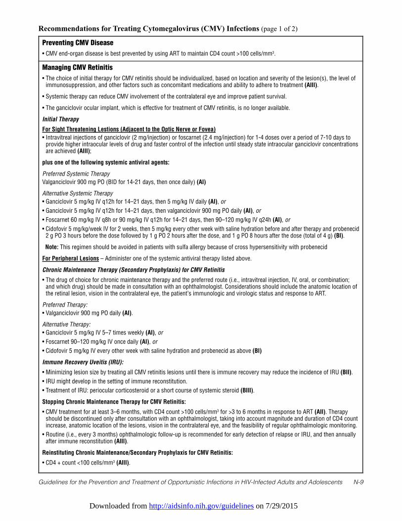
Downloaded from http://aidsinfo.nih.gov/guidelines on 7/29/2015
Guidelines for the Prevention and Treatment of Opportunistic Infections in HIV-Infected Adults and Adolescents N-9
Preventing CMV Disease
• CMV end-organ disease is best prevented by using ART to maintain CD4 count >100 cells/mm3.
Managing CMV Retinitis
• The choice of initial therapy for CMV retinitis should be individualized, based on location and severity of the lesion(s), the level ofimmunosuppression, and other factors such as concomitant medications and ability to adhere to treatment (AIII).
• Systemic therapy can reduce CMV involvement of the contralateral eye and improve patient survival.
• The ganciclovir ocular implant, which is effective for treatment of CMV retinitis, is no longer available.
Initial Therapy
For Sight Threatening Lestions (Adjacent to the Optic Nerve or Fovea)
• Intravitreal injections of ganciclovir (2 mg/injection) or foscarnet (2.4 mg/injection) for 1-4 doses over a period of 7-10 days toprovide higher intraocular levels of drug and faster control of the infection until steady state intraocular ganciclovir concentrationsare achieved (AIII);
plus one of the following systemic antiviral agents:
Preferred Systemic Therapy
Valganciclovir 900 mg PO (BID for 14-21 days, then once daily) (AI)
Alternative Systemic Therapy
• Ganciclovir 5 mg/kg IV q12h for 14–21 days, then 5 mg/kg IV daily (AI), or
• Ganciclovir 5 mg/kg IV q12h for 14–21 days, then valganciclovir 900 mg PO daily (AI), or
• Foscarnet 60 mg/kg IV q8h or 90 mg/kg IV q12h for 14–21 days, then 90–120 mg/kg IV q24h (AI), or
• Cidofovir 5 mg/kg/week IV for 2 weeks, then 5 mg/kg every other week with saline hydration before and after therapy and probenecid2 g PO 3 hours before the dose followed by 1 g PO 2 hours after the dose, and 1 g PO 8 hours after the dose (total of 4 g) (BI).
Note: This regimen should be avoided in patients with sulfa allergy because of cross hypersensitivity with probenecid
For Peripheral Lesions – Administer one of the systemic antiviral therapy listed above.
Chronic Maintenance Therapy (Secondary Prophylaxis) for CMV Retinitis
• The drug of choice for chronic maintenance therapy and the preferred route (i.e., intravitreal injection, IV, oral, or combination;and which drug) should be made in consultation with an ophthalmologist. Considerations should include the anatomic location ofthe retinal lesion, vision in the contralateral eye, the patient’s immunologic and virologic status and response to ART.
Preferred Therapy:
• Valganciclovir 900 mg PO daily (AI).
Alternative Therapy:
• Ganciclovir 5 mg/kg IV 5–7 times weekly (AI), or
• Foscarnet 90–120 mg/kg IV once daily (AI), or
• Cidofovir 5 mg/kg IV every other week with saline hydration and probenecid as above (BI)
Immune Recovery Uveitis (IRU):
• Minimizing lesion size by treating all CMV retinitis lesions until there is immune recovery may reduce the incidence of IRU (BII).
• IRU might develop in the setting of immune reconstitution.
• Treatment of IRU: periocular corticosteroid or a short course of systemic steroid (BIII).
Stopping Chronic Maintenance Therapy for CMV Retinitis:
• CMV treatment for at least 3–6 months, with CD4 count >100 cells/mm3 for >3 to 6 months in response to ART (AII). Therapyshould be discontinued only after consultation with an ophthalmologist, taking into account magnitude and duration of CD4 countincrease, anatomic location of the lesions, vision in the contralateral eye, and the feasibility of regular ophthalmologic monitoring.
• Routine (i.e., every 3 months) ophthalmologic follow-up is recommended for early detection of relapse or IRU, and then annuallyafter immune reconstitution (AIII).
Reinstituting Chronic Maintenance/Secondary Prophylaxis for CMV Retinitis:
• CD4 + count <100 cells/mm3 (AIII).
Recommendations for Treating Cytomegalovirus (CMV) Infections (page 1 of 2)
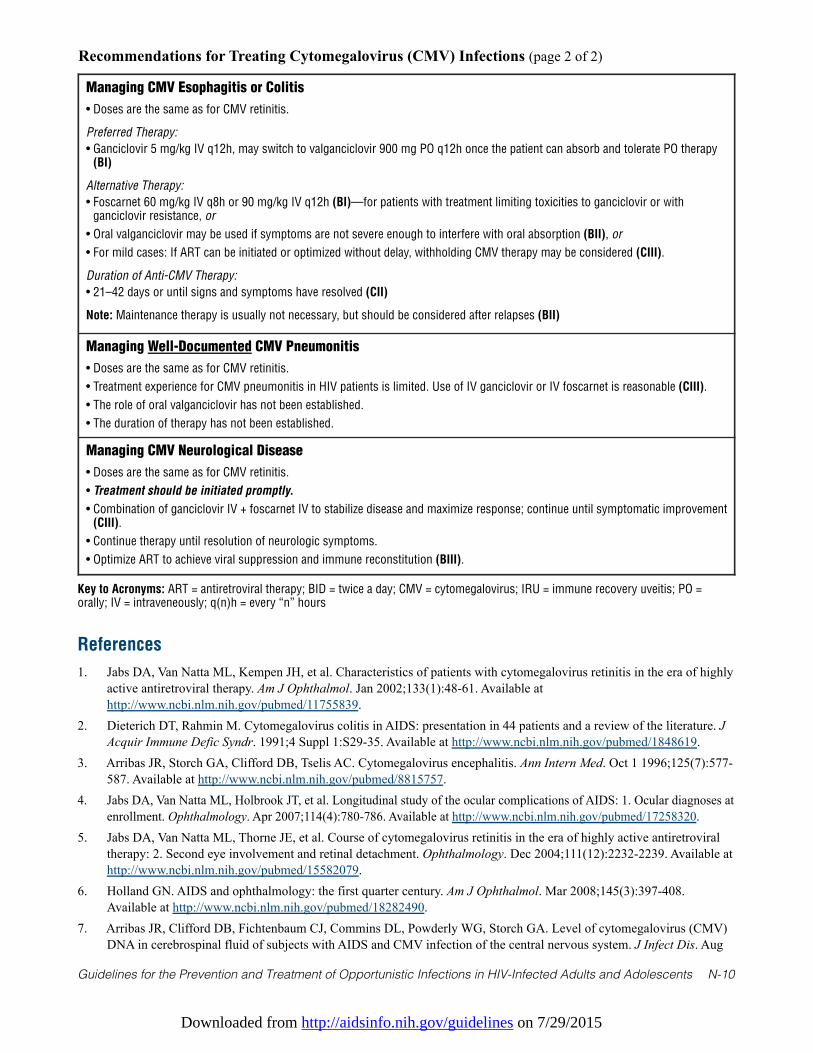
Downloaded from http://aidsinfo.nih.gov/guidelines on 7/29/2015
Guidelines for the Prevention and Treatment of Opportunistic Infections in HIV-Infected Adults and Adolescents N-10
Managing CMV Esophagitis or Colitis
• Doses are the same as for CMV retinitis.
Preferred Therapy:
• Ganciclovir 5 mg/kg IV q12h, may switch to valganciclovir 900 mg PO q12h once the patient can absorb and tolerate PO therapy(BI)
Alternative Therapy:
• Foscarnet 60 mg/kg IV q8h or 90 mg/kg IV q12h (BI)—for patients with treatment limiting toxicities to ganciclovir or withganciclovir resistance, or
• Oral valganciclovir may be used if symptoms are not severe enough to interfere with oral absorption (BII), or
• For mild cases: If ART can be initiated or optimized without delay, withholding CMV therapy may be considered (CIII).
Duration of Anti-CMV Therapy:
• 21–42 days or until signs and symptoms have resolved (CII)
Note: Maintenance therapy is usually not necessary, but should be considered after relapses (BII)
Managing Well-Documented CMV Pneumonitis
• Doses are the same as for CMV retinitis.
• Treatment experience for CMV pneumonitis in HIV patients is limited. Use of IV ganciclovir or IV foscarnet is reasonable (CIII).
• The role of oral valganciclovir has not been established.
• The duration of therapy has not been established.
Managing CMV Neurological Disease
• Doses are the same as for CMV retinitis.
• Treatment should be initiated promptly.
• Combination of ganciclovir IV + foscarnet IV to stabilize disease and maximize response; continue until symptomatic improvement(CIII).
• Continue therapy until resolution of neurologic symptoms.
• Optimize ART to achieve viral suppression and immune reconstitution (BIII).
Recommendations for Treating Cytomegalovirus (CMV) Infections (page 2 of 2)
Key to Acronyms: ART = antiretroviral therapy; BID = twice a day; CMV = cytomegalovirus; IRU = immune recovery uveitis; PO =orally; IV = intraveneously; q(n)h = every “n” hours
References
1. Jabs DA, Van Natta ML, Kempen JH, et al. Characteristics of patients with cytomegalovirus retinitis in the era of highlyactive antiretroviral therapy. Am J Ophthalmol. Jan 2002;133(1):48-61. Available athttp://www.ncbi.nlm.nih.gov/pubmed/11755839.
2. Dieterich DT, Rahmin M. Cytomegalovirus colitis in AIDS: presentation in 44 patients and a review of the literature. JAcquir Immune Defic Syndr. 1991;4 Suppl 1:S29-35. Available at http://www.ncbi.nlm.nih.gov/pubmed/1848619.
3. Arribas JR, Storch GA, Clifford DB, Tselis AC. Cytomegalovirus encephalitis. Ann Intern Med. Oct 1 1996;125(7):577-587. Available at http://www.ncbi.nlm.nih.gov/pubmed/8815757.
4. Jabs DA, Van Natta ML, Holbrook JT, et al. Longitudinal study of the ocular complications of AIDS: 1. Ocular diagnoses atenrollment. Ophthalmology. Apr 2007;114(4):780-786. Available at http://www.ncbi.nlm.nih.gov/pubmed/17258320.
5. Jabs DA, Van Natta ML, Thorne JE, et al. Course of cytomegalovirus retinitis in the era of highly active antiretroviraltherapy: 2. Second eye involvement and retinal detachment. Ophthalmology. Dec 2004;111(12):2232-2239. Available athttp://www.ncbi.nlm.nih.gov/pubmed/15582079.
6. Holland GN. AIDS and ophthalmology: the first quarter century. Am J Ophthalmol. Mar 2008;145(3):397-408.Available at http://www.ncbi.nlm.nih.gov/pubmed/18282490.
7. Arribas JR, Clifford DB, Fichtenbaum CJ, Commins DL, Powderly WG, Storch GA. Level of cytomegalovirus (CMV)DNA in cerebrospinal fluid of subjects with AIDS and CMV infection of the central nervous system. J Infect Dis. Aug
Downloaded from http://aidsinfo.nih.gov/guidelines on 7/29/2015
Guidelines for the Prevention and Treatment of Opportunistic Infections in HIV-Infected Adults and Adolescents N-11
1995;172(2):527-531. Available at http://www.ncbi.nlm.nih.gov/pubmed/7622897.8. Dodt KK, Jacobsen PH, Hofmann B, et al. Development of cytomegalovirus (CMV) disease may be predicted in HIV-
infected patients by CMV polymerase chain reaction and the antigenemia test. AIDS. Mar 1997;11(3):F21-28. Availableat http://www.ncbi.nlm.nih.gov/pubmed/9147416.
9. Zurlo JJ, O'Neill D, Polis MA, et al. Lack of clinical utility of cytomegalovirus blood and urine cultures in patients withHIV infection. Ann Intern Med. Jan 1 1993;118(1):12-17. Available at http://www.ncbi.nlm.nih.gov/pubmed/8093214.
10. Rodriguez-Barradas MC, Stool E, Musher DM, et al. Diagnosing and treating cytomegalovirus pneumonia in patientswith AIDS. Clin Infect Dis. Jul 1996;23(1):76-81. Available at http://www.ncbi.nlm.nih.gov/pubmed/8816133.
11. Wolf DG, Spector SA. Diagnosis of human cytomegalovirus central nervous system disease in AIDS patients by DNAamplification from cerebrospinal fluid. J Infect Dis. Dec 1992;166(6):1412-1415. Available athttp://www.ncbi.nlm.nih.gov/pubmed/1331254.
12. Deayton JR, Prof Sabin CA, Johnson MA, Emery VC, Wilson P, Griffiths PD. Importance of cytomegalovirus viraemiain risk of disease progression and death in HIV-infected patients receiving highly active antiretroviral therapy. Lancet.Jun 26 2004;363(9427):2116-2121. Available at http://www.ncbi.nlm.nih.gov/pubmed/15220032.
13. Hu H, Jabs DA, Forman MS, et al. Comparison of cytomegalovirus (CMV) UL97 gene sequences in the blood andvitreous of patients with acquired immunodeficiency syndrome and CMV retinitis. J Infect Dis. Apr 1 2002;185(7):861-867. Available at http://www.ncbi.nlm.nih.gov/pubmed/11920309.
14. Jabs DA, Martin BK, Forman MS, Ricks MO, Cytomegalovirus R, Viral Resistance Research G. Cytomegalovirus(CMV) blood DNA load, CMV retinitis progression, and occurrence of resistant CMV in patients with CMV retinitis. JInfect Dis. Aug 15 2005;192(4):640-649. Available at http://www.ncbi.nlm.nih.gov/pubmed/16028133.
15. Laine L, Bonacini M, Sattler F, Young T, Sherrod A. Cytomegalovirus and Candida esophagitis in patients with AIDS. JAcquir Immune Defic Syndr. 1992;5(6):605-609. Available at http://www.ncbi.nlm.nih.gov/pubmed/1316961.
16. Spector SA, McKinley GF, Lalezari JP, et al. Oral ganciclovir for the prevention of cytomegalovirus disease in personswith AIDS. Roche Cooperative Oral Ganciclovir Study Group. N Engl J Med. Jun 6 1996;334(23):1491-1497. Availableat http://www.ncbi.nlm.nih.gov/pubmed/8618603.
17. Wohl DA, Kendall MA, Andersen J, et al. Low rate of CMV end-organ disease in HIV-infected patients despite lowCD4+ cell counts and CMV viremia: results of ACTG protocol A5030. HIV Clin Trials. May-Jun 2009;10(3):143-152.Available at http://www.ncbi.nlm.nih.gov/pubmed/19632953.
18. Martin DF, Kuppermann BD, Wolitz RA, Palestine AG, Li H, Robinson CA. Oral ganciclovir for patients withcytomegalovirus retinitis treated with a ganciclovir implant. Roche Ganciclovir Study Group. N Engl J Med. Apr 81999;340(14):1063-1070. Available at http://www.ncbi.nlm.nih.gov/pubmed/10194235.
19. Kempen JH, Jabs DA, Wilson LA, Dunn JP, West SK, Tonascia J. Mortality risk for patients with cytomegalovirusretinitis and acquired immune deficiency syndrome. Clin Infect Dis. Nov 15 2003;37(10):1365-1373. Available athttp://www.ncbi.nlm.nih.gov/pubmed/14583871.
20. Studies of Ocular Complications of ARGTACTG. The ganciclovir implant plus oral ganciclovir versus parenteralcidofovir for the treatment of cytomegalovirus retinitis in patients with acquired immunodeficiency syndrome: TheGanciclovir Cidofovir Cytomegalovirus Retinitis Trial. Am J Ophthalmol. Apr 2001;131(4):457-467. Available athttp://www.ncbi.nlm.nih.gov/pubmed/11292409.
21. Musch DC, Martin DF, Gordon JF, Davis MD, Kuppermann BD. Treatment of cytomegalovirus retinitis with asustained-release ganciclovir implant. The Ganciclovir Implant Study Group. N Engl J Med. Jul 10 1997;337(2):83-90.Available at http://www.ncbi.nlm.nih.gov/pubmed/9211677.
22. Martin DF, Sierra-Madero J, Walmsley S, et al. A controlled trial of valganciclovir as induction therapy forcytomegalovirus retinitis. N Engl J Med. Apr 11 2002;346(15):1119-1126. Available athttp://www.ncbi.nlm.nih.gov/pubmed/11948271.
23. Kempen JH, Jabs DA, Wilson LA, Dunn JP, West SK, Tonascia JA. Risk of vision loss in patients with cytomegalovirusretinitis and the acquired immunodeficiency syndrome. Archives of ophthalmology. Apr 2003;121(4):466-476.Available at http://www.ncbi.nlm.nih.gov/pubmed/12695243.
24. Foscarnet-Ganciclovir Cytomegalovirus Retinitis Trial. 4. Visual outcomes. Studies of Ocular Complications of AIDSResearch Group in collaboration with the AIDS Clinical Trials Group. Ophthalmology. Jul 1994;101(7):1250-1261.Available at http://www.ncbi.nlm.nih.gov/pubmed/8035989.
Downloaded from http://aidsinfo.nih.gov/guidelines on 7/29/2015
Guidelines for the Prevention and Treatment of Opportunistic Infections in HIV-Infected Adults and Adolescents N-12
25. Bowen EF, Wilson P, Cope A, et al. Cytomegalovirus retinitis in AIDS patients: influence of cytomegaloviral load onresponse to ganciclovir, time to recurrence and survival. AIDS. Nov 1996;10(13):1515-1520. Available athttp://www.ncbi.nlm.nih.gov/pubmed/8931786.
26. Spector SA, Wong R, Hsia K, Pilcher M, Stempien MJ. Plasma cytomegalovirus (CMV) DNA load predicts CMVdisease and survival in AIDS patients. The Journal of clinical investigation. Jan 15 1998;101(2):497-502. Available athttp://www.ncbi.nlm.nih.gov/pubmed/9435323.
27. Jabs DA, Ahuja A, Van Natta M, et al. Course of cytomegalovirus retinitis in the era of highly active antiretroviraltherapy: five-year outcomes. Ophthalmology. Nov 2010;117(11):2152-2161 e2151-2152. Available athttp://www.ncbi.nlm.nih.gov/pubmed/20673591.
28. Ortega-Larrocea G, Espinosa E, Reyes-Teran G. Lower incidence and severity of cytomegalovirus-associated immunerecovery uveitis in HIV-infected patients with delayed highly active antiretroviral therapy. AIDS. Apr 292005;19(7):735-738. Available at http://www.ncbi.nlm.nih.gov/pubmed/15821403.
29. Dubreuil-Lemaire ML, Gori A, Vittecoq D, et al. Lenograstim for the treatment of neutropenia in patients receivingganciclovir for cytomegalovirus infection: a randomised, placebo-controlled trial in AIDS patients. European journal ofhaematology. Nov 2000;65(5):337-343. Available at http://www.ncbi.nlm.nih.gov/pubmed/11092465.
30. Kuritzkes DR, Parenti D, Ward DJ, et al. Filgrastim prevents severe neutropenia and reduces infective morbidity inpatients with advanced HIV infection: results of a randomized, multicenter, controlled trial. G-CSF 930101 StudyGroup. AIDS. Jan 1 1998;12(1):65-74. Available at http://www.ncbi.nlm.nih.gov/pubmed/9456256.
31. Nguyen QD, Kempen JH, Bolton SG, Dunn JP, Jabs DA. Immune recovery uveitis in patients with AIDS andcytomegalovirus retinitis after highly active antiretroviral therapy. Am J Ophthalmol. May 2000;129(5):634-639.Available at http://www.ncbi.nlm.nih.gov/pubmed/10844056.
32. Karavellas MP, Plummer DJ, Macdonald JC, et al. Incidence of immune recovery vitritis in cytomegalovirus retinitispatients following institution of successful highly active antiretroviral therapy. J Infect Dis. Mar 1999;179(3):697-700.Available at http://www.ncbi.nlm.nih.gov/pubmed/9952380.
33. Robinson MR, Reed G, Csaky KG, Polis MA, Whitcup SM. Immune-recovery uveitis in patients with cytomegalovirusretinitis taking highly active antiretroviral therapy. Am J Ophthalmol. Jul 2000;130(1):49-56. Available athttp://www.ncbi.nlm.nih.gov/pubmed/11004259.
34. Karavellas MP, Song M, Macdonald JC, Freeman WR. Long-term posterior and anterior segment complications ofimmune recovery uveitis associated with cytomegalovirus retinitis. Am J Ophthalmol. Jul 2000;130(1):57-64. Availableat http://www.ncbi.nlm.nih.gov/pubmed/11004260.
35. Kempen JH, Min YI, Freeman WR, et al. Risk of immune recovery uveitis in patients with AIDS and cytomegalovirusretinitis. Ophthalmology. Apr 2006;113(4):684-694. Available at http://www.ncbi.nlm.nih.gov/pubmed/16581429.
36. Morrison VL, Kozak I, LaBree LD, Azen SP, Kayicioglu OO, Freeman WR. Intravitreal triamcinolone acetonide for thetreatment of immune recovery uveitis macular edema. Ophthalmology. Feb 2007;114(2):334-339. Available athttp://www.ncbi.nlm.nih.gov/pubmed/17270681.
37. Holland GN, Vaudaux JD, Shiramizu KM, et al. Characteristics of untreated AIDS-related cytomegalovirus retinitis. II.Findings in the era of highly active antiretroviral therapy (1997 to 2000). Am J Ophthalmol. Jan 2008;145(1):12-22.Available at http://www.ncbi.nlm.nih.gov/pubmed/18154751.
38. Jabs DA, Wingard JR, de Bustros S, de Miranda P, Saral R, Santos GW. BW B759U for cytomegalovirus retinitis:intraocular drug penetration. Archives of ophthalmology. Oct 1986;104(10):1436-1437. Available athttp://www.ncbi.nlm.nih.gov/pubmed/3021090.
39. Kuppermann BD, Quiceno JI, Flores-Aguilar M, et al. Intravitreal ganciclovir concentration after intravenousadministration in AIDS patients with cytomegalovirus retinitis: implications for therapy. J Infect Dis. Dec1993;168(6):1506-1509. Available at http://www.ncbi.nlm.nih.gov/pubmed/8245536.
40. Arevalo JF, Gonzalez C, Capparelli EV, et al. Intravitreous and plasma concentrations of ganciclovir and foscarnet afterintravenous therapy in patients with AIDS and cytomegalovirus retinitis. J Infect Dis. Oct 1995;172(4):951-956.Available at http://www.ncbi.nlm.nih.gov/pubmed/7561215.
41. Combination foscarnet and ganciclovir therapy vs monotherapy for the treatment of relapsed cytomegalovirus retinitisin patients with AIDS. The Cytomegalovirus Retreatment Trial. The Studies of Ocular Complications of AIDS ResearchGroup in Collaboration with the AIDS Clinical Trials Group. Archives of ophthalmology. Jan 1996;114(1):23-33.
Downloaded from http://aidsinfo.nih.gov/guidelines on 7/29/2015
Guidelines for the Prevention and Treatment of Opportunistic Infections in HIV-Infected Adults and Adolescents N-13
Available at http://www.ncbi.nlm.nih.gov/pubmed/8540847.42. Jabs DA, Enger C, Dunn JP, Forman M. Cytomegalovirus retinitis and viral resistance: ganciclovir resistance. CMV
Retinitis and Viral Resistance Study Group. J Infect Dis. Mar 1998;177(3):770-773. Available athttp://www.ncbi.nlm.nih.gov/pubmed/9498461.
43. Jabs DA, Enger C, Forman M, Dunn JP. Incidence of foscarnet resistance and cidofovir resistance in patients treated forcytomegalovirus retinitis. The Cytomegalovirus Retinitis and Viral Resistance Study Group. Antimicrob AgentsChemother. Sep 1998;42(9):2240-2244. Available at http://www.ncbi.nlm.nih.gov/pubmed/9736542.
44. Jabs DA, Martin BK, Forman MS, et al. Mutations conferring ganciclovir resistance in a cohort of patients withacquired immunodeficiency syndrome and cytomegalovirus retinitis. J Infect Dis. Jan 15 2001;183(2):333-337.Available at http://www.ncbi.nlm.nih.gov/pubmed/11120934.
45. Emery VC, Griffiths PD. Prediction of cytomegalovirus load and resistance patterns after antiviral chemotherapy.Proceedings of the National Academy of Sciences of the United States of America. Jul 5 2000;97(14):8039-8044.Available at http://www.ncbi.nlm.nih.gov/pubmed/10859361.
46. Jabs DA, Enger C, Dunn JP, Forman M, Hubbard L. Cytomegalovirus retinitis and viral resistance: 3. Culture results.CMV Retinitis and Viral Resistance Study Group. Am J Ophthalmol. Oct 1998;126(4):543-549. Available athttp://www.ncbi.nlm.nih.gov/pubmed/9780099.
47. Weinberg A, Jabs DA, Chou S, et al. Mutations conferring foscarnet resistance in a cohort of patients with acquiredimmunodeficiency syndrome and cytomegalovirus retinitis. J Infect Dis. Mar 1 2003;187(5):777-784. Available athttp://www.ncbi.nlm.nih.gov/pubmed/12599051.
48. Martin BK, Ricks MO, Forman MS, Jabs DA, Cytomegalovirus R, Viral Resistance Study G. Change over time inincidence of ganciclovir resistance in patients with cytomegalovirus retinitis. Clin Infect Dis. Apr 1 2007;44(7):1001-1008. Available at http://www.ncbi.nlm.nih.gov/pubmed/17342657.
49. Chou S, Erice A, Jordan MC, et al. Analysis of the UL97 phosphotransferase coding sequence in clinicalcytomegalovirus isolates and identification of mutations conferring ganciclovir resistance. J Infect Dis. Mar1995;171(3):576-583. Available at http://www.ncbi.nlm.nih.gov/pubmed/7876604.
50. Chou S, Guentzel S, Michels KR, Miner RC, Drew WL. Frequency of UL97 phosphotransferase mutations related toganciclovir resistance in clinical cytomegalovirus isolates. J Infect Dis. Jul 1995;172(1):239-242. Available athttp://www.ncbi.nlm.nih.gov/pubmed/7797920.
51. Smith IL, Cherrington JM, Jiles RE, Fuller MD, Freeman WR, Spector SA. High-level resistance of cytomegalovirus toganciclovir is associated with alterations in both the UL97 and DNA polymerase genes. J Infect Dis. Jul1997;176(1):69-77. Available at http://www.ncbi.nlm.nih.gov/pubmed/9207351.
52. Chou S, Lurain NS, Thompson KD, Miner RC, Drew WL. Viral DNA polymerase mutations associated with drugresistance in human cytomegalovirus. J Infect Dis. Jul 1 2003;188(1):32-39. Available athttp://www.ncbi.nlm.nih.gov/pubmed/12825168.
53. Chou S, Van Wechel LC, Lichy HM, Marousek GI. Phenotyping of cytomegalovirus drug resistance mutations by usingrecombinant viruses incorporating a reporter gene. Antimicrob Agents Chemother. Jul 2005;49(7):2710-2715. Availableat http://www.ncbi.nlm.nih.gov/pubmed/15980340.
54. Wolf DG, Smith IL, Lee DJ, Freeman WR, Flores-Aguilar M, Spector SA. Mutations in human cytomegalovirus UL97gene confer clinical resistance to ganciclovir and can be detected directly in patient plasma. The Journal of clinicalinvestigation. Jan 1995;95(1):257-263. Available at http://www.ncbi.nlm.nih.gov/pubmed/7814623.
55. Vitravene Study G. Randomized dose-comparison studies of intravitreous fomivirsen for treatment of cytomegalovirusretinitis that has reactivated or is persistently active despite other therapies in patients with AIDS. Am J Ophthalmol.Apr 2002;133(4):475-483. Available at http://www.ncbi.nlm.nih.gov/pubmed/11931781.
56. Jabs DA, Martin BK, Ricks MO, Forman MS, Cytomegalovirus R, Viral Resistance Study G. Detection of ganciclovirresistance in patients with AIDS and cytomegalovirus retinitis: correlation of genotypic methods with viral phenotypeand clinical outcome. J Infect Dis. Jun 15 2006;193(12):1728-1737. Available athttp://www.ncbi.nlm.nih.gov/pubmed/16703517.
57. Jabs DA, Martin BK, Forman MS, et al. Cytomegalovirus resistance to ganciclovir and clinical outcomes of patientswith cytomegalovirus retinitis. Am J Ophthalmol. Jan 2003;135(1):26-34. Available athttp://www.ncbi.nlm.nih.gov/pubmed/12504693.
Downloaded from http://aidsinfo.nih.gov/guidelines on 7/29/2015
Guidelines for the Prevention and Treatment of Opportunistic Infections in HIV-Infected Adults and Adolescents N-14
58. Jabs DA, Martin BK, Forman MS, Cytomegalovirus R, Viral Resistance Research G. Mortality associated with resistantcytomegalovirus among patients with cytomegalovirus retinitis and AIDS. Ophthalmology. Jan 2010;117(1):128-132e122. Available at http://www.ncbi.nlm.nih.gov/pubmed/19818505.
59. AIDS Clinical Trials Group (ACTG) SoocoAF-GCRTR, design, and methods. AIDS Clinical Trials Group (ACTG),.Studies of ocular complications of AIDS Foscarnet-Ganciclovir Cytomegalovirus Retinitis Trial: 1. Rationale, design,and methods. AIDS Clinical Trials Group (ACTG). Controlled clinical trials. Feb 1992;13(1):22-39. Available athttp://www.ncbi.nlm.nih.gov/pubmed/1315661.
60. Taskintuna I, Rahhal FM, Rao NA, et al. Adverse events and autopsy findings after intravitreous cidofovir (HPMPC)therapy in patients with acquired immune deficiency syndrome (AIDS). Ophthalmology. Nov 1997;104(11):1827-1836;discussion 1836-1827. Available at http://www.ncbi.nlm.nih.gov/pubmed/9373113.
61. Holbrook JT, Colvin R, van Natta ML, et al. Evaluation of the United States public health service guidelines fordiscontinuation of anticytomegalovirus therapy after immune recovery in patients with cytomegalovirus retinitis. Am JOphthalmol. Oct 2011;152(4):628-637 e621. Available at http://www.ncbi.nlm.nih.gov/pubmed/21742304.
62. Tural C, Romeu J, Sirera G, et al. Long-lasting remission of cytomegalovirus retinitis without maintenance therapy inhuman immunodeficiency virus-infected patients. J Infect Dis. Apr 1998;177(4):1080-1083. Available athttp://www.ncbi.nlm.nih.gov/pubmed/9534987.
63. Vrabec TR, Baldassano VF, Whitcup SM. Discontinuation of maintenance therapy in patients with quiescentcytomegalovirus retinitis and elevated CD4+ counts. Ophthalmology. Jul 1998;105(7):1259-1264. Available athttp://www.ncbi.nlm.nih.gov/pubmed/9663231.
64. Macdonald JC, Torriani FJ, Morse LS, Karavellas MP, Reed JB, Freeman WR. Lack of reactivation of cytomegalovirus(CMV) retinitis after stopping CMV maintenance therapy in AIDS patients with sustained elevations in CD4 T cells inresponse to highly active antiretroviral therapy. J Infect Dis. May 1998;177(5):1182-1187. Available athttp://www.ncbi.nlm.nih.gov/pubmed/9593001.
65. Whitcup SM, Fortin E, Lindblad AS, et al. Discontinuation of anticytomegalovirus therapy in patients with HIVinfection and cytomegalovirus retinitis. JAMA. Nov 3 1999;282(17):1633-1637. Available athttp://www.ncbi.nlm.nih.gov/pubmed/10553789.
66. Jabs DA, Bolton SG, Dunn JP, Palestine AG. Discontinuing anticytomegalovirus therapy in patients with immunereconstitution after combination antiretroviral therapy. Am J Ophthalmol. Dec 1998;126(6):817-822. Available athttp://www.ncbi.nlm.nih.gov/pubmed/9860006.
67. Jouan M, Saves M, Tubiana R, et al. Discontinuation of maintenance therapy for cytomegalovirus retinitis in HIV-infected patients receiving highly active antiretroviral therapy. AIDS. Jan 5 2001;15(1):23-31. Available athttp://www.ncbi.nlm.nih.gov/pubmed/11192865.
68. Walmsley SL, Raboud J, Angel JB, et al. Long-term follow-up of a cohort of HIV-infected patients who discontinuedmaintenance therapy for cytomegalovirus retinitis. HIV Clin Trials. Jan-Feb 2006;7(1):1-9. Available athttp://www.ncbi.nlm.nih.gov/pubmed/16632459.
69. Torriani FJ, Freeman WR, Macdonald JC, et al. CMV retinitis recurs after stopping treatment in virological andimmunological failures of potent antiretroviral therapy. AIDS. Jan 28 2000;14(2):173-180. Available athttp://www.ncbi.nlm.nih.gov/pubmed/10708288.
70. Faqi AS, Klug A, Merker HJ, Chahoud I. Ganciclovir induces reproductive hazards in male rats after short-termexposure. Human & experimental toxicology. Sep 1997;16(9):505-511. Available athttp://www.ncbi.nlm.nih.gov/pubmed/9306137.
71. Miller BW, Howard TK, Goss JA, Mostello DJ, Holcomb WL, Jr., Brennan DC. Renal transplantation one week afterconception. Transplantation. Dec 15 1995;60(11):1353-1354. Available athttp://www.ncbi.nlm.nih.gov/pubmed/8525535.
72. Pescovitz MD. Absence of teratogenicity of oral ganciclovir used during early pregnancy in a liver transplant recipient.Transplantation. Mar 15 1999;67(5):758-759. Available at http://www.ncbi.nlm.nih.gov/pubmed/10096536.
73. Adler SP, Nigro G, Pereira L. Recent advances in the prevention and treatment of congenital cytomegalovirus infections.Seminars in perinatology. Feb 2007;31(1):10-18. Available at http://www.ncbi.nlm.nih.gov/pubmed/17317422.
74. Alvarez-McLeod A, Havlik J, Drew KE. Foscarnet treatment of genital infection due to acyclovir-resistant herpessimplex virus type 2 in a pregnant patient with AIDS: case report. Clin Infect Dis. Oct 1999;29(4):937-938. Available at
Downloaded from http://aidsinfo.nih.gov/guidelines on 7/29/2015
Guidelines for the Prevention and Treatment of Opportunistic Infections in HIV-Infected Adults and Adolescents N-15
http://www.ncbi.nlm.nih.gov/pubmed/10589917.75. Yamamoto AY, Mussi-Pinhata MM, Boppana SB, et al. Human cytomegalovirus reinfection is associated with
intrauterine transmission in a highly cytomegalovirus-immune maternal population. Am J Obstet Gynecol. Mar2010;202(3):297 e291-298. Available at http://www.ncbi.nlm.nih.gov/pubmed/20060091.
76. Stagno S, Pass RF, Cloud G, et al. Primary cytomegalovirus infection in pregnancy. Incidence, transmission to fetus,and clinical outcome. JAMA. Oct 10 1986;256(14):1904-1908. Available athttp://www.ncbi.nlm.nih.gov/pubmed/3020264.
77. Kovacs A, Schluchter M, Easley K, et al. Cytomegalovirus infection and HIV-1 disease progression in infants born toHIV-1-infected women. Pediatric Pulmonary and Cardiovascular Complications of Vertically Transmitted HIV InfectionStudy Group. N Engl J Med. Jul 8 1999;341(2):77-84. Available at http://www.ncbi.nlm.nih.gov/pubmed/10395631.
78. Quinn TC, Piot P, McCormick JB, et al. Serologic and immunologic studies in patients with AIDS in North Americaand Africa. The potential role of infectious agents as cofactors in human immunodeficiency virus infection. JAMA. May15 1987;257(19):2617-2621. Available at http://www.ncbi.nlm.nih.gov/pubmed/3494857.
79. Mussi-Pinhata MM, Yamamoto AY, Figueiredo LT, Cervi MC, Duarte G. Congenital and perinatal cytomegalovirusinfection in infants born to mothers infected with human immunodeficiency virus. J Pediatr. Feb 1998;132(2):285-290.Available at http://www.ncbi.nlm.nih.gov/pubmed/9506642.
80. Yow MD, Williamson DW, Leeds LJ, et al. Epidemiologic characteristics of cytomegalovirus infection in mothers andtheir infants. Am J Obstet Gynecol. May 1988;158(5):1189-1195. Available athttp://www.ncbi.nlm.nih.gov/pubmed/2835906.
81. Duryea EL, Sanchez PJ, Sheffield JS, et al. Maternal human immunodeficiency virus infection and congenitaltransmission of cytomegalovirus. Pediatr Infect Dis J. Oct 2010;29(10):915-918. Available athttp://www.ncbi.nlm.nih.gov/pubmed/20431424.
82. Guibert G, Warszawski J, Le Chenadec J, et al. Decreased risk of congenital cytomegalovirus infection in children bornto HIV-1-infected mothers in the era of highly active antiretroviral therapy. Clin Infect Dis. Jun 1 2009;48(11):1516-1525. Available at http://www.ncbi.nlm.nih.gov/pubmed/19388872.
83. Fowler KB, Boppana SB. Congenital cytomegalovirus (CMV) infection and hearing deficit. J Clin Virol. Feb2006;35(2):226-231. Available at http://www.ncbi.nlm.nih.gov/pubmed/16386462.
84. Mandelbrot L, Mayaux MJ, Bongain A, et al. Obstetric factors and mother-to-child transmission of humanimmunodeficiency virus type 1: the French perinatal cohorts. SEROGEST French Pediatric HIV Infection Study Group.Am J Obstet Gynecol. Sep 1996;175(3 Pt 1):661-667. Available at http://www.ncbi.nlm.nih.gov/pubmed/8828431.
85. Ekoukou D, Khuong-Josses MA, Ghibaudo N, Mechali D, Rotten D. Amniocentesis in pregnant HIV-infected patients.Absence of mother-to-child viral transmission in a series of selected patients. Eur J Obstet Gynecol Reprod Biol. Oct2008;140(2):212-217. Available at http://www.ncbi.nlm.nih.gov/pubmed/18584937.
86. Maiques V, Garcia-Tejedor A, Perales A, Cordoba J, Esteban RJ. HIV detection in amniotic fluid samples.Amniocentesis can be performed in HIV pregnant women? Eur J Obstet Gynecol Reprod Biol. Jun 10 2003;108(2):137-141. Available at http://www.ncbi.nlm.nih.gov/pubmed/12781400.
87. Nigro G, Adler SP, La Torre R, Best AM, Congenital Cytomegalovirus Collaborating G. Passive immunization duringpregnancy for congenital cytomegalovirus infection. N Engl J Med. Sep 29 2005;353(13):1350-1362. Available athttp://www.ncbi.nlm.nih.gov/pubmed/16192480.
88. Nigro G, Torre RL, Pentimalli H, et al. Regression of fetal cerebral abnormalities by primary cytomegalovirus infectionfollowing hyperimmunoglobulin therapy. Prenat Diagn. Jun 2008;28(6):512-517. Available athttp://www.ncbi.nlm.nih.gov/pubmed/18509871.
Downloaded from http://aidsinfo.nih.gov/guidelines on 7/29/2015
Guidelines for the Prevention and Treatment of Opportunistic Infections in HIV-Infected Adults and Adolescents O-1
Herpes Simplex Virus Disease (Last updated May 7, 2013; last reviewed May 7,2013)
Epidemiology
Infections with human herpes simplex virus type 1 (HSV-1) and type 2 (HSV-2) are common, with aseroprevalence of HSV-1 among adults in the United States of approximately 60% and a seroprevalence ofHSV-2 among persons aged ≥12 years of 17%.1 Approximately 70% of HIV-infected persons are HSV-2seropositive and 95% are seropositive for either HSV-1 or HSV-2.2 In most HSV-infected persons, HSVinfections are unrecognized clinically. However, regardless of the clinical severity of infection, re-activationon mucosal surfaces occurs frequently and can result in transmission. HSV-2 infection increases the risk ofHIV acquisition two- to three-fold, and HSV-2 reactivation results in increases in HIV RNA levels in bloodand genital secretions of coinfected patients.
Clinical Manifestations
Orolabial herpes (e.g., cold sores, fever blisters) is the most common manifestation of HSV-1 infection.Classic manifestations include a sensory prodrome in the affected area, rapidly followed by the evolution oflesions from papule to vesicle, ulcer, and crust stages on the lip. The course of illness in untreated patients is5 to 10 days. Lesions recur 1 to 12 times per year and can be triggered by sunlight or physiologic stress.
Genital herpes is the most common manifestation of HSV-2 infection. Typical genital mucosal or skin lesionsevolve through stages of papule, vesicle, ulcer, and crust. Ulcerative lesions are usually the only stageobserved on mucosal surfaces, but vesicles are commonly seen on genital skin (e.g., the penile shaft, thighs,pubis). Local symptoms might include a sensory prodrome consisting of pain and pruritis. Mucosal disease isoccasionally accompanied by dysuria or vaginal or urethral discharge. Inguinal lymphadenopathy is commonwith genital herpes, particularly in primary infection.3 These classic manifestations occur in some patients,but most individuals with genital herpes have mild and atypical lesions that are often unrecognized, notbrought to medical attention, and cannot reliably be diagnosed by physical examination. In profoundlyimmunocompromised patients, extensive, deep, nonhealing ulcerations can occur. These lesions have beenreported most often in those with CD4 T-lymphocyte (CD4) cell counts of <100 cells/µL and also may bemore commonly associated with acyclovir-resistant HSV.4
An episode of genital HSV-1 disease is indistinguishable from genital HSV-2 disease, but genital HSV-1recurrences and viral shedding occur less often than with genital HSV-2 infection.
Non-mucosal HSV infections, such as HSV keratitis, HSV encephalitis, HSV hepatitis, and herpetic whitlow,are similar in presentation to manifestations observed in HIV-seronegative individuals; disseminated HSVinfection is rare, even in profoundly immunosuppressed patients. HSV retinitis manifests as acute retinalnecrosis, which can lead rapidly to loss of vision.
Diagnosis
Because mucosal HSV infections cannot be diagnosed accurately by clinical examination, especially in HIV-seropositive patients, a laboratory diagnosis should be pursued in all cases.5 Viral culture, HSV DNAPolymerase chain reaction, and HSV antigen detection are available methods for diagnosis of mucocutaneousHSV lesions caused by HSV. Polymerase chain reaction is the most sensitive method. The virus detected ingenital lesions should be typed because of the prognostic significance—HSV-1 recurs less frequently thanHSV-2 in the genital area. Type-specific serologic assays are commercially available and can be used fordiagnosis in asymptomatic individuals or those with atypical lesions. Because of the poor sensitivity andspecificity of clinical diagnosis, the extensive interactions between HIV and HSV-2, and the availability ofeffective therapy for HSV-2, routine type-specific serologic screening for HSV-2 should be considered in
Downloaded from http://aidsinfo.nih.gov/guidelines on 7/29/2015
Guidelines for the Prevention and Treatment of Opportunistic Infections in HIV-Infected Adults and Adolescents O-2
patients seeking care for HIV. Diagnosis of HSV-2 should be accompanied by counseling that includesdiscussion of the risk of transmitting infection to sex partners. Guidelines for counseling are provided in the2010 Centers for Disease Control and Prevention sexually transmitted disease treatment guidelines.5
Preventing Exposure
The majority of HIV-infected patients have HSV-1 and HSV-2 infections. However, prevention of acquisitionof HSV is important for those who are uninfected. HSV-2-seronegative HIV-infected patients should asktheir partners to be tested using type-specific serology before initiating sexual activity, because disclosure ofHSV-2 in heterosexual HSV-2-discordant couples was associated with reduced risk of transmission of HSV-2(BII).6 Consistent use of latex condoms reduced HSV-2 acquisition from women to men and from men towomen, and their use should be encouraged for prevention of transmission of HSV-2 and other sexuallytransmitted pathogens (AII).7,8 HIV-infected individuals should specifically avoid sexual contact when theirpartners have overt (genital or orolabial) herpetic lesions (AII). However, most sexual transmission of HSVoccurs during asymptomatic viral shedding.
The use of suppressive antiviral therapy (i.e., valacyclovir 500 mg once daily) in patients with genital herpesreduced HSV-2 transmission to susceptible heterosexual partners by 50%;9 the effectiveness of this approachin reducing HSV-2 transmission to or from HIV-seropositive patients has not been evaluated.
Preventing Disease
Prophylaxis with antiviral drugs to prevent primary HSV infection is not recommended (BIII). The dose,duration, timing, and efficacy of antiviral prophylaxis after known or suspected exposure to HSV have notbeen evaluated. No vaccine for prevention of HSV infection is available.
Treating Disease
Patients with HSV infections can be treated with episodic therapy when symptomatic lesions occur or withdaily suppressive therapy to prevent recurrences. The management plan for genital HSV-2 disease in HIV-infected individuals should include consideration of several factors, such as frequency and severity of HSVrecurrences, the risk of HSV-2 transmission to susceptible partners, and the potential for interactions betweenHIV and HSV-2 that might result in increased HIV viral load in plasma and genital secretions. Episodictreatment for individual recurrences does not influence the natural history of genital HSV-2 infection and doesnot reduce the risk of HSV-2 transmission to sex partners, a major concern for patients with genital herpes.
Patients with orolabial lesions can be treated with oral valacyclovir, famciclovir, or acyclovir for 5 to 10 days(AIII). Severe mucocutaneous HSV lesions respond best to initial treatment with intravenous (IV) acyclovir(AIII).4,10 Patients can be switched to oral antiviral therapy after their lesions have begun to regress. Therapyshould be continued until the lesions have completely healed. Genital HSV episodes should be treated withoral valacyclovir, famciclovir, or acyclovir for 5 to 14 days (AI). Disseminated disease due to HSV is rare inHIV-seropositive patients, although HSV necrotizing retinitis can occur, which may be difficult to distinguishclinically from retinitis caused by VZV.
Special Considerations with Regard to Starting Antiretroviral TherapyIn most instances, orolabial HSV should not influence the decision about when to start antiretroviral therapy(ART). HIV-infected patients receiving ART who have immune reconstitution often have improvement in thefrequency and severity of their clinical episodes of genital herpes. However, immune reconstitution does notreduce the frequency of genital HSV shedding.11 Chronic cutaneous or muscosal HSV that is refractory totherapy and visceral or disseminated cases of HSV disease (which are uncommon) would be indications tohasten the initiation of ART (CIII).
Downloaded from http://aidsinfo.nih.gov/guidelines on 7/29/2015
Guidelines for the Prevention and Treatment of Opportunistic Infections in HIV-Infected Adults and Adolescents O-3
Monitoring of Response to Therapy and Adverse Events (Including ImmuneReconstitution Inflammatory Syndrome [IRIS])Acyclovir, valacyclovir, and famciclovir are occasionally associated with nausea or headache. No laboratorymonitoring is needed in patients receiving episodic or suppressive therapy unless they have advanced renalimpairment. For patients receiving high-dose IV acyclovir, monitoring of renal function and dose adjustmentas necessary are recommended at initiation of treatment and once or twice weekly for the duration oftreatment. Thrombotic thrombocytopenic purpura/hemolytic uremic syndrome has been reported in HIV-infected patients treated with high-dose (8 g/day) valacyclovir, but has not been reported at conventionaldoses recommended for therapy of HSV infection.12
Mucocutaneous lesions that are atypical and occasionally recalcitrant to therapy have been reported inindividuals initiating ART and have been attributed to IRIS.13
Managing Treatment FailureTreatment failure as a result of resistance to anti-HSV drugs should be suspected if lesions do not begin toresolve within 7 to 10 days after initiation of therapy. In immunocompromised patients with suspectedacyclovir-resistant HSV, viral culture of the lesion should be performed, and if virus is isolated, susceptibilitytesting done to confirm drug resistance (AII).14 Phenotypic testing of viral isolates has been the gold standardmethod for assessing HSV resistance; genotypic testing is under development.
The treatment of choice for acyclovir-resistant HSV is IV foscarnet (AI).15,16 IV cidofovir is a potentialalternative. Topical trifluridine, cidofovir, and imiquimod also have been used successfully for lesions onexternal surfaces, although prolonged application for 21 to 28 days or longer may be required (CIII).
Preventing Recurrence
Suppressive therapy with oral acyclovir, valacyclovir, or famciclovir is effective in preventing recurrencesand is preferred for patients who have severe HSV recurrences or who want to minimize the frequency ofrecurrences (AI).5,17 Suppressive anti-HSV therapy in HIV-infected individuals also results in a decrease inHIV viral load in plasma and anal and genital secretions and in a lower risk of HIV progression.18 Thisregimen does not decrease the risk of HIV transmission to sexual partners.19 Suppressive therapy for HSV isusually continued indefinitely, without regard for improved CD4 cell count.
The use of daily suppressive therapy (when compared to episodic therapy) was associated with a lower riskof development of acyclovir-resistant HSV in hematopoietic stem cell recipients;20 no specific data for HIV-infected individuals are available.
Special Considerations During Pregnancy
Diagnosis of mucocutaneous HSV infections is the same for pregnant women as for non-pregnant women.Episodic therapy for first-episode HSV disease and for recurrences can be offered during pregnancy. Visceraldisease is more likely to occur during pregnancy and can be fatal in rare cases. Acyclovir is the antiviral drugwith the most reported experience in pregnancy and appears to be safe (AIII).21 The use of valacyclovir andfamciclovir during pregnancy has been described and they appear to be safe and well tolerated.22
Valacyclovir use can be considered for treatment and suppressive therapy during pregnancy because of itssimplified dosing schedule (CIII).
An additional concern with HSV during pregnancy is the potential for HSV transmission to the fetus orneonate. The rate of HSV transmission to the newborn in HSV-2-seropositive pregnant women is low, exceptin those who acquire genital HSV late in pregnancy. The adverse sequelae for the fetus, however, can be verysignificant. The predominant risk for HSV transmission is maternal genital shedding of HSV at delivery.Cesarean delivery is recommended for women with a genital herpes prodrome or visible HSV genital lesionsat the onset of labor (BII).5 Use of acyclovir or valacyclovir in late pregnancy suppresses genital herpes
Downloaded from http://aidsinfo.nih.gov/guidelines on 7/29/2015
Guidelines for the Prevention and Treatment of Opportunistic Infections in HIV-Infected Adults and Adolescents O-4
outbreaks and reduces the need for cesarean delivery for recurrent HSV in HIV-seronegative women23 and islikely to have similar efficacy in HIV-seropositive women. The effect of antiviral therapy late in pregnancyon the incidence of neonatal herpes is unknown. Suppressive therapy with either valacyclovir or acyclovir isrecommended starting at 36 weeks’ gestation for pregnant women with recurrences of genital herpes duringpregnancy (BII).24 There is no known benefit of suppressive therapy for women who are only seropositivefor HSV-2 without a history of genital lesions. Maternal genital herpes was a risk factor for perinatal mother-to-child HIV transmission in the pre-highly active antiretroviral therapy era.25 Whether HSV facilitates HIVtransmission among pregnant women on HAART and whether HSV suppression reduces the risk for verticalHIV transmission during pregnancy, birth, or breastfeeding are unknown.
Recommendations for Treating Herpes Simplex Virus (HSV) Infections
Treating Orolabial Lesions (Duration: 5–10 days)
• Valacyclovir 1 g PO BID (AIII), or
• Famciclovir 500 mg PO BID (AIII), or
• Acyclovir 400 mg PO TID (AIII)
Treating Initial or Recurrent Genital Lesions (Duration: 5–14 Days)
• Valacyclovir 1 g PO BID (AI), or
• Famciclovir 500 mg PO BID (AI), or
• Acyclovir 400 mg PO TID (AI)
Treating Severe Mucocutaneous HSV Infections (AIII)
• Initial therapy acyclovir 5 mg/kg IV q8h
• After lesions begin to regress, change to oral therapy as above.
• Continue treatment until lesions have completely healed.
Chronic Suppressive Therapy
Indications:
• For patients with severe recurrences (AI), or
• Patients who want to minimize the frequency of recurrences (AI)
Treatment:
• Valacyclovir 500 mg PO BID (AI), or
• Famciclovir 500 mg PO BID (AI), or
• Acyclovir 400 mg PO BID (AI)
• Continue indefinitely without regard to CD4 count improvement.
For Acyclovir-Resistant Mucocutaneous HSV infections
Preferred Therapy:
• Foscarnet 80–120 mg/kg/day IV in 2–3 divided doses until clinical response (AI)
Alternative Therapy (Duration: 21–28 days or longer, based on clinical response) (CIII):
• Topical trifluridine, or
• Topical cidofovir, or
• Topical imiquimod, or
• IV cidofovir
Note:
• Topical formulations of trifluridine and cidofovir are not commercially available
• Extemporaneous compounding of topical products can be prepared using trifluridine ophthalmic solution and the IV formulationof cidofovir
Key to Acronyms: BID = twice daily; HSV = herpes simplex virus; IV = intraveneously; PO = orally; q(n)h = every "n" hours; TID = threetimes daily
Downloaded from http://aidsinfo.nih.gov/guidelines on 7/29/2015
Guidelines for the Prevention and Treatment of Opportunistic Infections in HIV-Infected Adults and Adolescents O-5
References
1. Xu F, Sternberg MR, Kottiri BJ, et al. Trends in herpes simplex virus type 1 and type 2 seroprevalence in the UnitedStates. JAMA. Aug 23 2006;296(8):964-973. Available at http://www.ncbi.nlm.nih.gov/pubmed/16926356.
2. Corey L, Wald A, Celum CL, Quinn TC. The effects of herpes simplex virus-2 on HIV-1 acquisition and transmission: areview of two overlapping epidemics. J Acquir Immune Defic Syndr. Apr 15 2004;35(5):435-445. Available athttp://www.ncbi.nlm.nih.gov/pubmed/15021308.
3. Corey L, Adams HG, Brown ZA, Holmes KK. Genital herpes simplex virus infections: clinical manifestations, course, andcomplications. Ann Intern Med. Jun 1983;98(6):958-972. Available at http://www.ncbi.nlm.nih.gov/pubmed/6344712.
4. Safrin S, Elbeik T, Phan L, et al. Correlation between response to acyclovir and foscarnet therapy and in vitrosusceptibility result for isolates of herpes simplex virus from human immunodeficiency virus-infected patients.Antimicrob Agents Chemother. Jun 1994;38(6):1246-1250. Available at http://www.ncbi.nlm.nih.gov/pubmed/8092821.
5. Workowski KA, Berman S, Centers for Disease C, Prevention. Sexually transmitted diseases treatment guidelines, 2010.MMWR Recomm Rep. Dec 17 2010;59(RR-12):1-110. Available at http://www.ncbi.nlm.nih.gov/pubmed/21160459.
6. Wald A, Krantz E, Selke S, Lairson E, Morrow RA, Zeh J. Knowledge of partners' genital herpes protects againstherpes simplex virus type 2 acquisition. J Infect Dis. Jul 1 2006;194(1):42-52. Available athttp://www.ncbi.nlm.nih.gov/pubmed/16741881.
7. Wald A, Langenberg AG, Krantz E, et al. The relationship between condom use and herpes simplex virus acquisition.Ann Intern Med. Nov 15 2005;143(10):707-713. Available at http://www.ncbi.nlm.nih.gov/pubmed/16287791.
8. Martin ET, Krantz E, Gottlieb SL, et al. A pooled analysis of the effect of condoms in preventing HSV-2 acquisition.Arch Intern Med. Jul 13 2009;169(13):1233-1240. Available at http://www.ncbi.nlm.nih.gov/pubmed/19597073.
9. Corey L, Wald A, Patel R, et al. Once-daily valacyclovir to reduce the risk of transmission of genital herpes. N Engl JMed. Jan 1 2004;350(1):11-20. Available at http://www.ncbi.nlm.nih.gov/pubmed/14702423.
10. Meyers JD, Wade JC, Mitchell CD, et al. Multicenter collaborative trial of intravenous acyclovir for treatment ofmucocutaneous herpes simplex virus infection in the immunocompromised host. Am J Med. Jul 20 1982;73(1A):229-235. Available at http://www.ncbi.nlm.nih.gov/pubmed/7048914.
11. Posavad CM, Wald A, Kuntz S, et al. Frequent reactivation of herpes simplex virus among HIV-1-infected patientstreated with highly active antiretroviral therapy. J Infect Dis. Aug 15 2004;190(4):693-696. Available athttp://www.ncbi.nlm.nih.gov/pubmed/15272395.
12. Bell WR, Chulay JD, Feinberg JE. Manifestations resembling thrombotic microangiopathy in patients with advancedhuman immunodeficiency virus (HIV) disease in a cytomegalovirus prophylaxis trial (ACTG 204). Medicine(Baltimore). Sep 1997;76(5):369-380. Available at http://www.ncbi.nlm.nih.gov/pubmed/9352739.
13. Couppie P, Sarazin F, Clyti E, et al. Increased incidence of genital herpes after HAART initiation: a frequentpresentation of immune reconstitution inflammatory syndrome (IRIS) in HIV-infected patients. AIDS Patient CareSTDS. Mar 2006;20(3):143-145. Available at http://www.ncbi.nlm.nih.gov/pubmed/16548710.
14. Balfour HH, Jr. Antiviral drugs. N Engl J Med. Apr 22 1999;340(16):1255-1268. Available athttp://www.ncbi.nlm.nih.gov/pubmed/10210711.
15. Safrin S, Crumpacker C, Chatis P, et al. A controlled trial comparing foscarnet with vidarabine for acyclovir-resistantmucocutaneous herpes simplex in the acquired immunodeficiency syndrome. The AIDS Clinical Trials Group. N Engl JMed. Aug 22 1991;325(8):551-555. Available at http://www.ncbi.nlm.nih.gov/pubmed/1649971.
16. Levin MJ, Bacon TH, Leary JJ. Resistance of herpes simplex virus infections to nucleoside analogues in HIV-infectedpatients. Clin Infect Dis. Nov 1 2004;39 Suppl 5:S248-257. Available athttp://www.ncbi.nlm.nih.gov/pubmed/15494896.
17. DeJesus E, Wald A, Warren T, et al. Valacyclovir for the suppression of recurrent genital herpes in humanimmunodeficiency virus-infected subjects. J Infect Dis. Oct 1 2003;188(7):1009-1016. Available athttp://www.ncbi.nlm.nih.gov/pubmed/14513421.
18. Lingappa JR, Baeten JM, Wald A, et al. Daily acyclovir for HIV-1 disease progression in people dually infected withHIV-1 and herpes simplex virus type 2: a randomised placebo-controlled trial. Lancet. Mar 6 2010;375(9717):824-833.Available at http://www.ncbi.nlm.nih.gov/pubmed/20153888.
Downloaded from http://aidsinfo.nih.gov/guidelines on 7/29/2015
Guidelines for the Prevention and Treatment of Opportunistic Infections in HIV-Infected Adults and Adolescents O-6
19. Celum C, Wald A, Lingappa JR, et al. Acyclovir and transmission of HIV-1 from persons infected with HIV-1 and HSV-2. N Engl J Med. Feb 4 2010;362(5):427-439. Available at http://www.ncbi.nlm.nih.gov/pubmed/20089951.
20. Erard V, Wald A, Corey L, Leisenring WM, Boeckh M. Use of long-term suppressive acyclovir after hematopoieticstem-cell transplantation: impact on herpes simplex virus (HSV) disease and drug-resistant HSV disease. J Infect Dis.Jul 15 2007;196(2):266-270. Available at http://www.ncbi.nlm.nih.gov/pubmed/17570114.
21. Stone KM, Reiff-Eldridge R, White AD, et al. Pregnancy outcomes following systemic prenatal acyclovir exposure:Conclusions from the international acyclovir pregnancy registry, 1984-1999. Birth defects research. Part A, Clinicaland molecular teratology. Apr 2004;70(4):201-207. Available at http://www.ncbi.nlm.nih.gov/pubmed/15108247.
22. Pasternak B, Hviid A. Use of acyclovir, valacyclovir, and famciclovir in the first trimester of pregnancy and the risk ofbirth defects. JAMA. Aug 25 2010;304(8):859-866. Available at http://www.ncbi.nlm.nih.gov/pubmed/20736469.
23. Sheffield JS, Hollier LM, Hill JB, Stuart GS, Wendel GD. Acyclovir prophylaxis to prevent herpes simplex virusrecurrence at delivery: a systematic review. Obstet Gynecol. Dec 2003;102(6):1396-1403. Available athttp://www.ncbi.nlm.nih.gov/pubmed/14662233.
24. Bulletins--Gynecology ACoP. ACOG Practice Bulletin No. 117: Gynecologic care for women with humanimmunodeficiency virus. Obstet Gynecol. Dec 2010;116(6):1492-1509. Available athttp://www.ncbi.nlm.nih.gov/pubmed/21099636.
25. Chen KT, Segu M, Lumey LH, et al. Genital herpes simplex virus infection and perinatal transmission of humanimmunodeficiency virus. Obstet Gynecol. Dec 2005;106(6):1341-1348. Available athttp://www.ncbi.nlm.nih.gov/pubmed/16319261.
Downloaded from http://aidsinfo.nih.gov/guidelines on 7/29/2015
Guidelines for the Prevention and Treatment of Opportunistic Infections in HIV-Infected Adults and Adolescents O-7
Varicella-Zoster Virus Diseases (Last updated July 8, 2013; last reviewed July 8,2013)
Epidemiology
More than 95% of adults (aged >20 years) born in the United States have immunity to varicella, the vastmajority due to primary VZV infection, known as varicella (or chickenpox). Reactivation of latent VZVresults in herpes zoster (or shingles). A person’s lifetime risk for herpes zoster is 15% to 20%, with thehighest incidence occurring in the elderly and immunocompromised individuals. The incidence of herpeszoster is >15-fold higher for HIV-infected adults than for age-matched controls.1 Herpes zoster can occur inHIV-infected adults at any CD4 T lymphocyte (CD4) cell count, but frequency of disease is highest withCD4 counts of <200 cells/µL.2-4 Antiretroviral therapy (ART) has not been shown to reduce the incidence ofherpes zoster in adult populations: in fact, rates appear to be higher in the period immediately after initiationof ART. Lower frequency of herpes zoster in pediatric patients treated with ART has been observed, but it isdifficult to separate ART effect from the impact of varicella vaccine.5,6
Clinical Manifestations
Varicella rash tends to have a central distribution with lesions first appearing on the head, then trunk, andfinally the extremities, evolving through stages of macules, papules, vesicles, pustules, and crusts. The rashis characterized by rapid evolution of lesions during the initial 8 to 12 hours and by successive crops of newlesions and by the presence of lesions in different stages of development at the same time. New vesicleformation continues for 2 to 4 days, accompanied by pruritus, fever, headache, malaise, and anorexia.7Primary varicella can cause substantial morbidity in HIV-seropositive adolescents and adults. Visceraldissemination, especially VZV pneumonitis, is well documented.7 Because most HIV-infected adults in theUnited States are VZV seropositive, primary varicella is an uncommon occurrence in this population.
Herpes zoster manifests as a painful cutaneous eruption in a dermatomal distribution, often preceded byprodromal pain. The most common sites for herpes zoster are the thoracic dermatomes (40%–50% of cases),followed by cranial nerve (20%–25%), cervical (15%–20%), lumbar (15%), and sacral (5%) dermatomes.Skin changes begin with an erythematous maculopapular rash, followed by the appearance of clear vesiclesand accompanied by pain (which may be severe). New vesicle formation typically continues for 3 to 5 days,followed by lesion pustulation and scabbing. Crusts typically persist for 2 to 3 weeks. About 20% to 30% ofHIV-infected patients have one or more subsequent episodes of herpes zoster, which may involve the same ordifferent dermatomes. The probability of a recurrence of herpes zoster within 1 year of the index episode isapproximately 10%.3,8 Approximately 10% to 15% of HIV-seropositive patients report post-herpeticneuralgia as a complication following herpes zoster.3,9
Most herpes zoster-related complications in HIV-seropositive patients, including disseminated herpes zoster,occur in patients with CD4 counts of <200 cells/µL.10 The CNS is the primary target organ for herpes zosterdissemination in patients coinfected with HIV. Various VZV-related neurologic syndromes occur in HIV-infected patients, including CNS vasculitis, multifocal leukoencephalitis, ventriculitis, myelitis andmyeloradiculitis, optic neuritis, cranial nerve palsies and focal brain-stem lesions, and aseptic meningitis.
Acute retinal necrosis (ARN) and progressive outer retinal necrosis (PORN) are variants of necrotizingretinopathy caused by VZV. Although ARN can occur in both immunocompetent and immunocompromisedpatients, PORN occurs almost exclusively in AIDS patients with CD4 counts <100 cells/µL.11 In contrast toARN, PORN is characterized by minimal inflammation in the aqueous and vitreous humor, absence of retinalvasculitis, and multiple discrete peripheral lesions in the outer retinal layer.12 PORN lesions rapidly coalesce,causing full-thickness retinal necrosis and subsequent retinal detachment.13 Both ARN and PORN areassociated with high rates of visual loss.
Downloaded from http://aidsinfo.nih.gov/guidelines on 7/29/2015
Guidelines for the Prevention and Treatment of Opportunistic Infections in HIV-Infected Adults and Adolescents O-8
Diagnosis
Varicella and herpes zoster are distinctive in appearance and diagnosis can usually be made clinically. Varicellacan also be diagnosed retrospectively by documenting seroconversion. Immunocompromised persons can haveatypical presentations and varicella may be difficult to distinguish from disseminated herpes zoster (as opposedto dermatomal herpes zoster); history of varicella or VZV exposure, a rash that began with a dermatomalpattern, and VZV serologic testing to assess prior VZV infection may be helpful. When lesions are atypical orthe diagnosis of VZV from other exanthems is uncertain, swabs from a fresh lesion or tissue biopsies can besubmitted for viral culture, direct fluorescent antigen testing, or polymerase chain reaction (PCR). Additionally,scabs are very good specimens for PCR testing. PCR of lesions is the most sensitive and specific method fordiagnosis of VZV infections. Histopathology and PCR (of blood or fluids such as cerebrospinal fluid or vitreoushumor) can aid with diagnosis of VZV infections of visceral organs (e.g., pneumonitis, encephalitis, retinitis).14
Routine serologic testing to determine the VZV serologic status of HIV-infected adults is not recommended.
Preventing Exposure
HIV-infected persons who are susceptible to VZV (i.e., persons who have no history of varicella or shingles,who are seronegative for VZV, and who have no history of vaccination against VZV) should avoid exposureto individuals with varicella or herpes zoster (AII).
If household contacts of HIV-infected persons without evidence of immunity to varicella are themselveswithout evidence of immunity, then these household contacts should be vaccinated to prevent acquisition ofvaricella and potential transmission of wild-type VZV to their susceptible HIV-infected contacts (BIII).
Preventing Disease
Long-term prophylaxis with antiviral drugs to prevent varicella is not recommended (AIII). Rather, for HIV-infected persons who are susceptible to VZV, post-exposure prophylaxis following known or suspected VZVexposure is recommended.
Vaccination To Prevent Primary InfectionThe live attenuated varicella vaccine has been documented to be safe and immunogenic in HIV-infectedchildren with relatively preserved immune systems (CD4 lymphocyte percentage ≥15%)15-18 and isrecommended for them (AI).19 Varicella vaccination of HIV-seropositive children also reduces the risk ofsubsequent herpes zoster.6,18 No studies have evaluated the vaccine in HIV-infected adolescents or adults, butvaricella vaccination (2 doses, administered 3 months apart) may be considered in HIV-seropositive/VZV-seronegative persons ≥8 years old with CD4 counts ≥200 cells/µL (CIII).20 If vaccination results in diseasecaused by vaccine virus (a rare event), therapy with acyclovir is recommended (AIII). Administration ofvaricella vaccine to more severely immunocompromised HIV-infected patients (CD4 counts <200 cells/µL)is not recommended (AIII). Because of the high prevalence of VZV seropositivity in adults, use of varicellavaccine in this population will be infrequent. If post-exposure varicella-zoster immune globulin (VariZIG™)has been administered, an interval of at least 5 months is recommended before varicella vaccination (CIII).21
If post-exposure acyclovir has been administered, an interval of at least 3 days is recommended beforevaricella vaccination (CIII).
Post-Exposure Prophylaxis To Prevent Primary InfectionAfter close contact with a person who has active varicella or herpes zoster, HIV-infected adolescents andadults who are susceptible to VZV should receive VariZIG as soon as possible, but within 10 days afterexposure (AIII).22 Risk for VZV transmission is higher following exposure to a person with varicella thanafter exposure to localized herpes zoster. In the United States, VariZIG can be obtained only under atreatment investigational new drugs application (IND) by contacting FFF Enterprises (Temecula, CA), at(800) 843-7477. The duration of protection is at least 3 weeks. Patients receiving monthly high-dose
Downloaded from http://aidsinfo.nih.gov/guidelines on 7/29/2015
Guidelines for the Prevention and Treatment of Opportunistic Infections in HIV-Infected Adults and Adolescents O-9
intravenous immune globulin (IVIG >400 mg/kg) are likely to be protected and probably do not requireVariZIG if the last dose of IVIG was administered <3 weeks before exposure. Short-term post-exposureadministration of acyclovir or valacyclovir beginning 7 to 10 days after exposure23 may be considered forpreventing varicella among susceptible HIV-infected adolescents or adults but this intervention has not beenstudied in these populations (BIII). Among VZV-susceptible immunocompetent children, post-exposurevaricella vaccination has been shown to reduce the risk for varicella and is more effective than pre-emptivetherapy with antiviral drugs; however the efficacy of post-exposure varicella vaccination for adolescents andadults has also not been established.
Treating Disease
VaricellaNo controlled prospective studies of antiviral therapy for varicella in HIV-infected adults have been reported.For uncomplicated varicella, the preferred treatment options are valacyclovir (1 g PO 3 times daily), orfamciclovir (500 mg PO 3 times daily) for 5 to 7 days (AII). Oral acyclovir (20 mg/kg body weight up to amaximum dose of 800 mg 5 times daily) can be an alternative (BII). Intravenous (IV) acyclovir for 7 to 10days is the recommended initial treatment for HIV-infected patients with severe varicella (AIII).7,24,25 If noevidence of visceral involvement with VZV is apparent, switching to oral antiviral therapy after the patienthas defervesced may be permissible (BIII).26
Herpes ZosterPrompt antiviral therapy should be instituted in all HIV-seropositive patients whose herpes zoster is diagnosedwithin 1 week of rash onset (or any time before full crusting of lesions). The recommended treatment optionsfor acute localized dermatomal herpes zoster in HIV-infected cpatients are oral valacyclovir (AII), famciclovir(AII), or acyclovir (BII) (doses as above) for 7 to 10 days, although longer durations of therapy should beconsidered if lesions resolve slowly. Valacyclovir or famciclovir are preferred because of their improvedpharmacokinetic properties and simplified dosing schedule. If cutaneous lesions are extensive or if visceralinvolvement is suspected, IV acyclovir should be initiated and continued until clinical improvement is evident(AII).27 A switch from IV acyclovir to oral antiviral therapy (to complete a 10- to 14-day treatment course) isreasonable when formation of new cutaneous lesions has ceased and the signs and symptoms of visceral VZVinfection are improving (BIII). Because of the absence of data to support benefit in this population, adjunctivecorticosteroid therapy for herpes zoster is not recommended (AIII). Optimization of ART is recommended forall patients with VZV infections that are difficult to treat (e.g., retinitis, encephalitis) (AIII).
Optimal antiviral therapy for PORN remains undefined.28-30 Outcomes with intravenous acyclovir organciclovir monotherapy were poor. Better results were obtained with intravenous ganciclovir (or thecombination of ganciclovir plus foscarnet), along with intravitreal antiviral drug injections.29 Specific treatmentshould include systemic therapy with at least one intravenous drug (selected from acyclovir, ganciclovir,foscarnet, and cidofovir) coupled with injections of at least one intravitreal drug (selected from ganciclovir andfoscarnet) (AIII).31,32 Treatment regimens for PORN recommended by certain specialists include a combinationof intravenous ganciclovir and/or foscarnet plus intravitreal injections of ganciclovir and/or foscarnet (AIII).The prognosis for visual preservation in involved eyes is poor, despite aggressive antiviral therapy.
Optimization of ART in HIV-infected patients with PORN is also recommended (AIII).32 Anecdotal reportshave described success with IV cidofovir for PORN. Intravitreal cidofovir should not be used because suchinjections may be associated with loss of intraocular pressure and other adverse effects. Ganciclovir ocularimplants, previously recommended by some experts, are no longer manufactured.
ARN appears to be more responsive than PORN to antiviral therapy. One recommended treatment is high-dose IV acyclovir (10–15 mg/kg every 8 hours for 10–14 days), followed by prolonged oral valacyclovir (1gram 3 times daily for 6 weeks) (AIII). Many experts would also include 1 or 2 doses of intravitrealganciclovir as part of the initial induction therapy (BIII). Involvement of an experienced ophthalmologist in
Downloaded from http://aidsinfo.nih.gov/guidelines on 7/29/2015
Guidelines for the Prevention and Treatment of Opportunistic Infections in HIV-Infected Adults and Adolescents O-10
management of patients with VZV retinitis is strongly recommended (AIII).
When to Start ARTA single uncomplicated episode of herpes zoster in an HIV-infected individual is not an indication to initiate ARTnor is it an indication to defer ART. Initiation of ART should be strongly considered in a patient who has multiplerecurrences of herpes zoster or who has a complication of VZV disease (e.g., PORN, encephalitis) (AIII).
Monitoring of Response to Therapy and Adverse Events (Including IRIS)For monitoring and adverse event recommendations related to anti-herpesvirus drugs, see preceding sectionson herpes simplex virus and cytomegalovirus.
Immune reconstitution following initiation of ART appears to be associated with an increased frequency ofVZV reactivation.33-36 Observational studies have shown the risk of zoster to increase 2- to 4-fold between 4and 16 weeks after initiating ART. The clinical presentation and natural history of herpes zoster in the settingof immune reconstitution do not differ from those observed in other HIV-infected patients and such episodesshould be managed in the same manner.
Managing Treatment FailureTreatment failure caused by resistance of VZV to acyclovir (and related drugs) is rare, but should besuspected if clinical findings do not improve within 10 days of initiation of therapy or if skin lesions have anatypical (e.g., verrucous) appearance. A viral culture should be obtained, and if VZV is isolated,susceptibility testing performed to establish antiviral drug susceptibility or resistance and to document theneed for alternative therapy. Among patients with suspected or proven acyclovir-resistant VZV infections,treatment with IV foscarnet is recommended (AII).37 IV cidofovir is a potential alternative (AIII).
Preventing Recurrence
The efficacy of long-term antiviral prophylaxis to prevent herpes zoster recurrences in HIV-seropositivepersons has not been evaluated and is not routinely recommended.
An attenuated virus vaccine for prevention of herpes zoster is FDA-approved for use in immunocompetentpersons aged ≥50 years, but is recommended for use beginning at age 60 years by the Advisory Committeeon Immunization Practices (ACIP). The zoster vaccine is contraindicated in persons with CD4 cell counts<200 cells/µL.
Special Considerations During Pregnancy
HIV-infected pregnant women who are susceptible to VZV and are in close contact with a person with activevaricella or herpes zoster should receive VariZIG as soon as possible (within 10 days)22 after exposure toVZV (AIII). If oral acyclovir is used for post-exposure prophylaxis, VZV serology should be performed sothat the drug can be discontinued if the patient is seropositive for VZV (CIII). Pregnant women should notreceive varicella vaccine (AIII).
Specific risks among HIV-infected women with varicella during pregnancy have not been reported. For HIV-seronegative women with varicella, the risk of transmitting VZV to the infant resulting in congenitalvaricella syndrome is 0.4% when infection occurs at or before 12 weeks’ gestation, 2.2% with infection at 13to 20 weeks, and is negligible after 20 weeks.38 Women with varicella during the first half of pregnancyshould be counseled about the risks and offered detailed ultrasound surveillance for findings indicative offetal congenital varicella syndrome.38 Administration of varicella-zoster immune globulin is recommendedprimarily to prevent complications in the mother; whether it has any benefit in prevention of congenitalvaricella syndrome is unknown. Infants born to women who have varicella from 5 days before until 2 daysafter delivery should receive VariZIG to reduce the severity and mortality of neonatal varicella acquired byexposure to maternal viremia (AIII).
Downloaded from http://aidsinfo.nih.gov/guidelines on 7/29/2015
Guidelines for the Prevention and Treatment of Opportunistic Infections in HIV-Infected Adults and Adolescents O-11
Oral acyclovir or valacyclovir are the preferred treatments for HIV-infected pregnant women who haveuncomplicated varicella during pregnancy (BIII). Pregnant women who have severe varicella or who exhibitsigns or symptoms of VZV pneumonitis should be hospitalized and treated with IV acyclovir (10 mg/kgevery 8 hours) (AII).
No controlled studies of antiviral therapy of herpes zoster during pregnancy have been reported.Recommended therapy for uncomplicated shingles in pregnant HIV-infected women is oral acyclovir orvalacyclovir (BIII). Pregnant women should not receive the herpes zoster vaccine (AIII).
Recommendations for Preventing and Treating Varicella Zoster Virus (VZV) Infections (page 1 of 2)
Pre-Exposure Prevention of VZV Primary Infection
Indications:
• Adult and adolescent patients with CD4 count ≥200 cells/mm3 without documentation of vaccination, health-care providerdiagnosis or verification of a history of varicella or herpes zoster, laboratory confirmation of disease, or persons who areseronegative for VZV (CIII)
Note: Routine VZV serologic testing in HIV-infected adults and adolescents is not recommended.
Vaccination:
• Primary varicella vaccination (Varivax™), 2 doses (0.5 mL SQ) administered 3 months apart (CIII)
• If vaccination results in disease because of vaccine virus, treatment with acyclovir is recommended (AIII).
• VZV-susceptible household contacts of susceptible HIV-infected persons should be vaccinated to prevent potential transmissionof VZV to their HIV-infected contacts (BIII).
• If post-exposure VariZIG has been administered, wait at least 5 months before varicella vaccination (CIII).
• If post-exposure acyclovir has been administered, wait at least 3 days before varicella vaccine (CIII).
Post-Exposure Prophylaxis:
Indication (AIII):
• Close contact with a person who has active varicella or herpes zoster, and
• Is susceptible to VZV (i.e., has no history of vaccination or of either condition, or is known to be VZV seronegative)
Preferred Prophylaxis:
• VariZIG 125 international units per 10 kg (maximum of 625 international units) IM, administered as soon as possible and within10 days after exposure to a person with active varicella or herpes zoster (AIII)
• VariZIG can be obtained only through an expanded access program under a treatment IND by contacting FFF Enterprise at (800)843-7477.
• If post-exposure VariZIG has been administered, wait at least 5 months before varicella vaccination (CIII).
Note: Patients receiving monthly high dose IVIG (i.e., > 400 mg/kg) are likely to be protected against VZV and probably do notrequire VariZIG if the last dose of IVIG was administered <3 weeks before VZV exposure.
Alternative Prophylaxis (Begin 7–10 Days After Exposure):
• Acyclovir 800 mg PO 5 times/day for 5–7 days (BIII), or
• Valacyclovir 1 g PO TID for 5–7 days (BIII)
Note:
• Neither these pre-emptive interventions nor post-exposure varicella vaccination have been studied in HIV-infected adults andadolescents.
• If acyclovir or valacyclovir is used, varicella vaccines should not be given until at least 72 hours after the last dose of the antiviraldrug.
Downloaded from http://aidsinfo.nih.gov/guidelines on 7/29/2015
Guidelines for the Prevention and Treatment of Opportunistic Infections in HIV-Infected Adults and Adolescents O-12
Recommendations for Preventing and Treating Varicella Zoster Virus (VZV) Infections (page 2 of 2)
Treatment of Varicella Infections
Primary Varicella Infection (Chickenpox)
Uncomplicated Cases
Preferred Therapy:
• Valacyclovir 1 g PO TID (AII), or
• Famciclovir 500 mg PO TID (AII)
Alternative Therapy:
• Acyclovir 800 mg PO 5 times daily (BII)
Duration:
• 5–7 days
Severe or Complicated Cases:
• Acyclovir 10–15 mg/kg IV q8h for 7–10 days (AIII)
• May switch to oral famciclovir, valacyclovir, or acyclovir after defervescence if no evidence of visceral involvement is evident (BIII)
Herpes Zoster (Shingles)
Acute Localized Dermatomal
Preferred Therapy:
• Valacyclovir 1000 mg PO TID (AII), or
• Famciclovir 500 mg PO TID (AII)
Alternative Therapy:
• Acyclovir 800 mg PO 5 times daily (BII)
Duration:
• 7–10 days, longer duration should be considered if lesions resolve slowly
Extensive Cutaneous Lesion or Visceral Involvement
• Acyclovir 10–15 mg/kg IV q8h until clinical improvement is evident (AII)
• Switch to oral therapy (valacyclovir 1 g TID, famciclovir 500 mg TID, or acyclovir 800 mg PO 5 times daily)—to complete a 10–14day course, when formation of new lesions has ceased and signs and symptoms of visceral VZV infection are improving (BIII)
PORN
• Involvement of an experienced ophthalmologist is strongly recommended (AIII)
• Ganciclovir 5 mg/kg and/or foscarnet 90 mg/kg IV q12h plus ganciclovir 2 mg/0.05mL and/or foscarnet 1.2 mg/0.05mLintravitreal twice weekly (AIII)
• Optimize ART regimen (AIII)
• Duration of therapy is not well defined and should be determined based on clinical, virologic, and immunologic responses inconsultation with ophthalmologist.
Note: ganciclovir ocular implants are no longer commercially available
ARN
• Acyclovir 10 - 15 mg/kg IV q8h for 10–14 days, followed by valacyclovir 1 g PO TID for 6 weeks PLUS ganciclovir 2 mg/0.05mLintravitreal twice weekly X 1-2 doses (AIII)
• Involvement of an experienced ophthalmologist is strongly recommended (AIII)
• Duration of therapy is not well defined and should be determined based on clinical, virologic, and immunologic responses inconsultation with ophthalmologist.
Key to Acronyms: ARN = acute retinal necrosis; CD4 = CD4 T lymphocyte cell; IND = investigational new drug application; IV =intraveneously; IVIG = intraveneous immunoglobulin; PO = orally; PORN = progressive outer retinal necrosis; q(n)h = every “n” hours;SQ = subcutaneously; TID = three times a day; VariZIG = varicella zoster immune globulin; VZV = varicella zoster virus
Downloaded from http://aidsinfo.nih.gov/guidelines on 7/29/2015
Guidelines for the Prevention and Treatment of Opportunistic Infections in HIV-Infected Adults and Adolescents O-13
References
1. Buchbinder SP, Katz MH, Hessol NA, et al. Herpes zoster and human immunodeficiency virus infection. J Infect Dis.Nov 1992;166(5):1153-1156. Available at http://www.ncbi.nlm.nih.gov/pubmed/1308664.
2. Engels EA, Rosenberg PS, Biggar RJ. Zoster incidence in human immunodeficiency virus-infected hemophiliacs andhomosexual men, 1984-1997. District of Columbia Gay Cohort Study. Multicenter Hemophilia Cohort Study. J InfectDis. Dec 1999;180(6):1784-1789. Available at http://www.ncbi.nlm.nih.gov/pubmed/10558932.
3. Gebo KA, Kalyani R, Moore RD, Polydefkis MJ. The incidence of, risk factors for, and sequelae of herpes zosteramong HIV patients in the highly active antiretroviral therapy era. J Acquir Immune Defic Syndr. Oct 1 2005;40(2):169-174. Available at http://www.ncbi.nlm.nih.gov/pubmed/16186734.
4. Vanhems P, Voisin L, Gayet-Ageron A, et al. The incidence of herpes zoster is less likely than other opportunisticinfections to be reduced by highly active antiretroviral therapy. J Acquir Immune Defic Syndr. Jan 1 2005;38(1):111-113. Available at http://www.ncbi.nlm.nih.gov/pubmed/15608535.
5. Levin MJ, Anderson JP, Seage GR, 3rd, Williams PL. Short-term and long-term effects of highly active antiretroviraltherapy on the incidence of herpes zoster in HIV-infected children. J Acquir Immune Defic Syndr. Feb 12009;50(2):182-191. Available at http://www.ncbi.nlm.nih.gov/pubmed/19131890.
6. Wood SM, Shah SS, Steenhoff AP, Rutstein RM. Primary varicella and herpes zoster among HIV-infected children from1989 to 2006. Pediatrics. Jan 2008;121(1):e150-156. Available at http://www.ncbi.nlm.nih.gov/pubmed/18086820.
7. Wallace MR, Hooper DG, Pyne JM, Graves SJ, Malone JL. Varicella immunity and clinical disease in HIV-infectedadults. South Med J. Jan 1994;87(1):74-76. Available at http://www.ncbi.nlm.nih.gov/pubmed/8284723.
8. Gnann JW, Jr., Crumpacker CS, Lalezari JP, et al. Sorivudine versus acyclovir for treatment of dermatomal herpeszoster in human immunodeficiency virus-infected patients: results from a randomized, controlled clinical trial.Collaborative Antiviral Study Group/AIDS Clinical Trials Group, Herpes Zoster Study Group. Antimicrob AgentsChemother. May 1998;42(5):1139-1145. Available at http://www.ncbi.nlm.nih.gov/pubmed/9593141.
9. Harrison RA, Soong S, Weiss HL, Gnann JW, Jr., Whitley RJ. A mixed model for factors predictive of pain in AIDSpatients with herpes zoster. J Pain Symptom Manage. Jun 1999;17(6):410-417. Available athttp://www.ncbi.nlm.nih.gov/pubmed/10388246.
10. Veenstra J, van Praag RM, Krol A, et al. Complications of varicella zoster virus reactivation in HIV-infectedhomosexual men. AIDS. Apr 1996;10(4):393-399. Available at http://www.ncbi.nlm.nih.gov/pubmed/8728043.
11. Engstrom RE, Jr., Holland GN, Margolis TP, et al. The progressive outer retinal necrosis syndrome. A variant ofnecrotizing herpetic retinopathy in patients with AIDS. Ophthalmology. Sep 1994;101(9):1488-1502. Available athttp://www.ncbi.nlm.nih.gov/pubmed/8090452.
12. Ormerod LD, Larkin JA, Margo CA, et al. Rapidly progressive herpetic retinal necrosis: a blinding diseasecharacteristic of advanced AIDS. Clin Infect Dis. Jan 1998;26(1):34-45; discussion 46-37. Available athttp://www.ncbi.nlm.nih.gov/pubmed/9455507.
13. Yin PD, Kurup SK, Fischer SH, et al. Progressive outer retinal necrosis in the era of highly active antiretroviral therapy:successful management with intravitreal injections and monitoring with quantitative PCR. J Clin Virol. Mar2007;38(3):254-259. Available at http://www.ncbi.nlm.nih.gov/pubmed/17280866.
14. Leung J, Harpaz R, Baughman AL, et al. Evaluation of laboratory methods for diagnosis of varicella. Clin Infect Dis.Jul 1 2010;51(1):23-32. Available at http://www.ncbi.nlm.nih.gov/pubmed/20504232.
15. Levin MJ, Gershon AA, Weinberg A, et al. Administration of live varicella vaccine to HIV-infected children withcurrent or past significant depression of CD4(+) T cells. J Infect Dis. Jul 15 2006;194(2):247-255. Available athttp://www.ncbi.nlm.nih.gov/pubmed/16779732.
16. Armenian SH, Han JY, Dunaway TM, Church JA. Safety and immunogenicity of live varicella virus vaccine in childrenwith human immunodeficiency virus type 1. Pediatr Infect Dis J. Apr 2006;25(4):368-370. Available athttp://www.ncbi.nlm.nih.gov/pubmed/16567993.
17. Bekker V, Westerlaken GH, Scherpbier H, et al. Varicella vaccination in HIV-1-infected children after immunereconstitution. AIDS. Nov 28 2006;20(18):2321-2329. Available at http://www.ncbi.nlm.nih.gov/pubmed/17117018.
18. Son M, Shapiro ED, LaRussa P, et al. Effectiveness of varicella vaccine in children infected with HIV. J Infect Dis. Jun15 2010;201(12):1806-1810. Available at http://www.ncbi.nlm.nih.gov/pubmed/20441519.
Downloaded from http://aidsinfo.nih.gov/guidelines on 7/29/2015
Guidelines for the Prevention and Treatment of Opportunistic Infections in HIV-Infected Adults and Adolescents O-14
19. Marin M, Guris D, Chaves SS, et al. Prevention of varicella: recommendations of the Advisory Committee onImmunization Practices (ACIP). MMWR Recomm Rep. Jun 22 2007;56(RR-4):1-40. Available athttp://www.ncbi.nlm.nih.gov/pubmed/17585291.
20. Centers for Disease Control and Prevention. Recommended adult immunization schedule: United States, 2010. AnnIntern Med. Jan 5 2010;152(1):36-39. Available at http://www.ncbi.nlm.nih.gov/pubmed/20048270.
21. Centers for Disease Control and Prevention. A new product (VariZIG) for postexposure prophylaxis of varicellaavailable under an investigational new drug application expanded access protocol. MMWR Morb Mortal Wkly Rep. Mar3 2006;55(8):209-210. Available at http://www.ncbi.nlm.nih.gov/pubmed/16511443.
22. Centers for Disease C, Prevention. FDA approval of an extended period for administering VariZIG for postexposureprophylaxis of varicella. MMWR Morb Mortal Wkly Rep. Mar 30 2012;61(12):212. Available athttp://www.ncbi.nlm.nih.gov/pubmed/22456121.
23. American Academy of Pediatrics. Red Book: 2012 Report of the Committee on Infectious Diseases. 29 ed2002.24. Prober CG, Kirk LE, Keeney RE. Acyclovir therapy of chickenpox in immunosuppressed children--a collaborative
study. J Pediatr. Oct 1982;101(4):622-625. Available at http://www.ncbi.nlm.nih.gov/pubmed/6750068.25. Arvin AM. Antiviral therapy for varicella and herpes zoster. Semin Pediatr Infect Dis. Jan 2002;13(1):12-21. Available
at http://www.ncbi.nlm.nih.gov/pubmed/12118839.26. Carcao MD, Lau RC, Gupta A, Huerter H, Koren G, King SM. Sequential use of intravenous and oral acyclovir in the
therapy of varicella in immunocompromised children. Pediatr Infect Dis J. Jul 1998;17(7):626-631. Available athttp://www.ncbi.nlm.nih.gov/pubmed/9686730.
27. Balfour HH, Jr., Bean B, Laskin OL, et al. Acyclovir halts progression of herpes zoster in immunocompromisedpatients. N Engl J Med. Jun 16 1983;308(24):1448-1453. Available at http://www.ncbi.nlm.nih.gov/pubmed/6343861.
28. Scott IU, Luu KM, Davis JL. Intravitreal antivirals in the management of patients with acquired immunodeficiencysyndrome with progressive outer retinal necrosis. Archives of ophthalmology. Sep 2002;120(9):1219-1222. Available athttp://www.ncbi.nlm.nih.gov/pubmed/12215102.
29. Moorthy RS, Weinberg DV, Teich SA, et al. Management of varicella zoster virus retinitis in AIDS. The British journalof ophthalmology. Mar 1997;81(3):189-194. Available at http://www.ncbi.nlm.nih.gov/pubmed/9135381.
30. Austin RB. Progressive outer retinal necrosis syndrome: a comprehensive review of its clinical presentation,relationship to immune system status, and management. Clin Eye Vis Care. Dec 2000;12(3-4):119-129. Available athttp://www.ncbi.nlm.nih.gov/pubmed/11137426.
31. Gore DM, Gore SK, Visser L. Progressive outer retinal necrosis: outcomes in the intravitreal era. Archives ofophthalmology. Jun 2012;130(6):700-706. Available at http://www.ncbi.nlm.nih.gov/pubmed/22801826.
32. Kim SJ, Equi R, Belair ML, Fine HF, Dunn JP. Long-term preservation of vision in progressive outer retinal necrosistreated with combination antiviral drugs and highly active antiretroviral therapy. Ocular immunology and inflammation.Nov-Dec 2007;15(6):425-427. Available at http://www.ncbi.nlm.nih.gov/pubmed/18085485.
33. Martinez E, Gatell J, Moran Y, et al. High incidence of herpes zoster in patients with AIDS soon after therapy with proteaseinhibitors. Clin Infect Dis. Dec 1998;27(6):1510-1513. Available at http://www.ncbi.nlm.nih.gov/pubmed/9868668.
34. Domingo P, Torres OH, Ris J, Vazquez G. Herpes zoster as an immune reconstitution disease after initiation ofcombination antiretroviral therapy in patients with human immunodeficiency virus type-1 infection. Am J Med. Jun 12001;110(8):605-609. Available at http://www.ncbi.nlm.nih.gov/pubmed/11382367.
35. Dunic I, Djurkovic-Djakovic O, Vesic S, Zerjav S, Jevtovic D. Herpes zoster as an immune restoration disease in AIDSpatients during therapy including protease inhibitors. Int J STD AIDS. Jul 2005;16(7):475-478. Available athttp://www.ncbi.nlm.nih.gov/pubmed/16004625.
36. Espinosa E, Pena-Jimenez A, Ormsby CE, Vega-Barrientos R, Reyes-Teran G. Later onset of herpes zoster-associatedimmune reconstitution inflammatory syndrome. HIV Med. Aug 2009;10(7):454-457. Available athttp://www.ncbi.nlm.nih.gov/pubmed/19490175.
37. Breton G, Fillet AM, Katlama C, Bricaire F, Caumes E. Acyclovir-resistant herpes zoster in human immunodeficiencyvirus-infected patients: results of foscarnet therapy. Clin Infect Dis. Dec 1998;27(6):1525-1527. Available athttp://www.ncbi.nlm.nih.gov/pubmed/9868672.
38. Pastuszak AL, Levy M, Schick B, et al. Outcome after maternal varicella infection in the first 20 weeks of pregnancy. NEngl J Med. Mar 31 1994;330(13):901-905. Available at http://www.ncbi.nlm.nih.gov/pubmed/8114861.
Downloaded from http://aidsinfo.nih.gov/guidelines on 7/29/2015
Guidelines for the Prevention and Treatment of Opportunistic Infections in HIV-Infected Adults and Adolescents O-15
Human Herpesvirus-8 Disease (Last updated May 7, 2013; last reviewed May 7,2013)
Epidemiology
Human herpesvirus-8 (HHV-8) seroprevalence among the general population in the United States is 1% to5%. The seroprevalence is greater among men who have sex with men (20%–77%),1 regardless of HIVinfection, and is also higher in certain Mediterranean countries (10%–20%) and in parts of sub-SaharanAfrica (30%–80%).2 HHV-8 is etiologically associated with all forms of Kaposi’s sarcoma ([KS] i.e., classic,endemic, transplant-related, and AIDS-related) and certain rare neoplastic disorders (such as primaryeffusion lymphoma) and lymphoproliferative disorders (multicentric Castleman’s disease) The precisepathogenesis is unclear even though seroconversion to HHV-8 precedes the development of these tumors.3Patients who are HHV-8 seropositive and have HHV-8 viremia have an increased risk (approximately nine-fold) for developing KS compared with HHV-8 seropositive men without HHV-8 viremia.4 HHV-8 viremiaalmost always accompanies symptomatic episodes of multicentric Castleman’s disease.5
The overall prevalence of KS was as high as 30% among patients with AIDS before the advent of effectiveantriretroviral therapy (ART).6 The incidence of KS, which increased nearly 10-fold in the United Statesbetween 1981 and 1987, began to gradually decline in 1987.7 Reasons for this reduction in KS incidenceprior to the widespread availability of ART are likely to be multiple, including the deaths of patients withadvanced AIDS who were most susceptible to KS, and the increasing use by HIV-infected individuals ofantiviral drugs that may have activity against HHV-8 (zidovudine for the treatment of HIV; ganciclovir,foscarnet, and cidofovir use for treatment of CMV disease).8 Supporting the latter hypothesis, observationalstudies indicate that patients receiving ganciclovir or foscarnet (but not acyclovir) develop KS at a reducedrate.9-12 A more marked reduction in KS incidence occurred in 1996, shortly after the introduction of proteaseinhibitor-containing ART in the United States. Today the incidence of KS in the United States remainsapproximately 3-fold higher than before the HIV pandemic, and notably KS incidence has not declined inregions of sub-Saharan Africa where ART coverage is increasing but incomplete.13 Primary effusion celllymphoma and multicentric Castleman’s disease remain rare.14
KS and primary effusion lymphoma are described most frequently among HIV-infected persons with moreadvanced immunosuppression (CD4 T lymphocyte [CD4] cell counts <200 cells/µL), although they canoccur at any CD4 cell count. Multicentric Castleman’s disease can present at any CD4 cell count. Recentreports of KS occurring at higher CD4 cell counts in the United States15,16 suggest that clinicians caring forpatients with HIV should be vigilant for the clinical manifestations of KS in patients at risk of HHV-8infection, regardless of CD4 cell count.
Clinical Manifestations
Most individuals with chronic HHV-8 infection are asymptomatic.17 Acquisition of HHV-8 inimmunocompetent children and organ transplant recipients has been associated with a primary infectionsyndrome consisting of fever, rash, lymphadenopathy, bone marrow failure, and occasional rapid progressionto KS.18,19 KS manifestations vary widely, but most patients have nontender, purplish, indurated skin lesions.Intraoral lesions are common and visceral dissemination can occur, occasionally without the presence of skinlesions. Multicentric Castleman’s disease manifests with generalized adenopathy and fever and can progressto multi-organ failure.14 Primary effusion lymphoma characteristically presents with effusions of the pleural,pericardial, or abdominal spaces; mass lesions can be seen but are less common manifestations.
Diagnosis
The diagnoses of KS, multicentric Castlemans disease and primary effusion lymphoma depend on cytologicand immunologic cell markers, as well as histology. Routine screening for HHV-8 by polymerese chain
Downloaded from http://aidsinfo.nih.gov/guidelines on 7/29/2015
Guidelines for the Prevention and Treatment of Opportunistic Infections in HIV-Infected Adults and Adolescents O-16
reaction (PCR) or serologic testing for HHV-8 antibody is not indicated for HIV-infected persons. Use ofPCR to quantify HHV-8 in the peripheral blood has no established role in the diagnosis of KS, multicentricCastleman’s disease and primary effusion lymphoma.5
Preventing Exposure
Asymptomatic HHV-8 infection is often associated with HHV-8 shedding in the saliva and occasionalshedding in genital secretions.1,17,20 Viral shedding may result in HHV-8 transmission to uninfected partnersthrough behaviors associated with exposure to saliva or genital secretions. Recommendations related topreventing exposure to HHV-8 do not exist; screening patients for HHV-8 serostatus and recommendingbehavioral modifications based on such information is not likely to be highly effective, has not beenvalidated, and is not currently recommended (CIII).
Preventing Disease
Despite observational evidence supporting a role for anti-HHV-8 therapy in preventing the development ofKS, the toxicity of current anti-HHV-8 therapy outweighs the potential benefits of administration (BIII).Because the strongest risk factor for the development of KS in HIV-positive individuals is a low CD4 cellcount,21 early initiation of ART is likely to be the most effective measure for the prevention of KS.
Treating Disease
Although ganciclovir, foscarnet, and cidofovir have in vitro activity against HHV-8 and limited studiesindicate these agents may be associated with reduced KS disease progression or lesion regression, larger andmore definitive studies are needed to determine whether antiviral therapy has a useful role in managingHHV-8-associated diseases. KS regression has been documented after ganciclovir or foscarnet therapy,although one study indicated cidofovir was ineffective.22
The use of IV ganciclovir or oral valganciclovir is an option for treatment of multicentric Castleman’sdisease (CII). A 3-week course of twice-daily IV ganciclovir or oral valganciclovir was associated withremissions in multicentric Castleman’s disease in one report,23 and a combination of valganciclovir and high-dose zidovudine given for 7 to 21 days led to durable clinical remissions of the disease (CII).24 Rituximabalso is an effective alternative to antiviral therapy in the treatment of multicentric Castleman’s disease(CII),25,26 though up to one-third of patients treated with rituximab may have subsequent exacerbations oremergence of KS.27,28
Chemotherapy, in combination with ART, should be administered to patients with primary effusion celllymphoma or visceral KS (AI) and is likely to be a useful adjunctive therapy in individuals with widelydisseminated cutaneous KS (BIII). Some clinicians recommend valganciclovir as adjunctive therapy in thetreatment of primary effusion lymphoma but there are no convincing data that it is useful (CIII).29,30
Detailed recommendations for treatment of HHV-8 malignancies (including chemotherapy and radiationtherapy) are beyond the scope of these guidelines. Treatment should be undertaken in consultation with anexperienced specialist (AIII).
Special Considerations When Starting ARTEarly initiation of ART is likely to prevent incident KS and primary effusion cell lymphoma, though nostudies have confirmed this hypothesis to date. ART that suppresses HIV replication should be administeredto all HIV-infected patients with KS, primary effusion cell lymphoma, or multicentric Castleman’s disease(AII), although insufficient evidence exists to support using one ART regimen over another.
Monitoring of Response to Therapy and Adverse Events (Including IRIS)Immune reconstitution inflammatory syndrome (IRIS) has been a reported complication among HHV-8-
Downloaded from http://aidsinfo.nih.gov/guidelines on 7/29/2015
Guidelines for the Prevention and Treatment of Opportunistic Infections in HIV-Infected Adults and Adolescents O-17
infected patients initiating ART.
KS: In one series, new onset KS or exacerbations of previously stable disease were the most common IRISsyndrome in a cohort of HIV-infected patients in Seattle.31 Over half of Ugandan patients with mild-to-moderate KS experienced an exacerbation when initating ART.32 Reliable predictors of KS-IRIS have notbeen identified.
Multicentric Castleman’s disease: A small number of patients with HIV-associated multicentricCastleman’s disease were also observed to have a clinical decompensation upon initiation of ART.33,34
Primary effusion lymphoma: No data exist on the frequency with which initiation of ART complicates thecourse of primary effusion lymphoma.
Taken together, it is clear that neither the incidence nor predictors of HHV-8-associated IRIS are well-described, but suppression of HIV replication and immune reconstitution are key components of therapy andinitiation of ART should not be delayed (AIII).
Preventing Recurrence
Effective suppression of HIV replication with ART in HIV-infected patients with KS may prevent KSprogression or occurrence of new lesions, and because KS is an AIDS-defining cancer, ART is indicated forall patients with active KS (AII). Suppression of HIV replication also is recommended for patients withmulticentric Castleman’s disease (AIII) and those with malignant lymphoproliferative disorders (AIII).
Special Considerations During Pregnancy
The seroprevalence of HHV-8 infection among HIV-infected pregnant women varies by geographic area,ranging from 1.7% among U.S.-born and 3.6% among Haitian-born women in New York City to 11.6%among pregnant women from 4 other U.S. cities.35 Pregnancy does not appear to affect the prevalence ofantibodies to HHV-8 or the antibody levels,36 although levels of HHV-8 DNA in the peripheral blood mayincrease late in pregnancy.37 HHV-8 seropositivity does not appear to influence pregnancy outcome. Routinescreening for HHV-8 by PCR or serology is not indicated for HIV-infected pregnant women (AIII). Antiviraltherapy for HHV-8 infection in pregnancy is not recommended (AIII).
In vitro models suggest that beta-human chorionic gonadotropin induces regression of KS tumors, butclinical reports on the incidence and natural history of KS in pregnancy are conflicting.38-41
Perinatal transmission of HHV-8 occurs infrequently. Evidence supporting vertical transmission duringpregnancy or the intrapartum period includes cases of KS occurring in the infant shortly after birth,42,43
higher risk for transmission with higher maternal antibody titer (and, by inference, higher maternal levels ofHHV-8),44 and detection of similar strains of HHV-8 DNA by PCR in specimens drawn at birth from HHV-8-seropositive mothers and their infants.45 Data indicate increased mortality through age 24 months amongHIV-infected infants born to HHV-8-seropositive compared with HHV-8-seronegative mothers,42-44,46-51 butthese studies could not completely account for other confounding factors affecting HIV-infected infants. Themajority of studies document a substantially higher rate of HHV-8 seropositivity among children born toHHV-8 antibody-positive compared with HHV-8 antibody-negative women.46-51
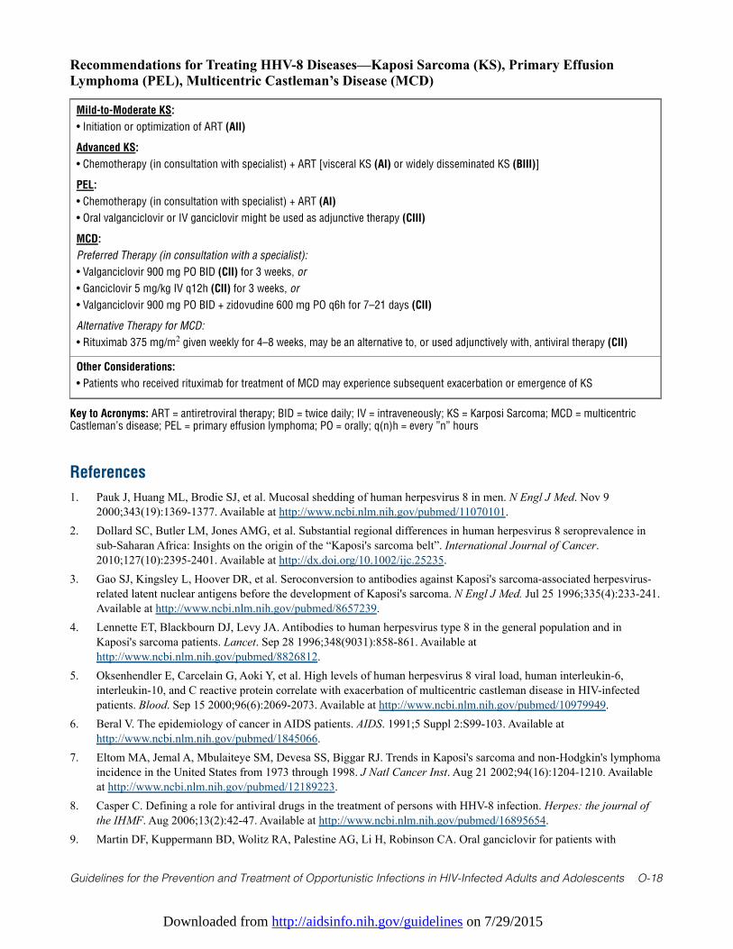
Downloaded from http://aidsinfo.nih.gov/guidelines on 7/29/2015
Guidelines for the Prevention and Treatment of Opportunistic Infections in HIV-Infected Adults and Adolescents O-18
Recommendations for Treating HHV-8 Diseases—Kaposi Sarcoma (KS), Primary EffusionLymphoma (PEL), Multicentric Castleman’s Disease (MCD)
Mild-to-Moderate KS:
• Initiation or optimization of ART (AII)
Advanced KS:
• Chemotherapy (in consultation with specialist) + ART [visceral KS (AI) or widely disseminated KS (BIII)]
PEL:
• Chemotherapy (in consultation with specialist) + ART (AI)
• Oral valganciclovir or IV ganciclovir might be used as adjunctive therapy (CIII)
MCD:
Preferred Therapy (in consultation with a specialist):
• Valganciclovir 900 mg PO BID (CII) for 3 weeks, or
• Ganciclovir 5 mg/kg IV q12h (CII) for 3 weeks, or
• Valganciclovir 900 mg PO BID + zidovudine 600 mg PO q6h for 7–21 days (CII)
Alternative Therapy for MCD:
• Rituximab 375 mg/m2 given weekly for 4–8 weeks, may be an alternative to, or used adjunctively with, antiviral therapy (CII)
Other Considerations:
• Patients who received rituximab for treatment of MCD may experience subsequent exacerbation or emergence of KS
Key to Acronyms: ART = antiretroviral therapy; BID = twice daily; IV = intraveneously; KS = Karposi Sarcoma; MCD = multicentricCastleman’s disease; PEL = primary effusion lymphoma; PO = orally; q(n)h = every ”n” hours
References
1. Pauk J, Huang ML, Brodie SJ, et al. Mucosal shedding of human herpesvirus 8 in men. N Engl J Med. Nov 92000;343(19):1369-1377. Available at http://www.ncbi.nlm.nih.gov/pubmed/11070101.
2. Dollard SC, Butler LM, Jones AMG, et al. Substantial regional differences in human herpesvirus 8 seroprevalence insub-Saharan Africa: Insights on the origin of the “Kaposi's sarcoma belt”. International Journal of Cancer.2010;127(10):2395-2401. Available at http://dx.doi.org/10.1002/ijc.25235.
3. Gao SJ, Kingsley L, Hoover DR, et al. Seroconversion to antibodies against Kaposi's sarcoma-associated herpesvirus-related latent nuclear antigens before the development of Kaposi's sarcoma. N Engl J Med. Jul 25 1996;335(4):233-241.Available at http://www.ncbi.nlm.nih.gov/pubmed/8657239.
4. Lennette ET, Blackbourn DJ, Levy JA. Antibodies to human herpesvirus type 8 in the general population and inKaposi's sarcoma patients. Lancet. Sep 28 1996;348(9031):858-861. Available athttp://www.ncbi.nlm.nih.gov/pubmed/8826812.
5. Oksenhendler E, Carcelain G, Aoki Y, et al. High levels of human herpesvirus 8 viral load, human interleukin-6,interleukin-10, and C reactive protein correlate with exacerbation of multicentric castleman disease in HIV-infectedpatients. Blood. Sep 15 2000;96(6):2069-2073. Available at http://www.ncbi.nlm.nih.gov/pubmed/10979949.
6. Beral V. The epidemiology of cancer in AIDS patients. AIDS. 1991;5 Suppl 2:S99-103. Available athttp://www.ncbi.nlm.nih.gov/pubmed/1845066.
7. Eltom MA, Jemal A, Mbulaiteye SM, Devesa SS, Biggar RJ. Trends in Kaposi's sarcoma and non-Hodgkin's lymphomaincidence in the United States from 1973 through 1998. J Natl Cancer Inst. Aug 21 2002;94(16):1204-1210. Availableat http://www.ncbi.nlm.nih.gov/pubmed/12189223.
8. Casper C. Defining a role for antiviral drugs in the treatment of persons with HHV-8 infection. Herpes: the journal ofthe IHMF. Aug 2006;13(2):42-47. Available at http://www.ncbi.nlm.nih.gov/pubmed/16895654.
9. Martin DF, Kuppermann BD, Wolitz RA, Palestine AG, Li H, Robinson CA. Oral ganciclovir for patients with
Downloaded from http://aidsinfo.nih.gov/guidelines on 7/29/2015
Guidelines for the Prevention and Treatment of Opportunistic Infections in HIV-Infected Adults and Adolescents O-19
cytomegalovirus retinitis treated with a ganciclovir implant. Roche Ganciclovir Study Group. N Engl J Med. Apr 81999;340(14):1063-1070. Available at http://www.ncbi.nlm.nih.gov/pubmed/10194235.
10. Ioannidis JP, Collier AC, Cooper DA, et al. Clinical efficacy of high-dose acyclovir in patients with humanimmunodeficiency virus infection: a meta-analysis of randomized individual patient data. J Infect Dis. Aug1998;178(2):349-359. Available at http://www.ncbi.nlm.nih.gov/pubmed/9697714.
11. Mocroft A, Youle M, Gazzard B, Morcinek J, Halai R, Phillips AN. Anti-herpesvirus treatment and risk of Kaposi'ssarcoma in HIV infection. Royal Free/Chelsea and Westminster Hospitals Collaborative Group. AIDS. Sep1996;10(10):1101-1105. Available at http://www.ncbi.nlm.nih.gov/pubmed/8874626.
12. Glesby MJ, Hoover DR, Weng S, et al. Use of antiherpes drugs and the risk of Kaposi's sarcoma: data from theMulticenter AIDS Cohort Study. J Infect Dis. Jun 1996;173(6):1477-1480. Available athttp://www.ncbi.nlm.nih.gov/pubmed/8648224.
13. Casper C. The increasing burden of HIV-associated malignancies in resource-limited regions. Annual review ofmedicine. 2011;62:157-170. Available at http://www.ncbi.nlm.nih.gov/pubmed/20868276.
14. Casper C. The aetiology and management of Castleman disease at 50 years: translating pathophysiology to patient care.British journal of haematology. Apr 2005;129(1):3-17. Available at http://www.ncbi.nlm.nih.gov/pubmed/15801951.
15. Maurer T, Ponte M, Leslie K. HIV-associated Kaposi's sarcoma with a high CD4 count and a low viral load. N Engl JMed. Sep 27 2007;357(13):1352-1353. Available at http://www.ncbi.nlm.nih.gov/pubmed/17898112.
16. Mani D, Neil N, Israel R, Aboulafia DM. A retrospective analysis of AIDS-associated Kaposi's sarcoma in patients withundetectable HIV viral loads and CD4 counts greater than 300 cells/mm3. J Int Assoc Physicians AIDS Care (Chic).Sep-Oct 2009;8(5):279-285. Available at http://www.ncbi.nlm.nih.gov/pubmed/19721098.
17. Casper C, Krantz E, Selke S, et al. Frequent and asymptomatic oropharyngeal shedding of human herpesvirus 8 amongimmunocompetent men. J Infect Dis. Jan 1 2007;195(1):30-36. Available athttp://www.ncbi.nlm.nih.gov/pubmed/17152006.
18. Andreoni M, Sarmati L, Nicastri E, et al. Primary human herpesvirus 8 infection in immunocompetent children. JAMA.Mar 13 2002;287(10):1295-1300. Available at http://www.ncbi.nlm.nih.gov/pubmed/11886321.
19. Luppi M, Barozzi P, Schulz TF, et al. Bone marrow failure associated with human herpesvirus 8 infection aftertransplantation. N Engl J Med. Nov 9 2000;343(19):1378-1385. Available athttp://www.ncbi.nlm.nih.gov/pubmed/11070102.
20. Casper C, Redman M, Huang ML, et al. HIV infection and human herpesvirus-8 oral shedding among men who havesex with men. J Acquir Immune Defic Syndr. Mar 1 2004;35(3):233-238. Available athttp://www.ncbi.nlm.nih.gov/pubmed/15076237.
21. Lodi S, Guiguet M, Costagliola D, et al. Kaposi sarcoma incidence and survival among HIV-infected homosexual menafter HIV seroconversion. J Natl Cancer Inst. Jun 2 2010;102(11):784-792. Available athttp://www.ncbi.nlm.nih.gov/pubmed/20442214.
22. Little RF, Merced-Galindez F, Staskus K, et al. A pilot study of cidofovir in patients with kaposi sarcoma. J Infect Dis.Jan 1 2003;187(1):149-153. Available at http://www.ncbi.nlm.nih.gov/pubmed/12508160.
23. Casper C, Nichols WG, Huang ML, Corey L, Wald A. Remission of HHV-8 and HIV-associated multicentric Castlemandisease with ganciclovir treatment. Blood. Mar 1 2004;103(5):1632-1634. Available athttp://www.ncbi.nlm.nih.gov/pubmed/14615380.
24. Uldrick TS, Polizzotto MN, Aleman K, et al. High-dose zidovudine plus valganciclovir for Kaposi sarcomaherpesvirus-associated multicentric Castleman disease: a pilot study of virus-activated cytotoxic therapy. Blood. Jun 302011;117(26):6977-6986. Available at http://www.ncbi.nlm.nih.gov/pubmed/21487108.
25. Bower M, Newsom-Davis T, Naresh K, et al. Clinical Features and Outcome in HIV-Associated MulticentricCastleman's Disease. J Clin Oncol. Jun 20 2011;29(18):2481-2486. Available athttp://www.ncbi.nlm.nih.gov/pubmed/21555697.
26. Marcelin AG, Aaron L, Mateus C, et al. Rituximab therapy for HIV-associated Castleman disease. Blood. Oct 152003;102(8):2786-2788. Available at http://www.ncbi.nlm.nih.gov/pubmed/12842986.
27. Gerard L, Berezne A, Galicier L, et al. Prospective Study of Rituximab in Chemotherapy-Dependent Human
Downloaded from http://aidsinfo.nih.gov/guidelines on 7/29/2015
Guidelines for the Prevention and Treatment of Opportunistic Infections in HIV-Infected Adults and Adolescents O-20
Immunodeficiency Virus Associated Multicentric Castleman's Disease: ANRS 117 CastlemaB Trial. J Clin Oncol.August 1, 2007 2007;25(22):3350-3356. Available at http://jco.ascopubs.org/cgi/content/abstract/25/22/3350.
28. Bower M, Powles T, Williams S, et al. Brief communication: rituximab in HIV-associated multicentric Castlemandisease. Ann Intern Med. Dec 18 2007;147(12):836-839. Available at http://www.ncbi.nlm.nih.gov/pubmed/18087054.
29. Aboulafia DM. Interleukin-2, ganciclovir, and high-dose zidovudine for the treatment of AIDS-associated primarycentral nervous system lymphoma. Clin Infect Dis. Jun 15 2002;34(12):1660-1662. Available athttp://www.ncbi.nlm.nih.gov/pubmed/12032910.
30. Crum-Cianflone NF, Wallace MR, Looney D. Successful secondary prophylaxis for primary effusion lymphoma with humanherpesvirus 8 therapy. AIDS. Jul 13 2006;20(11):1567-1569. Available at http://www.ncbi.nlm.nih.gov/pubmed/16847420.
31. Achenbach CJ, Harrington RD, Dhanireddy S, Crane HM, Casper C, Kitahata MM. Paradoxical immune reconstitutioninflammatory syndrome in HIV-infected patients treated with combination antiretroviral therapy after AIDS-definingopportunistic infection. Clin Infect Dis. Feb 1 2012;54(3):424-433. Available athttp://www.ncbi.nlm.nih.gov/pubmed/22095568.
32. Martin D, Gutkind JS. Kaposi's sarcoma virally encoded, G-protein-coupled receptor: a paradigm for paracrinetransformation. Methods Enzymol. 2009;460:125-150. Available at http://www.ncbi.nlm.nih.gov/pubmed/19446723.
33. Aaron L, Lidove O, Yousry C, Roudiere L, Dupont B, Viard JP. Human herpesvirus 8-positive Castleman disease inhuman immunodeficiency virus-infected patients: the impact of highly active antiretroviral therapy. Clin Infect Dis. Oct1 2002;35(7):880-882. Available at http://www.ncbi.nlm.nih.gov/pubmed/12228826.
34. Achenbach C, Kitahata MM. Recurrence or Worsening of AIDS-defining Opportunistic Infection (OI) due to ImmuneReconstitution Inflammatory Syndrome (IRIS) During Initial HAART Among a Clinic-Based Population. Paperpresented at: 48th ICAAC/IDSA 46th Annual Meeting; October 25-28, 2008; Washington, DC.
35. Goedert JJ, Kedes DH, Ganem D. Antibodies to human herpesvirus 8 in women and infants born in Haiti and the USA.Lancet. May 10 1997;349(9062):1368. Available at http://www.ncbi.nlm.nih.gov/pubmed/9149705.
36. Huang LM, Huang SY, Chen MY, et al. Geographical differences in human herpesvirus 8 seroepidemiology: a survey of1,201 individuals in Asia. J Med Virol. Mar 2000;60(3):290-293. Available athttp://www.ncbi.nlm.nih.gov/pubmed/10630961.
37. Lisco A, Barbierato M, Fiore JR, et al. Pregnancy and human herpesvirus 8 reactivation in human immunodeficiencyvirus type 1-infected women. J Clin Microbiol. Nov 2006;44(11):3863-3871. Available athttp://www.ncbi.nlm.nih.gov/pubmed/16943357.
38. Berger P, Dirnhofer S. Kaposi's sarcoma in pregnant women. Nature. Sep 7 1995;377(6544):21-22. Available athttp://www.ncbi.nlm.nih.gov/pubmed/7659155.
39. Lunardi-Iskandar Y, Bryant JL, Zeman RA, et al. Tumorigenesis and metastasis of neoplastic Kaposi's sarcoma cell linein immunodeficient mice blocked by a human pregnancy hormone. Nature. May 4 1995;375(6526):64-68. Available athttp://www.ncbi.nlm.nih.gov/pubmed/7723844.
40. Rabkin CS, Chibwe G, Muyunda K, Musaba E. Kaposi's sarcoma in pregnant women. Nature. Sep 71995;377(6544):21; author reply 22. Available at http://www.ncbi.nlm.nih.gov/pubmed/7659154.
41. Schulz TF, Weiss RA. Kaposi's sarcoma. A finger on the culprit. Nature. Jan 5 1995;373(6509):17-18. Available athttp://www.ncbi.nlm.nih.gov/pubmed/7800029.
42. Gutierrez-Ortega P, Hierro-Orozco S, Sanchez-Cisneros R, Montano LF. Kaposi's sarcoma in a 6-day-old infant withhuman immunodeficiency virus. Archives of dermatology. Mar 1989;125(3):432-433. Available athttp://www.ncbi.nlm.nih.gov/pubmed/2923454.
43. McCarthy GA, Kampmann B, Novelli V, Miller RF, Mercey DE, Gibb D. Vertical transmission of Kaposi's sarcoma.Archives of disease in childhood. May 1996;74(5):455-457. Available athttp://www.ncbi.nlm.nih.gov/pubmed/8669966.
44. Sitas F, Newton R, Boshoff C. Increasing probability of mother-to-child transmission of HHV-8 with increasingmaternal antibody titer for HHV-8. N Engl J Med. Jun 17 1999;340(24):1923. Available athttp://www.ncbi.nlm.nih.gov/pubmed/10375309.
45. Mbulaiteye S, Marshall V, Bagni RK, et al. Molecular evidence for mother-to-child transmission of Kaposi sarcoma-associated herpesvirus in Uganda and K1 gene evolution within the host. J Infect Dis. May 1 2006;193(9):1250-1257.
Downloaded from http://aidsinfo.nih.gov/guidelines on 7/29/2015
Guidelines for the Prevention and Treatment of Opportunistic Infections in HIV-Infected Adults and Adolescents O-21
Available at http://www.ncbi.nlm.nih.gov/pubmed/16586362.46. Mantina H, Kankasa C, Klaskala W, et al. Vertical transmission of Kaposi's sarcoma-associated herpesvirus. Int J
Cancer. Dec 1 2001;94(5):749-752. Available at http://www.ncbi.nlm.nih.gov/pubmed/11745472.47. Serraino D, Locatelli M, Songini M, et al. Human herpes virus-8 infection among pregnant women and their children:
results from the Sardinia-IDDM Study 2. Int J Cancer. Mar 1 2001;91(5):740-741. Available athttp://www.ncbi.nlm.nih.gov/pubmed/11267990.
48. Gessain A, Mauclere P, van Beveren M, et al. Human herpesvirus 8 primary infection occurs during childhood inCameroon, Central Africa. Int J Cancer. Apr 12 1999;81(2):189-192. Available athttp://www.ncbi.nlm.nih.gov/pubmed/10188717.
49. Bourboulia D, Whitby D, Boshoff C, et al. Serologic evidence for mother-to-child transmission of Kaposi sarcoma-associated herpesvirus infection. JAMA. Jul 1 1998;280(1):31-32. Available athttp://www.ncbi.nlm.nih.gov/pubmed/9660357.
50. Whitby D, Smith NA, Matthews S, et al. Human herpesvirus 8: seroepidemiology among women and detection in thegenital tract of seropositive women. J Infect Dis. Jan 1999;179(1):234-236. Available athttp://www.ncbi.nlm.nih.gov/pubmed/9841845.
51. Plancoulaine S, Abel L, van Beveren M, et al. Human herpesvirus 8 transmission from mother to child and betweensiblings in an endemic population. Lancet. Sep 23 2000;356(9235):1062-1065. Available athttp://www.ncbi.nlm.nih.gov/pubmed/11009141.
Downloaded from http://aidsinfo.nih.gov/guidelines on 7/29/2015
Guidelines for the Prevention and Treatment of Opportunistic Infections in HIV-Infected Adults and Adolescents P-1
Human Papillomavirus Disease (Last updated May 7, 2013; last reviewed May 7,2013)
Epidemiology
Human papillomavirus (HPV) infection is the major risk factor for the development of cervical cancer,1,2 thethird most common cancer in women worldwide.3 Nearly all cervical cancers test positive for HPV geneticsequences,4-6 most notably the E6 and E7 oncogenes,7-9 which are thought to play a major role inimmortalization of cervical epithelial cells.10
Cervical infection with HPV is common and occurs primarily through sexual transmission.11-15 Penetrativesexual intercourse is not strictly necessary for HPV transmission,16 but it is the primary risk factor for HPVinfection, and HPV prevalence is low in young women who report only non-penetrative sexual contact.16,17
The vast majority of cervical HPV infections resolve or become latent and undetectable, but in a subset ofwomen, infection persists.11,18,19 Persistence of oncogenic HPV infection is a necessary step in HPV-relatedcervical tumorigenesis,1,20,21 although it appears insufficient for final cell transformation.10 At least 12 HPVtypes are considered oncogenic, including HPV 16, 18, 31, 33, 35, 39, 45, 51, 52, 56, 58, and 59.21-23 HPV68is considered “probably oncogenic,” and several others are considered “possibly oncogenic.” HPV16 alone,though, accounts for approximately 50% of cervical cancers in the general population and HPV18 foranother 10% to 15%. The other oncogenic HPV types each individually account for fewer than 5% oftumors. HPV types 6 and 11 cause 90% of genital warts, but are not considered oncogenic.21-23
In the United States and Western Europe, women with HIV/AIDS have significantly higher rates of cervicalcancer than women in the general population,24-30 and recent cohort data show a direct relationship betweenlow CD4 T lymphocyte (CD4) cell count and cervical cancer risk.31 In Africa, the data are more limited andinconsistent,32 but the one prospective registry-based study found increased risk of cervical cancer in womenwith HIV/AIDS.33 HIV infection and low CD4 cell count also have been consistently and strongly associatedwith HPV infection itself and with precancerous cervical lesions, including low-grade cervical intraepithelialneoplasia (CIN), and the precursor to cervical cancer, CIN 3.34-46 Higher rates of HPV infection and CIN areseen in adolescents with HIV, regardless of whether HIV was acquired vertically or horizontally.35,47,48 Broglyand colleagues reported that 30% of female adolescents infected with HIV during the perinatal period had anabnormality (atypical squamous cells of uncertain significance [ASC-US] or greater) on their first Pap test;genital warts were also common in this group, with a cumulative rate of 12% by age 19 years.
Other cancers caused by HPV include most anal cancers and a subset of tumors of the vulva, vagina, penis, oralcavity, and oropharynx.1,22,49-51 HPV16 is the type present in most HPV-positive non-cervical cancers.1,22,49,52,53
Patients with HIV/AIDS also have significantly elevated incidence of these tumors relative to the generalpopulation,24,54,55 and CD4 cell count has been related to risk of anal cancer.31 Furthermore, high-grade analintraepithelial neoplasia (AIN), the likely anal cancer precursor lesion, is more common in HIV-seropositiveadults and adolescents than in HIV-seronegative adults and adolescents,56-58 as are anal and genital warts, and inwomen, vulvar intraepithelial neoplasia (VIN) and vaginal intraepithelial neoplasia (VAIN).59-61
Despite the associations between HIV and CD4 cell count with HPV-related cancers and pre-cancers, theimpact of antiretroviral therapy (ART) on the incidence of HPV-related tumors is uncertain, and it is possiblethat the impact differs by tumor type. Some studies found decreased persistence/progression of CIN withART,62 including the only study that distinguished between adherence and non-adherence to ART.63
Incidence of cervical cancer itself, however, has not changed significantly since ART was introduced,54 butanal cancer incidence appears to have increased.54 Use of ART did not affect CIN rates in adolescents withperinatally or horizontally acquired HIV.35,48 The incidence of high-grade VIN was not reduced with ARTuse, even though rates of low-grade vulvar lesions and anal or genital warts did decrease with ART,59 andsome64,65 but not other66,67 studies reported increased rates of oral warts following ART initiation. The burdenof HPV-related cancers can be expected to increase in HIV-seropositive patients, given successful
Downloaded from http://aidsinfo.nih.gov/guidelines on 7/29/2015
Guidelines for the Prevention and Treatment of Opportunistic Infections in HIV-Infected Adults and Adolescents P-2
prolongation of life with use of ART for HIV suppression, potentially longer duration of HPV persistence,and accumulation of somatic mutations and epigenetic changes that contribute to carcinogenesis. Thisclinical scenario may be of particular concern for HPV-related cancers, such as anal cancers, that are notcurrently subject to routine screening. However, increasing use of HPV vaccine in adolescents and youngadults may reduce the risk of HPV-associated cancers due to HPV types 16 and 18 in HIV-infected persons inlater life.
Clinical Manifestations
The principal clinical manifestations of mucosal HPV infection are genital, anal, and oral warts; CIN; VIN;VAIN; AIN; squamous cell cancers; and cervical adenocarcinomas. A subset of oropharyngeal cancers arecaused by HPV.68 Oral, genital, and anal warts (condyloma acuminata) are usually flat, papular, orpedunculated growths on the mucosa or epithelium. The lesions may measure a few millimeters to 1 to 2 cmin diameter. Most warts are asymptomatic, but warts can be associated with genital itching or discomfort.
No characteristic symptoms are associated with CIN, which is often asymptomatic but can manifest withbleeding. Cervical cancer also can be asymptomatic or may manifest with bleeding, pain, or a palpable mass.
No characteristic symptoms are associated with VAIN, VIN, and AIN; these lesions are often asymptomatic,but can manifest with bleeding or itching, and external lesions may be visible or palpable. Similarly,squamous cell cancers at these sites also can be asymptomatic or it may manifest with bleeding, pain, or avisible/palpable mass.
Diagnosis
Diagnosis of genital and oral warts is made by visual inspection and can be confirmed by biopsy, althoughbiopsy is needed only if the diagnosis is uncertain, the lesions do not respond to standard therapy, or wartsare pigmented, indurated, fixed, bleeding, or ulcerated. No data support the use of HPV testing for diagnosisor management of visible genital or oral warts.69
The same cytology (Pap test) and colposcopic techniques with biopsy are used to detect CIN among HIV-seronegative and HIV-seropositive patients (see section on Preventing Disease). The genitalia and anal canalshould be inspected carefully for visual signs of warts, intraepithelial neoplasia, or invasive cancer. A digitalexamination of the vaginal, vulvar, and perianal regions and the anal canal to feel for masses should beperformed as part of routine evaluation.
AIN, VAIN, and VIN are recognized through visual inspection, including high-resolution anoscospy,colposcopy, and biopsy as needed.
Indications for HPV TestingAvailable HPV tests can detect from 2 to 14 oncogenic HPV types in clinical specimens.70-72 The role ofcervical HPV testing for management of HIV-infected women has not been established.
Current indications for use of HPV tests are solely for the purpose of cervical cancer screening andmanagement; some professional organizations recommend using these tests for triage of women with ASC-US,screening of women older than age 30 years, and follow-up of certain cervical abnormalities.73,74 The AmericanSociety of Colposcopy and Cervical Pathology75 recommends that HPV tests be used similarly in HIV-infectedwomen, but available data are limited on use of such tests in this patient population.76,77 The utility of HPVtesting in HIV-infected women compared with women who are not HIV-infected may be sub-optimal, given thatmost studies demonstrated a high prevalence of oncogenic HPV in women with HIV. In this setting, the use ofHPV testing alone for screening or triage and for follow-up after treatment may lead to unnecessary colposcopy.
No recommendations are available for HPV testing of anal specimens or other noncervical specimens or forHPV testing prior to HPV vaccination.

Downloaded from http://aidsinfo.nih.gov/guidelines on 7/29/2015
Guidelines for the Prevention and Treatment of Opportunistic Infections in HIV-Infected Adults and Adolescents P-3
Preventing HPV Infection
HPV VaccineThe quadrivalent and bivalent HPV vaccines prevent HPV16 and HPV18 infections and prevent pre-cancersand cancers caused by HPV types 16 and 18 in females. The quadrivalent HPV vaccine also prevents HPV6and HPV11 infections. Clinical trials of both vaccines have demonstrated high efficacy for prevention ofcervical precancer in women.78,79 Clinical trials of the quadrivalent HPV vaccine have demonstrated highefficacy for prevention of vaginal and vulvar precancer in women. Thus, the CDC Advisory Committee onImmunization Practices has recommended routine vaccination with either the quadrivalent or bivalent HPVvaccines for 11 or 12 year-old girls. The vaccine series can be started at age 9 years. Catch-up vaccination isrecommended for 13 through 26 year-old females who have not completed the vaccine series (AI). Both brandsof HPV vaccine should be delivered through a series of 3 intra-muscular injections over a 6-month period. Thesecond and third doses should be given 2 and 6 months after the first dose. The quadrivalent vaccine has beenshown to prevent anal HPV6/11/16/18 infections and AIN related to these types.80-83 Thus, the AdvisoryCommittee also recommended routine quadrivalent HPV vaccination of previously unvaccinated males aged 11to 12 years, with catch-up vaccination up to the age of 21 years. Vaccination was also recommended for malesaged 22-26 years who are immunocompromised, or who test positive for HIV infection.84
Because HIV-infected patients have a substantial burden of HPV-related disease, they could also derivebenefit from a prophylactic HPV vaccine. No studies have been completed on the efficacy of either vaccineagainst HPV infection or related disease in HIV-infected individuals. Studies have been completed on thesafety and immunogenicity of the quadrivalent vaccine85,86 in HIV-infected individuals.
Limited data are available in HIV-infected girls and boys, but one randomized clinical trial of the quadrivalentHPV vaccine found it to be safe and immunogenic.85 One study of quadrivalent HPV vaccine in HIV-infectedboys and girls found that antibody titers to vaccine types 16 and 18 were moderately lower than what is foundin age-matched immunocompetent girls and boys. The clinical significance of this observation is unknown, butvaccine efficacy may be less than that in immunocompetent individuals. Nonetheless, either bivalent orquadrivalent HPV vaccine is strongly recommended for HIV-infected girls aged 9 through 12 years (AIII);quadrivalent vaccine is strongly recommended for HIV-infected boys aged 9 through 12 years (AIII). Ongoingstudies are evaluating the efficacy and duration of immune response in HIV-infected boys and girls.
Because the HPV vaccines work to prevent initial HPV infection, administration ideally should precedesexual exposure to HPV. Since some HIV-infected individuals have had many sex partners prior tovaccination, the vaccines may be of less benefit in these patients than in those with few or no lifetime sexpartners. Current data from HIV-infected individuals aged 13 to 26 years on prior exposure to HPV typesincluded on currently available vaccines are insufficient to determine the proportion that would benefit fromvaccination. Given existing evidence that the vaccine is safe and immunogenic,85,86 and because of thepotential benefit in preventing HPV-associated disease and cancer in this population, either the bivalent orquadrivalent HPV vaccine is recommended for HIV-infected females aged 13 through 26 years (BIII).Quadrivalent HPV vaccine is recommended for HIV-infected males aged 13 through 26 years (BIII).Vaccination is likely to be less effective in HIV-infected men and women aged 19 to 26 years than in thosewho are younger because of the strong possibility that they have already acquired HPV6, 11, 16, or 18infection through sexual activity. Some experts recommend basing vaccination on a discussion between thepatient and health care provider that includes the likelihood of previous HPV exposure and potential benefitof the vaccine (CIII). Data are insufficient to recommend vaccination for those older than age 26, andneither vaccine is approved for use in men or women older than age 26 years. HIV-infected women who havebeen vaccinated should also have routine cervical cancer screening because the vaccine does not prevent allHPV types that may be precursors to cervical cancer and because the vaccine may be less effective in HIV-infected women (especially those with low CD4 cell counts) than in HIV-uninfected women.
Downloaded from http://aidsinfo.nih.gov/guidelines on 7/29/2015
Guidelines for the Prevention and Treatment of Opportunistic Infections in HIV-Infected Adults and Adolescents P-4
Condom UseThe use of male latex condoms is strongly recommended for preventing transmission or acquisition of HPVinfection, as well as preventing HIV and other sexually transmitted diseases (STDs) (AII). Latex condomsprovide a sufficient barrier to prevent passage of particles the size of HPV.87 Consistent and proper use oflatex male condoms has been associated with 70% lower incidence of oncogenic HPV infection amongwomen.17 Similarly, recent cross-sectional data suggested that among heterosexual men, consistent condomuse was associated with 50% lower odds of HPV infection of the penis.88 A meta-analysis found that condomuse was associated with reduced risk of genital warts, and in women, with lower rates of CIN.89 Arandomized clinical trial of condom use in heterosexual couples found significantly more frequent clearanceof CIN and HPV among women randomized to condom use, and of penile lesions among their malepartners.90,91 In HIV-infected women, several studies have observed lower rates of HPV detection associatedwith use of condoms.34,92
Male condoms have benefits in reducing risk of transmission of nearly all STDs93 (including HIV infection)during heterosexual intercourse and same-sex intercourse between men. In circumstances when a malecondom cannot be used properly, a female condom (e.g., an FC1 or FC2 Female Condom®) should beconsidered for heterosexual vaginal intercourse (AII) and for heterosexual or male same-sex anal intercourse(BIII).94-97 Data on FC1 and FC2 Female Condoms suggest the devices are protective against STDs.96
Male CircumcisionEvidence is growing that male circumcision reduces rates of oncogenic HPV infection of the penis, basedupon data from randomized clinical trials98-101 and observational studies.102-107 Observational studies in thegeneral population also suggest that circumcision is associated with lower risk of penile cancer,108-111 and ofcervical cancer in sexual partners.112 Relevant data in HIV-seropositive men, however, are limited,100 and thefindings to date suggest that, while protective, the effects of circumcision against HPV infection may be lessin HIV-infected than in HIV-seronegative individuals.100,101 Furthermore, no clinical trials have assessedwhether circumcision of HIV-seropositive men reduces risk of genital or anal HPV-related cancer or pre-cancer (such as AIN) or oncogenic HPV infection of the anal or oral mucosa for them or their sexualpartners. Evidence is insufficient to recommend adult male circumcision solely for the purpose of reducingthe risk of oncogenic HPV infection in HIV-infected men, or their sex partners, in the United States.
Preventing Disease
Preventing Cervical CancerHIV-seropositive women who have initiated sexual intercourse should have a Pap test at 6-month intervalsduring the first year after diagnosis of HIV infection and, if the results are normal, annually thereafter. Newguidelines for adolescents and young women who are not infected with HIV recommend cervical cancerscreening start at age 21 years. Because of the reported high rate of progression of abnormal cytology inHIV-infected adolescents35 and young women who were infected through sexual intercourse, providersshould consider screening within 1 year of onset of sexual activity, regardless of age or mode of HIVinfection (e.g., perinatally acquired, sexually acquired) (BII). No similar prospective data are available foradolescents infected during the perinatal period, but as noted earlier, Brogly and colleagues reported that30% of such adolescents had ASC-US or greater on their first cervical Pap test.48 The mean age at the time ofthe first Pap was 16.7 years, with a range of 13 to 23 years. HIV-infected women remain at risk for cervicalcancer and should continue with annual screening throughout their lives.
If the Pap test results are abnormal, in general, care should be provided according to Guidelines forManagement of Women with Abnormal Cervical Cancer Screening Tests by the American Society forColposcopy and Cervical Pathology (ASCCP). However, HPV testing alone is not recommended forfollow-up of an abnormal Pap test in HIV-infected women (see Indications for HPV Testing section) (AIII).For ASC-US, at any age, either immediate referral to colposcopy or repeat cytology in 6 to 12 months is
Downloaded from http://aidsinfo.nih.gov/guidelines on 7/29/2015
Guidelines for the Prevention and Treatment of Opportunistic Infections in HIV-Infected Adults and Adolescents P-5
recommended (AIII). For any lesion greater than ASC-US on repeat cytology, referral for colposcopy isrecommended (AIII).
Preventing Vaginal and Vulvar CancerRoutine screening for vaginal cancer is not recommended for HIV-seropositive women following ahysterectomy for benign disease in the absence of prior documented CIN 2, CIN 3, or cancer. Women with ahistory of high-grade CIN or invasive cervical cancer are at increased risk and should be followed with aregular vaginal cuff Pap test (AIII).113,114 For patients with abnormal vaginal cuff Pap tests with no visiblevaginal colposcopic abnormalities, vaginal colposcopy and use of Lugol’s iodine to stain the vagina arerecommended (AIII). Vaginal colposcopy also is indicated in the presence of concomitant cervical andvulvar lesions.115,116 Classification of VAIN parallels that of the cervix, that is, VAIN 1, VAIN 2, and VAIN 3.
No screening procedure is available for vulvar cancer. However, biopsy or referral is indicated wheninspection/palpation identifies lesions suspicious for VIN or cancer.
Preventing Anal CancerCost-effectiveness evaluations indicate that in HIV-seropositive patients, screening for lesions using analcytology and treating anal precancerous lesions to reduce risk of anal cancer in HIV-infected patients mayprovide clinical benefits comparable to measures for prevention of other opportunistic infections.117 AlthoughAIN lesions are similar in many ways to CIN, there may be differences in natural history, optimal screening,and treatment approaches to prevent cancer. At this time, no national recommendations exist for routinescreening for anal cancer;69 some specialists recommend anal cytologic screening for HIV-seropositive menand women (CIII). An annual digital anal examination may be useful to detect masses on palpation thatcould be anal cancer (BIII).118 If anal cytology is performed and indicates ASC-US, atypical squamous cells;cannot rule out high-grade squamous intra-epithelial lesion (ASC-H), low grade squamous intraepitheliallesion (LSIL), or high grade squamous intraepithelial lesion (HSIL), then it should be followed by high-resolution anoscopy (BIII). Visible lesions should be biopsied to determine the level of histologic changesand to rule out invasive cancer (BIII) (see section on treatment for details on treating AIN).
Treating Disease
Preferred and Alternative Approaches for Treatment, Including Duration of TherapyTreating Genital and Oral Warts Patients with HIV may have larger or more numerous warts, may not respond as well to therapy for genitalwarts as individuals who are immunocompetent, and may have more frequent recurrences aftertreatment.60,119,120 Genital warts are not life-threatening, however, and they may regress without therapy, evenin patients with HIV, especially when immunity is relatively preserved. Treatments are available for genitalwarts but none is uniformly effective or uniformly preferred.121 Lacking randomized clinical trials specific tothe HIV-seropositive population and evidence suggesting that specific treatment options are less effective inHIV-infected individuals, guidelines should be followed for treatment of STDs in HIV-seronegativepatients.121 More than one treatment option may be required for refractory or recurrent lesions in patientswith HIV infection. Intra-anal, vaginal, or cervical warts should be treated and managed by a specialist.
Patient-applied treatments are generally recommended for uncomplicated external warts that can be easilyidentified and treated by the patient. Podophyllotoxin or podofilox (0.5% solution or gel), which is anantimitotic agent, should be applied topically to warts twice daily for 3 days, followed by 4 days of notherapy. Treatment can be repeated weekly for up to four cycles, until lesions are no longer visible (BIII). Asecond option is imiquimod (5% cream), a topical cytokine inducer that should be applied at bedtime on 3non-consecutive nights per week, for up to 16 weeks, until lesions are no longer visible. The treatment areashould be washed with soap and water 6 to 10 hours after the application (BII). A third option is
Downloaded from http://aidsinfo.nih.gov/guidelines on 7/29/2015
Guidelines for the Prevention and Treatment of Opportunistic Infections in HIV-Infected Adults and Adolescents P-6
sinecatechins (15% ointment), a topical botanical product that contains active catechins from green tea andshould be applied 3 times daily for up to 16 weeks, until warts are completely cleared and not visible (BIII).No clinical trials of this latter treatment option have been conducted in HIV-infected individuals.
Provider-applied treatments such as cryotherapy, trichloroacetic acid (TCA), bichloroacetic acid (BCA)podophyllin resin, and surgery, are typically recommended for complex or multicentric lesions, lesionsinaccessible to patient-applied therapy, or because of patient or provider preference.
Cryotherapy (liquid nitrogen or cryoprobe) destroys lesions by thermal-induced cytolysis and should beapplied until each lesion is thoroughly frozen, with treatment repeated every 1 to 2 weeks for up to 4 weeksuntil lesions are no longer visible (BIII). Some specialists recommend allowing the lesion to thaw andfreezing a second time in each session (BIII).
TCA and BCA (80%–90%) act as caustic agents to destroy wart tissue and should be applied to warts onlyand allowed to dry until a white frosting develops. If an excess amount of acid is applied, the treated areashould be powdered with talc, sodium bicarbonate, or liquid soap to remove un-reacted acid. The treatmentcan be repeated weekly for up to 6 weeks, until lesions are no longer visible (BIII).
Surgical treatments (e.g., tangential scissor excision, tangential shave excision, curettage, electrosurgery,electrocautery, infrared coagulation) can be used for external genital and anal warts (BIII). Laser surgery isan option, but is usually more expensive (CIII). Podophyllin resin (10–25% in tincture of benzoin) is a crudeextract that contains podophyllotoxin and other cytotoxins and induces wart necrosis after topicalapplication, and it should be applied to lesions (up to 10 cm2 of skin area) and then removed by washing afew hours later. Applications can be repeated weekly for up to 6 weeks until lesions are no longer visible(CIII). Podophyllin resin has inconsistent potency in topical preparations, and can have toxicity that maylimit routine use in clinical practice.
Topical application of cidofovir has reported activity against genital warts (CIII), but no topical formulationis commercially available. Intralesional interferon has been used for the treatment of genital warts butbecause of cost, difficulty of administration, and potential for systemic side effects such as fever, fatigue,myalgias, and leukopenia, it is not recommended for first-line treatment (CIII).
There is no consensus on optimal treatments of oral warts. Many treatments for anogenital warts cannot beused in the oral mucosa. Given the lack of randomized controlled trials, surgery is the most commontreatment for oral warts that interfere with function or need to be removed for aesthetic reasons.122
Treating CIN and Cervical CancerHIV-infected women with CIN should be managed by a clinician with experience in colposcopy andtreatment of cervical cancer precursors. In general, CIN in HIV-infected women should be managedaccording to ASCCP guidelines.75
Women with satisfactory colposcopy and biopsy-confirmed high-grade CIN can be treated with either ablation(i.e., cryotherapy, laser vaporization, electrocautery, diathermy, and cold coagulation) or excisional methods(e.g., loop electrosurgical excision procedure [LEEP], laser conization, cold knife conization), whereas womenwith unsatisfactory colposcopy should be treated only with excisional methods (AII). In patients with recurrenthigh-grade CIN, diagnostic excisional methods are recommended (AII). Hysterectomy is acceptable fortreatment of recurrent or persistent biopsy-confirmed high-grade CIN (BII). For HIV-infected adolescents, theASCCP guidelines for adolescents and young women should continue to be followed. In these patients,progression of lesions is more common, but so is recurrence. Therefore, close observation, as outlined in theguidelines, should be considered for management of CIN 1 and CIN 2 in HIV-infected adolescents. Ifcompliance is questionable, then it may be preferable to follow the treatment arm of management for CIN 2.
Management of invasive cervical, vaginal, and vulvar cancer should follow National Comprehensive CancerNetwork guidelines (http://www.nccn.org/professionals/physician_gls/f_guidelines.asp). Although
Downloaded from http://aidsinfo.nih.gov/guidelines on 7/29/2015
Guidelines for the Prevention and Treatment of Opportunistic Infections in HIV-Infected Adults and Adolescents P-7
complication and failure rates may be higher in HIV-infected women, standard treatment appears safe andefficacious for them.
Treating VIN, Vulvar Cancer, VAIN, and Vaginal CancerLow-grade VIN/VAIN (VIN/VAIN1) can be observed or managed as for vulvovaginal warts. TreatingVIN/VAIN should be individualized in consultation with a specialist and dependent upon the patient’smedical condition and the location and extent of the disease. Various treatment modalities are available forVIN, including local excision, laser vaporization, ablation, and imiquimod therapy. Treatment options forVAIN include topical 5-fluorouracil (5-FU), laser vaporization with CO2 laser, and excisional procedureswith electrosurgical loops or a scalpel excision.
Management of vulvar and vaginal cancer must be individualized in consultation with a specialist, followingNational Comprehensive Cancer Network (http://www.nccn.org/professionals/physician_gls/f_guidelines.asp)guidelines.
Treating AIN and Anal CancerFor AIN, no adequate randomized, controlled therapeutic trials have been reported and data are insufficientto recommend a specific treatment approach. Treatment decisions are based on assessment of the size andlocation of the lesion and the grade of histology. Several different treatments, including topical 5-FU,infrared coagulation, cryotherapy, laser therapy, and surgical excision, (local TCA has also been used forAIN) have been described in small open-label studies.57,123-127 Intra-anal imiquimod was evaluated in a small,randomized, placebo-controlled trial and demonstrated moderate efficacy for treatment of intra-anal AIN.128
In a retrospective analysis, infrared coagulation was proven to have moderate efficacy to treat AIN 2 or 3 inHIV-seropositive patients126 and it was safe and well tolerated in this population in a prospective AIDSMalignancy Consortium study.129 No indications exist for systemic chemotherapy or radiation therapy forpatients with AIN in the absence of evidence of invasive cancer.
The most commonly used treatment for anal cancer is combination radiation and chemotherapy.
Treating HPV-Associated Disease at Other Sites, Including the Penis and MouthPenile and some oropharyngeal cancers are associated with HPV infection. Treatment options do not differbetween HIV-seropositive and HIV-seronegative men and women. Data suggest a more favorable prognosisamong HPV-associated oropharyngeal cancers, compared with non-HPV-associated oropharyngeal cancers.130
Special Considerations With Regard To Starting ARTCurrently, there are no data to indicate that decisions about initiation of ART should be influenced bypresence of HPV-related oral, anal, or genital disease. Some studies have found decreased persistence orprogression of CIN during ART,62 including the only study that distinguished adherent from non-adherentART use.63 However, the incidence of cervical cancer itself has not changed significantly since theintroduction of ART,54 and anal cancer incidence appears to have increased.54 Use of ART did not affect ratesof CIN in adolescents with perinatally or horizontally acquired HIV.35,48 Similarly, use of ART was notassociated with a reduced incidence of high-grade vulvar neoplasia but it was associated with lower rates oflow-grade vulvar lesions and anal or genital warts.76 Some,64,65 but not other studies66,67 reported increasedrates of oral warts following ART initiation. Study results do not indicate that treatment for CIN or AINshould be modified for patients receiving ART. Conversely, no evidence indicates that ART should beinstituted or modified solely for the purpose of treating CIN or AIN, and the diagnosis of CIN or AIN inHIV-infected individuals should not be considered an indication for initiation of ART.
Monitoring Response to Therapy and Adverse Events (Including IRIS)Monitoring by physical examination is required during and after treatment of genital warts to detect toxicityor persistence or recurrence, all of which are common with each of the treatments.
Downloaded from http://aidsinfo.nih.gov/guidelines on 7/29/2015
Guidelines for the Prevention and Treatment of Opportunistic Infections in HIV-Infected Adults and Adolescents P-8
Because recurrences of CIN and cervical cancer after conventional therapy are more common in patientswho are HIV-seropositive, they should be followed after treatment with frequent cytologic screening andcolposcopic examination, according to published guidelines (AII) (see Preventing Disease and Treatingsections)75,131,132. Treatment of CIN with ablative and excisional modalities can be associated with severaladverse events, such as pain and discomfort, intraoperative hemorrhage, postoperative hemorrhage, infection,and cervical stenosis, and individualized treatment of adverse events is required.
Each of the treatment modalities for AIN described above is associated with adverse events, primarily pain,bleeding, ulceration, and in rare cases, development of abscesses, fissures, or fistulas. Patients can bemonitored for adverse events using the methods previously described.
Treatment for anal cancer with combination radiation and chemotherapy is associated with a high rate ofmorbidity, even when the treatment is successful. The most important complication is radiation-associatedproctitis.
Managing Treatment FailureFor persistent or recurrent genital warts, re-treatment with any of the modalities previously described shouldbe considered (AIII). Biopsy should be considered to exclude VIN. Genital warts often require more thanone course of treatment.
Recurrent cytologic and histologic abnormalities after therapy for CIN should be managed according toASCCP guidelines.75
There is no consensus on the treatment of biopsy-proven recurrent VIN and surgical excision can beconsidered.
Preventing Recurrence
Monitoring after therapy for cervical disease should follow ASCCP guidelines.75 In one study of HIV-seropositive women treated for high-grade CIN, low-dose intravaginal 5-FU (2 g twice weekly for 6 months)reduced the short-term risk of recurrence.133 Clinical experience with this therapy, however, is too limited toprovide a recommendation for use and no follow-up study to confirm these observations has been reported.No guidelines exist regarding frequency of monitoring after therapy for VIN, but twice-yearly vulvarinspection appears reasonable for women who have been treated for VIN. Women who have been treated forhigh-grade VAIN should be managed like those with CIN2, that is, with cytology at 6 and 12 months aftertherapy, and annually thereafter.
No indication exists for secondary prophylaxis (chronic maintenance therapy) with any of the conventionalmodalities to prevent recurrence of genital warts, CIN, or AIN.
Special Considerations During Pregnancy
HIV-infected pregnant women with genital warts or anogenital HPV-related neoplasia are best managed byan interdisciplinary team of specialists (such as an OB/GYN and an infectious disease physician). Pregnancymay be associated with an increased frequency and rate of growth of genital warts.134-136 Podophyllin andpodofilox should not be used during pregnancy (BIII). Systemic absorption of topically applied podophyllinhas been known to occur and the use of podophyllin has been associated with an increased risk of fetal deathin several animal models and case reports in humans, but not with congenital anomalies. At present, thereevidence is insufficient to recommend imiquimod use during pregnancy (CIII). No anomalies have beenobserved with the use of imiquimod in animals during pregnancy. There have been two case series describingthe use of imiquimod during pregnancy also without any significant adverse effects.137,138
Other topical treatments (such as bichloroacetic and trichloroacetic acid) and ablative therapies (i.e., laser,cryotherapy, and excision) can be used during pregnancy (AIII). Transmission of genital HPV6 and 11 from
Downloaded from http://aidsinfo.nih.gov/guidelines on 7/29/2015
Guidelines for the Prevention and Treatment of Opportunistic Infections in HIV-Infected Adults and Adolescents P-9
vaginal secretions at delivery is the presumed mechanism of early-onset recurrent laryngeal papillomatosis ininfants. This condition is rare but is more common among infants of women who have genital warts atdelivery.139 Cesarean delivery is not known to prevent this condition in infants and children.134-136,140 Nochange in obstetrical management is indicated for women with HPV infection unless extensive condylomataare present that might impede vaginal delivery or cause extensive bleeding (AIII).141-144
All HIV-infected female adults and adolescents should have a Pap test at least annually145 and all pregnantwomen should have a Pap test at their initial prenatal visit unless a normal cervical cytology result has beenobtained within the past year. Cytobrush sampling can be done during pregnancy.146 Pregnant women withabnormal cervical cytology results should undergo colposcopy and cervical biopsy of lesions suspicious forhigh-grade disease or cancer (BIII). Increased bleeding may occur with cervical biopsy during pregnancy.Endocervical curettage is unacceptable in pregnant women (AIII).
Pregnant women with ASC-US can be managed the same as non-pregnant women, with the exception that itis acceptable to defer colposcopy until at least 6 weeks postpartum (CIII). In the absence of invasive disease,treatment of CIN is not recommended during pregnancy. Re-evaluation with cytology and colposcopy isrecommended after 6 weeks postpartum. Women with CIN can deliver vaginally.
At present, vaccination with either commercially available HPV vaccine is not recommended duringpregnancy (CIII). However, in a combined analysis of 5 randomized controlled trials of the HPV6/11/16/18vaccine, administration of the vaccine to women who became pregnant during the course of the trial did notappear to negatively affect pregnancy outcomes.147
Pregnant women with suspected cervical cancer should be referred to a gynecologic oncologist for definitivediagnosis, treatment, and delivery planning. Vaginal delivery is not recommended for women with invasivecervical cancer.
The effects of treatment of AIN on pregnancy are unknown. Most experts recommend deferral of diagnosisand treatment of AIN until after delivery unless a strong clinical suspicion of anal cancer exists.
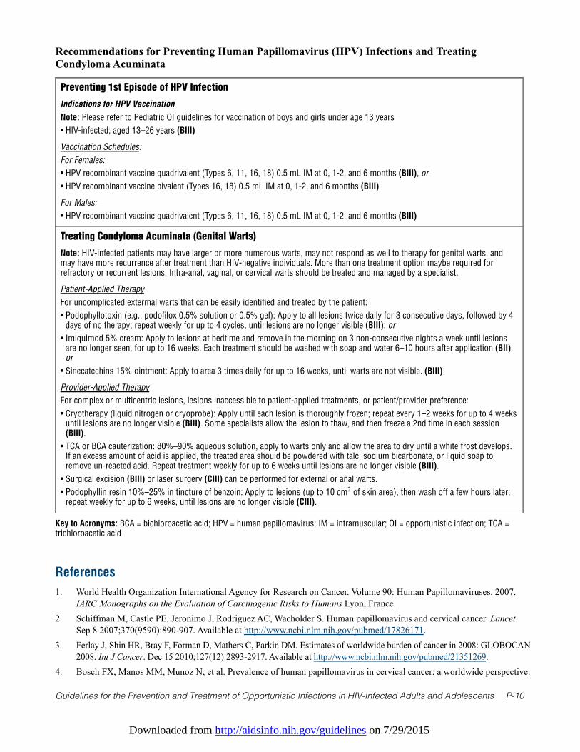
Downloaded from http://aidsinfo.nih.gov/guidelines on 7/29/2015
Guidelines for the Prevention and Treatment of Opportunistic Infections in HIV-Infected Adults and Adolescents P-10
Recommendations for Preventing Human Papillomavirus (HPV) Infections and TreatingCondyloma Acuminata
Preventing 1st Episode of HPV Infection
Indications for HPV Vaccination
Note: Please refer to Pediatric OI guidelines for vaccination of boys and girls under age 13 years
• HIV-infected; aged 13–26 years (BIII)
Vaccination Schedules:
For Females:
• HPV recombinant vaccine quadrivalent (Types 6, 11, 16, 18) 0.5 mL IM at 0, 1-2, and 6 months (BIII), or
• HPV recombinant vaccine bivalent (Types 16, 18) 0.5 mL IM at 0, 1-2, and 6 months (BIII)
For Males:
• HPV recombinant vaccine quadrivalent (Types 6, 11, 16, 18) 0.5 mL IM at 0, 1-2, and 6 months (BIII)
Treating Condyloma Acuminata (Genital Warts)
Note: HIV-infected patients may have larger or more numerous warts, may not respond as well to therapy for genital warts, andmay have more recurrence after treatment than HIV-negative individuals. More than one treatment option maybe required forrefractory or recurrent lesions. Intra-anal, vaginal, or cervical warts should be treated and managed by a specialist.
Patient-Applied Therapy
For uncomplicated extermal warts that can be easily identified and treated by the patient:
• Podophyllotoxin (e.g., podofilox 0.5% solution or 0.5% gel): Apply to all lesions twice daily for 3 consecutive days, followed by 4days of no therapy; repeat weekly for up to 4 cycles, until lesions are no longer visible (BIII); or
• Imiquimod 5% cream: Apply to lesions at bedtime and remove in the morning on 3 non-consecutive nights a week until lesionsare no longer seen, for up to 16 weeks. Each treatment should be washed with soap and water 6–10 hours after application (BII),or
• Sinecatechins 15% ointment: Apply to area 3 times daily for up to 16 weeks, until warts are not visible. (BIII)
Provider-Applied Therapy
For complex or multicentric lesions, lesions inaccessible to patient-applied treatments, or patient/provider preference:
• Cryotherapy (liquid nitrogen or cryoprobe): Apply until each lesion is thoroughly frozen; repeat every 1–2 weeks for up to 4 weeksuntil lesions are no longer visible (BIII). Some specialists allow the lesion to thaw, and then freeze a 2nd time in each session(BIII).
• TCA or BCA cauterization: 80%–90% aqueous solution, apply to warts only and allow the area to dry until a white frost develops.If an excess amount of acid is applied, the treated area should be powdered with talc, sodium bicarbonate, or liquid soap toremove un-reacted acid. Repeat treatment weekly for up to 6 weeks until lesions are no longer visible (BIII).
• Surgical excision (BIII) or laser surgery (CIII) can be performed for external or anal warts.
• Podophyllin resin 10%–25% in tincture of benzoin: Apply to lesions (up to 10 cm2 of skin area), then wash off a few hours later;repeat weekly for up to 6 weeks, until lesions are no longer visible (CIII).
Key to Acronyms: BCA = bichloroacetic acid; HPV = human papillomavirus; IM = intramuscular; OI = opportunistic infection; TCA =trichloroacetic acid
References
1. World Health Organization International Agency for Research on Cancer. Volume 90: Human Papillomaviruses. 2007.IARC Monographs on the Evaluation of Carcinogenic Risks to Humans Lyon, France.
2. Schiffman M, Castle PE, Jeronimo J, Rodriguez AC, Wacholder S. Human papillomavirus and cervical cancer. Lancet.Sep 8 2007;370(9590):890-907. Available at http://www.ncbi.nlm.nih.gov/pubmed/17826171.
3. Ferlay J, Shin HR, Bray F, Forman D, Mathers C, Parkin DM. Estimates of worldwide burden of cancer in 2008: GLOBOCAN2008. Int J Cancer. Dec 15 2010;127(12):2893-2917. Available at http://www.ncbi.nlm.nih.gov/pubmed/21351269.
4. Bosch FX, Manos MM, Munoz N, et al. Prevalence of human papillomavirus in cervical cancer: a worldwide perspective.
Downloaded from http://aidsinfo.nih.gov/guidelines on 7/29/2015
Guidelines for the Prevention and Treatment of Opportunistic Infections in HIV-Infected Adults and Adolescents P-11
International biological study on cervical cancer (IBSCC) Study Group. J Natl Cancer Inst. Jun 7 1995;87(11):796-802.Available at http://www.ncbi.nlm.nih.gov/pubmed/7791229.
5. Wheeler CM, Hunt WC, Joste NE, Key CR, Quint WG, Castle PE. Human papillomavirus genotype distributions:implications for vaccination and cancer screening in the United States. J Natl Cancer Inst. Apr 1 2009;101(7):475-487.Available at http://www.ncbi.nlm.nih.gov/pubmed/19318628.
6. Munoz N, Bosch FX, de Sanjose S, et al. Epidemiologic classification of human papillomavirus types associated withcervical cancer. N Engl J Med. Feb 6 2003;348(6):518-527. Available at http://www.ncbi.nlm.nih.gov/pubmed/12571259.
7. Kraus I, Molden T, Holm R, et al. Presence of E6 and E7 mRNA from human papillomavirus types 16, 18, 31, 33, and45 in the majority of cervical carcinomas. J Clin Microbiol. Apr 2006;44(4):1310-1317. Available athttp://www.ncbi.nlm.nih.gov/pubmed/16597856.
8. Castle PE, Dockter J, Giachetti C, et al. A cross-sectional study of a prototype carcinogenic human papillomavirusE6/E7 messenger RNA assay for detection of cervical precancer and cancer. Clin Cancer Res. May 1 2007;13(9):2599-2605. Available at http://www.ncbi.nlm.nih.gov/pubmed/17473189.
9. Ratnam S, Coutlee F, Fontaine D, et al. Clinical performance of the PreTect HPV-Proofer E6/E7 mRNA assay incomparison with that of the Hybrid Capture 2 test for identification of women at risk of cervical cancer. J ClinMicrobiol. Aug 2010;48(8):2779-2785. Available at http://www.ncbi.nlm.nih.gov/pubmed/20573862.
10. Doorbar J. Molecular biology of human papillomavirus infection and cervical cancer. Clinical science. May2006;110(5):525-541. Available at http://www.ncbi.nlm.nih.gov/pubmed/16597322.
11. Ho GY, Bierman R, Beardsley L, Chang CJ, Burk RD. Natural history of cervicovaginal papillomavirus infection in youngwomen. N Engl J Med. Feb 12 1998;338(7):423-428. Available at http://www.ncbi.nlm.nih.gov/pubmed/9459645.
12. Winer RL, Feng Q, Hughes JP, O'Reilly S, Kiviat NB, Koutsky LA. Risk of female human papillomavirus acquisitionassociated with first male sex partner. J Infect Dis. Jan 15 2008;197(2):279-282. Available athttp://www.ncbi.nlm.nih.gov/pubmed/18179386.
13. Bauer HM, Hildesheim A, Schiffman MH, et al. Determinants of genital human papillomavirus infection in low-riskwomen in Portland, Oregon. Sex Transm Dis. Sep-Oct 1993;20(5):274-278. Available athttp://www.ncbi.nlm.nih.gov/pubmed/8235925.
14. Wheeler CM, Parmenter CA, Hunt WC, et al. Determinants of genital human papillomavirus infection amongcytologically normal women attending the University of New Mexico student health center. Sex Transm Dis. Sep-Oct1993;20(5):286-289. Available at http://www.ncbi.nlm.nih.gov/pubmed/8235927.
15. Burk RD, Ho GY, Beardsley L, Lempa M, Peters M, Bierman R. Sexual behavior and partner characteristics are thepredominant risk factors for genital human papillomavirus infection in young women. J Infect Dis. Oct1996;174(4):679-689. Available at http://www.ncbi.nlm.nih.gov/pubmed/8843203.
16. Winer RL, Lee SK, Hughes JP, Adam DE, Kiviat NB, Koutsky LA. Genital human papillomavirus infection: incidenceand risk factors in a cohort of female university students. American journal of epidemiology. Feb 1 2003;157(3):218-226. Available at http://www.ncbi.nlm.nih.gov/pubmed/12543621.
17. Winer RL, Hughes JP, Feng Q, et al. Condom use and the risk of genital human papillomavirus infection in youngwomen. N Engl J Med. Jun 22 2006;354(25):2645-2654. Available at http://www.ncbi.nlm.nih.gov/pubmed/16790697.
18. Moscicki AB, Shiboski S, Broering J, et al. The natural history of human papillomavirus infection as measured byrepeated DNA testing in adolescent and young women. J Pediatr. Feb 1998;132(2):277-284. Available athttp://www.ncbi.nlm.nih.gov/pubmed/9506641.
19. Evander M, Edlund K, Gustafsson A, et al. Human papillomavirus infection is transient in young women: a population-based cohort study. J Infect Dis. Apr 1995;171(4):1026-1030. Available at http://www.ncbi.nlm.nih.gov/pubmed/7706782.
20. Rodriguez AC, Schiffman M, Herrero R, et al. Longitudinal study of human papillomavirus persistence and cervicalintraepithelial neoplasia grade 2/3: critical role of duration of infection. J Natl Cancer Inst. Mar 3 2010;102(5):315-324.Available at http://www.ncbi.nlm.nih.gov/pubmed/20157096.
21. Schiffman M, Clifford G, Buonaguro FM. Classification of weakly carcinogenic human papillomavirus types:addressing the limits of epidemiology at the borderline. Infectious agents and cancer. 2009;4:8. Available athttp://www.ncbi.nlm.nih.gov/pubmed/19486508.
22. Bouvard V, Baan R, Straif K, et al. A review of human carcinogens—Part B: biological agents. The lancet oncology.
Downloaded from http://aidsinfo.nih.gov/guidelines on 7/29/2015
Guidelines for the Prevention and Treatment of Opportunistic Infections in HIV-Infected Adults and Adolescents P-12
Apr 2009;10(4):321-322. Available at http://www.ncbi.nlm.nih.gov/pubmed/19350698.23. Castle PE. The evolving definition of carcinogenic human papillomavirus. Infectious agents and cancer. 2009;4:7.
Available at http://www.ncbi.nlm.nih.gov/pubmed/19432962.24. Frisch M, Biggar RJ, Goedert JJ. Human papillomavirus-associated cancers in patients with human immunodeficiency
virus infection and acquired immunodeficiency syndrome. J Natl Cancer Inst. Sep 20 2000;92(18):1500-1510.Available at http://www.ncbi.nlm.nih.gov/pubmed/10995805.
25. Chaturvedi AK, Madeleine MM, Biggar RJ, Engels EA. Risk of human papillomavirus-associated cancers amongpersons with AIDS. J Natl Cancer Inst. Aug 19 2009;101(16):1120-1130. Available athttp://www.ncbi.nlm.nih.gov/pubmed/19648510.
26. Simard EP, Engels EA. Cancer as a cause of death among people with AIDS in the United States. Clin Infect Dis. Oct15 2010;51(8):957-962. Available at http://www.ncbi.nlm.nih.gov/pubmed/20825305.
27. Clifford GM, Polesel J, Rickenbach M, et al. Cancer risk in the Swiss HIV Cohort Study: associations withimmunodeficiency, smoking, and highly active antiretroviral therapy. J Natl Cancer Inst. Mar 16 2005;97(6):425-432.Available at http://www.ncbi.nlm.nih.gov/pubmed/15770006.
28. Grulich AE, van Leeuwen MT, Falster MO, Vajdic CM. Incidence of cancers in people with HIV/AIDS compared withimmunosuppressed transplant recipients: a meta-analysis. Lancet. Jul 7 2007;370(9581):59-67. Available athttp://www.ncbi.nlm.nih.gov/pubmed/17617273.
29. Dal Maso L, Polesel J, Serraino D, et al. Pattern of cancer risk in persons with AIDS in Italy in the HAART era. Britishjournal of cancer. Mar 10 2009;100(5):840-847. Available at http://www.ncbi.nlm.nih.gov/pubmed/19223894.
30. Polesel J, Franceschi S, Suligoi B, et al. Cancer incidence in people with AIDS in Italy. Int J Cancer. Sep 12010;127(6):1437-1445. Available at http://www.ncbi.nlm.nih.gov/pubmed/20049835.
31. Guiguet M, Boue F, Cadranel J, et al. Effect of immunodeficiency, HIV viral load, and antiretroviral therapy on the riskof individual malignancies (FHDH-ANRS CO4): a prospective cohort study. The lancet oncology. Dec2009;10(12):1152-1159. Available at http://www.ncbi.nlm.nih.gov/pubmed/19818686.
32. Orem J, Otieno MW, Remick SC. AIDS-associated cancer in developing nations. Curr Opin Oncol. Sep2004;16(5):468-476. Available at http://www.ncbi.nlm.nih.gov/pubmed/15314517.
33. Mbulaiteye SM, Katabira ET, Wabinga H, et al. Spectrum of cancers among HIV-infected persons in Africa: the UgandaAIDS-Cancer Registry Match Study. Int J Cancer. Feb 15 2006;118(4):985-990. Available athttp://www.ncbi.nlm.nih.gov/pubmed/16106415.
34. Strickler HD, Burk RD, Fazzari M, et al. Natural history and possible reactivation of human papillomavirus in humanimmunodeficiency virus-positive women. J Natl Cancer Inst. Apr 20 2005;97(8):577-586. Available athttp://www.ncbi.nlm.nih.gov/pubmed/15840880.
35. Moscicki AB, Ellenberg JH, Crowley-Nowick P, Darragh TM, Xu J, Fahrat S. Risk of high-grade squamousintraepithelial lesion in HIV-infected adolescents. J Infect Dis. Oct 15 2004;190(8):1413-1421. Available athttp://www.ncbi.nlm.nih.gov/pubmed/15378433.
36. Schrager LK, Friedland GH, Maude D, et al. Cervical and vaginal squamous cell abnormalities in women infected withhuman immunodeficiency virus. J Acquir Immune Defic Syndr. 1989;2(6):570-575. Available athttp://www.ncbi.nlm.nih.gov/pubmed/2555473.
37. Maiman M, Fruchter RG, Serur E, Remy JC, Feuer G, Boyce J. Human immunodeficiency virus infection and cervicalneoplasia. Gynecol Oncol. Sep 1990;38(3):377-382. Available at http://www.ncbi.nlm.nih.gov/pubmed/2227552.
38. Ahdieh L, Klein RS, Burk R, et al. Prevalence, incidence, and type-specific persistence of human papillomavirus inhuman immunodeficiency virus (HIV)-positive and HIV-negative women. J Infect Dis. Sep 15 2001;184(6):682-690.Available at http://www.ncbi.nlm.nih.gov/pubmed/11517428.
39. Schuman P, Ohmit SE, Klein RS, et al. Longitudinal study of cervical squamous intraepithelial lesions in humanimmunodeficiency virus (HIV)-seropositive and at-risk HIV-seronegative women. J Infect Dis. Jul 1 2003;188(1):128-136. Available at http://www.ncbi.nlm.nih.gov/pubmed/12825181.
40. Massad LS, Riester KA, Anastos KM, et al. Prevalence and predictors of squamous cell abnormalities in Papanicolaousmears from women infected with HIV-1. Women's Interagency HIV Study Group. J Acquir Immune Defic Syndr. May1 1999;21(1):33-41. Available at http://www.ncbi.nlm.nih.gov/pubmed/10235512.
Downloaded from http://aidsinfo.nih.gov/guidelines on 7/29/2015
Guidelines for the Prevention and Treatment of Opportunistic Infections in HIV-Infected Adults and Adolescents P-13
41. Feingold AR, Vermund SH, Burk RD, et al. Cervical cytologic abnormalities and papillomavirus in women infectedwith human immunodeficiency virus. J Acquir Immune Defic Syndr. 1990;3(9):896-903. Available athttp://www.ncbi.nlm.nih.gov/pubmed/2166784.
42. Wright TC, Jr., Ellerbrock TV, Chiasson MA, Van Devanter N, Sun XW. Cervical intraepithelial neoplasia in women infectedwith human immunodeficiency virus: prevalence, risk factors, and validity of Papanicolaou smears. New York CervicalDisease Study. Obstet Gynecol. Oct 1994;84(4):591-597. Available at http://www.ncbi.nlm.nih.gov/pubmed/8090399.
43. Sun XW, Ellerbrock TV, Lungu O, Chiasson MA, Bush TJ, Wright TC, Jr. Human papillomavirus infection in humanimmunodeficiency virus-seropositive women. Obstet Gynecol. May 1995;85(5 Pt 1):680-686. Available athttp://www.ncbi.nlm.nih.gov/pubmed/7724095.
44. Heard I, Jeannel D, Bergeron C, Saada M, Henrion R, Kazatchkine MD. Lack of behavioural risk factors for squamousintraepithelial lesions (SIL) in HIV-infected women. Int J STD AIDS. Jun 1997;8(6):388-392. Available athttp://www.ncbi.nlm.nih.gov/pubmed/9179650.
45. Delmas MC, Larsen C, van Benthem B, et al. Cervical squamous intraepithelial lesions in HIV-infected women:prevalence, incidence and regression. European Study Group on Natural History of HIV Infection in Women. AIDS.Aug 18 2000;14(12):1775-1784. Available at http://www.ncbi.nlm.nih.gov/pubmed/10985315.
46. Six C, Heard I, Bergeron C, et al. Comparative prevalence, incidence and short-term prognosis of cervical squamousintraepithelial lesions amongst HIV-positive and HIV-negative women. AIDS. Jun 18 1998;12(9):1047-1056. Availableat http://www.ncbi.nlm.nih.gov/pubmed/9662202.
47. Moscicki AB, Ellenberg JH, Farhat S, Xu J. Persistence of human papillomavirus infection in HIV-infected and -uninfected adolescent girls: risk factors and differences, by phylogenetic type. J Infect Dis. Jul 1 2004;190(1):37-45.Available at http://www.ncbi.nlm.nih.gov/pubmed/15195241.
48. Brogly SB, Watts DH, Ylitalo N, et al. Reproductive health of adolescent girls perinatally infected with HIV. Am JPublic Health. Jun 2007;97(6):1047-1052. Available at http://www.ncbi.nlm.nih.gov/pubmed/17463385.
49. Parkin DM, Bray F. Chapter 2: The burden of HPV-related cancers. Vaccine. Aug 31 2006;24 Suppl 3:S3/11-25.Available at http://www.ncbi.nlm.nih.gov/pubmed/16949997.
50. Chaturvedi AK. Beyond cervical cancer: burden of other HPV-related cancers among men and women. The Journal ofadolescent health: official publication of the Society for Adolescent Medicine. Apr 2010;46(4 Suppl):S20-26. Availableat http://www.ncbi.nlm.nih.gov/pubmed/20307840.
51. Grulich AE, Jin F, Conway EL, Stein AN, Hocking J. Cancers attributable to human papillomavirus infection. Sexualhealth. Sep 2010;7(3):244-252. Available at http://www.ncbi.nlm.nih.gov/pubmed/20719211.
52. Smith JS, Backes DM, Hoots BE, Kurman RJ, Pimenta JM. Human papillomavirus type-distribution in vulvar andvaginal cancers and their associated precursors. Obstet Gynecol. Apr 2009;113(4):917-924. Available athttp://www.ncbi.nlm.nih.gov/pubmed/19305339.
53. De Vuyst H, Clifford GM, Nascimento MC, Madeleine MM, Franceschi S. Prevalence and type distribution of humanpapillomavirus in carcinoma and intraepithelial neoplasia of the vulva, vagina and anus: a meta-analysis. Int J Cancer.Apr 1 2009;124(7):1626-1636. Available at http://www.ncbi.nlm.nih.gov/pubmed/19115209.
54. Simard EP, Pfeiffer RM, Engels EA. Spectrum of cancer risk late after AIDS onset in the United States. Arch InternMed. Aug 9 2010;170(15):1337-1345. Available at http://www.ncbi.nlm.nih.gov/pubmed/20696958.
55. Engels EA, Biggar RJ, Hall HI, et al. Cancer risk in people infected with human immunodeficiency virus in the UnitedStates. Int J Cancer. Jul 1 2008;123(1):187-194. Available at http://www.ncbi.nlm.nih.gov/pubmed/18435450.
56. Wilkin TJ, Palmer S, Brudney KF, Chiasson MA, Wright TC. Anal intraepithelial neoplasia in heterosexual andhomosexual HIV-positive men with access to antiretroviral therapy. J Infect Dis. Nov 1 2004;190(9):1685-1691.Available at http://www.ncbi.nlm.nih.gov/pubmed/15478076.
57. Kreuter A, Brockmeyer NH, Hochdorfer B, et al. Clinical spectrum and virologic characteristics of anal intraepithelialneoplasia in HIV infection. Journal of the American Academy of Dermatology. Apr 2005;52(4):603-608. Available athttp://www.ncbi.nlm.nih.gov/pubmed/15793509.
58. Palefsky JM, Holly EA, Efirdc JT, et al. Anal intraepithelial neoplasia in the highly active antiretroviral therapy eraamong HIV-positive men who have sex with men. AIDS. Sep 2 2005;19(13):1407-1414. Available athttp://www.ncbi.nlm.nih.gov/pubmed/16103772.
Downloaded from http://aidsinfo.nih.gov/guidelines on 7/29/2015
Guidelines for the Prevention and Treatment of Opportunistic Infections in HIV-Infected Adults and Adolescents P-14
59. Massad LS, Silverberg MJ, Springer G, et al. Effect of antiretroviral therapy on the incidence of genital warts and vulvarneoplasia among women with the human immunodeficiency virus. Am J Obstet Gynecol. May 2004;190(5):1241-1248.Available at http://www.ncbi.nlm.nih.gov/pubmed/15167825.
60. Conley LJ, Ellerbrock TV, Bush TJ, Chiasson MA, Sawo D, Wright TC. HIV-1 infection and risk of vulvovaginal andperianal condylomata acuminata and intraepithelial neoplasia: a prospective cohort study. Lancet. Jan 122002;359(9301):108-113. Available at http://www.ncbi.nlm.nih.gov/pubmed/11809252.
61. Jamieson DJ, Paramsothy P, Cu-Uvin S, Duerr A, Group HIVERS. Vulvar, vaginal, and perianal intraepithelial neoplasiain women with or at risk for human immunodeficiency virus. Obstet Gynecol. May 2006;107(5):1023-1028. Available athttp://www.ncbi.nlm.nih.gov/pubmed/16648406.
62. Ahdieh-Grant L, Li R, Levine AM, et al. Highly active antiretroviral therapy and cervical squamous intraepitheliallesions in human immunodeficiency virus-positive women. J Natl Cancer Inst. Jul 21 2004;96(14):1070-1076. Availableat http://www.ncbi.nlm.nih.gov/pubmed/15265968.
63. Minkoff H, Zhong Y, Burk RD, et al. Influence of adherent and effective antiretroviral therapy use on humanpapillomavirus infection and squamous intraepithelial lesions in human immunodeficiency virus-positive women. JInfect Dis. Mar 2010;201(5):681-690. Available at http://www.ncbi.nlm.nih.gov/pubmed/20105077.
64. King MD, Reznik DA, O'Daniels CM, Larsen NM, Osterholt D, Blumberg HM. Human papillomavirus-associated oral wartsamong human immunodeficiency virus-seropositive patients in the era of highly active antiretroviral therapy: an emerginginfection. Clin Infect Dis. Mar 1 2002;34(5):641-648. Available at http://www.ncbi.nlm.nih.gov/pubmed/11803508.
65. Greenspan D, Canchola AJ, MacPhail LA, Cheikh B, Greenspan JS. Effect of highly active antiretroviral therapy on frequencyof oral warts. Lancet. May 5 2001;357(9266):1411-1412. Available at http://www.ncbi.nlm.nih.gov/pubmed/11356441.
66. Greenspan D, Gange SJ, Phelan JA, et al. Incidence of oral lesions in HIV-1-infected women: reduction with HAART.Journal of dental research. Feb 2004;83(2):145-150. Available at http://www.ncbi.nlm.nih.gov/pubmed/14742653.
67. Hamza OJ, Matee MI, Simon EN, et al. Oral manifestations of HIV infection in children and adults receiving highlyactive anti-retroviral therapy [HAART] in Dar es Salaam, Tanzania. BMC oral health. 2006;6:12. Available athttp://www.ncbi.nlm.nih.gov/pubmed/16916469.
68. D'Souza G, Kreimer AR, Viscidi R, et al. Case-control study of human papillomavirus and oropharyngeal cancer. N EnglJ Med. May 10 2007;356(19):1944-1956. Available at http://www.ncbi.nlm.nih.gov/pubmed/17494927.
69. CDC. Sexually transmitted diseases treatment guidelines 2002. Centers for Disease Control and Prevention. MMWRRecomm Rep. May 10 2002;51(RR-6):1-78. Available at http://www.ncbi.nlm.nih.gov/pubmed/12184549.
70. Food and Drug Administration. Product approval information – licensing action, package insert. hc2 High-Risk HPVDNA Test, Accessed April 23, 2010. Available at: http://www.accessdata.fda.gov/cdrh_docs/pdf/P890064S009c.pdf.
71. Food and Drug Administration. Product approval information – licensing action, package insert. Cervista™ HPV HRtest, Accessed April 23, 2010. Available at: http://www.accessdata.fda.gov/cdrh_docs/pdf8/P080014c.pdf.
72. Food and Drug Administration. Product approval information – licensing action, package insert. Cervista™ 16/18 test,Accessed April 23, 2010. Available at: http://www.accessdata.fda.gov/cdrh_docs/pdf8/P080015c.pdf.
73. Moyer VA, Force USPST. Screening for cervical cancer: U.S. Preventive Services Task Force recommendation statement.Ann Intern Med. Jun 19 2012;156(12):880-891, W312. Available at http://www.ncbi.nlm.nih.gov/pubmed/22711081.
74. Smith RA, Brooks D, Cokkinides V, Saslow D, Brawley OW. Cancer screening in the United States, 2013: A review ofcurrent american cancer society guidelines, current issues in cancer screening, and new guidance on cervical cancerscreening and lung cancer screening. CA: a cancer journal for clinicians. Mar 2013;63(2):87-105. Available athttp://www.ncbi.nlm.nih.gov/pubmed/23378235.
75. Wright TC, Jr., Massad LS, Dunton CJ, et al. 2006 consensus guidelines for the management of women with abnormalcervical cancer screening tests. Am J Obstet Gynecol. Oct 2007;197(4):346-355. Available athttp://www.ncbi.nlm.nih.gov/pubmed/17904957.
76. Massad LS, Schneider MF, Watts DH, et al. HPV testing for triage of HIV-infected women with papanicolaou smearsread as atypical squamous cells of uncertain significance. J Womens Health (Larchmt). Mar 2004;13(2):147-153.Available at http://www.ncbi.nlm.nih.gov/pubmed/15072728.
77. Kirby TO, Allen ME, Alvarez RD, Hoesley CJ, Huh WK. High-risk human papillomavirus and cervical intraepithelialneoplasia at time of atypical squamous cells of undetermined significance cytologic results in a population with human
Downloaded from http://aidsinfo.nih.gov/guidelines on 7/29/2015
Guidelines for the Prevention and Treatment of Opportunistic Infections in HIV-Infected Adults and Adolescents P-15
immunodeficiency virus. J Low Genit Tract Dis. Oct 2004;8(4):298-303. Available athttp://www.ncbi.nlm.nih.gov/pubmed/15874876.
78. Paavonen J, Naud P, Salmeron J, et al. Efficacy of human papillomavirus (HPV)-16/18 AS04-adjuvanted vaccineagainst cervical infection and precancer caused by oncogenic HPV types (PATRICIA): final analysis of a double-blind,randomised study in young women. Lancet. Jul 25 2009;374(9686):301-314. Available athttp://www.ncbi.nlm.nih.gov/pubmed/19586656.
79. Group FIS. Quadrivalent vaccine against human papillomavirus to prevent high-grade cervical lesions. N Engl J Med.May 10 2007;356(19):1915-1927. Available at http://www.ncbi.nlm.nih.gov/pubmed/17494925.
80. Palefsky JM, Giuliano AR, Goldstone S, et al. HPV vaccine against anal HPV infection and anal intraepithelial neoplasia. NEngl J Med. Oct 27 2011;365(17):1576-1585. Available at http://www.ncbi.nlm.nih.gov/pubmed/22029979.
81. Giuliano AR, Palefsky JM, Goldstone S, et al. Efficacy of quadrivalent HPV vaccine against HPV Infection and diseasein males. N Engl J Med. Feb 3 2011;364(5):401-411. Available at http://www.ncbi.nlm.nih.gov/pubmed/21288094.
82. Joura EA, Leodolter S, Hernandez-Avila M, et al. Efficacy of a quadrivalent prophylactic human papillomavirus (types6, 11, 16, and 18) L1 virus-like-particle vaccine against high-grade vulval and vaginal lesions: a combined analysis ofthree randomised clinical trials. Lancet. May 19 2007;369(9574):1693-1702. Available athttp://www.ncbi.nlm.nih.gov/pubmed/17512854.
83. Garland SM, Hernandez-Avila M, Wheeler CM, et al. Quadrivalent vaccine against human papillomavirus to preventanogenital diseases. N Engl J Med. May 10 2007;356(19):1928-1943. Available athttp://www.ncbi.nlm.nih.gov/pubmed/17494926.
84. Centers for Disease C, Prevention. Recommendations on the use of quadrivalent human papillomavirus vaccine inmales--Advisory Committee on Immunization Practices (ACIP), 2011. MMWR Morb Mortal Wkly Rep. Dec 232011;60(50):1705-1708. Available at http://www.ncbi.nlm.nih.gov/pubmed/22189893.
85. Levin MJ, Moscicki AB, Song LY, et al. Safety and immunogenicity of a quadrivalent human papillomavirus (types 6,11, 16, and 18) vaccine in HIV-infected children 7 to 12 years old. J Acquir Immune Defic Syndr. Oct 2010;55(2):197-204. Available at http://www.ncbi.nlm.nih.gov/pubmed/20574412.
86. Wilkin T, Lee JY, Lensing SY, et al. Safety and immunogenicity of the quadrivalent human papillomavirus vaccine in HIV-1-infected men. J Infect Dis. Oct 15 2010;202(8):1246-1253. Available at http://www.ncbi.nlm.nih.gov/pubmed/20812850.
87. Kish LS, McMahon JT, Bergfeld WF, Pelachyk JM. An ancient method and a modern scourge: the condom as a barrieragainst herpes. Journal of the American Academy of Dermatology. Nov 1983;9(5):769-770. Available athttp://www.ncbi.nlm.nih.gov/pubmed/6685737.
88. Nielson CM, Harris RB, Nyitray AG, Dunne EF, Stone KM, Giuliano AR. Consistent condom use is associated withlower prevalence of human papillomavirus infection in men. J Infect Dis. Aug 15 2010;202(3):445-451. Available athttp://www.ncbi.nlm.nih.gov/pubmed/20569156.
89. Manhart LE, Koutsky LA. Do condoms prevent genital HPV infection, external genital warts, or cervical neoplasia? Ameta-analysis. Sex Transm Dis. Nov 2002;29(11):725-735. Available at http://www.ncbi.nlm.nih.gov/pubmed/12438912.
90. Hogewoning CJ, Bleeker MC, van den Brule AJ, et al. Condom use promotes regression of cervical intraepithelialneoplasia and clearance of human papillomavirus: a randomized clinical trial. Int J Cancer. Dec 10 2003;107(5):811-816. Available at http://www.ncbi.nlm.nih.gov/pubmed/14566832.
91. Bleeker MC, Hogewoning CJ, Voorhorst FJ, et al. Condom use promotes regression of human papillomavirus-associated penile lesions in male sexual partners of women with cervical intraepithelial neoplasia. Int J Cancer. Dec 102003;107(5):804-810. Available at http://www.ncbi.nlm.nih.gov/pubmed/14566831.
92. Hankins C, Coutlee F, Lapointe N, et al. Prevalence of risk factors associated with human papillomavirus infection inwomen living with HIV. Canadian Women's HIV Study Group. CMAJ: Canadian Medical Association journal =journal de l'Association medicale canadienne. Jan 26 1999;160(2):185-191. Available athttp://www.ncbi.nlm.nih.gov/pubmed/9951439.
93. Holmes KK, Levine R, Weaver M. Effectiveness of condoms in preventing sexually transmitted infections. Bulletin ofthe World Health Organization. Jun 2004;82(6):454-461. Available at http://www.ncbi.nlm.nih.gov/pubmed/15356939.
94. Kelvin EA, Smith RA, Mantell JE, Stein ZA. Adding the female condom to the public health agenda on prevention ofHIV and other sexually transmitted infections among men and women during anal intercourse. Am J Public Health. Jun2009;99(6):985-987. Available at http://www.ncbi.nlm.nih.gov/pubmed/19372513.
Downloaded from http://aidsinfo.nih.gov/guidelines on 7/29/2015
Guidelines for the Prevention and Treatment of Opportunistic Infections in HIV-Infected Adults and Adolescents P-16
95. Macaluso M, Blackwell R, Jamieson DJ, et al. Efficacy of the male latex condom and of the female polyurethane condomas barriers to semen during intercourse: a randomized clinical trial. American journal of epidemiology. Jul 12007;166(1):88-96. Available at http://www.ncbi.nlm.nih.gov/pubmed/17420182.
96. French PP, Latka M, Gollub EL, Rogers C, Hoover DR, Stein ZA. Use-effectiveness of the female versus male condom inpreventing sexually transmitted disease in women. Sex Transm Dis. May 2003;30(5):433-439. Available athttp://www.ncbi.nlm.nih.gov/pubmed/12916135.
97. Waugh M. The role of condom use in sexually transmitted disease prevention: facts and controversies. Clin Dermatol.Sep-Oct 2010;28(5):549-552. Available at http://www.ncbi.nlm.nih.gov/pubmed/20797517.
98. Auvert B, Sobngwi-Tambekou J, Cutler E, et al. Effect of male circumcision on the prevalence of high-risk humanpapillomavirus in young men: results of a randomized controlled trial conducted in Orange Farm, South Africa. J InfectDis. Jan 1 2009;199(1):14-19. Available at http://www.ncbi.nlm.nih.gov/pubmed/19086814.
99. Tobian AA, Serwadda D, Quinn TC, et al. Male circumcision for the prevention of HSV-2 and HPV infections andsyphilis. N Engl J Med. Mar 26 2009;360(13):1298-1309. Available at http://www.ncbi.nlm.nih.gov/pubmed/19321868.
100. Serwadda D, Wawer MJ, Makumbi F, et al. Circumcision of HIV-infected men: effects on high-risk human papillomavirusinfections in a randomized trial in Rakai, Uganda. J Infect Dis. May 15 2010;201(10):1463-1469. Available athttp://www.ncbi.nlm.nih.gov/pubmed/20370481.
101. Gray RH, Serwadda D, Kong X, et al. Male circumcision decreases acquisition and increases clearance of high-riskhuman papillomavirus in HIV-negative men: a randomized trial in Rakai, Uganda. J Infect Dis. May 152010;201(10):1455-1462. Available at http://www.ncbi.nlm.nih.gov/pubmed/20370483.
102. Giuliano AR, Lazcano E, Villa LL, et al. Circumcision and sexual behavior: factors independently associated with humanpapillomavirus detection among men in the HIM study. Int J Cancer. Mar 15 2009;124(6):1251-1257. Available athttp://www.ncbi.nlm.nih.gov/pubmed/19089913.
103. Lu B, Wu Y, Nielson CM, et al. Factors associated with acquisition and clearance of human papillomavirus infection in acohort of US men: a prospective study. J Infect Dis. Feb 1 2009;199(3):362-371. Available athttp://www.ncbi.nlm.nih.gov/pubmed/19133808.
104. Nielson CM, Schiaffino MK, Dunne EF, Salemi JL, Giuliano AR. Associations between male anogenital humanpapillomavirus infection and circumcision by anatomic site sampled and lifetime number of female sex partners. J InfectDis. Jan 1 2009;199(1):7-13. Available at http://www.ncbi.nlm.nih.gov/pubmed/19086813.
105. Hernandez BY, Shvetsov YB, Goodman MT, et al. Reduced clearance of penile human papillomavirus infection inuncircumcised men. J Infect Dis. May 1 2010;201(9):1340-1343. Available at http://www.ncbi.nlm.nih.gov/pubmed/20350160.
106. Hernandez BY, Wilkens LR, Zhu X, et al. Circumcision and human papillomavirus infection in men: a site-specificcomparison. J Infect Dis. Mar 15 2008;197(6):787-794. Available at http://www.ncbi.nlm.nih.gov/pubmed/18284369.
107. Lajous M, Mueller N, Cruz-Valdez A, et al. Determinants of prevalence, acquisition, and persistence of humanpapillomavirus in healthy Mexican military men. Cancer epidemiology, biomarkers & prevention: a publication of theAmerican Association for Cancer Research, cosponsored by the American Society of Preventive Oncology. Jul2005;14(7):1710-1716. Available at http://www.ncbi.nlm.nih.gov/pubmed/16030106.
108. Saibishkumar EP, Crook J, Sweet J. Neonatal circumcision and invasive squamous cell carcinoma of the penis: a report of3 cases and a review of the literature. Canadian Urological Association journal = Journal de l'Association des urologuesdu Canada. Feb 2008;2(1):39-42. Available at http://www.ncbi.nlm.nih.gov/pubmed/18542727.
109. Schoen EJ, Oehrli M, Colby C, Machin G. The highly protective effect of newborn circumcision against invasive penilecancer. Pediatrics. Mar 2000;105(3):E36. Available at http://www.ncbi.nlm.nih.gov/pubmed/10699138.
110. Daling JR, Madeleine MM, Johnson LG, et al. Penile cancer: importance of circumcision, human papillomavirus andsmoking in in situ and invasive disease. Int J Cancer. Sep 10 2005;116(4):606-616. Available athttp://www.ncbi.nlm.nih.gov/entrez/query.fcgi?cmd=Retrieve&db=PubMed&dopt=Citation&list_uids=15825185.
111. Maden C, Sherman KJ, Beckmann AM, et al. History of circumcision, medical conditions, and sexual activity and risk ofpenile cancer. J Natl Cancer Inst. Jan 6 1993;85(1):19-24. Available at http://www.ncbi.nlm.nih.gov/pubmed/8380060.
112. Castellsague X, Bosch FX, Munoz N, et al. Male circumcision, penile human papillomavirus infection, and cervicalcancer in female partners. N Engl J Med. Apr 11 2002;346(15):1105-1112. Available athttp://www.ncbi.nlm.nih.gov/pubmed/11948269.
Downloaded from http://aidsinfo.nih.gov/guidelines on 7/29/2015
Guidelines for the Prevention and Treatment of Opportunistic Infections in HIV-Infected Adults and Adolescents P-17
113. Kalogirou D, Antoniou G, Karakitsos P, Botsis D, Papadimitriou A, Giannikos L. Vaginal intraepithelial neoplasia(VAIN) following hysterectomy in patients treated for carcinoma in situ of the cervix. European journal ofgynaecological oncology. 1997;18(3):188-191. Available at http://www.ncbi.nlm.nih.gov/pubmed/9174833.
114. Paramsothy P, Duerr A, Heilig CM, et al. Abnormal vaginal cytology in HIV-infected and at-risk women afterhysterectomy. J Acquir Immune Defic Syndr. Apr 15 2004;35(5):484-491. Available athttp://www.ncbi.nlm.nih.gov/pubmed/15021313.
115. Petry KU, Kochel H, Bode U, et al. Human papillomavirus is associated with the frequent detection of warty andbasaloid high-grade neoplasia of the vulva and cervical neoplasia among immunocompromised women. Gynecol Oncol.Jan 1996;60(1):30-34. Available at http://www.ncbi.nlm.nih.gov/pubmed/8557224.
116. Chiasson MA, Ellerbrock TV, Bush TJ, Sun XW, Wright TC, Jr. Increased prevalence of vulvovaginal condyloma andvulvar intraepithelial neoplasia in women infected with the human immunodeficiency virus. Obstet Gynecol. May1997;89(5 Pt 1):690-694. Available at http://www.ncbi.nlm.nih.gov/pubmed/9166302.
117. Goldie SJ, Kuntz KM, Weinstein MC, Freedberg KA, Welton ML, Palefsky JM. The clinical effectiveness and cost-effectiveness of screening for anal squamous intraepithelial lesions in homosexual and bisexual HIV-positive men.JAMA. May 19 1999;281(19):1822-1829. Available at http://www.ncbi.nlm.nih.gov/pubmed/10340370.
118. Chin-Hong PV, Palefsky JM. Human papillomavirus anogenital disease in HIV-infected individuals. Dermatologictherapy. Jan-Feb 2005;18(1):67-76. Available at http://www.ncbi.nlm.nih.gov/pubmed/15842614.
119. Silverberg MJ, Ahdieh L, Munoz A, et al. The impact of HIV infection and immunodeficiency on humanpapillomavirus type 6 or 11 infection and on genital warts. Sex Transm Dis. Aug 2002;29(8):427-435. Available athttp://www.ncbi.nlm.nih.gov/pubmed/12172526.
120. De Panfilis G, Melzani G, Mori G, Ghidini A, Graifemberghi S. Relapses after treatment of external genital warts aremore frequent in HIV-positive patients than in HIV-negative controls. Sex Transm Dis. Mar 2002;29(3):121-125.Available at http://www.ncbi.nlm.nih.gov/pubmed/11875372.
121. Workowski KA, Berman S, Centers for Disease C, Prevention. Sexually transmitted diseases treatment guidelines, 2010.MMWR Recomm Rep. Dec 17 2010;59(RR-12):1-110. Available at http://www.ncbi.nlm.nih.gov/pubmed/21160459.
122. Baccaglini L, Atkinson JC, Patton LL, Glick M, Ficarra G, Peterson DE. Management of oral lesions in HIV-positivepatients. Oral surgery, oral medicine, oral pathology, oral radiology, and endodontics. Mar 2007;103 Suppl:S50 e51-23. Available at http://www.ncbi.nlm.nih.gov/pubmed/17379155.
123. Cleary RK, Schaldenbrand JD, Fowler JJ, Schuler JM, Lampman RM. Perianal Bowen's disease and anal intraepithelialneoplasia: review of the literature. Diseases of the colon and rectum. Jul 1999;42(7):945-951. Available athttp://www.ncbi.nlm.nih.gov/pubmed/10411443.
124. Webber J, Fromm D. Photodynamic therapy for carcinoma in situ of the anus. Archives of surgery. Mar2004;139(3):259-261. Available at http://www.ncbi.nlm.nih.gov/pubmed/15006881.
125. Scholefield JH. Treatment of grade III anal intraepithelial neoplasia with photodynamic therapy: report of a case. DisColon Rectum, 2003; 46(11):1555-1559. Techniques in coloproctology. Nov 2004;8(3):200. Available athttp://www.ncbi.nlm.nih.gov/pubmed/15654532.
126. Goldstone SE, Kawalek AZ, Huyett JW. Infrared coagulator: a useful tool for treating anal squamous intraepitheliallesions. Diseases of the colon and rectum. May 2005;48(5):1042-1054. Available athttp://www.ncbi.nlm.nih.gov/pubmed/15868241.
127. Graham BD, Jetmore AB, Foote JE, Arnold LK. Topical 5-fluorouracil in the management of extensive anal Bowen'sdisease: a preferred approach. Diseases of the colon and rectum. Mar 2005;48(3):444-450. Available athttp://www.ncbi.nlm.nih.gov/pubmed/15747068.
128. Fox PA, Nathan M, Francis N, et al. A double-blind, randomized controlled trial of the use of imiquimod cream for thetreatment of anal canal high-grade anal intraepithelial neoplasia in HIV-positive MSM on HAART, with long-termfollow-up data including the use of open-label imiquimod. AIDS. Sep 24 2010;24(15):2331-2335. Available athttp://www.ncbi.nlm.nih.gov/pubmed/20729710.
129. Stier EA, Baranoski AS. Human papillomavirus-related diseases in HIV-infected individuals. Curr Opin Oncol. Sep2008;20(5):541-546. Available at http://www.ncbi.nlm.nih.gov/pubmed/19106657.
130. Licitra L, Perrone F, Bossi P, et al. High-risk human papillomavirus affects prognosis in patients with surgically treatedoropharyngeal squamous cell carcinoma. J Clin Oncol. Dec 20 2006;24(36):5630-5636. Available at
Downloaded from http://aidsinfo.nih.gov/guidelines on 7/29/2015
Guidelines for the Prevention and Treatment of Opportunistic Infections in HIV-Infected Adults and Adolescents P-18
http://www.ncbi.nlm.nih.gov/pubmed/17179101.131. Wright TC, Jr., Koulos J, Schnoll F, et al. Cervical intraepithelial neoplasia in women infected with the human
immunodeficiency virus: outcome after loop electrosurgical excision. Gynecol Oncol. Nov 1994;55(2):253-258.Available at http://www.ncbi.nlm.nih.gov/pubmed/7959293.
132. Fruchter RG, Maiman M, Sedlis A, Bartley L, Camilien L, Arrastia CD. Multiple recurrences of cervical intraepithelialneoplasia in women with the human immunodeficiency virus. Obstet Gynecol. Mar 1996;87(3):338-344. Available athttp://www.ncbi.nlm.nih.gov/pubmed/8598951.
133. Maiman M, Watts DH, Andersen J, Clax P, Merino M, Kendall MA. Vaginal 5-fluorouracil for high-grade cervicaldysplasia in human immunodeficiency virus infection: a randomized trial. Obstet Gynecol. Dec 1999;94(6):954-961.Available at http://www.ncbi.nlm.nih.gov/pubmed/10576182.
134. Shah K, Kashima H, Polk BF, Shah F, Abbey H, Abramson A. Rarity of cesarean delivery in cases of juvenile-onset respiratorypapillomatosis. Obstet Gynecol. Dec 1986;68(6):795-799. Available at http://www.ncbi.nlm.nih.gov/pubmed/3785792.
135. Morrison EA, Gammon MD, Goldberg GL, Vermund SH, Burk RD. Pregnancy and cervical infection with humanpapillomaviruses. Int J Gynaecol Obstet. Aug 1996;54(2):125-130. Available athttp://www.ncbi.nlm.nih.gov/pubmed/9236309.
136. Kjellberg L, Hallmans G, Ahren AM, et al. Smoking, diet, pregnancy and oral contraceptive use as risk factors forcervical intra-epithelial neoplasia in relation to human papillomavirus infection. British journal of cancer. Apr2000;82(7):1332-1338. Available at http://www.ncbi.nlm.nih.gov/pubmed/10755410.
137. Audisio T, Roca FC, Piatti C. Topical imiquimod therapy for external anogenital warts in pregnant women. Int JGynaecol Obstet. Mar 2008;100(3):275-276. Available at http://www.ncbi.nlm.nih.gov/pubmed/18035356.
138. Einarson A, Costei A, Kalra S, Rouleau M, Koren G. The use of topical 5% imiquimod during pregnancy: a case series.Reprod Toxicol. Jan 2006;21(1):1-2. Available at http://www.ncbi.nlm.nih.gov/pubmed/16039826.
139. Silverberg MJ, Thorsen P, Lindeberg H, Grant LA, Shah KV. Condyloma in pregnancy is strongly predictive of juvenile-onset recurrent respiratory papillomatosis. Obstet Gynecol. Apr 2003;101(4):645-652. Available athttp://www.ncbi.nlm.nih.gov/pubmed/12681865.
140. Fife KH, Katz BP, Brizendine EJ, Brown DR. Cervical human papillomavirus deoxyribonucleic acid persists throughoutpregnancy and decreases in the postpartum period. Am J Obstet Gynecol. May 1999;180(5):1110-1114. Available athttp://www.ncbi.nlm.nih.gov/pubmed/10329863.
141. Puranen MH, Yliskoski MH, Saarikoski SV, Syrjanen KJ, Syrjanen SM. Exposure of an infant to cervical humanpapillomavirus infection of the mother is common. Am J Obstet Gynecol. May 1997;176(5):1039-1045. Available athttp://www.ncbi.nlm.nih.gov/pubmed/9166165.
142. Watts DH, Koutsky LA, Holmes KK, et al. Low risk of perinatal transmission of human papillomavirus: results from aprospective cohort study. Am J Obstet Gynecol. Feb 1998;178(2):365-373. Available athttp://www.ncbi.nlm.nih.gov/pubmed/9500501.
143. Tseng CJ, Liang CC, Soong YK, Pao CC. Perinatal transmission of human papillomavirus in infants: relationshipbetween infection rate and mode of delivery. Obstet Gynecol. Jan 1998;91(1):92-96. Available athttp://www.ncbi.nlm.nih.gov/pubmed/9464728.
144. Tenti P, Zappatore R, Migliora P, Spinillo A, Belloni C, Carnevali L. Perinatal transmission of human papillomavirusfrom gravidas with latent infections. Obstet Gynecol. Apr 1999;93(4):475-479. Available athttp://www.ncbi.nlm.nih.gov/pubmed/10214817.
145. Bulletins—Gynecology ACoP. ACOG Practice Bulletin No. 117: Gynecologic care for women with humanimmunodeficiency virus. Obstet Gynecol. Dec 2010;116(6):1492-1509. Available athttp://www.ncbi.nlm.nih.gov/pubmed/21099636.
146. Orr JW, Jr., Barrett JM, Orr PF, Holloway RW, Holimon JL. The efficacy and safety of the cytobrush during pregnancy.Gynecol Oncol. Mar 1992;44(3):260-262. Available at http://www.ncbi.nlm.nih.gov/pubmed/1541438.
147. Garland SM, Ault KA, Gall SA, et al. Pregnancy and infant outcomes in the clinical trials of a human papillomavirus type6/11/16/18 vaccine: a combined analysis of five randomized controlled trials. Obstet Gynecol. Dec 2009;114(6):1179-1188. Available at http://www.ncbi.nlm.nih.gov/pubmed/19935017.
Downloaded from http://aidsinfo.nih.gov/guidelines on 7/29/2015
Guidelines for the Prevention and Treatment of Opportunistic Infections in HIV-Infected Adults and Adolescents Q-1
Hepatitis B Virus Infection (Last updated April 22, 2015; last reviewed April 22,2015)
Epidemiology
Hepatitis B virus (HBV) is the leading cause of chronic liver disease worldwide.1,2 Globally and in NorthAmerica, approximately 10% of HIV-infected patients have evidence of chronic HBV infection.3-5
In countries with a low prevalence of endemic chronic HBV infection, the virus is transmitted primarilythrough sexual contact and injection drug use, whereas perinatal and early childhood exposures areresponsible for most HBV transmission in higher prevalence regions.1,2,6 Although the general modes oftransmission are similar to HIV, HBV is transmitted more efficiently than HIV.1,2 HBV has an averageincubation period of 90 days (range 60–150 days) from exposure to onset of jaundice and 60 days (range 40–90 days) from exposure to onset of abnormal liver enzymes. Genotypes of HBV (A–H) have been identifiedwith different geographic distributions. Genotype A is most common among patients in North America andWestern Europe.
Clinical Manifestations
Acute infection is usually asymptomatic. When they manifest, symptoms may include right upper quadrantabdominal pain, nausea, vomiting, fever, and arthralgias with or without jaundice. Most patients with chronicHBV infection are asymptomatic or have nonspecific symptoms, such as fatigue, until they develop cirrhosisand signs of portal hypertension (i.e., ascites, variceal bleeding, coagulopathy, jaundice, or hepaticencephalopathy). Hepatocellular carcinoma (HCC) is asymptomatic in its early stages and usually, but notalways, occurs in the setting of HBV - or hepatitis C (HCV)-related cirrhosis.
Diagnosis
All HIV-infected patients should be tested for HBV infection because of shared routes of transmission. Initialtesting should include serologic testing for surface antigen (HBsAg), hepatitis B core antibody (anti-HBctotal), and hepatitis B surface antibody (anti-HBs). In acute infection, HBsAg can be detected 4 weeks (range1–9 weeks) after exposure and anti-HBc immunoglobulin M is usually detectable at the onset of symptoms.
Chronic HBV infection is defined as persistent HBsAg detected on 2 occasions at least 6 months apart.Patients with chronic HBV infection should be further tested for HBV e-antigen (HBeAg), antibody toHBeAg (anti-HBe), and HBV DNA. Active disease, which can be HBeAg-negative or HBeAG-positive, canbe distinguished from inactive disease by the presence of serum HBV DNA and persistent or fluctuatingalanine transaminase (ALT) elevation. Patients whose past infection has resolved are HBsAg-negative withpositive anti-HBs and anti-HBc, although cccDNA may remain in hepatocyte nuclei. Seroreversion mayoccur (becoming serum HBsAg-positive again) under severe immune suppression, as is seen with rituximabtherapy or after stem cell transplant.7,8
The presence of anti-HBc alone, often occurs on testing as an isolated anti-HBc test result, usually signifiesinfection with HBV in the past with subsequent loss of anti-HBs. It occurs in 6.6% to 58.6% of HIV-infectedpatients,9-11 Incidence of HBV viremia in HIV-infected patients with the isolated anti-HBc pattern rangesfrom 1% to 36%.10,12-15 The clinical significance of isolated anti-HBc is unknown16-18,20 but it may indicatechronic or, more likely, resolved infection in HIV-infected individuals.16,17,21 In a low-prevalence countrysuch as the United States, isolated anti-HBc may also represent a false-positive result.16-19 HIV-infectedpatients have a higher frequency of isolated anti-HBc, particularly those with underlying HCVcoinfection.19,22,23
Downloaded from http://aidsinfo.nih.gov/guidelines on 7/29/2015
Guidelines for the Prevention and Treatment of Opportunistic Infections in HIV-Infected Adults and Adolescents Q-2
Diagnosis of Disease Progression and the Role of Assessment of Liver FibrosisCompared with HIV-uninfected individuals, those who are HIV-infected have higher levels of HBV viremiaand lower likelihood of resolved infection following acute HBV infection.24 In HBV-monoinfectedindividuals, HBV DNA suppression, anti-HBe seroconversion (to anti-HBe-seronegativity), HBsAg loss, andacquisition of anti-HBs are all associated with a decreased incidence of cirrhosis, HCC,25-27 and improvedsurvival.28-31 In comparison, the predictive value of these parameters in persons with HIV/HBV coinfectionindicate they usually are more likely to have detectable HBeAg,24,32 lower rates of seroconversion to anti-HBe, and increased risk of HCC, liver-related mortality and morbidity.33,34
HBV infection can result in a dynamic disease with a number of phases that are associated with either activeor inactive chronic hepatitis. Duration of disease phases is different in those who acquire infection asneonates and young children compared with those who acquire infection as adults. Adults do not have an“immune-tolerant” phase (high levels of HBV DNA and low or rising ALT levels). Clinicians should beknowledgeable about these phases for HBV-monoinfected patients to determine who needs treatment andwho should be monitored. In HIV/HBV coinfection, monitoring and treatment are also focused on thesimultaneous treatment of both viruses.
HBV-monoinfected patients who are HBeAg-seropositive usually have high HBV DNA levels (>20,000IU/mL) and abnormal ALT levels. However, with perinatal infection or infection acquired in early childhood,patients initially have an immune tolerance phase, with the presence of HBeAg, normal ALT levels, and highlevels of HBV DNA but minimal or no liver disease. These patients may develop HBeAg-positive chronichepatitis B with elevated ALT levels and remain at risk for HCC, cirrhosis, and flares of HBV.35
Anti-HBe seroconversion usually implies a transition from active disease to an inactive carrier state.35 Thistransition can be spontaneous or associated with effective HBV treatment. In some instances, increasedlevels of ALT may precede a decline in HBV DNA that is accompanied by anti-HBe seroconversionp, that is,loss of HBeAg and development of anti-HBe. However, such spontaneous HBeAg conversion rates in HIV-infected patients appear to be lower than in monoinfected patients. The inactive chronic HBV state ischaracterized by a negative HBeAg, normal ALT levels, and an HBV DNA level <2,000 IU/mL. Patients inthe inactive state remain at risk of reactivation of HBV and development of HCC, but the risk is lower thanfor individuals with active HBV replication. In any patient, the re-emergence of abnormal liver enzyme testsmay reflect HBeAg-negative chronic HBV disease, a result of mutations in the basal core and precorepromoter regions. Although levels of HBV DNA are usually lower, HBeAg-negative patients experience anunrelenting but fluctuating course of disease progression, with fluctuating HBV DNA levels.35 Thus, even ina patient without HBeAg, serum ALT and HBV DNA levels still should be monitored.
Patients diagnosed with chronic HBV infection should have a complete blood count, ALT, aspartateaminotransferase (AST), albumin and bilirubin levels, and prothrombin time monitored at baseline and every6 months thereafter to assess severity and progression of liver disease. Patients with chronic HBV are atincreased risk of HCC and imaging studies every 6 months are recommended in those who are cirrhotic,Asian male and older than age 40, Asian female and older than age 50, or male older than age 20 and fromsub-Saharan Africa, as individuals in all of these groups are at increased risk of disease progression.35
Persistent low-level serum ALT abnormalities may be associated with significant liver disease, althoughnormal ALT levels also may be seen in the setting of cirrhosis. Transient or persistent elevations in serumALT levels can occur before loss of HBeAg, on discontinuation of anti-HBV therapy, and in association withemergence of HBV drug resistance.
Assessment of stage of liver fibrosis is important to know when to initiate esophageal variceal and HCCscreening in cirrhotic patients. Fibrosis stage can be determined by liver biopsy or by noninvasive methodssuch as transient elastography, The decision to perform a liver biopsy should be individualized, especiallygiven Department of Health and Human Services recommendations to initiate antiretroviral therapy (ART)-containing anti-HBV drugs regardless of CD4 T lymphocyte (CD4) cell count in HIV/HBV coinfected
Downloaded from http://aidsinfo.nih.gov/guidelines on 7/29/2015
Guidelines for the Prevention and Treatment of Opportunistic Infections in HIV-Infected Adults and Adolescents Q-3
patients.36 There is increasing evidence that noninvasive methods (i.e., elastometry and serum biochemicalindices) to evaluate liver fibrosis can be used to determine fibrosis in HBV.37-40 For example, one studydemonstrated that transient elastography was able to discriminate moderate to severe fibrosis from mildfibrosis in HIV/HBV coinfection.41
Preventing Exposure
HBV is primarily transmitted by percutaneous or mucosal exposure to infectious blood or body fluids.Therefore, HIV-infected patients should be counseled about transmission risks for HBV and avoidance ofbehaviors associated with such transmission (AIII). Such counseling should emphasize sexual transmissionas well as the risks associated with sharing needles and syringes, tattooing or body-piercing.
Preventing Disease
All household members and sexual contacts of patients with HBV should be screened and all susceptiblecontacts should receive both hepatitis A (HAV) and HBV vaccines regardless of whether they are HIV-infected (AII). HBV immunization is the most effective way to prevent HBV infection and its consequences.All HIV-infected patients without chronic HBV or immunity to HBV should be vaccinated with HBVvaccine (AII) or with the combined HAV and HBV vaccine (AII). This is of special importance in patientswith high-risk behaviors associated with HBV infection and not on cART containing HBV-active drugs.42 Allnon-immune patients should be tested annually for both anti-HBs (immunity to HBV) and HBsAg (forinfection).
Prevaccination screening should include HBsAg, anti-HBs, and anti-HBc. A patient who is seropositive foranti-HBc and anti-HBs has resolved infection and does not need vaccination. Similarly, the presence of anti-HBs alone at levels >10 IU/mL is consistent with seroprotection, usually from vaccination,43 and no furthervaccinations are required. The interpretation is less clear in individuals with isolated anti-HBc. Aside fromfalse-positive results, this pattern may signify infection in the distant past with subsequent loss of anti-HBs.44
Most HIV-infected patients with isolated anti-HBc are HBV DNA-negative and not immune to HBVinfection. They should be vaccinated with a complete series of HBV vaccine followed by anti-HBs testing(BII).23,45
The magnitude and duration of immunogenicity to HBV vaccination in HIV-infected adults is significantlylower than in HIV-seronegative healthy adults.46-49 Factors associated with poor response to vaccine includelow CD4 cell counts,47,50-55 presence of detectable HIV RNA,51,55,56 coinfection with HCV, occult HBVinfection (a rare situation of unclear clinical significance), and the general health status of the host.15,23,57-61
Based on these data, early vaccination is recommended in HIV-infected patients before CD4 cell countsdecline to <350 cells/mm3 (AII). However, in patients who present to care at a lower CD4 cell count,vaccination should not be deferred until CD4 counts increase to >350 cells/mm3 because some HIV-infectedpatients with CD4 counts <200 cells/mm3 do respond to vaccination (AII). Given decreased vaccineresponses among HIV-infected patients compared to HIV-uninfected individuals, anti-HBs titers should beobtained 1 month after completion of the vaccine series. For patients with anti-HBs levels <10 IU/mL, asecond vaccine series is recommended (BIII), although some specialists might delay revaccination until aftera sustained increase in CD4 cell count is achieved on ART (CIII). Two randomised controlled trials haveshown that using four doses of double-dose vaccine produces higher anti-HBs titers than 3 doses of standard-dose vaccine,62,63 and one study also showed a higher overall response rate.63 Some specialists consider thisapproach—four vaccinations—improves immunologic response in HIV-infected individuals either as aninitial vaccination schedule or in patients who are non-responders (BI). However, whether a schedule of fourdouble-dose vaccines is superior to 4 single-dose or 3 double-dose vaccines is still unclear. Another studysuggested that HIV-infected patients with CD4 counts >350 cells/mm3 had improved responses whenvaccinated with a double-dose vaccine on a 0-, 1-, and 6-month schedule.50 Although other approaches havebeen investigated to improve responses, such as the use of combined hepatitis A and B vaccine,64,65 or the use
Downloaded from http://aidsinfo.nih.gov/guidelines on 7/29/2015
Guidelines for the Prevention and Treatment of Opportunistic Infections in HIV-Infected Adults and Adolescents Q-4
of adjuvants;66 data are insufficient to support a broad recommendation for these approaches at this time.While additional studies are needed to determine optimal vaccination strategies in patients with advancedimmunosuppression, the vaccination series for HBV should be initiated at first visit regardless of CD4 cellcount.67
HAV vaccination is recommended for all HAV antibody-negative patients who have chronic liver disease, aremen who have sex with men, or who are injection drug users (AIII). Responses to the HAV vaccine arereduced in HIV-infected patients with CD4 counts <200 cells/mm3.68,69 Antibody response should be assessed1 month after vaccination is complete. If HAV antibody immunoglobulin (HAV Ab IgG) is negative, patientsshould be revaccinated when the CD4 cell count is >200 cells/mm3 (BIII).
Patients with chronic HBV disease should be advised to avoid alcohol consumption (AIII).
Treating Disease
The ultimate treatment goals in HIV/HBV coinfection are the same as for HBV monoinfection: to preventdisease progression and to reduce HBV-related morbidity and mortality. To this end, treatment for HBV isintertwined with that for HIV.
Special Considerations with Regard to Starting ARTRegardless of CD4 cell count or need for HBV treatment, ART that includes agents with activity against bothHIV and HBV is recommended for all patients coinfected with HIV and HBV. (AII). For HIV/HBVcoinfected individuals, ART MUST include two drugs active against HBV, preferably tenofovir andemtricitabine, regardless of the level of HBV DNA (AIII). Such a regimen will reduce the likelihood ofimmune reconstitution inflammatory syndrome (IRIS) against HBV and reduce risk of resistance that couldoccur with newer regimens such as with abacavir/lamivudine backbone.
If the patient refuses ART there are few options that can be used for treatment of HBV alone. Directly actingHBV drugs must not be given in the absence of a fully suppressive ART regimen. This is because most drugsactive against HBV also are active against HIV (anti-HBV drugs such as tenofovir, entecavir, emtricitabine,lamivudine, adefovir, and possibly telbivudine), but when given without more potent anti-HIV agents, canproduce drug-resistant HIV in the recipient (AII). Alternative HBV therapy for patients who refuse initiationof ART would be 48 weeks of pegylated interferon (IFN) (see below).
The Department of Health and Human Services guidelines for treatment of HIV infection recommend thefixed-dose coformulation of tenofovir/emtricitabine or abacavir/lamivudine as recommended nucleosidereverse transcriptase inhibitor (NRTI) backbones for ART-naive patients.36 Because both tenofovir andemtricitabine have anti-HBV activity, it is also the treatment of choice for HIV/HBV coinfected patients(AIII). Tenofovir is active against wild-type and lamivudine-resistant HBV strains. Studies in HBV/HIV-coinfected patients (most of them carrying lamivudine-resistant HBV) have shown, on average, 4 log10declines in HBV DNA levels.70-75 Tenofovir has a high genetic barrier for development of resistancemutations. However, the nephrotoxicity associated with tenofovir may limit its use in some patients. Inpatients who have renal dysfunction or are at high risk of developing renal dysfunction, entecavir can beadded to a fully suppressive ART regimen (BIII). Chronic administration of lamivudine or emtricitabine asthe only active drug against HBV should be avoided because of the high rate of selection of HBV drug-resistance mutations (AI).
Most patients receiving ART should continue HBV therapy indefinitely (CIII) because relapses afterresponse occur, particularly in those with lower CD4 cell counts, and because discontinuation ofnucleos(t)ide analogue therapy is associated with a HBV flare in approximately 30% of cases,76,77 with lossof the benefit accrued from previous anti-HBV treatment and possible decompensation of liver disease.47,78-80
If anti-HBV therapy and ART must be discontinued, transaminase levels should be monitored every 6 weeksfor 3 months and every 3 to 6 months thereafter. If a flare occurs, anti-HBV therapy and ART should be
Downloaded from http://aidsinfo.nih.gov/guidelines on 7/29/2015
Guidelines for the Prevention and Treatment of Opportunistic Infections in HIV-Infected Adults and Adolescents Q-5
reinstituted and can be potentially lifesaving (AIII).
Alternative Treatment of HBV in HIV-Infected Patients Who Are Not Receiving ARTIn general, HBV and HIV co-treatment is recommended. But if ART cannot be given or the patient refusesHIV treatment or is a long-term non-progressor, treatment for active HBV disease is indicated.35 Specifically,anti-HBV therapy is indicated for individuals with elevated ALT and elevated HBV DNA >2,000 IU/mL orsignificant fibrosis (AI).35 All patients with advanced liver disease or cirrhosis should also be treated.Additional information on HBV treatment indications is found in the American Association for the Study ofLiver Diseases (AASLD) guidelines.35
For HIV/HBV-coinfected patients not receiving ART who meet criteria for HBV therapy as described above,pegylated interferon-alfa-2a alone might be considered and is the only option that will not predispose toantiretroviral (ARV) drug resistance in HIV when used in the absence of ART (CIII). Adefovir alone is oflimited value in this setting because it is less potent and has a higher risk of selecting for resistance mutationsthan the preferred HBV nucleos(t)ides.35 However, data are limited on the use of these agents alone in theHIV/HBV-coinfected population. Patients who are HBeAg-positive, infected with HBV genotype A, in theearly stages of liver disease, and have high ALT levels are the most likely to benefit from pegylated IFN- alfa(CIII), which requires a defined course of 48 weeks. Tenofovir, entecavir, lamivudine, emtricitabine, andtelbivudine should not be used in the absence of ART because of the development of HIV-resistancemutations.81,82 If there is no indication for HBV treatment, continued monitoring and reassessment of risk ofliver-related morbidity and mortality is required because HBV is a dynamic disease that can change with time.
Some HIV/HBV-coinfected patients also have chronic HCV infection. There is scant information on thetreatment of HBV/HCV/HIV coinfection. Because patients with HBV, HCV, and HIV appear to haveaccelerated progression of liver fibrosis, higher risk of HCC, and increased mortality,83-85 attempts should bemade to treat both hepatitis viruses, if feasible. If ART is administered, then anti-HBV therapy must beincluded as part of the regimen (as above) and anti-HCV therapy can be introduced as needed (see HepatitisC Infection) (CIII). If ART is not desired, IFN-alfa-based therapy, which has activity against both HCV andHBV, should be considered (CIII).
Monitoring of Response to Therapy and Adverse Events (Including IRIS) In order to prevent emergence of drug-resistant variants and evaluate response for patients on nucleos(t)ideanalogues, treatment response should be monitored by testing for HBV DNA at 12-week intervals. HBeAgalso should be tested every 6 to12 months in patients who are HBeAg-positive. Treatment responses aredefined as follows:• Primary non-response is an HBV DNA <1 log10 decline at 12 weeks.86
• A complete virologic response is an undetectable HBV DNA by real-time polymerase chain reaction at24 to 48 weeks.86
• A partial virologic response is ≥1 log10 decline but still detectable HBV DNA at Week 24.86
• A maintained virologic response is a response that continues while on therapy, and a sustained virologicresponse is one that is still present 6 months after stopping therapy.
For patients who are HBeAg-positive, loss of HBeAg is also a measure of virologic response. Other markersthat indicate treatment success include improvement in liver histology based on biopsy or noninvasivemarkers, normalization of serum aminotransferases, and, in those with loss of HBeAg, the development ofanti-HBe. Sustained loss of HBsAg is considered by some to be a complete response; however, this desirableserologic response is uncommon.35
Major toxicities of IFN-alfa (pegylated or standard) are flu-like symptoms such as fatigue, pyrexia, myalgia,headache, and psychiatric reactions including depression, insomnia, irritability, anxiety. Other common reactionsare anorexia, nausea and vomiting, diarrhea, arthralgias, injection site reactions, alopecia, and pruritus.
Downloaded from http://aidsinfo.nih.gov/guidelines on 7/29/2015
Guidelines for the Prevention and Treatment of Opportunistic Infections in HIV-Infected Adults and Adolescents Q-6
Nucleos(t)ide analogs: Renal toxicity with tenofovir, including increased serum creatinine or renal tubulardysfunction, has been observed; both are more frequent in HIV-infected patients with underlying renalinsufficiency, older patients or those treated for prolonged periods.87 These biochemical changes are usuallyreversible on discontinuation of tenofovir.
Electrolytes and serum creatinine levels should be evaluated at baseline and every 3 to 6 months, andurinalysis every 6 months. Because renal toxicity may be reversible, alternative anti-HBV therapy should beused if renal toxicity occurs (AI). If tenofovir is used in patients with baseline renal insufficiency, doseadjustment as noted in the package insert is required.
Entecavir-associated lactic acidosis is uncommon but has been reported in HBV-monoinfected patients withadvanced cirrhosis.88 Telbivudine can cause creatine phosphokinase (CPK) elevations >7 times the upperlimit of normal, with some reports of myopathy.89 Thus, CPK should be measured at baseline, every 3 to 6months, and if musculoskeletal symptoms develop. If CPK levels are elevated, telbivudine should bediscontinued and replaced with another anti-HBV agent (AI).
Adefovir causes renal tubular disease at doses of 30 mg/day or higher, but this toxicity is uncommon at therecommended 10 mg/day dose. In HBV-monoinfected patients, incidence of increased creatinine levels with5 years of adefovir therapy ranges from 3% to 8%.90,91
Immune Reconstitution Inflammatory Syndrome (IRIS)Return of immune competence after ART (or after steroid withdrawal or chemotherapy) can lead toreactivation of HBV-associated liver disease. Any immune reconstitution can lead to a rise in serumaminotransferases, so called “hepatitis flare”,92 which constitutes IRIS in HIV/HBV-coinfected persons. IRISmay be manifested by dramatic increases in serum aminotransferase levels as CD4 cell counts rise within thefirst 6 to 12 weeks after starting ART, with signs and symptoms characteristic of acute hepatitis. Afterintroduction of ART, serum ALT levels should be monitored closely; some experts recommend ALT testing at6 and 12 weeks, then every 3 to 6 months thereafter. Any association between abnormal aminotransferasesand clinical jaundice or synthetic dysfunction (elevated International Normalized Ratio and low serumalbumin) should prompt consultation with a hepatologist.
Flares are worse in patients with more severe liver disease, especially in those with cirrhosis.93
Distinguishing between ARV-associated hepatotoxicity or other causes of hepatitis (acute hepatitis A, C, D orE virus, Epstein-Barr virus, herpes simplex virus, cytomegalovirus) and IRIS may be difficult. ARV-associated hepatotoxicity may be dose-dependent or idiosyncratic. The risk of hepatotoxicity has beenconsistently associated with elevated pre-ART aminotransferases (ALT, aspartate aminotransferase) and thepresence of HBV or HCV coinfection before initiation of ART.94-101 However, despite this increased risk ofhepatotoxicity in the setting of HCV or HBV coinfection, most (80%–90%) coinfected patients do not havehepatotoxicity,97 and clinically significant hepatotoxicity (elevated direct bilirubin) is rare; aminotransferaselevels return to baseline in most cases, even if the offending medication is continued.95,102 Therefore,discontinuing ART usually is not necessary in the presence of hepatotoxicity unless patients have symptomsof hypersensitivity (e.g., fever, lymphadenopathy, rash), symptomatic hepatitis (i.e., nausea, vomiting,abdominal pain, or jaundice), or elevations in serum aminotransferase levels >10 times the upper limit ofnormal. However, the development of jaundice is associated with severe morbidity and mortality and theoffending drug(s) should be discontinued (AIII).103
The major problem in managing ALT flares is distinguishing between drug-induced liver injury and HBVreactivation, IRIS, emergence of drug resistance, and HBeAg seroconversion. In drug-induced liver toxicity,determining the offending medication also can be challenging. A review of the medication history and testingfor serum HBV DNA, HBeAg, HIV RNA levels, and CD4 cell count can help distinguish between thesepossibilities. Liver histology also may help to differentiate drug toxicity (e.g., increased eosinophils) fromviral hepatitis (e.g., portal inflammation). If the flare is severe or HBV drug resistance is suspected, thenconsultation with a hepatologist is recommended. Other causes of abnormal liver tests should be sought,
Downloaded from http://aidsinfo.nih.gov/guidelines on 7/29/2015
Guidelines for the Prevention and Treatment of Opportunistic Infections in HIV-Infected Adults and Adolescents Q-7
including use of drugs or alcohol, other viral hepatitis infections (hepatitis A, C, D, and E), and nonalcoholicfatty liver disease.
Managing Treatment FailureHBV treatment failure on nucleos(t)ide analogues is defined as primary nonresponse after 12 weeks oftherapy in patients who consistently adhere to HBV therapy or an increase in HBV DNA levels greater than 1log10 above nadir. In either situation, treatment failure is generally due either to drug-resistant HBV if onlamivudine/emtricitabine monotherapy or noncompliance. If drug-resistant HBV is present, a change intreatment needs to be made (AII). Distinct resistance patterns exist with the different groups of anti-HBVdrugs: the L-nucleosides (telbivudine, lamivudine, emtricitabine); acyclic phosphonates/nucleotides (adefovirand tenofovir); and D-cyclopentane (entecavir), which shares some resistance mutations with the L-nucleosides. Many experts will obtain HBV-resistance testing because it has value in distinguishing betweennoncompliance and resistance, evaluating patients with unclear prior drug history, assessing differentadefovir-resistance pathways, and predicting the level of resistance to entecavir. However, tenofovir has notbeen associated with clinical resistance, although slow response has been noted as discussed above. Additionof entecavir has led to suppression of HBV DNA in these slow-to-respond patients.104
Lamivudine (or emtricitabine) monotherapy for HBV leads to emergence of drug-resistant HBV increasinglywith time on treatment and should not be used as the sole anti-HBV drug in an ART regimen (AII). The rateof development of lamivudine resistance is approximately 20% per year in HIV/HBV-coinfected patientstreated with lamivudine alone.105 If lamivudine resistance is suspected or documented, tenofovir should beadded (BIII).106-108 Because patients with lamivudine-resistant HBV will have cross-resistance to the other L-nucleosides (telbivudine, emtricitabine), and partial resistance to entecavir, those agents should not be usedin patients found to have lamivudine-resistant HBV (AI).109 All nucleoside analogs must be dose adjusted forrenal insufficiency per package insert guidelines and Table 8.
If treatment failure occurs on entecavir, then the only rational choice is replacement with tenofovir (+/-emtricitabine) because of the cross resistance that occurs with L-nucleosides (telbivudine, lamivudine,emtricitabine) (AI).
Patients whose HBV initially fails to respond to pegylated IFN-alfa can be given nucleos(t)ide analoguetherapy following the recommendations previously described (CIII).
If treatment failure with tenofovir occurs, particularly in lamivudine- or emtricitabine-experienced patients,then entecavir may be an active alternative, especially if higher doses of entecavir can be used (CIII).However, documented in vivo resistance to tenofovir has not yet been reported. Declines in HBV DNA levelscan be slow, especially when pretherapy HBV DNA levels are very high. HBV DNA levels usually dropquickly in patients who are receiving an HBV drug, with high potency and a high genetic barrier toresistance, such as tenofovir, but they may still be detectable for some years. Thus, in a compliant patientwith a partial virologic response to tenofovir, the drug should be continued with monitoring of HBV DNAlevels (BII). Improvement of response with the addition of entecavir has been reported, but whether such“intensification therapy” is required is unclear. Nonetheless, patients on drugs that are less potent or that havea lower barrier to resistance, such as adefovir or L-nucleosides, who have partial virologic responses (<2log10 drop in HBV DNA levels from baseline at 24 weeks) should be switched to a more potent regimensuch as tenofovir with emtricitabine or entecavir (if treatment-naive) because of the risk of development ofdrug resistance to the intial therapy (BII).
Special considerations for treating end-stage liver diseaseTreatment of end-stage liver disease in HIV/HBV-coinfected patients should be managed as it is in HIV-seronegative patients. These patients should be referred to a hepatologist. As with monoinfected patients,IFN-alfa is contraindicated in end-stage liver disease (AI), but nucleoside analogs are safe and efficacious(AI).105,110,111 All patients with ascites should undergo paracentesis to exclude spontaneous bacterial
Downloaded from http://aidsinfo.nih.gov/guidelines on 7/29/2015
Guidelines for the Prevention and Treatment of Opportunistic Infections in HIV-Infected Adults and Adolescents Q-8
peritonitis (SBP).112 Management of ascites includes sodium restriction (<2 g/day) and the recommendeddiuretic regimen is spironolactone combined with furosemide (ratio of 40 mg furosemide: 100 mgspironolactone) (AI). All patients who have had SBP and those with ascites total protein <1 g/dL shouldreceive prophylaxis against SBP with administration of oral antibiotics such as norfloxacin (400 mg/day),ciprofloxacin 750 mg/week or trimethoprim-sulfamethoxazole (one double-strength tablet/day) (AI).113
Esophagogastroduodenoscopy (EGD or upper endoscopy) should be performed on all patients with cirrhosisat the time of diagnosis and then every 1 to 2 years to identify substantial gastroesophageal varices (seeAmerican Association for the Study of Liver Diseases guidelines). Patients with varices require non-selectivebeta blockers, such as nadolol or propranolol, that are the mainstay of both primary and secondary preventionof variceal hemorrhage. Esophageal variceal banding is another preventive option, particularly for those whocannot tolerate beta blockers. Hepatic encephalopathy is treated with a 40-g protein diet and the use of non-absorbable disaccharides such as lactulose and/or non-absorbable antibiotics such as rifaximin.
Patients with HBV-related cirrhosis are at increased risk of HCC114 and should be screened every 6 to 12months with imaging studies, as recommended in HBV monoinfection. Choice of imaging (ultrasound,computed tomography, or magnetic resonance imaging) depends upon the expertise of the imaging centerand whether the patient has cirrhosis. Usually ultrasound is the initial preferred imaging modality. HCC canoccur without cirrhosis and HIV coinfection appears to increase the risk of HCC in HBV,115 but morefrequent screening in HIV/HBV coinfection has not been studied, and so is not recommended. HIV/HBV-coinfected patients with decompensated liver disease and/or early HCC are candidates for orthotopic livertransplantation. HIV infection is not a contraindication to organ transplantation with the use of effectiveART.116 Because transplantation does not cure HBV infection, post-transplant hepatitis B immune globulin(HBIG) and HBV treatment is required (AII).
Preventing Recurrence
As previously indicated, most patients should continue HBV therapy (with the exception of pegylated IFN)indefinitely (CIII) because relapses after response occur, particularly in those with lower CD4 cell counts,and because reports of hepatitis flares after discontinuation of lamivudine in those who have not reachedtreatment endpoints can be extrapolated to other HBV-active drugs.78-80
Special Considerations During Pregnancy
Pregnant women, including HIV-infected women, should be screened for HBsAg, anti-HBc, and anti-HBs.Those who are HBsAg- and anti-HBs-negative should be offered vaccination against HBV. Treatment ofsymptomatic acute HBV infection during pregnancy should be supportive, with special attention given tomaintaining blood glucose levels and normal clotting status. Risk of pre-term labor and delivery may beincreased with acute HBV infection.
High maternal HBV DNA levels correlate strongly with perinatal HBV transmission, including failures ofHBV passive-active immunoprophylaxis.117-120 Although a high viral load is clearly important, it is not theonly factor predisposing to prophylaxis failure, as demonstrated by a case report in which perinatal HBVtransmission occurred despite suppression of HBV DNA to undetectable levels in the mother withantepartum lamivudine and appropriate immunoprophylaxis of the infant.121,122
ART including drugs active against both HIV and HBV is recommended for all individuals with HIV/HBVcoinfection, including pregnant women, who require HBV treatment or who are initiating ART for their ownhealth. Because combination ART is recommended for all HIV-infected women during pregnancy to preventperinatal transmission of HIV, even if it is not required for their own health, all HIV/HBV-coinfectedpregnant women should receive an ART regimen containing HBV-active drugs. This is because of concernabout potential IRIS-related flare of HBV activity after initiation of ART, even in women with relatively highCD4 cell counts, if drugs without anti-HBV activity are used. In addition, using drugs with anti-HBV activity
Downloaded from http://aidsinfo.nih.gov/guidelines on 7/29/2015
Guidelines for the Prevention and Treatment of Opportunistic Infections in HIV-Infected Adults and Adolescents Q-9
during pregnancy will lower HBV levels and decrease the risk that HBIG and HBV vaccine will fail toprevent perinatal transmission of HBV. Following delivery, considerations regarding the continuation of ARVdrugs in mothers are the same as in other adults who are not pregnant. Therefore, once HBV therapy withnucleos(t)ide analogs is initiated, treatment is recommended to be continued indefinitely. However, if ARVdrugs are discontinued postpartum, frequent monitoring of liver function tests for potential HBV flare isrecommended, with prompt reinitiation of treatment for both HIV and HBV, should a flare occur.
Tenofovir given in combination with lamivudine or emtricitabine, is the preferred dual-NRTI backbone forpregnant women with chronic HBV infection (AIII), as it is in nonpregnant HIV/HBV-coinfectedindividuals.124 Because emtricitabine, lamivudine, and tenofovir have activity against both HIV and HBV, therecommended dual-NRTI backbone for HIV/HBV-coinfected individuals who are not pregnant istenofovir/emtricitabine or tenofovir/lamivudine (AI). Of the ARV agents with activity against hepatitis B, theone used most often in pregnancy is lamivudine. As of July 2013, more than 4,000 cases of pregnancyoutcomes after first-trimester exposure to lamivudine have been reported to the Antiretroviral PregnancyRegistry, with no indication of an increased risk of birth defects after exposure.123 Lamivudine has been welltolerated by pregnant women and is a recommended NRTI for use in pregnancy (AII).124 Similarly, noincrease in birth defects has been noted in 1400 cases of first-trimester exposure to emtricitabine, which is analternative NRTI for use in pregnancy (http://www.apregistry.com) (BII).123 Tenofovir was not teratogenic inanimals, but at high doses, reversible bone changes were seen in multiple animal species. A total of 1,982cases of first-trimester exposure to tenofovir have been reported to the Antiretroviral Pregnancy Registrywith no increase in birth defects noted.123
Several other ARV agents with activity against HBV, including adefovir and telbivudine, have been evaluatedand found not to be teratogenic in animals, but experience in the first trimester with these agents in humanpregnancy is limited. These drugs could be included in a regimen during pregnancy if other options areinappropriate. Each of these agents should be administered only in combination with a fully suppressiveARV regimen because of the risk of development of ARV drug resistance. Entecavir was associated withskeletal anomalies in rats and rabbits, but only at high, maternally-toxic doses. Data on use of entecavir andadefovir in human pregnancy are not available. Telbivudine was given to 95 HBV-seropositive, HIV-seronegative women during the third trimester in one study, and it was well tolerated with no birth defectsobserved. Cases of exposure during pregnancy to any of the ARV and HBV drugs listed should be reported tothe Antiretroviral Pregnancy Registry (800-258-4263; http://www.apregistry.com).
IFN-alfa formulations are not recommended for use in pregnancy. Although these agents are not teratogenic,they are abortifacient at high doses in monkeys and should not be used in pregnant women because of theirdirect antigrowth and antiproliferative effects (AII).125
Infants born to HBsAg-positive women should receive HBIG and HBV vaccine within 12 hours of delivery(AI). The second and third doses of vaccine should be administered at 1 and 6 months of age, respectively.
Infants born to HBsAg-positive women should receive hepatitis B immune globulin and hepatitis B vaccinewithin 12 hours of delivery (AI). The second and third doses of vaccine should be administered at 1 and 6months of age, respectively.
Downloaded from http://aidsinfo.nih.gov/guidelines on 7/29/2015
Guidelines for the Prevention and Treatment of Opportunistic Infections in HIV-Infected Adults and Adolescents Q-10
Preventing HBV Infection
Indications for HBV Vaccination:
• Patients without chronic HBV infection or without immunity to HBV (anti-HBs <10 IU/mL) (AII)
• Patients with isolated anti-HBc and with negative HBV DNA (BII).
• Early vaccination is recommended before CD4 count falls below 350 cells/mm3 (AII), as low CD4 count at time of vaccination hasbeen associated with poor response to the vaccine.
• However, in a patient with low baseline CD4 cell count, vaccination should not be deferred until CD4 reaches >350 cells/mm3, assome patients with CD4 <200 cells/mm3 do respond to vaccination (AII).
Vaccination Schedule:
• HBV vaccine IM (Engerix-B® 20 mcg/mL or Recombivax HB® 10 mcg/mL) at 0, 1, and 6 months (AII); or
• HBV vaccine IM (Engerix-B® 40 mcg/mL or Recombivax HB® 20 mcg/mL) at 0, 1, 2 and 6 months (BI); or
• Combined HAV and HBV vaccine (Twinrix®) 1 mL IM as a 3-dose series (at 0, 1, and 6 months) or as a 4-dose series (at days 0, 7,21 to 30, and 12 months) (AII)
• Anti-HBs should be obtained 1 month after completion of the vaccine series, anti-HBs <10 IU/mL will be considered as non-responders. (BIII)
For Vaccine Non-Responders:
• Revaccinate with a second vaccine series (BIII)
• For patients with low CD4 count at the time of first vaccination series, some experts might delay revaccination until after asustained increase in CD4 count with ART (CIII).
Alternative Vaccine Dose for Non-Responders:
• HBV vaccine IM (Engerix-B® 40 mcg/mL or Recombivax HB® 20 mcg/mL) at 0, 1, 2 and 6 months (BI),
Treating HBV Infection
Indication for Therapy:
• All HIV/HBV coinfected patients, regardless of CD4 count (AII). Treatment should be used for both HIV and HBV infections (AIII).
Preferred Therapy:
• The ART regimen must include 2 drugs active against HBV, preferably with tenofovir 300 mg + emtricitabine 200 mg (orlamivudine 300 mg) PO once daily (AIII).
Duration of Therapy:
• Most patients on treatment for HBV and HIV will receive therapy indefinitely (CIII).
Alternative Therapy
If ART cannot be given or if the patient refuses ART, or is a HIV long-term non-progressor:
• Anti-HBV therapy is indicated for elevated ALT, and HBV DNA >2,000 IU/mL, significant liver fibrosis, advanced liver disease orcirrhosis (AI).
• Peg-IFN-alfa 2a 180 mcg SQ once weekly for 48 weeks (CIII), or
• Peg-IFN- alfa 2b 1.5 mcg/kg SQ once weekly for 48 weeks (CIII)
If tenofovir cannot be used as part of the ART regimen because of current or high risk of renal dysfunction:
• A fully suppressive ART regimen without tenofovir should be used, with the addition of entecavir to the regimen (BIII)
Note: Chronic administration of emtricitabine or lamivudine monotherapy for HBV infection should be avoided in most cases dueto high rate of selection of HBV drug resistance mutation (AI).
Recommendations for Preventing and Treating Hepatitis B Virus (HBV) Infection (page 1 of 2)
Downloaded from http://aidsinfo.nih.gov/guidelines on 7/29/2015
Guidelines for the Prevention and Treatment of Opportunistic Infections in HIV-Infected Adults and Adolescents Q-11
References
1. Lee WM. Hepatitis B virus infection. N Engl J Med. 1997;337(24):1733-1745. Available athttp://www.ncbi.nlm.nih.gov/pubmed/9392700.
2. Levine OS, Vlahov D, Koehler J, Cohn S, Spronk AM, Nelson KE. Seroepidemiology of hepatitis B virus in apopulation of injecting drug users. Association with drug injection patterns. Am J Epidemiol. 1995;142(3):331-341.Available at http://www.ncbi.nlm.nih.gov/pubmed/7631637.
3. Alter MJ. Epidemiology of viral hepatitis and HIV coinfection. J Hepitol. 2006;44(1 Suppl):S6-9. Available athttp://www.ncbi.nlm.nih.gov/pubmed/16352363.
4. Thio CL. Hepatitis B and human immunodeficiency virus coinfection. Hepatology. 2009;49(5 Suppl):S138-145.Available at http://www.ncbi.nlm.nih.gov/pubmed/19399813.
5. Kourtis AP, Bulterys M, Hu DJ, Jamieson DJ. HIV-HBV coinfection--a global challenge. N Engl J Med.2012;366(19):1749-1752. Available at http://www.ncbi.nlm.nih.gov/pubmed/22571198.
6. Beasley RP, Trepo C, Stevens CE, Szmuness W. The e antigen and vertical transmission of hepatitis B surface antigen.Am J Epidemiol. 1977;105(2):94-98. Available at http://www.ncbi.nlm.nih.gov/pubmed/835566.
7. Mitka M. FDA: Increased HBV reactivation risk with ofatumumab or rituximab. JAMA. 2013;310(16):1664. Availableat http://www.ncbi.nlm.nih.gov/pubmed/24150447.
8. Kim SJ, Hsu C, Song YQ, et al. Hepatitis B virus reactivation in B-cell lymphoma patients treated with rituximab:analysis from the Asia Lymphoma Study Group. Eur J Cancer. 2013;49(16):3486-3496. Available athttp://www.ncbi.nlm.nih.gov/pubmed/23910494.
9. Palacios R, Mata R, Hidalgo A, et al. Very low prevalence and no clinical significance of occult hepatitis B in a cohortof HIV-infected patients with isolated anti-HBc seropositivity: the BHOI study. HIV Clin Trials. 2008;9(5):337-340.Available at http://www.ncbi.nlm.nih.gov/pubmed/18977722.
10. Neau D, Winnock M, Jouvencel AC, et al. Occult hepatitis B virus infection in HIV-infected patients with isolatedantibodies to hepatitis B core antigen: Aquitaine cohort, 2002-2003. Clin Infect Dis. 2005;40(5):750-753. Available athttp://www.ncbi.nlm.nih.gov/pubmed/15714424.
11. Lo Re V, 3rd, Wertheimer B, Localio AR, et al. Incidence of transaminitis among HIV-infected patients with occulthepatitis B. J Clin Virol. 2008;43(1):32-36. Available at http://www.ncbi.nlm.nih.gov/pubmed/18486540.
12. Tsui JI, French AL, Seaberg EC, et al. Prevalence and long-term effects of occult hepatitis B virus infection in HIV-infected women. Clin Infect Dis. 2007;45(6):736-740. Available at http://www.ncbi.nlm.nih.gov/pubmed/17712758.
Other Considerations:
• HAV vaccination is recommended for all HAV antibody-negative patients who have chronic liver disease, are men who have sexwith men, or who are injection drug users (AIII)
• Antibody responses to HAV should be assessed 1 month after completion of vaccination series. If HAV Ab IgG is negative,patients should be revaccinated when the CD4 count is >200 cells/mm3 (BIII).
• Directly acting HBV drugs (such as tenofovir, entecavir, emtricitabine, lamivudine, adefovir, and possibly telbivudine) must not begiven in the absence of a fully suppressive ART regimen to avoid selection of drug resistant HIV (AI).
• As patients with HBV/HCV/HIV coinfection appear to have accelerated liver fibrosis progression, high risk of HCC, and increasedmortality, treatment for both HBV and HCV infection should be initiated, if feasible.
• When changing ART regimens, it is crucial to continue agents with anti-HBV activity (BIII).
• If anti-HBV therapy must be discontinued, serum transaminase levels should be monitored every 6 weeks for 3 months, thenevery 3-6 months thereafter.
• If a hepatic flare occurs after drug discontinuation, HBV therapy should be re-instituted, as it can be potentially life saving (AIII).
Recommendations for Preventing and Treating Hepatitis B Virus (HBV) Infection (page 2 of 2)
Key to Acronyms: ab = antibody; anti-HBs = hepatitis B surface antibody; ALT = alanine transferease; ART = antiretroviral therapy; CD4= CD4 T-lymphocyte cell; HAV = hepatitis A virus; HBV = hepatitis B virus; HCC = hepatocellular carcinoma; HCV = hepatitis C virus;IFN = interferon; IgG = immunoglobulin; IM = intramuscular; PO = orally; SQ = subcutaneous
Downloaded from http://aidsinfo.nih.gov/guidelines on 7/29/2015
Guidelines for the Prevention and Treatment of Opportunistic Infections in HIV-Infected Adults and Adolescents Q-12
13. Tien PC, Kovacs A, Bacchetti P, et al. Association between syphilis, antibodies to herpes simplex virus type 2, andrecreational drug use and hepatitis B virus infection in the Women's Interagency HIV Study. Clin Infect Dis.2004;39(9):1363-1370. Available at http://www.ncbi.nlm.nih.gov/pubmed/15494914.
14. Filippini P, Coppola N, Pisapia R, et al. Impact of occult hepatitis B virus infection in HIV patients naive forantiretroviral therapy. AIDS. 2006;20(9):1253-1260. Available at http://www.ncbi.nlm.nih.gov/pubmed/16816553.
15. Shire NJ, Rouster SD, Rajicic N, Sherman KE. Occult hepatitis B in HIV-infected patients. J Acquir Immune DeficSyndr. 2004;36(3):869-875. Available at http://www.ncbi.nlm.nih.gov/pubmed/15213572.
16. Grob P, Jilg W, Bornhak H, et al. Serological pattern "anti-HBc alone": report on a workshop. J Med Virol.2000;62(4):450-455. Available at http://www.ncbi.nlm.nih.gov/pubmed/11074473.
17. Hofer M, Joller-Jemelka HI, Grob PJ, Luthy R, Opravil M. Frequent chronic hepatitis B virus infection in HIV-infectedpatients positive for antibody to hepatitis B core antigen only. Swiss HIV Cohort Study. Eur J Clin Microbiol Infect Dis.1998;17(1):6-13. Available at http://www.ncbi.nlm.nih.gov/pubmed/9512175.
18. Silva AE, McMahon BJ, Parkinson AJ, Sjogren MH, Hoofnagle JH, Di Bisceglie AM. Hepatitis B virus DNA inpersons with isolated antibody to hepatitis B core antigen who subsequently received hepatitis B vaccine. Clin InfectDis. 1998;26(4):895-897. Available at http://www.ncbi.nlm.nih.gov/pubmed/9564471.
19. Witt MD, Lewis RJ, Rieg G, Seaberg EC, Rinaldo CR, Thio CL. Predictors of the isolated hepatitis B core antibodypattern in HIV-infected and -uninfected men in the multicenter AIDS cohort study. Clin Infect Dis. 2013;56(4):606-612.Available at http://www.ncbi.nlm.nih.gov/pubmed/23090927.
20. Lok AS, Lai CL, Wu PC. Prevalence of isolated antibody to hepatitis B core antigen in an area endemic for hepatitis Bvirus infection: implications in hepatitis B vaccination programs. Hepatology. 1988;8(4):766-770. Available athttp://www.ncbi.nlm.nih.gov/pubmed/2968945.
21. Ponde RA, Cardoso DD, Ferro MO. The underlying mechanisms for the 'anti-HBc alone' serological profile. Arch Virol.2010;155(2):149-158. Available at http://www.ncbi.nlm.nih.gov/pubmed/20091193.
22. Gandhi RT, Wurcel A, Lee H, et al. Response to hepatitis B vaccine in HIV-1-positive subjects who test positive forisolated antibody to hepatitis B core antigen: implications for hepatitis B vaccine strategies. J Infect Dis.2005;191(9):1435-1441. Available at http://www.ncbi.nlm.nih.gov/pubmed/15809901.
23. Gandhi RT, Wurcel A, McGovern B, et al. Low prevalence of ongoing hepatitis B viremia in HIV-positive individualswith isolated antibody to hepatitis B core antigen. J Acquir Immune Defic Syndr. 2003;34(4):439-441. Available athttp://www.ncbi.nlm.nih.gov/pubmed/14615664.
24. Colin JF, Cazals-Hatem D, Loriot MA, et al. Influence of human immunodeficiency virus infection on chronic hepatitis Bin homosexual men. Hepatology. 1999;29(4):1306-1310. Available at http://www.ncbi.nlm.nih.gov/pubmed/10094979.
25. Harris RA, Chen G, Lin WY, Shen FM, London WT, Evans AA. Spontaneous clearance of high-titer serum HBV DNAand risk of hepatocellular carcinoma in a Chinese population. Cancer Causes Control. 2003;14(10):995-1000. Availableat http://www.ncbi.nlm.nih.gov/pubmed/14750539.
26. Iloeje UH, Yang HI, Su J, et al. Predicting cirrhosis risk based on the level of circulating hepatitis B viral load.Gastroenterology. 2006;130(3):678-686. Available at http://www.ncbi.nlm.nih.gov/pubmed/16530509.
27. Chen CJ, Yang HI, Su J, et al. Risk of hepatocellular carcinoma across a biological gradient of serum hepatitis B virusDNA level. JAMA. 2006;295(1):65-73. Available at http://www.ncbi.nlm.nih.gov/pubmed/16391218.
28. Fattovich G, Olivari N, Pasino M, D'Onofrio M, Martone E, Donato F. Long-term outcome of chronic hepatitis B inCaucasian patients: mortality after 25 years. Gut. 2008;57(1):84-90. Available athttp://www.ncbi.nlm.nih.gov/pubmed/17715267.
29. Hsu YS, Chien RN, Yeh CT, et al. Long-term outcome after spontaneous HBeAg seroconversion in patients withchronic hepatitis B. Hepatology. 2002;35(6):1522-1527. Available at http://www.ncbi.nlm.nih.gov/pubmed/12029639.
30. Niederau C, Heintges T, Lange S, et al. Long-term follow-up of HBeAg-positive patients treated with interferon alfa forchronic hepatitis B. N Engl J Med. 1996;334(22):1422-1427. Available at http://www.ncbi.nlm.nih.gov/pubmed/8618580.
31. Lau DT, Everhart J, Kleiner DE, et al. Long-term follow-up of patients with chronic hepatitis B treated with interferonalfa. Gastroenterology. 1997;113(5):1660-1667. Available at http://www.ncbi.nlm.nih.gov/pubmed/9352870.
32. Gilson RJ, Hawkins AE, Beecham MR, et al. Interactions between HIV and hepatitis B virus in homosexual men: effects onthe natural history of infection. AIDS. 1997;11(5):597-606. Available at http://www.ncbi.nlm.nih.gov/pubmed/9108941.
Downloaded from http://aidsinfo.nih.gov/guidelines on 7/29/2015
Guidelines for the Prevention and Treatment of Opportunistic Infections in HIV-Infected Adults and Adolescents Q-13
33. Thio CL, Seaberg EC, Skolasky R, Jr., et al. HIV-1, hepatitis B virus, and risk of liver-related mortality in theMulticenter Cohort Study (MACS). Lancet. 2002;360(9349):1921-1926. Available athttp://www.ncbi.nlm.nih.gov/pubmed/12493258.
34. Brau N, Fox RK, Xiao P, et al. Presentation and outcome of hepatocellular carcinoma in HIV-infected patients: a U.S.-Canadian multicenter study. J Hepitol. 2007;47(4):527-537. Available at http://www.ncbi.nlm.nih.gov/pubmed/17692986.
35. Lok AS, McMahon BJ. Chronic hepatitis B: update 2009. Hepatology. 2009;50(3):661-662. Available athttp://www.ncbi.nlm.nih.gov/pubmed/19714720.
36. Panel on Antiretroviral Guidelines for Adults and Adolescents. Guidelines for the use of antiretroviral agents in HIV-1-infected adults and adolescents. 2014. Available athttp://aidsinfo.nih.gov/contentfiles/lvguidelines/AdultandAdolescentGL.pdf. Accessed April 13, 2015.
37. Myers RP, Tainturier MH, Ratziu V, et al. Prediction of liver histological lesions with biochemical markers in patientswith chronic hepatitis B. J Hepitol. 2003;39(2):222-230. Available at http://www.ncbi.nlm.nih.gov/pubmed/12873819.
38. Poynard T, Vergniol J, Ngo Y, et al. Staging chronic hepatitis B into seven categories, defining inactive carriers andassessing treatment impact using a fibrosis biomarker (FibroTest(R)) and elastography (FibroScan(R)). J Hepitol.2014;61(5):994-1003. Available at http://www.ncbi.nlm.nih.gov/pubmed/25016224.
39. Lee HW, Yoo EJ, Kim BK, et al. Prediction of development of liver-related events by transient elastography in hepatitisB patients with complete virological response on antiviral therapy. Am J Gastroenterol. 2014;109(8):1241-1249.Available at http://www.ncbi.nlm.nih.gov/pubmed/24957159.
40. Leroy V, Sturm N, Faure P, et al. Prospective evaluation of FibroTest(R), FibroMeter(R), and HepaScore(R) for stagingliver fibrosis in chronic hepatitis B: comparison with hepatitis C. J Hepitol. 2014;61(1):28-34. Available athttp://www.ncbi.nlm.nih.gov/pubmed/24631902.
41. Miailhes P, Pradat P, Chevallier M, et al. Proficiency of transient elastography compared to liver biopsy for theassessment of fibrosis in HIV/HBV-coinfected patients. J Viral Hepat. 2011;18(1):61-69. Available athttp://www.ncbi.nlm.nih.gov/pubmed/20196798.
42. Heuft MM, Houba SM, van den Berk GE, et al. Protective effect of hepatitis B virus-active antiretroviral therapyagainst primary hepatitis B virus infection. AIDS. 2014;28(7):999-1005. Available athttp://www.ncbi.nlm.nih.gov/pubmed/24685742.
43. Hadler SC, Francis DP, Maynard JE, et al. Long-term immunogenicity and efficacy of hepatitis B vaccine inhomosexual men. N Engl J Med. 1986;315(4):209-214. Available at http://www.ncbi.nlm.nih.gov/pubmed/2941687.
44. Witt MD, Seaberg EC, Darilay A, et al. Incident hepatitis C virus infection in men who have sex with men: aprospective cohort analysis, 1984-2011. Clin Infect Dis. 2013;57(1):77-84. Available athttp://www.ncbi.nlm.nih.gov/pubmed/23532480.
45. Jongjirawisan Y, Ungulkraiwit P, Sungkanuparph S. Isolated antibody to hepatitis B core antigen in HIV-1 infectedpatients and a pilot study of vaccination to determine the anamnestic response. J Med Assoc Thai. 2006;89(12):2028-2034. Available at http://www.ncbi.nlm.nih.gov/pubmed/17214053.
46. Mast EE, Weinbaum CM, Fiore AE, et al. A comprehensive immunization strategy to eliminate transmission of hepatitisB virus infection in the United States: recommendations of the Advisory Committee on Immunization Practices (ACIP)Part II: immunization of adults. MMWR Recomm Rep. 2006;55(RR-16):1-33; quiz CE31-34. Available athttp://www.ncbi.nlm.nih.gov/pubmed/17159833.
47. Rey D, Krantz V, Partisani M, et al. Increasing the number of hepatitis B vaccine injections augments anti-HBsresponse rate in HIV-infected patients. Effects on HIV-1 viral load. Vaccine. 2000;18(13):1161-1165. Available athttp://www.ncbi.nlm.nih.gov/pubmed/10649616.
48. Loke RH, Murray-Lyon IM, Coleman JC, Evans BA, Zuckerman AJ. Diminished response to recombinant hepatitis Bvaccine in homosexual men with HIV antibody: an indicator of poor prognosis. J Med Virol. 1990;31(2):109-111.Available at http://www.ncbi.nlm.nih.gov/pubmed/2143776.
49. Tayal SC, Sankar KN. Impaired response to recombinant hepatitis B vaccine in asymptomatic HIV-infected individuals.AIDS. 1994;8(4):558-559. Available at http://www.ncbi.nlm.nih.gov/pubmed/7912087.
50. Fonseca MO, Pang LW, de Paula Cavalheiro N, Barone AA, Heloisa Lopes M. Randomized trial of recombinanthepatitis B vaccine in HIV-infected adult patients comparing a standard dose to a double dose. Vaccine.2005;23(22):2902-2908. Available at http://www.ncbi.nlm.nih.gov/pubmed/15780739.
Downloaded from http://aidsinfo.nih.gov/guidelines on 7/29/2015
Guidelines for the Prevention and Treatment of Opportunistic Infections in HIV-Infected Adults and Adolescents Q-14
51. Veiga AP, Casseb J, Duarte AJ. Humoral response to hepatitis B vaccination and its relationship with T CD45RA+(naive) and CD45RO+ (memory) subsets in HIV-1-infected subjects. Vaccine. 2006;24(49-50):7124-7128. Available athttp://www.ncbi.nlm.nih.gov/pubmed/16884833.
52. Bruguera M, Cremades M, Salinas R, Costa J, Grau M, Sans J. Impaired response to recombinant hepatitis B vaccine inHIV-infected persons. J Clin Gastroenterol. 1992;14(1):27-30. Available at http://www.ncbi.nlm.nih.gov/pubmed/1532609.
53. Keet IP, van Doornum G, Safary A, Coutinho RA. Insufficient response to hepatitis B vaccination in HIV-positivehomosexual men. AIDS. 1992;6(5):509-510. Available at http://www.ncbi.nlm.nih.gov/pubmed/1535502.
54. Ristola MA, Vuola JM, Valle M, von Reyn CF. Antibody responses to intradermal recombinant hepatitis Bimmunization among HIV-positive subjects. Vaccine. 2004;23(2):205-209. Available athttp://www.ncbi.nlm.nih.gov/pubmed/15531038.
55. Tedaldi EM, Baker RK, Moorman AC, et al. Hepatitis A and B vaccination practices for ambulatory patients infectedwith HIV. Clin Infect Dis. 2004;38(10):1478-1484. Available at http://www.ncbi.nlm.nih.gov/pubmed/15156488.
56. Overton ET, Sungkanuparph S, Powderly WG, Seyfried W, Groger RK, Aberg JA. Undetectable plasma HIV RNA loadpredicts success after hepatitis B vaccination in HIV-infected persons. Clin Infect Dis. 2005;41(7):1045-1048. Availableat http://www.ncbi.nlm.nih.gov/pubmed/16142673.
57. Lee SD, Chan CY, Yu MI, Lu RH, Chang FY, Lo KJ. Hepatitis B vaccination in patients with chronic hepatitis C. J MedVirol. 1999;59(4):463-468. Available at http://www.ncbi.nlm.nih.gov/pubmed/10534727.
58. Wiedmann M, Liebert UG, Oesen U, et al. Decreased immunogenicity of recombinant hepatitis B vaccine in chronichepatitis C. Hepatology. 2000;31(1):230-234. Available at http://www.ncbi.nlm.nih.gov/pubmed/10613751.
59. Anthony DD, Yonkers NL, Post AB, et al. Selective impairments in dendritic cell-associated function distinguishhepatitis C virus and HIV infection. J Immunol. 2004;172(8):4907-4916. Available athttp://www.ncbi.nlm.nih.gov/pubmed/15067070.
60. Sarobe P, Lasarte JJ, Casares N, et al. Abnormal priming of CD4(+) T cells by dendritic cells expressing hepatitis C viruscore and E1 proteins. J Virol. 2002;76(10):5062-5070. Available at http://www.ncbi.nlm.nih.gov/pubmed/11967322.
61. Auffermann-Gretzinger S, Keeffe EB, Levy S. Impaired dendritic cell maturation in patients with chronic, but notresolved, hepatitis C virus infection. Blood. 2001;97(10):3171-3176. Available athttp://www.ncbi.nlm.nih.gov/pubmed/11342445.
62. Chaiklang K, Wipasa J, Chaiwarith R, Praparattanapan J, Supparatpinyo K. Comparison of immunogenicity and safetyof four doses and four double doses vs. standard doses of hepatitis B vaccination in HIV-infected adults: a randomized,controlled trial. PloS one. 2013;8(11):e80409. Available at http://www.ncbi.nlm.nih.gov/pubmed/24265819.
63. Launay O, van der Vliet D, Rosenberg AR, et al. Safety and immunogenicity of 4 intramuscular double doses and 4intradermal low doses vs standard hepatitis B vaccine regimen in adults with HIV-1: a randomized controlled trial.JAMA. 2011;305(14):1432-1440. Available at http://www.ncbi.nlm.nih.gov/pubmed/21486976.
64. Wolters B, Muller T, Ross RS, et al. Comparative evaluation of the immunogenicity of combined hepatitis A and Bvaccine by a prospective and retrospective trial. Hum Vaccin. 2009;5(4):248-253. Available athttp://www.ncbi.nlm.nih.gov/pubmed/19276678.
65. Tung J, Carlisle E, Smieja M, Kim PT, Lee CH. A randomized clinical trial of immunization with combined hepatitis Aand B versus hepatitis B alone for hepatitis B seroprotection in hemodialysis patients. Am J Kidney Dis.2010;56(4):713-719. Available at http://www.ncbi.nlm.nih.gov/pubmed/20630640.
66. Cooper CL, Davis HL, Angel JB, et al. CPG 7909 adjuvant improves hepatitis B virus vaccine seroprotection inantiretroviral-treated HIV-infected adults. AIDS. 2005;19(14):1473-1479. Available athttp://www.ncbi.nlm.nih.gov/pubmed/16135900.
67. Whitaker JA, Rouphael NG, Edupuganti S, Lai L, Mulligan MJ. Strategies to increase responsiveness to hepatitis Bvaccination in adults with HIV-1. Lancet. 2012;12(12):966-976. Available athttp://www.ncbi.nlm.nih.gov/pubmed/23174382.
68. Weinberg A, Huang S, Fenton T, et al. Virologic and immunologic correlates with the magnitude of antibody responsesto the hepatitis A vaccine in HIV-infected children on highly active antiretroviral treatment. J Acquir Immune DeficSyndr. 2009;52(1):17-24. Available at http://www.ncbi.nlm.nih.gov/pubmed/19617848.
69. Laurence JC. Hepatitis A and B immunizations of individuals infected with human immunodeficiency virus. Am J Med.2005;118 Suppl 10A:75S-83S. Available at http://www.ncbi.nlm.nih.gov/pubmed/16271546.
Downloaded from http://aidsinfo.nih.gov/guidelines on 7/29/2015
Guidelines for the Prevention and Treatment of Opportunistic Infections in HIV-Infected Adults and Adolescents Q-15
70. van Bommel F, Wunsche T, Schurmann D, Berg T. Tenofovir treatment in patients with lamivudine-resistant hepatitis Bmutants strongly affects viral replication. Hepatology. 2002;36(2):507-508. Available athttp://www.ncbi.nlm.nih.gov/pubmed/12143063.
71. Nunez M, Perez-Olmeda M, Diaz B, Rios P, Gonzalez-Lahoz J, Soriano V. Activity of tenofovir on hepatitis B virusreplication in HIV-coinfected patients failing or partially responding to lamivudine. AIDS. 2002;16(17):2352-2354.Available at http://www.ncbi.nlm.nih.gov/pubmed/12441815.
72. Ristig MB, Crippin J, Aberg JA, et al. Tenofovir disoproxil fumarate therapy for chronic hepatitis B in humanimmunodeficiency virus/hepatitis B virus-coinfected individuals for whom interferon-alpha and lamivudine therapyhave failed. J Infect Dis. 2002;186(12):1844-1847. Available at http://www.ncbi.nlm.nih.gov/pubmed/12447773.
73. Nelson M, Portsmouth S, Stebbing J, et al. An open-label study of tenofovir in HIV-1 and Hepatitis B virus coinfectedindividuals. AIDS. 2003;17(1):F7-10. Available at http://www.ncbi.nlm.nih.gov/pubmed/12478090.
74. Benhamou Y, Tubiana R, Thibault V. Tenofovir disoproxil fumarate in patients with HIV and lamivudine-resistanthepatitis B virus. N Engl J Med. 2003;348(2):177-178. Available at http://www.ncbi.nlm.nih.gov/pubmed/12519935.
75. Peters MG, Andersen J, Lynch P, et al. Randomized controlled study of tenofovir and adefovir in chronic hepatitis Bvirus and HIV infection: ACTG A5127. Hepatology. 2006;44(5):1110-1116. Available athttp://www.ncbi.nlm.nih.gov/pubmed/17058225.
76. Dore GJ, Soriano V, Rockstroh J, et al. Frequent hepatitis B virus rebound among HIV-hepatitis B virus-coinfectedpatients following antiretroviral therapy interruption. AIDS. 2010;24(6):857-865. Available athttp://www.ncbi.nlm.nih.gov/pubmed/20216301.
77. Bellini C, Keiser O, Chave JP, et al. Liver enzyme elevation after lamivudine withdrawal in HIV-hepatitis B viruscoinfected patients: the Swiss HIV Cohort Study. HIV Med. 2009;10(1):12-18. Available athttp://www.ncbi.nlm.nih.gov/pubmed/18795964.
78. Bessesen M, Ives D, Condreay L, Lawrence S, Sherman KE. Chronic active hepatitis B exacerbations in humanimmunodeficiency virus-infected patients following development of resistance to or withdrawal of lamivudine. ClinInfect Dis. 1999;28(5):1032-1035. Available at http://www.ncbi.nlm.nih.gov/pubmed/10452630.
79. Proia LA, Ngui SL, Kaur S, Kessler HA, Trenholme GM. Reactivation of hepatitis B in patients with humanimmunodeficiency virus infection treated with combination antiretroviral therapy. Am J Med. 2000;108(3):249-251.Available at http://www.ncbi.nlm.nih.gov/pubmed/10723980.
80. Neau D, Schvoerer E, Robert D, et al. Hepatitis B exacerbation with a precore mutant virus following withdrawal oflamivudine in a human immunodeficiency virus-infected patient. J Infect. 2000;41(2):192-194. Available athttp://www.ncbi.nlm.nih.gov/pubmed/11023772.
81. McMahon MA, Jilek BL, Brennan TP, et al. The HBV drug entecavir - effects on HIV-1 replication and resistance. NEngl J Med. 2007;356(25):2614-2621. Available at http://www.ncbi.nlm.nih.gov/pubmed/17582071.
82. Koziel MJ, Peters MG. Viral hepatitis in HIV infection. N Engl J Med. 2007;356(14):1445-1454. Available athttp://www.ncbi.nlm.nih.gov/pubmed/17409326.
83. Puoti M, Spinetti A, Ghezzi A, et al. Mortality for liver disease in patients with HIV infection: a cohort study. J AcquirImmune Defic Syndr. 2000;24(3):211-217. Available at http://www.ncbi.nlm.nih.gov/pubmed/10969344.
84. Bonacini M, Louie S, Bzowej N, Wohl AR. Survival in patients with HIV infection and viral hepatitis B or C: a cohortstudy. AIDS. 2004;18(15):2039-2045. Available at http://www.ncbi.nlm.nih.gov/pubmed/15577625.
85. Donato F, Boffetta P, Puoti M. A meta-analysis of epidemiological studies on the combined effect of hepatitis B and Cvirus infections in causing hepatocellular carcinoma. Int J Cancer. 1998;75(3):347-354. Available athttp://www.ncbi.nlm.nih.gov/pubmed/9455792.
86. European Association For The Study Of The Liver. EASL Clinical Practice Guidelines: Management of chronichepatitis B virus infection. J Hepitol. 2012. Available at http://www.ncbi.nlm.nih.gov/pubmed/22436845.
87. Nishijima T, Kawasaki Y, Tanaka N, et al. Long-term exposure to tenofovir continuously decrease renal function inHIV-1-infected patients with low body weight: results from 10 years of follow-up. AIDS. 2014;28(13):1903-1910.Available at http://www.ncbi.nlm.nih.gov/pubmed/25259702.
88. Lange CM, Bojunga J, Hofmann WP, et al. Severe lactic acidosis during treatment of chronic hepatitis B with entecavirin patients with impaired liver function. Hepatology. 2009;50(6):2001-2006. Available athttp://www.ncbi.nlm.nih.gov/pubmed/19937695.
Downloaded from http://aidsinfo.nih.gov/guidelines on 7/29/2015
Guidelines for the Prevention and Treatment of Opportunistic Infections in HIV-Infected Adults and Adolescents Q-16
89. Liaw YF, Gane E, Leung N, et al. 2-Year GLOBE trial results: telbivudine Is superior to lamivudine in patients withchronic hepatitis B. Gastroenterology. 2009;136(2):486-495. Available athttp://www.ncbi.nlm.nih.gov/pubmed/19027013.
90. Hadziyannis SJ, Tassopoulos NC, Heathcote EJ, et al. Long-term therapy with adefovir dipivoxil for HBeAg-negativechronic hepatitis B for up to 5 years. Gastroenterology. 2006;131(6):1743-1751. Available athttp://www.ncbi.nlm.nih.gov/pubmed/17087951.
91. Marcellin P, Chang TT, Lim SG, et al. Long-term efficacy and safety of adefovir dipivoxil for the treatment of hepatitisB e antigen-positive chronic hepatitis B. Hepatology. 2008;48(3):750-758. Available athttp://www.ncbi.nlm.nih.gov/pubmed/18752330.
92. Lau GK. Does treatment with interferon-based therapy improve the natural history of chronic hepatitis B infection? JHepitol. 2007;46(1):6-8. Available at http://www.ncbi.nlm.nih.gov/pubmed/17112628.
93. Crane M, Oliver B, Matthews G, et al. Immunopathogenesis of hepatic flare in HIV/hepatitis B virus (HBV)-coinfectedindividuals after the initiation of HBV-active antiretroviral therapy. J Infect Dis. 2009;199(7):974-981. Available athttp://www.ncbi.nlm.nih.gov/pubmed/19231993.
94. Kontorinis N, Dieterich D. Hepatotoxicity of antiretroviral therapy. AIDS Rev. 2003;5(1):36-43. Available athttp://www.ncbi.nlm.nih.gov/pubmed/12875106.
95. Stern JO, Robinson PA, Love J, Lanes S, Imperiale MS, Mayers DL. A comprehensive hepatic safety analysis ofnevirapine in different populations of HIV infected patients. J Acquir Immune Defic Syndr. 2003;34 Suppl 1:S21-33.Available at http://www.ncbi.nlm.nih.gov/pubmed/14562855.
96. Sulkowski MS, Thomas DL, Chaisson RE, Moore RD. Hepatotoxicity associated with antiretroviral therapy in adultsinfected with human immunodeficiency virus and the role of hepatitis C or B virus infection. JAMA. 2000;283(1):74-80. Available at http://www.ncbi.nlm.nih.gov/pubmed/10632283.
97. Sulkowski MS, Mehta SH, Chaisson RE, Thomas DL, Moore RD. Hepatotoxicity associated with protease inhibitor-based antiretroviral regimens with or without concurrent ritonavir. AIDS. 2004;18(17):2277-2284. Available athttp://www.ncbi.nlm.nih.gov/pubmed/15577540.
98. Torti C, Lapadula G, Casari S, et al. Incidence and risk factors for liver enzyme elevation during highly activeantiretroviral therapy in HIV-HCV coinfected patients: results from the Italian EPOKA-MASTER Cohort. BMC InfectDis. 2005;5:58. Available at http://www.ncbi.nlm.nih.gov/pubmed/16018804.
99. Martinez E, Blanco JL, Arnaiz JA, et al. Hepatotoxicity in HIV-1-infected patients receiving nevirapine-containingantiretroviral therapy. AIDS. 2001;15(10):1261-1268. Available at http://www.ncbi.nlm.nih.gov/pubmed/11426070.
100. Meraviglia P, Schiavini M, Castagna A, et al. Lopinavir/ritonavir treatment in HIV antiretroviral-experienced patients:evaluation of risk factors for liver enzyme elevation. HIV Med. 2004;5(5):334-343. Available athttp://www.ncbi.nlm.nih.gov/pubmed/15369508.
101. Saves M, Vandentorren S, Daucourt V, et al. Severe hepatic cytolysis: incidence and risk factors in patients treated byantiretroviral combinations. Aquitaine Cohort, France, 1996-1998. Groupe dEpidemiologie Clinique de Sida enAquitaine (GECSA). AIDS. 1999;13(17):F115-121. Available at http://www.ncbi.nlm.nih.gov/pubmed/10597772.
102. Sherman KE, Shire NJ, Cernohous P, et al. Liver injury and changes in hepatitis C Virus (HCV) RNA load associatedwith protease inhibitor-based antiretroviral therapy for treatment-naive HCV-HIV-coinfected patients: lopinavir-ritonavir versus nelfinavir. Clin Infect Dis. 2005;41(8):1186-1195. Available athttp://www.ncbi.nlm.nih.gov/pubmed/16163639.
103. Reuben A. Hy's law. Hepatology. 2004;39(2):574-578. Available at http://www.ncbi.nlm.nih.gov/pubmed/14768020.104. Luetkemeyer AF, Charlebois ED, Hare CB, et al. Resistance patterns and response to entecavir intensification among
HIV-HBV-coinfected adults with persistent HBV viremia. J Acquir Immune Defic Syndr. 2011;58(3):e96-99. Availableat http://www.ncbi.nlm.nih.gov/pubmed/22005002.
105. Benhamou Y, Bochet M, Thibault V, et al. Long-term incidence of hepatitis B virus resistance to lamivudine in humanimmunodeficiency virus-infected patients. Hepatology. 1999;30(5):1302-1306. Available athttp://www.ncbi.nlm.nih.gov/pubmed/10534354.
106. Matthews GV, Seaberg E, Dore GJ, et al. Combination HBV therapy is linked to greater HBV DNA suppression in acohort of lamivudine-experienced HIV/HBV coinfected individuals. AIDS. 2009;23(13):1707-1715. Available athttp://www.ncbi.nlm.nih.gov/pubmed/19584701.
Downloaded from http://aidsinfo.nih.gov/guidelines on 7/29/2015
Guidelines for the Prevention and Treatment of Opportunistic Infections in HIV-Infected Adults and Adolescents Q-17
107. Vassiliadis TG, Giouleme O, Koumerkeridis G, et al. Adefovir plus lamivudine are more effective than adefovir alone inlamivudine-resistant HBeAg- chronic hepatitis B patients: a 4-year study. J Gastroenterol Hepatol. 2010;25(1):54-60.Available at http://www.ncbi.nlm.nih.gov/pubmed/19780875.
108. Lampertico P, Vigano M, Manenti E, Iavarone M, Sablon E, Colombo M. Low resistance to adefovir combined withlamivudine: a 3-year study of 145 lamivudine-resistant hepatitis B patients. Gastroenterology. 2007;133(5):1445-1451.Available at http://www.ncbi.nlm.nih.gov/pubmed/17983801.
109. Ze E, Baek EK, Lee JJ, et al. Long-term outcomes of two rescue therapies in lamivudine-refractory patients withchronic hepatitis B: combined lamivudine and adefovir, and 1-mg entecavir. Clin and Molec Hepatol. 2014;20(3):267-273. Available at http://www.ncbi.nlm.nih.gov/pubmed/25320730.
110. Lai CL, Chien RN, Leung NW, et al. A one-year trial of lamivudine for chronic hepatitis B. Asia Hepatitis LamivudineStudy Group. N Engl J Med. 1998;339(2):61-68. Available at http://www.ncbi.nlm.nih.gov/pubmed/9654535.
111. Dienstag JL, Schiff ER, Wright TL, et al. Lamivudine as initial treatment for chronic hepatitis B in the United States. NEngl J Med. 1999;341(17):1256-1263. Available at http://www.ncbi.nlm.nih.gov/pubmed/10528035.
112. Runyon BA, Practice Guidelines Committee AAftSoLD. Management of adult patients with ascites due to cirrhosis.Hepatology. 2004;39(3):841-856. Available at http://www.ncbi.nlm.nih.gov/pubmed/14999706.
113. Singh N, Gayowski T, Yu VL, Wagener MM. Trimethoprim-sulfamethoxazole for the prevention of spontaneousbacterial peritonitis in cirrhosis: a randomized trial. Ann Int Med. 1995;122(8):595-598. Available athttp://www.ncbi.nlm.nih.gov/pubmed/7887554.
114. Di Bisceglie AM. Hepatitis C and hepatocellular carcinoma. Hepatology. 1997;26(3 Suppl 1):34S-38S. Available athttp://www.ncbi.nlm.nih.gov/pubmed/9305661.
115. Salmon-Ceron D, Rosenthal E, Lewden C, et al. Emerging role of hepatocellular carcinoma among liver-related causesof deaths in HIV-infected patients: The French national Mortalite 2005 study. J Hepitol. 2009;50(4):736-745. Availableat http://www.ncbi.nlm.nih.gov/pubmed/19231018.
116. Miro JM, Laguno M, Moreno A, Rimola A, Hospital Clinic Olt In Hiv Working G. Management of end stage liverdisease (ESLD): what is the current role of orthotopic liver transplantation (OLT)? J Hepitol. 2006;44(1 Suppl):S140-145. Available at http://www.ncbi.nlm.nih.gov/pubmed/16352366.
117. del Canho R, Grosheide PM, Schalm SW, de Vries RR, Heijtink RA. Failure of neonatal hepatitis B vaccination: therole of HBV-DNA levels in hepatitis B carrier mothers and HLA antigens in neonates. J Hepitol. 1994;20(4):483-486.Available at http://www.ncbi.nlm.nih.gov/pubmed/8051386.
118. Ngui SL, Andrews NJ, Underhill GS, Heptonstall J, Teo CG. Failed postnatal immunoprophylaxis for hepatitis B:characteristics of maternal hepatitis B virus as risk factors. Clin Infect Dis. 1998;27(1):100-106. Available athttp://www.ncbi.nlm.nih.gov/pubmed/9675462.
119. Wiseman E, Fraser MA, Holden S, et al. Perinatal transmission of hepatitis B virus: an Australian experience. Med JAust. 2009;190(9):489-492. Available at http://www.ncbi.nlm.nih.gov/pubmed/19413519.
120. Kubo A, Shlager L, Marks AR, et al. Prevention of vertical transmission of hepatitis B: an observational study. Ann IntMed. 2014;160(12):828-835. Available at http://www.ncbi.nlm.nih.gov/pubmed/24862434.
121. Kazim SN, Wakil SM, Khan LA, Hasnain SE, Sarin SK. Vertical transmission of hepatitis B virus despite maternallamivudine therapy. Lancet. 2002;359(9316):1488-1489. Available at http://www.ncbi.nlm.nih.gov/pubmed/11988251.
122. Zhang H, Pan CQ, Pang Q, Tian R, Yan M, Liu X. Telbivudine or lamivudine use in late pregnancy safely reducesperinatal transmission of hepatitis B virus in real-life practice. Hepatology. 2014. Available athttp://www.ncbi.nlm.nih.gov/pubmed/25227594.
123. Antiretroviral Pregnancy Registry Steering Committee. Antiretroviral Pregnancy Registry international interim reportfor 1 January 1989 through 31 January 2012. 2012. Wilmington, NC.
124. Panel on Treatment of HIV-Infected Pregnant Women and Prevention of Perinatal Transmission. Recommendations foruse of antiretroviral drugs in pregnant HIV-1-infected women for maternal health and interventions to reduce perinatalHIV transmission in the United States. 2014. Available athttp://aidsinfo.nih.gov/contentfiles/lvguidelines/PerinatalGL.pdf. Accessed April 13, 2015
125. Boskovic R, Wide R, Wolpin J, Bauer DJ, Koren G. The reproductive effects of beta interferon therapy in pregnancy: alongitudinal cohort. Neurology. 2005;65(6):807-811. Available at http://www.ncbi.nlm.nih.gov/pubmed/16186517.
Downloaded from http://aidsinfo.nih.gov/guidelines on 7/29/2015
Guidelines for the Prevention and Treatment of Opportunistic Infections in HIV-Infected Adults and Adolescents R-1
Hepatitis C Virus Infection (Last updated October 28, 2014; last reviewed October 28, 2014)
Epidemiology
Hepatitis C virus (HCV) is a single-stranded RNA virus; the estimated worldwide prevalence of HCVinfection is 2% to 3%, which translates to an estimated 170 million infected individuals of whomapproximately 3.2 million live in the United States.1 Seven distinct HCV genotypes have been described.2Genotype 1 infection accounts for approximately 75% of infections in the United States and approximately90% of infections among blacks.3,4 Both HIV and HCV can be transmitted by percutaneous exposure toblood or blood products, through sexual intercourse, and from a mother to her infant; however, the relativeefficiency of transmission by these routes varies substantially. Approximately, 20% to 30% of HIV-infectedpatients in the United States are coinfected with HCV.5,6
HCV is approximately 10 times more infectious than HIV through percutaneous blood exposures and hasbeen shown to survive for weeks in syringes.7-9 Transmission via injection drug use remains the mostcommon mode of acquisition in the United States while transmission through contaminated blood products isnow rare. Health care-associated transmission of HCV also can occur as a result of improper reuse ofparenteral medications and equipment.10-12 Other factors that have been associated with HCV infectioninclude accidental occupation-related needlestick injuries, intranasal cocaine use, chronic hemodialysis, andtattoo placement.
Heterosexual transmission of HCV is uncommon but more likely in those whose partners are coinfected withHIV and HCV.13,14 Existing data also suggest that sexual contact is a relatively inefficient mode oftransmission between HIV seronegative men who have sex with men (MSM).15 However, in HIV-infectedMSM, multiple outbreaks of acute HCV infection demonstrate that sexual transmission is an important modeof acquisition in this population.16 Risk factors include unprotected receptive anal intercourse, use of sextoys, non-injection recreational drug use, and concurrent sexually transmitted diseases (STDs).15,17-19,20,21
Temporally, the increase in the incidence of sexual transmission of HCV among HIV-infected MSMscoincides with an increase in high-risk sexual behaviors following the introduction of antiretroviral therapy(ART).22,23
Mother-to-child transmission of HCV infection occurs in approximately 1% to 3% of infants born to HCV-seropositive mothers without and 4% to 7% of infants born to HCV-seropositive mothers with detectableplasma HCV RNA levels.24-27 Incidence of mother-to-child HCV transmission is increased when mothers areHIV-coinfected, reaching rates of 10% to 20%.28,29
Clinical Manifestations
Both acute and chronic HCV infections are usually minimally symptomatic or asymptomatic. Fewer than20% of patients with acute infection have characteristic symptoms, including low-grade fever, mild right-upper-quadrant pain, nausea, vomiting, anorexia, dark urine, and jaundice. Unexplained elevations in serumalanine aminotransferase (ALT) or aspartate aminotransferase (AST) levels may be the only laboratoryfinding during acute and chronic infection. Recognition of acute HCV infection in patients with new-onsetliver enzyme elevations is clinically important since HCV treatment during the early phases of infection ismore efficacious than treatment during the chronic phase.30,31
Cirrhosis develops in approximately 20% of patients with chronic HCV infection within 20 years afterinfection, although the risk for an individual is highly variable.32,33 Risk factors for development ofsignificant liver disease include older age at the time of infection, male sex, obesity, and concomitant alcoholuse.33,34 HIV coinfection adversely affects the course of HCV infection, resulting in significantly acceleratedprogression of liver disease to cirrhosis, particularly in those with advanced immunodeficiency (CD4 T-
Downloaded from http://aidsinfo.nih.gov/guidelines on 7/29/2015
Guidelines for the Prevention and Treatment of Opportunistic Infections in HIV-Infected Adults and Adolescents R-2
lymphocyte [CD4] count <200 cells/mm3).35,36 Further, coinfected patients with cirrhosis progress morerapidly to life-limiting outcomes such as end-stage liver disease and hepatocellular carcinoma (HCC) than dothose who are HCV-monoinfected.37,38 Because of its high prevalence and accelerated progression, HCVdisease is a leading non-AIDS cause of death in HIV-infected individuals.39-41 In addition to liver disease,HCV may be associated with symptomatic vasculitis due to cryoglobulinemia (largely affecting the skin),renal disease (membranoproliferative glomerulonephritis), and porphyria cutanea tarda.
Diagnosis
On entry into HIV care, all HIV-infected patients should undergo routine HCV screening. Initial testing forHCV should be performed using the most sensitive immunoassays licensed for detection of antibody to HCV(anti-HCV) in blood.42 For at risk HCV-seronegative individuals, HCV antibody testing is recommendedannually or as indicated by risk exposure.
False-negative anti-HCV antibody results are possible but are uncommon (<1%) in HIV-infected patientswith advanced immunosuppression.43,44 In addition, negative anti-HCV antibody results can occur duringacute infection. Following acute HCV infection, the duration of the window period prior to seroconversion ishighly variable, ranging from 2 weeks to 12 weeks. Serum ALT levels are frequently elevated early in thecourse of acute infection and high ALT levels should prompt testing for HCV RNA if serologic test resultsare negative or indeterminate in individuals at risk of HCV infection.45
Individuals who test positive for HCV antibody should undergo confirmatory testing by using a sensitivequantitative assay to measure plasma HCV RNA level. Importantly, plasma HCV RNA viral load does notcorrelate with HCV disease severity, and therefore, should not be monitored serially in patients not takingHCV treatment. Plasma HCV RNA levels do provide important prognostic information about the likelihoodof response to HCV treatment.
Preventing Exposure
The primary route of HCV transmission is drug injection via a syringe or other injection paraphernalia (i.e.,“cookers,” filters, or water) previously used by an infected person. HCV-seronegative injection drug usersshould be encouraged to stop using injection drugs by entering a substance abuse treatment program or, ifthey are unwilling or unable to stop, to reduce the risk of transmission by never sharing needles or injectionequipment.46-48 HCV also can be transmitted sexually, especially between HIV-infected MSM. HCV-seronegative patients must be counseled regarding the risk of sexual acquisition. The effectiveness of malecondoms in reducing HCV transmission is unknown, nonetheless, barrier precautions are stronglyrecommended to reduce the risk of STDs, including HCV (BIII).49
Preventing Disease
There is no vaccine or recommended post-exposure prophylaxis to prevent HCV infection. Following acuteHCV infection, chronic infection may be prevented within the first 6 to 12 months after infection throughantiviral treatment; relatively high rates of viral clearance have been observed with HCV treatment during theacute phase of infection.50,51 However, patients also have the potential for spontaneous clearance after acuteinfection; as such, some experts recommend observation of acutely infected patients—particularly those whoseinfection (e.g., those with C/C IL28B genotype) is more likely to resolve—for approximately 3 to 6 monthsbefore initiating HCV treatment.52 In the setting of evolving data, recommendations for management of acuteHCV infection in HIV-infected patients are expected to change rapidly. Clinicians should refer to the mostrecent HCV treatment guidelines (http://www.hcvguidelines.org) for the most up-to-date guidance.
HCV-infected individuals should be counseled about methods to prevent liver damage by avoiding anyalcohol consumption (as alcohol accelerates progression of liver disease), limiting ingestion of potentiallyhepatotoxic medications (e.g., acetaminophen should be limited to <2 g/day), and avoiding iron
Downloaded from http://aidsinfo.nih.gov/guidelines on 7/29/2015
Guidelines for the Prevention and Treatment of Opportunistic Infections in HIV-Infected Adults and Adolescents R-3
supplementation in the absence of documented iron deficiency.53 HCV-infected patients should be tested forprevious or concurrent hepatitis B virus (HBV) infection because co-infection with HBV is associated withincreased morbidity. Those without evidence of immunity to HBV should be vaccinated (see Hepatitis BVirus Infection section). Likewise, because acute hepatitis A virus (HAV) infection is more likely to befulminant in HCV-infected individuals, these patients should be screened for immunity (HAV IgG orantibody total) and those susceptible should be vaccinated (BIII).
Coinfected patients with cirrhosis are at risk of life-threatening complications and should be managed inconsultation with a gastroenterologist or hepatologist. In particular, individuals with cirrhosis should undergoserial screening for HCC;54 some experts recommend performing ultrasonography at 6- to 12-monthintervals, although the optimal screening strategy is unknown. Because of its relatively poor specificity andsensitivity, alfa-fetoprotein should not be the sole screening method. HIV infection is not an absolutecontraindication to liver transplantation; accordingly, coinfected patients with decompensated liver diseaseand/or early HCC may be considered for transplantation at specialized transplant centers.
Although earlier studies focused on the potential for antiretroviral (ARV)-associated liver injury with certainagents, more recent studies have found that effective HIV treatment is associated with reduced risk of liverdisease progression. Coinfected patients should be treated with ART in accordance with the Guidelines forthe Use of Antiretroviral Agents in HIV-1-Infected Adults and Adolescents developed by the Department ofHealth and Human Services Panel.55 Dose adjustment of certain ARV agents may be needed in patients withdecompensated cirrhosis.
Treating Disease
In general, the goals of therapy, treatment regimen, and monitoring parameters for HIV/HCV coinfectedpatients are similar to those recommended for HCV monoinfected patients. The field of HCV drugdevelopment is evolving rapidly. The armamenarium of approved drugs is likely to expand considerably inthe next few years. Clinicians should refer to the most recent HCV treatment guidelines(http://www.hcvguidelines.org) for the most up-to-date recommendations.
Special Considerations During Pregnancy
Pregnant HIV-infected women should be tested for HCV infection to allow appropriate management for themothers during pregnancy and after delivery, and also for their infants.56 HCV treatment with PegIFN andribavirin is contraindicated during pregnancy (AII). IFNs are abortifacient at high doses in monkeys andshould not be used in pregnant women because of their direct antigrowth and antiproliferative effects.57
Ribavirin is an FDA category X drug because of its teratogenicity at low doses in multiple animal species.Defects noted in animals include limb abnormalities, craniofacial defects, exencephaly, and anophthalmia.Ribavirin should not be used during pregnancy (AII). Women of childbearing potential and men receivingribavirin should be counseled about the risks and need for consistent contraceptive use during and for 6 monthsafter completion of ribavirin therapy (AIII). Inadvertent pregnancy during paternal exposure was notassociated with adverse events in two newborns.58 Pregnancies that occur in women taking ribavirin or those inwomen whose male partner is taking the drug should be reported to the Ribavirin Pregnancy Registry (800-593-2214 or http://www.ribavirinpregnancyregistry.com). Telaprevir, boceprevir, and sofosbuvir are PregnancyCategory B and simeprevir is Pregnancy Category C; however, these agents are often used in combination withPegIFN/ribavirin, which are not recommended in pregnancy. The FDA category assignment for these noveldrugs, though, is based on safety in animal studies as there are no human data available.
Evaluation of HCV-infected pregnant women, including liver biopsy, can be delayed until >3 months afterdelivery to allow potential pregnancy-related changes in disease activity to resolve. HAV and HBV vaccinescan be administered during pregnancy and women who have not previously been vaccinated should receivethem. Several studies have reported that perinatal transmission of HCV occurs more frequently in women withHIV/HCV-coinfection than in those with HCV monoinfection. However, data are limited regarding the role of
Downloaded from http://aidsinfo.nih.gov/guidelines on 7/29/2015
Guidelines for the Prevention and Treatment of Opportunistic Infections in HIV-Infected Adults and Adolescents R-4
medical or surgical interventions to reduce the risk of perinatal HCV transmission. Nearly all studies, includingthose in HIV-uninfected and HIV-infected women, have found that elective cesarean delivery does not reducethe risk of perinatal HCV transmission.26,59-61 Moreover, there is an increased risk of maternal morbidityassociated with cesarean compared with vaginal delivery, particularly in the setting of maternal HIVinfection.62-65 Thus, while elective cesarean delivery in HIV/HCV-coinfected women can be considered basedon HIV-related indications, data are insufficient to support its routine use for prevention of HCV transmission.
References
1. Alter MJ. Epidemiology of hepatitis C virus infection. World J Gastroenterol. 2007;13(17):2436-2441. Available athttp://www.ncbi.nlm.nih.gov/pubmed/17552026.
2. Scott JD, Gretch DR. Molecular diagnostics of hepatitis C virus infection: a systematic review. JAMA.2007;297(7):724-732. Available at http://www.ncbi.nlm.nih.gov/pubmed/17312292.
3. Armstrong GL, Wasley A, Simard EP, McQuillan GM, Kuhnert WL, Alter MJ. The prevalence of hepatitis C virusinfection in the United States, 1999 through 2002. Ann Intern Med. 2006;144(10):705-714. Available athttp://www.ncbi.nlm.nih.gov/pubmed/16702586.
4. Blatt LM, Mutchnick MG, Tong MJ, et al. Assessment of hepatitis C virus RNA and genotype from 6807 patients withchronic hepatitis C in the United States. J Viral Hepat. 2000;7(3):196-202. Available athttp://www.ncbi.nlm.nih.gov/pubmed/10849261.
5. Staples CT, Jr., Rimland D, Dudas D. Hepatitis C in the HIV (human immunodeficiency virus) Atlanta V.A. (VeteransAffairs Medical Center) Cohort Study (HAVACS): the effect of coinfection on survival. Clinical infectious diseases: anofficial publication of the Infectious Diseases Society of America. 1999;29(1):150-154. Available athttp://www.ncbi.nlm.nih.gov/pubmed/10433578.
6. Sherman KE, Rouster SD, Chung RT, Rajicic N. Hepatitis C Virus prevalence among patients infected with HumanImmunodeficiency Virus: a cross-sectional analysis of the US adult AIDS Clinical Trials Group. Clinical infectiousdiseases: an official publication of the Infectious Diseases Society of America. 2002;34(6):831-837. Available athttp://www.ncbi.nlm.nih.gov/pubmed/11833007.
7. Sulkowski MS, Moore RD, Mehta SH, Chaisson RE, Thomas DL. Hepatitis C and progression of HIV disease. JAMA.2002;288(2):199-206. Available at http://www.ncbi.nlm.nih.gov/pubmed/12095384.
8. Ciesek S, Friesland M, Steinmann J, et al. How stable is the hepatitis C virus (HCV)? Environmental stability of HCVand its susceptibility to chemical biocides. J Infect Dis. 2010;201(12):1859-1866. Available athttp://www.ncbi.nlm.nih.gov/pubmed/20441517.
9. Paintsil E, He H, Peters C, Lindenbach BD, Heimer R. Survival of hepatitis C virus in syringes: implication fortransmission among injection drug users. J Infect Dis. 2010;202(7):984-990. Available athttp://www.ncbi.nlm.nih.gov/pubmed/20726768.
10. Prati D. Transmission of hepatitis C virus by blood transfusions and other medical procedures: a global review. JHepatol. 2006;45(4):607-616. Available at http://www.ncbi.nlm.nih.gov/pubmed/16901579.
11. Alter MJ. Healthcare should not be a vehicle for transmission of hepatitis C virus. J Hepatol. 2008;48(1):2-4. Availableat http://www.ncbi.nlm.nih.gov/pubmed/18023493.
12. Centers for Disease C, Prevention. Acute hepatitis C virus infections attributed to unsafe injection practices at anendoscopy clinic--Nevada, 2007. MMWR. Morbidity and mortality weekly report. 2008;57(19):513-517. Available athttp://www.ncbi.nlm.nih.gov/pubmed/18480743.
13. Eyster ME, Alter HJ, Aledort LM, Quan S, Hatzakis A, Goedert JJ. Heterosexual co-transmission of hepatitis C virus(HCV) and human immunodeficiency virus (HIV). Ann Intern Med. 1991;115(10):764-768. Available athttp://www.ncbi.nlm.nih.gov/pubmed/1656825.
14. Lissen E, Alter HJ, Abad MA, et al. Hepatitis C virus infection among sexually promiscuous groups and theheterosexual partners of hepatitis C virus infected index cases. Eur J Clin Microbiol Infect Dis. 1993;12(11):827-831.Available at http://www.ncbi.nlm.nih.gov/pubmed/7509282.
15. van de Laar TJ, van der Bij AK, Prins M, et al. Increase in HCV incidence among men who have sex with men inAmsterdam most likely caused by sexual transmission. J Infect Dis. 2007;196(2):230-238. Available at
Downloaded from http://aidsinfo.nih.gov/guidelines on 7/29/2015
Guidelines for the Prevention and Treatment of Opportunistic Infections in HIV-Infected Adults and Adolescents R-5
http://www.ncbi.nlm.nih.gov/pubmed/17570110.16. van de Laar TJ, Matthews GV, Prins M, Danta M. Acute hepatitis C in HIV-infected men who have sex with men: an
emerging sexually transmitted infection. AIDS. 2010;24(12):1799-1812. Available athttp://www.ncbi.nlm.nih.gov/pubmed/20601854.
17. Rauch A, Rickenbach M, Weber R, et al. Unsafe sex and increased incidence of hepatitis C virus infection among HIV-infected men who have sex with men: the Swiss HIV Cohort Study. Clinical infectious diseases: an official publicationof the Infectious Diseases Society of America. 2005;41(3):395-402. Available athttp://www.ncbi.nlm.nih.gov/pubmed/16007539.
18. Danta M, Brown D, Bhagani S, et al. Recent epidemic of acute hepatitis C virus in HIV-positive men who have sex withmen linked to high-risk sexual behaviours. AIDS. 2007;21(8):983-991. Available athttp://www.ncbi.nlm.nih.gov/pubmed/17457092.
19. van de Laar T, Pybus O, Bruisten S, et al. Evidence of a large, international network of HCV transmission in HIV-positive men who have sex with men. Gastroenterology. 2009;136(5):1609-1617. Available athttp://www.ncbi.nlm.nih.gov/pubmed/19422083.
20. Fierer DS, Uriel AJ, Carriero DC, et al. Liver fibrosis during an outbreak of acute hepatitis C virus infection in HIV-infected men: a prospective cohort study. J Infect Dis. 2008;198(5):683-686. Available athttp://www.ncbi.nlm.nih.gov/pubmed/18627270.
21. Taylor LE, Holubar M, Wu K, et al. Incident hepatitis C virus infection among US HIV-infected men enrolled in clinicaltrials. Clinical infectious diseases: an official publication of the Infectious Diseases Society of America.2011;52(6):812-818. Available at http://www.ncbi.nlm.nih.gov/pubmed/21282184.
22. Crepaz N, Hart TA, Marks G. Highly active antiretroviral therapy and sexual risk behavior: a meta-analytic review.JAMA. 2004;292(2):224-236. Available at http://www.ncbi.nlm.nih.gov/pubmed/15249572.
23. Stolte IG, Dukers NH, Geskus RB, Coutinho RA, de Wit JB. Homosexual men change to risky sex when perceivingless threat of HIV/AIDS since availability of highly active antiretroviral therapy: a longitudinal study. AIDS.2004;18(2):303-309. Available at http://www.ncbi.nlm.nih.gov/pubmed/15075549.
24. Ohto H, Terazawa S, Sasaki N, et al. Transmission of hepatitis C virus from mothers to infants. The VerticalTransmission of Hepatitis C Virus Collaborative Study Group. The New England Journal of Medicine.1994;330(11):744-750. Available at http://www.ncbi.nlm.nih.gov/pubmed/8107740.
25. Roberts EA, Yeung L. Maternal-infant transmission of hepatitis C virus infection. Hepatology. 2002;36(5 Suppl1):S106-113. Available at http://www.ncbi.nlm.nih.gov/pubmed/12407583.
26. McMenamin MB, Jackson AD, Lambert J, et al. Obstetric management of hepatitis C-positive mothers: analysis ofvertical transmission in 559 mother-infant pairs. Am J Obstet Gynecol. 2008;199(3):315 e311-315. Available athttp://www.ncbi.nlm.nih.gov/pubmed/18771997.
27. Valladares G, Chacaltana A, Sjogren MH. The management of HCV-infected pregnant women. Ann Hepatol. 2010;9Suppl:92-97. Available at http://www.ncbi.nlm.nih.gov/pubmed/20714003.
28. Mast EE, Hwang LY, Seto DS, et al. Risk factors for perinatal transmission of hepatitis C virus (HCV) and the naturalhistory of HCV infection acquired in infancy. J Infect Dis. 2005;192(11):1880-1889. Available athttp://www.ncbi.nlm.nih.gov/pubmed/16267758.
29. Alter MJ. Epidemiology of viral hepatitis and HIV co-infection. J Hepatol. 2006;44(1 Suppl):S6-9. Available athttp://www.ncbi.nlm.nih.gov/pubmed/16352363.
30. Jaeckel E, Cornberg M, Wedemeyer H, et al. Treatment of acute hepatitis C with interferon alfa-2b. The New EnglandJournal of Medicine. 2001;345(20):1452-1457. Available at http://www.ncbi.nlm.nih.gov/pubmed/11794193.
31. Kamal SM, Fouly AE, Kamel RR, et al. Peginterferon alfa-2b therapy in acute hepatitis C: impact of onset of therapyon sustained virologic response. Gastroenterology. 2006;130(3):632-638. Available athttp://www.ncbi.nlm.nih.gov/pubmed/16530503.
32. Tong MJ, el-Farra NS, Reikes AR, Co RL. Clinical outcomes after transfusion-associated hepatitis C. The New EnglandJournal of Medicine. 1995;332(22):1463-1466. Available at http://www.ncbi.nlm.nih.gov/pubmed/7739682.
33. Poynard T, Bedossa P, Opolon P. Natural history of liver fibrosis progression in patients with chronic hepatitis C. TheOBSVIRC, METAVIR, CLINIVIR, and DOSVIRC groups. Lancet. 1997;349(9055):825-832. Available at
Downloaded from http://aidsinfo.nih.gov/guidelines on 7/29/2015
Guidelines for the Prevention and Treatment of Opportunistic Infections in HIV-Infected Adults and Adolescents R-6
http://www.ncbi.nlm.nih.gov/pubmed/9121257.34. Sulkowski MS, Thomas DL, Chaisson RE, Moore RD. Elevated liver enzymes following initiation of antiretroviral
therapy. JAMA. 2000;283(19):2526-2527. Available at http://www.ncbi.nlm.nih.gov/pubmed/10815113.35. Benhamou Y, Bochet M, Di Martino V, et al. Liver fibrosis progression in human immunodeficiency virus and hepatitis
C virus coinfected patients. The Multivirc Group. Hepatology. 1999;30(4):1054-1058. Available athttp://www.ncbi.nlm.nih.gov/pubmed/10498659.
36. Di Martino V, Rufat P, Boyer N, et al. The influence of human immunodeficiency virus coinfection on chronic hepatitisC in injection drug users: a long-term retrospective cohort study. Hepatology. 2001;34(6):1193-1199. Available athttp://www.ncbi.nlm.nih.gov/pubmed/11732009.
37. Pineda JA, Romero-Gomez M, Diaz-Garcia F, et al. HIV coinfection shortens the survival of patients with hepatitis Cvirus-related decompensated cirrhosis. Hepatology. 2005;41(4):779-789. Available athttp://www.ncbi.nlm.nih.gov/pubmed/15800956.
38. Ragni MV, Eghtesad B, Schlesinger KW, Dvorchik I, Fung JJ. Pretransplant survival is shorter in HIV-positive thanHIV-negative subjects with end-stage liver disease. Liver Transpl. 2005;11(11):1425-1430. Available athttp://www.ncbi.nlm.nih.gov/pubmed/16237709.
39. Salmon-Ceron D, Lewden C, Morlat P, et al. Liver disease as a major cause of death among HIV infected patients: roleof hepatitis C and B viruses and alcohol. J Hepatol. 2005;42(6):799-805. Available athttp://www.ncbi.nlm.nih.gov/pubmed/15973779.
40. Weber R, Sabin CA, Friis-Moller N, et al. Liver-related deaths in persons infected with the human immunodeficiencyvirus: the D:A:D study. Arch Intern Med. 2006;166(15):1632-1641. Available athttp://www.ncbi.nlm.nih.gov/pubmed/16908797.
41. Smith JA, Aberle JH, Fleming VM, et al. Dynamic coinfection with multiple viral subtypes in acute hepatitis C. J InfectDis. 2010;202(12):1770-1779. Available at http://www.ncbi.nlm.nih.gov/pubmed/21067369.
42. National Institutes of H. National Institutes of Health Consensus Development Conference Statement: Management ofhepatitis C: 2002--June 10-12, 2002. Hepatology. 2002;36(5 Suppl 1):S3-20. Available athttp://www.ncbi.nlm.nih.gov/pubmed/12407572.
43. Chamot E, Hirschel B, Wintsch J, et al. Loss of antibodies against hepatitis C virus in HIV-seropositive intravenousdrug users. AIDS. 1990;4(12):1275-1277. Available at http://www.ncbi.nlm.nih.gov/pubmed/1965126.
44. Thio CL, Nolt KR, Astemborski J, Vlahov D, Nelson KE, Thomas DL. Screening for hepatitis C virus in humanimmunodeficiency virus-infected individuals. J Clin Microbiol. 2000;38(2):575-577. Available athttp://www.ncbi.nlm.nih.gov/pubmed/10655348.
45. Sulkowski MS, Thomas DL. Hepatitis C in the HIV-infected patient. Clin Liver Dis. 2003;7(1):179-194. Available athttp://www.ncbi.nlm.nih.gov/pubmed/12691466.
46. Hagan H, Jarlais DC, Friedman SR, Purchase D, Alter MJ. Reduced risk of hepatitis B and hepatitis C among injectiondrug users in the Tacoma syringe exchange program. Am J Public Health. 1995;85(11):1531-1537. Available athttp://www.ncbi.nlm.nih.gov/pubmed/7485666.
47. Hagan H, McGough JP, Thiede H, Weiss NS, Hopkins S, Alexander ER. Syringe exchange and risk of infection withhepatitis B and C viruses. Am J Epidemiol. 1999;149(3):203-213. Available athttp://www.ncbi.nlm.nih.gov/pubmed/9927214.
48. Vlahov D, Junge B, Brookmeyer R, et al. Reductions in high-risk drug use behaviors among participants in theBaltimore needle exchange program. J Acquir Immune Defic Syndr Hum Retrovirol. 1997;16(5):400-406. Available athttp://www.ncbi.nlm.nih.gov/pubmed/9420320.
49. Centers for Disease C, Prevention. Sexual transmission of hepatitis C virus among HIV-infected men who have sex withmen--New York City, 2005-2010. MMWR. Morbidity and mortality weekly report. 2011;60(28):945-950. Available athttp://www.ncbi.nlm.nih.gov/pubmed/21775948.
50. Lambers FA, Brinkman K, Schinkel J, et al. Treatment of acute hepatitis C virus infection in HIV-infected MSM: theeffect of treatment duration. AIDS. 2011;25(10):1333-1336. Available athttp://www.ncbi.nlm.nih.gov/pubmed/21516025.
51. Piroth L, Larsen C, Binquet C, et al. Treatment of acute hepatitis C in human immunodeficiency virus-infected patients:
Downloaded from http://aidsinfo.nih.gov/guidelines on 7/29/2015
Guidelines for the Prevention and Treatment of Opportunistic Infections in HIV-Infected Adults and Adolescents R-7
the HEPAIG study. Hepatology. 2010;52(6):1915-1921. Available at http://www.ncbi.nlm.nih.gov/pubmed/21064156.52. Grebely J, Petoumenos K, Hellard M, et al. Potential role for interleukin-28B genotype in treatment decision-making in
recent hepatitis C virus infection. Hepatology. 2010;52(4):1216-1224. Available athttp://www.ncbi.nlm.nih.gov/pubmed/20803561.
53. Wiley TE, McCarthy M, Breidi L, McCarthy M, Layden TJ. Impact of alcohol on the histological and clinicalprogression of hepatitis C infection. Hepatology. 1998;28(3):805-809. Available athttp://www.ncbi.nlm.nih.gov/pubmed/9731576.
54. Forns X, Bruix J. Treating hepatitis C in patients with cirrhosis: the effort is worth it. J Hepatol. 2010;52(5):624-626.Available at http://www.ncbi.nlm.nih.gov/pubmed/20334945.
55. Panel on Antiretroviral Guidelines for Adults and Adolescents. Guidelines for the use of antiretroviral agents in HIV-1-infected adults and adolescents. Department of Health and Human Services. Available athttp://aidsinfo.nih.gov/ContentFiles/lvguidelines/AdultandAdolescentGL.pdf. Accessed June 1, 2012
56. ACOG educational bulletin. Viral hepatitis in pregnancy. Number 248, July 1998 (replaces No. 174, November 1992).American College of Obstetricians and Gynecologists. Int J Gynaecol Obstet. 1998;63(2):195-202. Available athttp://www.ncbi.nlm.nih.gov/pubmed/9856330.
57. Boskovic R, Wide R, Wolpin J, Bauer DJ, Koren G. The reproductive effects of beta interferon therapy in pregnancy: alongitudinal cohort. Neurology. 2005;65(6):807-811. Available at http://www.ncbi.nlm.nih.gov/pubmed/16186517.
58. Hegenbarth K, Maurer U, Kroisel PM, Fickert P, Trauner M, Stauber RE. No evidence for mutagenic effects ofribavirin: report of two normal pregnancies. The American Journal of Gastroenterology. 2001;96(7):2286-2287.Available at http://www.ncbi.nlm.nih.gov/pubmed/11467687.
59. Ghamar Chehreh ME, Tabatabaei SV, Khazanehdari S, Alavian SM. Effect of cesarean section on the risk of perinataltransmission of hepatitis C virus from HCV-RNA+/HIV- mothers: a meta-analysis. Arch Gynecol Obstet.2011;283(2):255-260. Available at http://www.ncbi.nlm.nih.gov/pubmed/20652289.
60. Marine-Barjoan E, Berrebi A, Giordanengo V, et al. HCV/HIV co-infection, HCV viral load and mode of delivery: riskfactors for mother-to-child transmission of hepatitis C virus? AIDS. 2007;21(13):1811-1815. Available athttp://www.ncbi.nlm.nih.gov/pubmed/17690581.
61. European Paediatric Hepatitis CVN. A significant sex--but not elective cesarean section--effect on mother-to-childtransmission of hepatitis C virus infection. J Infect Dis. 2005;192(11):1872-1879. Available athttp://www.ncbi.nlm.nih.gov/pubmed/16267757.
62. Read JS, Tuomala R, Kpamegan E, et al. Mode of delivery and postpartum morbidity among HIV-infected women: thewomen and infants transmission study. J Acquir Immune Defic Syndr. 2001;26(3):236-245. Available athttp://www.ncbi.nlm.nih.gov/pubmed/11242196.
63. Grubert TA, Reindell D, Kastner R, et al. Rates of postoperative complications among human immunodeficiency virus-infected women who have undergone obstetric and gynecologic surgical procedures. Clinical infectious diseases: anofficial publication of the Infectious Diseases Society of America. 2002;34(6):822-830. Available athttp://www.ncbi.nlm.nih.gov/pubmed/11850864.
64. Grubert TA, Reindell D, Kastner R, Lutz-Friedrich R, Belohradsky BH, Dathe O. Complications after caesarean sectionin HIV-1-infected women not taking antiretroviral treatment. Lancet. 1999;354(9190):1612-1613. Available athttp://www.ncbi.nlm.nih.gov/pubmed/10560681.
65. Fiore S, Newell ML, Thorne C, European HIViOG. Higher rates of post-partum complications in HIV-infected than inuninfected women irrespective of mode of delivery. AIDS. 2004;18(6):933-938. Available athttp://www.ncbi.nlm.nih.gov/pubmed/15060441.
Downloaded from http://aidsinfo.nih.gov/guidelines on 7/29/2015
Guidelines for the Prevention and Treatment of Opportunistic Infections in HIV-Infected Adults and Adolescents S-1
Progressive Multifocal Leukoencephalopathy/JC Virus Infection(Last updated May 7, 2013; last reviewed May 7, 2013)
Epidemiology
Progressive multifocal leukoencephalopathy (PML) is an opportunistic infection of the central nervoussystem (CNS), caused by the polyoma virus JC virus (JCV) and characterized by focal demyelination.1,2 Thevirus has worldwide distribution, with a seroprevalence of 39% to 69% among adults.3-6 Primary JCVinfection usually occurs in childhood, without identified symptoms, and establishes a chronic asymptomaticcarrier state in most individuals, which explains the detection of viral DNA in urine in 20% to 30% of adultswho are immunologically normal.4,7-11
Outside the context of HIV infection, PML is rare and characteristically manifests as a complication of otherimmunocompromising diseases or therapies.12-14 In recent years, PML has been reported in patients treatedwith immunomodulatory humanized antibodies, including natalizumab,15 efalizumab,16 infliximab,17 andrituximab.18 Concern has been raised about a possible increased risk of PML in HIV-infected patients treatedwith rituximab for non-Hodgkin lymphoma,19,20 but no reports have yet documented PML in that setting.
Before the advent of combination antiretroviral therapy (ART), PML developed in 3% to 7% of patients withAIDS21-23 and was almost invariably fatal; spontaneous remissions were rare.24 With the widespread use ofART in the developed world, incidence of PML has decreased substantially,25 whereas mortality in HIV-infected persons who develop the disease has remained high.26-28 Unlike some of the other CNS opportunisticinfections that are almost wholly prevented when CD4 T-lymphocyte (CD4 cell) counts are maintained above100 to 200 cells/mm3, PML can still appear in such patients and in those on ART.2,29,30 Moreover, PML candevelop in the setting of initiating ART and immune reconstitution, discussed below.2,31
Clinical Manifestations
PML manifests as focal neurological deficits, usually with insidious onset and steady progression. Becausethe demyelinating lesions can involve different brain regions, specific deficits vary from patient to patient.Any region of the CNS can be involved, although some areas seem to be more favored, including theoccipital lobes (with hemianopsia), frontal and parietal lobes (aphasia, hemiparesis, and hemisensorydeficits), and cerebellar peduncles and deep white matter (dysmetria and ataxia).12 Spinal cord involvementis rare.32 Although lesions can be multiple, one often is clinically predominant. Initial symptoms and signsoften begin as partial deficits (e.g., weakness in one leg) that worsen over time and involve a larger territory(e.g., evolution to hemiparesis) as individual lesions expand concentrically or along white matter tracts. Thefocal or multifocal nature of the pathology is responsible for the consistency of clinical presentations withdistinct focal symptoms and signs, rather than as a more diffuse encephalopathy, or isolated dementia orbehavioral syndrome, all of which are uncommon without concomitant focal findings.33
The time course of this evolving demyelination, with clinical progression over several weeks, often providesa clue to diagnosis because the other major opportunistic focal brain disorders (cerebral toxoplasmosis andprimary CNS lymphoma) characteristically progress in hours to days and cerebral infarcts begin even moreabruptly. Headache and fever are not characteristic of the disease, except in severe cases of inflammatoryPML (see below), but seizures develop in nearly 20% of PML patients and are associated with lesionsimmediately adjacent to the cortex.34
Diagnosis
Initial recognition of PML relies on a combination of clinical and neuroimaging findings. The first step isusually identifying the clinical picture of steady progression of focal neurological deficits. Magnetic resonanceimaging (MRI) almost always confirms distinct white matter lesions in areas of the brain corresponding to the
Downloaded from http://aidsinfo.nih.gov/guidelines on 7/29/2015
Guidelines for the Prevention and Treatment of Opportunistic Infections in HIV-Infected Adults and Adolescents S-2
clinical deficits. The lesions are hyperintense (white) on T2-weighted and fluid attenuated inversion recoverysequences and hypointense (dark) on T1-weighted sequences.2 The latter characteristic, though possibly subtle,helps to distinguish the PML lesion from other pathologies, including the white matter lesions of HIVencephalitis. In contrast to cerebral toxoplasmosis and primary CNS lymphoma, no mass effect or displacementof normal structures is usually evident. Although contrast enhancement is present in 10% to 15% of cases, it isusually sparse, with a thin or reticulated appearance adjacent to the edge of the lesions. Exceptions to thesecharacteristic imaging findings can occur when the inflammatory form of PML develops in the setting ofimmune reconstitution after initiation of ART (see below). Advanced neuroimaging techniques, such asdiffusion-weighted imaging and magenetic resonance (MR) spectroscopy, may provide additional diagnosticinformation.35-37 New PML lesions and the advancing edge of large lesions have high signal on diffusion-weighted imaging and normal-to-low apparent diffusion coefficient signifying restricted diffusion. Thesechanges relate to regions of active infection and oligodendrocyte swelling. Older lesions and the centers oflarger lesions have increased apparent diffusion coefficient values. MR spectroscopy typically shows decreasedN-acetylaspartate and increased choline, related to axonal loss and cell membrane and myelin breakdown,respectively, with the greatest changes at the center of lesions.38
In most cases of PML, the combined clinical and radiographic presentations support a presumptive diagnosis.Confirming the diagnosis, however, is invaluable. Certainly for atypical cases but even for typical cases,confirmation allows physicians to initiate ART rapidly and with certainty and prevents the need to revisitdiagnosis when disease progression continues. Confirmed diagnosis also informs discussions of prognosis.
The usual first step in confirming the diagnosis is to test cerebrospinal fluid (CSF) by polymerase chainreaction (PCR) for the presence of JCV DNA. The assay is positive in approximately 70% to 90% of patientsnot taking ART, for whom a positive result can be considered diagnostic in the appropriate clinical context,that is, those with subacute onset of focal neurological abnormalities and suggestive imaging findings.9,39
JCV may be detectable in the CSF of as few as 60% of ART-treated patients.40 In patients not taking ART, thenumber of JCV DNA copies can add additional information for prognosis, although the relationship betweencopy number and prognosis is less clear in patients taking ART.41,42 CSF analysis can be repeated if JCV PCRis negative yet suspicion of PML remains high and alternative diagnoses have been excluded (e.g., by PCRanalyses of CSF for varicella zoster virus and Epstein-Barr virus for varicella zoster virus encephalitis andprimary CNS lymphoma, respectively).
In some instances, brain biopsy is required to establish the diagnosis. PML can usually be identified by thecharacteristic tissue cytopathology, including oligodendrocytes with intranuclear inclusions, bizarreastrocytes, and lipid-laden macrophages, with identification of JCV or cross reacting polyoma virus byimmunohistochemistry, in situ nucleic acid hybridization, or electron microscopy.12,43,44
Serologic testing generally is not useful because of high anti-JCV seroprevalence in the general population.Recently, however, antibody testing has been assessed for stratifying risk of PML with natalizumabtreatment6 and it eventually may be applied to risk in HIV. Detection of intrathecally produced anti-JCVantibodies may prove useful for diagnostic testing45 but requires further prospective study.
Preventing Exposure
JCV has a worldwide distribution and, as previously noted, 20% to 60% of people exhibit serologic evidenceof exposure by their late teens.46 Currently, there is no known way to prevent exposure to the virus.
Preventing Disease
In many individuals, JCV likely continues as a latent and intermittently productive, although clinically silent,infection in the kidney or other systemic sites, and systemic infection may increase in the presence ofimmunosuppression. Whether JCV is also latent in the CNS or PML results from temporally more proximatehematogenous dissemination is the subject of debate.47,48 Protection is conferred by either preventing spread to

Downloaded from http://aidsinfo.nih.gov/guidelines on 7/29/2015
Guidelines for the Prevention and Treatment of Opportunistic Infections in HIV-Infected Adults and Adolescents S-3
the CNS or by preventing active viral replication with effective immunosurveillance. Therefore, the onlyeffective way to prevent disease is to prevent progression of HIV-related immunosuppression with ART (AII).
Treating Disease
No specific therapy exists for JCV infection or PML. The main approach to treatment involves ART toreverse the immunosuppression that interferes with the normal host response to this virus. Treatmentstrategies depend on the patient’s antiretroviral (ARV) treatment status and its effect. Thus, in patients withPML who are not on therapy, ART should be started immediately (AII). In this setting, approximately half ofHIV-infected PML patients experience a remission in which disease progression stops. Neurological deficitsoften persist, but some patients experience clinical improvement.27,49-55 In one retrospective study of 118consecutive patients with PML who received ART, 75 patients (63.6%) survived for a median of 114 weeks(2.2 years) after diagnosis of PML.55 Neurological function in the survivors was categorized as cure orimprovement in 33, stabilization or worsening in 40, and unknown in 2. Another recent retrospective caseseries reported that 42% of PML survivors on ART had moderate-to-severe disability.56 Peripheral bloodCD4 cell count at presentation was the only variable that predicted survival; the odds ratio for death was 2.7among patients with CD4 counts <100 cells/mm3 compared with patients who had higher CD4 cell counts. Inother case series, worse prognosis was also associated with high plasma HIV RNA levels at the time ofpresentation, poor virologic responses to ART, and the presence of lesions in the brain stem.30,49,51,52,54,55,57
Contrast enhancement on imaging may predict better outcome.29
ART should be optimized for virologic suppression in patients with PML who have received ART but remainHIV viremic because of inadequate adherence or ARV resistance (AIII). More problematic are patients whodevelop PML despite successful virologic suppression while taking ART. A preliminary report of PMLpatients treated intensively with four classes of ART (including enfuvirtide) suggested that the strategy mightoffer higher than anticipated survival,58 but it has not yet been followed by a full report or structured trial.Therefore, there is no evidence supporting ART intensification for PML.
The use of ARV drugs that better penetrate the CNS also has been proposed, with use of the CNS PenetrationEffectiveness (CPE) score of drug regimens as a guide. This score is based on the pharmacology of ARVdrugs with respect to their entry into the CNS (or, more often, the CSF) and, where available, on their CNSanti-HIV effects.59 One report found that at the beginning of the combination ART era, a high CPE score wasassociated with longer survival after a PML diagnosis, whereas in the late, more recent ART period, theeffect of the CPE score disappeared as more potent ARV regimens led to more effective plasma viral loadcontrol.60 Hence, in the current era, the effectiveness of selecting a treatment regimen with a high CPE scoreis not established. It seems likely that systemic rather than CNS efficacy is the salient aspect of ART in thissetting because ART’s most important effect on PML may be restoration of effective anti-JCV immunity thatcan limit CNS infection.
The history of more specifically targeted treatments for PML includes many anecdotal reports of success thathave not been confirmed by controlled studies. Based on case reports and demonstration of in vitro inhibitoryactivity against JCV, intravenous (IV) and intrathecal cytarabine (cytosine arabinoside) were tested in aclinical trial, but neither demonstrated clinical benefit.61 Therefore, treatment with cytarabine is notrecommended (AII). Similarly, cidofovir initially was reported to have a salutary clinical effect, but severallarge studies—including retrospective case-control studies, an open-label clinical trial, and a meta-analysisthat included patients from five large studies—demonstrated no benefit.40,53-55,62 Thus, treatment withcidofovir is also not recommended (AII). A lipid-ester derivative, hexadecyloxypropyl-cidofovir, recentlyhas been reported to suppress JCV replication in cell culture,63 but its efficacy in HIV-associated PML isunknown.
On the basis of a report indicating that the serotonergic 5HT2a receptor can serve as a cellular receptor forJCV in a glial cell culture system,64,65 drugs that block the 5HT2a receptor, including olanzapine,zisprasidone, mirtazapine, cyproheptadine, and risperidone, have been suggested as treatment for PML,66
Downloaded from http://aidsinfo.nih.gov/guidelines on 7/29/2015
Guidelines for the Prevention and Treatment of Opportunistic Infections in HIV-Infected Adults and Adolescents S-4
although the rationale for this practice has been questioned.67 Again, anecdotes about favorable outcomes1,68-
71 have not been substantiated by reports of genuine benefit in larger case series, cohort studies, or formalclinical trials. Thus, at this time, this class of drugs cannot be recommended (BIII).
After a cell-culture study indicated that JCV replication could be inhibited by a topoisomerase inhibitor,72 ananalogue, topotecan, was studied in a small trial. Results suggested a salutary effect in some patients,although the outcome likely was little different from the natural course in other patients with AIDS, and themain toxicities were hematologic.73 At this time, topotecan also is not recommended (BIII).
A Phase I/II clinical trial of the antimalarial drug mefloquine recently was initiated based on its demonstratedin vitro anti-JCV activity. The trial was later halted by the sponsoring pharmaceutical company, however,because of lack of demonstrable efficacy (http://clinicaltrials.gov/ct2/show/NCT00746941). To date, theresults have only been presented at a meeting and in abstract.74
Immunomodulatory approaches to the treatment of PML in HIV-infected patients also have been tried, but nonehas yet been studied in a prospective, controlled clinical trial. Although an initial retrospective analysissuggested that interferon-alpha might improve survival,75 a subsequent retrospective analysis did notdemonstrate benefit beyond that afforded by ART; therefore, interferon-alpha cannot be recommended.76 Asingle report described failure of interferon-beta treatment of HIV-associated PML77 and natalizumab-relatedPML developed in patients given interferon-beta for multiple sclerosis.15 Case reports have describedimprovement or recovery in PML-related neurological dysfunction in three patients who were not HIVinfected: one with Hodgkin lymphoma treated with autologous bone marrow transplantation, one with low-grade lymphoma and allogeneic stem cell transplantation, and one with myelodysplastic syndrome treated withinterleukin-2.78-80 Like the other reports, these, too, have not been followed up with more substantial trials.
Special Considerations with Regard to Starting ARTART should be started in patients not on HIV treatment as soon as PML is recognized (AII). For patientsalready on treatment who have demonstrated plasma viremia and are adherent to therapy, ART should beadjusted based on plasma virus susceptibility (AII).
Monitoring of Response to Therapy and Adverse Events (Including IRIS)Treatment response should be monitored with clinical examination and MRI. In patients with detectable JCVDNA in their CSF before initiation of ARV treatment, quantitation of CSF JCV DNA may prove useful as anindex to follow for assessing treatment response. No clear guidelines exist for the timing of follow-upassessments, but it is reasonable to be guided by clinical progress. In patients who appear stable or improved,neuroimaging can be obtained 6 to 8 weeks after ART initiation to screen for radiographic signs ofprogression or of immune response, and can serve as a further baseline for subsequent scans should thepatient begin to deteriorate. In patients who clinically worsen before or after this 6- to 8-week period, repeatneuroimaging should be obtained as soon as worsening is recognized (BIII).
PML-Immune Reconstitution Inflammatory SyndromePML has been reported to occur within the first weeks to months after initiating ART2,30,31,81,82 with clinicaland radiographic features that differ from classical PML, including lesions with contrast enhancement,edema and mass effect, and a more rapid clinical course. This presentation has been referred to asinflammatory PML or PML-immune reconstitution inflammatory syndrome (PML-IRIS). Both unmasking ofcryptic PML and paradoxical worsening in a patient with an established PML diagnosis have been observed.Histopathology typically demonstrates perivascular mononuclear inflammatory infiltration.83-86 Further studyis needed to determine whether the likelihood of detecting JCV in CSF is different in patients who havePML-IRIS than in those with classical PML.49,87 Unmasked PML-IRIS is presumed to represent the effects ofa restored immune response to JCV infection in the context of ART, with resultant local immune andinflammatory responses, but other undefined factors also may contribute to unmasked PML-IRIS. A similar,
Downloaded from http://aidsinfo.nih.gov/guidelines on 7/29/2015
Guidelines for the Prevention and Treatment of Opportunistic Infections in HIV-Infected Adults and Adolescents S-5
though often more fulminant, form of PML-IRIS has been reported after discontinuation of natalizumab andplasma exchange in patients with multiple sclerosis who develop PML.15,88,89
Because ART-induced immune reconstitution may be associated with both onset and paradoxical worseningof PML, corticosteroids have been used empirically in this setting, with reported benefit.2,82 Further study ofcorticosteroids for PML is needed to confirm efficacy and refine dosage and duration. At present, however,use of the drugs appears justified for PML-IRIS characterized by contrast enhancement, edema or masseffect, and clinical deterioration (BIII). The decision to use steroids can be difficult because it is the immuneresponse to JCV that controls the infection and treatments that blunt that response can be deleterious.Nevertheless, the inflammatory response against PML can, at times, be more damaging than the virus itself,and corticosteroids likely have a role in treatment of these patients.
The dosage and duration of corticosteroids for PML-IRIS have not been established. In the absence ofcomparative data, adjuvant corticosteroid therapy should be tailored to individual patients. One approach,modeled on treatment of multiple sclerosis flairs, is to begin with a 3- to 5 day course of IV methylprednisolonedosed at 1 g per day, followed by an oral prednisone taper, dosed according to clinical response. A taper maybegin with a dose of 60 mg per day in a single dose, tapered over 1 to 6 weeks. Clinical status should bemonitored carefully during this taper in an attempt to minimize systemic and immune effects while avoidingIRIS recrudescence. Contrast-enhanced MRI at 2 to 6 weeks may be helpful in documenting resolution ofinflammation and edema and to obtain a new baseline, recognizing that the MRI appearance may worsen despiteclinical improvement and that clinical status is likely the best indicator of treatment efficacy. Importantly, ARTshould be continued at the standard therapeutic doses during this period (AIII).
A single case report suggested that maraviroc might be beneficial for PML-IRIS,90 presumably related to theimmunomodulatory rather than ARV properties of the CCR5 inhibitor. However, it has not yet been followedby further studies.
Although some clinicians may want to use adjuvant corticosteroid therapy to treat all cases of PMLregardless of whether there is evidence of IRIS, this action is not justified and should be discouraged inpatients who have no evidence of substantial inflammation on contrast-enhanced neuroimaging or onpathological examination (CIII). In patients whose condition worsens, imaging can be repeated to monitorfor development of IRIS before initiating corticosteroids.
Managing Treatment FailureBecause PML remission can take several weeks, no strict criteria define treatment failure. However, a workingdefinition may be continued clinical worsening and continued detection of CSF JCV without substantialdecrease within 3 months. In the case of ART, plasma HIV RNA levels and blood CD4 cell count responsesprovide ancillary predictive information. Failing ART regimens should be changed based on standard guidelinesfor use of ART. When PML continues to worsen despite suppressive anti-HIV treatment, one of the unproventherapies described above can be considered, although the possibility of toxicity must be balanced against theunproven benefits of these treatments. Better treatments and rigorous assessment of them are needed.
Preventing Recurrence
Patients who experience remission of PML after ART rarely suffer subsequent recrudescence.53 The mainpreventive measure, based on its role in reversing the disease, is treatment with an effective ART regimenthat suppresses viremia and maintains CD4 cell counts (AII).
Special Considerations During Pregnancy
Diagnostic evaluation of PML should be the same in pregnant women as in women who are not pregnant.Therapy during pregnancy should consist of initiating or optimizing the ARV regimen.
Downloaded from http://aidsinfo.nih.gov/guidelines on 7/29/2015
Guidelines for the Prevention and Treatment of Opportunistic Infections in HIV-Infected Adults and Adolescents S-6
Recommendations for Preventing and Treating PML and JCV
References
1. Koralnik IJ. Progressive multifocal leukoencephalopathy revisited: Has the disease outgrown its name? Ann Neurol.Aug 2006;60(2):162-173. Available athttp://www.ncbi.nlm.nih.gov/entrez/query.fcgi?cmd=Retrieve&db=PubMed&dopt=Citation&list_uids=16862584.
2. Cinque P, Koralnik IJ, Gerevini S, Miro JM, Price RW. Progressive multifocal leukoencephalopathy in HIV-1 infection.Lancet Infect Dis. Oct 2009;9(10):625-636. Available athttp://www.ncbi.nlm.nih.gov/entrez/query.fcgi?cmd=Retrieve&db=PubMed&dopt=Citation&list_uids=19778765.
3. Kean JM, Rao S, Wang M, Garcea RL. Seroepidemiology of human polyomaviruses. PLoS Pathog. Mar2009;5(3):e1000363. Available athttp://www.ncbi.nlm.nih.gov/entrez/query.fcgi?cmd=Retrieve&db=PubMed&dopt=Citation&list_uids=19325891.
4. Egli A, Infanti L, Dumoulin A, et al. Prevalence of Polyomavirus BK and JC Infection and Replication in 400 HealthyBlood Donors. J Infect Dis. Jan 27 2009. Available athttp://www.ncbi.nlm.nih.gov/entrez/query.fcgi?cmd=Retrieve&db=PubMed&dopt=Citation&list_uids=19199540.
5. Antonsson A, Green AC, Mallitt KA, et al. Prevalence and stability of antibodies to the BK and JC polyomaviruses: along-term longitudinal study of Australians. J Gen Virol. Jul 2010;91(Pt 7):1849-1853. Available athttp://www.ncbi.nlm.nih.gov/entrez/query.fcgi?cmd=Retrieve&db=PubMed&dopt=Citation&list_uids=20219899.
6. Gorelik L, Lerner M, Bixler S, et al. Anti-JC virus antibodies: implications for PML risk stratification. Annals ofneurology. Sep 2010;68(3):295-303. Available at http://www.ncbi.nlm.nih.gov/pubmed/20737510.
7. Kitamura T, Aso Y, Kuniyoshi N, Hara K, Yogo Y. High incidence of urinary JC virus excretion innonimmunosuppressed older patients. J Infect Dis. Jun 1990;161(6):1128-1133. Available athttp://www.ncbi.nlm.nih.gov/entrez/query.fcgi?cmd=Retrieve&db=PubMed&dopt=Citation&list_uids=2161040.
8. Sundsfjord A, Flaegstad T, Flo R, et al. BK and JC viruses in human immunodeficiency virus type 1-infected persons:prevalence, excretion, viremia, and viral regulatory regions. J Infect Dis. Mar 1994;169(3):485-490. Available athttp://www.ncbi.nlm.nih.gov/entrez/query.fcgi?cmd=Retrieve&db=PubMed&dopt=Citation&list_uids=8158020.
9. Koralnik IJ, Boden D, Mai VX, Lord CI, Letvin NL. JC virus DNA load in patients with and without progressivemultifocal leukoencephalopathy. Neurology. Jan 15 1999;52(2):253-260. Available athttp://www.ncbi.nlm.nih.gov/entrez/query.fcgi?cmd=Retrieve&db=PubMed&dopt=Citation&list_uids=9932940.
10. Lednicky JA, Vilchez RA, Keitel WA, et al. Polyomavirus JCV excretion and genotype analysis in HIV-infectedpatients receiving highly active antiretroviral therapy. AIDS. Apr 11 2003;17(6):801-807. Available athttp://www.ncbi.nlm.nih.gov/entrez/query.fcgi?cmd=Retrieve&db=PubMed&dopt=Citation&list_uids=12660526.
11. Kato A, Kitamura T, Takasaka T, et al. Detection of the archetypal regulatory region of JC virus from the tonsil tissue ofpatients with tonsillitis and tonsilar hypertrophy. J Neurovirol. Aug 2004;10(4):244-249. Available athttp://www.ncbi.nlm.nih.gov/entrez/query.fcgi?cmd=Retrieve&db=PubMed&dopt=Citation&list_uids=15371154.
12. Richardson EP, Jr., Webster HD. Progressive multifocal leukoencephalopathy: its pathological features. Prog Clin BiolRes. 1983;105:191-203. Available athttp://www.ncbi.nlm.nih.gov/entrez/query.fcgi?cmd=Retrieve&db=PubMed&dopt=Citation&list_uids=6304757.
13. Garcia-Suarez J, de Miguel D, Krsnik I, Banas H, Arribas I, Burgaleta C. Changes in the natural history of progressivemultifocal leukoencephalopathy in HIV-negative lymphoproliferative disorders: impact of novel therapies. Am J
• There is no effective antiviral therapy for preventing or treating JCV infections or PML.
• The main approach to treatment is to preserve immune function or reverse HIV-associated immunosuppression with effectiveART.
• In ART-naive patients who are diagnosed with PML, ART should be started immediately (AII).
• In patients who are receiving ART but remains viremic because of inadequate adherence or drug resistance, ART should beoptimized to achieve HIV suppression (AIII).
Key to Acronyms: ART = antiretroviral therapy; JCV = JC virus; PML = progressive multifocal leukoencephalopathy.
Downloaded from http://aidsinfo.nih.gov/guidelines on 7/29/2015
Guidelines for the Prevention and Treatment of Opportunistic Infections in HIV-Infected Adults and Adolescents S-7
Hematol. Dec 2005;80(4):271-281. Available athttp://www.ncbi.nlm.nih.gov/entrez/query.fcgi?cmd=Retrieve&db=PubMed&dopt=Citation&list_uids=16315252.
14. Amend KL, Turnbull B, Foskett N, Napalkov P, Kurth T, Seeger J. Incidence of progressive multifocalleukoencephalopathy in patients without HIV. Neurology. Oct 12 2010;75(15):1326-1332. Available athttp://www.ncbi.nlm.nih.gov/pubmed/20938025.
15. Clifford DB, De Luca A, Simpson DM, Arendt G, Giovannoni G, Nath A. Natalizumab-associated progressivemultifocal leukoencephalopathy in patients with multiple sclerosis: lessons from 28 cases. Lancet Neurol. Apr2010;9(4):438-446. Available athttp://www.ncbi.nlm.nih.gov/entrez/query.fcgi?cmd=Retrieve&db=PubMed&dopt=Citation&list_uids=20298967.
16. Molloy ES, Calabrese LH. Therapy: Targeted but not trouble-free: efalizumab and PML. Nat Rev Rheumatol. Aug2009;5(8):418-419. Available athttp://www.ncbi.nlm.nih.gov/entrez/query.fcgi?cmd=Retrieve&db=PubMed&dopt=Citation&list_uids=19648939.
17. Kumar D, Bouldin TW, Berger RG. A case of progressive multifocal leukoencephalopathy in a patient treated withinfliximab. Arthritis and rheumatism. Nov 2010;62(11):3191-3195. Available athttp://www.ncbi.nlm.nih.gov/pubmed/20722036.
18. Carson KR, Focosi D, Major EO, et al. Monoclonal antibody-associated progressive multifocal leucoencephalopathy inpatients treated with rituximab, natalizumab, and efalizumab: a Review from the Research on Adverse Drug Events andReports (RADAR) Project. Lancet Oncol. Aug 2009;10(8):816-824. Available athttp://www.ncbi.nlm.nih.gov/entrez/query.fcgi?cmd=Retrieve&db=PubMed&dopt=Citation&list_uids=19647202.
19. Boue F, Gabarre J, Gisselbrecht C, et al. Phase II trial of CHOP plus rituximab in patients with HIV-associated non-Hodgkin's lymphoma. J Clin Oncol. Sep 1 2006;24(25):4123-4128. Available athttp://www.ncbi.nlm.nih.gov/entrez/query.fcgi?cmd=Retrieve&db=PubMed&dopt=Citation&list_uids=16896005.
20. Mounier N, Spina M, Gisselbrecht C. Modern management of non-Hodgkin lymphoma in HIV-infected patients. Br JHaematol. Mar 2007;136(5):685-698. Available athttp://www.ncbi.nlm.nih.gov/entrez/query.fcgi?cmd=Retrieve&db=PubMed&dopt=Citation&list_uids=17229246.
21. Petito CK, Cho ES, Lemann W, Navia BA, Price RW. Neuropathology of acquired immunodeficiency syndrome(AIDS): an autopsy review. J Neuropathol Exp Neurol. Nov 1986;45(6):635-646. Available athttp://www.ncbi.nlm.nih.gov/entrez/query.fcgi?cmd=Retrieve&db=PubMed&dopt=Citation&list_uids=3021914.
22. Anders KH, Guerra WF, Tomiyasu U, Verity MA, Vinters HV. The neuropathology of AIDS. UCLA experience andreview. Am J Pathol. Sep 1986;124(3):537-558. Available athttp://www.ncbi.nlm.nih.gov/entrez/query.fcgi?cmd=Retrieve&db=PubMed&dopt=Citation&list_uids=2876640.
23. Lang W, Miklossy J, Deruaz JP, et al. Neuropathology of the acquired immune deficiency syndrome (AIDS): a report of135 consecutive autopsy cases from Switzerland. Acta Neuropathol (Berl). 1989;77(4):379-390. Available athttp://www.ncbi.nlm.nih.gov/entrez/query.fcgi?cmd=Retrieve&db=PubMed&dopt=Citation&list_uids=2540610.
24. Berger JR, Mucke L. Prolonged survival and partial recovery in AIDS-associated progressive multifocalleukoencephalopathy. Neurology. Jul 1988;38(7):1060-1065. Available athttp://www.ncbi.nlm.nih.gov/entrez/query.fcgi?cmd=Retrieve&db=PubMed&dopt=Citation&list_uids=3386823.
25. d'Arminio Monforte A, Cinque P, Mocroft A, et al. Changing incidence of central nervous system diseases in theEuroSIDA cohort. Annals of neurology. Mar 2004;55(3):320-328. Available athttp://www.ncbi.nlm.nih.gov/pubmed/14991809.
26. Mocroft A, Collaboration AC. OIs, AIDS-Defining Conditions, and HIV-1 Disease Burden. Paper presented at: 14thConference on Retroviruses and Opportunistic Infections; February 27, 2007, 2007; Los Angeles.
27. Dworkin MS, Wan PC, Hanson DL, Jones JL. Progressive multifocal leukoencephalopathy: improved survival of humanimmunodeficiency virus-infected patients in the protease inhibitor era. J Infect Dis. Sep 1999;180(3):621-625. Available athttp://www.ncbi.nlm.nih.gov/entrez/query.fcgi?cmd=Retrieve&db=PubMed&dopt=Citation&list_uids=10438348.
28. Garvey L, Winston A, Walsh J, et al. HIV-associated central nervous system diseases in the recent combinationantiretroviral therapy era. Eur J Neurol. Mar 2011;18(3):527-534. Available athttp://www.ncbi.nlm.nih.gov/pubmed/21159073.
29. Berger JR, Levy RM, Flomenhoft D, Dobbs M. Predictive factors for prolonged survival in acquired immunodeficiencysyndrome-associated progressive multifocal leukoencephalopathy. Ann Neurol. Sep 1998;44(3):341-349. Available at
Downloaded from http://aidsinfo.nih.gov/guidelines on 7/29/2015
Guidelines for the Prevention and Treatment of Opportunistic Infections in HIV-Infected Adults and Adolescents S-8
http://www.ncbi.nlm.nih.gov/entrez/query.fcgi?cmd=Retrieve&db=PubMed&dopt=Citation&list_uids=9749600.30. Cinque P, Bossolasco S, Brambilla AM, et al. The effect of highly active antiretroviral therapy-induced immune
reconstitution on development and outcome of progressive multifocal leukoencephalopathy: study of 43 cases withreview of the literature. J Neurovirol. 2003;9 Suppl 1:73-80. Available athttp://www.ncbi.nlm.nih.gov/entrez/query.fcgi?cmd=Retrieve&db=PubMed&dopt=Citation&list_uids=12709876.
31. Du Pasquier RA, Koralnik IJ. Inflammatory reaction in progressive multifocal leukoencephalopathy: harmful orbeneficial? J Neurovirol. 2003;9 Suppl 1:25-31. Available athttp://www.ncbi.nlm.nih.gov/entrez/query.fcgi?cmd=Retrieve&db=PubMed&dopt=Citation&list_uids=12709868.
32. Bernal-Cano F, Joseph JT, Koralnik IJ. Spinal cord lesions of progressive multifocal leukoencephalopathy in anacquired immunodeficiency syndrome patient. Journal of neurovirology. Oct 2007;13(5):474-476. Available athttp://www.ncbi.nlm.nih.gov/pubmed/17994433.
33. Zunt JR, Tu RK, Anderson DM, Copass MC, Marra CM. Progressive multifocal leukoencephalopathy presenting ashuman immunodeficiency virus type 1 (HIV)-associated dementia. Neurology. Jul 1997;49(1):263-265. Available athttp://www.ncbi.nlm.nih.gov/entrez/query.fcgi?cmd=Retrieve&db=PubMed&dopt=Citation&list_uids=9222204.
34. Lima MA, Drislane FW, Koralnik IJ. Seizures and their outcome in progressive multifocal leukoencephalopathy.Neurology. Jan 24 2006;66(2):262-264. Available athttp://www.ncbi.nlm.nih.gov/entrez/query.fcgi?cmd=Retrieve&db=PubMed&dopt=Citation&list_uids=16434670.
35. Chang L, Ernst T, Tornatore C, et al. Metabolite abnormalities in progressive multifocal leukoencephalopathy by protonmagnetic resonance spectroscopy. Neurology. Apr 1997;48(4):836-845. Available athttp://www.ncbi.nlm.nih.gov/entrez/query.fcgi?cmd=Retrieve&db=PubMed&dopt=Citation&list_uids=9109865.
36. Mader I, Herrlinger U, Klose U, Schmidt F, Kuker W. Progressive multifocal leukoencephalopathy: analysis of lesiondevelopment with diffusion-weighted MRI. Neuroradiology. Oct 2003;45(10):717-721. Available athttp://www.ncbi.nlm.nih.gov/entrez/query.fcgi?cmd=Retrieve&db=PubMed&dopt=Citation&list_uids=12942223.
37. da Pozzo S, Manara R, Tonello S, Carollo C. Conventional and diffusion-weighted MRI in progressive multifocalleukoencephalopathy: new elements for identification and follow-up. Radiol Med. Oct 2006;111(7):971-977. Availableat http://www.ncbi.nlm.nih.gov/pubmed/17021685.
38. Shah R, Bag AK, Chapman PR, Cure JK. Imaging manifestations of progressive multifocal leukoencephalopathy. ClinRadiol. Jun 2010;65(6):431-439. Available at http://www.ncbi.nlm.nih.gov/pubmed/20451009.
39. Cinque P, Scarpellini P, Vago L, Linde A, Lazzarin A. Diagnosis of central nervous system complications in HIV-infected patients: cerebrospinal fluid analysis by the polymerase chain reaction. AIDS. Jan 1997;11(1):1-17. Available athttp://www.ncbi.nlm.nih.gov/pubmed/9110070.
40. De Luca A, Ammassari A, Pezzotti P, et al. Cidofovir in addition to antiretroviral treatment is not effective for AIDS-associated progressive multifocal leukoencephalopathy: a multicohort analysis. AIDS. Sep 12 2008;22(14):1759-1767.Available athttp://www.ncbi.nlm.nih.gov/entrez/query.fcgi?cmd=Retrieve&db=PubMed&dopt=Citation&list_uids=18753934.
41. Yiannoutsos CT, Major EO, Curfman B, et al. Relation of JC virus DNA in the cerebrospinal fluid to survival inacquired immunodeficiency syndrome patients with biopsy-proven progressive multifocal leukoencephalopathy. Annalsof neurology. Jun 1999;45(6):816-821. Available at http://www.ncbi.nlm.nih.gov/pubmed/10360779.
42. Bossolasco S, Calori G, Moretti F, et al. Prognostic significance of JC virus DNA levels in cerebrospinal fluid of patientswith HIV-associated progressive multifocal leukoencephalopathy. Clin Infect Dis. Mar 1 2005;40(5):738-744. Available athttp://www.ncbi.nlm.nih.gov/entrez/query.fcgi?cmd=Retrieve&db=PubMed&dopt=Citation&list_uids=15714422.
43. Silver SA, Arthur RR, Erozan YS, Sherman ME, McArthur JC, Uematsu S. Diagnosis of progressive multifocalleukoencephalopathy by stereotactic brain biopsy utilizing immunohistochemistry and the polymerase chain reaction.Acta Cytol. Jan-Feb 1995;39(1):35-44. Available athttp://www.ncbi.nlm.nih.gov/entrez/query.fcgi?cmd=Retrieve&db=PubMed&dopt=Citation&list_uids=7847007.
44. Jochum W, Weber T, Frye S, Hunsmann G, Luke W, Aguzzi A. Detection of JC virus by anti-VP1immunohistochemistry in brains with progressive multifocal leukoencephalopathy. Acta Neuropathol (Berl). Sep1997;94(3):226-231. Available athttp://www.ncbi.nlm.nih.gov/entrez/query.fcgi?cmd=Retrieve&db=PubMed&dopt=Citation&list_uids=9292691.
Downloaded from http://aidsinfo.nih.gov/guidelines on 7/29/2015
Guidelines for the Prevention and Treatment of Opportunistic Infections in HIV-Infected Adults and Adolescents S-9
45. Knowles WA, Luxton RW, Hand JF, Gardner SD, Brown DW. The JC virus antibody response in serum andcerebrospinal fluid in progressive multifocal leucoencephalopathy. Clin Diagn Virol. Aug 1995;4(2):183-194. Availableat http://www.ncbi.nlm.nih.gov/entrez/query.fcgi?cmd=Retrieve&db=PubMed&dopt=Citation&list_uids=15566839.
46. Knowles WA. Discovery and epidemiology of the human polyomaviruses BK virus (BKV) and JC virus (JCV). AdvExp Med Biol. 2006;577:19-45. Available at http://www.ncbi.nlm.nih.gov/pubmed/16626025.
47. Perez-Liz G, Del Valle L, Gentilella A, Croul S, Khalili K. Detection of JC virus DNA fragments but not proteins innormal brain tissue. Ann Neurol. Aug 7 2008. Available athttp://www.ncbi.nlm.nih.gov/entrez/query.fcgi?cmd=Retrieve&db=PubMed&dopt=Citation&list_uids=18688812.
48. Tan CS, Ellis LC, Wuthrich C, et al. JC virus latency in the brain and extraneural organs of patients with and withoutprogressive multifocal leukoencephalopathy. J Virol. Sep 2010;84(18):9200-9209. Available athttp://www.ncbi.nlm.nih.gov/entrez/query.fcgi?cmd=Retrieve&db=PubMed&dopt=Citation&list_uids=20610709.
49. Antinori A, Cingolani A, Lorenzini P, et al. Clinical epidemiology and survival of progressive multifocalleukoencephalopathy in the era of highly active antiretroviral therapy: data from the Italian Registry InvestigativeNeuro AIDS (IRINA). J Neurovirol. 2003;9 Suppl 1:47-53. Available athttp://www.ncbi.nlm.nih.gov/entrez/query.fcgi?cmd=Retrieve&db=PubMed&dopt=Citation&list_uids=12709872.
50. Clifford DB, Yiannoutsos C, Glicksman M, et al. HAART improves prognosis in HIV-associated progressive multifocalleukoencephalopathy. Neurology. Feb 1999;52(3):623-625. Available athttp://www.ncbi.nlm.nih.gov/entrez/query.fcgi?cmd=Retrieve&db=PubMed&dopt=Citation&list_uids=10025799.
51. Gasnault J, Taoufik Y, Goujard C, et al. Prolonged survival without neurological improvement in patients with AIDS-related progressive multifocal leukoencephalopathy on potent combined antiretroviral therapy. J Neurovirol. Aug1999;5(4):421-429. Available athttp://www.ncbi.nlm.nih.gov/entrez/query.fcgi?cmd=Retrieve&db=PubMed&dopt=Citation&list_uids=10463864.
52. Tassie JM, Gasnault J, Bentata M, et al. Survival improvement of AIDS-related progressive multifocalleukoencephalopathy in the era of protease inhibitors. Clinical Epidemiology Group. French Hospital Database on HIV.AIDS. Oct 1 1999;13(14):1881-1887. Available athttp://www.ncbi.nlm.nih.gov/entrez/query.fcgi?cmd=Retrieve&db=PubMed&dopt=Citation&list_uids=10513646.
53. Cinque P, Pierotti C, Vigano MG, et al. The good and evil of HAART in HIV-related progressive multifocalleukoencephalopathy. Journal of neurovirology. Aug 2001;7(4):358-363. Available athttp://www.ncbi.nlm.nih.gov/pubmed/11517417.
54. Marra CM, Rajicic N, Barker DE, et al. A pilot study of cidofovir for progressive multifocal leukoencephalopathy inAIDS. AIDS. Sep 6 2002;16(13):1791-1797. Available at http://www.ncbi.nlm.nih.gov/pubmed/12218391.
55. Berenguer J, Miralles P, Arrizabalaga J, et al. Clinical course and prognostic factors of progressive multifocalleukoencephalopathy in patients treated with highly active antiretroviral therapy. Clin Infect Dis. Apr 152003;36(8):1047-1052. Available athttp://www.ncbi.nlm.nih.gov/entrez/query.fcgi?cmd=Retrieve&db=PubMed&dopt=Citation&list_uids=12684918.
56. Lima MA, Bernal-Cano F, Clifford DB, Gandhi RT, Koralnik IJ. Clinical outcome of long-term survivors of progressivemultifocal leukoencephalopathy. J Neurol Neurosurg Psychiatry. Aug 14 2010. Available athttp://www.ncbi.nlm.nih.gov/entrez/query.fcgi?cmd=Retrieve&db=PubMed&dopt=Citation&list_uids=20710013.
57. Pazzi A, Galli L, Costenaro P, et al. The Relationship between Outcome of Progressive MultifocalLeukoencephalopathy and Type and Response to ART in Previously HAART-untreated Patients. Paper presented at:14th Conference on Retroviruses and Opportunistic Infections; February 25-28, 2007, 2007; Los Angeles.
58. Gasnault J, Hendel Chavez E, Dorofeev E, et al. Acceleration of immune recovery on intensified ART improvessurvival in patients with AIDS-related PML : preliminary reports of the ANRS 125 Trial. Paper presented at: CROI20072007; Los Angeles, CA.
59. Letendre S, Marquie-Beck J, Capparelli E, et al. Validation of the CNS Penetration-Effectiveness rank for quantifyingantiretroviral penetration into the central nervous system. Arch Neurol. Jan 2008;65(1):65-70. Available athttp://www.ncbi.nlm.nih.gov/entrez/query.fcgi?cmd=Retrieve&db=PubMed&dopt=Citation&list_uids=18195140.
60. Lanoy E, Guiguet M, Bentata M, et al. Survival after neuroAIDS: association with antiretroviral CNS Penetration-Effectiveness score. Neurology. Feb 15 2011;76(7):644-651. Available athttp://www.ncbi.nlm.nih.gov/pubmed/21248274.
Downloaded from http://aidsinfo.nih.gov/guidelines on 7/29/2015
Guidelines for the Prevention and Treatment of Opportunistic Infections in HIV-Infected Adults and Adolescents S-10
61. Hall CD, Dafni U, Simpson D, et al. Failure of cytarabine in progressive multifocal leukoencephalopathy associatedwith human immunodeficiency virus infection. AIDS Clinical Trials Group 243 Team. N Engl J Med. May 71998;338(19):1345-1351. Available athttp://www.ncbi.nlm.nih.gov/entrez/query.fcgi?cmd=Retrieve&db=PubMed&dopt=Citation&list_uids=9571254.
62. Gasnault J, Kousignian P, Kahraman M, et al. Cidofovir in AIDS-associated progressive multifocalleukoencephalopathy: a monocenter observational study with clinical and JC virus load monitoring. J Neurovirol. Aug2001;7(4):375-381. Available athttp://www.ncbi.nlm.nih.gov/entrez/query.fcgi?cmd=Retrieve&db=PubMed&dopt=Citation&list_uids=11517420.
63. Jiang ZG, Cohen J, Marshall LJ, Major EO. Hexadecyloxypropyl-cidofovir (CMX001) suppresses JC virus replicationin human fetal brain SVG cell cultures. Antimicrob Agents Chemother. Nov 2010;54(11):4723-4732. Available athttp://www.ncbi.nlm.nih.gov/pubmed/20823288.
64. Elphick GF, Querbes W, Jordan JA, et al. The human polyomavirus, JCV, uses serotonin receptors to infect cells.Science. Nov 19 2004;306(5700):1380-1383. Available athttp://www.ncbi.nlm.nih.gov/entrez/query.fcgi?cmd=Retrieve&db=PubMed&dopt=Citation&list_uids=15550673.
65. O'Hara BA, Atwood WJ. Interferon beta1-a and selective anti-5HT(2a) receptor antagonists inhibit infection of humanglial cells by JC virus. Virus Res. Mar 2008;132(1-2):97-103. Available athttp://www.ncbi.nlm.nih.gov/entrez/query.fcgi?cmd=Retrieve&db=PubMed&dopt=Citation&list_uids=18093678.
66. Altschuler EL, Kast RE. The atypical antipsychotic agents ziprasidone [correction of zisprasidone], risperdone andolanzapine as treatment for and prophylaxis against progressive multifocal leukoencephalopathy. Med Hypotheses.2005;65(3):585-586. Available athttp://www.ncbi.nlm.nih.gov/entrez/query.fcgi?cmd=Retrieve&db=PubMed&dopt=Citation&list_uids=16004936.
67. Santagata S, Kinney HC. Mechanism of JCV entry into oligodendrocytes. Science. Jul 15 2005;309(5733):381-382.Available athttp://www.ncbi.nlm.nih.gov/entrez/query.fcgi?cmd=Retrieve&db=PubMed&dopt=Citation&list_uids=16020715.
68. Focosi D, Fazzi R, Montanaro D, Emdin M, Petrini M. Progressive multifocal leukoencephalopathy in a haploidenticalstem cell transplant recipient: A clinical, neuroradiological and virological response after treatment with risperidone.Antiviral Res. Nov 27 2006. Available athttp://www.ncbi.nlm.nih.gov/entrez/query.fcgi?cmd=Retrieve&db=PubMed&dopt=Citation&list_uids=17140673.
69. Vulliemoz S, Lurati-Ruiz F, Borruat FX, et al. Favourable outcome of progressive multifocal leucoencephalopathy intwo patients with dermatomyositis. J Neurol Neurosurg Psychiatry. Sep 2006;77(9):1079-1082. Available athttp://www.ncbi.nlm.nih.gov/entrez/query.fcgi?cmd=Retrieve&db=PubMed&dopt=Citation&list_uids=16914758.
70. Lanzafame M, Ferrari S, Lattuada E, et al. Mirtazapine in an HIV-1 infected patient with progressive multifocalleukoencephalopathy. Infez Med. Mar 2009;17(1):35-37. Available athttp://www.ncbi.nlm.nih.gov/entrez/query.fcgi?cmd=Retrieve&db=PubMed&dopt=Citation&list_uids=19359824.
71. Cettomai D, McArthur JC. Mirtazapine use in human immunodeficiency virus-infected patients with progressivemultifocal leukoencephalopathy. Arch Neurol. Feb 2009;66(2):255-258. Available athttp://www.ncbi.nlm.nih.gov/entrez/query.fcgi?cmd=Retrieve&db=PubMed&dopt=Citation&list_uids=19204164.
72. Kerr DA, Chang CF, Gordon J, Bjornsti MA, Khalili K. Inhibition of human neurotropic virus (JCV) DNA replicationin glial cells by camptothecin. Virology. Oct 1993;196(2):612-618. Available athttp://www.ncbi.nlm.nih.gov/entrez/query.fcgi?cmd=Retrieve&db=PubMed&dopt=Citation&list_uids=8396804.
73. Royal W, 3rd, Dupont B, McGuire D, et al. Topotecan in the treatment of acquired immunodeficiency syndrome-relatedprogressive multifocal leukoencephalopathy. J Neurovirol. Jun 2003;9(3):411-419. Available athttp://www.ncbi.nlm.nih.gov/entrez/query.fcgi?cmd=Retrieve&db=PubMed&dopt=Citation&list_uids=12775425.
74. Clifford D, Nath A, Cinque P, et al. Mefloquine Treatment in Patients with Progressive MultifocalLeukoencephalopathy. Neurology. 2011;76:A28.
75. Huang SS, Skolasky RL, Dal Pan GJ, Royal W, 3rd, McArthur JC. Survival prolongation in HIV-associated progressivemultifocal leukoencephalopathy treated with alpha-interferon: an observational study. J Neurovirol. Jun 1998;4(3):324-332. Available athttp://www.ncbi.nlm.nih.gov/entrez/query.fcgi?cmd=Retrieve&db=PubMed&dopt=Citation&list_uids=9639075.
76. Geschwind MD, Skolasky RI, Royal WS, McArthur JC. The relative contributions of HAART and alpha-interferon for
Downloaded from http://aidsinfo.nih.gov/guidelines on 7/29/2015
Guidelines for the Prevention and Treatment of Opportunistic Infections in HIV-Infected Adults and Adolescents S-11
therapy of progressive multifocal leukoencephalopathy in AIDS. J Neurovirol. Aug 2001;7(4):353-357. Available athttp://www.ncbi.nlm.nih.gov/entrez/query.fcgi?cmd=Retrieve&db=PubMed&dopt=Citation&list_uids=11517416.
77. Nath A, Venkataramana A, Reich DS, Cortese I, Major EO. Progression of progressive multifocal leukoencephalopathydespite treatment with beta-interferon. Neurology. Jan 10 2006;66(1):149-150. Available athttp://www.ncbi.nlm.nih.gov/entrez/query.fcgi?cmd=Retrieve&db=PubMed&dopt=Citation&list_uids=16401874.
78. Przepiorka D, Jaeckle KA, Birdwell RR, et al. Successful treatment of progressive multifocal leukoencephalopathy withlow-dose interleukin-2. Bone Marrow Transplant. Dec 1997;20(11):983-987. Available athttp://www.ncbi.nlm.nih.gov/entrez/query.fcgi?cmd=Retrieve&db=PubMed&dopt=Citation&list_uids=9422479.
79. Buckanovich RJ, Liu G, Stricker C, et al. Nonmyeloablative allogeneic stem cell transplantation for refractoryHodgkin's lymphoma complicated by interleukin-2 responsive progressive multifocal leukoencephalopathy. AnnHematol. Jul 2002;81(7):410-413. Available athttp://www.ncbi.nlm.nih.gov/entrez/query.fcgi?cmd=Retrieve&db=PubMed&dopt=Citation&list_uids=12185517.
80. Kunschner L, Scott TF. Sustained recovery of progressive multifocal leukoencephalopathy after treatment with IL-2.Neurology. Nov 8 2005;65(9):1510. Available athttp://www.ncbi.nlm.nih.gov/entrez/query.fcgi?cmd=Retrieve&db=PubMed&dopt=Citation&list_uids=16275856.
81. Vendrely A, Bienvenu B, Gasnault J, Thiebault JB, Salmon D, Gray F. Fulminant inflammatory leukoencephalopathyassociated with HAART-induced immune restoration in AIDS-related progressive multifocal leukoencephalopathy. ActaNeuropathol (Berl). Apr 2005;109(4):449-455. Available athttp://www.ncbi.nlm.nih.gov/entrez/query.fcgi?cmd=Retrieve&db=PubMed&dopt=Citation&list_uids=15739098.
82. Tan K, Roda R, Ostrow L, McArthur J, Nath A. PML-IRIS in patients with HIV infection: clinical manifestations andtreatment with steroids. Neurology. Apr 28 2009;72(17):1458-1464. Available athttp://www.ncbi.nlm.nih.gov/entrez/query.fcgi?cmd=Retrieve&db=PubMed&dopt=Citation&list_uids=19129505.
83. Miralles P, Berenguer J, Lacruz C, et al. Inflammatory reactions in progressive multifocal leukoencephalopathy afterhighly active antiretroviral therapy. AIDS. Sep 28 2001;15(14):1900-1902. Available athttp://www.ncbi.nlm.nih.gov/pubmed/11579261.
84. Safdar A, Rubocki RJ, Horvath JA, Narayan KK, Waldron RL. Fatal immune restoration disease in humanimmunodeficiency virus type 1-infected patients with progressive multifocal leukoencephalopathy: impact ofantiretroviral therapy-associated immune reconstitution. Clin Infect Dis. Nov 15 2002;35(10):1250-1257. Available athttp://www.ncbi.nlm.nih.gov/entrez/query.fcgi?cmd=Retrieve&db=PubMed&dopt=Citation&list_uids=12410486.
85. Hoffmann C, Horst HA, Albrecht H, Schlote W. Progressive multifocal leucoencephalopathy with unusual inflammatoryresponse during antiretroviral treatment. J Neurol Neurosurg Psychiatry. Aug 2003;74(8):1142-1144. Available athttp://www.ncbi.nlm.nih.gov/entrez/query.fcgi?cmd=Retrieve&db=PubMed&dopt=Citation&list_uids=12876257.
86. Di Giambenedetto S, Vago G, Pompucci A, et al. Fatal inflammatory AIDS-associated PML with high CD4 counts onHAART: a new clinical entity? Neurology. Dec 28 2004;63(12):2452-2453. Available athttp://www.ncbi.nlm.nih.gov/pubmed/15623736.
87. Marzocchetti A, Di Giambenedetto S, Cingolani A, Ammassari A, Cauda R, De Luca A. Reduced rate of diagnosticpositive detection of JC virus DNA in cerebrospinal fluid in cases of suspected progressive multifocalleukoencephalopathy in the era of potent antiretroviral therapy. J Clin Microbiol. Aug 2005;43(8):4175-4177. Availableat http://www.ncbi.nlm.nih.gov/entrez/query.fcgi?cmd=Retrieve&db=PubMed&dopt=Citation&list_uids=16081969.
88. Khatri BO, Man S, Giovannoni G, et al. Effect of plasma exchange in accelerating natalizumab clearance and restoringleukocyte function. Neurology. Feb 3 2009;72(5):402-409. Available athttp://www.ncbi.nlm.nih.gov/entrez/query.fcgi?cmd=Retrieve&db=PubMed&dopt=Citation&list_uids=19188571.
89. Ransohoff RM. PML risk and natalizumab: more questions than answers. Lancet Neurol. Mar 2010;9(3):231-233. Availableat http://www.ncbi.nlm.nih.gov/entrez/query.fcgi?cmd=Retrieve&db=PubMed&dopt=Citation&list_uids=20117056.
90. Martin-Blondel G, Cuzin L, Delobel P, et al. Is maraviroc beneficial in paradoxical progressive multifocalleukoencephalopathy-immune reconstitution inflammatory syndrome management? AIDS. Nov 27 2009;23(18):2545-2546. Available at http://www.ncbi.nlm.nih.gov/pubmed/19907215.
Downloaded from http://aidsinfo.nih.gov/guidelines on 7/29/2015
Guidelines for the Prevention and Treatment of Opportunistic Infections in HIV-Infected Adults and Adolescents T-1
Malaria (Last updated May 7, 2013; last reviewed May 7, 2013)
Epidemiology
Malaria continues to contribute disproportionately to the global burden of infectious diseases, especially insub-Saharan Africa and Southeast Asia. In 2006, the World Health Organization estimated that out of a globalpopulation of 6.6 billion, 1.2 billion individuals live in areas where malaria is highly endemic (defined as 1or more cases per 1,000 people per year) and 2.1 billion individuals live in areas of some risk of malariatransmission.1 Of the nearly 250 million cases of malaria worldwide in 2006 (based on reports and models),between 152 million and 287 million occurred in Africa, the area of the world with the highest HIVprevalence.1 The global case-fatality rate was 4 deaths/10,000 infections per year, with ~90% of deathsoccurring in Africa and 85% of those deaths in children younger than 5 years of age. Current attributablemorbidity and mortality likely is an underestimate, given our limited understanding, surveillance, andreporting of non-falciparum infections.
Malaria typically is transmitted by the bite of an infected female Anopheles sp. mosquito. Reports of verticaltransmission and infection after blood transfusion do exist, but these routes of transmission are uncommon innon-endemic areas.2-5
Malaria in humans can be caused by any one of the five species: Plasmodium falciparum, Plasmodium vivax,Plasmodium ovale, Plasmodium malariae, and Plasmodium knowlesi (a zoonotic species that also infectsmacaques in Southeast Asia).5 Although P. vivax infections are more common and occur in a far widergeographic distribution,6 P. falciparum malaria represents the most serious public health problem because ofits tendency toward severe or fatal infections. P. vivax, however, should not be discounted as a risk fortravelers in many parts of the world.
Malaria and HIV both cause substantial morbidity and mortality, particularly in sub-Saharan Africa. Given thissubstantial overlap, even modest interactions between them have public health importance.7,8 Malaria influencesthe natural history of HIV infection, and HIV infection alters the natural history and severity of malaria.9
Many foreign-born individuals develop malaria in the United States because of distant exposure before theirarrival, or as a result of more recent travel for business or family reasons. Similarly, U.S.-born individuals candevelop malaria during travel to endemic areas.10-13 Failure to take appropriate chemoprophylaxis is a commonproblem for both groups of individuals.14,15 People who formerly lived in malarious areas may believe that theyare immune, and therefore, do not need to take prophylaxis.16 Such patients are at high risk of infection,however, because they likely have lost partial immunity within 6 months after leaving endemic regions.
Consideration of malaria in returning travelers who are febrile is important: Of the nearly 50 millionindividuals who travel to developing countries each year, between 5% and 11% develop a fever during orafter travel.17-20 Malaria is a surprisingly common cause of these fevers.21
Clinical Manifestations
The clinical syndromes caused by Plasmodium species depend on prior exposure.22 While many native U.S.travelers have no prior immunity, clinical manifestations in those who have resided in malarious areasdepend on whether they lived in an area with stable endemic malaria transmission (year round) or unstable(seasonal, infrequent or very low) transmission.23
In stable endemic areas, children younger than age 5 years may experience chronic infections with recurrentparasitemia, resulting in severe anemia and death. Children who survive these infections usually acquirepartial immunity by age 5 years, and if they remain in the area where malaria is endemic, maintain thisimmunity into adulthood. In stable endemic areas, adults usually experience asymptomatic or milderinfections as a result of this acquired immune response. However, as noted previously, patients who leave
Downloaded from http://aidsinfo.nih.gov/guidelines on 7/29/2015
Guidelines for the Prevention and Treatment of Opportunistic Infections in HIV-Infected Adults and Adolescents T-2
endemic areas and subsequently return may be at high risk of disease because they likely have lost partialimmunity 6 months after leaving endemic regions.
In unstable transmission areas, protective immunity is not acquired. For populations in these areas, theoverwhelming clinical manifestation is acute febrile disease that can be complicated by cerebral malaria,affecting persons of all ages.
When pregnant women in areas of unstable transmission develop acute malaria, the consequences mayinclude spontaneous abortion and stillbirth. In more stable transmission areas, pregnant women, particularlyprimigravidas, may lose some acquired immunity. Although infections may continue to be asymptomatic,infected pregnant women may acquire placental malaria that contributes to intrauterine growth retardation,low birth weight, and increased infant mortality.
Patients with malaria can exhibit various symptoms and a broad spectrum of severity, depending upon factorssuch as the infecting species and level of acquired immunity in the host. HIV-immunosuppressed patients inendemic areas may lose acquired malarial immunity, and HIV-immunosuppressed adults with little or noprevious malaria exposure (such as travelers) appear to be at increased risk of severe outcomes.24
The incubation period for P. falciparum is from a week to several months, but most often less than 60 days.Patients can present much later (>1 year), but this pattern is more common with other species, especially P.vivax. In non-immune patients, typical symptoms of malaria include fever, chills, myalgias and arthralgias,headache, diarrhea, vomiting, and other non-specific signs. Splenomegaly, anemia, thrombocytopenia,pulmonary or renal dysfunction, and neurologic findings also may be present. Classically, paroxysmal feversoccur every 48 hours for P. falciparum, P. vivax, and P. ovale malaria; those with P. malariae occur every 72hours. This classic presentation is highly variable, however, and may not be present. P. knowlesi, known to causehuman infection in Southeast Asia in travelers to jungle/forested areas, is clinically indistinguishable from otherspecies of malaria, and the overwhelming majority of patients present with uncomplicated disease (~90%).25
Uncomplicated malaria infection can progress to severe disease or death within hours. Malaria with centralnervous system symptoms can be particularly ominous. Cerebral malaria refers to unarousable coma notattributable to any other cause in patients infected with P. falciparum; in Africa, case fatality rates withcerebral malaria approach 40%.26-28 The risk of severe and complicated illness is increased in patients withhigh levels of parasitemia and without partial immunity. Metabolic acidosis is an important manifestation ofsevere malaria and an indicator of poor prognosis.29 Other acute complications include renal failure,hypoglycemia, disseminated intravascular coagulation, shock, and acute pulmonary edema.30 P. falciparum isthe species most commonly responsible for severe disease and death although the other species can causesevere disease and death too.25,31
Effect of HIV on Parasitemia and Clinical Severity HIV infection impairs acquired immunity to malaria that is present in older children and adults in stableendemic areas. Large cohort studies have demonstrated the increased frequency (with rates one- to two-foldhigher) of both parasitemia and clinical malaria in HIV-infected adults, with increasing risk and higher-density parasitemia associated with more advanced immunosuppression, particularly among those with CD4T-lymphocyte (CD4) cell counts <350 cells/mm3.32-34 Increased rates of malaria among individuals with HIVdo not appear to be as great as observed with classic opportunistic infections such as tuberculosis andPneumocystis jirovecii pneumonia.35
In a prospective cohort study in an area with unstable malaria transmission, HIV-infected non-immune adultswere found to be at increased risk of severe malaria, and the risk was associated with a low CD4 cell count.36
Non-immune HIV-infected patients were substantially more likely to have severe clinical malaria than werenon-immune patients without HIV. In KwaZulu Natal, an area of unstable malaria transmission, HIV-infectedadults hospitalized for malaria were substantially more likely to die or require an intensive care unitadmission than those who were not HIV-infected.37 In contrast, HIV infection did not confer an increased
Downloaded from http://aidsinfo.nih.gov/guidelines on 7/29/2015
Guidelines for the Prevention and Treatment of Opportunistic Infections in HIV-Infected Adults and Adolescents T-3
risk of poor outcomes among partially immune adults in areas with more stable transmission.32 In a cross-sectional study of travelers returning to France from malaria-endemic areas between 2000 and 2003,HIV-infected individuals with CD4 counts <350 cells/mm3 were at significantly higher risk of developingsevere malaria, compared with those who were HIV-negative.34
Effects of Malaria on Mother-to-Child HIV TransmissionPlacental malaria also has been associated with increased expression of CCR5 receptors in placentalmacrophages38 and increased viral load,39 raising the possibility of placental malaria leading to increasedmother-to-child transmission (MTCT) of HIV. However, data are conflicting concerning the effect of malariaduring pregnancy on risk of MTCT. One study in Uganda demonstrated increased MTCT in women withplacental malaria,40 but studies from Kenya did not demonstrate this association.41,42
Diagnosis
A malaria diagnosis must be considered in all febrile patients who have traveled to or lived in malaria-endemic areas or who have received blood products, tissues, or organs from individuals who have been tosuch areas.
Several diagnostic methods are available, including microscopic diagnosis, antigen detection tests,polymerase chain reaction based assays, and serologic tests.
Direct microscopic examination of intracellular parasites on stained blood films is the standard for definitivediagnosis in nearly all settings because it allows for identification of the species and provides a measure ofparasite density. Microscopic diagnosis of P. knowlesi is difficult because it is commonly misidentified as P.malariae, which tends to follow a more benign course. Providers should have a high index of suspicion for P.knowlesi in travelers returning from Southeast Asia.31
In non-immune patients with all types of malaria, symptoms may develop before detectable levels ofparasitemia are evident. For this reason, several blood smear examinations taken at 12– to 24-hour intervalsmay be needed to positively rule out a diagnosis of malaria in symptomatic patients. Guidelines forlaboratory diagnosis are summarized elsewhere and are available at Centers for Disease Control andPrevention (CDC)’s malaria website (http://www.cdc.gov/malaria). Rapid diagnostic tests, particularly forthe diagnosis of P. falciparum, can be used depending on the local expertise and practice and can facilitateprompt diagnosis and treatment of infected patients, but must be followed by microscopy.
Preventing Exposure
Pre-travel evaluation by a travel medicine specialist can provide specific education about risk of exposure invarious geographic locales, the utility of insecticide-impregnated bed nets in the setting where the individualwill be traveling or residing, and the use of DEET (N,N-diethyl-3-methyl-benzamide)-containing repellants.
Infection with P. falciparum can be more severe in HIV-infected patients with low CD4 cell counts and inpregnant women regardless of HIV infection than in other individuals. Because no chemoprophylacticregimen is completely effective, HIV-infected patients with low CD4 cell counts and women who arepregnant or likely to become pregnant should be advised to avoid travel to areas with malaria transmission ifpossible (AIII). If travel to an endemic area cannot be deferred, use of an effective chemoprophylaxisregimen is essential, along with careful attention to personal protective measures to prevent mosquito bites.
Preventing Disease
For United States travelers (including HIV-infected patients) to endemic areas, a combination ofchemoprophylaxis and personal protective measures can be highly effective in preventing malaria.Recommendations for prophylaxis are the same for HIV-infected patients as for those who are not HIV-
Downloaded from http://aidsinfo.nih.gov/guidelines on 7/29/2015
Guidelines for the Prevention and Treatment of Opportunistic Infections in HIV-Infected Adults and Adolescents T-4
infected and are available at CDC’s malaria website (AIII) (http://www.cdc.gov/malaria).
Malaria incidence has been markedly reduced in African adults with HIV who receive cotrimoxazole(trimethoprim-sulfamethoxazole) prophylaxis.43 A recent study of HIV-infected patients in Ugandademonstrated that malaria burden was reduced by 70% with cotrimoxazole, and then reduced another 50%when antiretroviral (ARV) drugs were provided, and finally reduced another 50% with provision ofinsecticide-treated nets.44 However, cotrimoxazole is not as effective an antimalarial prophylactic regimen asthe recommended antimalarials. Therefore, HIV-infected travelers should not rely on prophylaxis withcotrimoxazole for chemoprophylaxis against malaria (AIII).
Treating Disease
Because P. falciparum malaria can progress within hours from mild symptoms or low-grade fever to severedisease or death, all HIV-infected patients with confirmed or suspected P. falciparum infections should beadmitted to the hospital for evaluation, initiation of treatment, and observation of response to treatment(AIII). Diagnosis prior to treatment should always be pursued; however, treatment should not be delayedwhen malaria is strongly suspected but laboratory services are unavailable or results will be delayed (AIII).
Choice of treatment is guided by the degree of parasitemia and the species of Plasmodium identified, apatient’s clinical status, and the likely drug susceptibility of the infecting species (as determined by where theinfection was acquired).
For HIV-infected patients who do acquire Plasmodium infection, treatment recommendations are the same asfor HIV-uninfected patients (AIII). CDC posts current treatment recommendations on its website(http://www.cdc.gov/malaria) and has clinicians on call 24 hours to provide advice to clinicians ondiagnosing and treating malaria (CDC Malaria Hotline: (770) 488-7788; Monday through Friday. 8 a.m. to4:30 p.m. EST. (770) 488-7100 after hours).
Special Considerations with Regard to Starting Antiretroviral Therapy (ART) There is no reason to defer ART initiation after patients have recovered from acute malaria.
Monitoring of Response to Therapy and Adverse Events (Including IRIS)Careful monitoring of patients (especially those with P. falciparum malaria) is necessary, includingmeasurement of peripheral parasitemia and hemoglobin and blood glucose levels, as well as assessment ofcerebral, pulmonary, and renal function. Frequency of monitoring depends on severity of disease, a patient’simmune status, and the species of Plasmodium.
Chemoprophylaxis or treatment for malaria in patients receiving ARV agents requires attention to potential druginteractions (see Table 5). Several potential drug interactions can occur between antimalarial and HIV drugs.45
Providers are also encouraged to check for drug-drug interactions by using an interactive web-based resourcefrom the University of Liverpool at www.hiv-druginteractions.org. Mefloquine in repeated doses has beenobserved to reduce area under the concentration-time curve and maximal plasma concentrations of ritonavir by31% and 36%, respectively. Insufficient data are available to suggest that dose adjustments are needed.
Quinine levels may be increased by ritonavir-containing regimens; conversely, nevirapine and efavirenz canreduce plasma quinine levels. Potential interactions can occur between ritonavir and chloroquine, but theirclinical significance is unclear, and until further data are available, no dose adjustments are recommended.
Artemether-lumefantrine is now approved in the United States for treatment of uncomplicated P. falciparuminfection. Data in children suggest that this combination is well tolerated and safe in HIV-infected children,46
but data are lacking in HIV-infected adults. Artesunate is available for treatment of severe malaria through acompassionate use Investigational New Drug application. A trial in Uganda demonstrated the effectiveness ofartesunate plus amodiaquine in HIV-infected children, but treatment was associated with increased risk of
Downloaded from http://aidsinfo.nih.gov/guidelines on 7/29/2015
Guidelines for the Prevention and Treatment of Opportunistic Infections in HIV-Infected Adults and Adolescents T-5
neutropenia in those on ART, particularly zidovudine, which was attributed to the amodiaquine component oftherapy.47
Protease inhibitors and non-nucleoside reverse transcriptase inhibitors have the potential to affectmetabolism of artemisinin-containing drugs,48 but the overall effect and clinical significance remain unclear.No dose alterations currently are recommended.
No immune reconstitution inflammatory syndrome (IRIS) has been described in association with malaria.
Managing Treatment FailureHIV-infected individuals are at increased risk of malaria treatment failure.49 Management of treatment failureis the same in HIV-infected and HIV-uninfected patients, except for considerations about drug interactionsbetween ART and antimalarial drugs. Drug-resistant malaria and possible concomitant infections should beconsidered in HIV-infected patients whose malaria fails to respond to therapy.
Preventing Recurrence
If the species of malaria identified is P. vivax or P. ovale, which can cause recurrence due to hepatic phase ofinfection, then treatment with primaquine in addition to standard treatment is recommended to preventrecurrence (AI). Guidelines for primaquine treatment do not differ in HIV-infected individuals.
Special Considerations During Pregnancy
Malaria in pregnancy affects both mother and fetus. Infection with P. falciparum during pregnancy canincrease maternal risk of severe disease and anemia and risk for stillbirth, preterm birth, and low birthweight.50 The diagnosis of malaria in pregnant women is the same as in women who are not pregnant.
For pregnant women with a diagnosis of uncomplicated malaria caused by P. malariae, P. ovale,chloroquine-sensitive P. vivax, and chloroquine-sensitive P. falciparum, prompt treatment with chloroquine isrecommended.51 For pregnant women with a diagnosis of chloroquine-resistant P. vivax, treatment withquinine for 7 days is recommended. For pregnant women with a diagnosis of uncomplicated chloroquine-resistant P. falciparum malaria, prompt treatment with quinine and clindamycin is recommended.
On the basis of extensive experience with its use, chloroquine is considered the drug of choice forprophylaxis and treatment of sensitive strains of malaria in pregnancy. Although quinine at high doses hasbeen associated with an increased risk of birth defects (especially deafness) in some animal species andhumans (usually during attempted abortion), use of therapeutic doses in pregnancy is considered safe.51,52
Because of the potential for hypoglycemia, glucose levels should be monitored in pregnant women treatedwith quinine and their neonates. Clindamycin use has not been associated with birth defects. Animal andhuman data on use of prophylactic and treatment doses of mefloquine do not suggest teratogenicity and thedrug can be used safely during all trimesters.53 Because of limited data, atovaquone-proguanil is notrecommended for treatment in pregnancy and should be used only if quinine plus clindamycin, quininemonotherapy or mefloquine are unavailable or not tolerated.52 Tetracyclines are not recommended inpregnancy because of increased risk of maternal hepatotoxicity and staining of fetal teeth and bones.Primaquine use during pregnancy is not recommended because of limited experience with its use and thepotential for fetal glucose-6-phosphate dehydrogenase (G6PD) deficiency.
After treatment, all pregnant women with P. vivax and P. ovale should receive chloroquine prophylaxis forthe duration of pregnancy to avoid relapses. Once-weekly mefloquine can be used for prophylaxis inpregnant women with P. vivax acquired in an area with chloroquine-resistant strains. Women who havenormal G6PD screening tests can be treated with primaquine after delivery.
Downloaded from http://aidsinfo.nih.gov/guidelines on 7/29/2015
Guidelines for the Prevention and Treatment of Opportunistic Infections in HIV-Infected Adults and Adolescents T-6
Recommendations for Preventing and Treating Malaria
Preventing Malaria in Patients Traveling to Endemic Areas:
• Recommendations are the same for HIV-infected and HIV-uninfected patients.
• Specific recommendations are based on region of travel, malaria risks, and drug susceptibility in the region.
• Clinicians should refer to the following website for the most up-to-date recommendations: http://www.cdc.gov/malaria
• TMP-SMX has been shown to reduce malaria in HIV infected adults in Africa. However, it is not as effective as antimalarialprophylactic regimens. Therefore, HIV-infected travelers should not rely on TMP-SMX for prophylaxis against malaria (AIII).
Treating Malaria
• Because Plasmodium falciparum malaria can progress within hours from mild symptoms or low-grade fever to severe disease ordeath, all HIV-infected patients with confirmed or suspected P. falciparum infection should be admitted to the hospital forevaluation, initiation of treatment and observation of response to therapy (AIII).
• When suspicion of malaria is low, antimalarial treatment should not be initiated until the diagnosis has been confirmed bylaboratory investigations.
• Treatment should not be delayed when malaria is strongly suspected but laboratory services are unavailable or results will bedelayed (AIII).
• When malaria is strongly suspected, but not yet confirmed, clinicians are advised to consider and initiate treatment for otherpossible diagnoses in addition to malaria.
• Treatment recommendations for HIV-infected patients are the same as HIV-uninfected patients (AIII).
• Choice of therapy is guided by the degree of parasitemia, the species of Plasmodium, the patient’s clinical status, and the likelydrug susceptibility of the infected species.
• For treatment recommendations for specific region, clinicians should refer to
• The CDC malaria website: http://www.cdc.gov/malaria/
• The CDC Malaria Hotline: (770) 488-7788; Monday through Friday. 8 a.m. to 4:30 p.m. EST. (770) 488-7100 after hours.
Key to Acronyms: CDC = the Centers for Disease Control and Prevention; TMP-SMX = Trimethoprim-sulfamethoxazole
References:
1. World Health Organization. 2008 World Malaria Report. Available at http://www.who.int/malaria/publications/atoz/9789241563697/en/index.html. Accessed March 14, 2013.
2. Mungai M, Tegtmeier G, Chamberland M, Parise M. Transfusion-transmitted malaria in the United States from 1963 through1999. N Engl J Med. Jun 28 2001;344(26):1973-1978. Available at http://www.ncbi.nlm.nih.gov/pubmed/11430326.
3. Austin SC, Stolley PD, Lasky T. The history of malariotherapy for neurosyphilis. Modern parallels. JAMA. Jul 22-291992;268(4):516-519. Available at http://www.ncbi.nlm.nih.gov/pubmed/1619744.
4. Centers for Disease C. Update: self-induced malaria associated with malariotherapy for Lyme disease--Texas. MMWRMorb Mortal Wkly Rep. Oct 4 1991;40(39):665-666. Available at http://www.ncbi.nlm.nih.gov/pubmed/1896006.
5. Mali S, Steele S, Slutsker L, Arguin PM, Centers for Disease C, Prevention. Malaria surveillance - United States, 2008.MMWR Surveill Summ. Jun 25 2010;59(7):1-15. Available at http://www.ncbi.nlm.nih.gov/pubmed/20577158.
6. Guerra CA, Howes RE, Patil AP, et al. The international limits and population at risk of Plasmodium vivax transmissionin 2009. PLoS Negl Trop Dis. 2010;4(8):e774. Available at http://www.ncbi.nlm.nih.gov/pubmed/20689816.
7. Korenromp EL, Williams BG, de Vlas SJ, et al. Malaria attributable to the HIV-1 epidemic, sub-Saharan Africa. EmergInfect Dis. Sep 2005;11(9):1410-1419. Available at http://www.ncbi.nlm.nih.gov/pubmed/16229771.
8. Van Geertruyden JP, Menten J, Colebunders R, Korenromp E, D'Alessandro U. The impact of HIV-1 on the malariaparasite biomass in adults in sub-Saharan Africa contributes to the emergence of antimalarial drug resistance. Malar J.2008;7:134. Available at http://www.ncbi.nlm.nih.gov/pubmed/18647387.
9. Slutsker L, Marston BJ. HIV and malaria: interactions and implications. Curr Opin Infect Dis. Feb 2007;20(1):3-10.Available at http://www.ncbi.nlm.nih.gov/pubmed/17197875.
10. Kemper CA, Linett A, Kane C, Deresinski SC. Frequency of Travel of Adults Infected with HIV. J Travel Med. Jun 11995;2(2):85-88. Available at http://www.ncbi.nlm.nih.gov/pubmed/9815367.
11. Simons FM, Cobelens FG, Danner SA. Common health problems in HIV-infected travelers to the (sub)tropics. J Travel
Downloaded from http://aidsinfo.nih.gov/guidelines on 7/29/2015
Guidelines for the Prevention and Treatment of Opportunistic Infections in HIV-Infected Adults and Adolescents T-7
Med. Jun 1999;6(2):71-75. Available at http://www.ncbi.nlm.nih.gov/pubmed/10381957.12. Castelli F, Patroni A. The human immunodeficiency virus-infected traveler. Clin Infect Dis. Dec 2000;31(6):1403-1408.
Available at http://www.ncbi.nlm.nih.gov/pubmed/11096010.13. Bhadelia N, Klotman M, Caplivski D. The HIV-positive traveler. Am J Med. Jul 2007;120(7):574-580. Available at
http://www.ncbi.nlm.nih.gov/pubmed/17602926.14. Smego RA, Jr. Effectiveness of antimalarial drugs. N Engl J Med. Jul 28 2005;353(4):420-422; author reply 420-422.
Available at http://www.ncbi.nlm.nih.gov/pubmed/16050053.15. Suh KN, Mileno MD. Challenging scenarios in a travel clinic: advising the complex traveler. Infect Dis Clin North Am.
Mar 2005;19(1):15-47. Available at http://www.ncbi.nlm.nih.gov/pubmed/15701545.16. Sherrard AW, McCarthy AE. Travel patterns and health risks for patients infected with HIV. Travel Med Infect Dis. Sep
2009;7(5):291-295. Available at http://www.ncbi.nlm.nih.gov/pubmed/19747664.17. Ryan ET, Wilson ME, Kain KC. Illness after international travel. N Engl J Med. Aug 15 2002;347(7):505-516.
Available at http://www.ncbi.nlm.nih.gov/pubmed/12181406.18. Spira AM. Assessment of travellers who return home ill. Lancet. Apr 26 2003;361(9367):1459-1469. Available at
http://www.ncbi.nlm.nih.gov/pubmed/12727414.19. Steffen R, Rickenbach M, Wilhelm U, Helminger A, Schar M. Health problems after travel to developing countries. J
Infect Dis. Jul 1987;156(1):84-91. Available at http://www.ncbi.nlm.nih.gov/pubmed/3598228.20. Winer L, Alkan M. Incidence and precipitating factors of morbidity among Israeli travelers abroad. J Travel Med. Sep-
Oct 2002;9(5):227-232. Available at http://www.ncbi.nlm.nih.gov/pubmed/12962594.21. Wilson ME, Weld LH, Boggild A, et al. Fever in returned travelers: results from the GeoSentinel Surveillance Network.
Clin Infect Dis. Jun 15 2007;44(12):1560-1568. Available at http://www.ncbi.nlm.nih.gov/pubmed/17516399.22. Mackinnon MJ, Marsh K. The selection landscape of malaria parasites. Science. May 14 2010;328(5980):866-871.
Available at http://www.ncbi.nlm.nih.gov/pubmed/20466925.23. Snow RW, Marsh K. The consequences of reducing transmission of Plasmodium falciparum in Africa. Advances in
parasitology. 2002;52:235-264. Available at http://www.ncbi.nlm.nih.gov/pubmed/12521262.24. Matteelli A, Casalini C, Bussi G, et al. Imported malaria in an HIV-positive traveler: a case report with a fatal outcome.
J Travel Med. Jul-Aug 2005;12(4):222-224. Available at http://www.ncbi.nlm.nih.gov/pubmed/16086898.25. Daneshvar C, Davis TM, Cox-Singh J, et al. Clinical and laboratory features of human Plasmodium knowlesi infection.
Clin Infect Dis. Sep 15 2009;49(6):852-860. Available at http://www.ncbi.nlm.nih.gov/pubmed/19635025.26. Severe and complicated malaria. World Health Organization, Division of Control of Tropical Diseases. Trans R Soc
Trop Med Hyg. 1990;84 Suppl 2(Suppl 2):1-65. Available at http://www.ncbi.nlm.nih.gov/pubmed/2219249.27. Greenberg AE, Ntumbanzondo M, Ntula N, Mawa L, Howell J, Davachi F. Hospital-based surveillance of malaria-
related paediatric morbidity and mortality in Kinshasa, Zaire. Bulletin of the World Health Organization.1989;67(2):189-196. Available at http://www.ncbi.nlm.nih.gov/pubmed/2743538.
28. Molyneux ME, Taylor TE, Wirima JJ, Borgstein A. Clinical features and prognostic indicators in paediatric cerebralmalaria: a study of 131 comatose Malawian children. The Quarterly journal of medicine. May 1989;71(265):441-459.Available at http://www.ncbi.nlm.nih.gov/pubmed/2690177.
29. English M, Sauerwein R, Waruiru C, et al. Acidosis in severe childhood malaria. QJM: monthly journal of theAssociation of Physicians. Apr 1997;90(4):263-270. Available at http://www.ncbi.nlm.nih.gov/pubmed/9307760.
30. Marsh K, Forster D, Waruiru C, et al. Indicators of life-threatening malaria in African children. N Engl J Med. May 251995;332(21):1399-1404. Available at http://www.ncbi.nlm.nih.gov/pubmed/7723795.
31. Cox-Singh J, Davis TM, Lee KS, et al. Plasmodium knowlesi malaria in humans is widely distributed and potentially lifethreatening. Clin Infect Dis. Jan 15 2008;46(2):165-171. Available at http://www.ncbi.nlm.nih.gov/pubmed/18171245.
32. Whitworth J, Morgan D, Quigley M, et al. Effect of HIV-1 and increasing immunosuppression on malaria parasitaemiaand clinical episodes in adults in rural Uganda: a cohort study. Lancet. Sep 23 2000;356(9235):1051-1056. Available athttp://www.ncbi.nlm.nih.gov/pubmed/11009139.
33. Patnaik P, Jere CS, Miller WC, et al. Effects of HIV-1 serostatus, HIV-1 RNA concentration, and CD4 cell count on theincidence of malaria infection in a cohort of adults in rural Malawi. J Infect Dis. Sep 15 2005;192(6):984-991. Availableat http://www.ncbi.nlm.nih.gov/pubmed/16107950.
Downloaded from http://aidsinfo.nih.gov/guidelines on 7/29/2015
Guidelines for the Prevention and Treatment of Opportunistic Infections in HIV-Infected Adults and Adolescents T-8
34. Mouala C, Guiguet M, Houze S, et al. Impact of HIV infection on severity of imported malaria is restricted to patientswith CD4 cell counts < 350 cells/microl. AIDS. Sep 24 2009;23(15):1997-2004. Available athttp://www.ncbi.nlm.nih.gov/pubmed/19654499.
35. Laufer MK, van Oosterhout JJ, Thesing PC, et al. Impact of HIV-associated immunosuppression on malaria infection anddisease in Malawi. J Infect Dis. Mar 15 2006;193(6):872-878. Available at http://www.ncbi.nlm.nih.gov/pubmed/16479522.
36. Cohen C, Karstaedt A, Frean J, et al. Increased prevalence of severe malaria in HIV-infected adults in South Africa.Clin Infect Dis. Dec 1 2005;41(11):1631-1637. Available at http://www.ncbi.nlm.nih.gov/pubmed/16267737.
37. Grimwade K, French N, Mbatha DD, Zungu DD, Dedicoat M, Gilks CF. HIV infection as a cofactor for severefalciparum malaria in adults living in a region of unstable malaria transmission in South Africa. AIDS. Feb 202004;18(3):547-554. Available at http://www.ncbi.nlm.nih.gov/pubmed/15090809.
38. Tkachuk AN, Moormann AM, Poore JA, et al. Malaria enhances expression of CC chemokine receptor 5 on placentalmacrophages. J Infect Dis. Mar 15 2001;183(6):967-972. Available at http://www.ncbi.nlm.nih.gov/pubmed/11237815.
39. Mwapasa V, Rogerson SJ, Molyneux ME, et al. The effect of Plasmodium falciparum malaria on peripheral andplacental HIV-1 RNA concentrations in pregnant Malawian women. AIDS. Apr 30 2004;18(7):1051-1059. Available athttp://www.ncbi.nlm.nih.gov/pubmed/15096809.
40. Brahmbhatt H, Kigozi G, Wabwire-Mangen F, et al. The effects of placental malaria on mother-to-child HIV transmissionin Rakai, Uganda. AIDS. Nov 21 2003;17(17):2539-2541. Available at http://www.ncbi.nlm.nih.gov/pubmed/14600529.
41. Inion I, Mwanyumba F, Gaillard P, et al. Placental malaria and perinatal transmission of human immunodeficiency virustype 1. J Infect Dis. Dec 1 2003;188(11):1675-1678. Available at http://www.ncbi.nlm.nih.gov/pubmed/14639538.
42. Ayisi JG, van Eijk AM, Newman RD, et al. Maternal malaria and perinatal HIV transmission, western Kenya. EmergInfect Dis. Apr 2004;10(4):643-652. Available at http://www.ncbi.nlm.nih.gov/pubmed/15200854.
43. Anglaret X, Chene G, Attia A, et al. Early chemoprophylaxis with trimethoprim-sulphamethoxazole for HIV-1-infectedadults in Abidjan, Cote d'Ivoire: a randomised trial. Cotrimo-CI Study Group. Lancet. May 1 1999;353(9163):1463-1468. Available at http://www.ncbi.nlm.nih.gov/pubmed/10232311.
44. Mermin J, Ekwaru JP, Liechty CA, et al. Effect of co-trimoxazole prophylaxis, antiretroviral therapy, and insecticide-treated bednets on the frequency of malaria in HIV-1-infected adults in Uganda: a prospective cohort study. Lancet. Apr15 2006;367(9518):1256-1261. Available at http://www.ncbi.nlm.nih.gov/pubmed/16631881.
45. Khoo S, Back D, Winstanley P. The potential for interactions between antimalarial and antiretroviral drugs. AIDS. Jul 12005;19(10):995-1005. Available at http://www.ncbi.nlm.nih.gov/pubmed/15958830.
46. Katrak S, Gasasira A, Arinaitwe E, et al. Safety and tolerability of artemether-lumefantrine versus dihydroartemisinin-piperaquine for malaria in young HIV-infected and uninfected children. Malar J. 2009;8:272. Available athttp://www.ncbi.nlm.nih.gov/pubmed/19948038.
47. Gasasira AF, Kamya MR, Achan J, et al. High risk of neutropenia in HIV-infected children following treatment withartesunate plus amodiaquine for uncomplicated malaria in Uganda. Clin Infect Dis. Apr 1 2008;46(7):985-991.Available at http://www.ncbi.nlm.nih.gov/pubmed/18444813.
48. Parikh S, Gut J, Istvan E, Goldberg DE, Havlir DV, Rosenthal PJ. Antimalarial activity of human immunodeficiencyvirus type 1 protease inhibitors. Antimicrob Agents Chemother. Jul 2005;49(7):2983-2985. Available athttp://www.ncbi.nlm.nih.gov/pubmed/15980379.
49. Van Geertruyden JP, Mulenga M, Mwananyanda L, et al. HIV-1 immune suppression and antimalarial treatmentoutcome in Zambian adults with uncomplicated malaria. J Infect Dis. Oct 1 2006;194(7):917-925. Available athttp://www.ncbi.nlm.nih.gov/pubmed/16960779.
50. Desai M, ter Kuile FO, Nosten F, et al. Epidemiology and burden of malaria in pregnancy. Lancet Infect Dis. Feb2007;7(2):93-104. Available at http://www.ncbi.nlm.nih.gov/pubmed/17251080.
51. Griffith KS, Lewis LS, Mali S, Parise ME. Treatment of malaria in the United States: a systematic review. JAMA. May23 2007;297(20):2264-2277. Available at http://www.ncbi.nlm.nih.gov/pubmed/17519416.
52. McGready R, Thwai KL, Cho T, et al. The effects of quinine and chloroquine antimalarial treatments in the firsttrimester of pregnancy. Trans R Soc Trop Med Hyg. Mar-Apr 2002;96(2):180-184. Available athttp://www.ncbi.nlm.nih.gov/pubmed/12055810.
53. Centers for Disease Control and Prevention. Update: New Recommendations for Mefloquine Use in Pregnancy.http://www.cdc.gov/malaria/new_info/2011/mefloquine_pregnancy.html. Acessed March 14, 2013. 2011.
Downloaded from http://aidsinfo.nih.gov/guidelines on 7/29/2015
Guidelines for the Prevention and Treatment of Opportunistic Infections in HIV-Infected Adults and Adolescents T-9
Penicilliosis marneffei (Last updated May 7, 2013; last reviewed May 7, 2013)
Epidemiology
Penicilliosis is caused by the dimorphic fungus Penicillium marneffei, which is known to be endemic inSoutheast Asia (especially Northern Thailand and Vietnam) and southern China.1-3 More recently, indigenouscases of penicilliosis have been seen in several states of India, particularly Manipur, which is a new endemicarea for this fungus.4-6
Before the era of antiretroviral therapy (ART), penicilliosis was the presenting AIDS-defining illness in 6.8%of HIV-infected patients from the northern provinces of Thailand and less common elsewhere.7 Most cases ofpenicilliosis are observed in patients who have CD4 T lymphocyte (CD4) cell counts <100 cells/mm3.8 Theinfection is associated with a high mortality rate if timely treatment with appropriate antifungal drugs is notadministered.9
No data are available on acquisition and transmission of penicilliosis. However, like histoplasmosis, it isbelieved to be acquired by inhalation of microconidia from the mycelial phase of the organism. Reactivationof a silent focus of infection that was acquired years earlier can occur when cellular immunity wanes and it isthe presumed mechanism for disease occurrence in nonendemic areas. Evidence exists for seasonality inpenicilliosis infections; increased cases have been noted during the rainy months.10,11
Clinical Manifestations
The common clinical manifestations include fever, anemia, weight loss, and generalized skin papules with centralumbilication resembling molluscum contagiosum.1,5 Cutaneous penicilliosis lesions commonly appear on the face,ears, extremities, and occasionally the genitalia. Involvement of other organs, such as the central nervous system,bone marrow, lymph node, lung, liver, and intestine, has been reported. Patients with hepatic penicilliosis havefever, abdominal pain, hepatomegaly, and a marked increase in serum alkaline phosphatase levels.3
Diagnosis
The definitive diagnosis of penicilliosis is based on isolation of organisms from cultures of blood or other clinicalspecimens or by histopathologic demonstration of organisms in biopsy material. P. marneffei exibits dimorphicgrowth in culture. At 25°C, the fungus grows as a mold, demonstrating characteristic colonies that include a flatgreen surface and underlying deep red coloring. At 37°C the fungus grows as white colonies of yeast.12
An early presumptive diagnosis can be made several days before the results of fungal cultures are available bymicroscopic examination of Wright-stained samples of skin scrapings, bone marrow aspirate, or lymph node biopsyspecimens. Many intracellular and extracellular basophilic, spherical, oval, and elliptical yeast-like organisms canbe seen, some with clear central septation, which is a characteristic feature of P. marneffei.1,5 In some patients, thefungus can be identified by microscopic examination of a Wright’s-stained peripheral blood smear.13
Preventing Exposure
Available information does not support specific recommendations regarding exposure avoidance. However,patients with advanced HIV disease should avoid visiting endemic areas, and particularly rural areas in thoseregions (BIII).
Preventing Disease
A double-blind, placebo-controlled study from Chiang Mai, Thailand, demonstrated that oral itraconazole, 200mg daily for primary prophylaxis, significantly reduced occurrence of systemic fungal infections(cryptococcosis and penicilliosis) in HIV-infected patients with CD4 counts <200 cells/mm3.8 Fluconazole
Downloaded from http://aidsinfo.nih.gov/guidelines on 7/29/2015
Guidelines for the Prevention and Treatment of Opportunistic Infections in HIV-Infected Adults and Adolescents T-10
may also be effective prophylaxis.14 For most patients from the United States, such primary prophylaxiswould only be indicated in unusual situations in which those who are highly immunosuppressed have totravel to high-risk areas.
Indication for Primary ProphylaxisAll HIV-infected patients with CD4 counts <100 cells/mm3 who reside or stay for a long period in northernThailand, Vietnam, and southern China, and particularly in rural areas, should be administered primaryprophylaxis (BI). The preferred drug for prophylaxis is oral itraconazole, 200 mg/day (BI). An alternative drugis oral fluconazole 400 mg once weekly (BII). Primary prophylaxis is not indicated in other geographic areas.15
Discontinuation of Primary ProphylaxisNo randomized, controlled study has demonstrated the safety of discontinuation of primary prophylaxis forpenicilliosis. However, a retrospective cohort study reported no relapse in penicilliosis and invasive fungalinfections after discontinuation of itraconazole in patients receiving ART who had CD4 counts >100cells/mm3.16 Therefore, primary prophylaxis for penicilliosis can logically be discontinued in AIDS patientswho receive combination ART and have CD4 counts >100 cells/mm3 for ≥6 months but there are noconvincing data addressing this issue (CII). Primary prophylaxis should be reintroduced if the CD4 countdecreases to <100 cells/mm3 (BIII).
Treating Disease
The recommended treatment is liposomal amphotericin B, 3 to 5 mg/kg body weight/day intravenously for 2weeks, followed by oral itraconazole, 400 mg/day for a subsequent duration of 10 weeks (AII), followed bysecondary prophylaxis.17 Patients with mild disease can be initially treated with oral itraconazole 400 mg/day for8 weeks (BII),18 followed by 200 mg/day for prevention of recurrence. Itraconazole capsule is better absorbedwhen taken with or immediately after a meal. Itraconazole oral solution can be taken on an empty stomach.
The alternative drug for primary treatment in the hospital is IV voriconazole, 6 mg/kg every 12 hours on day1 and then 4 mg/kg every 12 hours for at least 3 days, followed by oral voriconazole, 200 mg twice daily fora maximum of 12 weeks. Patients with mild disease can be initially treated with oral voriconazole 400 mgtwice a day on day 1, and then 200 mg twice daily for 12 weeks (BII).19 The optimal dose of voriconazolefor secondary prophylaxis after 12 weeks has not been studied.
Special Considerations with Regard to Starting ARTNo studies exist regarding the optimal time to start ART in HIV-infected patients with acute penicilliosis, butanecdotal experience and information from clinical trials on other HIV associated opportunistic infectionssuggests that in those with active penicilliosis who have CD4 counts ≤50 cells/mm3, ART should be startedas soon as possible after the initiation of antifungal therapy (BIII). In patients with CD4 counts >50cells/mm3, it may be prudent to delay initiation of ART until after completion of the first 2 weeks ofinduction therapy for penicilliosis (CIII).
Monitoring of Response to Therapy and Adverse Events (Including IRIS)Patients treated with amphotericin B should be monitored for dose-dependent nephrotoxicity and electrolytedisturbances. Pre-infusion administration of 500 to 1000 mL of normal saline reduces the risk ofnephrotoxicity during treatment (BII). Infusion-related adverse reactions can be ameliorated by pretreatmentwith acetaminophen and diphenhydramine.
Because absorption of itraconazole can be erratic and because itraconazole can interact with some antiretroviraldrugs, serum itraconazole levels should be obtained in all patients to ensure adequate drug exposure (AIII). Theserum concentration should be >1 µg/mL. Itraconazole solution is recommended over the capsule formulationbecause of better bioavailability, but this has not been studied specifically in AIDS patients.
Downloaded from http://aidsinfo.nih.gov/guidelines on 7/29/2015
Guidelines for the Prevention and Treatment of Opportunistic Infections in HIV-Infected Adults and Adolescents T-11
Azoles and antiretroviral drugs such as protease ihibitors (PIs) and non-nucleoside reverse transciptase inhibitors(NNRTIs) do interact (see Table 5). Through the CYP3A4 mechanism, itraconazole and voriconazole can increaseblood levels and effects of PIs and NNRTIs. On the other hand, NNRTIs can slightly decrease blood levels ofitraconazole and voriconazole. Close monitoring should be done when using these drugs together.
The unmasking type of immune reconstitution inflammatory syndrome (IRIS) has been reported in severalpatients with penicilliosis.20,21 No paradoxical IRIS responses have been reported when ART is initiated inpatients with established penicilliosis. ART should not be withheld because of concern for possibledevelopment of IRIS (AIII).
Managing Treatment FailureVoriconazole has been reported to have good outcomes and can be used in patients whose infections fail torespond to initial therapy with amphotericin B followed by itraconazole (BII).19
Preventing Recurrence
When To Start Secondary ProphylaxisA study showed that more than 50% of patients not treated with ART had relapse of P. marneffei within 6months after discontinuation of antifungal therapy.18,22 A double-blind, placebo-controlled study from ChiangMai, Thailand, demonstrated that oral itraconazole 200 mg daily for secondary prophylaxis in AIDS patients,reduced the relapse rate for P. marneffei from 57% to 0% (P < 0.001).22 All patients who successfully completetreatment for penicilliosis should receive secondary prophylaxis (chronic maintenance therapy) with oralitraconazole 200 mg/day (AI) and should be started on ART if that was not done during acute disease (AIII).
When To Stop Secondary ProphylaxisNo randomized, controlled study has demonstrated the safety of discontinuation of secondary prophylaxis forpenicilliosis. However, a retrospective cohort study reported no relapse of penicilliosis after discontinuationof itraconazole in patients receiving ART whose CD4 cell counts were >100 cells/mm3.16 Therefore,secondary prophylaxis for penicilliosis can be discontinued in AIDS patients who receive combination ARTand have CD4 cell counts >100 cells/mm3 for at least 6 months (BII). Secondary prophylaxis should bereintroduced if the CD4 cell count decreases to <100 cells/mm3 (AIII)
Special Considerations During Pregnancy
Diagnosis and treatment of penicilliosis during pregnancy are similar to those in non-pregnant adults, withthe following considerations regarding antifungal use in pregnancy. Amphotericin B has not been shown tobe teratogenic in animals, and no increase in anomalies has been seen with its use in humans. Neonates bornto women on chronic amphotericin B at delivery should be evaluated for renal dysfunction and hypokalemia.
Itraconazole has been shown to be teratogenic in animals at high doses, but the metabolic mechanismaccounting for these defects is not present in humans, so the data are not applicable. Case series in humansdo not suggest an increased risk of birth defects with itraconazole, but experience is very limited.Voriconazole is Food and Drug Administration category D because of cleft palate and renal defects seen inrats and embryotoxicity in rabbits. No human data on use of voriconazole are available, so use in the firsttrimester is not recommended. No evidence of birth defects has been seen after episodic exposure to single,150-mg doses of fluconazole. With chronic use of doses ≥400 mg in pregnancy, however, 5 cases of asyndrome of craniosynostosis, characteristic facies, digital synostosis, and limb contractures have beenreported (fluconazole embryopathy).23
Substitution of amphotericin B for high-dose azoles in the first trimester is recommended (BIII). Women onsecondary prophylaxis with itraconazole or other azoles should postpone pregnancy until their CD4 cellcounts have been restored with ART, such that prophylaxis can be discontinued (BIII).
Downloaded from http://aidsinfo.nih.gov/guidelines on 7/29/2015
Guidelines for the Prevention and Treatment of Opportunistic Infections in HIV-Infected Adults and Adolescents T-12
Recommendations for Preventing and Treating Penicillium marneffei Infection Preventing 1st Episode of Penicilliosis (Primary Prophylaxis)
Indication for Primary Prophylaxis:
• Patients with CD4 count <100 cells/mm3 who reside or stay for a long period in northern Thailand, Vietnam, and Southern China,in particular in rural areas (BI)
Preferred Therapy:
• Itraconazolea 200 mg PO once daily (BI)
Alternative Therapy:
• Fluconazole 400 mg PO once weekly (BII)
Indication for Discontinuing Primary Prophylaxis:
• CD4 count >100 cells/mm3 for ≥6 months in response to ART (CII)
Indication for Restarting Primary Prophylaxis:
• CD4 count decreases to <100 cells/mm3 (BIII)
Treating Acute Infection in Severely Ill Patients
Preferred Therapy:
• Liposomal amphotericin B, 3 to 5 mg/kg/day IV for 2 weeks; followed by itraconazolea 200 mg PO BID for 10 weeks (AII),followed by chronic maintenance therapy (AII)
Alternative Therapy:
• Voriconazolea 6 mg/kg IV q12h for 1 day, then 4 mg/kg q12h for at least 3 days, followed by voriconazolea 200 mg PO BID for amaximum of 12 weeks (BII), followed by chronic maintenance therapy (BII)
Treating Mild Disease
Preferred Therapy:
• Itraconazolea 200 mg PO BID for 8 weeks (BII), followed by chronic maintenance therapy. (BII)
Alternative Therapy:
• Voriconazolea 400 mg PO BID for 1 day, then 200 mg BID for a maximum of 12 weeks (BII), followed by chronic maintenancetherapy. (BII)
Chronic Maintenance Therapy (Secondary Prophylaxis)
• Itraconazolea 200 mg PO daily (AI)
Criteria for Discontinuing Chronic Maintenance Therapy:
• CD4 count >100 cells/mm3 for ≥6 months in response to ART (BII)
Criteria for Restarting Chronic Maintenance Therapy:
• CD4 count <100 cells/mm3 (AIII), or
• If penicilliosis recurs at CD4 count >100 cells/mm3 (CIII)
Other Considerations:
• ART should be administered simultaneously with treatment for penicilliosis to improve outcome. (CIII)
• Because of the erratic absorption and potential for drug interactions with ARV therapy, itraconazole concentration should bemonitored, and serum concentration should be > 1 mcg/mL.
a Both itraconazole and voriconazole can have significant drug-drug interactions with various ARV drugs, dosage adjustment may benecessary, consider therapeutic drug monitoring to guide therapy. See Table 5 for drug interaction information
Key to Acronyms: CD4 = CD4 T lymphocyte; PO = orally; IV = intravenous; q(n)h = every “n” hours; BID = twice daily; ART =antiretroviral therapy, ARV = antriretroviral
Downloaded from http://aidsinfo.nih.gov/guidelines on 7/29/2015
Guidelines for the Prevention and Treatment of Opportunistic Infections in HIV-Infected Adults and Adolescents T-13
References
1. Supparatpinyo K, Khamwan C, Baosoung V, Nelson KE, Sirisanthana T. Disseminated Penicillium marneffei infectionin southeast Asia. Lancet. Jul 9 1994;344(8915):110-113. Available at http://www.ncbi.nlm.nih.gov/pubmed/7912350.
2. Clezy K, Sirisanthana T, Sirisanthana V, Brew B, Cooper DA. Late manifestations of HIV in Asia and the Pacific. AIDS.1994;8 Suppl 2(Suppl 2):S35-43. Available at http://www.ncbi.nlm.nih.gov/pubmed/7857567.
3. Kantipong P, Panich V, Pongsurachet V, Watt G. Hepatic penicilliosis in patients without skin lesions. Clin Infect Dis.May 1998;26(5):1215-1217. Available at http://www.ncbi.nlm.nih.gov/pubmed/9597255.
4. Singh PN, Ranjana K, Singh YI, et al. Indigenous disseminated Penicillium marneffei infection in the state of Manipur,India: report of four autochthonous cases. J Clin Microbiol. Aug 1999;37(8):2699-2702. Available athttp://www.ncbi.nlm.nih.gov/pubmed/10405425.
5. Ranjana KH, Priyokumar K, Singh TJ, et al. Disseminated Penicillium marneffei infection among HIV-infected patients inManipur state, India. J Infect. Nov 2002;45(4):268-271. Available at http://www.ncbi.nlm.nih.gov/pubmed/12423616.
6. Devi SB, Devi TS, Ningshen R, Devi Kh R, Singh TB, Singh NB. Penicillium morneffei, an emerging AIDS-relatedpathogen—a RIMS study. J Indian Med Assoc. Apr 2009;107(4):208-210. Available athttp://www.ncbi.nlm.nih.gov/pubmed/19810362.
7. Chariyalertsak S, Sirisanthana T, Saengwonloey O, Nelson KE. Clinical presentation and risk behaviors of patients withacquired immunodeficiency syndrome in Thailand, 1994–1998: regional variation and temporal trends. Clin Infect Dis.Mar 15 2001;32(6):955-962. Available at http://www.ncbi.nlm.nih.gov/pubmed/11247718.
8. Chariyalertsak S, Supparatpinyo K, Sirisanthana T, Nelson KE. A controlled trial of itraconazole as primary prophylaxisfor systemic fungal infections in patients with advanced human immunodeficiency virus infection in Thailand. ClinInfect Dis. Jan 15 2002;34(2):277-284. Available at http://www.ncbi.nlm.nih.gov/pubmed/11740718.
9. Supparatpinyo K, Nelson KE, Merz WG, et al. Response to antifungal therapy by human immunodeficiency virus-infected patients with disseminated Penicillium marneffei infections and in vitro susceptibilities of isolates from clinicalspecimens. Antimicrob Agents Chemother. Nov 1993;37(11):2407-2411. Available athttp://www.ncbi.nlm.nih.gov/pubmed/8285625.
10. Chariyalertsak S, Sirisanthana T, Supparatpinyo K, Nelson KE. Seasonal variation of disseminated Penicilliummarneffei infections in northern Thailand: a clue to the reservoir? J Infect Dis. Jun 1996;173(6):1490-1493. Available athttp://www.ncbi.nlm.nih.gov/pubmed/8648227.
11. Le T, Wolbers M, Chi NH, et al. Epidemiology, seasonality, and predictors of outcome of AIDS-associated Penicilliummarneffei infection in Ho Chi Minh City, Viet Nam. Clin Infect Dis. Apr 1 2011;52(7):945-952. Available athttp://www.ncbi.nlm.nih.gov/pubmed/21427403.
12. Vanittanakom N, Cooper CR, Jr., Fisher MC, Sirisanthana T. Penicillium marneffei infection and recent advances in theepidemiology and molecular biology aspects. Clin Microbiol Rev. Jan 2006;19(1):95-110. Available athttp://www.ncbi.nlm.nih.gov/pubmed/16418525.
13. Supparatpinyo K, Sirisanthana T. Disseminated Penicillium marneffei infection diagnosed on examination of aperipheral blood smear of a patient with human immunodeficiency virus infection. Clin Infect Dis. Feb 1994;18(2):246-247. Available at http://www.ncbi.nlm.nih.gov/pubmed/8161635.
14. Chaiwarith R, Fakthongyoo A, Praparattanapan J, Boonmee D, Sirisanthana T, Supparatpinyo K. Itraconazole vsfluconazole as a primary prophylaxis for fungal infections in HIV-infected patients in Thailand. Curr HIV Res. Jul2011;9(5):334-338. Available at http://www.ncbi.nlm.nih.gov/pubmed/21916838.
15. Hilmarsdottir I, Coutellier A, Elbaz J, et al. A French case of laboratory-acquired disseminated Penicillium marneffeiinfection in a patient with AIDS. Clin Infect Dis. Aug 1994;19(2):357-358. Available athttp://www.ncbi.nlm.nih.gov/pubmed/7986922.
16. Chaiwarith R, Charoenyos N, Sirisanthana T, Supparatpinyo K. Discontinuation of secondary prophylaxis againstpenicilliosis marneffei in AIDS patients after HAART. AIDS. Jan 30 2007;21(3):365-367. Available athttp://www.ncbi.nlm.nih.gov/pubmed/17255744.
17. Sirisanthana T, Supparatpinyo K, Perriens J, Nelson KE. Amphotericin B and itraconazole for treatment of disseminatedPenicillium marneffei infection in human immunodeficiency virus-infected patients. Clin Infect Dis. May1998;26(5):1107-1110. Available at http://www.ncbi.nlm.nih.gov/pubmed/9597237.
Downloaded from http://aidsinfo.nih.gov/guidelines on 7/29/2015
Guidelines for the Prevention and Treatment of Opportunistic Infections in HIV-Infected Adults and Adolescents T-14
18. Supparatpinyo K, Chiewchanvit S, Hirunsri P, et al. An efficacy study of itraconazole in the treatment of Penicilliummarneffei infection. Journal of the Medical Association of Thailand = Chotmaihet thangphaet. Dec 1992;75(12):688-691. Available at http://www.ncbi.nlm.nih.gov/pubmed/1339213.
19. Supparatpinyo K, Schlamm HT. Voriconazole as therapy for systemic Penicillium marneffei infections in AIDSpatients. Am J Trop Med Hyg. Aug 2007;77(2):350-353. Available at http://www.ncbi.nlm.nih.gov/pubmed/17690411.
20. Manosuthi W, Chaovavanich A, Tansuphaswadikul S, et al. Incidence and risk factors of major opportunistic infectionsafter initiation of antiretroviral therapy among advanced HIV-infected patients in a resource-limited setting. J Infect.Nov 2007;55(5):464-469. Available at http://www.ncbi.nlm.nih.gov/pubmed/17714788.
21. Gupta S, Mathur P, Maskey D, Wig N, Singh S. Immune restoration syndrome with disseminated Penicillium marneffeiand cytomegalovirus co-infections in an AIDS patient. AIDS Res Ther. 2007;4:21. Available athttp://www.ncbi.nlm.nih.gov/pubmed/17922912.
22. Supparatpinyo K, Perriens J, Nelson KE, Sirisanthana T. A controlled trial of itraconazole to prevent relapse ofPenicillium marneffei infection in patients infected with the human immunodeficiency virus. N Engl J Med. Dec 101998;339(24):1739-1743. Available at http://www.ncbi.nlm.nih.gov/pubmed/9845708.
23. Lopez-Rangel E, Van Allen MI. Prenatal exposure to fluconazole: an identifiable dysmorphic phenotype. Birth defectsresearch. Part A, Clinical and molecular teratology. Nov 2005;73(11):919-923. Available athttp://www.ncbi.nlm.nih.gov/pubmed/16265639.
Downloaded from http://aidsinfo.nih.gov/guidelines on 7/29/2015
Guidelines for the Prevention and Treatment of Opportunistic Infections in HIV-Infected Adults and Adolescents T-15
Leishmaniasis (Last updated May 9, 2014; last reviewed May 7, 2013)
Epidemiology
Leishmaniasis is caused by obligate intracellular protozoa that survive and replicate in intracellular vacuoleswithin macrophages and other mononuclear cells. The Leishmania genus has traditionally been differentiatedinto multiple species that cause cutaneous, mucosal, and/or visceral disease.1,2
Leishmaniasis occurs in 98 countries or territories in the tropics, subtropics, and southern Europe with anestimated incidence of 1.5 million new cases annually—as many as 1.2 million cases of cutaneousleishmaniasis and 0.4 million cases of visceral leishmaniasis.3 As of March 2010, HIV-leishmaniasis co-infection has been reported in 35 countries, predominantly as visceral leishmaniasis.3,4 The first cases ofHIV-leishmaniasis co-infection were described in Spain in the late 1980s. During the 1980s and 1990s, morethan 90% of co-infection cases were reported in southern Europe.3,5 After the introduction of combinationantiretroviral therapy (ART), the incidence has decreased substantially in developed countries,6,7 but HIV-leishmaniasis co-infection poses a growing problem in parts of Asia, Africa, and Latin America.3,4,8,9 In onelarge leishmaniasis specialty hospital in Bihar, India, the prevalence of HIV infection in patients withvisceral leishmaniasis has increased from 0.88% in 2000 to 2.18% in 2006.3 In a study in a treatment centerin Humera in northwestern Ethiopia, 31% of patients with visceral leishmaniasis were co-infected withHIV.10 Most leishmanial infections in immunocompetent hosts are asymptomatic. In many disease-endemicareas, 30% or more of the population has evidence of latent infection, as demonstrated by a positiveleishmanin skin test.11-13 After primary infection, Leishmania remain viable in healthy individuals for longperiods, leading to a population at risk of reactivation if immunosuppression occurs. In HIV-infected patientswithout severe immunosuppression, disease manifestations are similar to those in immunocompetentindividuals. In those with advanced immunosuppression (i.e., CD4 T lymphocyte (CD4) cell count <200cells/mm3), manifestations of leishmaniasis can be both atypical and more severe, and relapse aftertreatment—especially of visceral leishmaniasis—is common.14,15
In endemic areas, Leishmaniasis is usually spread by infected sand flies of the genera Phlebotomus andLutzomyia.2 However, in Southern Europe, HIV and Leishmania infantum visceral co-infections werereported in association with injection-drug use, suggesting that Leishmania also may be acquired by needlesharing.16 Leishmania parasites were demonstrated in 34% to 52% of used syringes discarded by injection-drug users in Madrid, and, based on molecular characteristics, investigators have described a new,epidemiologically significant leishmaniasis transmission cycle, relying on mechanical transfer of amastigotesvia syringe.17,18
Clinical Manifestations
The term leishmaniasis encompasses multiple syndromes—most notably, cutaneous and visceralleishmaniasis, but also related syndromes, such as mucosal (or mucocutaneous) leishmaniasis, disseminatedcutaneous leishmaniasis, diffuse cutaneous leishmaniasis (an anergic form), and post-kala-azar dermalleishmaniasis. The most common clinical presentation of leishmaniasis in HIV-infected individuals is asystemic visceral disease syndrome, but the distribution varies geographically, reflecting differences in thepredominant parasite species. In Europe, visceral disease has been reported in 95% of cases (87% typicalvisceral, 8% atypical visceral).4,5 In contrast, in Brazil, mucosal, visceral, and cutaneous forms haveaccounted for 43%, 37%, and 20% of reported cases, respectively.19
In patients with HIV and visceral disease, the most common clinical and laboratory findings are fever (65%–100%), systemic malaise (70%–90%), splenomegaly (usually moderate) (60%–90%), hepatomegaly withoutsplenomegaly (34%–85%), hepatosplenomegaly (68%–73%), lymphadenopathy (12%–57%), andpancytopenia (50%–80%).5,15 Anemia is usually marked, with <10g hemoglobin/dL (49%–100%);leukopenia moderate, with <2400 leukocytes/µL (56%–95%); and thrombocytopenia usually is present
Downloaded from http://aidsinfo.nih.gov/guidelines on 7/29/2015
Guidelines for the Prevention and Treatment of Opportunistic Infections in HIV-Infected Adults and Adolescents T-16
(52%–93%). Splenomegaly is less pronounced in HIV-co-infected patients than in immunocompetentpatients with visceral leishmaniasis.15 In those with more profound immunosuppression, atypicalmanifestations have been described, including involvement of the upper and lower gastrointestinal tract,lung, pleural and peritoneal cavities, and skin.4-6,15,20 Esophageal involvement can lead to dysphagia andodynophagia, and must be distinguished from other causes of esophagitis in HIV-infected patients, such ascandidiasis.5 Non-ulcerative cutaneous lesions that mimic Kaposi sarcoma (KS), nodular diffuseleishmaniasis, and post-kala-azar dermal leishmaniasis have been described.21-23 However, the presence ofLeishmania amastigotes in skin can occur in the absence of lesions or in combination with other pathology,such as KS, and does not prove that the parasite is the cause of the lesions.24,25
Disfiguring mucosal lesions associated with anergy to Leishmania antigens have been observed in Europeanswith AIDS, in contrast to mucocutaneous disease in immunocompetent patients, which is associated withstrong leishmanin skin-test responses.20,26,27
Diagnosis
Demonstration of Leishmania parasites by histopathology, cultures, and smears in tissue specimens (such asscrapings, aspirates, and biopsies) is the standard for diagnosing cutaneous leishmaniasis in HIV-co-infectedpatients.4,5
Visceral leishmaniasis also can be diagnosed by demonstration of leishmanial parasites in blood smears(approximately 50% sensitivity in expert hands), buffy-coat smear preparations, cultures from the peripheralblood, and smears or cultures from bone marrow or splenic aspirates. Other methods useful fordemonstrating Leishmania in the blood or tissue of co-infected patients include detection of Leishmanianucleic acid by PCR amplification (>95% sensitivity).18
Serologic tests to detect antibodies against Leishmania antigens have high sensitivity to diagnose visceralleishmaniasis in immunocompetent patients.28 Serology should not be used as a screening test as positiveserology can occur in individuals with asymptomatic infection. It should be used only as a confirmatory testin patients with a compatible clinical picture and exposure history suggestive of visceral leishmaniasis.Serology has a low sensitivity in HIV-infected patients, especially in Europe, such that parasitologicaldiagnosis should be sought when clinical suspicion has been raised.4,5,29
The use of recombinant antigen in ELISA assays may increase sensitivity, but a proportion of co-infectedpatients remain seronegative.30 Immunoblotting with Leishmania infantum soluble antigen has been successfulin detecting specific antileishmanial antibodies in up to 70% of European patients.29 Interestingly, reportssuggest that the serology sensitivity may remain fairly high in HIV-co-infected patients in Ethiopia (77%-89%in HIV-visceral leishmaniasis co-infected patients, versus 87%-95% in HIV-negative patients).31 Leishmanialskin tests are nearly always negative in active visceral leishmaniasis, with or without HIV co-infection.2
Preventing Exposure
Prevention of exposure to leishmanial infection relies on reservoir host control in areas with zoonotictransmission and vector control activities, such as indoor residual spraying and/or use of insecticide-treatedbed nets. The best way for travelers to leishmaniasis-endemic areas to prevent infection is to protectthemselves from sand fly bites. Personal protective measures include minimizing nocturnal outdooractivities, wearing protective clothing, and applying insect repellent to exposed skin.
Measures to decrease transmission of infectious agents in injection-drug users, such as the use of needleexchange programs, are appropriate.
Preventing Disease
Primary chemoprophylaxis to prevent leishmaniasis is not recommended, and no screening or preemptive
Downloaded from http://aidsinfo.nih.gov/guidelines on 7/29/2015
Guidelines for the Prevention and Treatment of Opportunistic Infections in HIV-Infected Adults and Adolescents T-17
therapy is appropriate for HIV-infected patients who may have been exposed to leishmanial infection. Novaccine against leishmaniasis is available.
Treating Disease
Visceral LeishmaniasisFor HIV-infected patients with visceral leishmaniasis, conventional and lipid formulations of amphotericin Bappear to be at least as effective as pentavalent antimonials.4,32-35 Liposomal and lipid complex preparationsof amphotericin B are typically better tolerated than conventional amphotericin B (amphotericin Bdeoxycholate) or pentavalent antimony (sodium stibogluconate).36-38 The equivalent efficacy and bettertoxicity profile have led most clinicians to regard liposomal amphotericin B as the drug of choice for visceralleishmaniasis in HIV-co-infected patients (AII).4,39 The optimal amphotericin B dosage has not beendetermined.39,40 Regimens with efficacy include liposomal preparations of 2 to 4 mg/kg body weightadministered on consecutive days or in an interrupted schedule (e.g., 4 mg/kg on days 1–5, 10, 17, 24, 31,and 38) to achieve a total cumulative dose of 20 to 60 mg/kg body weight (AII), or amphotericin Bdeoxycholate, 0.5 to 1.0 mg/kg body weight/day intravenously (IV), to achieve a total dose of 1.5 to 2.0 g(BII).32,35,39,41-43 Pentavalent antimony (sodium stibogluconate), which is available in the United Statesthrough the Centers for Disease Control and Prevention (CDC), 20 mg/kg/day IV or intramuscular (IM) for28 consecutive days, may be considered as an alternative (BII).
Additional treatment options for visceral leishmaniasis in HIV-co-infected patients include oral miltefosineand parenteral paromomycin. Miltefosine is an oral antileishmanial agent currently available outside theUnited States and may be used under individual investigational new drug protocols in the United States.Consultations and drug requests should be addressed to CDC Parasitic Diseases Inquiries (404-718-4745;[email protected]), the CDC Drug Service (404-639-3670), and; for emergencies after business hours, onweekends, and federal holidays; through the CDC Emergency Operations Center (770-488-7100).
Cure rates for visceral leishmaniasis in HIV-negative patients are reported to be approximately 95%.44 InEthiopia, HIV-co-infected patients treated with miltefosine had lower initial cure rates, compared with thosetreated with pentavalent antimony (sodium stibogluconate) (78% vs. 90%), but also lower mortality.45 Theadult dose is 100 mg daily for 4 weeks. Data supporting the use of miltefosine in HIV-co-infected patientsare limited, but it can be used for treatment of visceral leishmaniasis in Europe under a compassionate useprotocol (CIII).46 Gastrointestinal symptoms are common but they rarely limit treatment. Paromomycin, anaminoglycoside which is available outside the United States, has been shown to be used successfully in asmall number of HIV-negative visceral leishmaniasis patients in India and is now in use in severalcountries.40 No efficacy data currently are available for paromomycin in HIV-co-infected patients. A recenttrial of combination therapy (liposomal amphotericin plus miltefosine or paromomycin; miltefosine plusparomomycin) produced promising results in patients in India whose visceral leishmaniasis was not severe.47
Further research is needed to validate the efficacy of these regimens in severe disease in visceralleishmaniasis in other geographic regions, and in HIV-co-infected patients.
Cutaneous LeishmaniasisFew systematic data are available on the efficacy of treatment for cutaneous, mucocutaneous, or diffusecutaneous leishmaniasis in HIV-co-infected patients. On the basis of data in HIV-negative patients withcutaneous leishmaniasis and case reports in HIV-co-infected patients, HIV-infected patients should be treatedwith liposomal amphotericin B (BIII), as previously outlined,48 or pentavalent antimony (sodiumstibogluconate), depending on the form of the disease and the clinical response (BIII).2,49,50 However,pentavalent antimony can increase viral transcription and HIV replication in cultures of human peripheralblood mononuclear cells, raising concerns about its use in HIV-infected patients.51
Potential alternatives for cutaneous leishmaniasis include miltefosine, topical paromomycin, intralesionalpentavalent antimony, and local heat therapy; however, no data exist for co-infected patients and in
Downloaded from http://aidsinfo.nih.gov/guidelines on 7/29/2015
Guidelines for the Prevention and Treatment of Opportunistic Infections in HIV-Infected Adults and Adolescents T-18
immunocompetent patients, the effectiveness of these modalities is known to be dependent upon theinfecting species of Leishmania.40,52-54
Special Considerations with Regard to Starting ARTART should be initiated or optimized following standard practice for HIV-infected patients (AIII). There areno leishmaniasis-specific data on when to start ART. Appropriate use of ART has substantially improved thesurvival of co-infected patients in Europe and decreases the likelihood of relapse after antileishmanialtherapy.7,15,55 Therefore, ART should be started as soon as patients are able to tolerate it (AIII).
Monitoring of Response to Therapy and Adverse Events (Including IRIS)Patients treated with liposomal amphotericin B should be monitored for dose-dependent nephrotoxicity,electrolyte disturbances, and infusion-related adverse reactions (AII). Infusional adverse events areameliorated by pretreatment with acetaminophen, diphenhydramine, or limited doses of corticosteroids (BII).Infusion of 1 L of saline over an hour before drug infusion can help reduce the risk of glomerular functiondecline during treatment (BIII). The frequency of nephrotoxicity is lower for liposomal or lipid-associatedpreparations than for amphotericin B deoxycholate.37 Amphotericin B deoxycholate treatment is alsoassociated with an increased risk of anemia.33
Patients receiving pentavalent antimony (sodium stibogluconate) should be monitored closely for adversereactions.49 Overall, at a dose of 20 mg/kg of body weight per day, greater than 60% of patients have 1 ormore of the following reactions: thrombophlebitis, anorexia, myalgia, arthralgia, abdominal pain, elevationof liver transaminases, amylase or lipase, and (in some patients) clinical pancreatitis. Weeklyelectrocardiograms are recommended during treatment, with vigilance for changes that may indicate earlycardiotoxicity, such as prolonged QT intervals and T-wave inversion (CIII). Rarely, arrhythmias and suddendeath have occurred.33,41 Severe adverse reactions to pentavalent antimony (sodium stibogluconate),including acute pancreatitis and leukopenia, appear to be more common in co-infected patients than in thosewho are not infected with HIV.56
Cases of newly symptomatic visceral and cutaneous leishmaniasis have been reported in association with theimmune reconstitution inflammatory syndrome (IRIS) following initiation of ART.57,58 Several of these caseshave resembled post-kala-azar dermal leishmaniasis or disseminated cutaneous leishmaniasis.59-62 Existingexperience with IRIS-associated leishmaniasis, however, is insufficient to provide data for specificmanagement guidelines.
Managing Treatment Failure For patients who fail to respond to initial therapy or experience a relapse after initial treatment, a repeatcourse of the initial regimen, or one of the recommended alternatives for initial therapy should be used, aspreviously outlined (AIII). The response rate for retreatment appears to be similar to that for initial therapy,although some patients evolve to a chronic disease state with serial relapses despite aggressive acute andmaintenance therapies.
Immunotherapy, including interferon-gamma and recombinant human granulocyte macrophage colonystimulating factor (GM-CSF), has been used experimentally as an adjunct to antileishmanial treatment forrefractory cases.63,64 However, a clinical trial of pentavalent antimony (sodium stibogluconate) plusinterferon-gamma for visceral leishmaniasis in HIV-co-infected patients was suspended when an interimanalysis indicated that there was no advantage over pentavalent antimony (sodium stibogluconate) alone.41 Inaddition, the use of interferon-gamma was reported to be associated with acceleration of KS in two patientswith visceral leishmaniasis and HIV co-infection.24
Downloaded from http://aidsinfo.nih.gov/guidelines on 7/29/2015
Guidelines for the Prevention and Treatment of Opportunistic Infections in HIV-Infected Adults and Adolescents T-19
Preventing Recurrence
Relapses, particularly of visceral leishmaniasis and disseminated cutaneous leishmaniasis, are common aftercessation of anti-leishmanial therapy in HIV-infected patients, and frequency of relapse is inversely related toCD4 cell count. In HIV-co-infected patients with visceral leishmaniasis who were not receiving orresponding to ART, the risk of relapse at 6 and 12 months was 60% and 90%, respectively, in the absence ofsecondary prophylaxis (chronic maintenance therapy).5,65 Therefore, secondary prophylaxis with an effectiveantileishmanial drug, administered at least every 2 to 4 weeks, is recommended, particularly for patients withvisceral leishmaniasis and CD4 cell counts <200 cells/µL (AII).5,15,34,65
The only published, randomized trial of secondary prophylaxis compared amphotericin B lipid complex (3mg/kg every 21 days) in 8 patients to no prophylaxis in 9 patients; this trial reported relapse rates of 50%versus 78%, respectively, after 1 year of follow-up.34 In retrospective observational studies, monthlypentavalent antimony (sodium stibogluconate) or lipid formulations of amphotericin every 2 to 4 weeks werealso associated with decreased relapse rates.15,65 Liposomal amphotericin B (4 mg/kg every 2–4 weeks) oramphotericin B lipid complex (3 mg/kg every 21 days) should be used for secondary prophylaxis (AII).Pentavalent antimony (sodium stibogluconate), 20 mg/kg IV or IM every 4 weeks, is an alternative (BII).Although pentamidine is no longer recommended to treat primary visceral leishmaniasis, it has beensuggested as another alternative for secondary prophylaxis in a dosage of 6 mg/kg IV every 2 to 4 weeks(CIII).66 Allopurinol, in a dose of 300 mg orally 3 times daily, used for maintenance therapy is less effectivethan monthly pentavalent antimony and is not recommended (BII).65 Although no published data onefficacy are available, maintenance therapy may be indicated for immunocompromised patients withcutaneous leishmaniasis who have multiple relapses after adequate treatment (CIII).
When to Stop Secondary ProphylaxisSome investigators suggest that secondary antileishmanial prophylaxis can be discontinued in patients whoseCD4 count is >200 to 350 cells/mm3 in response to ART.67 Others, however, suggest that secondaryprophylaxis should be maintained indefinitely. In one study, a positive peripheral blood PCR for leishmaniacorrelated with a high risk of relapse.68 Thus, because there are so little published data or clinical trialexperience, no recommendation can be made regarding discontinuation of secondary prophylaxis in HIV-leishmania-co-infected persons.
Special Considerations During Pregnancy
Diagnostic considerations are the same in pregnant women as in women who are not pregnant. One studysuggests that lesions of cutaneous leishmaniasis may be larger and more likely to be exophytic in pregnancy,and that untreated cutaneous leishmaniasis may be associated with an increased risk of preterm delivery andstillbirth.69 Labels for pentavalent antimony compounds (sodium stibogluconate, available in the UnitedStates through CDC, and meglumine antimoniate) state that these drugs are contraindicated for use inpregnant women, although various antimonial compounds were not teratogenic in chickens, rats, or sheep.70-
72 Good clinical and pregnancy outcomes have been reported for small series of pregnant women treated withmeglumine antimoniate, amphotericin B deoxycholate, or liposomal amphotericin B.73-76 Retrospectiveanalyses suggest that rates of preterm birth and spontaneous abortion may be increased in women withvisceral leishmaniasis during pregnancy, especially in the first trimester and when antimonial drugs areused.77,78 Because visceral leishmaniasis is a potentially lethal disease, postponing treatment until afterdelivery is not an option. Liposomal amphotericin B is the first choice for therapy of visceral leishmaniasis inpregnancy because of concerns about toxicity and lack of experience with use of pentavalent antimonycompounds in human pregnancy (AIII).74 The alternatives are amphotericin B deoxycholate (AIII) orpentavalent antimony (sodium stibogluconate) (AIII). Miltefosine is teratogenic and is contraindicated inpregnancy.40 Perinatal transmission of Leishmania spp. is rare; 13 documented cases have been reported.77,79-
81 No data are available on the risk of transmission of Leishmania spp. in HIV-infected pregnant women.
Downloaded from http://aidsinfo.nih.gov/guidelines on 7/29/2015
Guidelines for the Prevention and Treatment of Opportunistic Infections in HIV-Infected Adults and Adolescents T-20
Recommendations for Treating Visceral and Cutaneous Leishmaniasis
Treating Visceral Leishmaniasis
Preferred Therapy:
• Liposomal amphotericin B 2–4 mg/kg IV daily (AII), or
• Liposomal amphotericin B interrupted schedule (e.g., 4 mg/kg on days 1–5, 10, 17, 24, 31, 38) (AII)
• Achieve a total dose of 20–60 mg/kg (AII)
Alternative Therapy:
• Other amphotericin B lipid complex dosed as above, or
• Amphotericin B deoxycholate 0.5–1.0 mg/kg IV daily for total dose of 1.5–2.0 grams (BII), or
• Pentavalent antimony (Sodium stibogluconate) 20 mg/kg IV or IM daily for 28 days (BII). (Contact the CDC Drug Service at 404-639-3670; [email protected]; for emergencies, call 770-488-7100)
• Miltefosine 100 mg PO daily for 4 weeks (CIII). Requires individual IND; consultation should be addressed to CDC ParasiticDiseases Inquiries (404-718-4745; [email protected]) or the CDC Drug Service (404-639-3670; [email protected]; foremergencies, call 770-488-7100)
Chronic Maintenance Therapy for Visceral Leishmaniasis
Indication:
• For patients with visceral leishmaniasis and CD4 count <200 cells/mm3 (AII)
Preferred Therapy:
• Liposomal amphotericin B 4 mg/kg every 2–4 weeks (AII), or
• Amphotericin B Lipid Complex 3 mg/kg every 21 days (AII)
Alternative Therapy:
• Pentavalent antimony (Sodium stibogluconate) 20 mg/kg IV or IM every 4 weeks (BII)
Discontinuation of Chronic Maintenance Therapy
Some investigators suggest that therapy can be discontinued after sustained (>3 to 6 months) increase in CD4 count to >200 to350 cells/mm3 in response to ART, but others suggest that therapy should be continued indefinitely. Therefore, no recommendationcan be made regarding discontinuation of chronic maintenance therapy.
Treating Cutaneous Leishmaniasis
Preferred Therapy:
• Liposomal amphotericin B 2–4 mg/kg IV daily for 10 days or interrupted schedule (e.g., 4 mg/kg on days 1–5, 10, 17, 24, 31, 38)to achieve total dose of 20–60 mg/kg (BIII), or
• Pentavalent antimony (Sodium stibogluconate) 20 mg/kg IV or IM daily for 28 days (BIII)
Alternative Therapy:
• Other options include oral miltefosine (can be obtained in the United States through a treatment IND), topical paromomycin,intralesional pentavalent antimony (sodium stibogluconate), or local heat therapy
Chronic Maintenance Therapy for Cutaneous Leishmaniasis
• May be indicated for immunocompromised patients with multiple relapses (CIII)
Key to Acronyms: ART = antiretroviral therapy; CD4 = CD4 T lymphocyte cell; CDC = the Centers for Disease Control and Prevention;IM = intramuscular; IND = investigational new drug; IV = intravenous
References
1. Desjeux P. Leishmaniasis: current situation and new perspectives. Comparative immunology, microbiology andinfectious diseases. Sep 2004;27(5):305-318. Available at http://www.ncbi.nlm.nih.gov/pubmed/15225981.
2. Jeronimo SMB, de Queiroz Sousa A, Pearson RD. Leishmaniasis. In: Guerrant RL, Walker DH, Weller PF, eds.Tropical infectious diseases: principles, pathogens and practice. Edinburgh, Scotland: Churchill Livingstone Elsevier;2006:1095-1113.
Downloaded from http://aidsinfo.nih.gov/guidelines on 7/29/2015
Guidelines for the Prevention and Treatment of Opportunistic Infections in HIV-Infected Adults and Adolescents T-21
3. World Health Organization. Leishmaniasis. Available at http://www.who.int/leishmaniasis/burden/en/. Accessed March21, 2013.
4. Murray HW. Leishmaniasis in the United States: treatment in 2012. Am J Trop Med Hyg. Mar 2012;86(3):434-440.Available at http://www.ncbi.nlm.nih.gov/pubmed/22403313.
5. Alvar J, Canavate C, Gutierrez-Solar B, et al. Leishmania and human immunodeficiency virus coinfection: the first 10years. Clin Microbiol Rev. Apr 1997;10(2):298-319. Available at http://www.ncbi.nlm.nih.gov/pubmed/9105756.
6. Rosenthal E, Marty P, del Giudice P, et al. HIV and Leishmania coinfection: a review of 91 cases with focus on atypicallocations of Leishmania. Clin Infect Dis. Oct 2000;31(4):1093-1095. Available athttp://www.ncbi.nlm.nih.gov/pubmed/11049794.
7. Tortajada C, Perez-Cuevas B, Moreno A, et al. Highly active antiretroviral therapy (HAART) modifies the incidenceand outcome of visceral leishmaniasis in HIV-infected patients. J Acquir Immune Defic Syndr. Jul 1 2002;30(3):364-366. Available at http://www.ncbi.nlm.nih.gov/pubmed/12131576.
8. Mathur P, Samantaray JC, Vajpayee M, Samanta P. Visceral leishmaniasis/human immunodeficiency virus co-infectionin India: the focus of two epidemics. Journal of medical microbiology. Jul 2006;55(Pt 7):919-922. Available athttp://www.ncbi.nlm.nih.gov/pubmed/16772420.
9. Wolday D, Berhe N, Akuffo H, Desjeux P, Britton S. Emerging Leishmania/HIV co-infection in Africa. Medicalmicrobiology and immunology. Nov 2001;190(1-2):65-67. Available at http://www.ncbi.nlm.nih.gov/pubmed/11770113.
10. ter Horst R, Collin SM, Ritmeijer K, Bogale A, Davidson RN. Concordant HIV infection and visceral leishmaniasis inEthiopia: the influence of antiretroviral treatment and other factors on outcome. Clin Infect Dis. Jun 12008;46(11):1702-1709. Available at http://www.ncbi.nlm.nih.gov/pubmed/18419422.
11. Marty P, Le Fichoux Y, Giordana D, Brugnetti A. Leishmanin reaction in the human population of a highly endemicfocus of canine leishmaniasis in Alpes-Maritimes, France. Trans R Soc Trop Med Hyg. May-Jun 1992;86(3):249-250.Available at http://www.ncbi.nlm.nih.gov/pubmed/1412644.
12. Moral L, Rubio EM, Moya M. A leishmanin skin test survey in the human population of l'Alacanti region (Spain):implications for the epidemiology of Leishmania infantum infection in southern Europe. Trans R Soc Trop Med Hyg.Mar-Apr 2002;96(2):129-132. Available at http://www.ncbi.nlm.nih.gov/pubmed/12055798.
13. Werneck GL, Rodrigues L, Santos MV, et al. The burden of Leishmania chagasi infection during an urban outbreak ofvisceral leishmaniasis in Brazil. Acta Trop. Jul 2002;83(1):13-18. Available athttp://www.ncbi.nlm.nih.gov/pubmed/12062788.
14. Lopez-Velez R, Perez-Molina JA, Guerrero A, et al. Clinicoepidemiologic characteristics, prognostic factors, andsurvival analysis of patients coinfected with human immunodeficiency virus and Leishmania in an area of Madrid,Spain. Am J Trop Med Hyg. Apr 1998;58(4):436-443. Available at http://www.ncbi.nlm.nih.gov/pubmed/9574788.
15. Pintado V, Martin-Rabadan P, Rivera ML, Moreno S, Bouza E. Visceral leishmaniasis in human immunodeficiencyvirus (HIV)-infected and non-HIV-infected patients. A comparative study. Medicine (Baltimore). Jan 2001;80(1):54-73.Available at http://www.ncbi.nlm.nih.gov/pubmed/11204503.
16. Alvar J, Jimenez M. Could infected drug-users be potential Leishmania infantum reservoirs? AIDS. Jun 1994;8(6):854.Available at http://www.ncbi.nlm.nih.gov/pubmed/8086149.
17. Chicharro C, Morales MA, Serra T, Ares M, Salas A, Alvar J. Molecular epidemiology of Leishmania infantum on theisland of Majorca: a comparison of phenotypic and genotypic tools. Trans R Soc Trop Med Hyg. Apr 2002;96 Suppl1:S93-99. Available at http://www.ncbi.nlm.nih.gov/pubmed/12055859.
18. Cruz I, Morales MA, Noguer I, Rodriguez A, Alvar J. Leishmania in discarded syringes from intravenous drug users.Lancet. Mar 30 2002;359(9312):1124-1125. Available at http://www.ncbi.nlm.nih.gov/pubmed/11943264.
19. Rabello A, Orsini M, Disch J. Leishmania/HIV co-infection in Brazil: an appraisal. Ann Trop Med Parasitol. Oct2003;97 Suppl 1:17-28. Available at http://www.ncbi.nlm.nih.gov/pubmed/14678630.
20. Mota Sasaki M, Matsumo Carvalho M, Schmitz Ferreira ML, Machado MP. Cutaneous Leishmaniasis Coinfection inAIDS Patients: Case Report and Literature Review. The Brazilian journal of infectious diseases: an official publicationof the Brazilian Society of Infectious Diseases. Jun 1997;1(3):142-144. Available athttp://www.ncbi.nlm.nih.gov/pubmed/11105130.
21. Gonzalez-Beato MJ, Moyano B, Sanchez C, et al. Kaposi's sarcoma-like lesions and other nodules as cutaneous
Downloaded from http://aidsinfo.nih.gov/guidelines on 7/29/2015
Guidelines for the Prevention and Treatment of Opportunistic Infections in HIV-Infected Adults and Adolescents T-22
involvement in AIDS-related visceral leishmaniasis. The British journal of dermatology. Dec 2000;143(6):1316-1318.Available at http://www.ncbi.nlm.nih.gov/pubmed/11122042.
22. Carnauba D, Jr., Konishi CT, Petri V, Martinez IC, Shimizu L, Pereira-Chioccola VL. Atypical disseminatedleishmaniasis similar to post-kala-azar dermal leishmaniasis in a Brazilian AIDS patient infected with Leishmania(Leishmania) infantum chagasi: a case report. Int J Infect Dis. Nov 2009;13(6):e504-507. Available athttp://www.ncbi.nlm.nih.gov/pubmed/19447660.
23. Lindoso JA, Barbosa RN, Posada-Vergara MP, et al. Unusual manifestations of tegumentary leishmaniasis in AIDSpatients from the New World. The British journal of dermatology. Feb 2009;160(2):311-318. Available athttp://www.ncbi.nlm.nih.gov/pubmed/19187345.
24. Albrecht H, Stellbrink HJ, Gross G, Berg B, Helmchen U, Mensing H. Treatment of atypical leishmaniasis withinterferon gamma resulting in progression of Kaposi's sarcoma in an AIDS patient. The Clinical investigator. Dec1994;72(12):1041-1047. Available at http://www.ncbi.nlm.nih.gov/pubmed/7711412.
25. Bosch RJ, Rodrigo AB, Sanchez P, de Galvez MV, Herrera E. Presence of Leishmania organisms in specific and non-specific skin lesions in HIV-infected individuals with visceral leishmaniasis. International journal of dermatology. Oct2002;41(10):670-675. Available at http://www.ncbi.nlm.nih.gov/pubmed/12390190.
26. Canovas DL, Carbonell J, Torres J, Altes J, Buades J. Laryngeal leishmaniasis as initial opportunistic disease in HIVinfection. The Journal of laryngology and otology. Dec 1994;108(12):1089-1092. Available athttp://www.ncbi.nlm.nih.gov/pubmed/7861090.
27. Miralles ES, Nunez M, Hilara Y, Harto A, Moreno R, Ledo A. Mucocutaneous leishmaniasis and HIV. Dermatology.1994;189(3):275-277. Available at http://www.ncbi.nlm.nih.gov/pubmed/7949483.
28. Sundar S, Rai M. Laboratory diagnosis of visceral leishmaniasis. Clin Diagn Lab Immunol. Sep 2002;9(5):951-958.Available at http://www.ncbi.nlm.nih.gov/pubmed/12204943.
29. Medrano FJ, Canavate C, Leal M, Rey C, Lissen E, Alvar J. The role of serology in the diagnosis and prognosis ofvisceral leishmaniasis in patients coinfected with human immunodeficiency virus type-1. Am J Trop Med Hyg. Jul1998;59(1):155-162. Available at http://www.ncbi.nlm.nih.gov/pubmed/9684645.
30. Houghton RL, Petrescu M, Benson DR, et al. A cloned antigen (recombinant K39) of Leishmania chagasi diagnostic forvisceral leishmaniasis in human immunodeficiency virus type 1 patients and a prognostic indicator for monitoringpatients undergoing drug therapy. J Infect Dis. May 1998;177(5):1339-1344. Available athttp://www.ncbi.nlm.nih.gov/pubmed/9593022.
31. ter Horst R, Tefera T, Assefa G, Ebrahim AZ, Davidson RN, Ritmeijer K. Field evaluation of rK39 test and directagglutination test for diagnosis of visceral leishmaniasis in a population with high prevalence of humanimmunodeficiency virus in Ethiopia. Am J Trop Med Hyg. Jun 2009;80(6):929-934. Available athttp://www.ncbi.nlm.nih.gov/pubmed/19478251.
32. Davidson RN, Di Martino L, Gradoni L, et al. Liposomal amphotericin B (AmBisome) in Mediterranean visceralleishmaniasis: a multi-centre trial. The Quarterly journal of medicine. Feb 1994;87(2):75-81. Available athttp://www.ncbi.nlm.nih.gov/pubmed/8153291.
33. Laguna F, Lopez-Velez R, Pulido F, et al. Treatment of visceral leishmaniasis in HIV-infected patients: a randomizedtrial comparing meglumine antimoniate with amphotericin B. Spanish HIV-Leishmania Study Group. AIDS. Jun 181999;13(9):1063-1069. Available at http://www.ncbi.nlm.nih.gov/pubmed/10397536.
34. Lopez-Velez R, Videla S, Marquez M, et al. Amphotericin B lipid complex versus no treatment in the secondaryprophylaxis of visceral leishmaniasis in HIV-infected patients. J Antimicrob Chemother. Mar 2004;53(3):540-543.Available at http://www.ncbi.nlm.nih.gov/pubmed/14739148.
35. Russo R, Nigro LC, Minniti S, et al. Visceral leishmaniasis in HIV infected patients: treatment with high dose liposomalamphotericin B (AmBisome). J Infect. Mar 1996;32(2):133-137. Available athttp://www.ncbi.nlm.nih.gov/pubmed/8708370.
36. Lazanas MC, Tsekes GA, Papandreou S, et al. Liposomal amphotericin B for leishmaniasis treatment of AIDS patientsunresponsive to antimonium compounds. AIDS. Jul 1993;7(7):1018-1019. Available athttp://www.ncbi.nlm.nih.gov/pubmed/8357549.
37. Sundar S, Mehta H, Suresh AV, Singh SP, Rai M, Murray HW. Amphotericin B treatment for Indian visceral
Downloaded from http://aidsinfo.nih.gov/guidelines on 7/29/2015
Guidelines for the Prevention and Treatment of Opportunistic Infections in HIV-Infected Adults and Adolescents T-23
leishmaniasis: conventional versus lipid formulations. Clin Infect Dis. Feb 1 2004;38(3):377-383. Available athttp://www.ncbi.nlm.nih.gov/pubmed/14727208.
38. Torre-Cisneros J, Villanueva JL, Kindelan JM, Jurado R, Sanchez-Guijo P. Successful treatment of antimony-resistantvisceral leishmaniasis with liposomal amphotericin B in patients infected with human immunodeficiency virus. ClinInfect Dis. Oct 1993;17(4):625-627. Available at http://www.ncbi.nlm.nih.gov/pubmed/8268341.
39. Bern C, Adler-Moore J, Berenguer J, et al. Liposomal amphotericin B for the treatment of visceral leishmaniasis. ClinInfect Dis. Oct 1 2006;43(7):917-924. Available at http://www.ncbi.nlm.nih.gov/pubmed/16941377.
40. Alvar J, Croft S, Olliaro P. Chemotherapy in the treatment and control of leishmaniasis. Advances in parasitology.2006;61:223-274. Available at http://www.ncbi.nlm.nih.gov/pubmed/16735166.
41. Laguna F, Videla S, Jimenez-Mejias ME, et al. Amphotericin B lipid complex versus meglumine antimoniate in thetreatment of visceral leishmaniasis in patients infected with HIV: a randomized pilot study. J Antimicrob Chemother.Sep 2003;52(3):464-468. Available at http://www.ncbi.nlm.nih.gov/pubmed/12888588.
42. Meyerhoff A. U.S. Food and Drug Administration approval of AmBisome (liposomal amphotericin B) for treatment ofvisceral leishmaniasis. Clin Infect Dis. Jan 1999;28(1):42-48; discussion 49-51. Available athttp://www.ncbi.nlm.nih.gov/pubmed/10028069.
43. Laguna F, Torre-Cisneros J, Moreno V, Villanueva JL, Valencia E. Efficacy of intermittent liposomal amphotericin B inthe treatment of visceral leishmaniasis in patients infected with human immunodeficiency virus. Clin Infect Dis. Sep1995;21(3):711-712. Available at http://www.ncbi.nlm.nih.gov/pubmed/8527591.
44. Sundar S, Jha TK, Thakur CP, Bhattacharya SK, Rai M. Oral miltefosine for the treatment of Indian visceralleishmaniasis. Trans R Soc Trop Med Hyg. Dec 2006;100 Suppl 1:S26-33. Available athttp://www.ncbi.nlm.nih.gov/pubmed/16730038.
45. Ritmeijer K, Dejenie A, Assefa Y, et al. A comparison of miltefosine and sodium stibogluconate for treatment ofvisceral leishmaniasis in an Ethiopian population with high prevalence of HIV infection. Clin Infect Dis. Aug 12006;43(3):357-364. Available at http://www.ncbi.nlm.nih.gov/pubmed/16804852.
46. Sindermann H, Engel KR, Fischer C, Bommer W, Miltefosine Compassionate Use P. Oral miltefosine for leishmaniasisin immunocompromised patients: compassionate use in 39 patients with HIV infection. Clin Infect Dis. Nov 152004;39(10):1520-1523. Available at http://www.ncbi.nlm.nih.gov/pubmed/15546090.
47. Sundar S, Sinha PK, Rai M, et al. Comparison of short-course multidrug treatment with standard therapy for visceralleishmaniasis in India: an open-label, non-inferiority, randomised controlled trial. Lancet. Feb 5 2011;377(9764):477-486. Available at http://www.ncbi.nlm.nih.gov/pubmed/21255828.
48. Wortmann G, Zapor M, Ressner R, et al. Lipsosomal amphotericin B for treatment of cutaneous leishmaniasis. Am JTrop Med Hyg. Nov 2010;83(5):1028-1033. Available at http://www.ncbi.nlm.nih.gov/pubmed/21036832.
49. Herwaldt BL, Berman JD. Recommendations for treating leishmaniasis with sodium stibogluconate (Pentostam) andreview of pertinent clinical studies. Am J Trop Med Hyg. Mar 1992;46(3):296-306. Available athttp://www.ncbi.nlm.nih.gov/pubmed/1313656.
50. Reithinger R, Dujardin JC, Louzir H, Pirmez C, Alexander B, Brooker S. Cutaneous leishmaniasis. Lancet Infect Dis.Sep 2007;7(9):581-596. Available at http://www.ncbi.nlm.nih.gov/pubmed/17714672.
51. Barat C, Zhao C, Ouellette M, Tremblay MJ. HIV-1 replication is stimulated by sodium stibogluconate, the therapeuticmainstay in the treatment of leishmaniasis. J Infect Dis. Jan 15 2007;195(2):236-245. Available athttp://www.ncbi.nlm.nih.gov/pubmed/17191169.
52. Belay AD, Asafa Y, Mesure J, Davidson RN. Successful miltefosine treatment of post-kala-azar dermal leishmaniasisoccurring during antiretroviral therapy. Ann Trop Med Parasitol. Apr 2006;100(3):223-227. Available athttp://www.ncbi.nlm.nih.gov/pubmed/16630379.
53. Reithinger R, Mohsen M, Wahid M, et al. Efficacy of thermotherapy to treat cutaneous leishmaniasis caused byLeishmania tropica in Kabul, Afghanistan: a randomized, controlled trial. Clin Infect Dis. Apr 15 2005;40(8):1148-1155. Available at http://www.ncbi.nlm.nih.gov/pubmed/15791515.
54. Soto J, Arana BA, Toledo J, et al. Miltefosine for new world cutaneous leishmaniasis. Clin Infect Dis. May 12004;38(9):1266-1272. Available at http://www.ncbi.nlm.nih.gov/pubmed/15127339.
55. de la Rosa R, Pineda JA, Delgado J, et al. Influence of highly active antiretroviral therapy on the outcome of subclinical
Downloaded from http://aidsinfo.nih.gov/guidelines on 7/29/2015
Guidelines for the Prevention and Treatment of Opportunistic Infections in HIV-Infected Adults and Adolescents T-24
visceral leishmaniasis in human immunodeficiency virus-infected patients. Clin Infect Dis. Feb 15 2001;32(4):633-635.Available at http://www.ncbi.nlm.nih.gov/pubmed/11181128.
56. Delgado J, Macias J, Pineda JA, et al. High frequency of serious side effects from meglumine antimoniate given withoutan upper limit dose for the treatment of visceral leishmaniasis in human immunodeficiency virus type-1-infected patients.Am J Trop Med Hyg. Nov 1999;61(5):766-769. Available at http://www.ncbi.nlm.nih.gov/pubmed/10586909.
57. Berry A, Abraham B, Dereure J, Pinzani V, Bastien P, Reynes J. Two case reports of symptomatic visceral leishmaniasisin AIDS patients concomitant with immune reconstitution due to antiretroviral therapy. Scandinavian journal ofinfectious diseases. 2004;36(3):225-227. Available at http://www.ncbi.nlm.nih.gov/pubmed/15119371.
58. Posada-Vergara MP, Lindoso JA, Tolezano JE, Pereira-Chioccola VL, Silva MV, Goto H. Tegumentary leishmaniasis asa manifestation of immune reconstitution inflammatory syndrome in 2 patients with AIDS. J Infect Dis. Nov 152005;192(10):1819-1822. Available at http://www.ncbi.nlm.nih.gov/pubmed/16235183.
59. Chrusciak-Talhari A, Ribeiro-Rodrigues R, Talhari C, et al. Tegumentary leishmaniasis as the cause of immunereconstitution inflammatory syndrome in a patient co-infected with human immunodeficiency virus and Leishmaniaguyanensis. Am J Trop Med Hyg. Oct 2009;81(4):559-564. Available at http://www.ncbi.nlm.nih.gov/pubmed/19815866.
60. Sinha S, Fernandez G, Kapila R, Lambert WC, Schwartz RA. Diffuse cutaneous leishmaniasis associated with theimmune reconstitution inflammatory syndrome. International journal of dermatology. Dec 2008;47(12):1263-1270.Available at http://www.ncbi.nlm.nih.gov/pubmed/19126013.
61. Tadesse A, Hurissa Z. Leishmaniasis (PKDL) as a case of immune reconstitution inflammatory syndrome (IRIS) inHIV-positive patient after initiation of anti-retroviral therapy (ART). Ethiopian medical journal. Jan 2009;47(1):77-79.Available at http://www.ncbi.nlm.nih.gov/pubmed/19743785.
62. Antinori S, Longhi E, Bestetti G, et al. Post-kala-azar dermal leishmaniasis as an immune reconstitution inflammatorysyndrome in a patient with acquired immune deficiency syndrome. The British journal of dermatology. Nov2007;157(5):1032-1036. Available at http://www.ncbi.nlm.nih.gov/pubmed/17854365.
63. Badaro R, Johnson WD, Jr. The role of interferon-gamma in the treatment of visceral and diffuse cutaneousleishmaniasis. J Infect Dis. Mar 1993;167 Suppl 1(Suppl 1):S13-17. Available athttp://www.ncbi.nlm.nih.gov/pubmed/8433014.
64. Badaro R, Nascimento C, Carvalho JS, et al. Granulocyte-macrophage colony-stimulating factor in combination withpentavalent antimony for the treatment of visceral Leishmaniasis. Eur J Clin Microbiol Infect Dis. 1994;13 Suppl2:S23-28. Available at http://www.ncbi.nlm.nih.gov/pubmed/7875148.
65. Ribera E, Ocana I, de Otero J, Cortes E, Gasser I, Pahissa A. Prophylaxis of visceral leishmaniasis in humanimmunodeficiency virus-infected patients. Am J Med. May 1996;100(5):496-501. Available athttp://www.ncbi.nlm.nih.gov/pubmed/8644760.
66. Patel TA, Lockwood DN. Pentamidine as secondary prophylaxis for visceral leishmaniasis in the immunocompromisedhost: report of four cases. Tropical medicine & international health: TM & IH. Sep 2009;14(9):1064-1070. Available athttp://www.ncbi.nlm.nih.gov/pubmed/19552658.
67. Berenguer J, Cosin J, Miralles P, Lopez JC, Padilla B. Discontinuation of secondary anti-leishmania prophylaxis inHIV-infected patients who have responded to highly active antiretroviral therapy. AIDS. Dec 22 2000;14(18):2946-2948. Available at http://www.ncbi.nlm.nih.gov/pubmed/11153679.
68. Bourgeois N, Bastien P, Reynes J, Makinson A, Rouanet I, Lachaud L. 'Active chronic visceral leishmaniasis' in HIV-1-infected patients demonstrated by biological and clinical long-term follow-up of 10 patients. HIV Med. Nov2010;11(10):670-673. Available at http://www.ncbi.nlm.nih.gov/pubmed/20500233.
69. Morgan DJ, Guimaraes LH, Machado PR, et al. Cutaneous leishmaniasis during pregnancy: exuberant lesions andpotential fetal complications. Clin Infect Dis. Aug 15 2007;45(4):478-482. Available athttp://www.ncbi.nlm.nih.gov/pubmed/17638198.
70. James LF, Lazar VA, Binns W. Effects of sublethal doses of certain minerals on pregnant ewes and fetal development.American journal of veterinary research. Jan 1966;27(116):132-135. Available athttp://www.ncbi.nlm.nih.gov/pubmed/5913019.
71. Ridgway LP, Karnofsky DA. The effects of metals on the chick embryo: toxicity and production of abnormalities indevelopment. Annals of the New York Academy of Sciences. Aug 8 1952;55(2):203-215. Available at
Downloaded from http://aidsinfo.nih.gov/guidelines on 7/29/2015
Guidelines for the Prevention and Treatment of Opportunistic Infections in HIV-Infected Adults and Adolescents T-25
http://www.ncbi.nlm.nih.gov/pubmed/12977037.72. Rossi F, Acampora R, Vacca C, et al. Prenatal and postnatal antimony exposure in rats: effect on vasomotor reactivity
development of pups. Teratogenesis, carcinogenesis, and mutagenesis. 1987;7(5):491-496. Available athttp://www.ncbi.nlm.nih.gov/pubmed/2893463.
73. Gradoni L, Gaeta GB, Pellizzer G, Maisto A, Scalone A. Mediterranean visceral leishmaniasis in pregnancy.Scandinavian journal of infectious diseases. 1994;26(5):627-629. Available athttp://www.ncbi.nlm.nih.gov/pubmed/7855563.
74. Pagliano P, Carannante N, Rossi M, et al. Visceral leishmaniasis in pregnancy: a case series and a systematic review ofthe literature. J Antimicrob Chemother. Feb 2005;55(2):229-233. Available athttp://www.ncbi.nlm.nih.gov/pubmed/15649998.
75. Topno RK, Pandey K, Das VN, et al. Visceral leishmaniasis in pregnancy - the role of amphotericin B. Ann Trop MedParasitol. Apr 2008;102(3):267-270. Available at http://www.ncbi.nlm.nih.gov/pubmed/18348781.
76. Utili R, Rambaldi A, Tripodi MF, Andreana A. Visceral leishmaniasis during pregnancy treated with meglumineantimoniate. Infection. May-Jun 1995;23(3):182-183. Available at http://www.ncbi.nlm.nih.gov/pubmed/7499009.
77. Adam GK, Abdulla MA, Ahmed AA, Adam I. Maternal and perinatal outcomes of visceral leishmaniasis (kala-azar)treated with sodium stibogluconate in eastern Sudan. Int J Gynaecol Obstet. Dec 2009;107(3):208-210. Available athttp://www.ncbi.nlm.nih.gov/pubmed/19766208.
78. Mueller M, Balasegaram M, Koummuki Y, Ritmeijer K, Santana MR, Davidson R. A comparison of liposomalamphotericin B with sodium stibogluconate for the treatment of visceral leishmaniasis in pregnancy in Sudan. JAntimicrob Chemother. Oct 2006;58(4):811-815. Available at http://www.ncbi.nlm.nih.gov/pubmed/16916865.
79. Boehme CC, Hain U, Novosel A, Eichenlaub S, Fleischmann E, Loscher T. Congenital visceral leishmaniasis. EmergInfect Dis. Feb 2006;12(2):359-360. Available at http://www.ncbi.nlm.nih.gov/pubmed/17080586.
80. Meinecke CK, Schottelius J, Oskam L, Fleischer B. Congenital transmission of visceral leishmaniasis (Kala Azar) froman asymptomatic mother to her child. Pediatrics. Nov 1999;104(5):e65. Available athttp://www.ncbi.nlm.nih.gov/pubmed/10545591.
81. Zinchuk A, Nadraga A. Congenital visceral leishmaniasis in Ukraine: case report. Ann Trop Paediatr. 2010;30(2):161-164. Available at http://www.ncbi.nlm.nih.gov/pubmed/20522305.
Downloaded from http://aidsinfo.nih.gov/guidelines on 7/29/2015
Guidelines for the Prevention and Treatment of Opportunistic Infections in HIV-Infected Adults and Adolescents T-26
Chagas Disease (Last updated May 7, 2013; last reviewed May 7, 2013)
Epidemiology
Chagas disease (American trypanosomiasis) is caused by the protozoan parasite Trypanosoma cruzi, andtransmitted to humans by infected triatomine bugs, and less commonly by transfusion, organ transplant, frommother to infant, and in rare instances, by ingestion of contaminated food or drink.1-4 The hematophagoustriatomine vectors defecate during or immediately after feeding on a person. The parasite is present in largenumbers in the feces of infected bugs, and enters the human body through the bite wound, or through theintact conjunctiva or other mucous membrane.
Vector-borne transmission occurs only in the Americas, where an estimated 8 to 10 million people haveChagas disease.5 Historically, transmission occurred largely in rural areas in Latin America, where housesbuilt of mud brick are vulnerable to colonization by the triatomine vectors.4 In such areas, Chagas diseaseusually is acquired in childhood. In the last several decades, successful vector control programs havesubstantially decreased transmission rates in much of Latin America, and large-scale migration has broughtinfected individuals to cities both within and outside of Latin America.4,6,7
Infected triatomine vectors and T. cruzi-infected domestic and wild animals are found across the southern half ofthe United States, and rare cases of autochthonous vector-borne transmission have been documented.8-10
However, the risk of vector-borne infection within the United States appears to be very low, probably because ofbetter housing conditions and less efficient vectors.11 T. cruzi also can be transmitted in blood; screening of blooddonations for anti-T. cruzi antibodies was introduced in 2007 after the U.S. Food and Drug Administrationapproved a serological test for that purpose.12,13 Currently an estimated 90% of the U.S. blood supply is screened.
For these reasons, the vast majority of the estimated 300,000 individuals in the United States with Chagasdisease are thought to be immigrants who acquired the infection while living in endemic areas in LatinAmerica.14 In patients chronically infected with T. cruzi as a result of prior infection, profoundimmunosuppression (for instance, due to advanced HIV disease) may lead to reactivation diseasecharacterized by parasitemia, associated with increased intracellular parasite replication and lack ofimmunological control of the infection.15-17
Clinical Manifestations
The acute phase of T. cruzi infection, which typically goes unrecognized, lasts up to 90 days and ischaracterized by circulating trypomastigotes detectable on microscopy of fresh blood or buffy coat smears.2,4
If the portal of infection was the conjunctiva, patients may develop the characteristic Romaña’s sign—unilateral painless swelling of the upper and lower eyelids—which usually lasts several weeks. The othersymptoms of acute infection are usually limited to a non-specific febrile illness. In a small proportion ofpatients, however, acute, life-threatening myocarditis or meningoencephalitis may occur.2,4 At the end of theacute phase, typically 60 to 90 days after infection, parasitemia falls below levels detectable by microscopy,and in the absence of effective etiologic treatment, T. cruzi infection passes into the chronic phase.2,18
Most patients with chronic T. cruzi infection have no signs or symptoms, and are said to have theindeterminate form of the disease. Over the course of their lives, 20% to 30% of them will progress toclinically evident Chagas disease, most commonly cardiomyopathy.2,18 The earliest manifestations are usuallyconduction system abnormalities, such as right bundle branch block, alone or in combination with frequentpremature ventricular contractions, which may develop years to decades after infection.4,19 Over time, thedisease may progress to higher-grade heart block and complex ventricular arrhythmias. In patients with moreadvanced cardiomyopathy, congestive heart failure, ventricular aneurysm, and complete heart block are poorprognostic signs, associated with high rates of short-term mortality, including sudden death.20 Chagasdigestive disease is much less common than cardiomyopathy, and seen predominantly in infected patients inparts of Brazil and Bolivia.21 Dysphagia is the characteristic symptom of megaesophagus, and prolonged
Downloaded from http://aidsinfo.nih.gov/guidelines on 7/29/2015
Guidelines for the Prevention and Treatment of Opportunistic Infections in HIV-Infected Adults and Adolescents T-27
constipation is the most common complaint associated with megacolon.
T. cruzi reactivation during the chronic phase of Chagas disease is characterized by a return to high levels ofparasite replication and parasitemia, usually detectable by microscopy, and can occur in the settings ofimmunosuppressive therapy to prevent transplant rejection and cancer chemotherapy, as well as in HIV-infected patients.16,22-26 Even in the absence of symptoms, patients with chronic Chagas disease who areHIV-co-infected have significantly higher levels of T. cruzi parasitemia than their immunocompetentcounterparts.25 Most cases of clinically apparent reactivation occur in patients with CD4 T lymphocyte cellcounts <200 cells/mm3, a history of prior opportunistic infections, or both.16
The clinical features of reactivated Chagas disease in patients with HIV infection differ from those observed inindividuals who are immunosuppressed for other reasons. The most common manifestations consist of T. cruzimeningoencephalitis, with or without brain abscesses (chagomas).15,16,27,28 The presentation may be confused withcentral nervous system (CNS) toxoplasmosis and should be considered in the differential diagnosis of AIDSpatients with CNS symptoms or mass lesions on imaging. The second most frequently reported manifestation ofreactivation in HIV-infected patients is acute myocarditis, sometimes superimposed on pre-existing chronic Chagasheart disease.16,17 Patients may present with new arrhythmias, pericardial effusion, acute cardiac decompensation orrapid progression of existing chronic cardiomyopathy.16,29 Less frequent manifestations of reactivation include skinlesions, erythema nodosum, and parasitic invasion of the peritoneum, stomach or intestine.16,29
Diagnosis
Most patients infected with Chagas disease, including those in the United States, are in the chronic phase andtypically unaware of their infection. Screening for infection in patients with the indeterminate or early clinicalforms of chronic Chagas disease is important to identify those who might benefit from antiparasitic treatmentand counseling regarding potential transmission of T. cruzi to others (e.g., blood donation, organ donation).This is particularly important for HIV-infected patients because of the risk of reactivation disease. Diagnosisof chronic infection relies on serological methods to detect immunoglobulin G antibodies to T. cruzi, mostcommonly enzyme-linked immunosorbent assay (ELISA) and immunofluorescent antibody assay (IFA). Noavailable assay has sufficient sensitivity and specificity to be used alone; a single positive result does notconstitute a confirmed diagnosis. Two serological tests based on different antigens (i.e., whole parasite lysateand recombinant antigens) and/or techniques (e.g., ELISA and IFA) are used in parallel to improve theaccuracy. In some cases, the infection status remains difficult to resolve even after a third test, because there isno true gold standard assay for chronic T. cruzi infection.30,31 Data suggest that the sensitivity of serologicalassays varies by geographical location, possibly because of T. cruzi strain differences and resulting antibodyresponses.32,33 Options for T. cruzi serological testing in the United States include diagnostic ELISA kits basedon parasite lysate or recombinant antigens.30,34 In general, polymerase chain reaction (PCR) is not a usefuldiagnostic test for chronic T. cruzi infection. The sensitivity is highly variable and depends on patientcharacteristics as well as PCR primers and methods.35,36
In HIV-infected patients with epidemiologic risk factors for Chagas disease, co-infection with T. cruzi andreactivation disease should be considered in the differential diagnosis of CNS mass lesions,meningoencephalitis, arrhythmias or heart failure.16,26,27 The imaging pattern of brain chagoma is similar tothat of cerebral toxoplasmosis, although chagomas tend to be larger than Toxoplasma lesions.17,27,28 Computedtomography and magnetic resonance imaging show subcortical hypodense lesions that enhance with contrastor gadolinium. These lesions most often involve brain white matter. Histopathology shows inflammation andthe presence of T. cruzi amastigotes in glial cells, and less often, in neurons. CSF shows a mild pleocytosis(lymphocyte predominance), increased protein, and T. cruzi trypomastigotes.16,17,27,28 In a case series thatincluded 15 HIV and T. cruzi-co-infected patients with clinical meningoencephalitis, trypomastigotes werevisualized in cerebrospinal fluid (CSF) in 85%.15,16,27,28
A definitive diagnosis of re-activation is established by identification of the parasite or its products in tissue,such as on brain biopsy, in CSF or in blood.16 Circulating parasites are rarely detected microscopically in
Downloaded from http://aidsinfo.nih.gov/guidelines on 7/29/2015
Guidelines for the Prevention and Treatment of Opportunistic Infections in HIV-Infected Adults and Adolescents T-28
immunocompetent patients with chronic Chagas disease or in HIV-co-infected patients in the absence ofreactivation.25 If observed in an HIV-T. cruzi-co-infected patient, circulating parasites suggest reactivation andthe need for treatment. Blood concentration techniques, such as capillary centrifugation, can improvesensitivity.37 In centrifuged blood, T. cruzi trypomastigotes are found just above the buffy coat. Centrifugationand microscopic examination of CSF also can be employed for patients with suspected CNS Chagas disease.Parasites also may be observed in lymph nodes, bone marrow, skin lesions, or pericardial fluid. Hemoculture issomewhat more sensitive than direct methods, but takes 2 to 8 weeks to demonstrate parasites.
Conventional PCR is not useful for diagnosing re-activation, because the method can yield a positive result inchronic T. cruzi infection in the absence of re-activation.35,36 However, quantitative PCR assays (real-timePCR) performed on serial blood specimens that show rising parasite numbers over time provide the earliestand most sensitive indicator of reactivation.38,39 Few published data exist on PCR of CSF, but it would beexpected to have high sensitivity for the diagnosis of reactivation in the CNS.
Preventing Exposure
Travelers to endemic countries may be at risk for infection with T. cruzi if they visit rural areas and stay in rusticlodging. The triatomine vector typically infests cracks in walls and roofing of poor-quality buildings constructedof adobe brick, mud, or thatch.40 Because the insects feed at night, individuals who live in or visit Chagasdisease-endemic areas should avoid sleeping in such dwellings or outdoors. Control programs in endemic areasrely on spraying infested dwellings with residual-action insecticide. If sleeping outdoors or in suspect dwellingscannot be avoided, sleeping under insecticide-treated bed nets provides significant protection.41
Most blood products in the United States are screened routinely for T. cruzi but screening is not universal inthe United States or in others areas, including parts of Latin America.42
Although transfusion-acquired cases have been uncommon in the United States, transfusion with infectedblood products remains a risk for acquiring Chagas disease. No drugs or vaccines for preventing T. cruziinfection are available.
Preventing Disease
Clinical manifestations of Chagas disease in HIV-positive patients usually represent reactivation and not acuteinfection with T. cruzi. All HIV-infected patients with epidemiologic risk factors for Chagas disease should betested for antibody to T. cruzi to detect latent infection.18 A single course of treatment with benznidazole ornifurtimox can be considered for T. cruzi-infected individuals who have not been previously treated and whodo not have advanced Chagas cardiomyopathy (CIII). However, the efficacy of currently available drugs inthe chronic phase is suboptimal, there is no useful test of cure, and treated individuals are still considered atrisk for reactivation.31,43 Although direct data are lacking, optimization of antiretroviral therapy (ART) mayhelp prevent Chagas reactivation in co-infected patients (BIII). Most symptomatic reactivation cases haveoccurred in patients who were not taking ART.16
Treating Disease
Chemotherapy for Chagas disease with benznidazole or nifurtimox is effective in reducing parasitemia andpreventing clinical manifestations or slowing progression in patients with acute, early-chronic, and re-activateddisease.43,44 These drugs have limited efficacy, however, in achieving parasitological cure. Consultation with aspecialist should be sought. Benznidazole (5 to 8 mg/kg/day for 30 to 60 days) is the initial treatment mostcommonly recommended (BIII). Nifurtimox (8 to 10 mg/kg/day, administered for 90 to 120 days) is an alternative(CIII). The duration of therapy with either of these agents has not been studied in patients co-infected with HIV.Mortality is high for symptomatic reactivated T. cruzi infection, even in patients who receive chemotherapy.16,27
Limited data suggest that early recognition and treatment of reactivation may improve prognosis.16
Neither anti-trypanosomal drug is licensed in the United States; however, the drugs are available from the
Downloaded from http://aidsinfo.nih.gov/guidelines on 7/29/2015
Guidelines for the Prevention and Treatment of Opportunistic Infections in HIV-Infected Adults and Adolescents T-29
CDC Drug Service for use under investigational protocols. Consultations and drug requests should beaddressed to Division of Parasitic Diseases and Malaria Public Inquiries line (404-718-4745;[email protected]), the CDC Drug Service (404-639-3670), and for emergencies after business hours, onweekends, and federal holidays through the CDC Emergency Operations Center (770-488-7100).
Special Considerations with Regard to Starting ARTAs with other parasite infections that localize in the CNS, the decision to initiate ART must be carefullyconsidered in HIV-infected patients with reactivated T. cruzi infection involving the brain. Only anecdotalinformation exists on the consequences of starting ART after a diagnosis of CNS Chagas disease, but thereare no cases of Chagas-related immune reconstitution inflammatory syndrome (IRIS) that have been welldescribed. Therefore, there is no known contraindication to starting or optimizing ART in patients with CNSChagas disease as soon as their CNS disease is clinically stable (AIII).
Monitoring of Response to Therapy and Adverse Events (Including IRIS)Patients undergoing treatment should be monitored closely because both benznidazole and nifurtimox areassociated with significant toxicities.45 Benznidazole causes peripheral neuropathy, rash, and granulocytopenia.Nifurtimox causes anorexia, nausea, vomiting, abdominal pain and weight loss, restlessness, tremors, andperipheral neuropathy. The adverse effects of both drugs wane when the drugs are discontinued.
As stated above, no reports are available regarding T. cruzi infection and IRIS.
Managing Treatment Failure Although no efficacy data are available, retreatment with benznidazole or nifurtimox is recommended forHIV-infected patients with T. cruzi reactivation who fail to respond or who relapse after initialantitrypanosomal therapy (AIII). A publication documents a single case of a T. cruzi-infected patient onimmunosuppressive therapy for systemic lupus erythematosus who had a good response to posaconazoleafter failure of benznidazole treatment; failure of benznidazole and response to posaconazole weredocumented by real-time PCR assays in serial specimens.46 Posaconazole is not currently licensed for use inT. cruzi infection, but a clinical trial is underway (NCT01162967 in http://www.clinicaltrials.gov).
Preventing Recurrence
Patients with HIV infection are at risk for recurrent or relapsing clinical manifestations because ofintermittent reactivation of chronic infection.16 The drugs are only partially effective in the chronic phase ofT. cruzi infection and may be suppressive rather than curative.43 Because the drugs are toxic and experiencewith their use in HIV-infected patients is limited, expert advice should be sought.44 Whether secondaryprophylaxis or chronic maintenance therapy should be used in HIV-infected patients with latent Chagasdisease is unclear, particularly when potent ART is used.
Special Considerations During Pregnancy
As recommended for all individuals with epidemiological risk of Chagas disease, screening of pregnantwomen who have lived in endemic areas should be considered to identify maternal infection and possiblerisk of infection in their offspring. In pregnant women in areas where the disease is endemic in LatinAmerica, the seroprevalence of T. cruzi infection can be as high as 30%.14,47 In the United States, one studyof 3,765 pregnant women in Houston, Texas, confirmed antibody to T. cruzi in 0.4% of Hispanic women and0.1% of non-Hispanic women.48
From 1% to 10% of infants of T. cruzi-infected mothers are born with acute T. cruzi infection.14,47 Most congenitalT. cruzi infections are asymptomatic or cause non-specific signs; laboratory screening is required for detection ofthese cases. Studies from the 1980s suggest that congenital transmission of T. cruzi may increase the risk ofspontaneous abortion, stillbirth, and low birthweight.49 In a small proportion of patients, congenital infectioncauses severe morbidity, including low birthweight, hepatosplenomegaly, anemia, meningoencephalitis, and/or
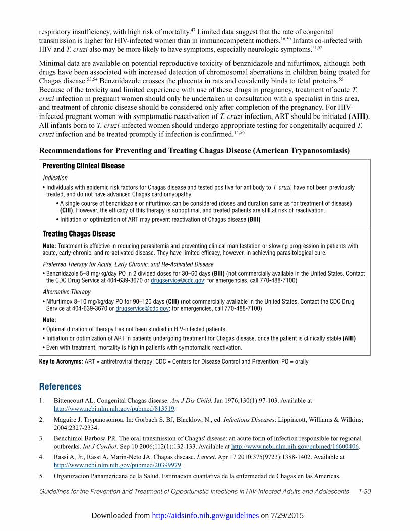
Downloaded from http://aidsinfo.nih.gov/guidelines on 7/29/2015
Guidelines for the Prevention and Treatment of Opportunistic Infections in HIV-Infected Adults and Adolescents T-30
respiratory insufficiency, with high risk of mortality.47 Limited data suggest that the rate of congenitaltransmission is higher for HIV-infected women than in immunocompetent mothers.16,50 Infants co-infected withHIV and T. cruzi also may be more likely to have symptoms, especially neurologic symptoms.51,52
Minimal data are available on potential reproductive toxicity of benznidazole and nifurtimox, although bothdrugs have been associated with increased detection of chromosomal aberrations in children being treated forChagas disease.53,54 Benznidazole crosses the placenta in rats and covalently binds to fetal proteins.55
Because of the toxicity and limited experience with use of these drugs in pregnancy, treatment of acute T.cruzi infection in pregnant women should only be undertaken in consultation with a specialist in this area,and treatment of chronic disease should be considered only after completion of the pregnancy. For HIV-infected pregnant women with symptomatic reactivation of T. cruzi infection, ART should be initiated (AIII).All infants born to T. cruzi-infected women should undergo appropriate testing for congenitally acquired T.cruzi infection and be treated promptly if infection is confirmed.14,56
Recommendations for Preventing and Treating Chagas Disease (American Trypanosomiasis)
Preventing Clinical Disease
Indication
• Individuals with epidemic risk factors for Chagas disease and tested positive for antibody to T. cruzi, have not been previouslytreated, and do not have advanced Chagas cardiomyopathy.
• A single course of benznidazole or nifurtimox can be considered (doses and duration same as for treatment of disease)(CIII). However, the efficacy of this therapy is suboptimal, and treated patients are still at risk of reactivation.
• Initiation or optimization of ART may prevent reactivation of Chagas disease (BIII)
Treating Chagas Disease
Note: Treatment is effective in reducing parasitemia and preventing clinical manifestation or slowing progression in patients withacute, early-chronic, and re-activated disease. They have limited efficacy, however, in achieving parasitological cure.
Preferred Therapy for Acute, Early Chronic, and Re-Activated Disease
• Benznidazole 5–8 mg/kg/day PO in 2 divided doses for 30–60 days (BIII) (not commercially available in the United States. Contactthe CDC Drug Service at 404-639-3670 or [email protected]; for emergencies, call 770-488-7100)
Alternative Therapy
• Nifurtimox 8–10 mg/kg/day PO for 90–120 days (CIII) (not commercially available in the United States. Contact the CDC DrugService at 404-639-3670 or [email protected]; for emergencies, call 770-488-7100)
Note:
• Optimal duration of therapy has not been studied in HIV-infected patients.
• Initiation or optimization of ART in patients undergoing treatment for Chagas disease, once the patient is clinically stable (AIII)
• Even with treatment, mortality is high in patients with symptomatic reactivation.
Key to Acronyms: ART = antiretroviral therapy; CDC = Centers for Disease Control and Prevention; PO = orally
References
1. Bittencourt AL. Congenital Chagas disease. Am J Dis Child. Jan 1976;130(1):97-103. Available athttp://www.ncbi.nlm.nih.gov/pubmed/813519.
2. Maguire J. Trypanosomoa. In: Gorbach S. BJ, Blacklow, N., ed. Infectious Diseases: Lippincott, Williams & Wilkins;2004:2327-2334.
3. Benchimol Barbosa PR. The oral transmission of Chagas' disease: an acute form of infection responsible for regionaloutbreaks. Int J Cardiol. Sep 10 2006;112(1):132-133. Available at http://www.ncbi.nlm.nih.gov/pubmed/16600406.
4. Rassi A, Jr., Rassi A, Marin-Neto JA. Chagas disease. Lancet. Apr 17 2010;375(9723):1388-1402. Available athttp://www.ncbi.nlm.nih.gov/pubmed/20399979.
5. Organizacion Panamericana de la Salud. Estimacion cuantativa de la enfermedad de Chagas en las Americas.
Downloaded from http://aidsinfo.nih.gov/guidelines on 7/29/2015
Guidelines for the Prevention and Treatment of Opportunistic Infections in HIV-Infected Adults and Adolescents T-31
Montevideo, Uraguay, Organizacion Panamericana de la Salud. 2006. 6. Gascon J, Bern C, Pinazo MJ. Chagas disease in Spain, the United States and other non-endemic countries. Acta Trop.
Jul-Aug 2010;115(1-2):22-27. Available at http://www.ncbi.nlm.nih.gov/pubmed/19646412.7. Moncayo A. Chagas disease: current epidemiological trends after the interruption of vectorial and transfusional
transmission in the Southern Cone countries. Mem Inst Oswaldo Cruz. Jul 2003;98(5):577-591. Available athttp://www.ncbi.nlm.nih.gov/pubmed/12973523.
8. Dorn PL, Perniciaro L, Yabsley MJ, et al. Autochthonous transmission of Trypanosoma cruzi, Louisiana. Emerg InfectDis. Apr 2007;13(4):605-607. Available at http://www.ncbi.nlm.nih.gov/pubmed/17553277.
9. Herwaldt BL, Grijalva MJ, Newsome AL, et al. Use of polymerase chain reaction to diagnose the fifth reported US caseof autochthonous transmission of Trypanosoma cruzi, in Tennessee, 1998. J Infect Dis. Jan 2000;181(1):395-399.Available at http://www.ncbi.nlm.nih.gov/pubmed/10608796.
10. Kjos SA, Snowden KF, Craig TM, Lewis B, Ronald N, Olson JK. Distribution and characterization of canine Chagas diseasein Texas. Vet Parasitol. Apr 15 2008;152(3-4):249-256. Available at http://www.ncbi.nlm.nih.gov/pubmed/18255233.
11. Navin TR, Miller KD, Satriale RF, Lobel HO. Adverse reactions associated with pyrimethamine-sulfadoxineprophylaxis for Pneumocystis carinii infections in AIDS. Lancet. Jun 8 1985;1(8441):1332. Available athttp://www.ncbi.nlm.nih.gov/pubmed/2860516.
12. Centers for Disease C, Prevention. Blood donor screening for chagas disease—United States, 2006-2007. MMWR MorbMortal Wkly Rep. Feb 23 2007;56(7):141-143. Available at http://www.ncbi.nlm.nih.gov/pubmed/17318113.
13. Bern C, Montgomery SP, Katz L, Caglioti S, Stramer SL. Chagas disease and the US blood supply. Curr Opin InfectDis. Oct 2008;21(5):476-482. Available at http://www.ncbi.nlm.nih.gov/pubmed/18725796.
14. Bern C, Verastegui M, Gilman RH, et al. Congenital Trypanosoma cruzi transmission in Santa Cruz, Bolivia. Clin InfectDis. Dec 1 2009;49(11):1667-1674. Available at http://www.ncbi.nlm.nih.gov/pubmed/19877966.
15. Rocha A, de Meneses AC, da Silva AM, et al. Pathology of patients with Chagas' disease and acquiredimmunodeficiency syndrome. Am J Trop Med Hyg. Mar 1994;50(3):261-268. Available athttp://www.ncbi.nlm.nih.gov/pubmed/8147485.
16. Sartori AM, Ibrahim KY, Nunes Westphalen EV, et al. Manifestations of Chagas disease (American trypanosomiasis) inpatients with HIV/AIDS. Ann Trop Med Parasitol. Jan 2007;101(1):31-50. Available athttp://www.ncbi.nlm.nih.gov/pubmed/17244408.
17. Vaidian AK, Weiss LM, Tanowitz HB. Chagas' disease and AIDS. Kinetoplastid biology and disease. May 132004;3(1):2. Available at http://www.ncbi.nlm.nih.gov/pubmed/15142278.
18. Committee WHOE. Control of Chagas disease. World Health Organ Tech Rep Ser. 2002;905:i-vi, 1-109, back cover.Available at http://www.ncbi.nlm.nih.gov/pubmed/12092045.
19. Rassi A, Jr., Rassi A, Little WC. Chagas' heart disease. Clin Cardiol. Dec 2000;23(12):883-889. Available athttp://www.ncbi.nlm.nih.gov/pubmed/11129673.
20. Rassi A, Jr., Rassi SG, Rassi A. Sudden death in Chagas' disease. Arq Bras Cardiol. Jan 2001;76(1):75-96. Available athttp://www.ncbi.nlm.nih.gov/pubmed/11175486.
21. de Oliveira RB, Troncon LE, Dantas RO, Menghelli UG. Gastrointestinal manifestations of Chagas' disease. Am JGastroenterol. Jun 1998;93(6):884-889. Available at http://www.ncbi.nlm.nih.gov/pubmed/9647012.
22. Campos SV, Strabelli TM, Amato Neto V, et al. Risk factors for Chagas' disease reactivation after heart transplantation.J Heart Lung Transplant. Jun 2008;27(6):597-602. Available at http://www.ncbi.nlm.nih.gov/pubmed/18503957.
23. Kohl S, Pickering LK, Frankel LS, Yaeger RG. Reactivation of Chagas' disease during therapy of acute lymphocyticleukemia. Cancer. Sep 1 1982;50(5):827-828. Available at http://www.ncbi.nlm.nih.gov/pubmed/6807527.
24. Riarte A, Luna C, Sabatiello R, et al. Chagas' disease in patients with kidney transplants: 7 years of experience 1989-1996. Clin Infect Dis. Sep 1999;29(3):561-567. Available at http://www.ncbi.nlm.nih.gov/pubmed/10530448.
25. Sartori AM, Neto JE, Nunes EV, et al. Trypanosoma cruzi parasitemia in chronic Chagas disease: comparison betweenhuman immunodeficiency virus (HIV)-positive and HIV-negative patients. J Infect Dis. Sep 15 2002;186(6):872-875.Available at http://www.ncbi.nlm.nih.gov/pubmed/12198628.
26. Sartori AM, Lopes MH, Benvenuti LA, et al. Reactivation of Chagas' disease in a human immunodeficiency virus-
Downloaded from http://aidsinfo.nih.gov/guidelines on 7/29/2015
Guidelines for the Prevention and Treatment of Opportunistic Infections in HIV-Infected Adults and Adolescents T-32
infected patient leading to severe heart disease with a late positive direct microscopic examination of the blood. Am JTrop Med Hyg. Nov 1998;59(5):784-786. Available at http://www.ncbi.nlm.nih.gov/pubmed/9840598.
27. Cordova E, Boschi A, Ambrosioni J, Cudos C, Corti M. Reactivation of Chagas disease with central nervous systeminvolvement in HIV-infected patients in Argentina, 1992-2007. Int J Infect Dis. Nov 2008;12(6):587-592. Available athttp://www.ncbi.nlm.nih.gov/pubmed/18337139.
28. Diazgranados CA, Saavedra-Trujillo CH, Mantilla M, Valderrama SL, Alquichire C, Franco-Paredes C. Chagasicencephalitis in HIV patients: common presentation of an evolving epidemiological and clinical association. LancetInfect Dis. May 2009;9(5):324-330. Available at http://www.ncbi.nlm.nih.gov/pubmed/19393962.
29. Ferreira MS, Nishioka Sde A, Silvestre MT, Borges AS, Nunes-Araujo FR, Rocha A. Reactivation of Chagas' disease inpatients with AIDS: report of three new cases and review of the literature. Clin Infect Dis. Dec 1997;25(6):1397-1400.Available at http://www.ncbi.nlm.nih.gov/pubmed/9431385.
30. Leiby DA, Wendel S, Takaoka DT, Fachini RM, Oliveira LC, Tibbals MA. Serologic testing for Trypanosoma cruzi:comparison of radioimmunoprecipitation assay with commercially available indirect immunofluorescence assay,indirect hemagglutination assay, and enzyme-linked immunosorbent assay kits. J Clin Microbiol. Feb 2000;38(2):639-642. Available at http://www.ncbi.nlm.nih.gov/pubmed/10655360.
31. Tarleton RL, Reithinger R, Urbina JA, Kitron U, Gurtler RE. The challenges of Chagas Disease—grim outlook orglimmer of hope. PLoS Med. Dec 2007;4(12):e332. Available at http://www.ncbi.nlm.nih.gov/pubmed/18162039.
32. Sosa-Estani S, Gamboa-Leon MR, Del Cid-Lemus J, et al. Use of a rapid test on umbilical cord blood to screen forTrypanosoma cruzi infection in pregnant women in Argentina, Bolivia, Honduras, and Mexico. Am J Trop Med Hyg.Nov 2008;79(5):755-759. Available at http://www.ncbi.nlm.nih.gov/pubmed/18981518.
33. Verani JR, Seitz A, Gilman RH, et al. Geographic variation in the sensitivity of recombinant antigen-based rapid testsfor chronic Trypanosoma cruzi infection. Am J Trop Med Hyg. Mar 2009;80(3):410-415. Available athttp://www.ncbi.nlm.nih.gov/pubmed/19270291.
34. Gorlin J, Rossmann S, Robertson G, et al. Evaluation of a new Trypanosoma cruzi antibody assay for blood donorscreening. Transfusion. Mar 2008;48(3):531-540. Available at http://www.ncbi.nlm.nih.gov/pubmed/18067497.
35. Junqueira AC, Chiari E, Wincker P. Comparison of the polymerase chain reaction with two classical parasitologicalmethods for the diagnosis of Chagas disease in an endemic region of north-eastern Brazil. Trans R Soc Trop Med Hyg.Mar-Apr 1996;90(2):129-132. Available at http://www.ncbi.nlm.nih.gov/pubmed/8761570.
36. Wincker P, Telleria J, Bosseno MF, et al. PCR-based diagnosis for Chagas' disease in Bolivian children living in anactive transmission area: comparison with conventional serological and parasitological diagnosis. Parasitology. Apr1997;114 ( Pt 4):367-373. Available at http://www.ncbi.nlm.nih.gov/pubmed/9107023.
37. Feilij H, Muller L, Gonzalez Cappa SM. Direct micromethod for diagnosis of acute and congenital Chagas' disease. JClin Microbiol. Aug 1983;18(2):327-330. Available at http://www.ncbi.nlm.nih.gov/pubmed/6413530.
38. Duffy T, Bisio M, Altcheh J, et al. Accurate real-time PCR strategy for monitoring bloodstream parasitic loads in chagasdisease patients. PLoS Negl Trop Dis. 2009;3(4):e419. Available at http://www.ncbi.nlm.nih.gov/pubmed/19381287.
39. Schijman AG, Vigliano C, Burgos J, et al. Early diagnosis of recurrence of Trypanosoma cruzi infection by polymerasechain reaction after heart transplantation of a chronic Chagas' heart disease patient. J Heart Lung Transplant. Nov2000;19(11):1114-1117. Available at http://www.ncbi.nlm.nih.gov/pubmed/11077230.
40. Mott KE, Muniz TM, Lehman JS, Jr., et al. House construction, triatomine distribution, and household distribution ofseroreactivity to Trypanosoma cruzi in a rural community in northeast Brazil. Am J Trop Med Hyg. Nov1978;27(6):1116-1122. Available at http://www.ncbi.nlm.nih.gov/pubmed/103445.
41. Kroeger A, Villegas E, Ordonez-Gonzalez J, Pabon E, Scorza JV. Prevention of the transmission of Chagas' disease withpyrethroid-impregnated materials. Am J Trop Med Hyg. Mar 2003;68(3):307-311. Available athttp://www.ncbi.nlm.nih.gov/pubmed/12685636.
42. Schmunis GA, Cruz JR. Safety of the blood supply in Latin America. Clin Microbiol Rev. Jan 2005;18(1):12-29.Available at http://www.ncbi.nlm.nih.gov/pubmed/15653816.
43. Rodriques Coura J, de Castro SL. A critical review on Chagas disease chemotherapy. Mem Inst Oswaldo Cruz. Jan2002;97(1):3-24. Available at http://www.ncbi.nlm.nih.gov/pubmed/11992141.
44. Bern C, Montgomery SP, Herwaldt BL, et al. Evaluation and treatment of chagas disease in the United States: a systematic
Downloaded from http://aidsinfo.nih.gov/guidelines on 7/29/2015
Guidelines for the Prevention and Treatment of Opportunistic Infections in HIV-Infected Adults and Adolescents T-33
review. JAMA. Nov 14 2007;298(18):2171-2181. Available at http://www.ncbi.nlm.nih.gov/pubmed/18000201.45. Castro JA, de Mecca MM, Bartel LC. Toxic side effects of drugs used to treat Chagas' disease (American
trypanosomiasis). Human & experimental toxicology. Aug 2006;25(8):471-479. Available athttp://www.ncbi.nlm.nih.gov/pubmed/16937919.
46. Pinazo MJ, Espinosa G, Gallego M, Lopez-Chejade PL, Urbina JA, Gascon J. Successful treatment with posaconazole ofa patient with chronic Chagas disease and systemic lupus erythematosus. Am J Trop Med Hyg. Apr 2010;82(4):583-587.Available at http://www.ncbi.nlm.nih.gov/pubmed/20348503.
47. Torrico F, Alonso-Vega C, Suarez E, et al. Maternal Trypanosoma cruzi infection, pregnancy outcome, morbidity, andmortality of congenitally infected and non-infected newborns in Bolivia. Am J Trop Med Hyg. Feb 2004;70(2):201-209.Available at http://www.ncbi.nlm.nih.gov/pubmed/14993634.
48. Di Pentima MC, Hwang LY, Skeeter CM, Edwards MS. Prevalence of antibody to Trypanosoma cruzi in pregnant Hispanicwomen in Houston. Clin Infect Dis. Jun 1999;28(6):1281-1285. Available at http://www.ncbi.nlm.nih.gov/pubmed/10451166.
49. Bittencourt AL. Possible risk factors for vertical transmission of Chagas' disease. Revista do Instituto de MedicinaTropical de Sao Paulo. Sep-Oct 1992;34(5):403-408. Available at http://www.ncbi.nlm.nih.gov/pubmed/1342103.
50. Scapellato PG, Bottaro EG, Rodriguez-Brieschke MT. Mother-child transmission of Chagas disease: could coinfectionwith human immunodeficiency virus increase the risk? Rev Soc Bras Med Trop. Mar-Apr 2009;42(2):107-109. Availableat http://www.ncbi.nlm.nih.gov/pubmed/19448923.
51. Freilij H, Altcheh J. Congenital Chagas' disease: diagnostic and clinical aspects. Clin Infect Dis. Sep 1995;21(3):551-555. Available at http://www.ncbi.nlm.nih.gov/pubmed/8527542.
52. Freilij H, Altcheh J, Muchinik G. Perinatal human immunodeficiency virus infection and congenital Chagas' disease.Pediatr Infect Dis J. Feb 1995;14(2):161-162. Available at http://www.ncbi.nlm.nih.gov/pubmed/7746707.
53. Gorla NB, Ledesma OS, Barbieri GP, Larripa IB. Assessment of cytogenetic damage in chagasic children treated withbenznidazole. Mutation research. Oct 1988;206(2):217-220. Available at http://www.ncbi.nlm.nih.gov/pubmed/3140001.
54. Gorla NB, Ledesma OS, Barbieri GP, Larripa IB. Thirteenfold increase of chromosomal aberrations non-randomlydistributed in chagasic children treated with nifurtimox. Mutation research. Oct 1989;224(2):263-267. Available athttp://www.ncbi.nlm.nih.gov/pubmed/2507913.
55. de Toranzo EG, Masana M, Castro JA. Administration of benznidazole, a chemotherapeutic agent against Chagas disease, topregnant rats. Covalent binding of reactive metabolites to fetal and maternal proteins. Archives internationales depharmacodynamie et de therapie. Nov 1984;272(1):17-23. Available at http://www.ncbi.nlm.nih.gov/pubmed/6440493.
56. Oliveira I, Torrico F, Munoz J, Gascon J. Congenital transmission of Chagas disease: a clinical approach. Expert RevAnti Infect Ther. Aug 2010;8(8):945-956. Available at http://www.ncbi.nlm.nih.gov/pubmed/20695749.
Downloaded from http://aidsinfo.nih.gov/guidelines on 7/29/2015
Guidelines for the Prevention and Treatment of Opportunistic Infections in HIV-Infected Adults and Adolescents T-34
Isosporiasis (Cystoisosporiasis) (Last updated May 7, 2013; last reviewedMay 7, 2013)
Epidemiology
Isosporiasis, also known as cystoisosporiasis, occurs worldwide but predominantly in tropical andsubtropical regions. Immunocompromised patients, including those who are HIV-infected, are at increasedrisk for chronic, debilitating illness.1-7 Although Isospora (Cystoisospora) belli completes its life cycle inhumans, the oocysts shed in the feces of infected individuals must mature (sporulate) outside the host, in theenvironment, to become infective. On the basis of limited data, the maturation process is completed inapproximately 1 to 2 days but might occur more rapidly in some settings.2 Infection results from ingestion ofsporulated oocysts, such as from contaminated food or water. After ingestion, the parasite invadesenterocytes in the small intestine. Ultimately, immature oocysts are produced and shed in stool.
Clinical Manifestations
The most common manifestation is watery, non-bloody diarrhea, which may be associated with abdominalpain, cramping, anorexia, nausea, vomiting, and low-grade fever. The diarrhea can be profuse and prolonged,particularly in immunocompromised patients, resulting in severe dehydration, electrolyte abnormalities suchas hypokalemia, weight loss, and malabsorption.6-12 Acalculous cholecystitis/cholangiopathy2,13-15 andreactive arthritis16 also have been reported.
Diagnosis
Typically, infection is diagnosed by detecting Isospora oocysts (dimensions, 23–36 µm by 12–17 µm) infecal specimens.2 Oocysts may be shed intermittently and at low levels, even by patients with profusediarrhea. Diagnosis can be facilitated by repeated stool examinations with sensitive methods, such asmodified acid-fast techniques, on which oocysts stain bright red, and UV fluorescence microscopy, underwhich they autofluoresce.2,17 Infection also can be diagnosed by detecting oocysts in duodenalaspirates/mucus or developmental stages of the parasite in intestinal biopsy specimens.2,10 Extraintestinalinfection, such as in the biliary tract, lymph nodes, spleen, and liver, has been documented in postmortemexaminations of HIV-infected patients.2,18-20
Preventing Exposure
Because I. belli is acquired by ingesting infected water or food, avoiding potentially contaminated food orwater in isosporiasis-endemic areas may help prevent infection.
Preventing Disease
In some settings, chemoprophylaxis with trimethoprim-sulfamethoxazole (TMP-SMX) has been associatedwith a lower incidence or prevalence of isosporiasis.1,3,4,21 In a randomized, placebo-controlled trial, dailyTMP-SMX (160/800 mg) was protective against isosporiasis in persons with early-stage HIV infection(World Health Organization clinical stage 2 or 3 at enrollment).1 In an observational study, incidence ofisosporiasis decreased after widespread introduction of antiretroviral therapy (ART), except in patients withCD4 counts <50 cells/mm3.3 After adjustment for the CD4 T lymphocyte (CD4) cell count, the risk ofisosporiasis was substantially lower in those receiving prophylaxis with TMP-SMX, sulfadiazine, orpyrimethamine (unspecified regimens). In analyses of data from a Los Angeles county AIDS surveillanceregistry during the pre-ART era, the prevalence of isosporiasis was lower in patients with versus without ahistory of Pneumocystis pneumonia—indirect evidence of a protective effect from use of TMP-SMX forPneumocystis pneumonia.4 Insufficient evidence is available, however, to support a general recommendationfor primary prophylaxis for isosporiasis per se, especially for U.S. travelers in isoporiasis-endemic areas.
Downloaded from http://aidsinfo.nih.gov/guidelines on 7/29/2015
Guidelines for the Prevention and Treatment of Opportunistic Infections in HIV-Infected Adults and Adolescents T-35
Treating Disease
Clinical management includes fluid and electrolyte support for dehydrated patients and nutritionalsupplementation for malnourished patients (AIII). TMP-SMX is the antimicrobial agent of choice fortreatment of isosporiasis (AI). It is the only agent whose use is supported by substantial published data andclinical experience. Therefore, potential alternative therapies should be reserved for patients withdocumented sulfa intolerance or in whom treatment fails (AIII).
Three studies in HIV-infected patients in Haiti have demonstrated the effectiveness of various treatmentregimens of TMP-SMX.6,7,22 The patients were not receiving ART, and laboratory indicators ofimmunodeficiency (such as CD4 cell counts) were not specified. On the basis of the initial studies,6,7 thetraditional treatment regimen has been a 10-day course of TMP-SMX (160/800 mg) administered orally fourtimes daily (AII).23 In another study, TMP-SMX (160/800 mg) administered twice daily was also effective(BI).22 Although published experience using two daily doses of TMP-SMX (160/800 mg) is limited, oneapproach would be to start with this regimen but to increase the daily dose and the duration of therapy (up to3–4 weeks)6,10 if symptoms worsen or persist (BIII). Intravenous administration of TMP-SMX should beconsidered for patients with potential or documented malabsorption.
Limited data suggest that therapy with pyrimethamine–sulfadiazine and pyrimethamine–sulfadoxine may beeffective.2,9,10,24-26 However, the combination of pyrimethamine plus sulfadoxine is not typicallyrecommended for use in the United States (CIII); it has been associated with an increased risk of severecutaneous reactions, including Stevens-Johnson syndrome,27 and pyrimethamine and sulfadoxine clearslowly from the body after therapy is discontinued.
Single-agent therapy with pyrimethamine has been used, with anecdotal success for treatment and preventionof isosporiasis.3,28,29 Pyrimethamine (50–75 mg/day) plus leucovorin (10–25 mg/day) to preventmyelosuppression may be an effective treatment alternative; it is the option for sulfa-intolerant patients (BIII).
Special Considerations with Regard to Starting ARTOnly limited data address the utility of ART in the setting of Isospora and HIV co-infection.3,14,21 Immunereconstitution with ART may result in fewer relapses of isosporiasis, and no cases of immune reconstitutioninflammatory syndrome (IRIS) have been reported. Therefore, the potential benefits of ART likely outweighthe risks. For patients with isosporiasis who otherwise fulfill criteria for ART, TMP-SMX therapy and ARTcan be started simultaneously; there is no known reason to defer initiation of ART other than the potential forpoor ART absorption (AIII).
Monitoring of Response to Therapy and Adverse Events (Including IRIS)Patients should be monitored for clinical response and adverse events. In HIV-infected patients, TMP-SMXtherapy is commonly associated with side effects, such as rash, fever, leukopenia, thrombocytopenia, andelevated transaminase levels. IRIS has not been described.
Managing Treatment FailureIf symptoms worsen or persist despite approximately 5 to 7 days of TMP-SMX therapy, the possibilities ofnoncompliance, malabsorption, and concurrent infections/enteropathies should be considered; the TMP-SMX regimen (daily dose, duration, and mode of administration) also should be reevaluated. For patientswith documented sulfa intolerance or in whom treatment fails, use of a potential alternative agent (typicallypyrimethamine) should be considered. Ciprofloxacin is a second-line agent (CI). On the basis of limited datafrom a randomized, controlled trial in Haiti, ciprofloxacin (500 mg twice daily for 7 days) is less effectivethan TMP-SMX but may have modest activity against I. belli.22
Unsubstantiated or mixed data are available for albendazole,29-31 nitazoxanide,32,33 doxycycline,34 themacrolides roxithromycin and spiramycin,25,35,36 and the veterinary anticoccidial agent diclazuril (CIII).37,38
Downloaded from http://aidsinfo.nih.gov/guidelines on 7/29/2015
Guidelines for the Prevention and Treatment of Opportunistic Infections in HIV-Infected Adults and Adolescents T-36
Limited data suggest that drugs such as metronidazole, quinacrine, iodoquinol, paromomycin, andfurazolidone are ineffective.8,25,26,28,35,37 Apparent or partial responses, if noted, may be attributable totreatment of concomitant infections or to nonspecific effects.
Preventing Recurrence
Patients with CD4 cell counts <200 cells/mm3 should receive secondary prophylaxis (chronic maintenancetherapy) with TMP-SMX, which is also protective against Pneumocystis jirovecii and Toxoplasma gondiiinfections (AI). In studies in Haiti, approximately 50% of patients who did not receive secondaryprophylaxis had symptomatic recurrences approximately 2 months after completing a course of TMP-SMXtherapy, relapses rapidly responded to retreatment, and secondary prophylaxis decreased the risk ofrelapse.6,7,22 In a randomized, placebo-controlled trial, no symptomatic recurrences were noted in patientswho received maintenance therapy with thrice-weekly TMP-SMX (160/800 mg) (AI).7 Daily TMP-SMX(160/800 mg) and thrice-weekly TMP-SMX (320/1600 mg) have been effective (BIII);5,10 however, clinicaland parasitologic relapses despite maintenance TMP-SMX therapy and ART have been reported.14
In sulfa-intolerant patients, pyrimethamine (25 mg/day) with leucovorin (5–10 mg/day) has been used(BIII).28 On the basis of limited data, ciprofloxacin (500 mg thrice weekly) is considered a second-linealternative (CI).22
When To Stop Secondary ProphylaxisThe issue of discontinuing prophylaxis has not been evaluated in a clinical trial. Chemoprophylaxis probablycan be safely discontinued in patients without evidence of active I. belli infection who have a sustainedincrease in the CD4 cell count to levels >200 cells/mm3 for >6 months after initiation of ART (BIII).
Special Considerations During PregnancyTMP-SMX is the agent of choice for primary treatment and secondary prophylaxis in pregnant women, as itis in persons who are not pregnant. Although first-trimester exposure to trimethoprim has been associatedwith a small increased risk of birth defects,39-42 TMP-SMX therapy should be provided in the setting ofmaternal symptomatic I. belli infection. Because of concerns about possible teratogenicity associated withfirst-trimester drug exposure, clinicians may withhold secondary prophylaxis during the first trimester andtreat only symptomatic infection (CIII). Although pyrimethamine has been associated with birth defects inanimals, limited human data have not suggested an increased risk of defects.43 Human data about the use ofciprofloxacin during several hundred pregnancies have not suggested an increased risk of birth defects orcartilage abnormalities.44
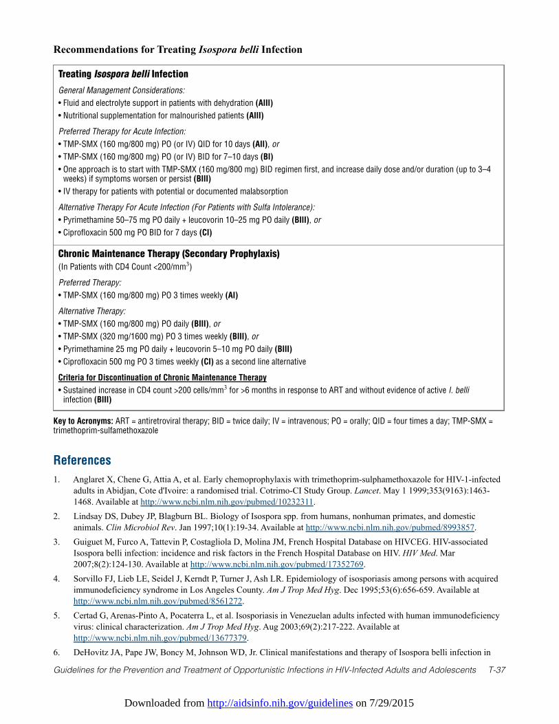
Downloaded from http://aidsinfo.nih.gov/guidelines on 7/29/2015
Guidelines for the Prevention and Treatment of Opportunistic Infections in HIV-Infected Adults and Adolescents T-37
Treating Isospora belli Infection
General Management Considerations:
• Fluid and electrolyte support in patients with dehydration (AIII)
• Nutritional supplementation for malnourished patients (AIII)
Preferred Therapy for Acute Infection:
• TMP-SMX (160 mg/800 mg) PO (or IV) QID for 10 days (AII), or
• TMP-SMX (160 mg/800 mg) PO (or IV) BID for 7–10 days (BI)
• One approach is to start with TMP-SMX (160 mg/800 mg) BID regimen first, and increase daily dose and/or duration (up to 3–4weeks) if symptoms worsen or persist (BIII)
• IV therapy for patients with potential or documented malabsorption
Alternative Therapy For Acute Infection (For Patients with Sulfa Intolerance):
• Pyrimethamine 50–75 mg PO daily + leucovorin 10–25 mg PO daily (BIII), or
• Ciprofloxacin 500 mg PO BID for 7 days (CI)
Chronic Maintenance Therapy (Secondary Prophylaxis)
(In Patients with CD4 Count <200/mm3)
Preferred Therapy:
• TMP-SMX (160 mg/800 mg) PO 3 times weekly (AI)
Alternative Therapy:
• TMP-SMX (160 mg/800 mg) PO daily (BIII), or
• TMP-SMX (320 mg/1600 mg) PO 3 times weekly (BIII), or
• Pyrimethamine 25 mg PO daily + leucovorin 5–10 mg PO daily (BIII)
• Ciprofloxacin 500 mg PO 3 times weekly (CI) as a second line alternative
Criteria for Discontinuation of Chronic Maintenance Therapy
• Sustained increase in CD4 count >200 cells/mm3 for >6 months in response to ART and without evidence of active I. belliinfection (BIII)
Recommendations for Treating Isospora belli Infection
Key to Acronyms: ART = antiretroviral therapy; BID = twice daily; IV = intravenous; PO = orally; QID = four times a day; TMP-SMX =trimethoprim-sulfamethoxazole
References
1. Anglaret X, Chene G, Attia A, et al. Early chemoprophylaxis with trimethoprim-sulphamethoxazole for HIV-1-infectedadults in Abidjan, Cote d'Ivoire: a randomised trial. Cotrimo-CI Study Group. Lancet. May 1 1999;353(9163):1463-1468. Available at http://www.ncbi.nlm.nih.gov/pubmed/10232311.
2. Lindsay DS, Dubey JP, Blagburn BL. Biology of Isospora spp. from humans, nonhuman primates, and domesticanimals. Clin Microbiol Rev. Jan 1997;10(1):19-34. Available at http://www.ncbi.nlm.nih.gov/pubmed/8993857.
3. Guiguet M, Furco A, Tattevin P, Costagliola D, Molina JM, French Hospital Database on HIVCEG. HIV-associatedIsospora belli infection: incidence and risk factors in the French Hospital Database on HIV. HIV Med. Mar2007;8(2):124-130. Available at http://www.ncbi.nlm.nih.gov/pubmed/17352769.
4. Sorvillo FJ, Lieb LE, Seidel J, Kerndt P, Turner J, Ash LR. Epidemiology of isosporiasis among persons with acquiredimmunodeficiency syndrome in Los Angeles County. Am J Trop Med Hyg. Dec 1995;53(6):656-659. Available athttp://www.ncbi.nlm.nih.gov/pubmed/8561272.
5. Certad G, Arenas-Pinto A, Pocaterra L, et al. Isosporiasis in Venezuelan adults infected with human immunodeficiencyvirus: clinical characterization. Am J Trop Med Hyg. Aug 2003;69(2):217-222. Available athttp://www.ncbi.nlm.nih.gov/pubmed/13677379.
6. DeHovitz JA, Pape JW, Boncy M, Johnson WD, Jr. Clinical manifestations and therapy of Isospora belli infection in
Downloaded from http://aidsinfo.nih.gov/guidelines on 7/29/2015
Guidelines for the Prevention and Treatment of Opportunistic Infections in HIV-Infected Adults and Adolescents T-38
patients with the acquired immunodeficiency syndrome. N Engl J Med. Jul 10 1986;315(2):87-90. Available athttp://www.ncbi.nlm.nih.gov/pubmed/3487730.
7. Pape JW, Verdier RI, Johnson WD, Jr. Treatment and prophylaxis of Isospora belli infection in patients with theacquired immunodeficiency syndrome. N Engl J Med. Apr 20 1989;320(16):1044-1047. Available athttp://www.ncbi.nlm.nih.gov/pubmed/2927483.
8. Forthal DN, Guest SS. Isospora belli enteritis in three homosexual men. Am J Trop Med Hyg. Nov 1984;33(6):1060-1064. Available at http://www.ncbi.nlm.nih.gov/pubmed/6507724.
9. Modigliani R, Bories C, Le Charpentier Y, et al. Diarrhoea and malabsorption in acquired immune deficiencysyndrome: a study of four cases with special emphasis on opportunistic protozoan infestations. Gut. Feb1985;26(2):179-187. Available at http://www.ncbi.nlm.nih.gov/pubmed/4038492.
10. Whiteside ME, Barkin JS, May RG, Weiss SD, Fischl MA, MacLeod CL. Enteric coccidiosis among patients with theacquired immunodeficiency syndrome. Am J Trop Med Hyg. Nov 1984;33(6):1065-1072. Available athttp://www.ncbi.nlm.nih.gov/pubmed/6334448.
11. Bialek R, Overkamp D, Rettig I, Knobloch J. Case report: Nitazoxanide treatment failure in chronic isosporiasis. Am JTrop Med Hyg. Aug 2001;65(2):94-95. Available at http://www.ncbi.nlm.nih.gov/pubmed/11508398.
12. Williams DT, Smith RS, Mallon WK. Severe hypokalemia, paralysis, and AIDS-associated isospora belli diarrhea. JEmerg Med. Dec 2011;41(6):e129-132. Available at http://www.ncbi.nlm.nih.gov/pubmed/18993015.
13. Benator DA, French AL, Beaudet LM, Levy CS, Orenstein JM. Isospora belli infection associated with acalculouscholecystitis in a patient with AIDS. Ann Intern Med. Nov 1 1994;121(9):663-664. Available athttp://www.ncbi.nlm.nih.gov/pubmed/7944075.
14. Lagrange-Xelot M, Porcher R, Sarfati C, et al. Isosporiasis in patients with HIV infection in the highly active antiretroviraltherapy era in France. HIV Med. Feb 2008;9(2):126-130. Available at http://www.ncbi.nlm.nih.gov/pubmed/18257775.
15. Walther Z, Topazian MD. Isospora cholangiopathy: case study with histologic characterization and molecularconfirmation. Hum Pathol. Sep 2009;40(9):1342-1346. Available at http://www.ncbi.nlm.nih.gov/pubmed/19447468.
16. Gonzalez-Dominguez J, Roldan R, Villanueva JL, Kindelan JM, Jurado R, Torre-Cisneros J. Isospora belli reactivearthritis in a patient with AIDS. Annals of the rheumatic diseases. Sep 1994;53(9):618-619. Available athttp://www.ncbi.nlm.nih.gov/pubmed/7979603.
17. Bialek R, Binder N, Dietz K, Knobloch J, Zelck UE. Comparison of autofluorescence and iodine staining for detectionof Isospora belli in feces. Am J Trop Med Hyg. Sep 2002;67(3):304-305. Available athttp://www.ncbi.nlm.nih.gov/pubmed/12408672.
18. Frenkel JK, Silva MB, Saldanha J, et al. Isospora belli infection: observation of unicellular cysts in mesentericlymphoid tissues of a Brazilian patient with AIDS and animal inoculation. The Journal of eukaryotic microbiology.2003;50 Suppl:682-684. Available at http://www.ncbi.nlm.nih.gov/pubmed/14736218.
19. Restrepo C, Macher AM, Radany EH. Disseminated extraintestinal isosporiasis in a patient with acquired immunedeficiency syndrome. Am J Clin Pathol. Apr 1987;87(4):536-542. Available athttp://www.ncbi.nlm.nih.gov/pubmed/3826017.
20. Bernard E, Delgiudice P, Carles M, et al. Disseminated isosporiasis in an AIDS patient. Eur J Clin Microbiol Infect Dis.Sep 1997;16(9):699-701. Available at http://www.ncbi.nlm.nih.gov/pubmed/9352268.
21. Dillingham RA, Pinkerton R, Leger P, et al. High early mortality in patients with chronic acquired immunodeficiencysyndrome diarrhea initiating antiretroviral therapy in Haiti: a case-control study. Am J Trop Med Hyg. Jun2009;80(6):1060-1064. Available at http://www.ncbi.nlm.nih.gov/pubmed/19478276.
22. Verdier RI, Fitzgerald DW, Johnson WD, Jr., Pape JW. Trimethoprim-sulfamethoxazole compared with ciprofloxacinfor treatment and prophylaxis of Isospora belli and Cyclospora cayetanensis infection in HIV-infected patients. Arandomized, controlled trial. Ann Intern Med. Jun 6 2000;132(11):885-888. Available athttp://www.ncbi.nlm.nih.gov/pubmed/10836915.
23. Guerrant RL, Van Gilder T, Steiner TS, et al. Practice guidelines for the management of infectious diarrhea. Clin InfectDis. Feb 1 2001;32(3):331-351. Available at http://www.ncbi.nlm.nih.gov/pubmed/11170940.
24. Mojon M, Coudert J, E.O. dL. Serious isosporosis by Isospora belli: a case report treated by Fansidar [Abstract].Southeast Asian J Trop Med Public Health. 12:449-500. 1981.
Downloaded from http://aidsinfo.nih.gov/guidelines on 7/29/2015
Guidelines for the Prevention and Treatment of Opportunistic Infections in HIV-Infected Adults and Adolescents T-39
25. Ebrahimzadeh A, Bottone EJ. Persistent diarrhea caused by Isospora belli: therapeutic response to pyrimethamine andsulfadiazine. Diagn Microbiol Infect Dis. Oct 1996;26(2):87-89. Available at http://www.ncbi.nlm.nih.gov/pubmed/8985661.
26. Trier JS, Moxey PC, Schimmel EM, Robles E. Chronic intestinal coccidiosis in man: intestinal morphology and response totreatment. Gastroenterology. May 1974;66(5):923-935. Available at http://www.ncbi.nlm.nih.gov/pubmed/4826994.
27. Navin TR, Miller KD, Satriale RF, Lobel HO. Adverse reactions associated with pyrimethamine-sulfadoxine prophylaxisfor Pneumocystis carinii infections in AIDS. Lancet. Jun 8 1985;1(8441):1332. Available athttp://www.ncbi.nlm.nih.gov/pubmed/2860516.
28. Weiss LM, Perlman DC, Sherman J, Tanowitz H, Wittner M. Isospora belli infection: treatment with pyrimethamine. AnnIntern Med. Sep 15 1988;109(6):474-475. Available at http://www.ncbi.nlm.nih.gov/pubmed/3261956.
29. Jongwutiwes S, Sampatanukul P, Putaporntip C. Recurrent isosporiasis over a decade in an immunocompetent hostsuccessfully treated with pyrimethamine. Scandinavian journal of infectious diseases. 2002;34(11):859-862. Available athttp://www.ncbi.nlm.nih.gov/pubmed/12578164.
30. Dionisio D, Sterrantino G, Meli M, Leoncini F, Orsi A, Nicoletti P. Treatment of isosporiasis with combined albendazoleand ornidazole in patients with AIDS. AIDS. Sep 1996;10(11):1301-1302. Available athttp://www.ncbi.nlm.nih.gov/pubmed/8883600.
31. Zulu I, Veitch A, Sianongo S, et al. Albendazole chemotherapy for AIDS-related diarrhoea in Zambia--clinical,parasitological and mucosal responses. Alimentary pharmacology & therapeutics. 2002; 16(3):595-601. Available athttp://www.ncbi.nlm.nih.gov/entrez/query.fcgi?cmd=Retrieve&db=PubMed&dopt=Citation&list_uids=11876715.
32. Romero Cabello R, Guerrero LR, Munoz Garcia MR, Geyne Cruz A. Nitazoxanide for the treatment of intestinal protozoanand helminthic infections in Mexico. Trans R Soc Trop Med Hyg. Nov-Dec 1997;91(6):701-703. Available athttp://www.ncbi.nlm.nih.gov/pubmed/9580117.
33. Doumbo O, Rossignol JF, Pichard E, et al. Nitazoxanide in the treatment of cryptosporidial diarrhea and other intestinalparasitic infections associated with acquired immunodeficiency syndrome in tropical Africa. Am J Trop Med Hyg. Jun1997;56(6):637-639. Available at http://www.ncbi.nlm.nih.gov/pubmed/9230795.
34. Meyohas MC, Capella F, Poirot JL, et al. [Treatment with doxycycline and nifuroxazide of Isospora belli infection in AIDS].Pathologie-biologie. Jun 1990;38(5 ( Pt 2)):589-591. Available at http://www.ncbi.nlm.nih.gov/pubmed/2385457.
35. Gaska JA, Tietze KJ, Cosgrove EM. Unsuccessful treatment of enteritis due to Isospora belli with spiramycin: a casereport. J Infect Dis. Dec 1985;152(6):1336-1338. Available at http://www.ncbi.nlm.nih.gov/pubmed/4067332.
36. Musey KL, Chidiac C, Beaucaire G, Houriez S, Fourrier A. Effectiveness of roxithromycin for treating Isospora belliinfection. J Infect Dis. Sep 1988;158(3):646. Available at http://www.ncbi.nlm.nih.gov/pubmed/3411149.
37. Limson-Pobre RN, Merrick S, Gruen D, Soave R. Use of diclazuril for the treatment of isosporiasis in patients with AIDS.Clin Infect Dis. Jan 1995;20(1):201-202. Available at http://www.ncbi.nlm.nih.gov/pubmed/7727660.
38. Kayembe K, Desmet P, Henry MC, Stoffels P. Diclazuril for Isospora belli infection in AIDS. Lancet. Jun 171989;1(8651):1397-1398. Available at http://www.ncbi.nlm.nih.gov/pubmed/2567420.
39. Czeizel AE, Rockenbauer M, Sorensen HT, Olsen J. The teratogenic risk of trimethoprim-sulfonamides: a population based case-control study. Reprod Toxicol. Nov-Dec 2001;15(6):637-646. Available at http://www.ncbi.nlm.nih.gov/pubmed/11738517.
40. Hernandez-Diaz S, Werler MM, Walker AM, Mitchell AA. Folic acid antagonists during pregnancy and the risk of birthdefects. N Engl J Med. Nov 30 2000;343(22):1608-1614. Available at http://www.ncbi.nlm.nih.gov/pubmed/11096168.
41. Hernandez-Diaz S, Werler MM, Walker AM, Mitchell AA. Neural tube defects in relation to use of folic acid antagonistsduring pregnancy. American journal of epidemiology. May 15 2001;153(10):961-968. Available athttp://www.ncbi.nlm.nih.gov/pubmed/11384952.
42. Jungmann EM, Mercey D, DeRuiter A, et al. Is first trimester exposure to the combination of antiretroviral therapy andfolate antagonists a risk factor for congenital abnormalities? Sexually transmitted infections. Dec 2001;77(6):441-443.Available at http://www.ncbi.nlm.nih.gov/pubmed/11714944.
43. Deen JL, von Seidlein L, Pinder M, Walraven GE, Greenwood BM. The safety of the combination artesunate andpyrimethamine-sulfadoxine given during pregnancy. Trans R Soc Trop Med Hyg. Jul-Aug 2001;95(4):424-428. Available athttp://www.ncbi.nlm.nih.gov/pubmed/11579889.
44. Nahum GG, Uhl K, Kennedy DL. Antibiotic use in pregnancy and lactation: what is and is not known about teratogenic andtoxic risks. Obstet Gynecol. May 2006;107(5):1120-1138. Available at http://www.ncbi.nlm.nih.gov/pubmed/16648419.
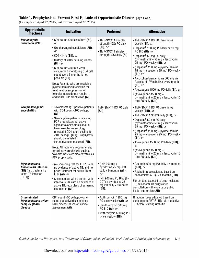
Downloaded from http://aidsinfo.nih.gov/guidelines on 7/29/2015
Guidelines for the Prevention and Treatment of Opportunistic Infections in HIV-Infected Adults and Adolescents U-1
Table 1. Prophylaxis to Prevent First Episode of Opportunistic Disease (page 1 of 5) (Last updated April 22, 2015; last reviewed April 22, 2015)
OpportunisticInfections
Indication Preferred Alternative
Pneumocystispneumonia (PCP)
• CD4 count <200 cells/mm3 (AI),or
• Oropharyngeal candidiasis (AII),or
• CD4 <14% (BII), or
• History of AIDS-defining illness(BII), or
• CD4 count >200 but <250cells/mm3 if monitoring CD4 cellcount every 3 months is notpossible (BII)
Note: Patients who are receivingpyrimethamine/sulfadiazine fortreatment or suppression oftoxoplasmosis do not requireadditional PCP prophylaxis (AII).
• TMP-SMXa 1 double-strength (DS) PO daily(AI), or
• TMP-SMXa 1 single-strength (SS) daily (AI)
• TMP-SMXa 1 DS PO three timesweekly (BI), or
• Dapsoneb 100 mg PO daily or 50 mgPO BID (BI), or
• Dapsoneb 50 mg PO daily +(pyrimethamine 50 mg + leucovorin25 mg) PO weekly (BI), or
• (Dapsoneb 200 mg + pyrimethamine75 mg + leucovorin 25 mg) PO weekly(BI); or
• Aerosolized pentamidine 300 mg viaRespigard II™ nebulizer every month(BI), or
• Atovaquone 1500 mg PO daily (BI), or
• (Atovaquone 1500 mg +pyrimethamine 25 mg + leucovorin 10mg) PO daily (CIII)
Toxoplasma gondiiencephalitis
• Toxoplasma IgG-positive patientswith CD4 count <100 cells/µL(AII);
• Seronegative patients receivingPCP prophylaxis not activeagainst toxoplasmosis shouldhave toxoplasma serologyretested if CD4 count decline to<100 cells/µL (CIII). Prophylaxisshould be initiated ifseroconversion occurred (AII).
Note: All regimens recommendedfor primary prophylaxis againsttoxoplasmosis are also effective asPCP prophylaxis.
TMP-SMXa 1 DS PO daily(AII)
• TMP-SMXa 1 DS PO three timesweekly (BIII), or
• TMP-SMXa 1 SS PO daily (BIII), or
• Dapsoneb 50 mg PO daily +(pyrimethamine 50 mg + leucovorin25 mg) PO weekly (BI), or
• (Dapsoneb 200 mg + pyrimethamine75 mg + leucovorin 25 mg) PO weekly(BI); or
• Atovaquone 1500 mg PO daily (CIII);or
• (Atovaquone 1500 mg +pyrimethamine 25 mg + leucovorin 10mg) PO daily (CIII)
Mycobacteriumtuberculosis infection(TB) (i.e., treatment oflatent TB infection[LTBI])
• (+) screening test for LTBIc, withno evidence of active TB, and noprior treatment for active TB orLTBI (AI), or
• Close contact with a person withinfectious TB, with no evidence ofactive TB, regardless of screeningtest results (AII).
• (INH 300 mg +pyridoxine 25 mg) POdaily x 9 months (AII),or
• INH 900 mg PO BIW (byDOT) + pyridoxine 25mg PO daily x 9 months(BII).
• Rifampin 600 mg PO daily x 4 months(BIII), or
• Rifabutin (dose adjusted based onconcomitant ART)d x 4 months (BIII).
For persons exposed to drug-resistantTB, select anti-TB drugs afterconsultation with experts or publichealth authorities (AII).
DisseminatedMycobacterium aviumcomplex (MAC)disease
CD4 count <50 cells/µL—afterruling out active disseminatedMAC disease based on clinicalassessment (AI).
• Azithromycin 1200 mgPO once weekly (AI), or
• Clarithromycin 500 mgPO BID (AI), or
• Azithromycin 600 mg POtwice weekly (BIII)
Rifabutin (dose adjusted based onconcomitant ART)d (BI); rule out activeTB before starting rifabutin
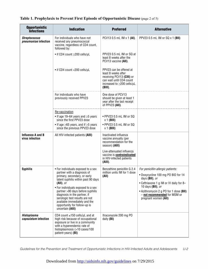
Downloaded from http://aidsinfo.nih.gov/guidelines on 7/29/2015
Guidelines for the Prevention and Treatment of Opportunistic Infections in HIV-Infected Adults and Adolescents U-2
Table 1. Prophylaxis to Prevent First Episode of Opportunistic Disease (page 2 of 5)
OpportunisticInfections
Indication Preferred Alternative
Streptococcuspneumoniae infection
For individuals who have notreceived any pneumococcalvaccine, regardless of CD4 count,followed by:
• if CD4 count ≥200 cells/µL
• if CD4 count <200 cells/µL
PCV13 0.5 mL IM x 1 (AI).
PPV23 0.5 mL IM or SQ atleast 8 weeks after thePCV13 vaccine (AII).
PPV23 can be offered atleast 8 weeks afterreceiving PCV13 (CIII) orcan wait until CD4 countincreased to ≥200 cells/µL(BIII).
PPV23 0.5 mL IM or SQ x 1 (BII)
For individuals who havepreviously received PPV23
One dose of PCV13should be given at least 1year after the last receiptof PPV23 (AII).
Re-vaccination
• If age 19–64 years and ≥5 yearssince the first PPV23 dose
• If age ≥65 years, and if ≥5 yearssince the previous PPV23 dose
• PPV23 0.5 mL IM or SQx 1 (BIII)
• PPV23 0.5 mL IM or SQx 1 (BIII)
Influenza A and Bvirus infection
All HIV-infected patients (AIII) Inactivated influenzavaccine annually (perrecommendation for theseason) (AIII)
Live-attenuated influenzavaccine is contraindicatedin HIV-infected patients(AIII).
Syphilis • For individuals exposed to a sexpartner with a diagnosis ofprimary, secondary, or earlylatent syphilis within past 90 days(AII), or
• For individuals exposed to a sexpartner >90 days before syphilisdiagnosis in the partner, ifserologic test results are notavailable immediately and theopportunity for follow-up isuncertain (AIII)
Benzathine penicillin G 2.4million units IM for 1 dose(AII)
For penicillin-allergic patients:
• Doxycycline 100 mg PO BID for 14days (BII), or
• Ceftriaxone 1 g IM or IV daily for 8–10 days (BII), or
• Azithromycin 2 g PO for 1 dose (BII)– not recommended for MSM orpregnant women (AII)
Histoplasmacapsulatum infection
CD4 count ≤150 cells/µL and athigh risk because of occupationalexposure or live in a communitywith a hyperendemic rate ofhistoplasmosis (>10 cases/100patient-years) (BI)
Itraconazole 200 mg POdaily (BI)
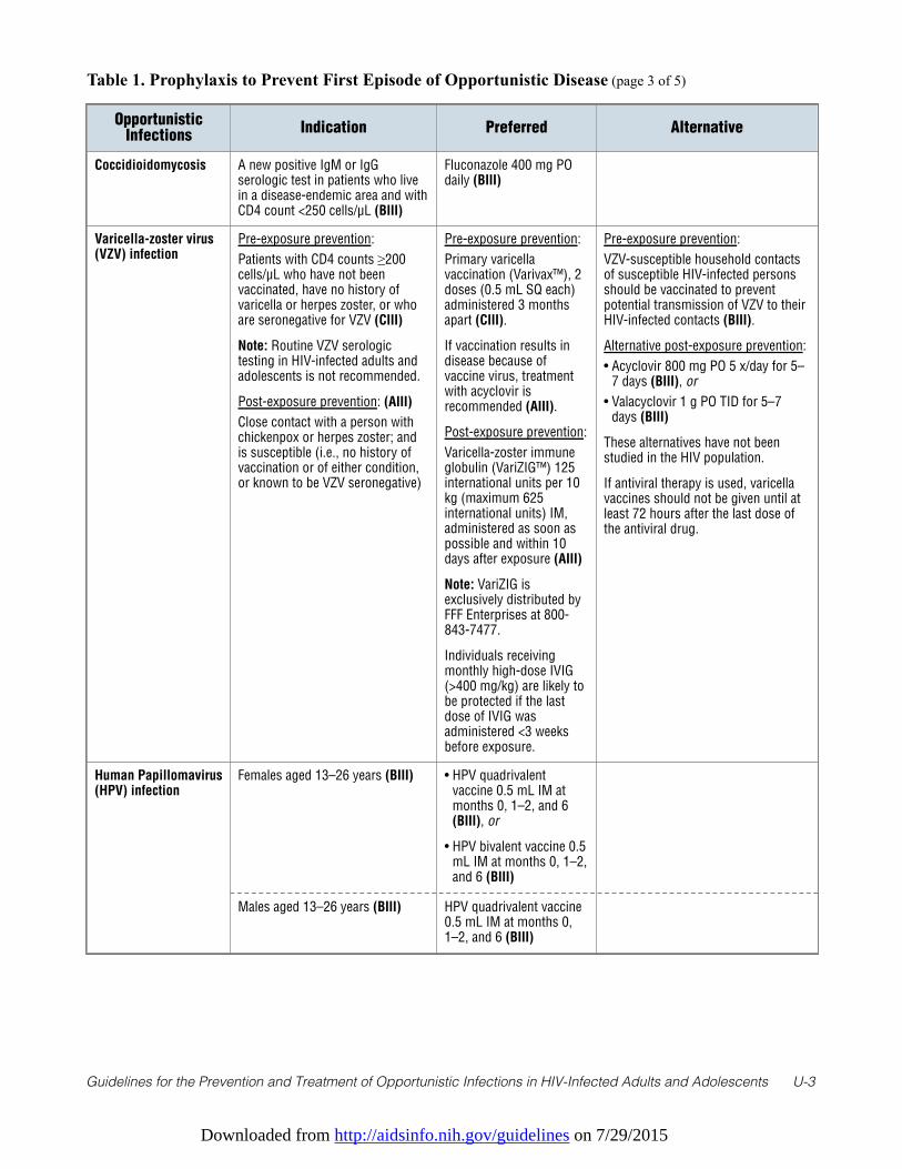
Downloaded from http://aidsinfo.nih.gov/guidelines on 7/29/2015
Guidelines for the Prevention and Treatment of Opportunistic Infections in HIV-Infected Adults and Adolescents U-3
Table 1. Prophylaxis to Prevent First Episode of Opportunistic Disease (page 3 of 5)
OpportunisticInfections
Indication Preferred Alternative
Coccidioidomycosis A new positive IgM or IgGserologic test in patients who livein a disease-endemic area and withCD4 count <250 cells/µL (BIII)
Fluconazole 400 mg POdaily (BIII)
Varicella-zoster virus(VZV) infection
Pre-exposure prevention:
Patients with CD4 counts ≥200cells/µL who have not beenvaccinated, have no history ofvaricella or herpes zoster, or whoare seronegative for VZV (CIII)
Note: Routine VZV serologictesting in HIV-infected adults andadolescents is not recommended.
Post-exposure prevention: (AIII)
Close contact with a person withchickenpox or herpes zoster; andis susceptible (i.e., no history ofvaccination or of either condition,or known to be VZV seronegative)
Pre-exposure prevention:
Primary varicellavaccination (Varivax™), 2doses (0.5 mL SQ each)administered 3 monthsapart (CIII).
If vaccination results indisease because ofvaccine virus, treatmentwith acyclovir isrecommended (AIII).
Post-exposure prevention:
Varicella-zoster immuneglobulin (VariZIG™) 125international units per 10kg (maximum 625international units) IM,administered as soon aspossible and within 10days after exposure (AIII)
Note: VariZIG isexclusively distributed byFFF Enterprises at 800-843-7477.
Individuals receivingmonthly high-dose IVIG(>400 mg/kg) are likely tobe protected if the lastdose of IVIG wasadministered <3 weeksbefore exposure.
Pre-exposure prevention:
VZV-susceptible household contactsof susceptible HIV-infected personsshould be vaccinated to preventpotential transmission of VZV to theirHIV-infected contacts (BIII).
Alternative post-exposure prevention:
• Acyclovir 800 mg PO 5 x/day for 5–7 days (BIII), or
• Valacyclovir 1 g PO TID for 5–7days (BIII)
These alternatives have not beenstudied in the HIV population.
If antiviral therapy is used, varicellavaccines should not be given until atleast 72 hours after the last dose ofthe antiviral drug.
Human Papillomavirus(HPV) infection
Females aged 13–26 years (BIII) • HPV quadrivalentvaccine 0.5 mL IM atmonths 0, 1–2, and 6(BIII), or
• HPV bivalent vaccine 0.5mL IM at months 0, 1–2,and 6 (BIII)
Males aged 13–26 years (BIII) HPV quadrivalent vaccine0.5 mL IM at months 0,1–2, and 6 (BIII)
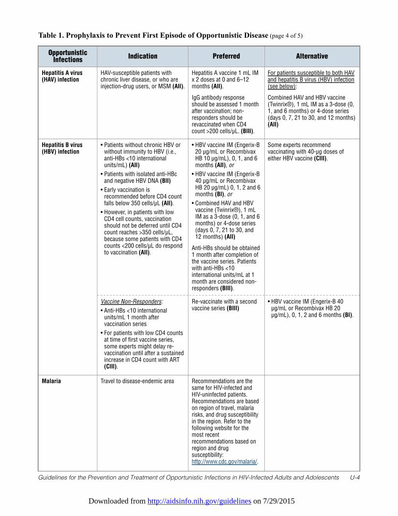
Downloaded from http://aidsinfo.nih.gov/guidelines on 7/29/2015
Guidelines for the Prevention and Treatment of Opportunistic Infections in HIV-Infected Adults and Adolescents U-4
Table 1. Prophylaxis to Prevent First Episode of Opportunistic Disease (page 4 of 5)
OpportunisticInfections
Indication Preferred Alternative
Hepatitis A virus(HAV) infection
HAV-susceptible patients withchronic liver disease, or who areinjection-drug users, or MSM (AII).
Hepatitis A vaccine 1 mL IMx 2 doses at 0 and 6–12months (AII).
IgG antibody responseshould be assessed 1 monthafter vaccination; non-responders should berevaccinated when CD4count >200 cells/µL. (BIII).
For patients susceptible to both HAVand hepatitis B virus (HBV) infection(see below):
Combined HAV and HBV vaccine(Twinrix®), 1 mL IM as a 3-dose (0,1, and 6 months) or 4-dose series(days 0, 7, 21 to 30, and 12 months)(AII)
Hepatitis B virus(HBV) infection
• Patients without chronic HBV orwithout immunity to HBV (i.e.,anti-HBs <10 internationalunits/mL) (AII)
• Patients with isolated anti-HBcand negative HBV DNA (BII)
• Early vaccination isrecommended before CD4 countfalls below 350 cells/µL (AII).
• However, in patients with lowCD4 cell counts, vaccinationshould not be deferred until CD4count reaches >350 cells/µL,because some patients with CD4counts <200 cells/µL do respondto vaccination (AII).
• HBV vaccine IM (Engerix-B20 µg/mL or RecombivaxHB 10 µg/mL), 0, 1, and 6months (AII), or
• HBV vaccine IM (Engerix-B40 µg/mL or RecombivaxHB 20 µg/mL) 0, 1, 2 and 6months (BI), or
• Combined HAV and HBVvaccine (Twinrix®), 1 mLIM as a 3-dose (0, 1, and 6months) or 4-dose series(days 0, 7, 21 to 30, and12 months) (AII)
Anti-HBs should be obtained1 month after completion ofthe vaccine series. Patientswith anti-HBs <10international units/mL at 1month are considered non-responders (BIII).
Some experts recommendvaccinating with 40-µg doses ofeither HBV vaccine (CIII).
Vaccine Non-Responders:
• Anti-HBs <10 internationalunits/mL 1 month aftervaccination series
• For patients with low CD4 countsat time of first vaccine series,some experts might delay re-vaccination until after a sustainedincrease in CD4 count with ART(CIII).
Re-vaccinate with a secondvaccine series (BIII)
• HBV vaccine IM (Engerix-B 40µg/mL or Recombivax HB 20µg/mL), 0, 1, 2 and 6 months (BI).
Malaria Travel to disease-endemic area Recommendations are thesame for HIV-infected andHIV-uninfected patients.Recommendations are basedon region of travel, malariarisks, and drug susceptibilityin the region. Refer to thefollowing website for themost recentrecommendations based onregion and drugsusceptibility:http://www.cdc.gov/malaria/.
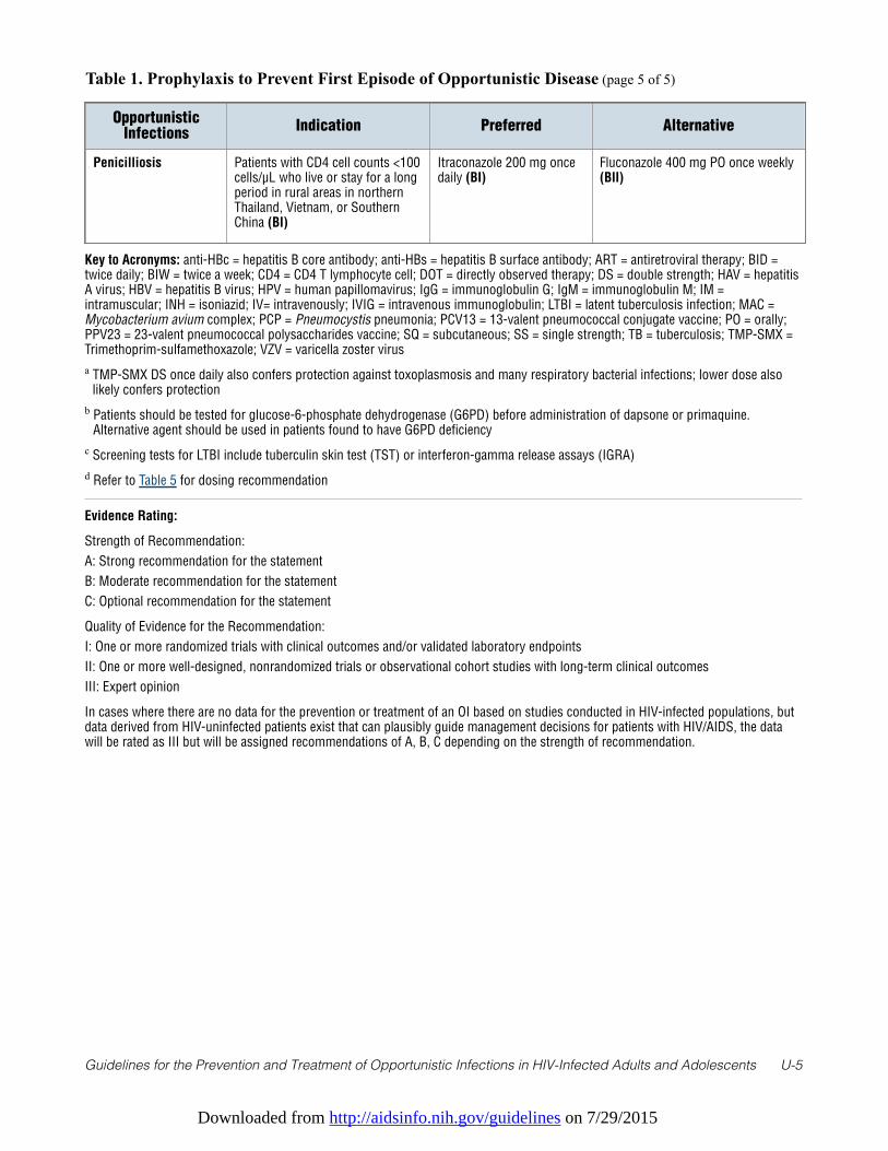
Downloaded from http://aidsinfo.nih.gov/guidelines on 7/29/2015
Guidelines for the Prevention and Treatment of Opportunistic Infections in HIV-Infected Adults and Adolescents U-5
Table 1. Prophylaxis to Prevent First Episode of Opportunistic Disease (page 5 of 5)
OpportunisticInfections
Indication Preferred Alternative
Penicilliosis Patients with CD4 cell counts <100cells/µL who live or stay for a longperiod in rural areas in northernThailand, Vietnam, or SouthernChina (BI)
Itraconazole 200 mg oncedaily (BI)
Fluconazole 400 mg PO once weekly(BII)
Key to Acronyms: anti-HBc = hepatitis B core antibody; anti-HBs = hepatitis B surface antibody; ART = antiretroviral therapy; BID =twice daily; BIW = twice a week; CD4 = CD4 T lymphocyte cell; DOT = directly observed therapy; DS = double strength; HAV = hepatitisA virus; HBV = hepatitis B virus; HPV = human papillomavirus; IgG = immunoglobulin G; IgM = immunoglobulin M; IM =intramuscular; INH = isoniazid; IV= intravenously; IVIG = intravenous immunoglobulin; LTBI = latent tuberculosis infection; MAC =Mycobacterium avium complex; PCP = Pneumocystis pneumonia; PCV13 = 13-valent pneumococcal conjugate vaccine; PO = orally;PPV23 = 23-valent pneumococcal polysaccharides vaccine; SQ = subcutaneous; SS = single strength; TB = tuberculosis; TMP-SMX =Trimethoprim-sulfamethoxazole; VZV = varicella zoster virus
a TMP-SMX DS once daily also confers protection against toxoplasmosis and many respiratory bacterial infections; lower dose alsolikely confers protection
b Patients should be tested for glucose-6-phosphate dehydrogenase (G6PD) before administration of dapsone or primaquine.Alternative agent should be used in patients found to have G6PD deficiency
c Screening tests for LTBI include tuberculin skin test (TST) or interferon-gamma release assays (IGRA)
d Refer to Table 5 for dosing recommendation
Evidence Rating:
Strength of Recommendation:
A: Strong recommendation for the statement
B: Moderate recommendation for the statement
C: Optional recommendation for the statement
Quality of Evidence for the Recommendation:
I: One or more randomized trials with clinical outcomes and/or validated laboratory endpoints
II: One or more well-designed, nonrandomized trials or observational cohort studies with long-term clinical outcomes
III: Expert opinion
In cases where there are no data for the prevention or treatment of an OI based on studies conducted in HIV-infected populations, butdata derived from HIV-uninfected patients exist that can plausibly guide management decisions for patients with HIV/AIDS, the datawill be rated as III but will be assigned recommendations of A, B, C depending on the strength of recommendation.
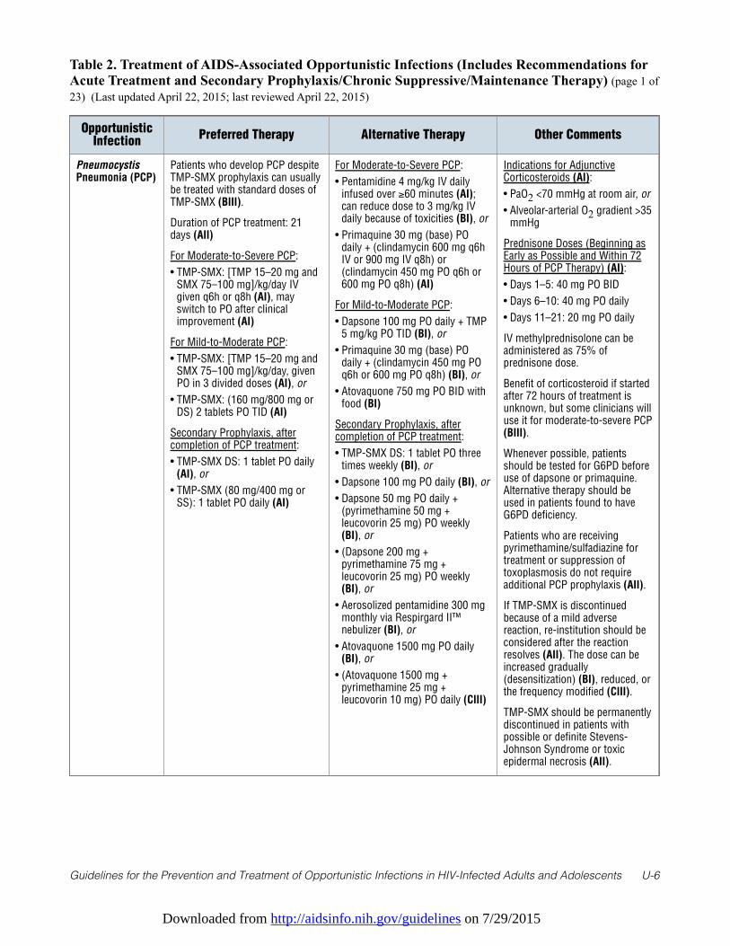
Downloaded from http://aidsinfo.nih.gov/guidelines on 7/29/2015
Guidelines for the Prevention and Treatment of Opportunistic Infections in HIV-Infected Adults and Adolescents U-6
Table 2. Treatment of AIDS-Associated Opportunistic Infections (Includes Recommendations forAcute Treatment and Secondary Prophylaxis/Chronic Suppressive/Maintenance Therapy) (page 1 of23) (Last updated April 22, 2015; last reviewed April 22, 2015)
OpportunisticInfection
Preferred Therapy Alternative Therapy Other Comments
PneumocystisPneumonia (PCP)
Patients who develop PCP despiteTMP-SMX prophylaxis can usuallybe treated with standard doses ofTMP-SMX (BIII).
Duration of PCP treatment: 21days (AII)
For Moderate-to-Severe PCP:
• TMP-SMX: [TMP 15–20 mg andSMX 75–100 mg]/kg/day IVgiven q6h or q8h (AI), mayswitch to PO after clinicalimprovement (AI)
For Mild-to-Moderate PCP:
• TMP-SMX: [TMP 15–20 mg andSMX 75–100 mg]/kg/day, givenPO in 3 divided doses (AI), or
• TMP-SMX: (160 mg/800 mg orDS) 2 tablets PO TID (AI)
Secondary Prophylaxis, aftercompletion of PCP treatment:
• TMP-SMX DS: 1 tablet PO daily(AI), or
• TMP-SMX (80 mg/400 mg orSS): 1 tablet PO daily (AI)
For Moderate-to-Severe PCP:
• Pentamidine 4 mg/kg IV dailyinfused over ≥60 minutes (AI);can reduce dose to 3 mg/kg IVdaily because of toxicities (BI), or
• Primaquine 30 mg (base) POdaily + (clindamycin 600 mg q6hIV or 900 mg IV q8h) or(clindamycin 450 mg PO q6h or600 mg PO q8h) (AI)
For Mild-to-Moderate PCP:
• Dapsone 100 mg PO daily + TMP5 mg/kg PO TID (BI), or
• Primaquine 30 mg (base) POdaily + (clindamycin 450 mg POq6h or 600 mg PO q8h) (BI), or
• Atovaquone 750 mg PO BID withfood (BI)
Secondary Prophylaxis, aftercompletion of PCP treatment:
• TMP-SMX DS: 1 tablet PO threetimes weekly (BI), or
• Dapsone 100 mg PO daily (BI), or
• Dapsone 50 mg PO daily +(pyrimethamine 50 mg +leucovorin 25 mg) PO weekly(BI), or
• (Dapsone 200 mg +pyrimethamine 75 mg +leucovorin 25 mg) PO weekly(BI), or
• Aerosolized pentamidine 300 mgmonthly via Respirgard II™nebulizer (BI), or
• Atovaquone 1500 mg PO daily(BI), or
• (Atovaquone 1500 mg +pyrimethamine 25 mg +leucovorin 10 mg) PO daily (CIII)
Indications for AdjunctiveCorticosteroids (AI):
• PaO2 <70 mmHg at room air, or
• Alveolar-arterial O2 gradient >35mmHg
Prednisone Doses (Beginning asEarly as Possible and Within 72Hours of PCP Therapy) (AI):
• Days 1–5: 40 mg PO BID
• Days 6–10: 40 mg PO daily
• Days 11–21: 20 mg PO daily
IV methylprednisolone can beadministered as 75% ofprednisone dose.
Benefit of corticosteroid if startedafter 72 hours of treatment isunknown, but some clinicians willuse it for moderate-to-severe PCP(BIII).
Whenever possible, patientsshould be tested for G6PD beforeuse of dapsone or primaquine.Alternative therapy should beused in patients found to haveG6PD deficiency.
Patients who are receivingpyrimethamine/sulfadiazine fortreatment or suppression oftoxoplasmosis do not requireadditional PCP prophylaxis (AII).
If TMP-SMX is discontinuedbecause of a mild adversereaction, re-institution should beconsidered after the reactionresolves (AII). The dose can beincreased gradually(desensitization) (BI), reduced, orthe frequency modified (CIII).
TMP-SMX should be permanentlydiscontinued in patients withpossible or definite Stevens-Johnson Syndrome or toxicepidermal necrosis (AII).
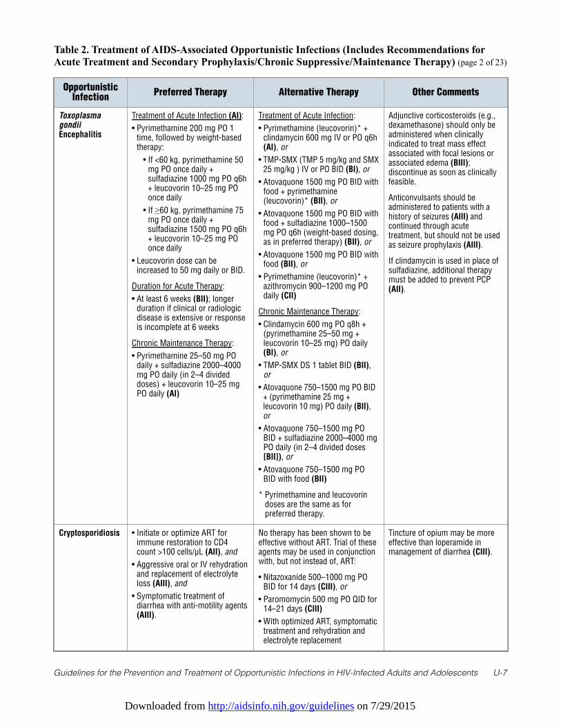
Downloaded from http://aidsinfo.nih.gov/guidelines on 7/29/2015
Guidelines for the Prevention and Treatment of Opportunistic Infections in HIV-Infected Adults and Adolescents U-7
Table 2. Treatment of AIDS-Associated Opportunistic Infections (Includes Recommendations forAcute Treatment and Secondary Prophylaxis/Chronic Suppressive/Maintenance Therapy) (page 2 of 23)
OpportunisticInfection
Preferred Therapy Alternative Therapy Other Comments
ToxoplasmagondiiEncephalitis
Treatment of Acute Infection (AI):
• Pyrimethamine 200 mg PO 1time, followed by weight-basedtherapy:
• If <60 kg, pyrimethamine 50mg PO once daily +sulfadiazine 1000 mg PO q6h+ leucovorin 10–25 mg POonce daily
• If ≥60 kg, pyrimethamine 75mg PO once daily +sulfadiazine 1500 mg PO q6h+ leucovorin 10–25 mg POonce daily
• Leucovorin dose can beincreased to 50 mg daily or BID.
Duration for Acute Therapy:
• At least 6 weeks (BII); longerduration if clinical or radiologicdisease is extensive or responseis incomplete at 6 weeks
Chronic Maintenance Therapy:
• Pyrimethamine 25–50 mg POdaily + sulfadiazine 2000–4000mg PO daily (in 2–4 divideddoses) + leucovorin 10–25 mgPO daily (AI)
Treatment of Acute Infection:
• Pyrimethamine (leucovorin)* +clindamycin 600 mg IV or PO q6h(AI), or
• TMP-SMX (TMP 5 mg/kg and SMX25 mg/kg ) IV or PO BID (BI), or
• Atovaquone 1500 mg PO BID withfood + pyrimethamine(leucovorin)* (BII), or
• Atovaquone 1500 mg PO BID withfood + sulfadiazine 1000–1500mg PO q6h (weight-based dosing,as in preferred therapy) (BII), or
• Atovaquone 1500 mg PO BID withfood (BII), or
• Pyrimethamine (leucovorin)* +azithromycin 900–1200 mg POdaily (CII)
Chronic Maintenance Therapy:
• Clindamycin 600 mg PO q8h +(pyrimethamine 25–50 mg +leucovorin 10–25 mg) PO daily(BI), or
• TMP-SMX DS 1 tablet BID (BII),or
• Atovaquone 750–1500 mg PO BID+ (pyrimethamine 25 mg +leucovorin 10 mg) PO daily (BII),or
• Atovaquone 750–1500 mg POBID + sulfadiazine 2000–4000 mgPO daily (in 2–4 divided doses[BII]), or
• Atovaquone 750–1500 mg POBID with food (BII)
* Pyrimethamine and leucovorindoses are the same as forpreferred therapy.
Adjunctive corticosteroids (e.g.,dexamethasone) should only beadministered when clinicallyindicated to treat mass effectassociated with focal lesions orassociated edema (BIII);discontinue as soon as clinicallyfeasible.
Anticonvulsants should beadministered to patients with ahistory of seizures (AIII) andcontinued through acutetreatment, but should not be usedas seizure prophylaxis (AIII).
If clindamycin is used in place ofsulfadiazine, additional therapymust be added to prevent PCP(AII).
Cryptosporidiosis • Initiate or optimize ART forimmune restoration to CD4count >100 cells/µL (AII), and
• Aggressive oral or IV rehydrationand replacement of electrolyteloss (AIII), and
• Symptomatic treatment ofdiarrhea with anti-motility agents(AIII).
No therapy has been shown to beeffective without ART. Trial of theseagents may be used in conjunctionwith, but not instead of, ART:
• Nitazoxanide 500–1000 mg POBID for 14 days (CIII), or
• Paromomycin 500 mg PO QID for14–21 days (CIII)
• With optimized ART, symptomatictreatment and rehydration andelectrolyte replacement
Tincture of opium may be moreeffective than loperamide inmanagement of diarrhea (CIII).
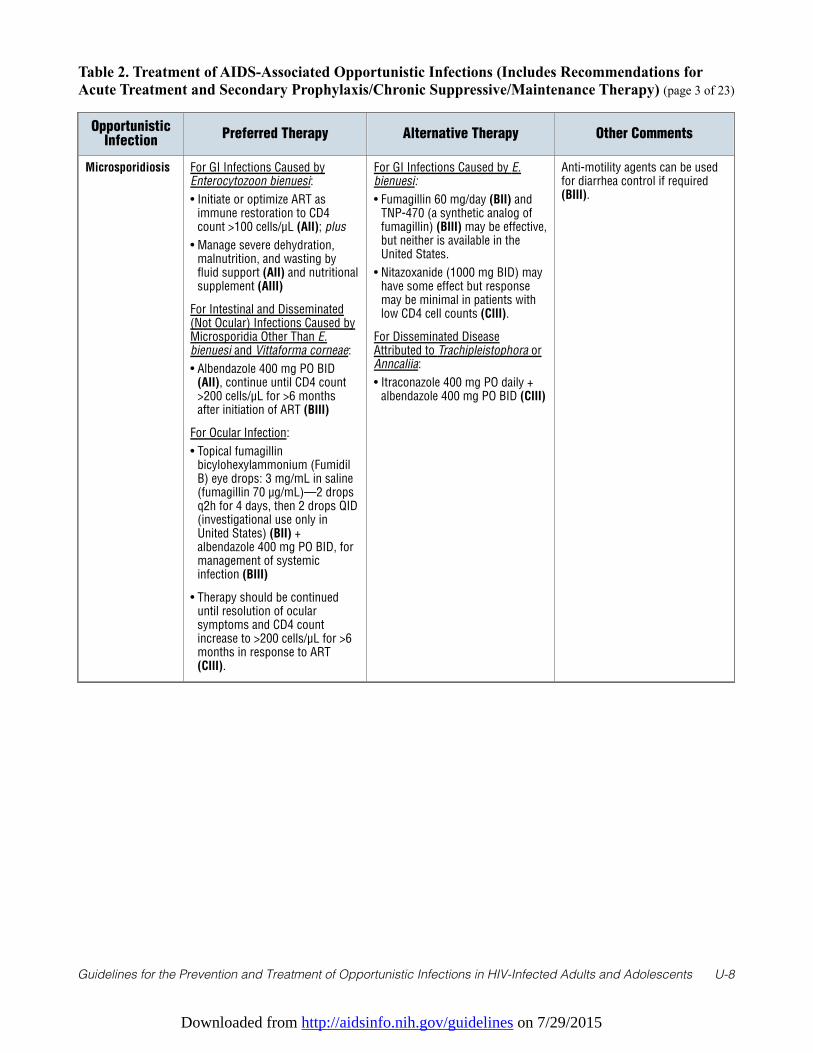
Downloaded from http://aidsinfo.nih.gov/guidelines on 7/29/2015
Guidelines for the Prevention and Treatment of Opportunistic Infections in HIV-Infected Adults and Adolescents U-8
Table 2. Treatment of AIDS-Associated Opportunistic Infections (Includes Recommendations forAcute Treatment and Secondary Prophylaxis/Chronic Suppressive/Maintenance Therapy) (page 3 of 23)
OpportunisticInfection
Preferred Therapy Alternative Therapy Other Comments
Microsporidiosis For GI Infections Caused byEnterocytozoon bienuesi:
• Initiate or optimize ART asimmune restoration to CD4count >100 cells/µL (AII); plus
• Manage severe dehydration,malnutrition, and wasting byfluid support (AII) and nutritionalsupplement (AIII)
For Intestinal and Disseminated(Not Ocular) Infections Caused byMicrosporidia Other Than E.bienuesi and Vittaforma corneae:
• Albendazole 400 mg PO BID(AII), continue until CD4 count>200 cells/µL for >6 monthsafter initiation of ART (BIII)
For Ocular Infection:
• Topical fumagillinbicylohexylammonium (FumidilB) eye drops: 3 mg/mL in saline(fumagillin 70 µg/mL)—2 dropsq2h for 4 days, then 2 drops QID(investigational use only inUnited States) (BII) +albendazole 400 mg PO BID, formanagement of systemicinfection (BIII)
• Therapy should be continueduntil resolution of ocularsymptoms and CD4 countincrease to >200 cells/µL for >6months in response to ART(CIII).
For GI Infections Caused by E.bienuesi:
• Fumagillin 60 mg/day (BII) andTNP-470 (a synthetic analog offumagillin) (BIII) may be effective,but neither is available in theUnited States.
• Nitazoxanide (1000 mg BID) mayhave some effect but responsemay be minimal in patients withlow CD4 cell counts (CIII).
For Disseminated DiseaseAttributed to Trachipleistophora orAnncaliia:
• Itraconazole 400 mg PO daily +albendazole 400 mg PO BID (CIII)
Anti-motility agents can be usedfor diarrhea control if required(BIII).
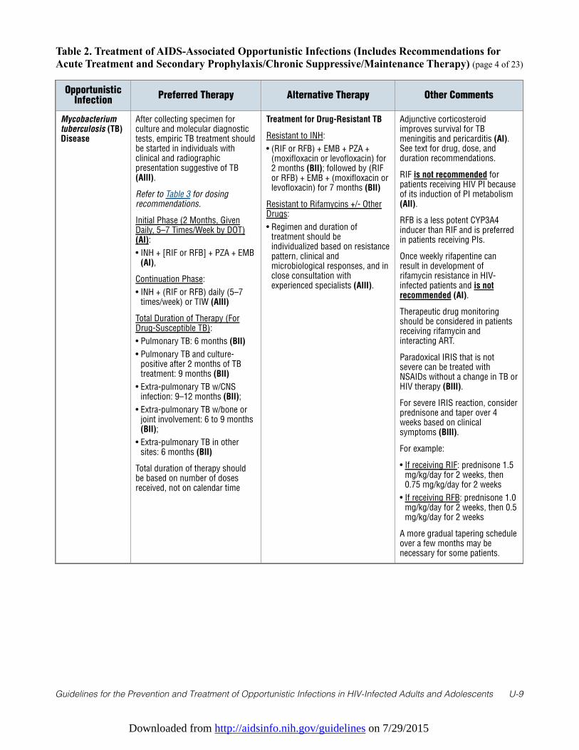
Downloaded from http://aidsinfo.nih.gov/guidelines on 7/29/2015
Guidelines for the Prevention and Treatment of Opportunistic Infections in HIV-Infected Adults and Adolescents U-9
Table 2. Treatment of AIDS-Associated Opportunistic Infections (Includes Recommendations forAcute Treatment and Secondary Prophylaxis/Chronic Suppressive/Maintenance Therapy) (page 4 of 23)
OpportunisticInfection
Preferred Therapy Alternative Therapy Other Comments
Mycobacteriumtuberculosis (TB)Disease
After collecting specimen forculture and molecular diagnostictests, empiric TB treatment shouldbe started in individuals withclinical and radiographicpresentation suggestive of TB(AIII).
Refer to Table 3 for dosingrecommendations.
Initial Phase (2 Months, GivenDaily, 5–7 Times/Week by DOT)(AI):
• INH + [RIF or RFB] + PZA + EMB(AI),
Continuation Phase:
• INH + (RIF or RFB) daily (5–7times/week) or TIW (AIII)
Total Duration of Therapy (ForDrug-Susceptible TB):
• Pulmonary TB: 6 months (BII)
• Pulmonary TB and culture-positive after 2 months of TBtreatment: 9 months (BII)
• Extra-pulmonary TB w/CNSinfection: 9–12 months (BII);
• Extra-pulmonary TB w/bone orjoint involvement: 6 to 9 months(BII);
• Extra-pulmonary TB in othersites: 6 months (BII)
Total duration of therapy shouldbe based on number of dosesreceived, not on calendar time
Treatment for Drug-Resistant TB
Resistant to INH:
• (RIF or RFB) + EMB + PZA +(moxifloxacin or levofloxacin) for2 months (BII); followed by (RIFor RFB) + EMB + (moxifloxacin orlevofloxacin) for 7 months (BII)
Resistant to Rifamycins +/- OtherDrugs:
• Regimen and duration oftreatment should beindividualized based on resistancepattern, clinical andmicrobiological responses, and inclose consultation withexperienced specialists (AIII).
Adjunctive corticosteroidimproves survival for TBmeningitis and pericarditis (AI).See text for drug, dose, andduration recommendations.
RIF is not recommended forpatients receiving HIV PI becauseof its induction of PI metabolism(AII).
RFB is a less potent CYP3A4inducer than RIF and is preferredin patients receiving PIs.
Once weekly rifapentine canresult in development ofrifamycin resistance in HIV-infected patients and is notrecommended (AI).
Therapeutic drug monitoringshould be considered in patientsreceiving rifamycin andinteracting ART.
Paradoxical IRIS that is notsevere can be treated withNSAIDs without a change in TB orHIV therapy (BIII).
For severe IRIS reaction, considerprednisone and taper over 4weeks based on clinicalsymptoms (BIII).
For example:
• If receiving RIF: prednisone 1.5mg/kg/day for 2 weeks, then0.75 mg/kg/day for 2 weeks
• If receiving RFB: prednisone 1.0mg/kg/day for 2 weeks, then 0.5mg/kg/day for 2 weeks
A more gradual tapering scheduleover a few months may benecessary for some patients.
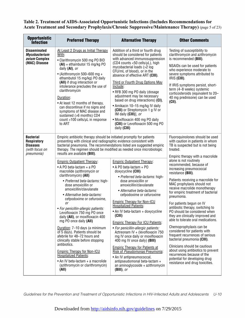
Downloaded from http://aidsinfo.nih.gov/guidelines on 7/29/2015
Guidelines for the Prevention and Treatment of Opportunistic Infections in HIV-Infected Adults and Adolescents U-10
Table 2. Treatment of AIDS-Associated Opportunistic Infections (Includes Recommendations forAcute Treatment and Secondary Prophylaxis/Chronic Suppressive/Maintenance Therapy) (page 5 of 23)
OpportunisticInfection
Preferred Therapy Alternative Therapy Other Comments
DisseminatedMycobacteriumavium Complex(MAC) Disease
At Least 2 Drugs as Initial TherapyWith:
• Clarithromycin 500 mg PO BID(AI) + ethambutol 15 mg/kg POdaily (AI), or
• (Azithromycin 500–600 mg +ethambutol 15 mg/kg) PO daily(AII) if drug interaction orintolerance precludes the use ofclarithromycin
Duration:
• At least 12 months of therapy,can discontinue if no signs andsymptoms of MAC disease andsustained (>6 months) CD4count >100 cells/µL in responseto ART
Addition of a third or fourth drugshould be considered for patientswith advanced immunosuppression(CD4 counts <50 cells/µL), highmycobacterial loads (>2 logCFU/mL of blood), or in theabsence of effective ART (CIII).
Third or Fourth Drug Options MayInclude:
• RFB 300 mg PO daily (dosageadjustment may be necessarybased on drug interactions) (CI),
• Amikacin 10–15 mg/kg IV daily(CIII) or Streptomycin 1 g IV orIM daily (CIII)], or
• Moxifloxacin 400 mg PO daily(CIII) or Levofloxacin 500 mg POdaily (CIII)
Testing of susceptibility toclarithromycin and azithromycinis recommended (BIII).
NSAIDs can be used for patientswho experience moderate tosevere symptoms attributed toIRIS (CIII).
If IRIS symptoms persist, short-term (4–8 weeks) systemiccorticosteroids (equivalent to 20–40 mg prednisone) can be used(CII).
BacterialRespiratoryDiseases(with focus onpneumonia)
Empiric antibiotic therapy should be initiated promptly for patientspresenting with clinical and radiographic evidence consistent withbacterial pneumonia. The recommendations listed are suggested empirictherapy. The regimen should be modified as needed once microbiologicresults are available (BIII).
Fluoroquinolones should be usedwith caution in patients in whomTB is suspected but is not beingtreated.
Empiric therapy with a macrolidealone is not routinelyrecommended, because ofincreasing pneumococcalresistance (BIII).
Patients receiving a macrolide forMAC prophylaxis should notreceive macrolide monotherapyfor empiric treatment of bacterialpneumonia.
For patients begun on IVantibiotic therapy, switching toPO should be considered whenthey are clinically improved andable to tolerate oral medications.
Chemoprophylaxis can beconsidered for patients withfrequent recurrences of seriousbacterial pneumonia (CIII).
Clinicians should be cautiousabout using antibiotics to preventrecurrences because of thepotential for developing drugresistance and drug toxicities.
Empiric Outpatient Therapy:
• A PO beta-lactam + a POmacrolide (azithromycin orclarithromycin) (AII)
• Preferred beta-lactams: high-dose amoxicillin oramoxicillin/clavulanate
• Alternative beta-lactams:cefpodoxime or cefuroxime,or
• For penicillin-allergic patients:Levofloxacin 750 mg PO oncedaily (AII), or moxifloxacin 400mg PO once daily (AII)
Duration: 7–10 days (a minimumof 5 days). Patients should beafebrile for 48–72 hours andclinically stable before stoppingantibiotics.
Empiric Therapy for Non-ICUHospitalized Patients:
• An IV beta-lactam + a macrolide(azithromycin or clarithromycin)(AII)
Empiric Outpatient Therapy:
• A PO beta-lactam + POdoxycycline (CIII)
• Preferred beta-lactams: high-dose amoxicillin oramoxicillin/clavulanate
• Alternative beta-lactams:cefpodoxime or cefuroxime
Empiric Therapy for Non-ICUHospitalized Patients:
• An IV beta-lactam + doxycycline(CIII)
Empiric Therapy For ICU Patients:
• For penicillin-allergic patients:Aztreonam IV + (levofloxacin 750mg IV once daily or moxifloxacin400 mg IV once daily) (BIII)
Empiric Therapy for Patients atRisk of Pseudomonas Pneumonia:
• An IV antipneumococcal,antipseudomonal beta-lactam +an aminoglycoside + azithromycin(BIII), or
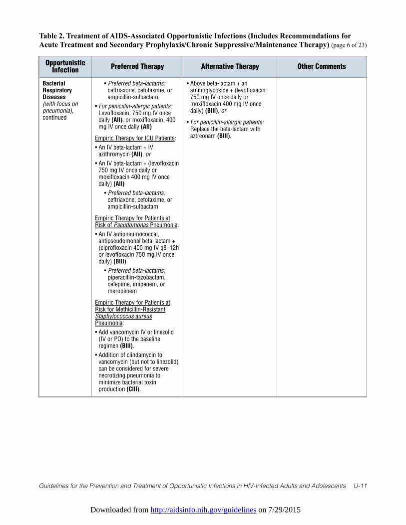
Downloaded from http://aidsinfo.nih.gov/guidelines on 7/29/2015
Guidelines for the Prevention and Treatment of Opportunistic Infections in HIV-Infected Adults and Adolescents U-11
Table 2. Treatment of AIDS-Associated Opportunistic Infections (Includes Recommendations forAcute Treatment and Secondary Prophylaxis/Chronic Suppressive/Maintenance Therapy) (page 6 of 23)
OpportunisticInfection
Preferred Therapy Alternative Therapy Other Comments
BacterialRespiratoryDiseases(with focus onpneumonia),continued
• Preferred beta-lactams:ceftriaxone, cefotaxime, orampicillin-sulbactam
• For penicillin-allergic patients:Levofloxacin, 750 mg IV oncedaily (AII), or moxifloxacin, 400mg IV once daily (AII)
Empiric Therapy for ICU Patients:
• An IV beta-lactam + IVazithromycin (AII), or
• An IV beta-lactam + (levofloxacin750 mg IV once daily ormoxifloxacin 400 mg IV oncedaily) (AII)
• Preferred beta-lactams:ceftriaxone, cefotaxime, orampicillin-sulbactam
Empiric Therapy for Patients atRisk of Pseudomonas Pneumonia:
• An IV antipneumococcal,antipseudomonal beta-lactam +(ciprofloxacin 400 mg IV q8–12hor levofloxacin 750 mg IV oncedaily) (BIII)
• Preferred beta-lactams:piperacillin-tazobactam,cefepime, imipenem, ormeropenem
Empiric Therapy for Patients atRisk for Methicillin-ResistantStaphylococcus aureusPneumonia:
• Add vancomycin IV or linezolid(IV or PO) to the baselineregimen (BIII).
• Addition of clindamycin tovancomycin (but not to linezolid)can be considered for severenecrotizing pneumonia tominimize bacterial toxinproduction (CIII).
• Above beta-lactam + anaminoglycoside + (levofloxacin750 mg IV once daily ormoxifloxacin 400 mg IV oncedaily) (BIII), or
• For penicillin-allergic patients:Replace the beta-lactam withaztreonam (BIII).
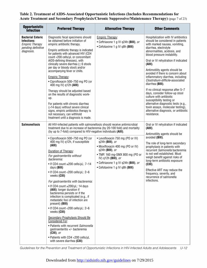
Downloaded from http://aidsinfo.nih.gov/guidelines on 7/29/2015
Guidelines for the Prevention and Treatment of Opportunistic Infections in HIV-Infected Adults and Adolescents U-12
Table 2. Treatment of AIDS-Associated Opportunistic Infections (Includes Recommendations forAcute Treatment and Secondary Prophylaxis/Chronic Suppressive/Maintenance Therapy) (page 7 of 23)
OpportunisticInfection
Preferred Therapy Alternative Therapy Other Comments
Bacterial EntericInfections:Empiric Therapypending definitivediagnosis.
Diagnostic fecal specimens shouldbe obtained before initiation ofempiric antibiotic therapy.
Empiric antibiotic therapy is indicatedfor patients with advanced HIV (CD4count <200 cells/µL or concomitantAIDS-defining illnesses), withclinically severe diarrhea (≥ 6 stoolsper day or bloody stool) and/oraccompanying fever or chills.
Empiric Therapy:
• Ciprofloxacin 500–750 mg PO (or400 mg IV) q12h (AIII)
Therapy should be adjusted basedon the results of diagnostic work-up.
For patients with chronic diarrhea(>14 days) without severe clinicalsigns, empiric antibiotics therapy isnot necessary, can withholdtreatment until a diagnosis is made.
Empiric Therapy:
• Ceftriaxone 1 g IV q24h (BIII), or
• Cefotaxime 1 g IV q8h (BIII)
Hospitalization with IV antibioticsshould be considered in patientswith marked nausea, vomiting,diarrhea, electrolyteabnormalities, acidosis, andblood pressure instability.
Oral or IV rehydration if indicated(AIII).
Antimotility agents should beavoided if there is concern aboutinflammatory diarrhea, includingClostridium-difficile-associateddiarrhea (BIII).
If no clinical response after 5–7days, consider follow-up stoolculture with antibioticsusceptibility testing oralternative diagnostic tests (e.g.,toxin assays, molecular testing),alternative diagnosis, or antibioticresistance.
Salmonellosis All HIV-infected patients with salmonellosis should receive antimicrobialtreatment due to an increase of bacteremia (by 20-100 fold) and mortality(by up to 7-fold) compared to HIV-negative individuals (AIII).
Oral or IV rehydration if indicated(AIII).
Antimotility agents should beavoided (BIII).
The role of long-term secondaryprophylaxis in patients withrecurrent Salmonella bacteremiais not well established. Mustweigh benefit against risks oflong-term antibiotic exposure(CIII).
Effective ART may reduce thefrequency, severity, andrecurrence of salmonellainfections.
• Ciprofloxacin 500–750 mg PO (or400 mg IV) q12h, if susceptible(AIII)
Duration of Therapy:
For gastroenteritis withoutbacteremia:
• If CD4 count ≥200 cells/µL: 7–14days (BIII)
• If CD4 count <200 cells/µL: 2–6weeks (CIII)
For gastroenteritis with bacteremia:
• If CD4 count ≥200/µL: 14 days(AIII); longer duration ifbacteremia persists or if theinfection is complicated (e.g., ifmetastatic foci of infection arepresent) (BIII)
• If CD4 count <200 cells/µL: 2–6weeks (CIII)
Secondary Prophylaxis Should BeConsidered For:
• Patients with recurrent Salmonellagastroenteritis +/- bacteremia(CIII), or
• Patients with CD4 <200 cells/µLwith severe diarrhea (CIII)
• Levofloxacin 750 mg (PO or IV)q24h (BIII), or
• Moxifloxacin 400 mg (PO or IV)q24h (BIII), or
• TMP, 160 mg-SMX 800 mg (PO orIV) q12h (BIII), or
• Ceftriaxone 1 g IV q24h (BIII), or
• Cefotaxime 1 g IV q8h (BIII)
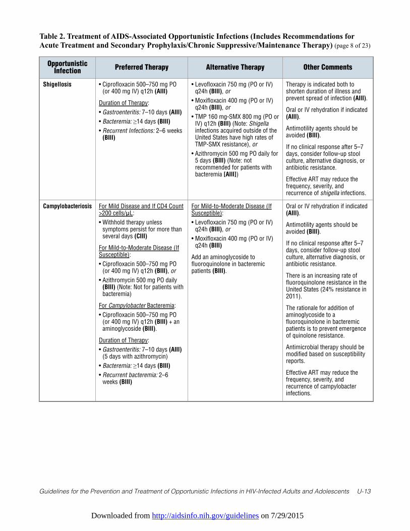
Downloaded from http://aidsinfo.nih.gov/guidelines on 7/29/2015
Guidelines for the Prevention and Treatment of Opportunistic Infections in HIV-Infected Adults and Adolescents U-13
Table 2. Treatment of AIDS-Associated Opportunistic Infections (Includes Recommendations forAcute Treatment and Secondary Prophylaxis/Chronic Suppressive/Maintenance Therapy) (page 8 of 23)
OpportunisticInfection
Preferred Therapy Alternative Therapy Other Comments
Shigellosis • Ciprofloxacin 500–750 mg PO(or 400 mg IV) q12h (AIII)
Duration of Therapy:
• Gastroenteritis: 7–10 days (AIII)
• Bacteremia: ≥14 days (BIII)
• Recurrent Infections: 2–6 weeks(BIII)
• Levofloxacin 750 mg (PO or IV)q24h (BIII), or
• Moxifloxacin 400 mg (PO or IV)q24h (BIII), or
• TMP 160 mg-SMX 800 mg (PO orIV) q12h (BIII) (Note: Shigellainfections acquired outside of theUnited States have high rates ofTMP-SMX resistance), or
• Azithromycin 500 mg PO daily for5 days (BIII) (Note: notrecommended for patients withbacteremia [AIII])
Therapy is indicated both toshorten duration of illness andprevent spread of infection (AIII).
Oral or IV rehydration if indicated(AIII).
Antimotility agents should beavoided (BIII).
If no clinical response after 5–7days, consider follow-up stoolculture, alternative diagnosis, orantibiotic resistance.
Effective ART may reduce thefrequency, severity, andrecurrence of shigella infections.
Campylobacteriosis For Mild Disease and If CD4 Count>200 cells/μL:
• Withhold therapy unlesssymptoms persist for more thanseveral days (CIII)
For Mild-to-Moderate Disease (IfSusceptible):
• Ciprofloxacin 500–750 mg PO(or 400 mg IV) q12h (BIII), or
• Azithromycin 500 mg PO daily(BIII) (Note: Not for patients withbacteremia)
For Campylobacter Bacteremia:
• Ciprofloxacin 500–750 mg PO(or 400 mg IV) q12h (BIII) + anaminoglycoside (BIII).
Duration of Therapy:
• Gastroenteritis: 7–10 days (AIII)(5 days with azithromycin)
• Bacteremia: ≥14 days (BIII)
• Recurrent bacteremia: 2–6weeks (BIII)
For Mild-to-Moderate Disease (IfSusceptible):
• Levofloxacin 750 mg (PO or IV)q24h (BIII), or
• Moxifloxacin 400 mg (PO or IV)q24h (BIII)
Add an aminoglycoside tofluoroquinolone in bacteremicpatients (BIII).
Oral or IV rehydration if indicated(AIII).
Antimotility agents should beavoided (BIII).
If no clinical response after 5–7days, consider follow-up stoolculture, alternative diagnosis, orantibiotic resistance.
There is an increasing rate offluoroquinolone resistance in theUnited States (24% resistance in2011).
The rationale for addition ofaminoglycoside to afluoroquinolone in bacteremicpatients is to prevent emergenceof quinolone resistance.
Antimicrobial therapy should bemodified based on susceptibilityreports.
Effective ART may reduce thefrequency, severity, andrecurrence of campylobacterinfections.
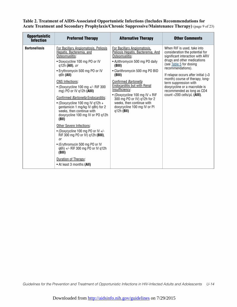
Downloaded from http://aidsinfo.nih.gov/guidelines on 7/29/2015
Guidelines for the Prevention and Treatment of Opportunistic Infections in HIV-Infected Adults and Adolescents U-14
Table 2. Treatment of AIDS-Associated Opportunistic Infections (Includes Recommendations forAcute Treatment and Secondary Prophylaxis/Chronic Suppressive/Maintenance Therapy) (page 9 of 23)
OpportunisticInfection
Preferred Therapy Alternative Therapy Other Comments
Bartonellosis For Bacillary Angiomatosis, PeliosisHepatis, Bacteremia, andOsteomyelitis:
• Doxycycline 100 mg PO or IVq12h (AII), or
• Erythromycin 500 mg PO or IVq6h (AII)
CNS Infections:
• (Doxycycline 100 mg +/- RIF 300mg) PO or IV q12h (AIII)
Confirmed Bartonella Endocarditis:
• (Doxycycline 100 mg IV q12h +gentamicin 1 mg/kg IV q8h) for 2weeks, then continue withdoxycycline 100 mg IV or PO q12h(BII)
Other Severe Infections:
• (Doxycycline 100 mg PO or IV +/-RIF 300 mg PO or IV) q12h (BIII),or
• (Erythromycin 500 mg PO or IVq6h) +/- RIF 300 mg PO or IV q12h(BIII)
Duration of Therapy:
• At least 3 months (AII)
For Bacillary Angiomatosis,Peliosis Hepatis, Bacteremia, AndOsteomyelitis:
• Azithromycin 500 mg PO daily(BIII)
• Clarithromycin 500 mg PO BID(BIII)
Confirmed BartonellaEndocarditis but with RenalInsufficiency:
• (Doxycycline 100 mg IV + RIF300 mg PO or IV) q12h for 2weeks, then continue withdoxycycline 100 mg IV or PIq12h (BII)
When RIF is used, take intoconsideration the potential forsignificant interaction with ARVdrugs and other medications(see Table 5 for dosingrecommendations).
If relapse occurs after initial (>3month) course of therapy, long-term suppression withdoxycycline or a macrolide isrecommended as long as CD4count <200 cells/µL (AIII).
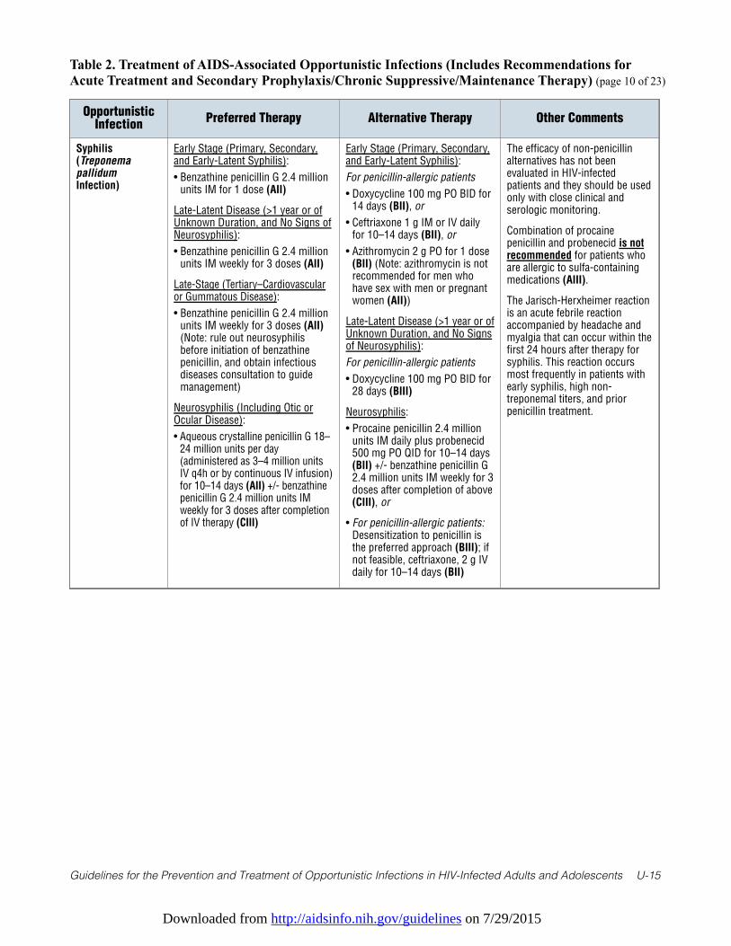
Downloaded from http://aidsinfo.nih.gov/guidelines on 7/29/2015
Guidelines for the Prevention and Treatment of Opportunistic Infections in HIV-Infected Adults and Adolescents U-15
Table 2. Treatment of AIDS-Associated Opportunistic Infections (Includes Recommendations forAcute Treatment and Secondary Prophylaxis/Chronic Suppressive/Maintenance Therapy) (page 10 of 23)
OpportunisticInfection
Preferred Therapy Alternative Therapy Other Comments
Syphilis(TreponemapallidumInfection)
Early Stage (Primary, Secondary,and Early-Latent Syphilis):
• Benzathine penicillin G 2.4 millionunits IM for 1 dose (AII)
Late-Latent Disease (>1 year or ofUnknown Duration, and No Signs ofNeurosyphilis):
• Benzathine penicillin G 2.4 millionunits IM weekly for 3 doses (AII)
Late-Stage (Tertiary–Cardiovascularor Gummatous Disease):
• Benzathine penicillin G 2.4 millionunits IM weekly for 3 doses (AII)(Note: rule out neurosyphilisbefore initiation of benzathinepenicillin, and obtain infectiousdiseases consultation to guidemanagement)
Neurosyphilis (Including Otic orOcular Disease):
• Aqueous crystalline penicillin G 18–24 million units per day(administered as 3–4 million unitsIV q4h or by continuous IV infusion)for 10–14 days (AII) +/- benzathinepenicillin G 2.4 million units IMweekly for 3 doses after completionof IV therapy (CIII)
Early Stage (Primary, Secondary,and Early-Latent Syphilis):
For penicillin-allergic patients
• Doxycycline 100 mg PO BID for14 days (BII), or
• Ceftriaxone 1 g IM or IV dailyfor 10–14 days (BII), or
• Azithromycin 2 g PO for 1 dose(BII) (Note: azithromycin is notrecommended for men whohave sex with men or pregnantwomen (AII))
Late-Latent Disease (>1 year or ofUnknown Duration, and No Signsof Neurosyphilis):
For penicillin-allergic patients
• Doxycycline 100 mg PO BID for28 days (BIII)
Neurosyphilis:
• Procaine penicillin 2.4 millionunits IM daily plus probenecid500 mg PO QID for 10–14 days(BII) +/- benzathine penicillin G2.4 million units IM weekly for 3doses after completion of above(CIII), or
• For penicillin-allergic patients:Desensitization to penicillin isthe preferred approach (BIII); ifnot feasible, ceftriaxone, 2 g IVdaily for 10–14 days (BII)
The efficacy of non-penicillinalternatives has not beenevaluated in HIV-infectedpatients and they should be usedonly with close clinical andserologic monitoring.
Combination of procainepenicillin and probenecid is notrecommended for patients whoare allergic to sulfa-containingmedications (AIII).
The Jarisch-Herxheimer reactionis an acute febrile reactionaccompanied by headache andmyalgia that can occur within thefirst 24 hours after therapy forsyphilis. This reaction occursmost frequently in patients withearly syphilis, high non-treponemal titers, and priorpenicillin treatment.
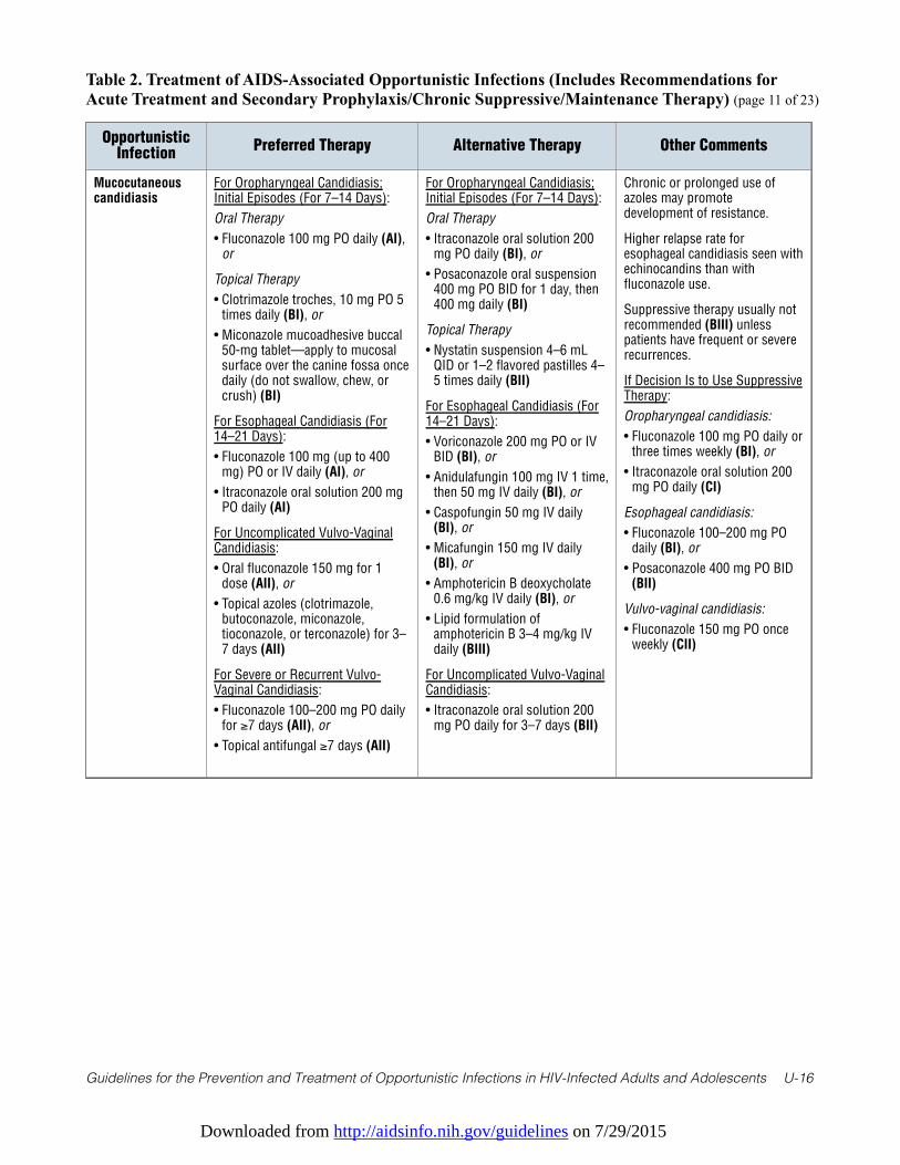
Downloaded from http://aidsinfo.nih.gov/guidelines on 7/29/2015
Guidelines for the Prevention and Treatment of Opportunistic Infections in HIV-Infected Adults and Adolescents U-16
Table 2. Treatment of AIDS-Associated Opportunistic Infections (Includes Recommendations forAcute Treatment and Secondary Prophylaxis/Chronic Suppressive/Maintenance Therapy) (page 11 of 23)
OpportunisticInfection
Preferred Therapy Alternative Therapy Other Comments
Mucocutaneouscandidiasis
For Oropharyngeal Candidiasis;Initial Episodes (For 7–14 Days):
Oral Therapy
• Fluconazole 100 mg PO daily (AI),or
Topical Therapy
• Clotrimazole troches, 10 mg PO 5times daily (BI), or
• Miconazole mucoadhesive buccal50-mg tablet—apply to mucosalsurface over the canine fossa oncedaily (do not swallow, chew, orcrush) (BI)
For Esophageal Candidiasis (For14–21 Days):
• Fluconazole 100 mg (up to 400mg) PO or IV daily (AI), or
• Itraconazole oral solution 200 mgPO daily (AI)
For Uncomplicated Vulvo-VaginalCandidiasis:
• Oral fluconazole 150 mg for 1dose (AII), or
• Topical azoles (clotrimazole,butoconazole, miconazole,tioconazole, or terconazole) for 3–7 days (AII)
For Severe or Recurrent Vulvo-Vaginal Candidiasis:
• Fluconazole 100–200 mg PO dailyfor ≥7 days (AII), or
• Topical antifungal ≥7 days (AII)
For Oropharyngeal Candidiasis;Initial Episodes (For 7–14 Days):
Oral Therapy
• Itraconazole oral solution 200mg PO daily (BI), or
• Posaconazole oral suspension400 mg PO BID for 1 day, then400 mg daily (BI)
Topical Therapy
• Nystatin suspension 4–6 mLQID or 1–2 flavored pastilles 4–5 times daily (BII)
For Esophageal Candidiasis (For14–21 Days):
• Voriconazole 200 mg PO or IVBID (BI), or
• Anidulafungin 100 mg IV 1 time,then 50 mg IV daily (BI), or
• Caspofungin 50 mg IV daily(BI), or
• Micafungin 150 mg IV daily(BI), or
• Amphotericin B deoxycholate0.6 mg/kg IV daily (BI), or
• Lipid formulation ofamphotericin B 3–4 mg/kg IVdaily (BIII)
For Uncomplicated Vulvo-VaginalCandidiasis:
• Itraconazole oral solution 200mg PO daily for 3–7 days (BII)
Chronic or prolonged use ofazoles may promotedevelopment of resistance.
Higher relapse rate foresophageal candidiasis seen withechinocandins than withfluconazole use.
Suppressive therapy usually notrecommended (BIII) unlesspatients have frequent or severerecurrences.
If Decision Is to Use SuppressiveTherapy:
Oropharyngeal candidiasis:
• Fluconazole 100 mg PO daily orthree times weekly (BI), or
• Itraconazole oral solution 200mg PO daily (CI)
Esophageal candidiasis:
• Fluconazole 100–200 mg POdaily (BI), or
• Posaconazole 400 mg PO BID(BII)
Vulvo-vaginal candidiasis:
• Fluconazole 150 mg PO onceweekly (CII)
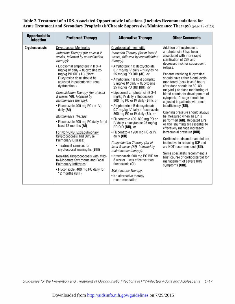
Downloaded from http://aidsinfo.nih.gov/guidelines on 7/29/2015
Guidelines for the Prevention and Treatment of Opportunistic Infections in HIV-Infected Adults and Adolescents U-17
Table 2. Treatment of AIDS-Associated Opportunistic Infections (Includes Recommendations forAcute Treatment and Secondary Prophylaxis/Chronic Suppressive/Maintenance Therapy) (page 12 of 23)
OpportunisticInfection
Preferred Therapy Alternative Therapy Other Comments
Cryptococcosis Cryptococcal Meningitis
Induction Therapy (for at least 2weeks, followed by consolidationtherapy):
• Liposomal amphotericin B 3–4mg/kg IV daily + flucytosine 25mg/kg PO QID (AI) (Note:Flucytosine dose should beadjusted in patients with renaldysfunction.)
Consolidation Therapy (for at least8 weeks (AI), followed bymaintenance therapy):
• Fluconazole 400 mg PO (or IV)daily (AI)
Maintenance Therapy:
• Fluconazole 200 mg PO daily for atleast 12 months (AI)
For Non-CNS, ExtrapulmonaryCryptococcosis and DiffusePulmonary Disease:
• Treatment same as forcryptococcal meningitis (BIII)
Non-CNS Cryptococcosis with Mild-to-Moderate Symptoms and FocalPulmonary Infiltrates:
• Fluconazole, 400 mg PO daily for12 months (BIII)
Cryptococcal meningitis
Induction Therapy (for at least 2weeks, followed by consolidationtherapy):
• Amphotericin B deoxycholate0.7 mg/kg IV daily + flucytosine25 mg/kg PO QID (AI), or
• Amphotericin B lipid complex 5 mg/kg IV daily + flucytosine25 mg/kg PO QID (BII), or
• Liposomal amphotericin B 3-4mg/kg IV daily + fluconazole800 mg PO or IV daily (BIII), or
• Amphotericin B deoxycholate0.7 mg/kg IV daily + fluconazole800 mg PO or IV daily (BI), or
• Fluconazole 400–800 mg PO orIV daily + flucytosine 25 mg/kgPO QID (BII), or
• Fluconazole 1200 mg PO or IVdaily (CII)
Consolidation Therapy (for atleast 8 weeks (AI), followed bymaintenance therapy):
• Itraconazole 200 mg PO BID for8 weeks—less effective thanfluconazole (CI)
Maintenance Therapy:
• No alternative therapyrecommendation
Addition of flucytosine toamphotericin B has beenassociated with more rapidsterilization of CSF anddecreased risk for subsequentrelapse.
Patients receiving flucytosineshould have either blood levelsmonitored (peak level 2 hoursafter dose should be 30–80mcg/mL) or close monitoring ofblood counts for development ofcytopenia. Dosage should beadjusted in patients with renalinsufficiency (BII).
Opening pressure should alwaysbe measured when an LP isperformed (AII). Repeated LPsor CSF shunting are essential toeffectively manage increasedintracranial pressure (BIII).
Corticosteroids and mannitol areineffective in reducing ICP andare NOT recommended (BII).
Some specialists recommend abrief course of corticosteroid formanagement of severe IRISsymptoms (CIII).
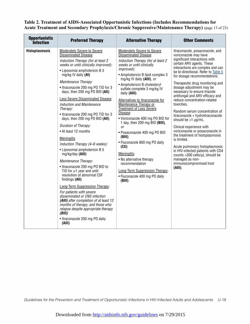
Downloaded from http://aidsinfo.nih.gov/guidelines on 7/29/2015
Guidelines for the Prevention and Treatment of Opportunistic Infections in HIV-Infected Adults and Adolescents U-18
Table 2. Treatment of AIDS-Associated Opportunistic Infections (Includes Recommendations forAcute Treatment and Secondary Prophylaxis/Chronic Suppressive/Maintenance Therapy) (page 13 of 23)
OpportunisticInfection
Preferred Therapy Alternative Therapy Other Comments
Histoplasmosis Moderately Severe to SevereDisseminated Disease
Induction Therapy (for at least 2weeks or until clinically improved):
• Liposomal amphotericin B 3mg/kg IV daily (AI)
Maintenance Therapy
• Itraconazole 200 mg PO TID for 3days, then 200 mg PO BID (AII)
Less Severe Disseminated Disease
Induction and MaintenanceTherapy:
• Itraconazole 200 mg PO TID for 3days, then 200 mg PO BID (AII)
Duration of Therapy:
• At least 12 months
Meningitis
Induction Therapy (4–6 weeks):
• Liposomal amphotericin B 5mg/kg/day (AIII)
Maintenance Therapy:
• Itraconazole 200 mg PO BID toTID for ≥1 year and untilresolution of abnormal CSFfindings (AII)
Long-Term Suppression Therapy:
For patients with severedisseminated or CNS infection(AIII) after completion of at least 12months of therapy; and those whorelapse despite appropriate therapy(BIII):
• Itraconazole 200 mg PO daily(AIII)
Moderately Severe to SevereDisseminated Disease
Induction Therapy (for at least 2weeks or until clinicallyimproved):
• Amphotericin B lipid complex 3mg/kg IV daily (AIII), or
• Amphotericin B cholesterylsulfate complete 3 mg/kg IVdaily (AIII)
Alternatives to Itraconazole forMaintenance Therapy orTreatment of Less SevereDisease:
• Voriconazole 400 mg PO BID for1 day, then 200 mg BID (BIII),or
• Posaconazole 400 mg PO BID(BIII)
• Fluconazole 800 mg PO daily(CII)
Meningitis:
• No alternative therapyrecommendation
Long-Term Suppression Therapy:
• Fluconazole 400 mg PO daily(BIII)
Itraconazole, posaconazole, andvoriconazole may havesignificant interactions withcertain ARV agents. Theseinteractions are complex and canbe bi-directional. Refer to Table 5for dosage recommendations.
Therapeutic drug monitoring anddosage adjustment may benecessary to ensure triazoleantifungal and ARV efficacy andreduce concentration-relatedtoxicities.
Random serum concentration ofitraconazole + hydroitraconazoleshould be >1 µg/mL.
Clinical experience withvoriconazole or posaconazole inthe treatment of histoplasmosisis limited.
Acute pulmonary histoplasmosisin HIV-infected patients with CD4counts >300 cells/µL should bemanaged as non-immunocompromised host(AIII).
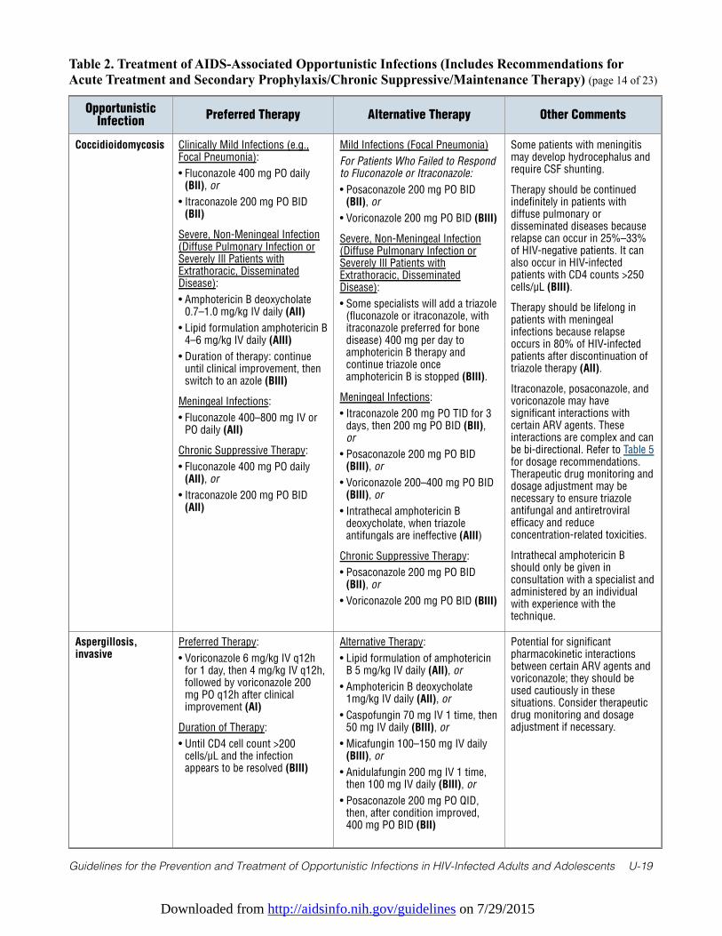
Downloaded from http://aidsinfo.nih.gov/guidelines on 7/29/2015
Guidelines for the Prevention and Treatment of Opportunistic Infections in HIV-Infected Adults and Adolescents U-19
Table 2. Treatment of AIDS-Associated Opportunistic Infections (Includes Recommendations forAcute Treatment and Secondary Prophylaxis/Chronic Suppressive/Maintenance Therapy) (page 14 of 23)
OpportunisticInfection
Preferred Therapy Alternative Therapy Other Comments
Coccidioidomycosis Clinically Mild Infections (e.g.,Focal Pneumonia):
• Fluconazole 400 mg PO daily(BII), or
• Itraconazole 200 mg PO BID(BII)
Severe, Non-Meningeal Infection(Diffuse Pulmonary Infection orSeverely Ill Patients withExtrathoracic, DisseminatedDisease):
• Amphotericin B deoxycholate0.7–1.0 mg/kg IV daily (AII)
• Lipid formulation amphotericin B4–6 mg/kg IV daily (AIII)
• Duration of therapy: continueuntil clinical improvement, thenswitch to an azole (BIII)
Meningeal Infections:
• Fluconazole 400–800 mg IV orPO daily (AII)
Chronic Suppressive Therapy:
• Fluconazole 400 mg PO daily(AII), or
• Itraconazole 200 mg PO BID(AII)
Mild Infections (Focal Pneumonia)
For Patients Who Failed to Respondto Fluconazole or Itraconazole:
• Posaconazole 200 mg PO BID(BII), or
• Voriconazole 200 mg PO BID (BIII)
Severe, Non-Meningeal Infection(Diffuse Pulmonary Infection orSeverely Ill Patients withExtrathoracic, DisseminatedDisease):
• Some specialists will add a triazole(fluconazole or itraconazole, withitraconazole preferred for bonedisease) 400 mg per day toamphotericin B therapy andcontinue triazole onceamphotericin B is stopped (BIII).
Meningeal Infections:
• Itraconazole 200 mg PO TID for 3days, then 200 mg PO BID (BII),or
• Posaconazole 200 mg PO BID(BIII), or
• Voriconazole 200–400 mg PO BID(BIII), or
• Intrathecal amphotericin Bdeoxycholate, when triazoleantifungals are ineffective (AIII)
Chronic Suppressive Therapy:
• Posaconazole 200 mg PO BID(BII), or
• Voriconazole 200 mg PO BID (BIII)
Some patients with meningitismay develop hydrocephalus andrequire CSF shunting.
Therapy should be continuedindefinitely in patients withdiffuse pulmonary ordisseminated diseases becauserelapse can occur in 25%–33%of HIV-negative patients. It canalso occur in HIV-infectedpatients with CD4 counts >250cells/µL (BIII).
Therapy should be lifelong inpatients with meningealinfections because relapseoccurs in 80% of HIV-infectedpatients after discontinuation oftriazole therapy (AII).
Itraconazole, posaconazole, andvoriconazole may havesignificant interactions withcertain ARV agents. Theseinteractions are complex and canbe bi-directional. Refer to Table 5for dosage recommendations.Therapeutic drug monitoring anddosage adjustment may benecessary to ensure triazoleantifungal and antiretroviralefficacy and reduceconcentration-related toxicities.
Intrathecal amphotericin Bshould only be given inconsultation with a specialist andadministered by an individualwith experience with thetechnique.
Aspergillosis,invasive
Preferred Therapy:
• Voriconazole 6 mg/kg IV q12hfor 1 day, then 4 mg/kg IV q12h,followed by voriconazole 200mg PO q12h after clinicalimprovement (AI)
Duration of Therapy:
• Until CD4 cell count >200cells/µL and the infectionappears to be resolved (BIII)
Alternative Therapy:
• Lipid formulation of amphotericinB 5 mg/kg IV daily (AII), or
• Amphotericin B deoxycholate1mg/kg IV daily (AII), or
• Caspofungin 70 mg IV 1 time, then50 mg IV daily (BIII), or
• Micafungin 100–150 mg IV daily(BIII), or
• Anidulafungin 200 mg IV 1 time,then 100 mg IV daily (BIII), or
• Posaconazole 200 mg PO QID,then, after condition improved,400 mg PO BID (BII)
Potential for significantpharmacokinetic interactionsbetween certain ARV agents andvoriconazole; they should beused cautiously in thesesituations. Consider therapeuticdrug monitoring and dosageadjustment if necessary.
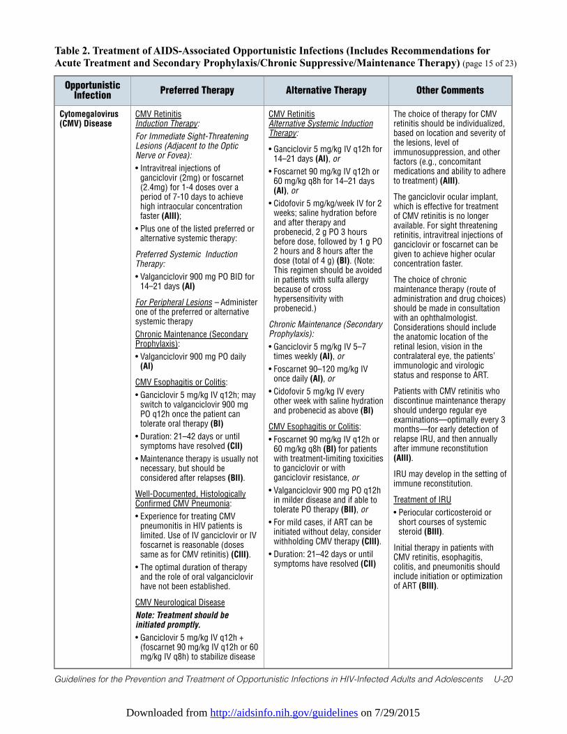
Downloaded from http://aidsinfo.nih.gov/guidelines on 7/29/2015
Guidelines for the Prevention and Treatment of Opportunistic Infections in HIV-Infected Adults and Adolescents U-20
Table 2. Treatment of AIDS-Associated Opportunistic Infections (Includes Recommendations forAcute Treatment and Secondary Prophylaxis/Chronic Suppressive/Maintenance Therapy) (page 15 of 23)
OpportunisticInfection
Preferred Therapy Alternative Therapy Other Comments
Cytomegalovirus(CMV) Disease
CMV RetinitisInduction Therapy:
For Immediate Sight-ThreateningLesions (Adjacent to the OpticNerve or Fovea):
• Intravitreal injections ofganciclovir (2mg) or foscarnet(2.4mg) for 1-4 doses over aperiod of 7-10 days to achievehigh intraocular concentrationfaster (AIII);
• Plus one of the listed preferred oralternative systemic therapy:
Preferred Systemic InductionTherapy:
• Valganciclovir 900 mg PO BID for14–21 days (AI)
For Peripheral Lesions – Administerone of the preferred or alternativesystemic therapy
Chronic Maintenance (SecondaryProphylaxis):
• Valganciclovir 900 mg PO daily(AI)
CMV Esophagitis or Colitis:
• Ganciclovir 5 mg/kg IV q12h; mayswitch to valganciclovir 900 mgPO q12h once the patient cantolerate oral therapy (BI)
• Duration: 21–42 days or untilsymptoms have resolved (CII)
• Maintenance therapy is usually notnecessary, but should beconsidered after relapses (BII).
Well-Documented, HistologicallyConfirmed CMV Pneumonia:
• Experience for treating CMVpneumonitis in HIV patients islimited. Use of IV ganciclovir or IVfoscarnet is reasonable (dosessame as for CMV retinitis) (CIII).
• The optimal duration of therapyand the role of oral valganciclovirhave not been established.
CMV Neurological Disease
Note: Treatment should beinitiated promptly.
• Ganciclovir 5 mg/kg IV q12h +(foscarnet 90 mg/kg IV q12h or 60mg/kg IV q8h) to stabilize disease
CMV RetinitisAlternative Systemic InductionTherapy:
• Ganciclovir 5 mg/kg IV q12h for14–21 days (AI), or
• Foscarnet 90 mg/kg IV q12h or60 mg/kg q8h for 14–21 days(AI), or
• Cidofovir 5 mg/kg/week IV for 2weeks; saline hydration beforeand after therapy andprobenecid, 2 g PO 3 hoursbefore dose, followed by 1 g PO2 hours and 8 hours after thedose (total of 4 g) (BI). (Note:This regimen should be avoidedin patients with sulfa allergybecause of crosshypersensitivity withprobenecid.)
Chronic Maintenance (SecondaryProphylaxis):
• Ganciclovir 5 mg/kg IV 5–7times weekly (AI), or
• Foscarnet 90–120 mg/kg IVonce daily (AI), or
• Cidofovir 5 mg/kg IV everyother week with saline hydrationand probenecid as above (BI)
CMV Esophagitis or Colitis:
• Foscarnet 90 mg/kg IV q12h or60 mg/kg q8h (BI) for patientswith treatment-limiting toxicitiesto ganciclovir or withganciclovir resistance, or
• Valganciclovir 900 mg PO q12hin milder disease and if able totolerate PO therapy (BII), or
• For mild cases, if ART can beinitiated without delay, considerwithholding CMV therapy (CIII).
• Duration: 21–42 days or untilsymptoms have resolved (CII)
The choice of therapy for CMVretinitis should be individualized,based on location and severity ofthe lesions, level ofimmunosuppression, and otherfactors (e.g., concomitantmedications and ability to adhereto treatment) (AIII).
The ganciclovir ocular implant,which is effective for treatmentof CMV retinitis is no longeravailable. For sight threateningretinitis, intravitreal injections ofganciclovir or foscarnet can begiven to achieve higher ocularconcentration faster.
The choice of chronicmaintenance therapy (route ofadministration and drug choices)should be made in consultationwith an ophthalmologist.Considerations should includethe anatomic location of theretinal lesion, vision in thecontralateral eye, the patients’immunologic and virologicstatus and response to ART.
Patients with CMV retinitis whodiscontinue maintenance therapyshould undergo regular eyeexaminations—optimally every 3months—for early detection ofrelapse IRU, and then annuallyafter immune reconstitution(AIII).
IRU may develop in the setting ofimmune reconstitution.
Treatment of IRU
• Periocular corticosteroid orshort courses of systemicsteroid (BIII).
Initial therapy in patients withCMV retinitis, esophagitis,colitis, and pneumonitis shouldinclude initiation or optimizationof ART (BIII).
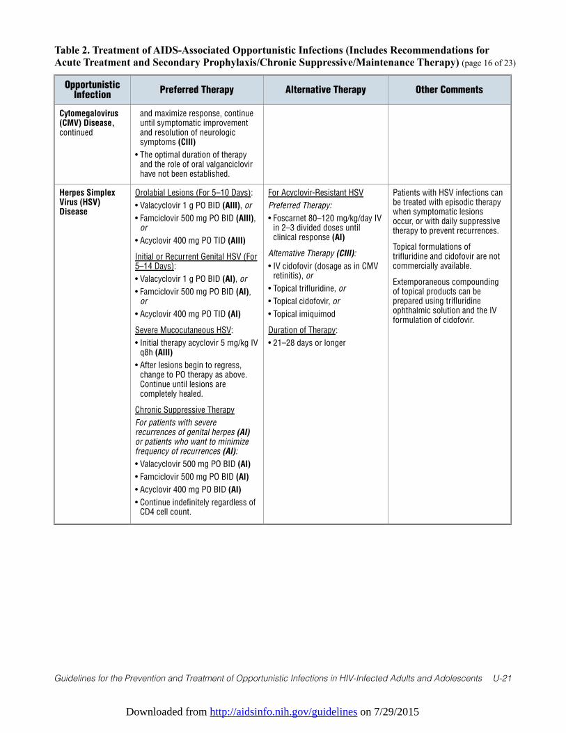
Downloaded from http://aidsinfo.nih.gov/guidelines on 7/29/2015
Guidelines for the Prevention and Treatment of Opportunistic Infections in HIV-Infected Adults and Adolescents U-21
Table 2. Treatment of AIDS-Associated Opportunistic Infections (Includes Recommendations forAcute Treatment and Secondary Prophylaxis/Chronic Suppressive/Maintenance Therapy) (page 16 of 23)
OpportunisticInfection
Preferred Therapy Alternative Therapy Other Comments
Cytomegalovirus(CMV) Disease,continued
and maximize response, continueuntil symptomatic improvementand resolution of neurologicsymptoms (CIII)
• The optimal duration of therapyand the role of oral valganciclovirhave not been established.
Herpes SimplexVirus (HSV)Disease
Orolabial Lesions (For 5–10 Days):
• Valacyclovir 1 g PO BID (AIII), or
• Famciclovir 500 mg PO BID (AIII),or
• Acyclovir 400 mg PO TID (AIII)
Initial or Recurrent Genital HSV (For5–14 Days):
• Valacyclovir 1 g PO BID (AI), or
• Famciclovir 500 mg PO BID (AI),or
• Acyclovir 400 mg PO TID (AI)
Severe Mucocutaneous HSV:
• Initial therapy acyclovir 5 mg/kg IVq8h (AIII)
• After lesions begin to regress,change to PO therapy as above.Continue until lesions arecompletely healed.
Chronic Suppressive Therapy
For patients with severerecurrences of genital herpes (AI)or patients who want to minimizefrequency of recurrences (AI):
• Valacyclovir 500 mg PO BID (AI)
• Famciclovir 500 mg PO BID (AI)
• Acyclovir 400 mg PO BID (AI)
• Continue indefinitely regardless ofCD4 cell count.
For Acyclovir-Resistant HSV
Preferred Therapy:
• Foscarnet 80–120 mg/kg/day IVin 2–3 divided doses untilclinical response (AI)
Alternative Therapy (CIII):
• IV cidofovir (dosage as in CMVretinitis), or
• Topical trifluridine, or
• Topical cidofovir, or
• Topical imiquimod
Duration of Therapy:
• 21–28 days or longer
Patients with HSV infections canbe treated with episodic therapywhen symptomatic lesionsoccur, or with daily suppressivetherapy to prevent recurrences.
Topical formulations oftrifluridine and cidofovir are notcommercially available.
Extemporaneous compoundingof topical products can beprepared using trifluridineophthalmic solution and the IVformulation of cidofovir.
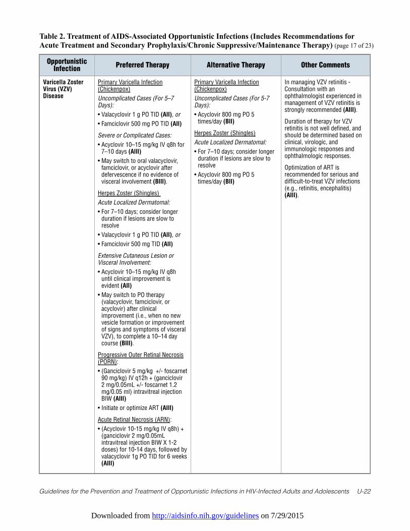
Downloaded from http://aidsinfo.nih.gov/guidelines on 7/29/2015
Guidelines for the Prevention and Treatment of Opportunistic Infections in HIV-Infected Adults and Adolescents U-22
Table 2. Treatment of AIDS-Associated Opportunistic Infections (Includes Recommendations forAcute Treatment and Secondary Prophylaxis/Chronic Suppressive/Maintenance Therapy) (page 17 of 23)
OpportunisticInfection
Preferred Therapy Alternative Therapy Other Comments
Varicella ZosterVirus (VZV)Disease
Primary Varicella Infection(Chickenpox)
Uncomplicated Cases (For 5–7Days):
• Valacyclovir 1 g PO TID (AII), or
• Famciclovir 500 mg PO TID (AII)
Severe or Complicated Cases:
• Acyclovir 10–15 mg/kg IV q8h for7–10 days (AIII)
• May switch to oral valacyclovir,famciclovir, or acyclovir afterdefervescence if no evidence ofvisceral involvement (BIII).
Herpes Zoster (Shingles)
Acute Localized Dermatomal:
• For 7–10 days; consider longerduration if lesions are slow toresolve
• Valacyclovir 1 g PO TID (AII), or
• Famciclovir 500 mg TID (AII)
Extensive Cutaneous Lesion orVisceral Involvement:
• Acyclovir 10–15 mg/kg IV q8huntil clinical improvement isevident (AII)
• May switch to PO therapy(valacyclovir, famciclovir, oracyclovir) after clinicalimprovement (i.e., when no newvesicle formation or improvementof signs and symptoms of visceralVZV), to complete a 10–14 daycourse (BIII).
Progressive Outer Retinal Necrosis(PORN):
• (Ganciclovir 5 mg/kg +/- foscarnet90 mg/kg) IV q12h + (ganciclovir2 mg/0.05mL +/- foscarnet 1.2mg/0.05 ml) intravitreal injectionBIW (AIII)
• Initiate or optimize ART (AIII)
Acute Retinal Necrosis (ARN):
• (Acyclovir 10-15 mg/kg IV q8h) +(ganciclovir 2 mg/0.05mLintravitreal injection BIW X 1-2doses) for 10-14 days, followed byvalacyclovir 1g PO TID for 6 weeks(AIII)
Primary Varicella Infection(Chickenpox)
Uncomplicated Cases (For 5-7Days):
• Acyclovir 800 mg PO 5times/day (BII)
Herpes Zoster (Shingles)
Acute Localized Dermatomal:
• For 7–10 days; consider longerduration if lesions are slow toresolve
• Acyclovir 800 mg PO 5times/day (BII)
In managing VZV retinitis -Consultation with anophthalmologist experienced inmanagement of VZV retinitis isstrongly recommended (AIII).
Duration of therapy for VZVretinitis is not well defined, andshould be determined based onclinical, virologic, andimmunologic responses andophthalmologic responses.
Optimization of ART isrecommended for serious anddifficult-to-treat VZV infections(e.g., retinitis, encephalitis)(AIII).
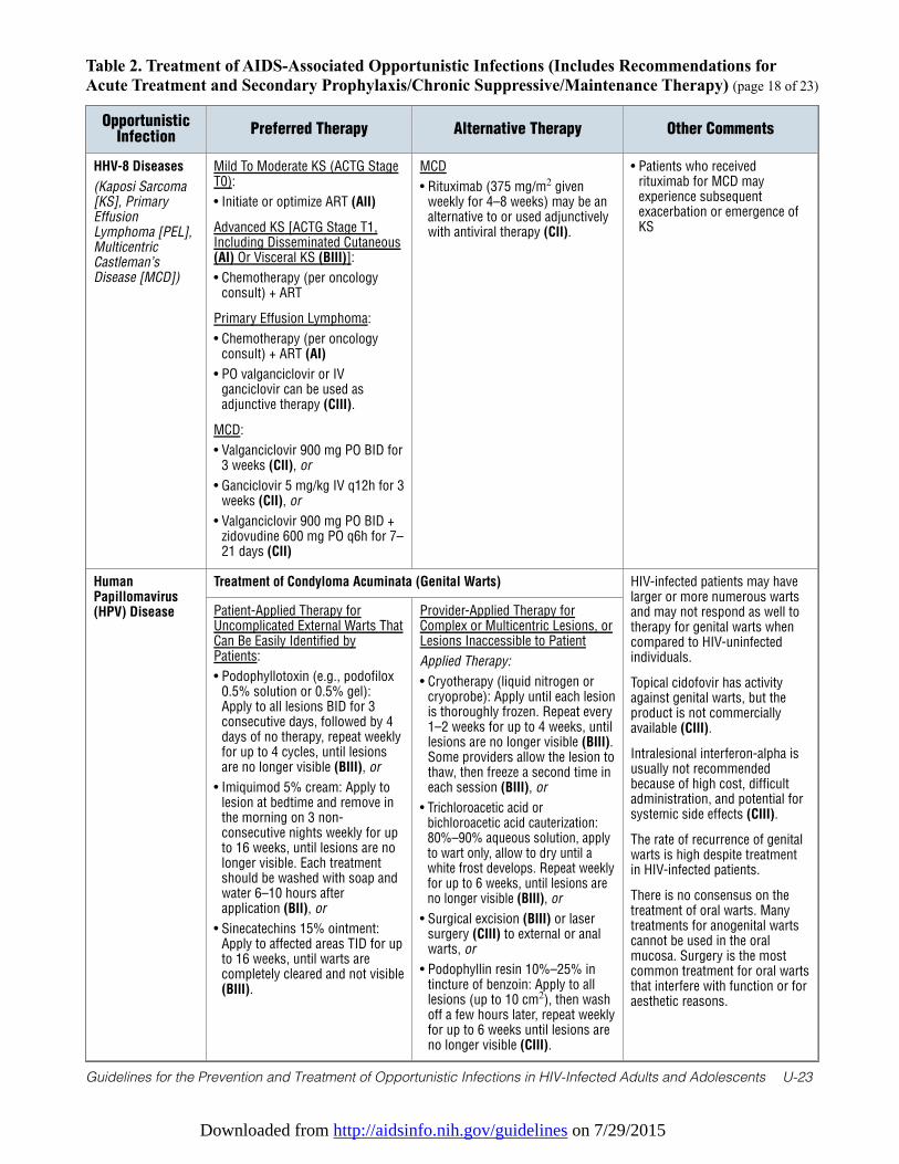
Downloaded from http://aidsinfo.nih.gov/guidelines on 7/29/2015
Guidelines for the Prevention and Treatment of Opportunistic Infections in HIV-Infected Adults and Adolescents U-23
Table 2. Treatment of AIDS-Associated Opportunistic Infections (Includes Recommendations forAcute Treatment and Secondary Prophylaxis/Chronic Suppressive/Maintenance Therapy) (page 18 of 23)
OpportunisticInfection
Preferred Therapy Alternative Therapy Other Comments
HHV-8 Diseases
(Kaposi Sarcoma[KS], PrimaryEffusionLymphoma [PEL],MulticentricCastleman’sDisease [MCD])
Mild To Moderate KS (ACTG StageT0):
• Initiate or optimize ART (AII)
Advanced KS [ACTG Stage T1,Including Disseminated Cutaneous(AI) Or Visceral KS (BIII)]:
• Chemotherapy (per oncologyconsult) + ART
Primary Effusion Lymphoma:
• Chemotherapy (per oncologyconsult) + ART (AI)
• PO valganciclovir or IVganciclovir can be used asadjunctive therapy (CIII).
MCD:
• Valganciclovir 900 mg PO BID for3 weeks (CII), or
• Ganciclovir 5 mg/kg IV q12h for 3weeks (CII), or
• Valganciclovir 900 mg PO BID +zidovudine 600 mg PO q6h for 7–21 days (CII)
MCD
• Rituximab (375 mg/m2 givenweekly for 4–8 weeks) may be analternative to or used adjunctivelywith antiviral therapy (CII).
• Patients who receivedrituximab for MCD mayexperience subsequentexacerbation or emergence ofKS
HumanPapillomavirus(HPV) Disease
Treatment of Condyloma Acuminata (Genital Warts) HIV-infected patients may havelarger or more numerous wartsand may not respond as well totherapy for genital warts whencompared to HIV-uninfectedindividuals.
Topical cidofovir has activityagainst genital warts, but theproduct is not commerciallyavailable (CIII).
Intralesional interferon-alpha isusually not recommendedbecause of high cost, difficultadministration, and potential forsystemic side effects (CIII).
The rate of recurrence of genitalwarts is high despite treatmentin HIV-infected patients.
There is no consensus on thetreatment of oral warts. Manytreatments for anogenital wartscannot be used in the oralmucosa. Surgery is the mostcommon treatment for oral wartsthat interfere with function or foraesthetic reasons.
Patient-Applied Therapy forUncomplicated External Warts ThatCan Be Easily Identified byPatients:
• Podophyllotoxin (e.g., podofilox0.5% solution or 0.5% gel):Apply to all lesions BID for 3consecutive days, followed by 4days of no therapy, repeat weeklyfor up to 4 cycles, until lesionsare no longer visible (BIII), or
• Imiquimod 5% cream: Apply tolesion at bedtime and remove inthe morning on 3 non-consecutive nights weekly for upto 16 weeks, until lesions are nolonger visible. Each treatmentshould be washed with soap andwater 6–10 hours afterapplication (BII), or
• Sinecatechins 15% ointment:Apply to affected areas TID for upto 16 weeks, until warts arecompletely cleared and not visible(BIII).
Provider-Applied Therapy forComplex or Multicentric Lesions, orLesions Inaccessible to Patient
Applied Therapy:
• Cryotherapy (liquid nitrogen orcryoprobe): Apply until each lesionis thoroughly frozen. Repeat every1–2 weeks for up to 4 weeks, untillesions are no longer visible (BIII).Some providers allow the lesion tothaw, then freeze a second time ineach session (BIII), or
• Trichloroacetic acid orbichloroacetic acid cauterization:80%–90% aqueous solution, applyto wart only, allow to dry until awhite frost develops. Repeat weeklyfor up to 6 weeks, until lesions areno longer visible (BIII), or
• Surgical excision (BIII) or lasersurgery (CIII) to external or analwarts, or
• Podophyllin resin 10%–25% intincture of benzoin: Apply to alllesions (up to 10 cm2), then washoff a few hours later, repeat weeklyfor up to 6 weeks until lesions areno longer visible (CIII).
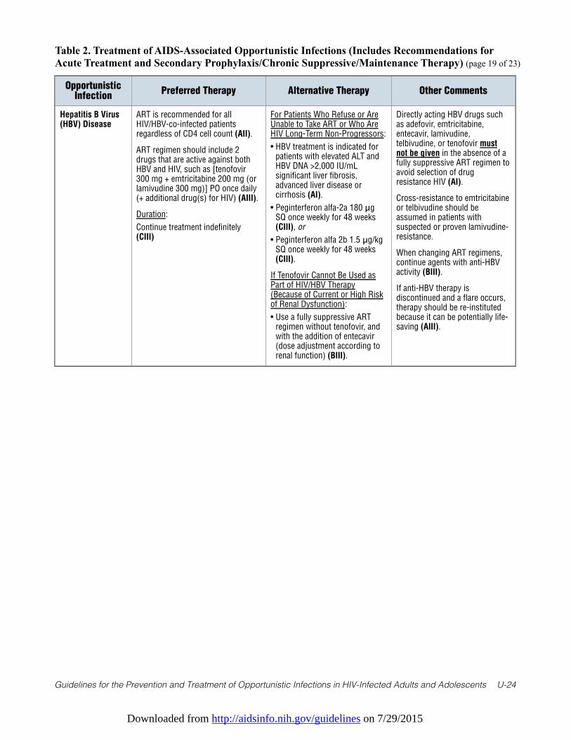
Downloaded from http://aidsinfo.nih.gov/guidelines on 7/29/2015
Guidelines for the Prevention and Treatment of Opportunistic Infections in HIV-Infected Adults and Adolescents U-24
Table 2. Treatment of AIDS-Associated Opportunistic Infections (Includes Recommendations forAcute Treatment and Secondary Prophylaxis/Chronic Suppressive/Maintenance Therapy) (page 19 of 23)
OpportunisticInfection
Preferred Therapy Alternative Therapy Other Comments
Hepatitis B Virus(HBV) Disease
ART is recommended for allHIV/HBV-co-infected patientsregardless of CD4 cell count (AII).
ART regimen should include 2drugs that are active against bothHBV and HIV, such as [tenofovir300 mg + emtricitabine 200 mg (orlamivudine 300 mg)] PO once daily(+ additional drug(s) for HIV) (AIII).
Duration:
Continue treatment indefinitely(CIII)
For Patients Who Refuse or AreUnable to Take ART or Who AreHIV Long-Term Non-Progressors:
• HBV treatment is indicated forpatients with elevated ALT andHBV DNA >2,000 IU/mLsignificant liver fibrosis,advanced liver disease orcirrhosis (AI).
• Peginterferon alfa-2a 180 μgSQ once weekly for 48 weeks(CIII), or
• Peginterferon alfa 2b 1.5 μg/kgSQ once weekly for 48 weeks(CIII).
If Tenofovir Cannot Be Used asPart of HIV/HBV Therapy(Because of Current or High Riskof Renal Dysfunction):
• Use a fully suppressive ARTregimen without tenofovir, andwith the addition of entecavir(dose adjustment according torenal function) (BIII).
Directly acting HBV drugs suchas adefovir, emtricitabine,entecavir, lamivudine,telbivudine, or tenofovir mustnot be given in the absence of afully suppressive ART regimen toavoid selection of drugresistance HIV (AI).
Cross-resistance to emtricitabineor telbivudine should beassumed in patients withsuspected or proven lamivudine-resistance.
When changing ART regimens,continue agents with anti-HBVactivity (BIII).
If anti-HBV therapy isdiscontinued and a flare occurs,therapy should be re-institutedbecause it can be potentially life-saving (AIII).
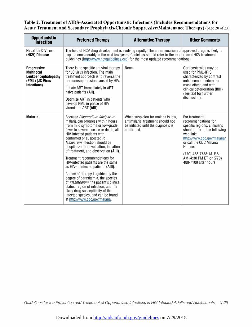
Downloaded from http://aidsinfo.nih.gov/guidelines on 7/29/2015
Guidelines for the Prevention and Treatment of Opportunistic Infections in HIV-Infected Adults and Adolescents U-25
Table 2. Treatment of AIDS-Associated Opportunistic Infections (Includes Recommendations forAcute Treatment and Secondary Prophylaxis/Chronic Suppressive/Maintenance Therapy) (page 20 of 23)
OpportunisticInfection
Preferred Therapy Alternative Therapy Other Comments
Hepatitis C Virus(HCV) Disease
The field of HCV drug development is evolving rapidly. The armamenarium of approved drugs is likely toexpand considerably in the next few years. Clinicians should refer to the most recent HCV treatmentguidelines (http://www.hcvguidelines.org) for the most updated recommendations.
ProgressiveMultifocalLeukoencephalopathy(PML) (JC VirusInfections)
There is no specific antiviral therapyfor JC virus infection. The maintreatment approach is to reverse theimmunosuppression caused by HIV.
Initiate ART immediately in ART-naive patients (AII).
Optimize ART in patients whodevelop PML in phase of HIVviremia on ART (AIII)
None. Corticosteroids may beused for PML-IRIScharacterized by contrastenhancement, edema ormass effect, and withclinical deterioration (BIII)(see text for furtherdiscussion).
Malaria Because Plasmodium falciparummalaria can progress within hoursfrom mild symptoms or low-gradefever to severe disease or death, allHIV-infected patients withconfirmed or suspected P.falciparum infection should behospitalized for evaluation, initiationof treatment, and observation (AIII).
Treatment recommendations forHIV-infected patients are the sameas HIV-uninfected patients (AIII).
Choice of therapy is guided by thedegree of parasitemia, the speciesof Plasmodium, the patient’s clinicalstatus, region of infection, and thelikely drug susceptibility of theinfected species, and can be foundat http://www.cdc.gov/malaria.
When suspicion for malaria is low,antimalarial treatment should notbe initiated until the diagnosis isconfirmed.
For treatmentrecommendations forspecific regions, cliniciansshould refer to the followingweb link:http://www.cdc.gov/malaria/or call the CDC MalariaHotline:
(770) 488-7788: M–F 8AM–4:30 PM ET, or (770)488-7100 after hours
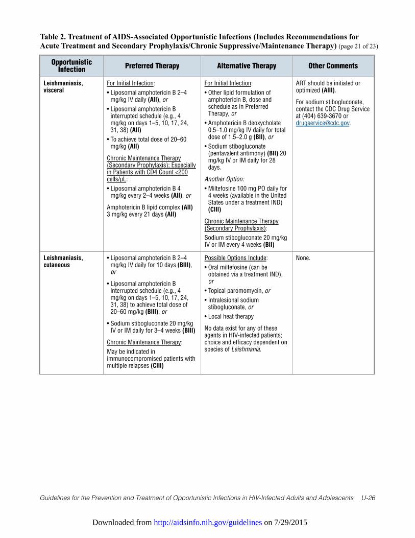
Downloaded from http://aidsinfo.nih.gov/guidelines on 7/29/2015
Guidelines for the Prevention and Treatment of Opportunistic Infections in HIV-Infected Adults and Adolescents U-26
Table 2. Treatment of AIDS-Associated Opportunistic Infections (Includes Recommendations forAcute Treatment and Secondary Prophylaxis/Chronic Suppressive/Maintenance Therapy) (page 21 of 23)
OpportunisticInfection
Preferred Therapy Alternative Therapy Other Comments
Leishmaniasis,visceral
For Initial Infection:
• Liposomal amphotericin B 2–4mg/kg IV daily (AII), or
• Liposomal amphotericin Binterrupted schedule (e.g., 4mg/kg on days 1–5, 10, 17, 24,31, 38) (AII)
• To achieve total dose of 20–60mg/kg (AII)
Chronic Maintenance Therapy(Secondary Prophylaxis); Especiallyin Patients with CD4 Count <200cells/µL:
• Liposomal amphotericin B 4mg/kg every 2–4 weeks (AII), or
Amphotericin B lipid complex (AII)3 mg/kg every 21 days (AII)
For Initial Infection:
• Other lipid formulation ofamphotericin B, dose andschedule as in PreferredTherapy, or
• Amphotericin B deoxycholate0.5–1.0 mg/kg IV daily for totaldose of 1.5–2.0 g (BII), or
• Sodium stibogluconate(pentavalent antimony) (BII) 20mg/kg IV or IM daily for 28days.
Another Option:
• Miltefosine 100 mg PO daily for4 weeks (available in the UnitedStates under a treatment IND)(CIII)
Chronic Maintenance Therapy(Secondary Prophylaxis):
Sodium stibogluconate 20 mg/kgIV or IM every 4 weeks (BII)
ART should be initiated oroptimized (AIII).
For sodium stibogluconate,contact the CDC Drug Serviceat (404) 639-3670 [email protected].
Leishmaniasis,cutaneous
• Liposomal amphotericin B 2–4mg/kg IV daily for 10 days (BIII),or
• Liposomal amphotericin Binterrupted schedule (e.g., 4mg/kg on days 1–5, 10, 17, 24,31, 38) to achieve total dose of20–60 mg/kg (BIII), or
• Sodium stibogluconate 20 mg/kgIV or IM daily for 3–4 weeks (BIII)
Chronic Maintenance Therapy:
May be indicated inimmunocompromised patients withmultiple relapses (CIII)
Possible Options Include:
• Oral miltefosine (can beobtained via a treatment IND),or
• Topical paromomycin, or
• Intralesional sodiumstibogluconate, or
• Local heat therapy
No data exist for any of theseagents in HIV-infected patients;choice and efficacy dependent onspecies of Leishmania.
None.
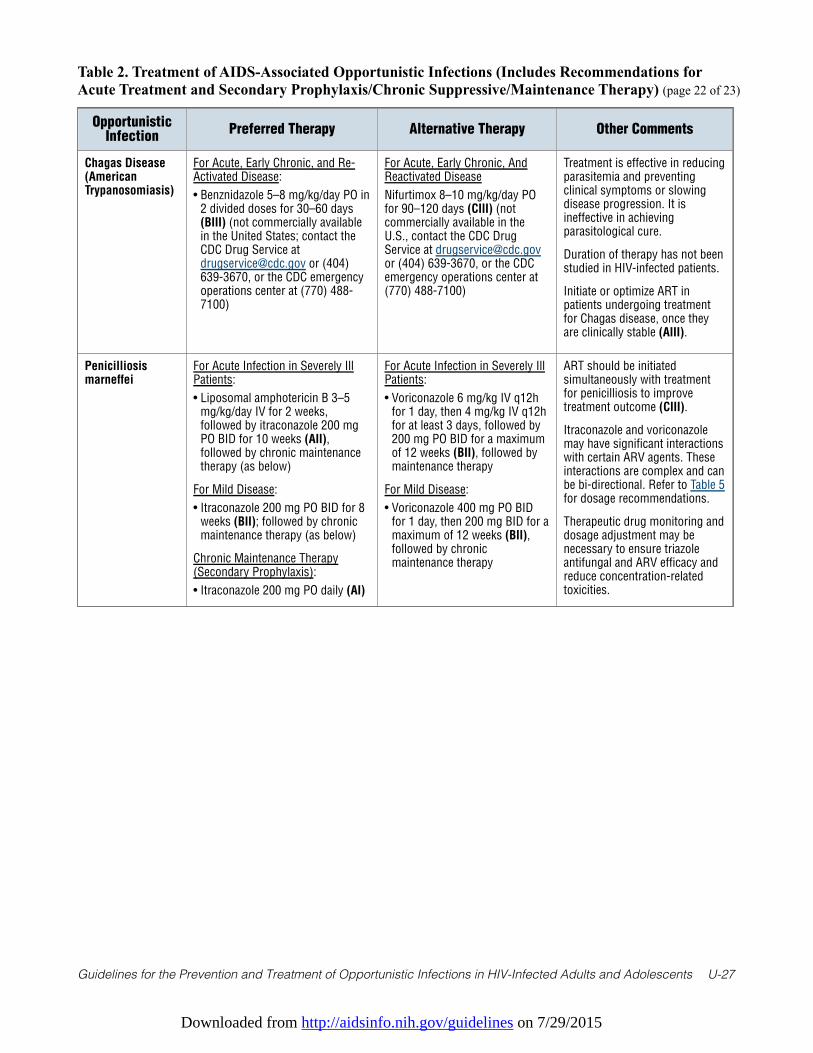
Downloaded from http://aidsinfo.nih.gov/guidelines on 7/29/2015
Guidelines for the Prevention and Treatment of Opportunistic Infections in HIV-Infected Adults and Adolescents U-27
Table 2. Treatment of AIDS-Associated Opportunistic Infections (Includes Recommendations forAcute Treatment and Secondary Prophylaxis/Chronic Suppressive/Maintenance Therapy) (page 22 of 23)
OpportunisticInfection
Preferred Therapy Alternative Therapy Other Comments
Chagas Disease(AmericanTrypanosomiasis)
For Acute, Early Chronic, and Re-Activated Disease:
• Benznidazole 5–8 mg/kg/day PO in2 divided doses for 30–60 days(BIII) (not commercially availablein the United States; contact theCDC Drug Service [email protected] or (404)639-3670, or the CDC emergencyoperations center at (770) 488-7100)
For Acute, Early Chronic, AndReactivated Disease
Nifurtimox 8–10 mg/kg/day POfor 90–120 days (CIII) (notcommercially available in theU.S., contact the CDC DrugService at [email protected] (404) 639-3670, or the CDCemergency operations center at(770) 488-7100)
Treatment is effective in reducingparasitemia and preventingclinical symptoms or slowingdisease progression. It isineffective in achievingparasitological cure.
Duration of therapy has not beenstudied in HIV-infected patients.
Initiate or optimize ART inpatients undergoing treatmentfor Chagas disease, once theyare clinically stable (AIII).
Penicilliosismarneffei
For Acute Infection in Severely IllPatients:
• Liposomal amphotericin B 3–5mg/kg/day IV for 2 weeks,followed by itraconazole 200 mgPO BID for 10 weeks (AII),followed by chronic maintenancetherapy (as below)
For Mild Disease:
• Itraconazole 200 mg PO BID for 8weeks (BII); followed by chronicmaintenance therapy (as below)
Chronic Maintenance Therapy(Secondary Prophylaxis):
• Itraconazole 200 mg PO daily (AI)
For Acute Infection in Severely IllPatients:
• Voriconazole 6 mg/kg IV q12hfor 1 day, then 4 mg/kg IV q12hfor at least 3 days, followed by200 mg PO BID for a maximumof 12 weeks (BII), followed bymaintenance therapy
For Mild Disease:
• Voriconazole 400 mg PO BIDfor 1 day, then 200 mg BID for amaximum of 12 weeks (BII),followed by chronicmaintenance therapy
ART should be initiatedsimultaneously with treatmentfor penicilliosis to improvetreatment outcome (CIII).
Itraconazole and voriconazolemay have significant interactionswith certain ARV agents. Theseinteractions are complex and canbe bi-directional. Refer to Table 5for dosage recommendations.
Therapeutic drug monitoring anddosage adjustment may benecessary to ensure triazoleantifungal and ARV efficacy andreduce concentration-relatedtoxicities.
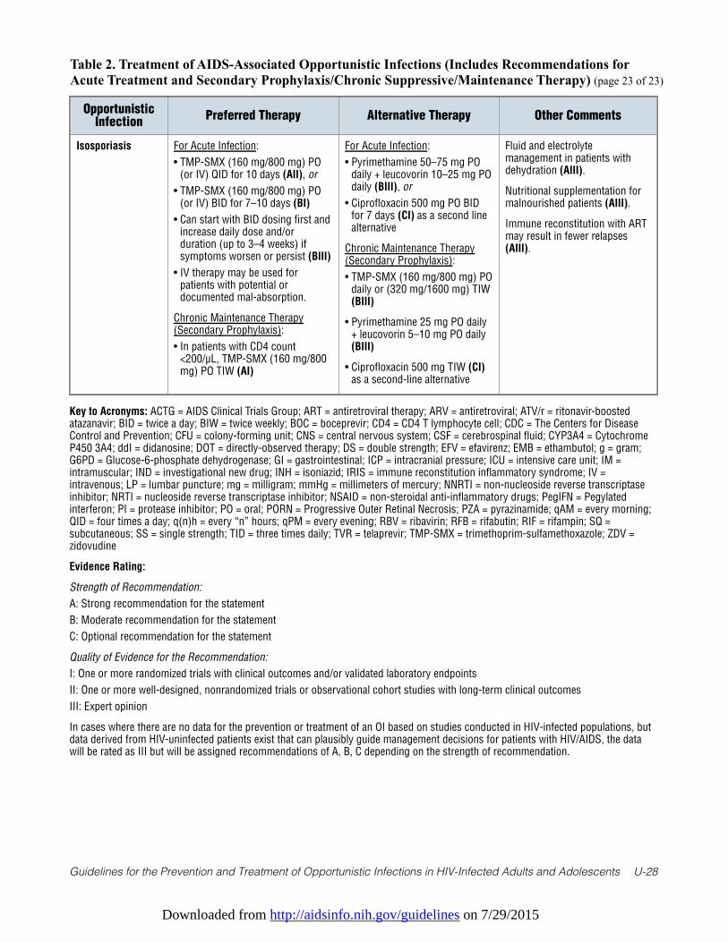
Downloaded from http://aidsinfo.nih.gov/guidelines on 7/29/2015
Guidelines for the Prevention and Treatment of Opportunistic Infections in HIV-Infected Adults and Adolescents U-28
Table 2. Treatment of AIDS-Associated Opportunistic Infections (Includes Recommendations forAcute Treatment and Secondary Prophylaxis/Chronic Suppressive/Maintenance Therapy) (page 23 of 23)
OpportunisticInfection
Preferred Therapy Alternative Therapy Other Comments
Isosporiasis For Acute Infection:
• TMP-SMX (160 mg/800 mg) PO(or IV) QID for 10 days (AII), or
• TMP-SMX (160 mg/800 mg) PO(or IV) BID for 7–10 days (BI)
• Can start with BID dosing first andincrease daily dose and/orduration (up to 3–4 weeks) ifsymptoms worsen or persist (BIII)
• IV therapy may be used forpatients with potential ordocumented mal-absorption.
Chronic Maintenance Therapy(Secondary Prophylaxis):
• In patients with CD4 count<200/µL, TMP-SMX (160 mg/800mg) PO TIW (AI)
For Acute Infection:
• Pyrimethamine 50–75 mg POdaily + leucovorin 10–25 mg POdaily (BIII), or
• Ciprofloxacin 500 mg PO BIDfor 7 days (CI) as a second linealternative
Chronic Maintenance Therapy(Secondary Prophylaxis):
• TMP-SMX (160 mg/800 mg) POdaily or (320 mg/1600 mg) TIW(BIII)
• Pyrimethamine 25 mg PO daily+ leucovorin 5–10 mg PO daily(BIII)
• Ciprofloxacin 500 mg TIW (CI)as a second-line alternative
Fluid and electrolytemanagement in patients withdehydration (AIII).
Nutritional supplementation formalnourished patients (AIII).
Immune reconstitution with ARTmay result in fewer relapses(AIII).
Key to Acronyms: ACTG = AIDS Clinical Trials Group; ART = antiretroviral therapy; ARV = antiretroviral; ATV/r = ritonavir-boostedatazanavir; BID = twice a day; BIW = twice weekly; BOC = boceprevir; CD4 = CD4 T lymphocyte cell; CDC = The Centers for DiseaseControl and Prevention; CFU = colony-forming unit; CNS = central nervous system; CSF = cerebrospinal fluid; CYP3A4 = CytochromeP450 3A4; ddI = didanosine; DOT = directly-observed therapy; DS = double strength; EFV = efavirenz; EMB = ethambutol; g = gram;G6PD = Glucose-6-phosphate dehydrogenase; GI = gastrointestinal; ICP = intracranial pressure; ICU = intensive care unit; IM =intramuscular; IND = investigational new drug; INH = isoniazid; IRIS = immune reconstitution inflammatory syndrome; IV =intravenous; LP = lumbar puncture; mg = milligram; mmHg = millimeters of mercury; NNRTI = non-nucleoside reverse transcriptaseinhibitor; NRTI = nucleoside reverse transcriptase inhibitor; NSAID = non-steroidal anti-inflammatory drugs; PegIFN = Pegylatedinterferon; PI = protease inhibitor; PO = oral; PORN = Progressive Outer Retinal Necrosis; PZA = pyrazinamide; qAM = every morning;QID = four times a day; q(n)h = every “n” hours; qPM = every evening; RBV = ribavirin; RFB = rifabutin; RIF = rifampin; SQ =subcutaneous; SS = single strength; TID = three times daily; TVR = telaprevir; TMP-SMX = trimethoprim-sulfamethoxazole; ZDV =zidovudine
Evidence Rating:
Strength of Recommendation:
A: Strong recommendation for the statement
B: Moderate recommendation for the statement
C: Optional recommendation for the statement
Quality of Evidence for the Recommendation:
I: One or more randomized trials with clinical outcomes and/or validated laboratory endpoints
II: One or more well-designed, nonrandomized trials or observational cohort studies with long-term clinical outcomes
III: Expert opinion
In cases where there are no data for the prevention or treatment of an OI based on studies conducted in HIV-infected populations, butdata derived from HIV-uninfected patients exist that can plausibly guide management decisions for patients with HIV/AIDS, the datawill be rated as III but will be assigned recommendations of A, B, C depending on the strength of recommendation.
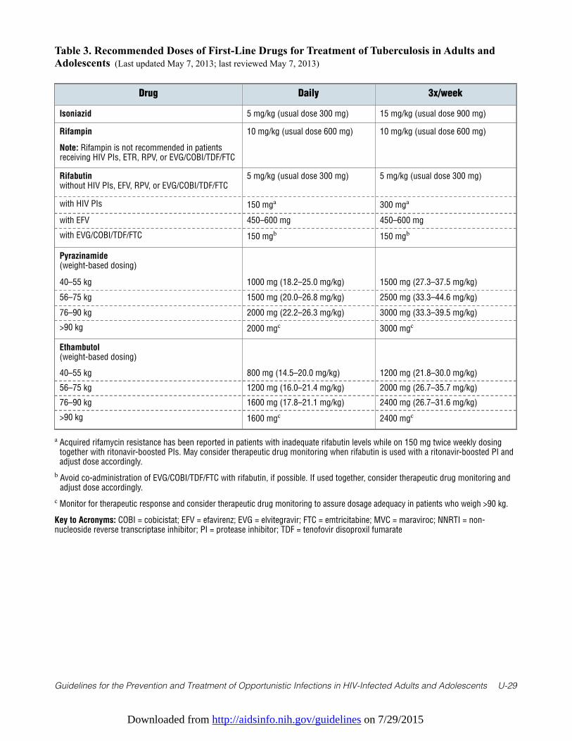
Downloaded from http://aidsinfo.nih.gov/guidelines on 7/29/2015
Guidelines for the Prevention and Treatment of Opportunistic Infections in HIV-Infected Adults and Adolescents U-29
Table 3. Recommended Doses of First-Line Drugs for Treatment of Tuberculosis in Adults andAdolescents (Last updated May 7, 2013; last reviewed May 7, 2013)
Drug Daily 3x/week
Isoniazid 5 mg/kg (usual dose 300 mg) 15 mg/kg (usual dose 900 mg)
Rifampin
Note: Rifampin is not recommended in patientsreceiving HIV PIs, ETR, RPV, or EVG/COBI/TDF/FTC
10 mg/kg (usual dose 600 mg) 10 mg/kg (usual dose 600 mg)
Rifabutinwithout HIV PIs, EFV, RPV, or EVG/COBI/TDF/FTC
5 mg/kg (usual dose 300 mg) 5 mg/kg (usual dose 300 mg)
with HIV PIs 150 mga 300 mga
with EFV 450–600 mg 450–600 mg
with EVG/COBI/TDF/FTC 150 mgb 150 mgb
Pyrazinamide(weight-based dosing)
40–55 kg 1000 mg (18.2–25.0 mg/kg) 1500 mg (27.3–37.5 mg/kg)
56–75 kg 1500 mg (20.0–26.8 mg/kg) 2500 mg (33.3–44.6 mg/kg)
76–90 kg 2000 mg (22.2–26.3 mg/kg) 3000 mg (33.3–39.5 mg/kg)
>90 kg 2000 mgc 3000 mgc
Ethambutol(weight-based dosing)
40–55 kg 800 mg (14.5–20.0 mg/kg) 1200 mg (21.8–30.0 mg/kg)
56–75 kg 1200 mg (16.0–21.4 mg/kg) 2000 mg (26.7–35.7 mg/kg)
76–90 kg 1600 mg (17.8–21.1 mg/kg) 2400 mg (26.7–31.6 mg/kg)
>90 kg 1600 mgc 2400 mgc
a Acquired rifamycin resistance has been reported in patients with inadequate rifabutin levels while on 150 mg twice weekly dosingtogether with ritonavir-boosted PIs. May consider therapeutic drug monitoring when rifabutin is used with a ritonavir-boosted PI andadjust dose accordingly.
b Avoid co-administration of EVG/COBI/TDF/FTC with rifabutin, if possible. If used together, consider therapeutic drug monitoring andadjust dose accordingly.
c Monitor for therapeutic response and consider therapeutic drug monitoring to assure dosage adequacy in patients who weigh >90 kg.
Key to Acronyms: COBI = cobicistat; EFV = efavirenz; EVG = elvitegravir; FTC = emtricitabine; MVC = maraviroc; NNRTI = non-nucleoside reverse transcriptase inhibitor; PI = protease inhibitor; TDF = tenofovir disoproxil fumarate
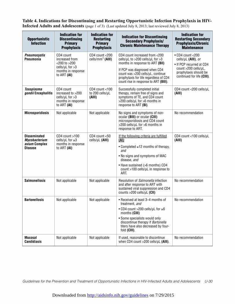
Downloaded from http://aidsinfo.nih.gov/guidelines on 7/29/2015
Guidelines for the Prevention and Treatment of Opportunistic Infections in HIV-Infected Adults and Adolescents U-30
Table 4. Indications for Discontinuing and Restarting Opportunistic Infection Prophylaxis in HIV-Infected Adults and Adolescents (page 1 of 3) (Last updated July 8, 2013; last reviewed July 8, 2013)
OpportunisticInfection
Indication forDiscontinuing
PrimaryProphylaxis
Indication forRestarting
PrimaryProphylaxis
Indication for DiscontinuingSecondary Prophylaxis/
Chronic Maintenance Therapy
Indication forRestarting SecondaryProphylaxis/Chronic
Maintenance
PneumocystisPneumonia
CD4 countincreased from<200 to >200cells/µL for >3months in responseto ART (AI)
CD4 count <200cells/mm3 (AIII)
CD4 count increased from <200cells/µL to >200 cells/µL for >3months in response to ART (BII)
If PCP was diagnosed when CD4count was >200 cells/µL, continueprophylaxis for life regardless of CD4count rise in response to ART (BIII).
• CD4 count <200cells/µL (AIII), or
• If PCP recurred at CD4count >200 cells/µL,prophylaxis should becontinued for life (CIII).
Toxoplasmagondii Encephalitis
CD4 countincreased to >200cells/µL for >3months in responseto ART (AI)
CD4 count <100to 200 cells/µL(AIII)
Successfully completed initialtherapy, remain free of signs andsymptoms of TE, and CD4 count>200 cells/µL for >6 months inresponse to ART (BI).
CD4 count <200 cells/µL(AIII)
Microsporidiosis Not applicable Not applicable No signs and symptoms of non-ocular (BIII) or ocular (CIII)microsporidiosis and CD4 count>200 cells/µL for >6 months inresponse to ART.
No recommendation
DisseminatedMycobacteriumavium ComplexDisease
CD4 count >100cells/µL for ≥3months in responseto ART (AI)
CD4 count <50cells/µL (AIII)
If the following criteria are fulfilled(AI):
• Completed ≥12 months of therapy,and
• No signs and symptoms of MACdisease, and
• Have sustained (>6 months) CD4count >100 cells/µL in response toART.
CD4 count <100 cells/µL(AIII)
Salmonellosis Not applicable Not applicable Resolution of Salmonella infectionand after response to ART withsustained viral suppression and CD4counts >200 cells/µL (CII)
No recommendation
Bartonellosis Not applicable Not applicable • Received at least 3–4 months oftreatment, and
• CD4 count >200 cells/µL for ≥6months (CIII)
• Some specialists would onlydiscontinue therapy if Bartonellatiters have also decreased by four-fold (CIII).
No recommendation
MucosalCandidiasis
Not applicable Not applicable If used, reasonable to discontinuewhen CD4 count >200 cells/µL (AIII).
No recommendation
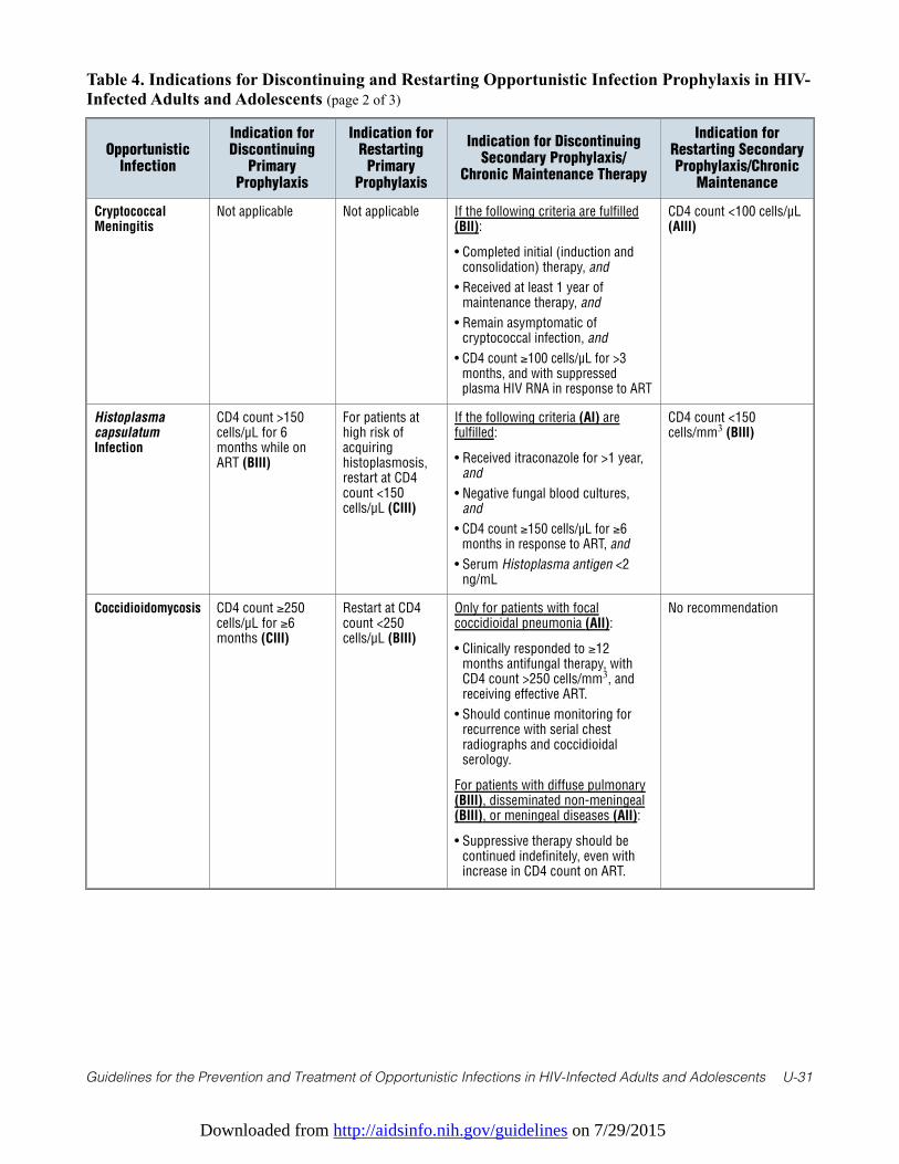
Downloaded from http://aidsinfo.nih.gov/guidelines on 7/29/2015
Guidelines for the Prevention and Treatment of Opportunistic Infections in HIV-Infected Adults and Adolescents U-31
Table 4. Indications for Discontinuing and Restarting Opportunistic Infection Prophylaxis in HIV-Infected Adults and Adolescents (page 2 of 3)
OpportunisticInfection
Indication forDiscontinuing
PrimaryProphylaxis
Indication forRestarting
PrimaryProphylaxis
Indication for DiscontinuingSecondary Prophylaxis/
Chronic Maintenance Therapy
Indication forRestarting SecondaryProphylaxis/Chronic
Maintenance
CryptococcalMeningitis
Not applicable Not applicable If the following criteria are fulfilled(BII):
• Completed initial (induction andconsolidation) therapy, and
• Received at least 1 year ofmaintenance therapy, and
• Remain asymptomatic ofcryptococcal infection, and
• CD4 count ≥100 cells/µL for >3months, and with suppressedplasma HIV RNA in response to ART
CD4 count <100 cells/µL(AIII)
HistoplasmacapsulatumInfection
CD4 count >150cells/µL for 6months while onART (BIII)
For patients athigh risk ofacquiringhistoplasmosis,restart at CD4count <150cells/µL (CIII)
If the following criteria (AI) arefulfilled:
• Received itraconazole for >1 year,and
• Negative fungal blood cultures,and
• CD4 count ≥150 cells/µL for ≥6months in response to ART, and
• Serum Histoplasma antigen <2ng/mL
CD4 count <150cells/mm3 (BIII)
Coccidioidomycosis CD4 count ≥250cells/µL for ≥6months (CIII)
Restart at CD4count <250cells/µL (BIII)
Only for patients with focalcoccidioidal pneumonia (AII):
• Clinically responded to ≥12months antifungal therapy, withCD4 count >250 cells/mm3, andreceiving effective ART.
• Should continue monitoring forrecurrence with serial chestradiographs and coccidioidalserology.
For patients with diffuse pulmonary(BIII), disseminated non-meningeal(BIII), or meningeal diseases (AII):
• Suppressive therapy should becontinued indefinitely, even withincrease in CD4 count on ART.
No recommendation
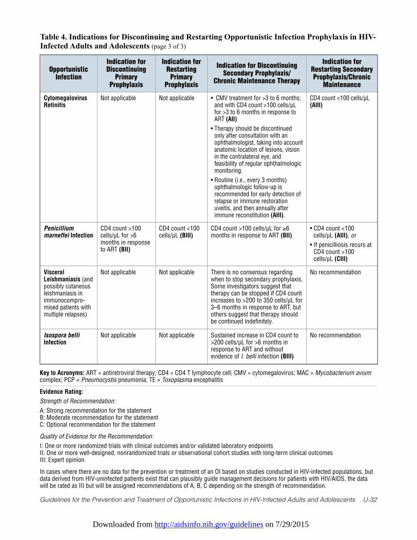
Downloaded from http://aidsinfo.nih.gov/guidelines on 7/29/2015
Guidelines for the Prevention and Treatment of Opportunistic Infections in HIV-Infected Adults and Adolescents U-32
Table 4. Indications for Discontinuing and Restarting Opportunistic Infection Prophylaxis in HIV-Infected Adults and Adolescents (page 3 of 3)
OpportunisticInfection
Indication forDiscontinuing
PrimaryProphylaxis
Indication forRestarting
PrimaryProphylaxis
Indication for DiscontinuingSecondary Prophylaxis/
Chronic Maintenance Therapy
Indication forRestarting SecondaryProphylaxis/Chronic
Maintenance
CytomegalovirusRetinitis
Not applicable Not applicable • CMV treatment for >3 to 6 months;and with CD4 count >100 cells/µLfor >3 to 6 months in response toART (AII)
• Therapy should be discontinuedonly after consultation with anophthalmologist, taking into accountanatomic location of lesions, visionin the contralateral eye, andfeasibility of regular ophthalmologicmonitoring.
• Routine (i.e., every 3 months)ophthalmologic follow-up isrecommended for early detection ofrelapse or immune restorationuveitis, and then annually afterimmune reconstitution (AIII).
CD4 count <100 cells/µL(AIII)
Penicilliummarneffei Infection
CD4 count >100cells/µL for >6months in responseto ART (BII)
CD4 count <100cells/µL (BIII)
CD4 count >100 cells/µL for ≥6months in response to ART (BII)
• CD4 count <100cells/µL (AIII), or
• If penicilliosis recurs atCD4 count >100cells/µL (CIII)
VisceralLeishmaniasis (andpossibly cutaneousleishmaniasis inimmunocompro-mised patients withmultiple relapses)
Not applicable Not applicable There is no consensus regardingwhen to stop secondary prophylaxis.Some investigators suggest thattherapy can be stopped if CD4 countincreases to >200 to 350 cells/µL for3–6 months in response to ART, butothers suggest that therapy shouldbe continued indefinitely.
No recommendation
Isospora belliInfection
Not applicable Not applicable Sustained increase in CD4 count to>200 cells/µL for >6 months inresponse to ART and withoutevidence of I. belli infection (BIII)
No recommendation
Key to Acronyms: ART = antiretroviral therapy; CD4 = CD4 T lymphocyte cell; CMV = cytomegalovirus; MAC = Mycobacterium aviumcomplex; PCP = Pneumocystis pneumonia; TE = Toxoplasma encephalitis
Evidence Rating:
Strength of Recommendation:
A: Strong recommendation for the statementB: Moderate recommendation for the statementC: Optional recommendation for the statement
Quality of Evidence for the Recommendation:
I: One or more randomized trials with clinical outcomes and/or validated laboratory endpointsII: One or more well-designed, nonrandomized trials or observational cohort studies with long-term clinical outcomesIII: Expert opinion
In cases where there are no data for the prevention or treatment of an OI based on studies conducted in HIV-infected populations, butdata derived from HIV-uninfected patients exist that can plausibly guide management decisions for patients with HIV/AIDS, the datawill be rated as III but will be assigned recommendations of A, B, C depending on the strength of recommendation.
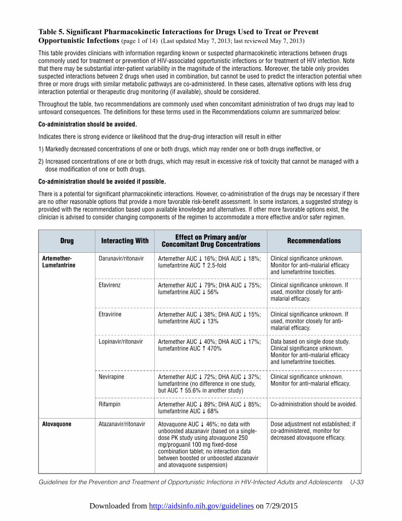
Downloaded from http://aidsinfo.nih.gov/guidelines on 7/29/2015
Guidelines for the Prevention and Treatment of Opportunistic Infections in HIV-Infected Adults and Adolescents U-33
Table 5. Significant Pharmacokinetic Interactions for Drugs Used to Treat or PreventOpportunistic Infections (page 1 of 14) (Last updated May 7, 2013; last reviewed May 7, 2013)
This table provides clinicians with information regarding known or suspected pharmacokinetic interactions between drugs
commonly used for treatment or prevention of HIV-associated opportunistic infections or for treatment of HIV infection. Note
that there may be substantial inter-patient variability in the magnitude of the interactions. Moreover, the table only provides
suspected interactions between 2 drugs when used in combination, but cannot be used to predict the interaction potential when
three or more drugs with similar metabolic pathways are co-administered. In these cases, alternative options with less drug
interaction potential or therapeutic drug monitoring (if available), should be considered.
Throughout the table, two recommendations are commonly used when concomitant administration of two drugs may lead to
untoward consequences. The definitions for these terms used in the Recommendations column are summarized below:
Co-administration should be avoided.
Indicates there is strong evidence or likelihood that the drug-drug interaction will result in either
1) Markedly decreased concentrations of one or both drugs, which may render one or both drugs ineffective, or
2) Increased concentrations of one or both drugs, which may result in excessive risk of toxicity that cannot be managed with a
dose modification of one or both drugs.
Co-administration should be avoided if possible.
There is a potential for significant pharmacokinetic interactions. However, co-administration of the drugs may be necessary if there
are no other reasonable options that provide a more favorable risk-benefit assessment. In some instances, a suggested strategy is
provided with the recommendation based upon available knowledge and alternatives. If other more favorable options exist, the
clinician is advised to consider changing components of the regimen to accommodate a more effective and/or safer regimen.
Drug Interacting WithEffect on Primary and/or
Concomitant Drug ConcentrationsRecommendations
Artemether-Lumefantrine
Darunavir/ritonavir Artemether AUC ↓ 16%; DHA AUC ↓ 18%;lumefantrine AUC ↑ 2.5-fold
Clinical significance unknown.Monitor for anti-malarial efficacyand lumefantrine toxicities.
Efavirenz Artemether AUC ↓ 79%; DHA AUC ↓ 75%;lumefantrine AUC ↓ 56%
Clinical significance unknown. Ifused, monitor closely for anti-malarial efficacy.
Etravirine Artemether AUC ↓ 38%; DHA AUC ↓ 15%;lumefantrine AUC ↓ 13%
Clinical significance unknown. Ifused, monitor closely for anti-malarial efficacy.
Lopinavir/ritonavir Artemether AUC ↓ 40%; DHA AUC ↓ 17%;lumefantrine AUC ↑ 470%
Data based on single dose study.Clinical significance unknown.Monitor for anti-malarial efficacyand lumefantrine toxicities.
Nevirapine Artemether AUC ↓ 72%; DHA AUC ↓ 37%;lumefantrine (no difference in one study,but AUC ↑ 55.6% in another study)
Clinical significance unknown.Monitor for anti-malarial efficacy.
Rifampin Artemether AUC ↓ 89%; DHA AUC ↓ 85%;lumefantrine AUC ↓ 68%
Co-administration should be avoided.
Atovaquone Atazanavir/ritonavir Atovaquone AUC ↓ 46%; no data withunboosted atazanavir (based on a single-dose PK study using atovaquone 250mg/proguanil 100 mg fixed-dosecombination tablet; no interaction databetween boosted or unboosted atazanavirand atovaquone suspension)
Dose adjustment not established; ifco-administered, monitor fordecreased atovaquone efficacy.
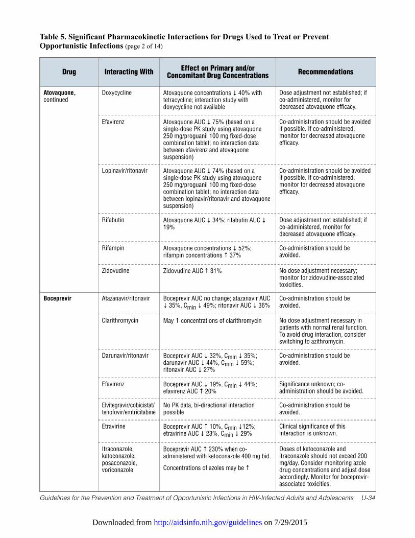
Downloaded from http://aidsinfo.nih.gov/guidelines on 7/29/2015
Guidelines for the Prevention and Treatment of Opportunistic Infections in HIV-Infected Adults and Adolescents U-34
Table 5. Significant Pharmacokinetic Interactions for Drugs Used to Treat or PreventOpportunistic Infections (page 2 of 14)
Drug Interacting WithEffect on Primary and/or
Concomitant Drug ConcentrationsRecommendations
Atovaquone,continued
Doxycycline Atovaquone concentrations ↓ 40% withtetracycline; interaction study withdoxycycline not available
Dose adjustment not established; ifco-administered, monitor fordecreased atovaquone efficacy.
Efavirenz Atovaquone AUC ↓ 75% (based on asingle-dose PK study using atovaquone250 mg/proguanil 100 mg fixed-dosecombination tablet; no interaction databetween efavirenz and atovaquonesuspension)
Co-administration should be avoidedif possible. If co-administered,monitor for decreased atovaquoneefficacy.
Lopinavir/ritonavir Atovaquone AUC ↓ 74% (based on asingle-dose PK study using atovaquone250 mg/proguanil 100 mg fixed-dosecombination tablet; no interaction databetween lopinavir/ritonavir and atovaquonesuspension)
Co-administration should be avoidedif possible. If co-administered,monitor for decreased atovaquoneefficacy.
Rifabutin Atovaquone AUC ↓ 34%; rifabutin AUC ↓19%
Dose adjustment not established; ifco-administered, monitor fordecreased atovaquone efficacy.
Rifampin Atovaquone concentrations ↓ 52%;rifampin concentrations ↑ 37%
Co-administration should beavoided.
Zidovudine Zidovudine AUC ↑ 31% No dose adjustment necessary;monitor for zidovudine-associatedtoxicities.
Boceprevir Atazanavir/ritonavir Boceprevir AUC no change; atazanavir AUC↓ 35%, Cmin ↓ 49%; ritonavir AUC ↓ 36%
Co-administration should beavoided.
Clarithromycin May ↑ concentrations of clarithromycin No dose adjustment necessary inpatients with normal renal function.To avoid drug interaction, considerswitching to azithromycin.
Darunavir/ritonavir Boceprevir AUC ↓ 32%, Cmin ↓ 35%;darunavir AUC ↓ 44%, Cmin ↓ 59%;ritonavir AUC ↓ 27%
Co-administration should beavoided.
Efavirenz Boceprevir AUC ↓ 19%, Cmin ↓ 44%;efavirenz AUC ↑ 20%
Significance unknown; co-administration should be avoided.
Elvitegravir/cobicistat/tenofovir/emtricitabine
No PK data, bi-directional interactionpossible
Co-administration should beavoided.
Etravirine Boceprevir AUC ↑ 10%, Cmin ↓12%;etravirine AUC ↓ 23%, Cmin ↓ 29%
Clinical significance of thisinteraction is unknown.
Itraconazole,ketoconazole,posaconazole,voriconazole
Boceprevir AUC ↑ 230% when co-administered with ketoconazole 400 mg bid.
Concentrations of azoles may be ↑
Doses of ketoconazole anditraconazole should not exceed 200mg/day. Consider monitoring azoledrug concentrations and adjust doseaccordingly. Monitor for boceprevir-associated toxicities.
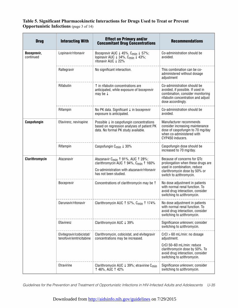
Downloaded from http://aidsinfo.nih.gov/guidelines on 7/29/2015
Guidelines for the Prevention and Treatment of Opportunistic Infections in HIV-Infected Adults and Adolescents U-35
Table 5. Significant Pharmacokinetic Interactions for Drugs Used to Treat or PreventOpportunistic Infections (page 3 of 14)
Drug Interacting WithEffect on Primary and/or
Concomitant Drug ConcentrationsRecommendations
Boceprevir,continued
Lopinavir/ritonavir Boceprevir AUC ↓ 45%, Cmin ↓ 57%;lopinavir AUC ↓ 34%, Cmin ↓ 43%;ritonavir AUC ↓ 22%
Co-administration should beavoided.
Raltegravir No significant interaction. This combination can be co-administered without dosageadjustment
Rifabutin ↑ in rifabutin concentrations areanticipated, while exposure of boceprevirmay be ↓
Co-administration should beavoided, if possible. If used incombination, consider monitoringrifabutin concentration and adjustdose accordingly.
Rifampin No PK data. Significant ↓ in boceprevirexposure is anticipated.
Co-administration should beavoided.
Caspofungin Efavirenz, nevirapine Possible ↓ in caspofungin concentrationsbased on regression analyses of patient PKdata. No formal PK study available.
Manufacturer recommendsconsider increasing maintenancedose of caspofungin to 70 mg/daywhen co-administered withCYP450 inducers.
Rifampin Caspofungin Cmin ↓ 30% Caspofungin dose should beincreased to 70 mg/day.
Clarithromycin Atazanavir Atazanavir Cmin ↑ 91%, AUC ↑ 28%;clarithromycin AUC ↑ 94%, Cmin ↑ 160%
Co-administration with atazanavir/ritonavirhas not been studied.
Because of concerns for QTcprolongation when these drugs areused in combination, reduceclarithromycin dose by 50% orswitch to azithromycin.
Boceprevir Concentrations of clarithromycin may be ↑ No dose adjustment in patientswith normal renal function. Toavoid drug interaction, considerswitching to azithromycin.
Darunavir/ritonavir Clarithromycin AUC ↑ 57%, Cmin ↑ 174% No dose adjustment in patientswith normal renal function. Toavoid drug interaction, considerswitching to azithromycin.
Efavirenz Clarithromycin AUC ↓ 39% Significance unknown; considerswitching to azithromycin.
Elvitegravir/cobicistat/tenofovir/emtricitabine
Clarithromycin, cobicistat, and elvitegravirconcentrations may be increased.
CrCl > 60 mL/min: no dosageadjustment.
CrCl 50–60 mL/min: reduceclarithromycin dose by 50%. Toavoid drug interaction, considerswitching to azithromycin.
Etravirine Clarithromycin AUC ↓ 39%; etravirine Cmin↑ 46%, AUC ↑ 42%
Significance unknown; considerswitching to azithromycin.
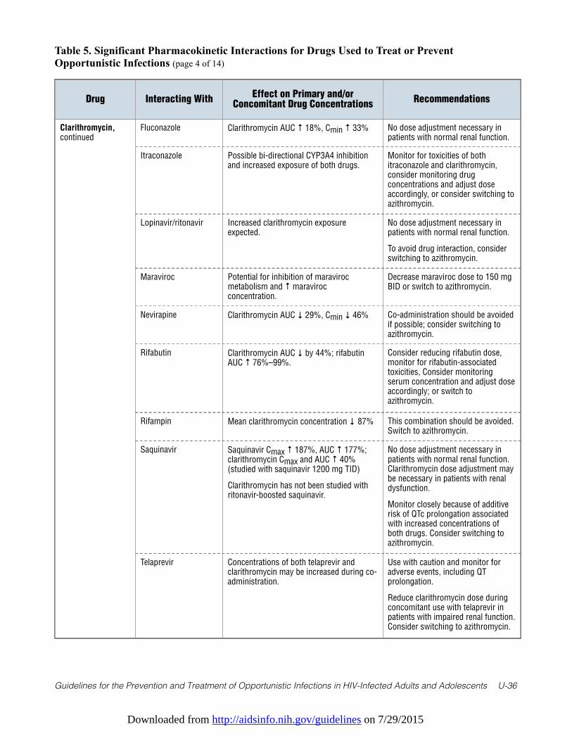
Downloaded from http://aidsinfo.nih.gov/guidelines on 7/29/2015
Guidelines for the Prevention and Treatment of Opportunistic Infections in HIV-Infected Adults and Adolescents U-36
Table 5. Significant Pharmacokinetic Interactions for Drugs Used to Treat or PreventOpportunistic Infections (page 4 of 14)
Drug Interacting WithEffect on Primary and/or
Concomitant Drug ConcentrationsRecommendations
Clarithromycin,continued
Fluconazole Clarithromycin AUC ↑ 18%, Cmin ↑ 33% No dose adjustment necessary inpatients with normal renal function.
Itraconazole Possible bi-directional CYP3A4 inhibitionand increased exposure of both drugs.
Monitor for toxicities of bothitraconazole and clarithromycin,consider monitoring drugconcentrations and adjust doseaccordingly, or consider switching toazithromycin.
Lopinavir/ritonavir Increased clarithromycin exposureexpected.
No dose adjustment necessary inpatients with normal renal function.
To avoid drug interaction, considerswitching to azithromycin.
Maraviroc Potential for inhibition of maravirocmetabolism and ↑ maravirocconcentration.
Decrease maraviroc dose to 150 mgBID or switch to azithromycin.
Nevirapine Clarithromycin AUC ↓ 29%, Cmin ↓ 46% Co-administration should be avoidedif possible; consider switching toazithromycin.
Rifabutin Clarithromycin AUC ↓ by 44%; rifabutinAUC ↑ 76%–99%.
Consider reducing rifabutin dose,monitor for rifabutin-associatedtoxicities, Consider monitoringserum concentration and adjust doseaccordingly; or switch toazithromycin.
Rifampin Mean clarithromycin concentration ↓ 87% This combination should be avoided.Switch to azithromycin.
Saquinavir Saquinavir Cmax ↑ 187%, AUC ↑ 177%;clarithromycin Cmax and AUC ↑ 40%(studied with saquinavir 1200 mg TID)
Clarithromycin has not been studied withritonavir-boosted saquinavir.
No dose adjustment necessary inpatients with normal renal function.Clarithromycin dose adjustment maybe necessary in patients with renaldysfunction.
Monitor closely because of additiverisk of QTc prolongation associatedwith increased concentrations ofboth drugs. Consider switching toazithromycin.
Telaprevir Concentrations of both telaprevir andclarithromycin may be increased during co-administration.
Use with caution and monitor foradverse events, including QTprolongation.
Reduce clarithromycin dose duringconcomitant use with telaprevir inpatients with impaired renal function.Consider switching to azithromycin.
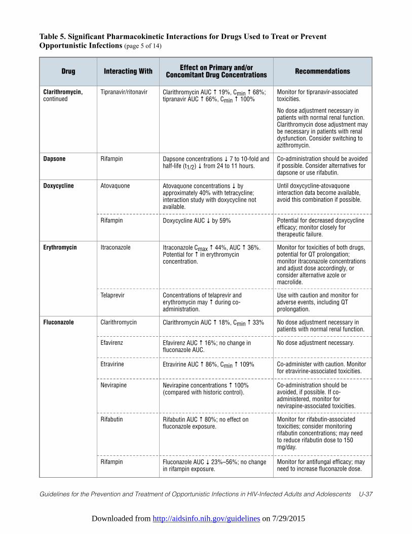
Downloaded from http://aidsinfo.nih.gov/guidelines on 7/29/2015
Guidelines for the Prevention and Treatment of Opportunistic Infections in HIV-Infected Adults and Adolescents U-37
Table 5. Significant Pharmacokinetic Interactions for Drugs Used to Treat or PreventOpportunistic Infections (page 5 of 14)
Drug Interacting WithEffect on Primary and/or
Concomitant Drug ConcentrationsRecommendations
Clarithromycin,continued
Tipranavir/ritonavir Clarithromycin AUC ↑ 19%, Cmin ↑ 68%;tipranavir AUC ↑ 66%, Cmin ↑ 100%
Monitor for tipranavir-associatedtoxicities.
No dose adjustment necessary inpatients with normal renal function.Clarithromycin dose adjustment maybe necessary in patients with renaldysfunction. Consider switching toazithromycin.
Dapsone Rifampin Dapsone concentrations ↓ 7 to 10-fold andhalf-life (t1/2) ↓ from 24 to 11 hours.
Co-administration should be avoidedif possible. Consider alternatives fordapsone or use rifabutin.
Doxycycline Atovaquone Atovaquone concentrations ↓ byapproximately 40% with tetracycline;interaction study with doxycycline notavailable.
Until doxycycline-atovaquoneinteraction data become available,avoid this combination if possible.
Rifampin Doxycycline AUC ↓ by 59% Potential for decreased doxycyclineefficacy; monitor closely fortherapeutic failure.
Erythromycin Itraconazole Itraconazole Cmax ↑ 44%, AUC ↑ 36%.Potential for ↑ in erythromycinconcentration.
Monitor for toxicities of both drugs,potential for QT prolongation;monitor itraconazole concentrationsand adjust dose accordingly, orconsider alternative azole ormacrolide.
Telaprevir Concentrations of telaprevir anderythromycin may ↑ during co-administration.
Use with caution and monitor foradverse events, including QTprolongation.
Fluconazole Clarithromycin Clarithromycin AUC ↑ 18%, Cmin ↑ 33% No dose adjustment necessary inpatients with normal renal function.
Efavirenz Efavirenz AUC ↑ 16%; no change influconazole AUC.
No dose adjustment necessary.
Etravirine Etravirine AUC ↑ 86%, Cmin ↑ 109% Co-administer with caution. Monitorfor etravirine-associated toxicities.
Nevirapine Nevirapine concentrations ↑ 100%(compared with historic control).
Co-administration should beavoided, if possible. If co-administered, monitor fornevirapine-associated toxicities.
Rifabutin Rifabutin AUC ↑ 80%; no effect onfluconazole exposure.
Monitor for rifabutin-associatedtoxicities; consider monitoringrifabutin concentrations; may needto reduce rifabutin dose to 150mg/day.
Rifampin Fluconazole AUC ↓ 23%–56%; no changein rifampin exposure.
Monitor for antifungal efficacy; mayneed to increase fluconazole dose.
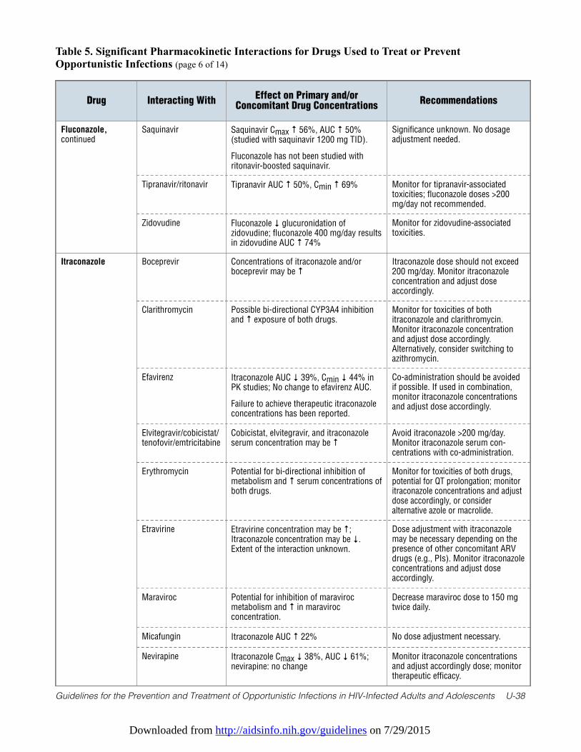
Downloaded from http://aidsinfo.nih.gov/guidelines on 7/29/2015
Guidelines for the Prevention and Treatment of Opportunistic Infections in HIV-Infected Adults and Adolescents U-38
Table 5. Significant Pharmacokinetic Interactions for Drugs Used to Treat or PreventOpportunistic Infections (page 6 of 14)
Drug Interacting WithEffect on Primary and/or
Concomitant Drug ConcentrationsRecommendations
Fluconazole,continued
Saquinavir Saquinavir Cmax ↑ 56%, AUC ↑ 50%(studied with saquinavir 1200 mg TID).
Fluconazole has not been studied withritonavir-boosted saquinavir.
Significance unknown. No dosageadjustment needed.
Tipranavir/ritonavir Tipranavir AUC ↑ 50%, Cmin ↑ 69% Monitor for tipranavir-associatedtoxicities; fluconazole doses >200mg/day not recommended.
Zidovudine Fluconazole ↓ glucuronidation ofzidovudine; fluconazole 400 mg/day resultsin zidovudine AUC ↑ 74%
Monitor for zidovudine-associatedtoxicities.
Itraconazole Boceprevir Concentrations of itraconazole and/orboceprevir may be ↑
Itraconazole dose should not exceed200 mg/day. Monitor itraconazoleconcentration and adjust doseaccordingly.
Clarithromycin Possible bi-directional CYP3A4 inhibitionand ↑ exposure of both drugs.
Monitor for toxicities of bothitraconazole and clarithromycin.Monitor itraconazole concentrationand adjust dose accordingly.Alternatively, consider switching toazithromycin.
Efavirenz Itraconazole AUC ↓ 39%, Cmin ↓ 44% inPK studies; No change to efavirenz AUC.
Failure to achieve therapeutic itraconazoleconcentrations has been reported.
Co-administration should be avoidedif possible. If used in combination,monitor itraconazole concentrationsand adjust dose accordingly.
Elvitegravir/cobicistat/tenofovir/emtricitabine
Cobicistat, elvitegravir, and itraconazoleserum concentration may be ↑
Avoid itraconazole >200 mg/day.Monitor itraconazole serum con-centrations with co-administration.
Erythromycin Potential for bi-directional inhibition ofmetabolism and ↑ serum concentrations ofboth drugs.
Monitor for toxicities of both drugs,potential for QT prolongation; monitoritraconazole concentrations and adjustdose accordingly, or consideralternative azole or macrolide.
Etravirine Etravirine concentration may be ↑;Itraconazole concentration may be ↓.Extent of the interaction unknown.
Dose adjustment with itraconazolemay be necessary depending on thepresence of other concomitant ARVdrugs (e.g., PIs). Monitor itraconazoleconcentrations and adjust doseaccordingly.
Maraviroc Potential for inhibition of maravirocmetabolism and ↑ in maravirocconcentration.
Decrease maraviroc dose to 150 mgtwice daily.
Micafungin Itraconazole AUC ↑ 22% No dose adjustment necessary.
Nevirapine Itraconazole Cmax ↓ 38%, AUC ↓ 61%;nevirapine: no change
Monitor itraconazole concentrationsand adjust accordingly dose; monitortherapeutic efficacy.
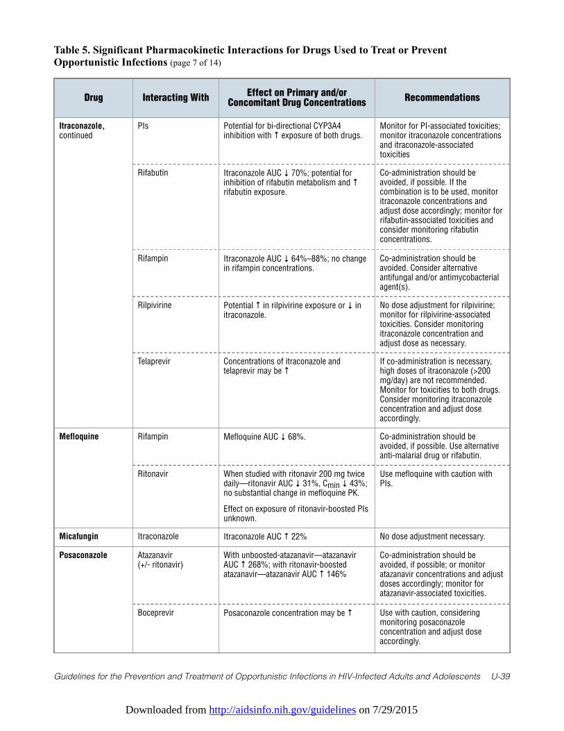
Downloaded from http://aidsinfo.nih.gov/guidelines on 7/29/2015
Guidelines for the Prevention and Treatment of Opportunistic Infections in HIV-Infected Adults and Adolescents U-39
Table 5. Significant Pharmacokinetic Interactions for Drugs Used to Treat or PreventOpportunistic Infections (page 7 of 14)
Drug Interacting WithEffect on Primary and/or
Concomitant Drug ConcentrationsRecommendations
Itraconazole,continued
PIs Potential for bi-directional CYP3A4inhibition with ↑ exposure of both drugs.
Monitor for PI-associated toxicities;monitor itraconazole concentrationsand itraconazole-associatedtoxicities
Rifabutin Itraconazole AUC ↓ 70%; potential forinhibition of rifabutin metabolism and ↑rifabutin exposure.
Co-administration should beavoided, if possible. If thecombination is to be used, monitoritraconazole concentrations andadjust dose accordingly; monitor forrifabutin-associated toxicities andconsider monitoring rifabutinconcentrations.
Rifampin Itraconazole AUC ↓ 64%–88%; no changein rifampin concentrations.
Co-administration should beavoided. Consider alternativeantifungal and/or antimycobacterialagent(s).
Rilpivirine Potential ↑ in rilpivirine exposure or ↓ initraconazole.
No dose adjustment for rilpivirine;monitor for rilpivirine-associatedtoxicities. Consider monitoringitraconazole concentration andadjust dose as necessary.
Telaprevir Concentrations of itraconazole andtelaprevir may be ↑
If co-administration is necessary,high doses of itraconazole (>200mg/day) are not recommended.Monitor for toxicities to both drugs.Consider monitoring itraconazoleconcentration and adjust doseaccordingly.
Mefloquine Rifampin Mefloquine AUC ↓ 68%. Co-administration should beavoided, if possible. Use alternativeanti-malarial drug or rifabutin.
Ritonavir When studied with ritonavir 200 mg twicedaily—ritonavir AUC ↓ 31%, Cmin ↓ 43%;no substantial change in mefloquine PK.
Effect on exposure of ritonavir-boosted PIsunknown.
Use mefloquine with caution withPIs.
Micafungin Itraconazole Itraconazole AUC ↑ 22% No dose adjustment necessary.
Posaconazole Atazanavir (+/- ritonavir)
With unboosted-atazanavir—atazanavirAUC ↑ 268%; with ritonavir-boostedatazanavir—atazanavir AUC ↑ 146%
Co-administration should beavoided, if possible; or monitoratazanavir concentrations and adjustdoses accordingly; monitor foratazanavir-associated toxicities.
Boceprevir Posaconazole concentration may be ↑ Use with caution, consideringmonitoring posaconazoleconcentration and adjust doseaccordingly.
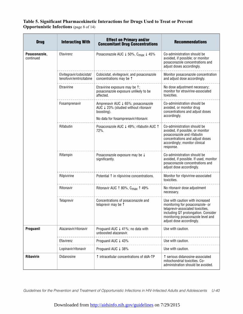
Downloaded from http://aidsinfo.nih.gov/guidelines on 7/29/2015
Guidelines for the Prevention and Treatment of Opportunistic Infections in HIV-Infected Adults and Adolescents U-40
Table 5. Significant Pharmacokinetic Interactions for Drugs Used to Treat or PreventOpportunistic Infections (page 8 of 14)
Drug Interacting WithEffect on Primary and/or
Concomitant Drug ConcentrationsRecommendations
Posaconazole,continued
Efavirenz Posaconazole AUC ↓ 50%, Cmax ↓ 45% Co-administration should beavoided, if possible; or monitorposaconazole concentrations andadjust doses accordingly.
Elvitegravir/cobicistat/tenofovir/emtricitabine
Cobicistat, elvitegravir, and posaconazoleconcentrations may be ↑
Monitor posaconazole concentrationand adjust dose accordingly.
Etravirine Etravirine exposure may be ↑;posaconazole exposure unlikely to beaffected.
No dose adjustment necessary;monitor for etravirine-associatedtoxicities.
Fosamprenavir Amprenavir AUC ↓ 65%; posaconazoleAUC ↓ 23% (studied without ritonavirboosting).
No data for fosamprenavir/ritonavir.
Co-administration should beavoided, or monitor drugconcentrations and adjust dosesaccordingly.
Rifabutin Posaconazole AUC ↓ 49%; rifabutin AUC ↑72%.
Co-administration should beavoided, if possible, or monitorposaconazole and rifabutinconcentrations and adjust dosesaccordingly; monitor clinicalresponse.
Rifampin Posaconazole exposure may be ↓significantly.
Co-administration should beavoided, if possible. If used, monitorposaconazole concentrations andadjust dose accordingly.
Rilpivirine Potential ↑ in rilpivirine concentrations. Monitor for rilpivirine-associatedtoxicities.
Ritonavir Ritonavir AUC ↑ 80%, Cmax ↑ 49% No ritonavir dose adjustmentnecessary.
Telaprevir Concentrations of posaconazole andtelaprevir may be ↑
Use with caution with increasedmonitoring for posaconazole- ortelaprevir-associated toxicities,including QT prolongation. Considermonitoring posaconazole level andadjust dose accordingly.
Proguanil Atazanavir/ritonavir Proguanil AUC ↓ 41%; no data withunboosted atazanavir.
Use with caution.
Efavirenz Proguanil AUC ↓ 43% Use with caution.
Lopinavir/ritonavir Proguanil AUC ↓ 38% Use with caution.
Ribavirin Didanosine ↑ intracellular concentrations of ddA-TP ↑ serious didanosine-associatedmitochondrial toxicities. Co-administration should be avoided.
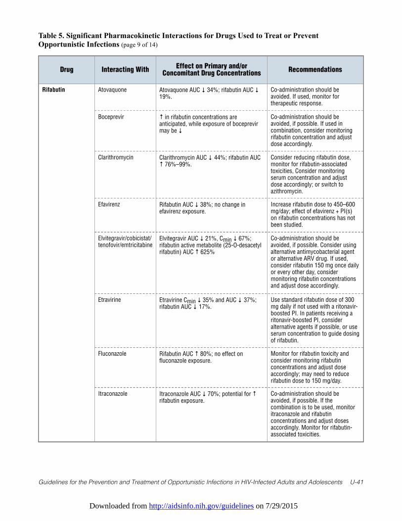
Downloaded from http://aidsinfo.nih.gov/guidelines on 7/29/2015
Guidelines for the Prevention and Treatment of Opportunistic Infections in HIV-Infected Adults and Adolescents U-41
Table 5. Significant Pharmacokinetic Interactions for Drugs Used to Treat or PreventOpportunistic Infections (page 9 of 14)
Drug Interacting WithEffect on Primary and/or
Concomitant Drug ConcentrationsRecommendations
Rifabutin Atovaquone Atovaquone AUC ↓ 34%; rifabutin AUC ↓19%.
Co-administration should beavoided. If used, monitor fortherapeutic response.
Boceprevir ↑ in rifabutin concentrations areanticipated, while exposure of boceprevirmay be ↓
Co-administration should beavoided, if possible. If used incombination, consider monitoringrifabutin concentration and adjustdose accordingly.
Clarithromycin Clarithromycin AUC ↓ 44%; rifabutin AUC↑ 76%–99%.
Consider reducing rifabutin dose,monitor for rifabutin-associatedtoxicities, Consider monitoringserum concentration and adjustdose accordingly; or switch toazithromycin.
Efavirenz Rifabutin AUC ↓ 38%; no change inefavirenz exposure.
Increase rifabutin dose to 450–600mg/day; effect of efavirenz + PI(s)on rifabutin concentrations has notbeen studied.
Elvitegravir/cobicistat/tenofovir/emtricitabine
Elvitegravir AUC ↓ 21%, Cmin ↓ 67%;rifabutin active metabolite (25-O-desacetylrifabutin) AUC ↑ 625%
Co-administration should beavoided, if possible. Consider usingalternative antimycobacterial agentor alternative ARV drug. If used,consider rifabutin 150 mg once dailyor every other day, considermonitoring rifabutin concentrationsand adjust dose accordingly.
Etravirine Etravirine Cmin ↓ 35% and AUC ↓ 37%;rifabutin AUC ↓ 17%.
Use standard rifabutin dose of 300mg daily if not used with a ritonavir-boosted PI. In patients receiving aritonavir-boosted PI, consideralternative agents if possible, or useserum concentration to guide dosingof rifabutin.
Fluconazole Rifabutin AUC ↑ 80%; no effect onfluconazole exposure.
Monitor for rifabutin toxicity andconsider monitoring rifabutinconcentrations and adjust doseaccordingly; may need to reducerifabutin dose to 150 mg/day.
Itraconazole Itraconazole AUC ↓ 70%; potential for ↑rifabutin exposure.
Co-administration should beavoided, if possible. If thecombination is to be used, monitoritraconazole and rifabutinconcentrations and adjust dosesaccordingly. Monitor for rifabutin-associated toxicities.
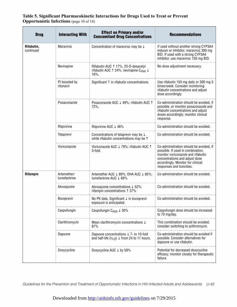
Downloaded from http://aidsinfo.nih.gov/guidelines on 7/29/2015
Guidelines for the Prevention and Treatment of Opportunistic Infections in HIV-Infected Adults and Adolescents U-42
Table 5. Significant Pharmacokinetic Interactions for Drugs Used to Treat or PreventOpportunistic Infections (page 10 of 14)
Drug Interacting WithEffect on Primary and/or
Concomitant Drug ConcentrationsRecommendations
Rifabutin,continued
Maraviroc Concentration of maraviroc may be ↓ If used without another strong CYP3A4inducer or inhibitor, maraviroc 300 mgBID. If used with a strong CYP3A4inhibitor, use maraviroc 150 mg BID.
Nevirapine Rifabutin AUC ↑ 17%, 25-O-desacetylrifabutin AUC ↑ 24%; nevirapine Cmin ↓16%.
No dose adjustment necessary.
PI boosted byritonavir
Significant ↑ in rifabutin concentrations. Use rifabutin 150 mg daily or 300 mg 3times/week. Consider monitoringrifabutin concentrations and adjustdose accordingly.
Posaconazole Posaconazole AUC ↓ 49%; rifabutin AUC ↑72%.
Co-administration should be avoided, ifpossible, or monitor posaconazole andrifabutin concentrations and adjustdoses accordingly; monitor clinicalresponse.
Rilpivirine Rilpivirine AUC ↓ 46% Co-administration should be avoided.
Telaprevir Concentrations of telaprevir may be ↓,while rifabutin concentrations may be ↑
Co-administration should be avoided.
Voriconazole Voriconazole AUC ↓ 79%; rifabutin AUC ↑3-fold.
Co-administration should be avoided, ifpossible. If used in combination,monitor voriconazole and rifabutinconcentrations and adjust doseaccordingly. Monitor for clinicalresponses and toxicities.
Rifampin Artemether/lumefantrine
Artemether AUC ↓ 89%; DHA AUC ↓ 85%;lumefantrine AUC ↓ 68%
Co-administration should be avoided.
Atovaquone Atovaquone concentrations ↓ 52%;rifampin concentrations ↑ 37%
Co-administration should be avoided.
Boceprevir No PK data. Significant ↓ in boceprevirexposure is anticipated.
Co-administration should be avoided.
Caspofungin Caspofungin Cmin ↓ 30% Caspofungin dose should be increasedto 70 mg/day.
Clarithromycin Mean clarithromycin concentrations ↓87%
This combination should be avoided;consider switching to azithromycin.
Dapsone Dapsone concentrations ↓ 7- to 10-foldand half-life (t1/2) ↓ from 24 to 11 hours.
Co-administration should be avoided ifpossible. Consider alternatives fordapsone or use rifabutin.
Doxycycline Doxycycline AUC ↓ by 59% Potential for decreased doxycyclineefficacy; monitor closely for therapeuticfailure.
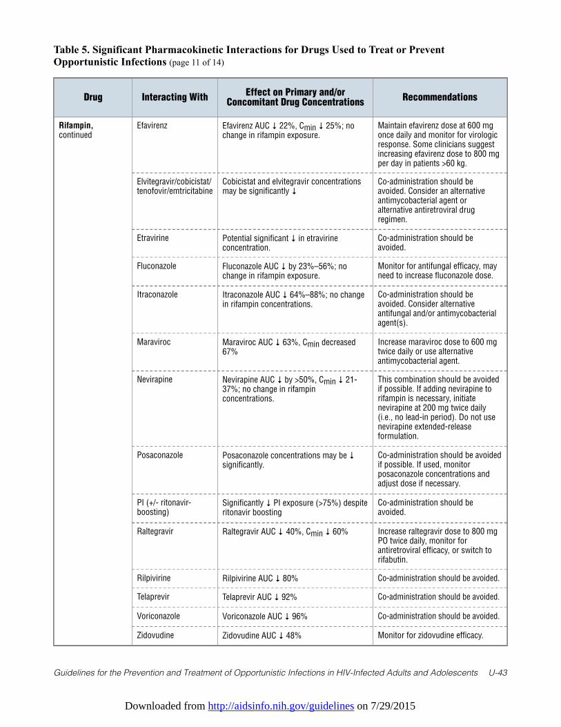
Downloaded from http://aidsinfo.nih.gov/guidelines on 7/29/2015
Guidelines for the Prevention and Treatment of Opportunistic Infections in HIV-Infected Adults and Adolescents U-43
Table 5. Significant Pharmacokinetic Interactions for Drugs Used to Treat or PreventOpportunistic Infections (page 11 of 14)
Drug Interacting WithEffect on Primary and/or
Concomitant Drug ConcentrationsRecommendations
Rifampin,continued
Efavirenz Efavirenz AUC ↓ 22%, Cmin ↓ 25%; nochange in rifampin exposure.
Maintain efavirenz dose at 600 mgonce daily and monitor for virologicresponse. Some clinicians suggestincreasing efavirenz dose to 800 mgper day in patients >60 kg.
Elvitegravir/cobicistat/tenofovir/emtricitabine
Cobicistat and elvitegravir concentrationsmay be significantly ↓
Co-administration should beavoided. Consider an alternativeantimycobacterial agent oralternative antiretroviral drugregimen.
Etravirine Potential significant ↓ in etravirineconcentration.
Co-administration should beavoided.
Fluconazole Fluconazole AUC ↓ by 23%–56%; nochange in rifampin exposure.
Monitor for antifungal efficacy, mayneed to increase fluconazole dose.
Itraconazole Itraconazole AUC ↓ 64%–88%; no changein rifampin concentrations.
Co-administration should beavoided. Consider alternativeantifungal and/or antimycobacterialagent(s).
Maraviroc Maraviroc AUC ↓ 63%, Cmin decreased67%
Increase maraviroc dose to 600 mgtwice daily or use alternativeantimycobacterial agent.
Nevirapine Nevirapine AUC ↓ by >50%, Cmin ↓ 21-37%; no change in rifampinconcentrations.
This combination should be avoidedif possible. If adding nevirapine torifampin is necessary, initiatenevirapine at 200 mg twice daily(i.e., no lead-in period). Do not usenevirapine extended-releaseformulation.
Posaconazole Posaconazole concentrations may be ↓significantly.
Co-administration should be avoidedif possible. If used, monitorposaconazole concentrations andadjust dose if necessary.
PI (+/- ritonavir-boosting)
Significantly ↓ PI exposure (>75%) despiteritonavir boosting
Co-administration should beavoided.
Raltegravir Raltegravir AUC ↓ 40%, Cmin ↓ 60% Increase raltegravir dose to 800 mgPO twice daily, monitor forantiretroviral efficacy, or switch torifabutin.
Rilpivirine Rilpivirine AUC ↓ 80% Co-administration should be avoided.
Telaprevir Telaprevir AUC ↓ 92% Co-administration should be avoided.
Voriconazole Voriconazole AUC ↓ 96% Co-administration should be avoided.
Zidovudine Zidovudine AUC ↓ 48% Monitor for zidovudine efficacy.
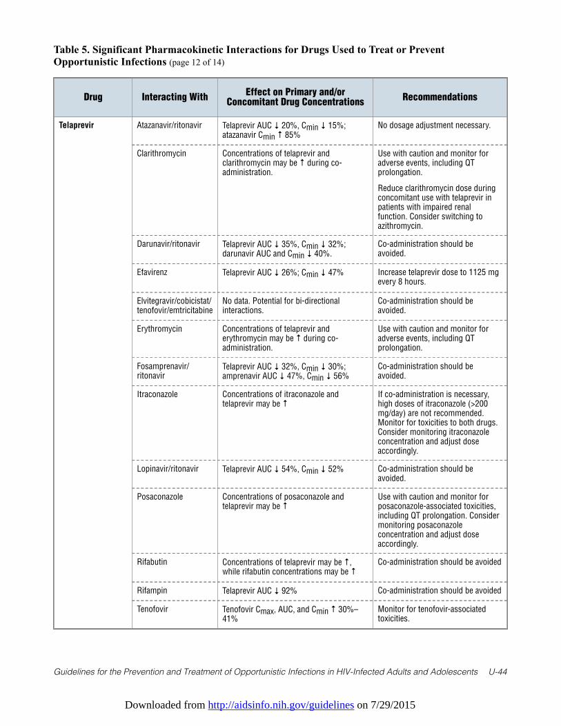
Downloaded from http://aidsinfo.nih.gov/guidelines on 7/29/2015
Guidelines for the Prevention and Treatment of Opportunistic Infections in HIV-Infected Adults and Adolescents U-44
Table 5. Significant Pharmacokinetic Interactions for Drugs Used to Treat or PreventOpportunistic Infections (page 12 of 14)
Drug Interacting WithEffect on Primary and/or
Concomitant Drug ConcentrationsRecommendations
Telaprevir Atazanavir/ritonavir Telaprevir AUC ↓ 20%, Cmin ↓ 15%;atazanavir Cmin ↑ 85%
No dosage adjustment necessary.
Clarithromycin Concentrations of telaprevir andclarithromycin may be ↑ during co-administration.
Use with caution and monitor foradverse events, including QTprolongation.
Reduce clarithromycin dose duringconcomitant use with telaprevir inpatients with impaired renalfunction. Consider switching toazithromycin.
Darunavir/ritonavir Telaprevir AUC ↓ 35%, Cmin ↓ 32%;darunavir AUC and Cmin ↓ 40%.
Co-administration should beavoided.
Efavirenz Telaprevir AUC ↓ 26%; Cmin ↓ 47% Increase telaprevir dose to 1125 mgevery 8 hours.
Elvitegravir/cobicistat/tenofovir/emtricitabine
No data. Potential for bi-directionalinteractions.
Co-administration should beavoided.
Erythromycin Concentrations of telaprevir anderythromycin may be ↑ during co-administration.
Use with caution and monitor foradverse events, including QTprolongation.
Fosamprenavir/ritonavir
Telaprevir AUC ↓ 32%, Cmin ↓ 30%;amprenavir AUC ↓ 47%, Cmin ↓ 56%
Co-administration should beavoided.
Itraconazole Concentrations of itraconazole andtelaprevir may be ↑
If co-administration is necessary,high doses of itraconazole (>200mg/day) are not recommended.Monitor for toxicities to both drugs.Consider monitoring itraconazoleconcentration and adjust doseaccordingly.
Lopinavir/ritonavir Telaprevir AUC ↓ 54%, Cmin ↓ 52% Co-administration should beavoided.
Posaconazole Concentrations of posaconazole andtelaprevir may be ↑
Use with caution and monitor forposaconazole-associated toxicities,including QT prolongation. Considermonitoring posaconazoleconcentration and adjust doseaccordingly.
Rifabutin Concentrations of telaprevir may be ↑,while rifabutin concentrations may be ↑
Co-administration should be avoided
Rifampin Telaprevir AUC ↓ 92% Co-administration should be avoided
Tenofovir Tenofovir Cmax, AUC, and Cmin ↑ 30%–41%
Monitor for tenofovir-associatedtoxicities.
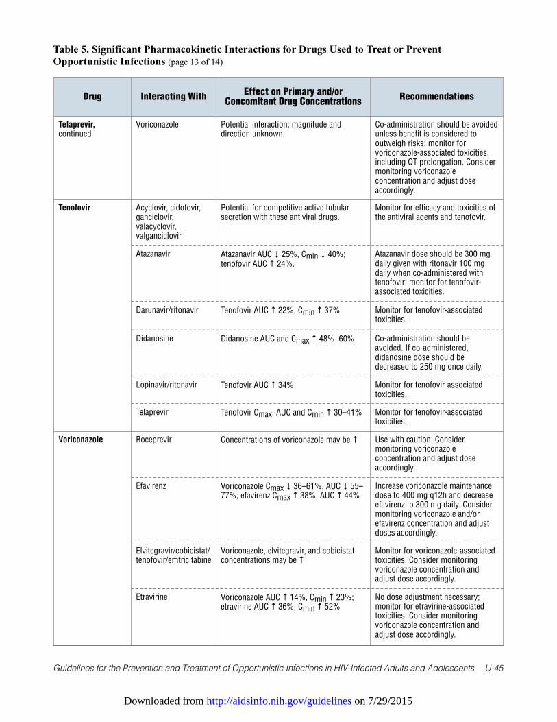
Downloaded from http://aidsinfo.nih.gov/guidelines on 7/29/2015
Guidelines for the Prevention and Treatment of Opportunistic Infections in HIV-Infected Adults and Adolescents U-45
Table 5. Significant Pharmacokinetic Interactions for Drugs Used to Treat or PreventOpportunistic Infections (page 13 of 14)
Drug Interacting WithEffect on Primary and/or
Concomitant Drug ConcentrationsRecommendations
Telaprevir,continued
Voriconazole Potential interaction; magnitude anddirection unknown.
Co-administration should be avoidedunless benefit is considered tooutweigh risks; monitor forvoriconazole-associated toxicities,including QT prolongation. Considermonitoring voriconazoleconcentration and adjust doseaccordingly.
Tenofovir Acyclovir, cidofovir,ganciclovir,valacyclovir,valganciclovir
Potential for competitive active tubularsecretion with these antiviral drugs.
Monitor for efficacy and toxicities ofthe antiviral agents and tenofovir.
Atazanavir Atazanavir AUC ↓ 25%, Cmin ↓ 40%;tenofovir AUC ↑ 24%.
Atazanavir dose should be 300 mgdaily given with ritonavir 100 mgdaily when co-administered withtenofovir; monitor for tenofovir-associated toxicities.
Darunavir/ritonavir Tenofovir AUC ↑ 22%, Cmin ↑ 37% Monitor for tenofovir-associatedtoxicities.
Didanosine Didanosine AUC and Cmax ↑ 48%–60% Co-administration should beavoided. If co-administered,didanosine dose should bedecreased to 250 mg once daily.
Lopinavir/ritonavir Tenofovir AUC ↑ 34% Monitor for tenofovir-associatedtoxicities.
Telaprevir Tenofovir Cmax, AUC and Cmin ↑ 30–41% Monitor for tenofovir-associatedtoxicities.
Voriconazole Boceprevir Concentrations of voriconazole may be ↑ Use with caution. Considermonitoring voriconazoleconcentration and adjust doseaccordingly.
Efavirenz Voriconazole Cmax ↓ 36–61%, AUC ↓ 55–77%; efavirenz Cmax ↑ 38%, AUC ↑ 44%
Increase voriconazole maintenancedose to 400 mg q12h and decreaseefavirenz to 300 mg daily. Considermonitoring voriconazole and/orefavirenz concentration and adjustdoses accordingly.
Elvitegravir/cobicistat/tenofovir/emtricitabine
Voriconazole, elvitegravir, and cobicistatconcentrations may be ↑
Monitor for voriconazole-associatedtoxicities. Consider monitoringvoriconazole concentration andadjust dose accordingly.
Etravirine Voriconazole AUC ↑ 14%, Cmin ↑ 23%;etravirine AUC ↑ 36%, Cmin ↑ 52%
No dose adjustment necessary;monitor for etravirine-associatedtoxicities. Consider monitoringvoriconazole concentration andadjust dose accordingly.
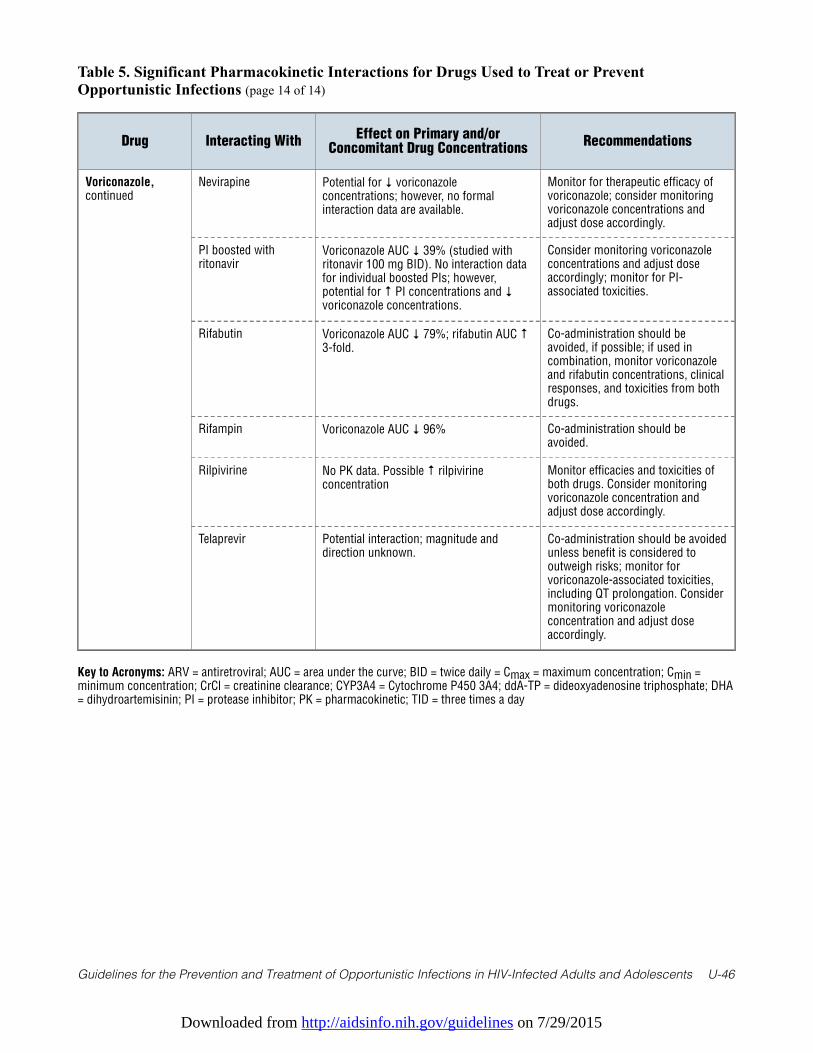
Downloaded from http://aidsinfo.nih.gov/guidelines on 7/29/2015
Guidelines for the Prevention and Treatment of Opportunistic Infections in HIV-Infected Adults and Adolescents U-46
Table 5. Significant Pharmacokinetic Interactions for Drugs Used to Treat or PreventOpportunistic Infections (page 14 of 14)
Drug Interacting WithEffect on Primary and/or
Concomitant Drug ConcentrationsRecommendations
Voriconazole,continued
Nevirapine Potential for ↓ voriconazoleconcentrations; however, no formalinteraction data are available.
Monitor for therapeutic efficacy ofvoriconazole; consider monitoringvoriconazole concentrations andadjust dose accordingly.
PI boosted withritonavir
Voriconazole AUC ↓ 39% (studied withritonavir 100 mg BID). No interaction datafor individual boosted PIs; however,potential for ↑ PI concentrations and ↓voriconazole concentrations.
Consider monitoring voriconazoleconcentrations and adjust doseaccordingly; monitor for PI-associated toxicities.
Rifabutin Voriconazole AUC ↓ 79%; rifabutin AUC ↑3-fold.
Co-administration should beavoided, if possible; if used incombination, monitor voriconazoleand rifabutin concentrations, clinicalresponses, and toxicities from bothdrugs.
Rifampin Voriconazole AUC ↓ 96% Co-administration should beavoided.
Rilpivirine No PK data. Possible ↑ rilpivirineconcentration
Monitor efficacies and toxicities ofboth drugs. Consider monitoringvoriconazole concentration andadjust dose accordingly.
Telaprevir Potential interaction; magnitude anddirection unknown.
Co-administration should be avoidedunless benefit is considered tooutweigh risks; monitor forvoriconazole-associated toxicities,including QT prolongation. Considermonitoring voriconazoleconcentration and adjust doseaccordingly.
Key to Acronyms: ARV = antiretroviral; AUC = area under the curve; BID = twice daily = Cmax = maximum concentration; Cmin =minimum concentration; CrCl = creatinine clearance; CYP3A4 = Cytochrome P450 3A4; ddA-TP = dideoxyadenosine triphosphate; DHA= dihydroartemisinin; PI = protease inhibitor; PK = pharmacokinetic; TID = three times a day
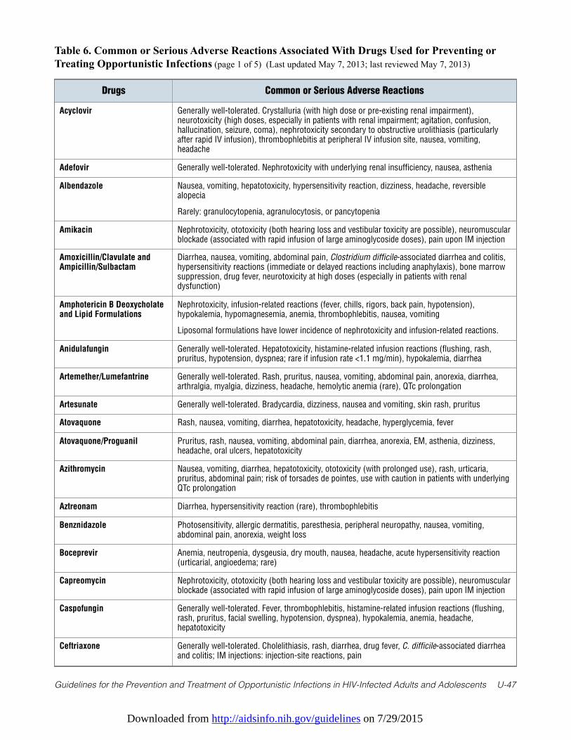
Downloaded from http://aidsinfo.nih.gov/guidelines on 7/29/2015
Guidelines for the Prevention and Treatment of Opportunistic Infections in HIV-Infected Adults and Adolescents U-47
Table 6. Common or Serious Adverse Reactions Associated With Drugs Used for Preventing orTreating Opportunistic Infections (page 1 of 5) (Last updated May 7, 2013; last reviewed May 7, 2013)
Drugs Common or Serious Adverse Reactions
Acyclovir Generally well-tolerated. Crystalluria (with high dose or pre-existing renal impairment),neurotoxicity (high doses, especially in patients with renal impairment; agitation, confusion,hallucination, seizure, coma), nephrotoxicity secondary to obstructive urolithiasis (particularlyafter rapid IV infusion), thrombophlebitis at peripheral IV infusion site, nausea, vomiting,headache
Adefovir Generally well-tolerated. Nephrotoxicity with underlying renal insufficiency, nausea, asthenia
Albendazole Nausea, vomiting, hepatotoxicity, hypersensitivity reaction, dizziness, headache, reversiblealopecia
Rarely: granulocytopenia, agranulocytosis, or pancytopenia
Amikacin Nephrotoxicity, ototoxicity (both hearing loss and vestibular toxicity are possible), neuromuscularblockade (associated with rapid infusion of large aminoglycoside doses), pain upon IM injection
Amoxicillin/Clavulate andAmpicillin/Sulbactam
Diarrhea, nausea, vomiting, abdominal pain, Clostridium difficile-associated diarrhea and colitis,hypersensitivity reactions (immediate or delayed reactions including anaphylaxis), bone marrowsuppression, drug fever, neurotoxicity at high doses (especially in patients with renaldysfunction)
Amphotericin B Deoxycholateand Lipid Formulations
Nephrotoxicity, infusion-related reactions (fever, chills, rigors, back pain, hypotension),hypokalemia, hypomagnesemia, anemia, thrombophlebitis, nausea, vomiting
Liposomal formulations have lower incidence of nephrotoxicity and infusion-related reactions.
Anidulafungin Generally well-tolerated. Hepatotoxicity, histamine-related infusion reactions (flushing, rash,pruritus, hypotension, dyspnea; rare if infusion rate <1.1 mg/min), hypokalemia, diarrhea
Artemether/Lumefantrine Generally well-tolerated. Rash, pruritus, nausea, vomiting, abdominal pain, anorexia, diarrhea,arthralgia, myalgia, dizziness, headache, hemolytic anemia (rare), QTc prolongation
Artesunate Generally well-tolerated. Bradycardia, dizziness, nausea and vomiting, skin rash, pruritus
Atovaquone Rash, nausea, vomiting, diarrhea, hepatotoxicity, headache, hyperglycemia, fever
Atovaquone/Proguanil Pruritus, rash, nausea, vomiting, abdominal pain, diarrhea, anorexia, EM, asthenia, dizziness,headache, oral ulcers, hepatotoxicity
Azithromycin Nausea, vomiting, diarrhea, hepatotoxicity, ototoxicity (with prolonged use), rash, urticaria,pruritus, abdominal pain; risk of torsades de pointes, use with caution in patients with underlyingQTc prolongation
Aztreonam Diarrhea, hypersensitivity reaction (rare), thrombophlebitis
Benznidazole Photosensitivity, allergic dermatitis, paresthesia, peripheral neuropathy, nausea, vomiting,abdominal pain, anorexia, weight loss
Boceprevir Anemia, neutropenia, dysgeusia, dry mouth, nausea, headache, acute hypersensitivity reaction(urticarial, angioedema; rare)
Capreomycin Nephrotoxicity, ototoxicity (both hearing loss and vestibular toxicity are possible), neuromuscularblockade (associated with rapid infusion of large aminoglycoside doses), pain upon IM injection
Caspofungin Generally well-tolerated. Fever, thrombophlebitis, histamine-related infusion reactions (flushing,rash, pruritus, facial swelling, hypotension, dyspnea), hypokalemia, anemia, headache,hepatotoxicity
Ceftriaxone Generally well-tolerated. Cholelithiasis, rash, diarrhea, drug fever, C. difficile-associated diarrheaand colitis; IM injections: injection-site reactions, pain
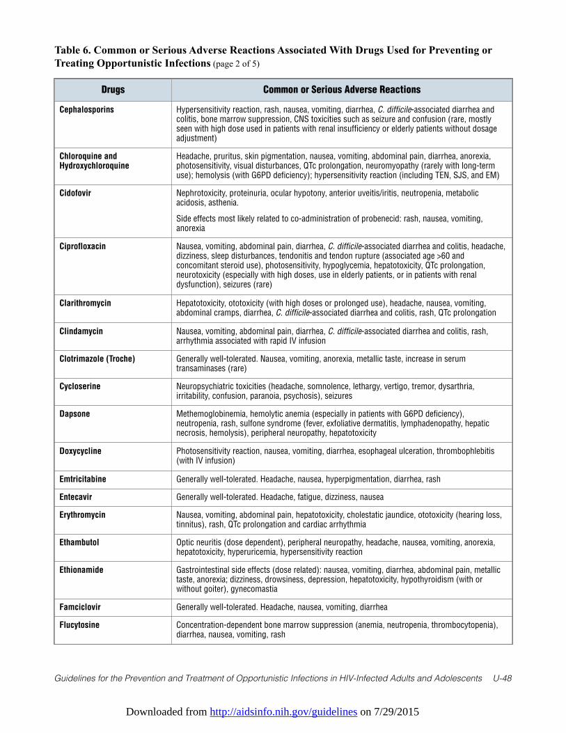
Downloaded from http://aidsinfo.nih.gov/guidelines on 7/29/2015
Guidelines for the Prevention and Treatment of Opportunistic Infections in HIV-Infected Adults and Adolescents U-48
Table 6. Common or Serious Adverse Reactions Associated With Drugs Used for Preventing orTreating Opportunistic Infections (page 2 of 5)
Drugs Common or Serious Adverse Reactions
Cephalosporins Hypersensitivity reaction, rash, nausea, vomiting, diarrhea, C. difficile-associated diarrhea andcolitis, bone marrow suppression, CNS toxicities such as seizure and confusion (rare, mostlyseen with high dose used in patients with renal insufficiency or elderly patients without dosageadjustment)
Chloroquine andHydroxychloroquine
Headache, pruritus, skin pigmentation, nausea, vomiting, abdominal pain, diarrhea, anorexia,photosensitivity, visual disturbances, QTc prolongation, neuromyopathy (rarely with long-termuse); hemolysis (with G6PD deficiency); hypersensitivity reaction (including TEN, SJS, and EM)
Cidofovir Nephrotoxicity, proteinuria, ocular hypotony, anterior uveitis/iritis, neutropenia, metabolicacidosis, asthenia.
Side effects most likely related to co-administration of probenecid: rash, nausea, vomiting,anorexia
Ciprofloxacin Nausea, vomiting, abdominal pain, diarrhea, C. difficile-associated diarrhea and colitis, headache,dizziness, sleep disturbances, tendonitis and tendon rupture (associated age >60 andconcomitant steroid use), photosensitivity, hypoglycemia, hepatotoxicity, QTc prolongation,neurotoxicity (especially with high doses, use in elderly patients, or in patients with renaldysfunction), seizures (rare)
Clarithromycin Hepatotoxicity, ototoxicity (with high doses or prolonged use), headache, nausea, vomiting,abdominal cramps, diarrhea, C. difficile-associated diarrhea and colitis, rash, QTc prolongation
Clindamycin Nausea, vomiting, abdominal pain, diarrhea, C. difficile-associated diarrhea and colitis, rash,arrhythmia associated with rapid IV infusion
Clotrimazole (Troche) Generally well-tolerated. Nausea, vomiting, anorexia, metallic taste, increase in serumtransaminases (rare)
Cycloserine Neuropsychiatric toxicities (headache, somnolence, lethargy, vertigo, tremor, dysarthria,irritability, confusion, paranoia, psychosis), seizures
Dapsone Methemoglobinemia, hemolytic anemia (especially in patients with G6PD deficiency),neutropenia, rash, sulfone syndrome (fever, exfoliative dermatitis, lymphadenopathy, hepaticnecrosis, hemolysis), peripheral neuropathy, hepatotoxicity
Doxycycline Photosensitivity reaction, nausea, vomiting, diarrhea, esophageal ulceration, thrombophlebitis(with IV infusion)
Emtricitabine Generally well-tolerated. Headache, nausea, hyperpigmentation, diarrhea, rash
Entecavir Generally well-tolerated. Headache, fatigue, dizziness, nausea
Erythromycin Nausea, vomiting, abdominal pain, hepatotoxicity, cholestatic jaundice, ototoxicity (hearing loss,tinnitus), rash, QTc prolongation and cardiac arrhythmia
Ethambutol Optic neuritis (dose dependent), peripheral neuropathy, headache, nausea, vomiting, anorexia,hepatotoxicity, hyperuricemia, hypersensitivity reaction
Ethionamide Gastrointestinal side effects (dose related): nausea, vomiting, diarrhea, abdominal pain, metallictaste, anorexia; dizziness, drowsiness, depression, hepatotoxicity, hypothyroidism (with orwithout goiter), gynecomastia
Famciclovir Generally well-tolerated. Headache, nausea, vomiting, diarrhea
Flucytosine Concentration-dependent bone marrow suppression (anemia, neutropenia, thrombocytopenia),diarrhea, nausea, vomiting, rash

Downloaded from http://aidsinfo.nih.gov/guidelines on 7/29/2015
Guidelines for the Prevention and Treatment of Opportunistic Infections in HIV-Infected Adults and Adolescents U-49
Table 6. Common or Serious Adverse Reactions Associated With Drugs Used for Preventing orTreating Opportunistic Infections (page 3 of 5)
Drugs Common or Serious Adverse Reactions
Fluconazole Hepatotoxicity, rash, nausea, vomiting, diarrhea, abdominal discomfort, reversible alopecia (withdoses ≥400 mg/d for >2 months)
Foscarnet Nephrotoxicity, electrolyte imbalances (hypocalcemia, hypomagnesemia, hypophosphatemia,hyperphosphatemia, hypokalemia), penile ulceration, nausea, vomiting, anorexia, headache,seizure (associated with electrolyte imbalances), anemia, injection-site associatedthrombophlebitis
Fumagillin (Investigational) Oral therapy: Neutropenia, thrombocytopenia, vertigo, nausea, vomiting, diarrhea, anorexia,abdominal cramps
Ocular therapy: Minimal systemic effect or local effect
Ganciclovir Neutropenia, thrombocytopenia, anemia, injection-site-associated thrombophlebitis, confusion
Imipenem/Cilastatin Hypersensitivity reaction (immediate or delayed); CNS effects—seizure, myoclonus, confusion(more frequent with impenem than meropenem and dorepenem [especially with higher doses, inpatients with underlying CNS disorders, or with renal insufficiency]), nausea, vomiting, diarrhea,C. difficile-associated diarrhea and colitis, thrombophlebitis, headache, bone marrowsuppression, drug fever
Interferon-Alfa andPeginterferon-Alfa
Influenza-like syndrome (fever, headache, fatigue, and myalgia), neuropsychiatric disorders(depression and suicidal ideation), neutropenia, anemia, thrombocytopenia, thyroid dysfunction,injection-site reactions, alopecia, nausea, anorexia, diarrhea, weight loss, development orexacerbation of autoimmune disorders, ocular effects (retinal hemorrhage, retinal artery or veinobstructions, and cotton wool spots)
Isoniazid Hepatotoxicity, peripheral neuropathy, optic neuritis, psychosis (rare)
Itraconazole Hepatotoxicity, congestive heart failure, edema, hypokalemia, nausea, vomiting, diarrhea,abdominal pain, rash
Lamivudine Generally well-tolerated. Nausea, vomiting
Levofloxacin Nausea, vomiting, abdominal pain, diarrhea, C. difficile-associated diarrhea and colitis, headache,dizziness, sleep disturbances, tendonitis and tendon rupture (associated >60 years of age andconcomitant steroid use), photosensitivity, hypoglycemia, hepatotoxicity, QTc prolongation,neurotoxicity (especially with high doses, use in elderly patients, or in patients with renaldysfunction), seizures (rare)
Linezolid Anemia, neutropenia, thrombocytopenia (especially with >2- to 4-week treatment), peripheralneuropathy, optic neuritis with long-term (months) therapy, serotonin syndrome (especially inpatients receiving concomitant serotonergic agents), seizure (in patients with a history of seizureor with risk factors for seizure), lactic acidosis (rare)
Mefloquine Depression, psychosis, rash (reports of TEN and SJS), nausea, vomiting, diarrhea, epigastricpain, agitation, dizziness, headache, insomnia, abnormal dreams, QTc prolongation, arrhythmias(extrasystole, sinus bradycardia)
Meropenem Generally well-tolerated. Hypersensitivity reaction (immediate or delayed), nausea, vomiting,diarrhea, C. difficile-associated diarrhea and colitis, thrombophlebitis, headache, bone marrowsuppression, drug fever
Micafungin Generally well-tolerated. Histamine-related infusion reactions (such as flushing, rash, pruritus,hypotension, dyspnea) may occur, but it is rare if infused over 1 hour; anaphylaxis andanaphylactoid reaction; hepatotoxicity, thrombophlebitis, nausea, vomiting, diarrhea,hypokalemia, hemolysis (rare)
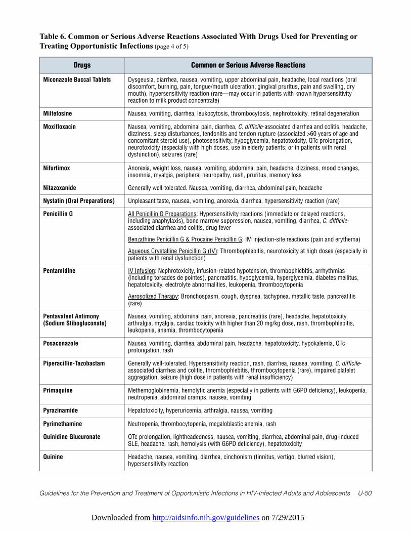
Downloaded from http://aidsinfo.nih.gov/guidelines on 7/29/2015
Guidelines for the Prevention and Treatment of Opportunistic Infections in HIV-Infected Adults and Adolescents U-50
Table 6. Common or Serious Adverse Reactions Associated With Drugs Used for Preventing orTreating Opportunistic Infections (page 4 of 5)
Drugs Common or Serious Adverse Reactions
Miconazole Buccal Tablets Dysgeusia, diarrhea, nausea, vomiting, upper abdominal pain, headache, local reactions (oraldiscomfort, burning, pain, tongue/mouth ulceration, gingival pruritus, pain and swelling, drymouth), hypersensitivity reaction (rare—may occur in patients with known hypersensitivityreaction to milk product concentrate)
Miltefosine Nausea, vomiting, diarrhea, leukocytosis, thrombocytosis, nephrotoxicity, retinal degeneration
Moxifloxacin Nausea, vomiting, abdominal pain, diarrhea, C. difficile-associated diarrhea and colitis, headache,dizziness, sleep disturbances, tendonitis and tendon rupture (associated >60 years of age andconcomitant steroid use), photosensitivity, hypoglycemia, hepatotoxicity, QTc prolongation,neurotoxicity (especially with high doses, use in elderly patients, or in patients with renaldysfunction), seizures (rare)
Nifurtimox Anorexia, weight loss, nausea, vomiting, abdominal pain, headache, dizziness, mood changes,insomnia, myalgia, peripheral neuropathy, rash, pruritus, memory loss
Nitazoxanide Generally well-tolerated. Nausea, vomiting, diarrhea, abdominal pain, headache
Nystatin (Oral Preparations) Unpleasant taste, nausea, vomiting, anorexia, diarrhea, hypersensitivity reaction (rare)
Penicillin G All Penicillin G Preparations: Hypersensitivity reactions (immediate or delayed reactions,including anaphylaxis), bone marrow suppression, nausea, vomiting, diarrhea, C. difficile-associated diarrhea and colitis, drug fever
Benzathine Penicillin G & Procaine Penicillin G: IM injection-site reactions (pain and erythema)
Aqueous Crystalline Penicillin G (IV): Thrombophlebitis, neurotoxicity at high doses (especially inpatients with renal dysfunction)
Pentamidine IV Infusion: Nephrotoxicity, infusion-related hypotension, thrombophlebitis, arrhythmias(including torsades de pointes), pancreatitis, hypoglycemia, hyperglycemia, diabetes mellitus,hepatotoxicity, electrolyte abnormalities, leukopenia, thrombocytopenia
Aerosolized Therapy: Bronchospasm, cough, dyspnea, tachypnea, metallic taste, pancreatitis(rare)
Pentavalent Antimony(Sodium Stibogluconate)
Nausea, vomiting, abdominal pain, anorexia, pancreatitis (rare), headache, hepatotoxicity,arthralgia, myalgia, cardiac toxicity with higher than 20 mg/kg dose, rash, thrombophlebitis,leukopenia, anemia, thrombocytopenia
Posaconazole Nausea, vomiting, diarrhea, abdominal pain, headache, hepatotoxicity, hypokalemia, QTcprolongation, rash
Piperacillin-Tazobactam Generally well-tolerated. Hypersensitivity reaction, rash, diarrhea, nausea, vomiting, C. difficile-associated diarrhea and colitis, thrombophlebitis, thrombocytopenia (rare), impaired plateletaggregation, seizure (high dose in patients with renal insufficiency)
Primaquine Methemoglobinemia, hemolytic anemia (especially in patients with G6PD deficiency), leukopenia,neutropenia, abdominal cramps, nausea, vomiting
Pyrazinamide Hepatotoxicity, hyperuricemia, arthralgia, nausea, vomiting
Pyrimethamine Neutropenia, thrombocytopenia, megaloblastic anemia, rash
Quinidine Glucuronate QTc prolongation, lightheadedness, nausea, vomiting, diarrhea, abdominal pain, drug-inducedSLE, headache, rash, hemolysis (with G6PD deficiency), hepatotoxicity
Quinine Headache, nausea, vomiting, diarrhea, cinchonism (tinnitus, vertigo, blurred vision),hypersensitivity reaction
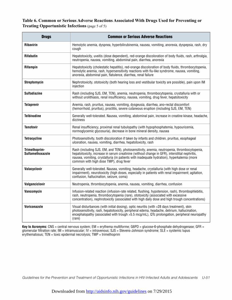
Downloaded from http://aidsinfo.nih.gov/guidelines on 7/29/2015
Guidelines for the Prevention and Treatment of Opportunistic Infections in HIV-Infected Adults and Adolescents U-51
Table 6. Common or Serious Adverse Reactions Associated With Drugs Used for Preventing orTreating Opportunistic Infections (page 5 of 5)
Drugs Common or Serious Adverse Reactions
Ribavirin Hemolytic anemia, dyspnea, hyperbilirubinemia, nausea, vomiting, anorexia, dyspepsia, rash, drycough
Rifabutin Hepatotoxicity, uveitis (dose dependent), red-orange discoloration of body fluids, rash, arthralgia,neutropenia, nausea, vomiting, abdominal pain, diarrhea, anorexia
Rifampin Hepatotoxicity (cholestatic hepatitis), red-orange discoloration of body fluids, thrombocytopenia,hemolytic anemia, rash, hypersensitivity reactions with flu-like syndrome, nausea, vomiting,anorexia, abdominal pain, flatulence, diarrhea, renal failure
Streptomycin Nephrotoxicity, ototoxicity (both hearing loss and vestibular toxicity are possible), pain upon IMinjection
Sulfadiazine Rash (including SJS, EM, TEN), anemia, neutropenia, thrombocytopenia, crystalluria with orwithout urolithiasis, renal insufficiency, nausea, vomiting, drug fever, hepatotoxicity
Telaprevir Anemia, rash, pruritus, nausea, vomiting, dysgeusia, diarrhea, ano-rectal discomfort(hemorrhoid, pruritus), proctitis, severe cutaneous eruption (including SJS, EM, TEN)
Telbivudine Generally well-tolerated. Nausea, vomiting, abdominal pain, increase in creatine kinase, headache,dizziness
Tenofovir Renal insufficiency, proximal renal tubulopathy (with hypophosphatemia, hypouricemia,normoglycemic glycosuria), decrease in bone mineral density, nausea
Tetracycline Photosensitivity, tooth discoloration if taken by infants and children, pruritus, esophagealulceration, nausea, vomiting, diarrhea, hepatotoxicity, rash
Trimethoprim-Sulfamethoxazole
Rash (including SJS, EM, and TEN), photosensitivity, anemia, neutropenia, thrombocytopenia,hepatotoxicity, increase in serum creatinine (without change in GFR), interstitial nephritis,nausea, vomiting, crystalluria (in patients with inadequate hydration), hyperkalemia (morecommon with high dose TMP), drug fever
Valacyclovir Generally well-tolerated. Nausea, vomiting, headache, crystalluria (with high dose or renalimpairment), neurotoxicity (high doses, especially in patients with renal impairment; agitation,confusion, hallucination, seizure, coma)
Valganciclovir Neutropenia, thrombocytopenia, anemia, nausea, vomiting, diarrhea, confusion
Vancomycin Infusion-related reaction (infusion-rate related, flushing, hypotension, rash), thrombophlebitis,rash, neutropenia, thrombocytopenia (rare), ototoxicity (associated with excessiveconcentration), nephrotoxicity (associated with high daily dose and high trough concentrations)
Voriconazole Visual disturbances (with initial dosing), optic neuritis (with >28 days treatment), skinphotosensitivity, rash, hepatotoxicity, peripheral edema, headache, delirium, hallucination,encephalopathy (associated with trough >5.5 mcg/mL), QTc prolongation, peripheral neuropathy(rare)
Key to Acronyms: CNS = central nervous system; EM = erythema multiforme; G6PD = glucose-6-phosphate dehydrogenase; GFR =glomerular filtration rate; IM = intramuscular; IV = intravenous; SJS = Stevens-Johnson syndrome; SLE = systemic lupuserythematosus; TEN = toxic epidermal necrolysis; TMP = trimethoprim
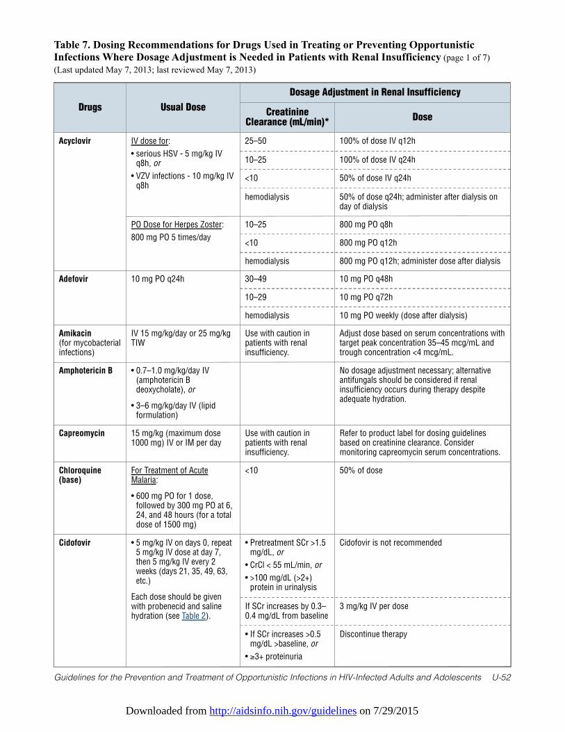
Downloaded from http://aidsinfo.nih.gov/guidelines on 7/29/2015
Guidelines for the Prevention and Treatment of Opportunistic Infections in HIV-Infected Adults and Adolescents U-52
Table 7. Dosing Recommendations for Drugs Used in Treating or Preventing OpportunisticInfections Where Dosage Adjustment is Needed in Patients with Renal Insufficiency (page 1 of 7)(Last updated May 7, 2013; last reviewed May 7, 2013)
Drugs Usual Dose
Dosage Adjustment in Renal Insufficiency
CreatinineClearance (mL/min)*
Dose
Acyclovir IV dose for:
• serious HSV - 5 mg/kg IVq8h, or
• VZV infections - 10 mg/kg IVq8h
25–50 100% of dose IV q12h
10–25 100% of dose IV q24h
<10 50% of dose IV q24h
hemodialysis 50% of dose q24h; administer after dialysis onday of dialysis
PO Dose for Herpes Zoster:
800 mg PO 5 times/day
10–25 800 mg PO q8h
<10 800 mg PO q12h
hemodialysis 800 mg PO q12h; administer dose after dialysis
Adefovir 10 mg PO q24h 30–49 10 mg PO q48h
10–29 10 mg PO q72h
hemodialysis 10 mg PO weekly (dose after dialysis)
Amikacin(for mycobacterialinfections)
IV 15 mg/kg/day or 25 mg/kgTIW
Use with caution inpatients with renalinsufficiency.
Adjust dose based on serum concentrations withtarget peak concentration 35–45 mcg/mL andtrough concentration <4 mcg/mL.
Amphotericin B • 0.7–1.0 mg/kg/day IV(amphotericin Bdeoxycholate), or
• 3–6 mg/kg/day IV (lipidformulation)
No dosage adjustment necessary; alternativeantifungals should be considered if renalinsufficiency occurs during therapy despiteadequate hydration.
Capreomycin 15 mg/kg (maximum dose1000 mg) IV or IM per day
Use with caution inpatients with renalinsufficiency.
Refer to product label for dosing guidelinesbased on creatinine clearance. Considermonitoring capreomycin serum concentrations.
Chloroquine(base)
For Treatment of AcuteMalaria:
• 600 mg PO for 1 dose,followed by 300 mg PO at 6,24, and 48 hours (for a totaldose of 1500 mg)
<10 50% of dose
Cidofovir • 5 mg/kg IV on days 0, repeat5 mg/kg IV dose at day 7,then 5 mg/kg IV every 2weeks (days 21, 35, 49, 63,etc.)
Each dose should be givenwith probenecid and salinehydration (see Table 2).
• Pretreatment SCr >1.5mg/dL, or
• CrCl < 55 mL/min, or
• >100 mg/dL (>2+)protein in urinalysis
Cidofovir is not recommended
If SCr increases by 0.3–0.4 mg/dL from baseline
3 mg/kg IV per dose
• If SCr increases >0.5mg/dL >baseline, or
• ≥3+ proteinuria
Discontinue therapy
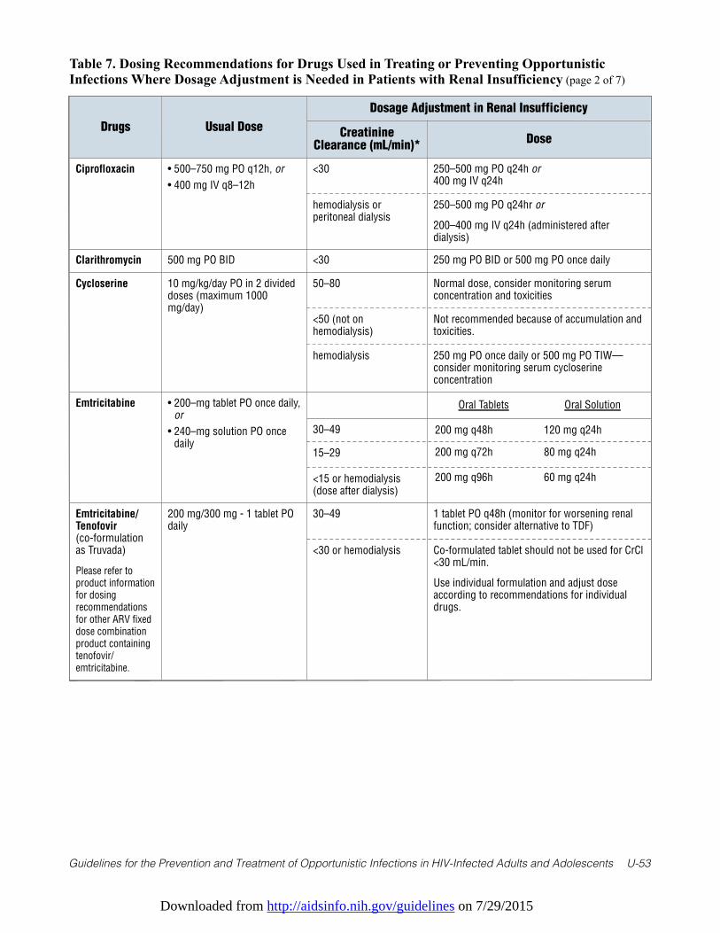
Downloaded from http://aidsinfo.nih.gov/guidelines on 7/29/2015
Guidelines for the Prevention and Treatment of Opportunistic Infections in HIV-Infected Adults and Adolescents U-53
Table 7. Dosing Recommendations for Drugs Used in Treating or Preventing OpportunisticInfections Where Dosage Adjustment is Needed in Patients with Renal Insufficiency (page 2 of 7)
Drugs Usual Dose
Dosage Adjustment in Renal Insufficiency
CreatinineClearance (mL/min)*
Dose
Ciprofloxacin • 500–750 mg PO q12h, or
• 400 mg IV q8–12h
<30 250–500 mg PO q24h or400 mg IV q24h
hemodialysis orperitoneal dialysis
250–500 mg PO q24hr or
200–400 mg IV q24h (administered afterdialysis)
Clarithromycin 500 mg PO BID <30 250 mg PO BID or 500 mg PO once daily
Cycloserine 10 mg/kg/day PO in 2 divideddoses (maximum 1000mg/day)
50–80 Normal dose, consider monitoring serumconcentration and toxicities
<50 (not onhemodialysis)
Not recommended because of accumulation andtoxicities.
hemodialysis 250 mg PO once daily or 500 mg PO TIW—consider monitoring serum cycloserineconcentration
Emtricitabine • 200–mg tablet PO once daily,or
• 240–mg solution PO oncedaily
30–49
15–29
<15 or hemodialysis(dose after dialysis)
Emtricitabine/Tenofovir(co-formulationas Truvada)
Please refer toproduct informationfor dosingrecommendationsfor other ARV fixeddose combinationproduct containingtenofovir/emtricitabine.
200 mg/300 mg - 1 tablet POdaily
30–49 1 tablet PO q48h (monitor for worsening renalfunction; consider alternative to TDF)
<30 or hemodialysis Co-formulated tablet should not be used for CrCl<30 mL/min.
Use individual formulation and adjust doseaccording to recommendations for individualdrugs.
Oral Tablets Oral Solution
200 mg q48h 120 mg q24h
200 mg q72h 80 mg q24h
200 mg q96h 60 mg q24h
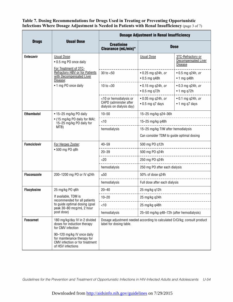
Downloaded from http://aidsinfo.nih.gov/guidelines on 7/29/2015
Guidelines for the Prevention and Treatment of Opportunistic Infections in HIV-Infected Adults and Adolescents U-54
Table 7. Dosing Recommendations for Drugs Used in Treating or Preventing OpportunisticInfections Where Dosage Adjustment is Needed in Patients with Renal Insufficiency (page 3 of 7)
Drugs Usual Dose
Dosage Adjustment in Renal Insufficiency
CreatinineClearance (mL/min)*
Dose
Entecavir Usual Dose:
• 0.5 mg PO once daily
For Treatment of 3TC-Refractory HBV or for Patientswith Decompensated LiverDisease:
• 1 mg PO once daily
Usual Dose 3TC-Refractory orDecompensated LiverDisease
30 to <50 • 0.25 mg q24h, or
• 0.5 mg q48h
• 0.5 mg q24h, or
• 1 mg q48h
10 to <30 • 0.15 mg q24h, or
• 0.5 mg q72h
• 0.3 mg q24h, or
• 1 mg q72h
<10 or hemodialysis orCAPD (administer afterdialysis on dialysis day)
• 0.05 mg q24h, or
• 0.5 mg q7 days
• 0.1 mg q24h, or
• 1 mg q7 days
Ethambutol • 15–25 mg/kg PO daily
• (15 mg/kg PO daily for MAI;15–25 mg/kg PO daily forMTB)
10–50 15–25 mg/kg q24–36h
<10 15–25 mg/kg q48h
hemodialysis 15–25 mg/kg TIW after hemodialysis
Can consider TDM to guide optimal dosing
Famciclovir For Herpes Zoster:
• 500 mg PO q8h
40–59 500 mg PO q12h
20–39 500 mg PO q24h
<20 250 mg PO q24h
hemodialysis 250 mg PO after each dialysis
Fluconazole 200–1200 mg PO or IV q24h ≤50 50% of dose q24h
hemodialysis Full dose after each dialysis
Flucytosine 25 mg/kg PO q6h
If available, TDM isrecommended for all patientsto guide optimal dosing (goalpeak 30–80 mcg/mL 2 hourpost dose)
20–40 25 mg/kg q12h
10–20 25 mg/kg q24h
<10 25 mg/kg q48h
hemodialysis 25–50 mg/kg q48–72h (after hemodialysis)
Foscarnet 180 mg/kg/day IV in 2 divideddoses for induction therapyfor CMV infection
90–120 mg/kg IV once dailyfor maintenance therapy forCMV infection or for treatmentof HSV infections
Dosage adjustment needed according to calculated CrCl/kg; consult productlabel for dosing table.
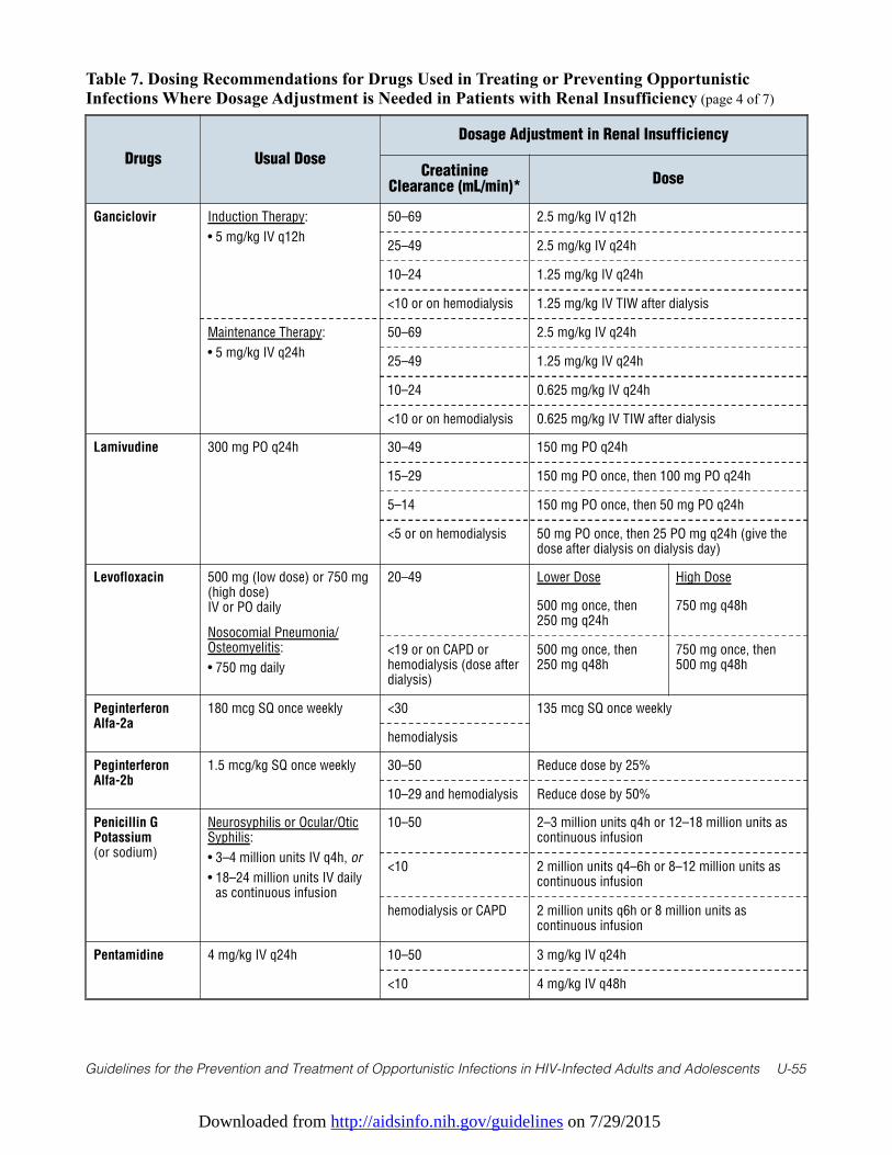
Downloaded from http://aidsinfo.nih.gov/guidelines on 7/29/2015
Guidelines for the Prevention and Treatment of Opportunistic Infections in HIV-Infected Adults and Adolescents U-55
Table 7. Dosing Recommendations for Drugs Used in Treating or Preventing OpportunisticInfections Where Dosage Adjustment is Needed in Patients with Renal Insufficiency (page 4 of 7)
Drugs Usual Dose
Dosage Adjustment in Renal Insufficiency
CreatinineClearance (mL/min)*
Dose
Ganciclovir Induction Therapy:
• 5 mg/kg IV q12h
50–69 2.5 mg/kg IV q12h
25–49 2.5 mg/kg IV q24h
10–24 1.25 mg/kg IV q24h
<10 or on hemodialysis 1.25 mg/kg IV TIW after dialysis
Maintenance Therapy:
• 5 mg/kg IV q24h
50–69 2.5 mg/kg IV q24h
25–49 1.25 mg/kg IV q24h
10–24 0.625 mg/kg IV q24h
<10 or on hemodialysis 0.625 mg/kg IV TIW after dialysis
Lamivudine 300 mg PO q24h 30–49 150 mg PO q24h
15–29 150 mg PO once, then 100 mg PO q24h
5–14 150 mg PO once, then 50 mg PO q24h
<5 or on hemodialysis 50 mg PO once, then 25 PO mg q24h (give thedose after dialysis on dialysis day)
Levofloxacin 500 mg (low dose) or 750 mg(high dose) IV or PO daily
Nosocomial Pneumonia/Osteomyelitis:
• 750 mg daily
20–49 Lower Dose High Dose
500 mg once, then250 mg q24h
750 mg q48h
<19 or on CAPD orhemodialysis (dose afterdialysis)
500 mg once, then250 mg q48h
750 mg once, then500 mg q48h
PeginterferonAlfa-2a
180 mcg SQ once weekly <30 135 mcg SQ once weekly
hemodialysis
PeginterferonAlfa-2b
1.5 mcg/kg SQ once weekly 30–50 Reduce dose by 25%
10–29 and hemodialysis Reduce dose by 50%
Penicillin GPotassium(or sodium)
Neurosyphilis or Ocular/OticSyphilis:
• 3–4 million units IV q4h, or
• 18–24 million units IV dailyas continuous infusion
10–50 2–3 million units q4h or 12–18 million units ascontinuous infusion
<10 2 million units q4–6h or 8–12 million units ascontinuous infusion
hemodialysis or CAPD 2 million units q6h or 8 million units ascontinuous infusion
Pentamidine 4 mg/kg IV q24h 10–50 3 mg/kg IV q24h
<10 4 mg/kg IV q48h
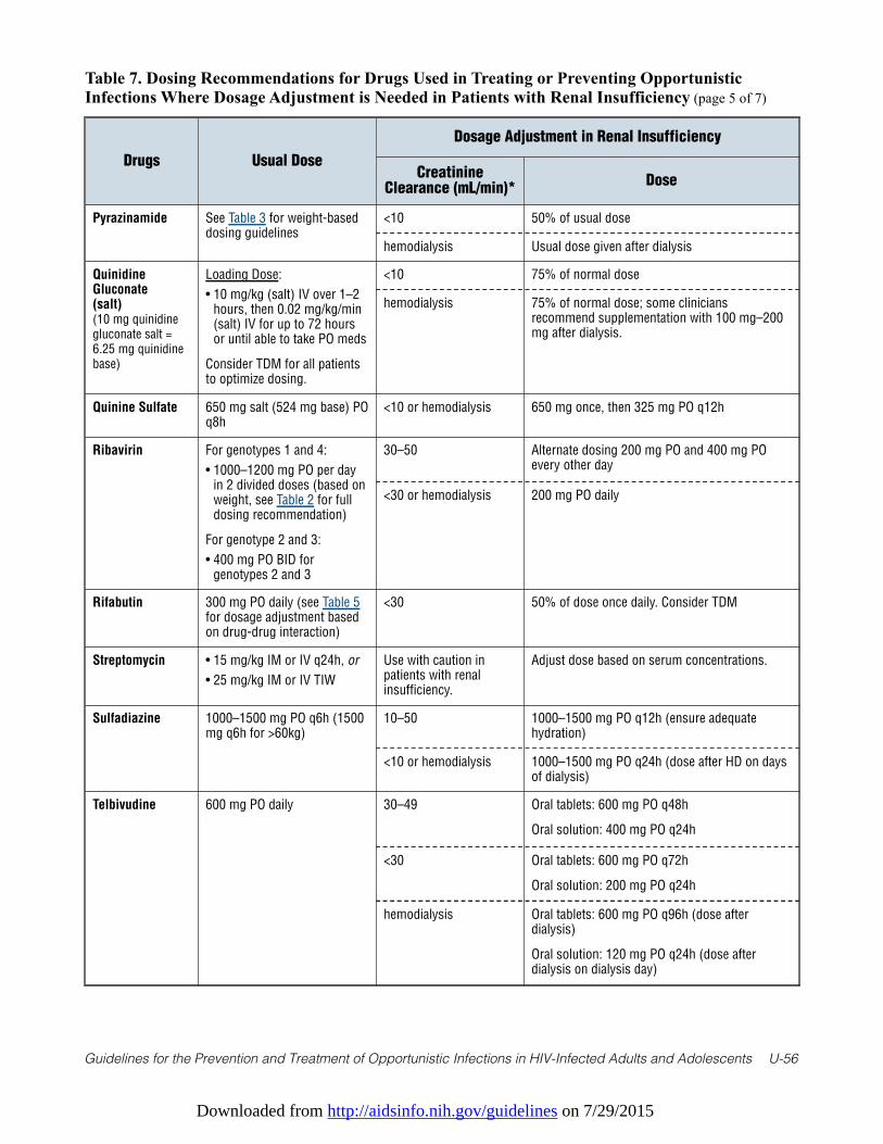
Downloaded from http://aidsinfo.nih.gov/guidelines on 7/29/2015
Guidelines for the Prevention and Treatment of Opportunistic Infections in HIV-Infected Adults and Adolescents U-56
Table 7. Dosing Recommendations for Drugs Used in Treating or Preventing OpportunisticInfections Where Dosage Adjustment is Needed in Patients with Renal Insufficiency (page 5 of 7)
Drugs Usual Dose
Dosage Adjustment in Renal Insufficiency
CreatinineClearance (mL/min)*
Dose
Pyrazinamide See Table 3 for weight-baseddosing guidelines
<10 50% of usual dose
hemodialysis Usual dose given after dialysis
QuinidineGluconate (salt)(10 mg quinidinegluconate salt =6.25 mg quinidinebase)
Loading Dose:
• 10 mg/kg (salt) IV over 1–2hours, then 0.02 mg/kg/min(salt) IV for up to 72 hoursor until able to take PO meds
Consider TDM for all patientsto optimize dosing.
<10 75% of normal dose
hemodialysis 75% of normal dose; some cliniciansrecommend supplementation with 100 mg–200mg after dialysis.
Quinine Sulfate 650 mg salt (524 mg base) POq8h
<10 or hemodialysis 650 mg once, then 325 mg PO q12h
Ribavirin For genotypes 1 and 4:
• 1000–1200 mg PO per dayin 2 divided doses (based onweight, see Table 2 for fulldosing recommendation)
For genotype 2 and 3:
• 400 mg PO BID forgenotypes 2 and 3
30–50 Alternate dosing 200 mg PO and 400 mg POevery other day
<30 or hemodialysis 200 mg PO daily
Rifabutin 300 mg PO daily (see Table 5for dosage adjustment basedon drug-drug interaction)
<30 50% of dose once daily. Consider TDM
Streptomycin • 15 mg/kg IM or IV q24h, or
• 25 mg/kg IM or IV TIW
Use with caution inpatients with renalinsufficiency.
Adjust dose based on serum concentrations.
Sulfadiazine 1000–1500 mg PO q6h (1500mg q6h for >60kg)
10–50 1000–1500 mg PO q12h (ensure adequatehydration)
<10 or hemodialysis 1000–1500 mg PO q24h (dose after HD on daysof dialysis)
Telbivudine 600 mg PO daily 30–49 Oral tablets: 600 mg PO q48h
Oral solution: 400 mg PO q24h
<30 Oral tablets: 600 mg PO q72h
Oral solution: 200 mg PO q24h
hemodialysis Oral tablets: 600 mg PO q96h (dose afterdialysis)
Oral solution: 120 mg PO q24h (dose afterdialysis on dialysis day)
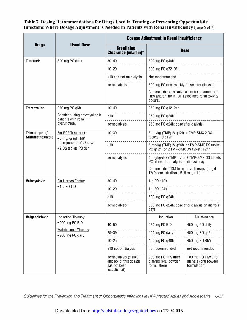
Downloaded from http://aidsinfo.nih.gov/guidelines on 7/29/2015
Guidelines for the Prevention and Treatment of Opportunistic Infections in HIV-Infected Adults and Adolescents U-57
Table 7. Dosing Recommendations for Drugs Used in Treating or Preventing OpportunisticInfections Where Dosage Adjustment is Needed in Patients with Renal Insufficiency (page 6 of 7)
Drugs Usual Dose
Dosage Adjustment in Renal Insufficiency
CreatinineClearance (mL/min)*
Dose
Tenofovir 300 mg PO daily 30–49 300 mg PO q48h
10–29 300 mg PO q72–96h
<10 and not on dialysis Not recommended
hemodialysis 300 mg PO once weekly (dose after dialysis)
Can consider alternative agent for treatment ofHBV and/or HIV if TDF-associated renal toxicityoccurs.
Tetracycline 250 mg PO q6h
Consider using doxycycline inpatients with renaldysfunction.
10–49 250 mg PO q12–24h
<10 250 mg PO q24h
hemodialysis 250 mg PO q24h; dose after dialysis
Trimethoprim/Sulfamethoxazole
For PCP Treatment:
• 5 mg/kg (of TMPcomponent) IV q8h, or
• 2 DS tablets PO q8h
10–30 5 mg/kg (TMP) IV q12h or TMP-SMX 2 DStablets PO q12h
<10 5 mg/kg (TMP) IV q24h, or TMP-SMX DS tabletPO q12h (or 2 TMP-SMX DS tablets q24h)
hemodialysis 5 mg/kg/day (TMP) IV or 2 TMP-SMX DS tabletsPO; dose after dialysis on dialysis day
Can consider TDM to optimize therapy (targetTMP concentrations: 5–8 mcg/mL)
Valacyclovir For Herpes Zoster:
• 1 g PO TID
30–49 1 g PO q12h
10–29 1 g PO q24h
<10 500 mg PO q24h
hemodialysis 500 mg PO q24h; dose after dialysis on dialysisdays
Valganciclovir Induction Therapy:
• 900 mg PO BID
Maintenance Therapy:
• 900 mg PO daily
Induction Maintenance
40–59 450 mg PO BID 450 mg PO daily
25–39 450 mg PO daily 450 mg PO q48h
10–25 450 mg PO q48h 450 mg PO BIW
<10 not on dialysis not recommended not recommended
hemodialysis (clinicalefficacy of this dosagehas not beenestablished)
200 mg PO TIW afterdialysis (oral powderformulation)
100 mg PO TIW afterdialysis (oral powderformulation)
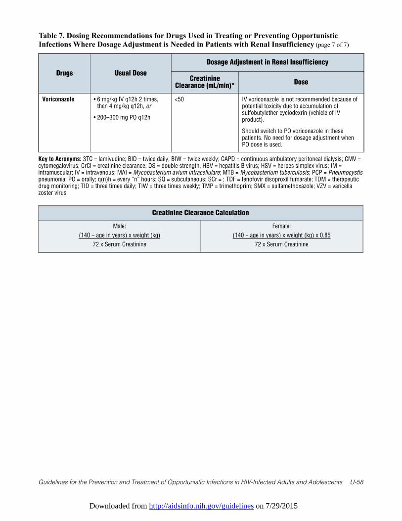
Downloaded from http://aidsinfo.nih.gov/guidelines on 7/29/2015
Guidelines for the Prevention and Treatment of Opportunistic Infections in HIV-Infected Adults and Adolescents U-58
Table 7. Dosing Recommendations for Drugs Used in Treating or Preventing OpportunisticInfections Where Dosage Adjustment is Needed in Patients with Renal Insufficiency (page 7 of 7)
Drugs Usual Dose
Dosage Adjustment in Renal Insufficiency
CreatinineClearance (mL/min)*
Dose
Voriconazole • 6 mg/kg IV q12h 2 times,then 4 mg/kg q12h, or
• 200–300 mg PO q12h
<50 IV voriconazole is not recommended because ofpotential toxicity due to accumulation ofsulfobutylether cyclodexrin (vehicle of IVproduct).
Should switch to PO voriconazole in thesepatients. No need for dosage adjustment whenPO dose is used.
Key to Acronyms: 3TC = lamivudine; BID = twice daily; BIW = twice weekly; CAPD = continuous ambulatory peritoneal dialysis; CMV =cytomegalovirus; CrCl = creatinine clearance; DS = double strength, HBV = hepatitis B virus; HSV = herpes simplex virus; IM =intramuscular; IV = intravenous; MAI = Mycobacterium avium intracellulare; MTB = Mycobacterium tuberculosis; PCP = Pneumocystispneumonia; PO = orally; q(n)h = every “n” hours; SQ = subcutaneous; SCr = ; TDF = tenofovir disoproxil fumarate; TDM = therapeuticdrug monitoring; TID = three times daily; TIW = three times weekly; TMP = trimethoprim; SMX = sulfamethoxazole; VZV = varicellazoster virus
Creatinine Clearance Calculation
Male:
(140 − age in years) x weight (kg)
72 x Serum Creatinine
Female:
(140 − age in years) x weight (kg) x 0.85
72 x Serum Creatinine
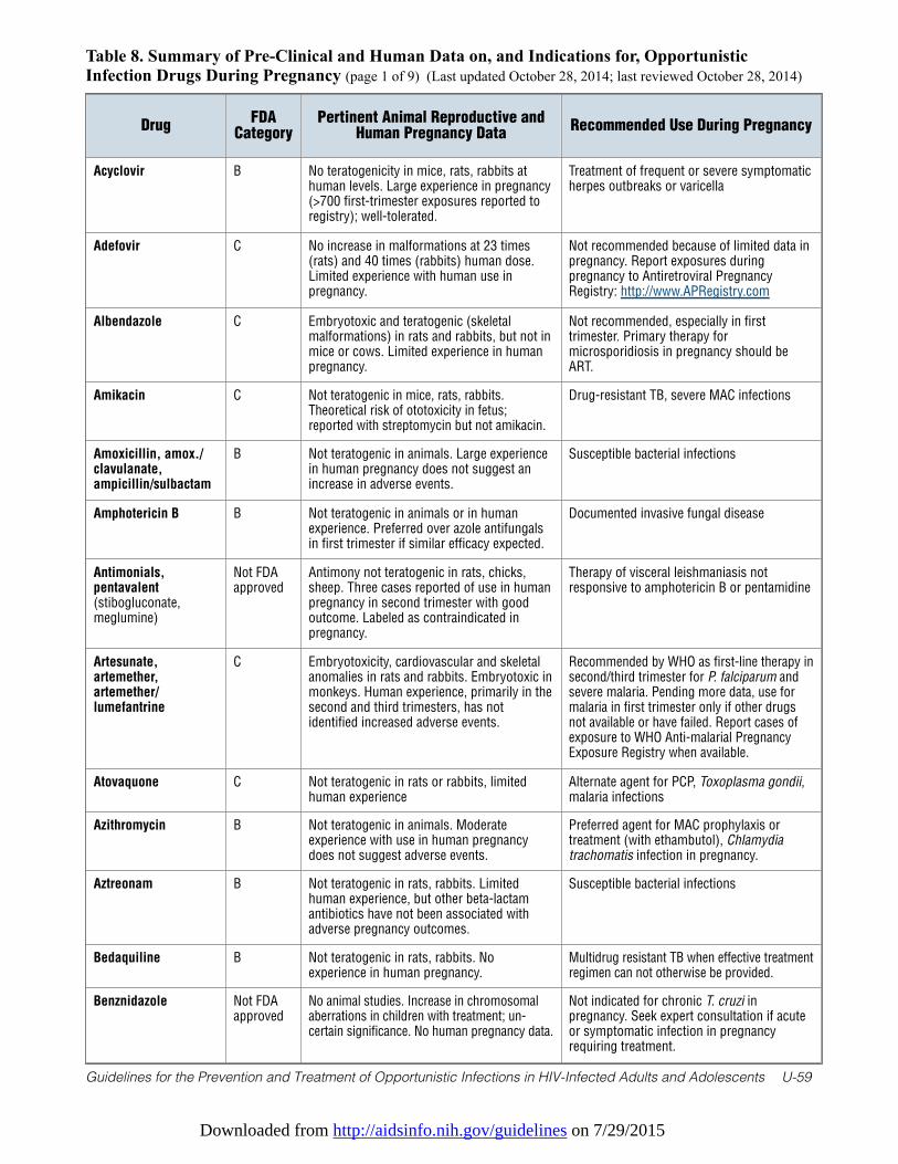
Downloaded from http://aidsinfo.nih.gov/guidelines on 7/29/2015
Guidelines for the Prevention and Treatment of Opportunistic Infections in HIV-Infected Adults and Adolescents U-59
Table 8. Summary of Pre-Clinical and Human Data on, and Indications for, OpportunisticInfection Drugs During Pregnancy (page 1 of 9) (Last updated October 28, 2014; last reviewed October 28, 2014)
DrugFDA
CategoryPertinent Animal Reproductive and
Human Pregnancy DataRecommended Use During Pregnancy
Acyclovir B No teratogenicity in mice, rats, rabbits athuman levels. Large experience in pregnancy(>700 first-trimester exposures reported toregistry); well-tolerated.
Treatment of frequent or severe symptomaticherpes outbreaks or varicella
Adefovir C No increase in malformations at 23 times(rats) and 40 times (rabbits) human dose.Limited experience with human use inpregnancy.
Not recommended because of limited data inpregnancy. Report exposures duringpregnancy to Antiretroviral PregnancyRegistry: http://www.APRegistry.com
Albendazole C Embryotoxic and teratogenic (skeletalmalformations) in rats and rabbits, but not inmice or cows. Limited experience in humanpregnancy.
Not recommended, especially in firsttrimester. Primary therapy formicrosporidiosis in pregnancy should beART.
Amikacin C Not teratogenic in mice, rats, rabbits.Theoretical risk of ototoxicity in fetus;reported with streptomycin but not amikacin.
Drug-resistant TB, severe MAC infections
Amoxicillin, amox./clavulanate,ampicillin/sulbactam
B Not teratogenic in animals. Large experiencein human pregnancy does not suggest anincrease in adverse events.
Susceptible bacterial infections
Amphotericin B B Not teratogenic in animals or in humanexperience. Preferred over azole antifungalsin first trimester if similar efficacy expected.
Documented invasive fungal disease
Antimonials,pentavalent(stibogluconate,meglumine)
Not FDAapproved
Antimony not teratogenic in rats, chicks,sheep. Three cases reported of use in humanpregnancy in second trimester with goodoutcome. Labeled as contraindicated inpregnancy.
Therapy of visceral leishmaniasis notresponsive to amphotericin B or pentamidine
Artesunate,artemether,artemether/lumefantrine
C Embryotoxicity, cardiovascular and skeletalanomalies in rats and rabbits. Embryotoxic inmonkeys. Human experience, primarily in thesecond and third trimesters, has notidentified increased adverse events.
Recommended by WHO as first-line therapy insecond/third trimester for P. falciparum andsevere malaria. Pending more data, use formalaria in first trimester only if other drugsnot available or have failed. Report cases ofexposure to WHO Anti-malarial PregnancyExposure Registry when available.
Atovaquone C Not teratogenic in rats or rabbits, limitedhuman experience
Alternate agent for PCP, Toxoplasma gondii,malaria infections
Azithromycin B Not teratogenic in animals. Moderateexperience with use in human pregnancydoes not suggest adverse events.
Preferred agent for MAC prophylaxis ortreatment (with ethambutol), Chlamydiatrachomatis infection in pregnancy.
Aztreonam B Not teratogenic in rats, rabbits. Limitedhuman experience, but other beta-lactamantibiotics have not been associated withadverse pregnancy outcomes.
Susceptible bacterial infections
Bedaquiline B Not teratogenic in rats, rabbits. Noexperience in human pregnancy.
Multidrug resistant TB when effective treatmentregimen can not otherwise be provided.
Benznidazole Not FDAapproved
No animal studies. Increase in chromosomalaberrations in children with treatment; un-certain significance. No human pregnancy data.
Not indicated for chronic T. cruzi inpregnancy. Seek expert consultation if acuteor symptomatic infection in pregnancyrequiring treatment.
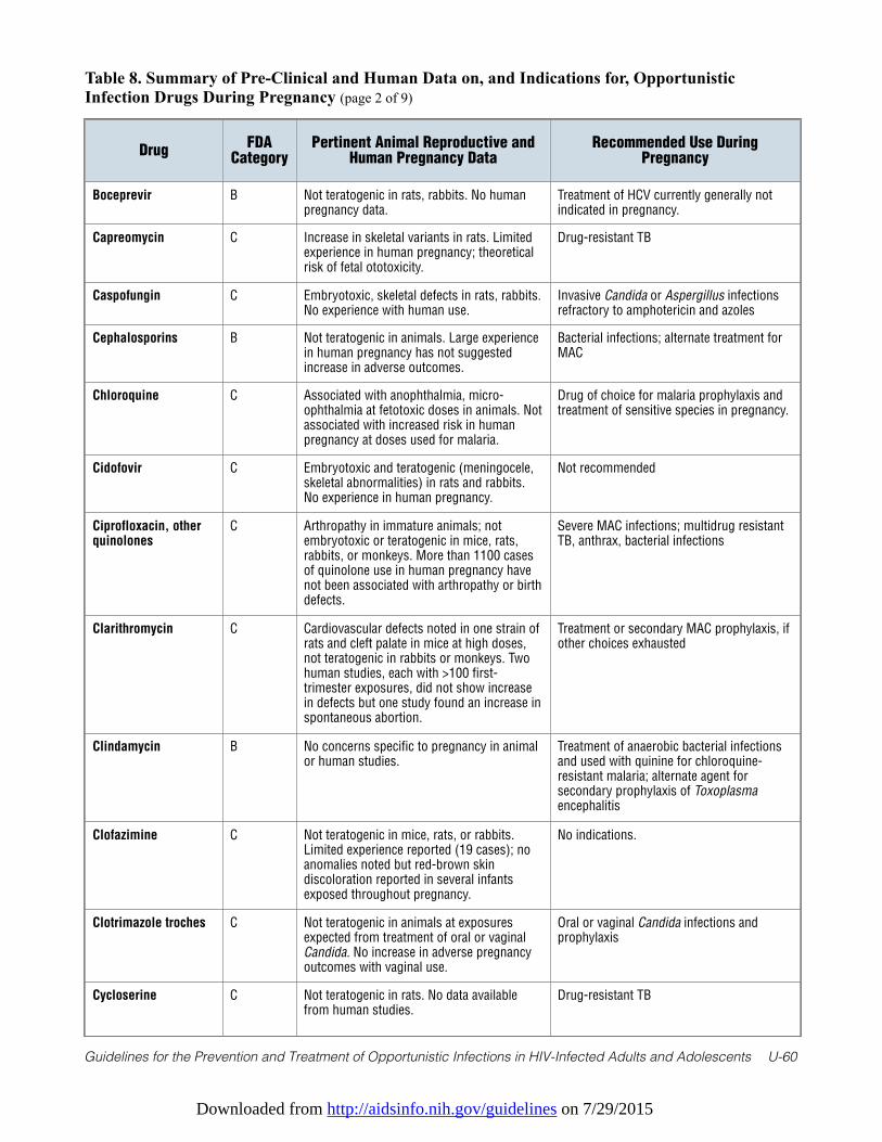
Downloaded from http://aidsinfo.nih.gov/guidelines on 7/29/2015
Guidelines for the Prevention and Treatment of Opportunistic Infections in HIV-Infected Adults and Adolescents U-60
Table 8. Summary of Pre-Clinical and Human Data on, and Indications for, OpportunisticInfection Drugs During Pregnancy (page 2 of 9)
DrugFDA
CategoryPertinent Animal Reproductive and
Human Pregnancy DataRecommended Use During
Pregnancy
Boceprevir B Not teratogenic in rats, rabbits. No humanpregnancy data.
Treatment of HCV currently generally notindicated in pregnancy.
Capreomycin C Increase in skeletal variants in rats. Limitedexperience in human pregnancy; theoreticalrisk of fetal ototoxicity.
Drug-resistant TB
Caspofungin C Embryotoxic, skeletal defects in rats, rabbits.No experience with human use.
Invasive Candida or Aspergillus infectionsrefractory to amphotericin and azoles
Cephalosporins B Not teratogenic in animals. Large experiencein human pregnancy has not suggestedincrease in adverse outcomes.
Bacterial infections; alternate treatment forMAC
Chloroquine C Associated with anophthalmia, micro-ophthalmia at fetotoxic doses in animals. Notassociated with increased risk in humanpregnancy at doses used for malaria.
Drug of choice for malaria prophylaxis andtreatment of sensitive species in pregnancy.
Cidofovir C Embryotoxic and teratogenic (meningocele,skeletal abnormalities) in rats and rabbits.No experience in human pregnancy.
Not recommended
Ciprofloxacin, otherquinolones
C Arthropathy in immature animals; notembryotoxic or teratogenic in mice, rats,rabbits, or monkeys. More than 1100 casesof quinolone use in human pregnancy havenot been associated with arthropathy or birthdefects.
Severe MAC infections; multidrug resistantTB, anthrax, bacterial infections
Clarithromycin C Cardiovascular defects noted in one strain ofrats and cleft palate in mice at high doses,not teratogenic in rabbits or monkeys. Twohuman studies, each with >100 first-trimester exposures, did not show increasein defects but one study found an increase inspontaneous abortion.
Treatment or secondary MAC prophylaxis, ifother choices exhausted
Clindamycin B No concerns specific to pregnancy in animalor human studies.
Treatment of anaerobic bacterial infectionsand used with quinine for chloroquine-resistant malaria; alternate agent forsecondary prophylaxis of Toxoplasmaencephalitis
Clofazimine C Not teratogenic in mice, rats, or rabbits.Limited experience reported (19 cases); noanomalies noted but red-brown skindiscoloration reported in several infantsexposed throughout pregnancy.
No indications.
Clotrimazole troches C Not teratogenic in animals at exposuresexpected from treatment of oral or vaginalCandida. No increase in adverse pregnancyoutcomes with vaginal use.
Oral or vaginal Candida infections andprophylaxis
Cycloserine C Not teratogenic in rats. No data availablefrom human studies.
Drug-resistant TB
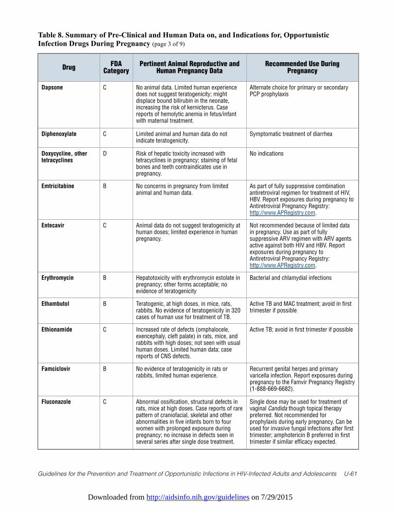
Downloaded from http://aidsinfo.nih.gov/guidelines on 7/29/2015
Guidelines for the Prevention and Treatment of Opportunistic Infections in HIV-Infected Adults and Adolescents U-61
Table 8. Summary of Pre-Clinical and Human Data on, and Indications for, OpportunisticInfection Drugs During Pregnancy (page 3 of 9)
DrugFDA
CategoryPertinent Animal Reproductive and
Human Pregnancy DataRecommended Use During
Pregnancy
Dapsone C No animal data. Limited human experiencedoes not suggest teratogenicity; mightdisplace bound bilirubin in the neonate,increasing the risk of kernicterus. Casereports of hemolytic anemia in fetus/infantwith maternal treatment.
Alternate choice for primary or secondaryPCP prophylaxis
Diphenoxylate C Limited animal and human data do notindicate teratogenicity.
Symptomatic treatment of diarrhea
Doxycycline, othertetracyclines
D Risk of hepatic toxicity increased withtetracyclines in pregnancy; staining of fetalbones and teeth contraindicates use inpregnancy.
No indications
Emtricitabine B No concerns in pregnancy from limitedanimal and human data.
As part of fully suppressive combinationantiretroviral regimen for treatment of HIV,HBV. Report exposures during pregnancy toAntiretroviral Pregnancy Registry:http://www.APRegistry.com.
Entecavir C Animal data do not suggest teratogenicity athuman doses; limited experience in humanpregnancy.
Not recommended because of limited datain pregnancy. Use as part of fullysuppressive ARV regimen with ARV agentsactive against both HIV and HBV. Reportexposures during pregnancy toAntiretroviral Pregnancy Registry:http://www.APRegistry.com.
Erythromycin B Hepatotoxicity with erythromycin estolate inpregnancy; other forms acceptable; noevidence of teratogenicity
Bacterial and chlamydial infections
Ethambutol B Teratogenic, at high doses, in mice, rats,rabbits. No evidence of teratogenicity in 320cases of human use for treatment of TB.
Active TB and MAC treatment; avoid in firsttrimester if possible
Ethionamide C Increased rate of defects (omphalocele,exencephaly, cleft palate) in rats, mice, andrabbits with high doses; not seen with usualhuman doses. Limited human data; casereports of CNS defects.
Active TB; avoid in first trimester if possible
Famciclovir B No evidence of teratogenicity in rats orrabbits, limited human experience.
Recurrent genital herpes and primaryvaricella infection. Report exposures duringpregnancy to the Famvir Pregnancy Registry(1-888-669-6682).
Fluconazole C Abnormal ossification, structural defects inrats, mice at high doses. Case reports of rarepattern of craniofacial, skeletal and otherabnormalities in five infants born to fourwomen with prolonged exposure duringpregnancy; no increase in defects seen inseveral series after single dose treatment.
Single dose may be used for treatment ofvaginal Candida though topical therapypreferred. Not recommended forprophylaxis during early pregnancy. Can beused for invasive fungal infections after firsttrimester; amphotericin B preferred in firsttrimester if similar efficacy expected.
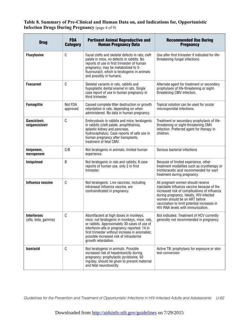
Downloaded from http://aidsinfo.nih.gov/guidelines on 7/29/2015
Guidelines for the Prevention and Treatment of Opportunistic Infections in HIV-Infected Adults and Adolescents U-62
Table 8. Summary of Pre-Clinical and Human Data on, and Indications for, OpportunisticInfection Drugs During Pregnancy (page 4 of 9)
DrugFDA
CategoryPertinent Animal Reproductive and
Human Pregnancy DataRecommended Use During
Pregnancy
Flucytosine C Facial clefts and skeletal defects in rats; cleftpalate in mice, no defects in rabbits. Noreports of use in first trimester of humanpregnancy; may be metabolized to 5-fluorouracil, which is teratogenic in animalsand possibly in humans.
Use after first trimester if indicated for life-threatening fungal infections.
Foscarnet C Skeletal variants in rats, rabbits andhypoplastic dental enamel in rats. Singlecase report of use in human pregnancy inthird trimester.
Alternate agent for treatment or secondaryprophylaxis of life-threatening or sight-threatening CMV infection.
Fumagillin Not FDAapproved
Caused complete litter destruction or growthretardation in rats, depending on whenadministered. No data in human pregnancy.
Topical solution can be used for ocularmicrosporidial infections.
Ganciclovir,valganciclovir
C Embryotoxic in rabbits and mice; teratogenicin rabbits (cleft palate, anophthalmia,aplastic kidney and pancreas,hydrocephalus). Case reports of safe use inhuman pregnancy after transplants,treatment of fetal CMV.
Treatment or secondary prophylaxis of life-threatening or sight-threatening CMVinfection. Preferred agent for therapy inchildren.
Imipenem,meropenem
C/B Not teratogenic in animals; limited humanexperience.
Serious bacterial infections
Imiquimod B Not teratogenic in rats and rabbits; 8 casereports of human use, only 2 in firsttrimester.
Because of limited experience, othertreatment modalities such as cryotherapy ortrichloracetic acid recommended for warttreatment during pregnancy.
Influenza vaccine C Not teratogenic. Live vaccines, includingintranasal influenza vaccine, arecontraindicated in pregnancy.
All pregnant women should receiveinjectable influenza vaccine because of theincreased risk of complications of influenzaduring pregnancy. Ideally, HIV-infectedwomen should be on ART beforevaccination to limit potential increases inHIV RNA levels with immunization.
Interferons(alfa, beta, gamma)
C Abortifacient at high doses in monkeys,mice; not teratogenic in monkeys, mice, rats,or rabbits. Approximately 30 cases of use ofinterferon-alfa in pregnancy reported; 14 infirst trimester without increase in anomalies;possible increased risk of intrauterinegrowth retardation.
Not indicated. Treatment of HCV currentlygenerally not recommended in pregnancy.
Isoniazid C Not teratogenic in animals. Possibleincreased risk of hepatotoxicity duringpregnancy; prophylactic pyridoxine, 50mg/day, should be given to prevent maternaland fetal neurotoxicity.
Active TB; prophylaxis for exposure or skintest conversion
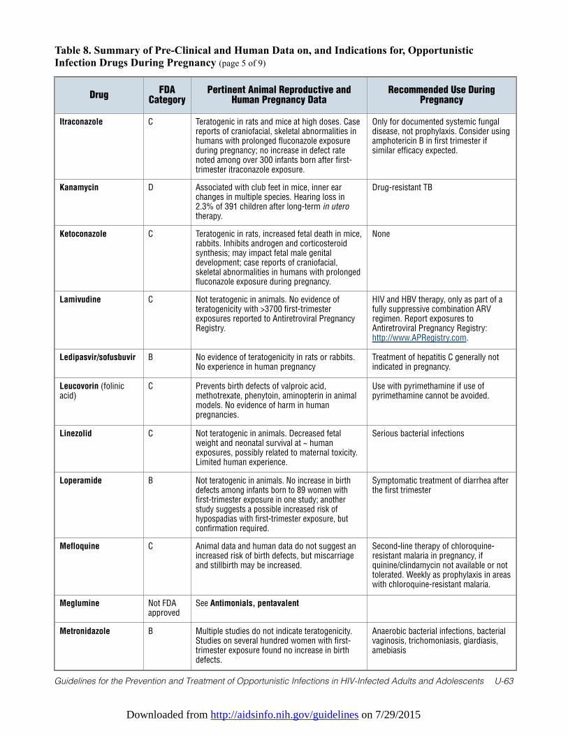
Downloaded from http://aidsinfo.nih.gov/guidelines on 7/29/2015
Guidelines for the Prevention and Treatment of Opportunistic Infections in HIV-Infected Adults and Adolescents U-63
Table 8. Summary of Pre-Clinical and Human Data on, and Indications for, OpportunisticInfection Drugs During Pregnancy (page 5 of 9)
DrugFDA
CategoryPertinent Animal Reproductive and
Human Pregnancy DataRecommended Use During
Pregnancy
Itraconazole C Teratogenic in rats and mice at high doses. Casereports of craniofacial, skeletal abnormalities inhumans with prolonged fluconazole exposureduring pregnancy; no increase in defect ratenoted among over 300 infants born after first-trimester itraconazole exposure.
Only for documented systemic fungaldisease, not prophylaxis. Consider usingamphotericin B in first trimester ifsimilar efficacy expected.
Kanamycin D Associated with club feet in mice, inner earchanges in multiple species. Hearing loss in2.3% of 391 children after long-term in uterotherapy.
Drug-resistant TB
Ketoconazole C Teratogenic in rats, increased fetal death in mice,rabbits. Inhibits androgen and corticosteroidsynthesis; may impact fetal male genitaldevelopment; case reports of craniofacial,skeletal abnormalities in humans with prolongedfluconazole exposure during pregnancy.
None
Lamivudine C Not teratogenic in animals. No evidence ofteratogenicity with >3700 first-trimesterexposures reported to Antiretroviral PregnancyRegistry.
HIV and HBV therapy, only as part of afully suppressive combination ARVregimen. Report exposures toAntiretroviral Pregnancy Registry:http://www.APRegistry.com.
Ledipasvir/sofusbuvir B No evidence of teratogenicity in rats or rabbits.No experience in human pregnancy
Treatment of hepatitis C generally notindicated in pregnancy.
Leucovorin (folinicacid)
C Prevents birth defects of valproic acid,methotrexate, phenytoin, aminopterin in animalmodels. No evidence of harm in humanpregnancies.
Use with pyrimethamine if use ofpyrimethamine cannot be avoided.
Linezolid C Not teratogenic in animals. Decreased fetalweight and neonatal survival at ~ humanexposures, possibly related to maternal toxicity.Limited human experience.
Serious bacterial infections
Loperamide B Not teratogenic in animals. No increase in birthdefects among infants born to 89 women withfirst-trimester exposure in one study; anotherstudy suggests a possible increased risk ofhypospadias with first-trimester exposure, butconfirmation required.
Symptomatic treatment of diarrhea afterthe first trimester
Mefloquine C Animal data and human data do not suggest anincreased risk of birth defects, but miscarriageand stillbirth may be increased.
Second-line therapy of chloroquine-resistant malaria in pregnancy, ifquinine/clindamycin not available or nottolerated. Weekly as prophylaxis in areaswith chloroquine-resistant malaria.
Meglumine Not FDAapproved
See Antimonials, pentavalent
Metronidazole B Multiple studies do not indicate teratogenicity.Studies on several hundred women with first-trimester exposure found no increase in birthdefects.
Anaerobic bacterial infections, bacterialvaginosis, trichomoniasis, giardiasis,amebiasis
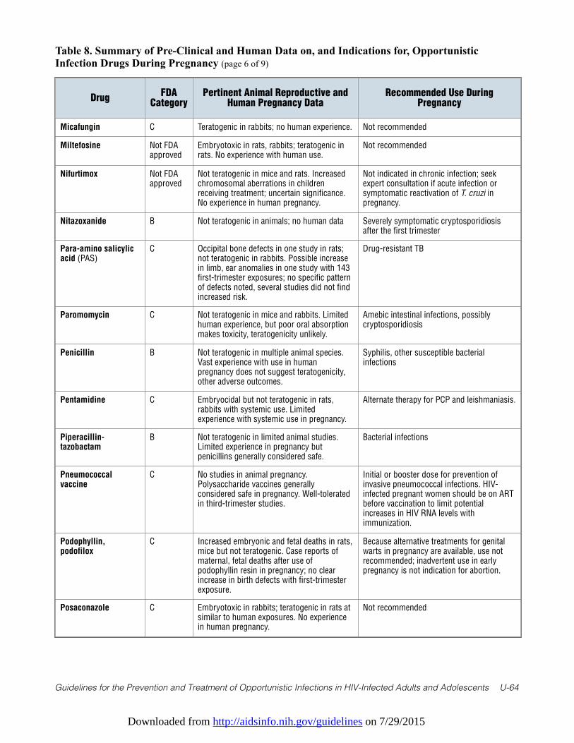
Downloaded from http://aidsinfo.nih.gov/guidelines on 7/29/2015
Guidelines for the Prevention and Treatment of Opportunistic Infections in HIV-Infected Adults and Adolescents U-64
Table 8. Summary of Pre-Clinical and Human Data on, and Indications for, OpportunisticInfection Drugs During Pregnancy (page 6 of 9)
DrugFDA
CategoryPertinent Animal Reproductive and
Human Pregnancy DataRecommended Use During
Pregnancy
Micafungin C Teratogenic in rabbits; no human experience. Not recommended
Miltefosine Not FDAapproved
Embryotoxic in rats, rabbits; teratogenic inrats. No experience with human use.
Not recommended
Nifurtimox Not FDAapproved
Not teratogenic in mice and rats. Increasedchromosomal aberrations in childrenreceiving treatment; uncertain significance.No experience in human pregnancy.
Not indicated in chronic infection; seekexpert consultation if acute infection orsymptomatic reactivation of T. cruzi inpregnancy.
Nitazoxanide B Not teratogenic in animals; no human data Severely symptomatic cryptosporidiosisafter the first trimester
Para-amino salicylicacid (PAS)
C Occipital bone defects in one study in rats;not teratogenic in rabbits. Possible increasein limb, ear anomalies in one study with 143first-trimester exposures; no specific patternof defects noted, several studies did not findincreased risk.
Drug-resistant TB
Paromomycin C Not teratogenic in mice and rabbits. Limitedhuman experience, but poor oral absorptionmakes toxicity, teratogenicity unlikely.
Amebic intestinal infections, possiblycryptosporidiosis
Penicillin B Not teratogenic in multiple animal species.Vast experience with use in humanpregnancy does not suggest teratogenicity,other adverse outcomes.
Syphilis, other susceptible bacterialinfections
Pentamidine C Embryocidal but not teratogenic in rats,rabbits with systemic use. Limitedexperience with systemic use in pregnancy.
Alternate therapy for PCP and leishmaniasis.
Piperacillin-tazobactam
B Not teratogenic in limited animal studies.Limited experience in pregnancy butpenicillins generally considered safe.
Bacterial infections
Pneumococcalvaccine
C No studies in animal pregnancy.Polysaccharide vaccines generallyconsidered safe in pregnancy. Well-toleratedin third-trimester studies.
Initial or booster dose for prevention ofinvasive pneumococcal infections. HIV-infected pregnant women should be on ARTbefore vaccination to limit potentialincreases in HIV RNA levels withimmunization.
Podophyllin,podofilox
C Increased embryonic and fetal deaths in rats,mice but not teratogenic. Case reports ofmaternal, fetal deaths after use ofpodophyllin resin in pregnancy; no clearincrease in birth defects with first-trimesterexposure.
Because alternative treatments for genitalwarts in pregnancy are available, use notrecommended; inadvertent use in earlypregnancy is not indication for abortion.
Posaconazole C Embryotoxic in rabbits; teratogenic in rats atsimilar to human exposures. No experiencein human pregnancy.
Not recommended
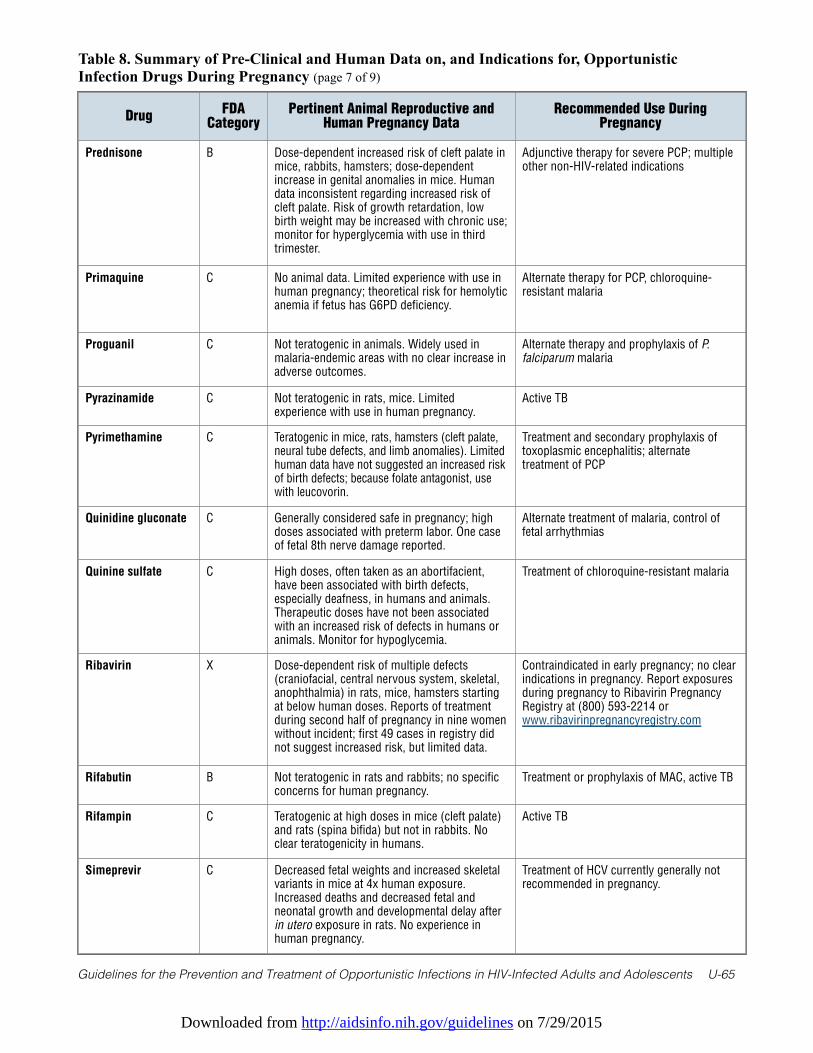
Downloaded from http://aidsinfo.nih.gov/guidelines on 7/29/2015
Guidelines for the Prevention and Treatment of Opportunistic Infections in HIV-Infected Adults and Adolescents U-65
Table 8. Summary of Pre-Clinical and Human Data on, and Indications for, OpportunisticInfection Drugs During Pregnancy (page 7 of 9)
DrugFDA
CategoryPertinent Animal Reproductive and
Human Pregnancy DataRecommended Use During
Pregnancy
Prednisone B Dose-dependent increased risk of cleft palate inmice, rabbits, hamsters; dose-dependentincrease in genital anomalies in mice. Humandata inconsistent regarding increased risk ofcleft palate. Risk of growth retardation, lowbirth weight may be increased with chronic use;monitor for hyperglycemia with use in thirdtrimester.
Adjunctive therapy for severe PCP; multipleother non-HIV-related indications
Primaquine C No animal data. Limited experience with use inhuman pregnancy; theoretical risk for hemolyticanemia if fetus has G6PD deficiency.
Alternate therapy for PCP, chloroquine-resistant malaria
Proguanil C Not teratogenic in animals. Widely used inmalaria-endemic areas with no clear increase inadverse outcomes.
Alternate therapy and prophylaxis of P.falciparum malaria
Pyrazinamide C Not teratogenic in rats, mice. Limitedexperience with use in human pregnancy.
Active TB
Pyrimethamine C Teratogenic in mice, rats, hamsters (cleft palate,neural tube defects, and limb anomalies). Limitedhuman data have not suggested an increased riskof birth defects; because folate antagonist, usewith leucovorin.
Treatment and secondary prophylaxis oftoxoplasmic encephalitis; alternatetreatment of PCP
Quinidine gluconate C Generally considered safe in pregnancy; highdoses associated with preterm labor. One caseof fetal 8th nerve damage reported.
Alternate treatment of malaria, control offetal arrhythmias
Quinine sulfate C High doses, often taken as an abortifacient,have been associated with birth defects,especially deafness, in humans and animals.Therapeutic doses have not been associatedwith an increased risk of defects in humans oranimals. Monitor for hypoglycemia.
Treatment of chloroquine-resistant malaria
Ribavirin X Dose-dependent risk of multiple defects(craniofacial, central nervous system, skeletal,anophthalmia) in rats, mice, hamsters startingat below human doses. Reports of treatmentduring second half of pregnancy in nine womenwithout incident; first 49 cases in registry didnot suggest increased risk, but limited data.
Contraindicated in early pregnancy; no clearindications in pregnancy. Report exposuresduring pregnancy to Ribavirin PregnancyRegistry at (800) 593-2214 orwww.ribavirinpregnancyregistry.com
Rifabutin B Not teratogenic in rats and rabbits; no specificconcerns for human pregnancy.
Treatment or prophylaxis of MAC, active TB
Rifampin C Teratogenic at high doses in mice (cleft palate)and rats (spina bifida) but not in rabbits. Noclear teratogenicity in humans.
Active TB
Simeprevir C Decreased fetal weights and increased skeletalvariants in mice at 4x human exposure.Increased deaths and decreased fetal andneonatal growth and developmental delay afterin utero exposure in rats. No experience inhuman pregnancy.
Treatment of HCV currently generally notrecommended in pregnancy.
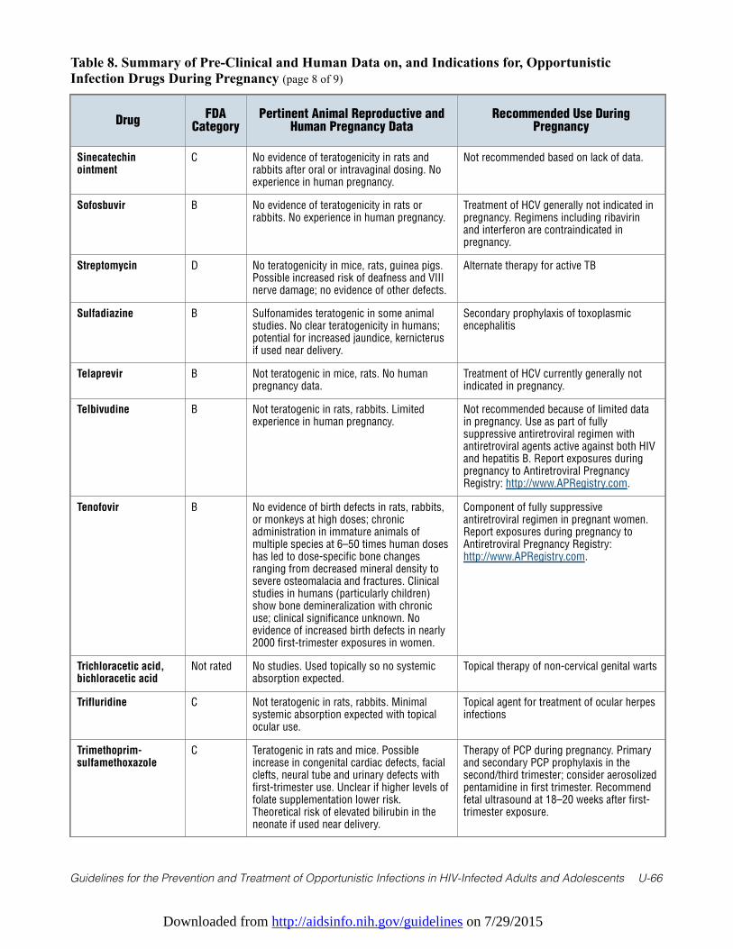
Downloaded from http://aidsinfo.nih.gov/guidelines on 7/29/2015
Guidelines for the Prevention and Treatment of Opportunistic Infections in HIV-Infected Adults and Adolescents U-66
Table 8. Summary of Pre-Clinical and Human Data on, and Indications for, OpportunisticInfection Drugs During Pregnancy (page 8 of 9)
DrugFDA
CategoryPertinent Animal Reproductive and
Human Pregnancy DataRecommended Use During
Pregnancy
Sinecatechinointment
C No evidence of teratogenicity in rats andrabbits after oral or intravaginal dosing. Noexperience in human pregnancy.
Not recommended based on lack of data.
Sofosbuvir B No evidence of teratogenicity in rats orrabbits. No experience in human pregnancy.
Treatment of HCV generally not indicated inpregnancy. Regimens including ribavirinand interferon are contraindicated inpregnancy.
Streptomycin D No teratogenicity in mice, rats, guinea pigs.Possible increased risk of deafness and VIIInerve damage; no evidence of other defects.
Alternate therapy for active TB
Sulfadiazine B Sulfonamides teratogenic in some animalstudies. No clear teratogenicity in humans;potential for increased jaundice, kernicterusif used near delivery.
Secondary prophylaxis of toxoplasmicencephalitis
Telaprevir B Not teratogenic in mice, rats. No humanpregnancy data.
Treatment of HCV currently generally notindicated in pregnancy.
Telbivudine B Not teratogenic in rats, rabbits. Limitedexperience in human pregnancy.
Not recommended because of limited datain pregnancy. Use as part of fullysuppressive antiretroviral regimen withantiretroviral agents active against both HIVand hepatitis B. Report exposures duringpregnancy to Antiretroviral PregnancyRegistry: http://www.APRegistry.com.
Tenofovir B No evidence of birth defects in rats, rabbits,or monkeys at high doses; chronicadministration in immature animals ofmultiple species at 6–50 times human doseshas led to dose-specific bone changesranging from decreased mineral density tosevere osteomalacia and fractures. Clinicalstudies in humans (particularly children)show bone demineralization with chronicuse; clinical significance unknown. Noevidence of increased birth defects in nearly2000 first-trimester exposures in women.
Component of fully suppressiveantiretroviral regimen in pregnant women.Report exposures during pregnancy toAntiretroviral Pregnancy Registry:http://www.APRegistry.com.
Trichloracetic acid,bichloracetic acid
Not rated No studies. Used topically so no systemicabsorption expected.
Topical therapy of non-cervical genital warts
Trifluridine C Not teratogenic in rats, rabbits. Minimalsystemic absorption expected with topicalocular use.
Topical agent for treatment of ocular herpesinfections
Trimethoprim-sulfamethoxazole
C Teratogenic in rats and mice. Possibleincrease in congenital cardiac defects, facialclefts, neural tube and urinary defects withfirst-trimester use. Unclear if higher levels offolate supplementation lower risk.Theoretical risk of elevated bilirubin in theneonate if used near delivery.
Therapy of PCP during pregnancy. Primaryand secondary PCP prophylaxis in thesecond/third trimester; consider aerosolizedpentamidine in first trimester. Recommendfetal ultrasound at 18–20 weeks after first-trimester exposure.

Downloaded from http://aidsinfo.nih.gov/guidelines on 7/29/2015
Guidelines for the Prevention and Treatment of Opportunistic Infections in HIV-Infected Adults and Adolescents U-67
Table 8. Summary of Pre-Clinical and Human Data on, and Indications for, OpportunisticInfection Drugs During Pregnancy (page 9 of 9)
DrugFDA
CategoryPertinent Animal Reproductive and
Human Pregnancy DataRecommended Use During
Pregnancy
Valacyclovir B Not teratogenic in mice, rats, and rabbits.Experience with valacyclovir in pregnancylimited; prodrug of acyclovir, which isconsidered safe for use in pregnancy.
Treatment of HSV and varicella infections inpregnancy
Vancomycin C Not teratogenic in rats, rabbits. Limitedhuman experience.
Serious bacterial infections
Voriconazole D Embryotoxic in rats, rabbits. Teratogenic inrats (cleft palate, hydronephrosis, andossification defects). No experience withhuman use.
Not recommended
Key to Acronyms: ART = antiretroviral therapy; ARV = antiretroviral; CMV = cytomegalovirus; CNS = central nervous system; FDA =Food and Drug Administration; G6PD = Glucose-6-phosphate dehydrogenase; HBV = hepatitis B virus; HCV = hepatitis C virus; HSV =herpes simplex virus; MAC = Mycobacterium avium complex; PCP = Pneumocystis pneumonia; TB = tuberculosis; VIII nerve =vestibulocochlear nerve; WHO = World Health Organization
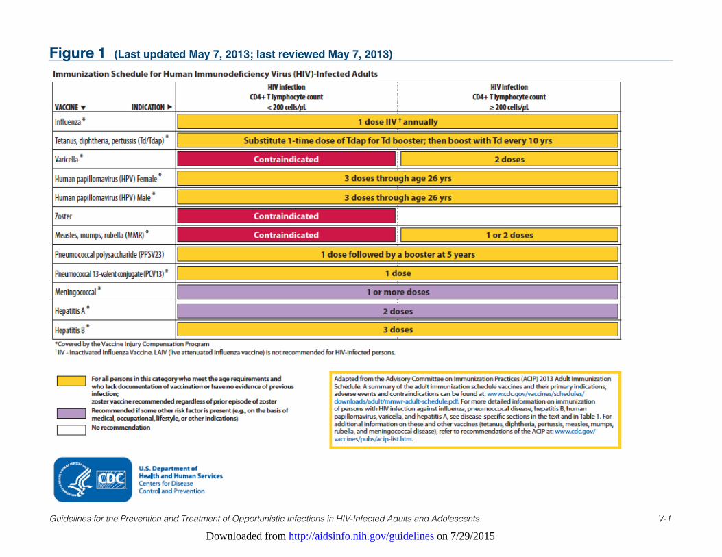
Downloaded from http://aidsinfo.nih.gov/guidelines on 7/29/2015
Guidelines for the Prevention and Treatment of Opportunistic Infections in HIV-Infected Adults and Adolescents V-1
Figure 1 (Last updated May 7, 2013; last reviewed May 7, 2013)
Downloaded from http://aidsinfo.nih.gov/guidelines on 7/29/2015
Guidelines for the Prevention and Treatment of Opportunistic Infections in HIV-Infected Adults and Adolescents W-1
Appendix A. Recommendations to Help HIV-infected PatientsAvoid Exposure to, or Infection from, Opportunistic Pathogens(Last updated May 7, 2013; last reviewed May 7, 2013)
Sexual Exposures
Male latex condoms, when used consistently and correctly during every act of sexual intercourse, are highlyeffective in preventing the sexual transmission of HIV and can reduce the risk for acquiring other sexuallytransmitted diseases (STDs), including chlamydia, gonorrhea, and trichomoniasis(http://www.cdc.gov/condomeffectiveness/latex.htm). Correct and consistent use of male latex condoms notonly reduces the risk of HIV transmission but might reduce the risk for transmission of herpes simplex virus,syphilis, and chancroid when the infected area or potential site of exposure is covered, although data for thiseffect are more limited.1,2 Male condoms also appear to reduce the risk for human papillomavirus associateddiseases (i.e., genital warts, cervical cancer) and thereby mitigate the adverse consequences of infection withHPV. Although data for female condoms are limited, women should consider using them to prevent theacquisition of STDs and reduce their risk of transmitting HIV.3 Spermicides containing nonoxynol-9 are noteffective for HIV/STD prevention4-6 and may increase risk of transmission to uninfected partners;7,8
nonoxynol-9 should not be used as a microbicide or lubricant during vaginal or anal intercourse.
As with many non-sexually transmitted opportunistic infections, intercurrent infections with sexuallytransmitted pathogens (especially pathogens that cause genital ulcers such as herpes simplex, syphilis, andchancroid) can, if untreated, stimulate increases in HIV viral load and consequent declines in CD4 Tlymphocyte (CD4) count.9 Furthermore, acquisition of STDs by HIV-infected patients indicates participationin high-risk sexual behavior that is capable of transmitting HIV to others, the risk for which is substantiallyincreased in the presence of genital tract inflammation (e.g., from gonorrhea or chlamydia) and genital ulcerdisease (e.g., herpes simplex virus-2 infection, syphilis).9-14 All HIV-infected persons, including those whoare asymptomatic, should be tested at initial evaluation for trichomoniasis in women; syphilis, urogenitalgonorrhea, and chlamydia in men and women; and oral gonorrhea, rectal gonorrhea, and rectal chlamydia formale patients reporting receptive sex at these anatomic sites.15-17 Nucleic acid amplification testing methodsare the most sensitive and specific method for the diagnosis of anogenital, oral, and rectal chlamydia andgonorrhea infection. Detailed recommendations for specific testing in HIV-infected persons can be found atthe following site: http://www.cdc.gov/std/treatment. For all sexually active patients, screening should berepeated at least annually and more frequently depending on individual risk or symptoms. In addition toidentifying and treating STDs, providers should communicate prevention messages, discuss sexual and drug-use behaviors, positively reinforce safer behaviors, refer patients for services such as substance abusetreatment, and facilitate partner notification, counseling, and testing.
Specific sex practices should be avoided that might result in oral exposure to feces (e.g., oral-anal contact) toreduce the risk for intestinal infections (e.g., cryptosporidiosis, shigellosis, campylobacteriosis, amebiasis,giardiasis, lymphogranuloma venereum [LGV] serovars of C. trachomatis, hepatitis A [HAV]). Persons whowish to reduce their risk for exposure might consider using dental dams or similar barrier methods for oral-anal and oral-genital contact, changing condoms after anal intercourse, and wearing latex gloves duringdigital-anal contact. Frequent washing of hands and genitals with warm soapy water during and afteractivities that might bring these body parts in contact with feces might further reduce risk for illness.
Sexual transmission of hepatitis C virus (HCV) and infection can occur, especially among HIV-infected menwho have sex with men (MSM).18-20 HIV-infected MSM not known to be infected with HCV, and whopresent with new and unexplained increases in alanine aminotransferase, should be tested for HCV virusinfection. Routine (e.g., annual) HCV testing should be considered for MSM with high risk sexual behaviorsor with a diagnosis of an ulcerative STD.16
HAV can be transmitted sexually, therefore vaccination is recommended for all susceptible MSM, as well as
Downloaded from http://aidsinfo.nih.gov/guidelines on 7/29/2015
Guidelines for the Prevention and Treatment of Opportunistic Infections in HIV-Infected Adults and Adolescents W-2
others with indications for HAV vaccination (e.g., injection-drug users, persons with chronic liver disease orwho are infected with hepatitis B [HBV]). HAV vaccination is also recommended for other HIV-infectedpersons (e.g., injection-drug users, persons with chronic liver disease or who are infected with HBV orHCV). HBV vaccination is recommended for all susceptible HIV-infected patients. HBV infection can occurwhen mucous membranes are exposed to blood or body fluids that contain blood, which might occur duringsome types of sexual contact. HIV-infected patients coinfected with HBV or HCV should be reminded thatuse of latex condoms not only reduces their risk of transmitting HIV to sexual partners but reduces their riskof transmitting these viral hepatitis infections as well.
Injection-Drug-Use Exposures
Injection-drug use is a complex behavior that puts HIV-infected persons at risk for HBV and HCV infection,additional possibly drug-resistant strains of HIV, and other bloodborne pathogens. Providers should assess aperson’s readiness to change this practice and encourage activities to provide education and support directedat recovery. Patients should be counseled to stop using injection drugs and to enter and complete substanceabuse treatment, including relapse prevention programs.21
For patients who continue to inject drugs, health-care providers should advise them to adhere to thefollowing practices:
• Never reuse or share syringes, needles, water, or drug-preparation equipment; if injection equipment thathas been used by other persons is shared, the implements should first be cleaned with bleach and waterbefore use.
• Use only sterile syringes and needles obtained from a reliable source (e.g., pharmacies or syringe-exchange programs).
• Use sterile (e.g., boiled) water to prepare drugs, and if this is not feasible, use clean water from a reliablesource (e.g., fresh tap water); use a new or disinfected container (i.e., cooker) and a new filter (i.e.,cotton) to prepare drugs.
• Clean the injection site with a new alcohol swab before injection.• Safely dispose of syringes and needles after one use.
All susceptible injection-drug–users should be vaccinated against HBV and HAV infection. HIV-infectedinjection drug users not known to be HCV infected who present with new and unexplained increases inalanine aminotransferase should be tested for HCV infection. Routine (e.g., annual) HCV testing should beconsidered for injection drug users who continue to inject drugs.
Environmental and Occupational Exposures
Certain activities or types of employment might increase the risk for exposure to tuberculosis (TB). Theseinclude residency or occupation in correctional institutions and shelters for the homeless, other settingsidentified as high risk by local health authorities, as well as volunteer work or employment in health-carefacilities where patients with TB are treated. Decisions regarding the risk of occupational exposure to TBshould be made in conjunction with a health-care provider and should be based on such factors as thepatient’s specific duties in the workplace, the prevalence of TB in the community, and the degree to whichprecautions designed to prevent the transmission of TB are taken in the workplace. These decisions willaffect the frequency with which the patient should be screened for TB.
Day care providers and parents of children in child care are at increased risk for acquiring cytomegalovirusinfection, cryptosporidiosis, and other infections (e.g., HAV, giardiasis) from children. The risk for acquiringinfection can be diminished by practicing optimal hygienic practices (e.g., washing hands with soap andwater, or alcohol-based hand sanitizers if soap and water are unavailable) after fecal contact (e.g., during
Downloaded from http://aidsinfo.nih.gov/guidelines on 7/29/2015
Guidelines for the Prevention and Treatment of Opportunistic Infections in HIV-Infected Adults and Adolescents W-3
diaper changing) and after contact with urine or saliva.
Occupations involving contact with animals (e.g., veterinary work and employment in pet stores, farms, orslaughterhouses) might pose a risk for toxoplasmosis, cryptosporidiosis, salmonellosis, campylobacteriosis,Bartonella infection, E. coli infection, and other infections of concern to any immunocompromised host(e.g., leptospirosis, brucellosis, Capnocytophaga spp.). However, available data are insufficient to justify arecommendation against HIV-infected persons working in such settings. Wearing gloves and good handhygiene can reduce the risk of infection.
Contact with young farm animals, specifically animals with diarrhea, should be avoided to reduce the risk forcryptosporidiosis. Since soils and sands can be contaminated with Toxoplasma gondii and Cryptosporidiumparvum, persons who have extended contact with these materials (e.g., gardening; playing in or cleaningsandboxes) should wash their hands thoroughly with soap and water following exposure. In areas wherehistoplasmosis is endemic, patients should avoid activities known to be associated with increased risk (e.g.,creating dust when working with surface soil; cleaning chicken coops that are heavily contaminated withcompost droppings; disturbing soil beneath bird-roosting sites; cleaning, remodeling or demolishing oldbuildings; and cave exploring). In areas where coccidioidomycosis is endemic, when possible, patientsshould avoid activities associated with increased risk, including extensive exposure to disturbed native soil(e.g., building excavation sites, during dust storms).
Pet-Related Exposures
Health-care providers should advise HIV-infected persons of the potential risk posed by pet ownership.However, they should be sensitive to the psychological benefits of pet ownership and should not routinelyadvise HIV-infected persons to part with their pets. Specifically, providers should advise HIV-infectedpatients of the following precautions.
GeneralHIV-infected persons should avoid direct contact with stool from pets or stray animals. Veterinary careshould be sought when a pet develops diarrheal illness. If possible, HIV-infected persons should avoidcontact with animals that have diarrhea.
When obtaining a new pet, HIV-infected patients should avoid animals aged <6 months (or <1 year for cats)and specifically animals with diarrhea. Because the hygienic and sanitary conditions in pet-breedingfacilities, pet stores, and animal shelters vary, patients should be cautious when obtaining pets from thesesources. Stray animals should also be avoided, and specifically those with diarrhea.
Gloves should always be worn when handling feces or cleaning areas that might have been contaminated byfeces from pets. Patients should wash their hands after handling pets and also before eating. Patients,especially those with CD4 cell counts < 200 cells/µL should avoid direct contact with all animal feces toreduce the risk for toxoplasmosis, cryptosporidiosis, salmonellosis, campylobacteriosis, E. coli infection, andother infectious illnesses. HIV-infected persons should limit or avoid direct exposure to calves and lambs(e.g., farms, petting zoos). Paying attention to hand hygiene (i.e., washing hands with soap and water, oralcohol-based hand sanitizers if soap and water are unavailable) and avoiding direct contact with stool areimportant when visiting premises where these animals are housed or exhibited.
Patients should not allow pets, particularly cats, to lick patients’ open cuts or wounds and should take care toavoid any animal bites. Patients should wash all animal bites, animal scratches, or wounds licked by animalspromptly with soap and water and seek medical attention. A course of antimicrobial therapy might berecommended if the wounds are moderate or severe, demonstrate crush injury and edema, involve the bonesof a joint, involve a puncture of the skin near a joint, or involve a puncture of a joint directly.
Downloaded from http://aidsinfo.nih.gov/guidelines on 7/29/2015
Guidelines for the Prevention and Treatment of Opportunistic Infections in HIV-Infected Adults and Adolescents W-4
CatsPatients should be aware that cat ownership may under some circumstances increase their risk fortoxoplasmosis and Bartonella infection, and enteric infections. Patients who elect to obtain a cat shouldadopt or purchase an animal aged >1 year and in good health to reduce the risk for cryptosporidiosis,Bartonella infection, salmonellosis, campylobacteriosis, and E. coli infection.
Litter boxes should be cleaned daily, preferably by an HIV-negative, non-pregnant person; if HIV-infectedpatients perform this task, they should wear gloves and wash their hands thoroughly afterward to reduce therisk for toxoplasmosis. To further reduce the risk for toxoplasmosis, HIV-infected patients should keep catsindoors, not allow them to hunt, and not feed them raw or undercooked meat. Although declawing is notusually advised, patients should avoid activities that might result in cat scratches or bites to reduce the riskfor Bartonella infection. Patients should also wash sites of cat scratches or bites promptly and should notallow cats to lick patients’ open cuts or wounds. Care of cats should include flea control to reduce the risk forBartonella infection. Testing cats for toxoplasmosis or Bartonella infection is not recommended, as suchtests cannot accurately identify animals that pose a current risk for human infection.
BirdsScreening healthy birds for Cryptococcus neoformans, Mycobacterium avium, or Histoplasma capsulatum isnot recommended.
OtherHIV-infected persons should avoid or limit contact with reptiles (e.g., snakes, lizards, iguanas, and turtles)and chicks and ducklings because of the high risk for exposure to Salmonella spp. Gloves should be usedduring aquarium cleaning to reduce the risk for infection with Mycobacterium marinum. Contact with exoticpets (e.g., nonhuman primates) should be avoided.
Food- and Water-Related Exposures
FoodContaminated food is a common source of enteric infections. Transmission most often occurs by ingestion ofundercooked foods or by cross-contamination of foods in the kitchen.
Health-care providers should advise HIV-infected persons, particularly those with a CD4 count <200cells/µL, not to eat raw or undercooked eggs, including specific foods that might contain raw eggs (e.g.,certain preparation of Hollandaise sauce, Caesar salad dressings, homemade mayonnaises, uncooked cookieand cake batter, eggnog); raw or undercooked poultry, meat, and seafood (raw shellfish in particular);unpasteurized dairy products (including milk and cheese); unpasteurized fruit juices; and raw seed sprouts(e.g., alfalfa sprouts or mung bean sprouts).
Meat and poultry are safest when adequate cooking is confirmed by thermometer. Current U.S. Department ofAgriculture (USDA) guidance (http://www.fsis.usda.gov/Factsheets/Keep_Food_Safe_Food_Safety_Basics/index.asp) is that the internal temperature be at least 145°F (63°C) for whole cuts of meat, 160°F (71°C) forground meat excluding poultry, and 165°F (74°C) for poultry; whole cuts of meat and poultry should rest at leastthree minutes before carving and consuming. Immunocompromised persons who wish to maximally ensure theircooked meats are safe to eat may choose to use the following recommendations: the internal temperature shouldbe at least 165°F (74°C) for all types of red meats and 180°F (82°C) for poultry. If a thermometer is not usedwhen cooking meats, the risk for illness is decreased by eating poultry and meat that have no trace of pink color.However, color change of the meat (e.g., absence of pink) does not always correlate with internal temperature.Irradiated meats, if available, are predicted to eliminate the risk of foodborne enteric infection. Use ofmicrowaves as a primary means of cooking of potentially contaminated foods (e.g., meats, hot dogs) should beavoided because microwave cooking is not uniform.
Downloaded from http://aidsinfo.nih.gov/guidelines on 7/29/2015
Guidelines for the Prevention and Treatment of Opportunistic Infections in HIV-Infected Adults and Adolescents W-5
Produce items should be washed thoroughly; providers may wish to advise patients that produce is safestwhen cooked.
Health-care providers should advise HIV-infected persons to avoid cross-contamination of foods. Saladpreparation prior to handling of raw meats or other uncooked, potentially contaminated foods decreases risk.Uncooked meats, including hot dogs, and their juices should not come into contact with other foods. Hands,cutting boards, counters, knives, and other utensils should be washed thoroughly (preferably in a dish washeron hot cycle) after contact with uncooked foods.
Soft cheeses (e.g., feta, Brie, Camembert, blue-veined, and Mexican-style cheese such as queso fresco) andprepared deli foods (including coldcuts, salads, hummus, hot dogs, pâtés) are potential sources of Listeriamonocytogenes infection, which can lead to serious, even fatal, systemic infection in HIV-infected patientswith low CD4 cell counts; consumption of these foods should be avoided.
Hard cheeses, processed cheeses, cream cheese, including slices and spreads; cottage cheese or yogurt; andcanned or shelf-stable pâté and meat spreads need not be avoided. Avoid raw or unpasteurized milk,including goat’s milk, or foods that contain unpasteurized milk or milk products.
Additional and more detailed information on the safe handling and preparation of food for persons with HIVinfection can be found through the websites of the Food and Drug Administration (http://www.fda.gov/ForConsumers/ByAudience/ForPatientAdvocates/HIVandAIDSActivities/ucm135844.htm) and the USDA(http://www.fsis.usda.gov/pdf/food_safety_for_people_with_hiv.pdf).
WaterPatients should not drink water directly from lakes or rivers because of the risk for cryptosporidiosis,giardiasis, and toxoplasmosis. Waterborne infection can also result from swallowing water duringrecreational activities. All HIV-infected patients should avoid swimming in water that is probablycontaminated with human or animal waste and should avoid swallowing water during swimming. Patients,especially those with CD4 cell counts <200 cells/µL, should also be made aware that swimming or playing inlakes, rivers, and oceans as well as some swimming pools, recreational water parks, and ornamental waterfountains can expose them to enteric pathogens (e.g., Cryptosporidium, Giardia, norovirus, Shiga toxin-producing E. coli) that cause diarrheal illness and to which their HIV infection makes them more susceptible.
Outbreaks of diarrheal illness have been linked to drinking water from municipal water supplies. Duringoutbreaks or in other situations in which a community boil-water advisory is issued, boiling water for >1minute will eliminate the risk for most viral, bacterial, and parasitic causes of diarrhea, includingcryptosporidiosis. Using submicron, personal-use water filters (home/office types) or drinking bottled watermight also reduce the risk from municipal and from well water.
Available data are inadequate to support a recommendation that all HIV-infected persons boil or otherwiseavoid drinking tap water in non-outbreak settings. However, persons who wish to take independent action toreduce their risk for waterborne cryptosporidiosis might take precautions similar to those recommendedduring outbreaks. Such decisions are best made in conjunction with a health-care provider. Persons whochoose to use a personal-use filter or bottled water should be aware of the complexities involved in selectingthe appropriate products, the lack of enforceable standards for destruction or removal of oocysts, productcost, and the difficulty of using these products consistently.
Patients taking precautions to avoid acquiring pathogens from drinking water should be advised that icemade from contaminated tap water also can be a source of infection. Patients should also be made aware thatfountain beverages served in restaurants, bars, theaters, and other public places also might pose a risk,because these beverages, and the ice they might contain, are usually made from tap water. Nationallydistributed brands of bottled or canned water and carbonated soft drinks are safe to drink. Commerciallypackaged (i.e., sealed at the factory and unopened), non-carbonated soft drinks and fruit juices that do notrequire refrigeration until after they are opened (i.e., those that can be stored unrefrigerated on grocery
Downloaded from http://aidsinfo.nih.gov/guidelines on 7/29/2015
Guidelines for the Prevention and Treatment of Opportunistic Infections in HIV-Infected Adults and Adolescents W-6
shelves) also are safe. Nationally distributed brands of frozen fruit juice concentrate are safe if they arereconstituted by users with water from a safe source. Fruit juices that must be kept refrigerated from the timethey are processed to the time they are consumed might be either fresh (i.e., unpasteurized) or heat treated(i.e., pasteurized); only juices labeled as pasteurized should be considered safe to consume. Other pasteurizedbeverages and beers also are considered safe.
Travel-Related Exposures
HIV-infected travelers to developing countries, especially travelers who are severely immunosuppressed, riskexposure to both opportunistic and non-opportunistic pathogens not prevalent in the United States. Health-care providers or specialists in travel medicine (a list can be found at http://www.istm.com) should beconsulted 4 to 6 weeks in advance of travel to fully review and implement all measures necessary to preventillness abroad. The Centers for Disease Control and Prevention (CDC) maintain a website accessible totravelers and their care providers at http://www.cdc.gov/travel and regularly publishes recommendations forprevention of disease while traveling in the CDC’s Yellow Book (Health Information for InternationalTravel).22 The CDC’s travel website allows users to locate prevention recommendations according togeographic destination and to find updates on international disease outbreaks that might pose a health threatto travelers. A detailed review of concerns faced by immunocompromised persons traveling abroad isavailable at http://wwwnc.cdc.gov/travel/yellowbook/2012/chapter-8-advising-travelers-with-specific-needs/immunocompromised-travelers.htm in the Yellow Book.
The following summary advice should be considered for all HIV-infected travelers but does substitute fordestination-specific consultation with a travel medicine specialist.
The risk for foodborne and waterborne infections among HIV-infected persons is magnified during travel toeconomically developing countries. Travelers to such countries may wish to additionally consult the sectionFood- and Water-Related Exposures, above, as well as recommendations for food and water precautions andwater disinfection in the CDC Yellow Book (Health Information for Travelers).22 Specifically, persons whotravel to economically developing areas should avoid foods and beverages that might be contaminated, aswell as tap water, ice made with tap water, and items sold by street vendors. Raw fruits or vegetables thatmight have been washed in tap water should be avoided. Foods and beverages that are usually safe includesteaming hot foods, fruits that are peeled by the traveler, unopened and properly bottled (includingcarbonated) beverages, hot coffee and tea, beer, wine, and water that is brought to a rolling boil for 1 minute.Treating water with iodine or chlorine can be as effective as boiling for preventing infections with mostpathogens. Iodine and chlorine treatments may not prevent infection with Cryptosporidium; however thesetreatments can be used when boiling is not practical.
Waterborne infections might result from swallowing water during recreational activities. To reduce the riskfor parasitic (e.g., cryptosporidiosis, giardiasis, toxoplasmosis) and bacterial infections, patients should avoidswallowing water during swimming and should not swim in water that might be contaminated (e.g., withsewage or animal waste). HIV-infected persons traveling to developing countries should also be advised tonot use tap water to brush their teeth.
Scrupulous attention to safe food and water consumption and good hygiene (i.e., regularly washing hands withsoap and water, or alcohol-based hand sanitizers if soap and water are unavailable) are the most effectivemethods for reducing risk of travelers’ diarrhea. Antimicrobial prophylaxis for travelers’ diarrhea is notrecommended routinely for HIV-infected persons traveling to developing countries. Such preventive therapycan have adverse effects, can promote the emergence of drug-resistant organisms, and can increase the risk of C.difficile-associated diarrhea. Nonetheless, studies (none involving an HIV-infected population) have reportedthat prophylaxis can reduce the risk for diarrhea among travelers. Under selected circumstances (e.g., those inwhich the risk for infection is high and the period of travel brief), the health-care provider and patient mightweigh the potential risks and benefits and decide that antibiotic prophylaxis is warranted.
Downloaded from http://aidsinfo.nih.gov/guidelines on 7/29/2015
Guidelines for the Prevention and Treatment of Opportunistic Infections in HIV-Infected Adults and Adolescents W-7
HIV-infected travelers to developing countries should consider carrying a sufficient supply of anantimicrobial agent to be taken empirically if diarrhea occurs. Antimicrobial resistance among entericbacterial pathogens outside the United States is a growing public health problem; therefore, the choice ofantibiotic should be made in consultation with a clinician based on the traveler’s destination. Travelersshould consult a physician if they develop severe diarrhea that does not respond to empirical therapy, if theirstools contain blood, they develop fever with shaking chills, or dehydration occurs. Antiperistaltic agents(e.g., diphenoxylate and loperamide) are used for treating diarrhea; however, they should not be used bypatients with high fever or with blood in the stool, and their use should be discontinued if symptoms persistfor more than 48 hours.
Live-virus vaccines should, in general, not be used. An exception is measles vaccine, which is recommendedfor non-immune persons. However, measles vaccine is not recommended for persons who are severelyimmunosuppressed. Severely immunosuppressed persons who must travel to measles-endemic countries shouldconsult a travel medicine specialist regarding possible utility of prophylaxis with immune globulin. Anotherexception is varicella vaccine, which can be administered to asymptomatic susceptible persons with a CD4 cellcount ≥200 cells/µL. For adults and adolescents with CD4 cell counts <200 cells/µL, varicella-zoster immuneglobulin (VariZIG™) is indicated after close contact with a person who has active varicella or zoster and anti-herpetic antiviral therapy (e.g., acyclovir, famciclovir, valacyclovir) is recommended in the event vaccinationor exposure results in clinical disease (for further details, see Varicella-Zoster Virus Diseases chapter). Personsat risk for and non-immune to polio and typhoid fever or who require influenza vaccination should beadministered only inactivated formulations of these vaccines not live-attenuated preparations.
Yellow fever vaccine is a live-virus vaccine with uncertain safety and efficacy among HIV-infected persons.Travelers with asymptomatic HIV infection who cannot avoid potential exposure to yellow fever should beoffered vaccination. If travel to a zone with yellow fever is necessary and vaccination is not administered,patients should be advised of the risk, instructed in methods for avoiding the bites of vector mosquitoes, andprovided a vaccination waiver letter. Preparation for travel should include a review and updating of routinevaccinations, including diphtheria, tetanus, acellular pertussis, and influenza.
Killed and recombinant vaccines (e.g., influenza, diphtheria, tetanus, rabies, HAV, HBV, Japaneseencephalitis, meningococcal vaccines) should usually be used for HIV-infected persons just as they would beused for non-HIV–infected persons anticipating travel. Comprehensive and regularly updated informationregarding recommended vaccinations and recommendations when a vaccination is contraindicated are listedby vaccine at http://www.cdc.gov/vaccines/pubs/acip-list.htm.
References
1. Martin ET, Krantz E, Gottlieb SL, et al. A pooled analysis of the effect of condoms in preventing HSV-2 acquisition.Arch Intern Med. Jul 13 2009;169(13):1233-1240. Available at http://www.ncbi.nlm.nih.gov/pubmed/19597073.
2. Koss CA, Dunne EF, Warner L. A systematic review of epidemiologic studies assessing condom use and risk ofsyphilis. Sex Transm Dis. Jul 2009;36(7):401-405. Available at http://www.ncbi.nlm.nih.gov/pubmed/19455075.
3. Minnis AM, Padian NS. Effectiveness of female controlled barrier methods in preventing sexually transmittedinfections and HIV: current evidence and future research directions. Sex Transm Infect. Jun 2005;81(3):193-200.Available at http://www.ncbi.nlm.nih.gov/pubmed/15923284.
4. Roddy RE, Zekeng L, Ryan KA, Tamoufe U, Weir SS, Wong EL. A controlled trial of nonoxynol 9 film to reduce male-to-female transmission of sexually transmitted diseases. N Engl J Med. Aug 20 1998;339(8):504-510. Available athttp://www.ncbi.nlm.nih.gov/pubmed/9709043.
5. Roddy RE, Zekeng L, Ryan KA, Tamoufe U, Tweedy KG. Effect of nonoxynol-9 gel on urogenital gonorrhea andchlamydial infection: a randomized controlled trial. JAMA. Mar 6 2002;287(9):1117-1122. Available athttp://www.ncbi.nlm.nih.gov/pubmed/11879108.
6. Richardson BA, Lavreys L, Martin HL, Jr., et al. Evaluation of a low-dose nonoxynol-9 gel for the prevention ofsexually transmitted diseases: a randomized clinical trial. Sex Transm Dis. Jul 2001;28(7):394-400. Available at
Downloaded from http://aidsinfo.nih.gov/guidelines on 7/29/2015
Guidelines for the Prevention and Treatment of Opportunistic Infections in HIV-Infected Adults and Adolescents W-8
http://www.ncbi.nlm.nih.gov/pubmed/11460023.7. Van Damme L, Ramjee G, Alary M, et al. Effectiveness of COL-1492, a nonoxynol-9 vaginal gel, on HIV-1
transmission in female sex workers: a randomised controlled trial. Lancet. Sep 28 2002;360(9338):971-977. Availableat http://www.ncbi.nlm.nih.gov/pubmed/12383665.
8. Phillips DM, Taylor CL, Zacharopoulos VR, Maguire RA. Nonoxynol-9 causes rapid exfoliation of sheets of rectalepithelium. Contraception. Sep 2000;62(3):149-154. Available at http://www.ncbi.nlm.nih.gov/pubmed/11124363.
9. Modjarrad K, Vermund SH. Effect of treating co-infections on HIV-1 viral load: a systematic review. Lancet Infect Dis.Jul 2010;10(7):455-463. Available at http://www.ncbi.nlm.nih.gov/pubmed/20610327.
10. Fleming DT, Wasserheit JN. From epidemiological synergy to public health policy and practice: the contribution ofother sexually transmitted diseases to sexual transmission of HIV infection. Sex Transm Infect. Feb 1999;75(1):3-17.Available at http://www.ncbi.nlm.nih.gov/pubmed/10448335.
11. Rottingen JA, Cameron DW, Garnett GP. A systematic review of the epidemiologic interactions between classicsexually transmitted diseases and HIV: how much really is known? Sex Transm Dis. Oct 2001;28(10):579-597.Available at http://www.ncbi.nlm.nih.gov/pubmed/11689757.
12. McClelland RS, Wang CC, Mandaliya K, et al. Treatment of cervicitis is associated with decreased cervical shedding ofHIV-1. AIDS. Jan 5 2001;15(1):105-110. Available at http://www.ncbi.nlm.nih.gov/pubmed/11192850.
13. Cohen MS, Hoffman IF, Royce RA, et al. Reduction of concentration of HIV-1 in semen after treatment of urethritis:implications for prevention of sexual transmission of HIV-1. AIDSCAP Malawi Research Group. Lancet. Jun 281997;349(9069):1868-1873. Available at http://www.ncbi.nlm.nih.gov/pubmed/9217758.
14. Ghys PD, Fransen K, Diallo MO, et al. The associations between cervicovaginal HIV shedding, sexually transmitteddiseases and immunosuppression in female sex workers in Abidjan, Cote d'Ivoire. AIDS. Oct 1997;11(12):F85-93.Available at http://www.ncbi.nlm.nih.gov/pubmed/9342059.
15. Centers for Disease C, Prevention, Health R, Services A, National Institutes of H, America HIVMAotIDSo.Incorporating HIV prevention into the medical care of persons living with HIV. Recommendations of CDC, the HealthResources and Services Administration, the National Institutes of Health, and the HIV Medicine Association of theInfectious Diseases Society of America. MMWR Recomm Rep. Jul 18 2003;52(RR-12):1-24. Available athttp://www.ncbi.nlm.nih.gov/pubmed/12875251.
16. Workowski KA, Berman S, Centers for Disease C, Prevention. Sexually transmitted diseases treatment guidelines, 2010.MMWR Recomm Rep. Dec 17 2010;59(RR-12):1-110. Available at http://www.ncbi.nlm.nih.gov/pubmed/21160459.
17. Aberg JA, Kaplan JE, Libman H, et al. Primary care guidelines for the management of persons infected with humanimmunodeficiency virus: 2009 update by the HIV medicine Association of the Infectious Diseases Society of America.Clin Infect Dis. Sep 1 2009;49(5):651-681. Available at http://www.ncbi.nlm.nih.gov/pubmed/19640227.
18. Danta M, Brown D, Bhagani S, et al. Recent epidemic of acute hepatitis C virus in HIV-positive men who have sex withmen linked to high-risk sexual behaviours. AIDS. May 11 2007;21(8):983-991. Available athttp://www.ncbi.nlm.nih.gov/pubmed/17457092.
19. Urbanus AT, van de Laar TJ, Stolte IG, et al. Hepatitis C virus infections among HIV-infected men who have sex with men:an expanding epidemic. AIDS. Jul 31 2009;23(12):F1-7. Available at http://www.ncbi.nlm.nih.gov/pubmed/19542864.
20. Centers for Disease C, Prevention. Sexual transmission of hepatitis C virus among HIV-infected men who have sex withmen--New York City, 2005-2010. MMWR Morb Mortal Wkly Rep. Jul 22 2011;60(28):945-950. Available athttp://www.ncbi.nlm.nih.gov/pubmed/21775948.
21. CDC. HIV prevention bulletin: medical advice for persons who inject illicit drugs - May 9, 1997.22. Centers for Disease C, Prevention. CDC Health Information for International Travel, 2012. Atlanta, GA: US
Department of Heath and Human Services, Public Health Service; 2012.
Downloaded from http://aidsinfo.nih.gov/guidelines on 7/29/2015
Guidelines for the Prevention and Treatment of Opportunistic Infections in HIV-Infected Adults and Adolescents X-1
Appendix B. List of Abbreviations (Last updated May 7, 2013; last reviewedMay 7, 2013)
Acronym/Abbreviation Definition
ABGs arterial blood gassesACTG AIDS Clinical Trials GroupAFB acid-fact bacilliAIN anal intraepithelial neoplasiaALT alanine aminotransferaseanti-HBc hepatitis B core antibodyanti-HBs hepatitis B surface antibodyART antiretroviral therapyARV antiretroviralASCCP American Society for Colposcopy and Cervical PathologyASC-H atypical squamous cells—cannot exclude high grade cervical
squamous intraepithelial lesionASC-US atypical squamous cells of uncertain significanceAST serum aspartate aminotransferaseAUC area under the curveBA bacillary angiomatosisBAL bronchoalveolar lavageBID twice a dayBIW twice a weekCAP community-acquired pneumoniaCAPD continuous ambulatory peritoneal dialysisCD4 CD4 T lymphocyte cellCDC the Centers for Disease Control and PreventionCDI Clostridium difficile-associated infectionCES-D Center for Epidemiologic Studies Depression ScaleCFU colony-forming unitCIA chemiluminescence immunoassays CIN cervical intraepithelial neoplasiaCmax maximum concentrationCmin minimum concentrationCMV cytomegalovirusCNS central nervous systemCPE central nervous system penetration effectivenessCrCl creatinine clearanceCSF cerebrospinal fluidCT computed tomography
Downloaded from http://aidsinfo.nih.gov/guidelines on 7/29/2015
Guidelines for the Prevention and Treatment of Opportunistic Infections in HIV-Infected Adults and Adolescents X-2
CYP3A4 Cytochrome P450 3A4DAAs direct acting antiviral agentsDOT directly observed therapyDS double strengthEDTA ethylenediaminetetraacetic acidEIAs enzyme immunoassays EM erythema multiformeFDA Food and Drug AdministrationFTA-ABS fluorescent treponemal antibody absorbed g gramG6PD Glucose-6-phosphate dehydrogenaseGFR glomerular filtration rateGI gastrointestinalHAV hepatitis A virusHBV hepatitis B virusHCV hepatitis C virusHHV-8 human herpesvirus-8HPA hypothalamic-pituitary-adrenalHPV human papillomavirusHSIL high grade cervical squamous intraepithelial lesionHSV herpes simplex virusHSV-1 herpes simplex virus 1HSV-2 herpes simplex virus 2ICP intracranial pressureICU intensive care unitIFN interferonIgG immunoglobulin GIgM immunoglobulin MIGRA interferon-gamma release assaysIM intramuscularIND investigational new drugIRIS immune reconstitution inflammatory syndromeIRU immune recovery uveitisIV intravenousIVIG intravenous immunoglobulinJCV JC virusKS Kaposi SarcomaLEEP loop electrosurgical excision procedureLP lumbar punctureLSIL low grade squamous intraepithelial lesion
Downloaded from http://aidsinfo.nih.gov/guidelines on 7/29/2015
Guidelines for the Prevention and Treatment of Opportunistic Infections in HIV-Infected Adults and Adolescents X-3
LTBI latent tuberculosis infectionMAC Mycobacterium avium complexMAI Mycobacterium avium intracellulareMCD multicentric Castleman’s diseaseMDR TB multi-drug-resistant tuberculosismg milligrammmHg millimeters of mercuryMSM men who have sex with menMTB Mycobacterium tuberculosisNAA nucleic acid amplificationNNRTI non-nucleoside reverse transcriptase inhibitorNRTI nucleoside reverse transcriptase inhibitorsNSAID non-steroidal anti-inflammatory drugsNVP nevirapineOI opportunistic infectionPCP Pneumocystis pneumoniaPCR polymerase chain reactionPEL primary effusion lymphomaPK pharmacokineticPML progressive multifocal LeukoencephalopathyPO orallyPORN Progressive Outer Retinal NecrosisPPV polysaccharide vaccinePSI pneumonia severity indexq(n)h every ”n” hoursqAM every morningQID four times a dayqPM every eveningRPR rapid plasma reagin RVR rapid virological responseSCr serum creatinineSJS Stevens-Johnson syndromeSLE systemic lupus erythematosusSQ subcutaneousSS single strengthSTD sexually transmitted diseaseSVR sustained virologic responseTB tuberculosisTDM therapeutic drug monitoringTE Toxoplasma encephalitis
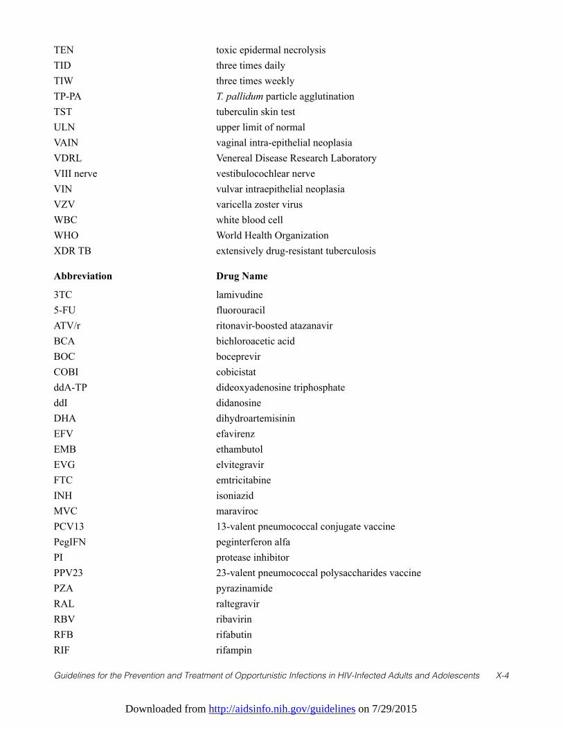
Downloaded from http://aidsinfo.nih.gov/guidelines on 7/29/2015
Guidelines for the Prevention and Treatment of Opportunistic Infections in HIV-Infected Adults and Adolescents X-4
TEN toxic epidermal necrolysisTID three times dailyTIW three times weeklyTP-PA T. pallidum particle agglutination TST tuberculin skin testULN upper limit of normalVAIN vaginal intra-epithelial neoplasiaVDRL Venereal Disease Research Laboratory VIII nerve vestibulocochlear nerveVIN vulvar intraepithelial neoplasiaVZV varicella zoster virusWBC white blood cellWHO World Health OrganizationXDR TB extensively drug-resistant tuberculosis
Abbreviation Drug Name
3TC lamivudine5-FU fluorouracilATV/r ritonavir-boosted atazanavirBCA bichloroacetic acidBOC boceprevirCOBI cobicistatddA-TP dideoxyadenosine triphosphateddI didanosineDHA dihydroartemisininEFV efavirenzEMB ethambutolEVG elvitegravirFTC emtricitabineINH isoniazidMVC maravirocPCV13 13-valent pneumococcal conjugate vaccinePegIFN peginterferon alfaPI protease inhibitorPPV23 23-valent pneumococcal polysaccharides vaccinePZA pyrazinamideRAL raltegravirRBV ribavirinRFB rifabutinRIF rifampin
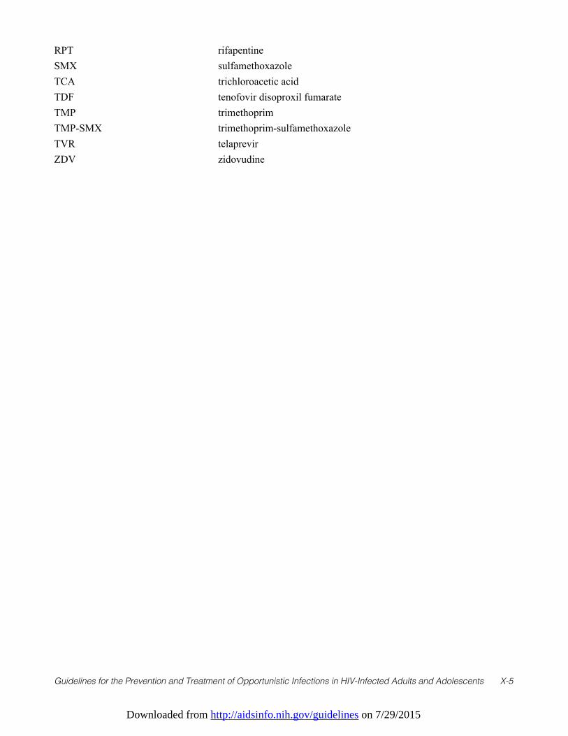
Downloaded from http://aidsinfo.nih.gov/guidelines on 7/29/2015
Guidelines for the Prevention and Treatment of Opportunistic Infections in HIV-Infected Adults and Adolescents X-5
RPT rifapentineSMX sulfamethoxazoleTCA trichloroacetic acidTDF tenofovir disoproxil fumarateTMP trimethoprimTMP-SMX trimethoprim-sulfamethoxazoleTVR telaprevirZDV zidovudine
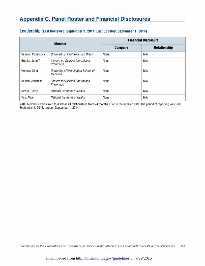
Downloaded from http://aidsinfo.nih.gov/guidelines on 7/29/2015
Guidelines for the Prevention and Treatment of Opportunistic Infections in HIV-Infected Adults and Adolescents Y-1
Appendix C. Panel Roster and Financial Disclosures
Leadership (Last Reviewed: September 1, 2014; Last Updated: September 1, 2014)
MemberFinancial Disclosure
Company Relationship
Benson, Constance University of California, San Diego None N/A
Brooks, John T. Centers for Disease Control andPrevention
None N/A
Holmes, King University of Washington School ofMedicine
None N/A
Kaplan, Jonathan Centers for Disease Control andPrevention
None N/A
Masur, Henry National Institutes of Health None N/A
Pau, Alice National Institutes of Health None N/A
Note: Members were asked to disclose all relationships from 24 months prior to the updated date. The period of reporting was fromSeptember 1, 2012, through September 1, 2014.
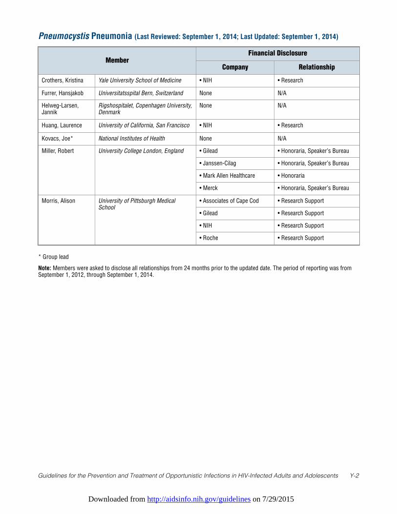
Downloaded from http://aidsinfo.nih.gov/guidelines on 7/29/2015
Guidelines for the Prevention and Treatment of Opportunistic Infections in HIV-Infected Adults and Adolescents Y-2
Pneumocystis Pneumonia (Last Reviewed: September 1, 2014; Last Updated: September 1, 2014)
MemberFinancial Disclosure
Company Relationship
Crothers, Kristina Yale University School of Medicine • NIH • Research
Furrer, Hansjakob Universitatsspital Bern, Switzerland None N/A
Helweg-Larsen,Jannik
Rigshospitalet, Copenhagen University,Denmark
None N/A
Huang, Laurence University of California, San Francisco • NIH • Research
Kovacs, Joe* National Institutes of Health None N/A
Miller, Robert University College London, England • Gilead • Honoraria, Speaker’s Bureau
• Janssen-Cilag • Honoraria, Speaker’s Bureau
• Mark Allen Healthcare • Honoraria
• Merck • Honoraria, Speaker’s Bureau
Morris, Alison University of Pittsburgh MedicalSchool
• Associates of Cape Cod • Research Support
• Gilead • Research Support
• NIH • Research Support
• Roche • Research Support
* Group lead
Note: Members were asked to disclose all relationships from 24 months prior to the updated date. The period of reporting was fromSeptember 1, 2012, through September 1, 2014.
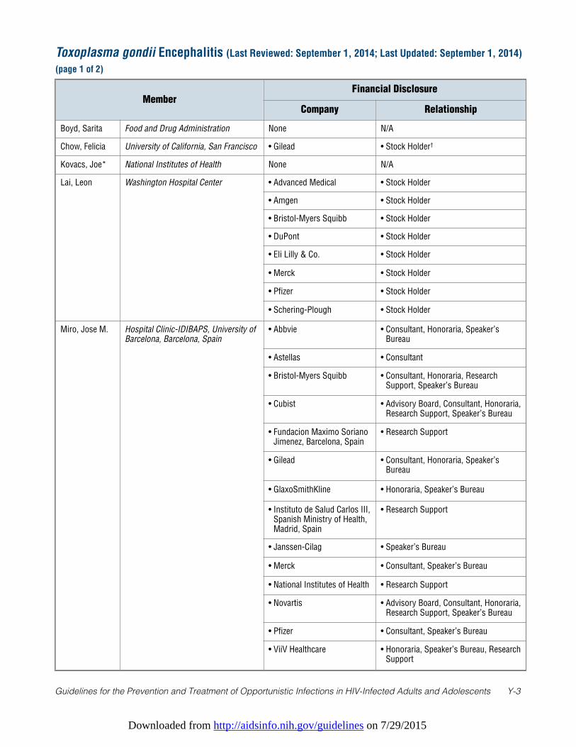
Downloaded from http://aidsinfo.nih.gov/guidelines on 7/29/2015
Guidelines for the Prevention and Treatment of Opportunistic Infections in HIV-Infected Adults and Adolescents Y-3
Toxoplasma gondii Encephalitis (Last Reviewed: September 1, 2014; Last Updated: September 1, 2014)
(page 1 of 2)
MemberFinancial Disclosure
Company Relationship
Boyd, Sarita Food and Drug Administration None N/A
Chow, Felicia University of California, San Francisco • Gilead • Stock Holder†
Kovacs, Joe* National Institutes of Health None N/A
Lai, Leon Washington Hospital Center • Advanced Medical • Stock Holder
• Amgen • Stock Holder
• Bristol-Myers Squibb • Stock Holder
• DuPont • Stock Holder
• Eli Lilly & Co. • Stock Holder
• Merck • Stock Holder
• Pfizer • Stock Holder
• Schering-Plough • Stock Holder
Miro, Jose M. Hospital Clinic-IDIBAPS, University ofBarcelona, Barcelona, Spain
• Abbvie • Consultant, Honoraria, Speaker’sBureau
• Astellas • Consultant
• Bristol-Myers Squibb • Consultant, Honoraria, ResearchSupport, Speaker’s Bureau
• Cubist • Advisory Board, Consultant, Honoraria,Research Support, Speaker’s Bureau
• Fundacion Maximo SorianoJimenez, Barcelona, Spain
• Research Support
• Gilead • Consultant, Honoraria, Speaker’sBureau
• GlaxoSmithKline • Honoraria, Speaker’s Bureau
• Instituto de Salud Carlos III,Spanish Ministry of Health,Madrid, Spain
• Research Support
• Janssen-Cilag • Speaker’s Bureau
• Merck • Consultant, Speaker’s Bureau
• National Institutes of Health • Research Support
• Novartis • Advisory Board, Consultant, Honoraria,Research Support, Speaker’s Bureau
• Pfizer • Consultant, Speaker’s Bureau
• ViiV Healthcare • Honoraria, Speaker’s Bureau, ResearchSupport
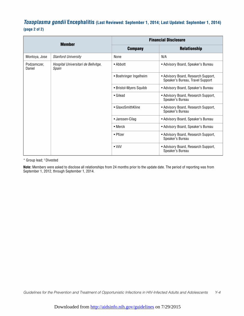
Downloaded from http://aidsinfo.nih.gov/guidelines on 7/29/2015
Guidelines for the Prevention and Treatment of Opportunistic Infections in HIV-Infected Adults and Adolescents Y-4
* Group lead; † Divested
Note: Members were asked to disclose all relationships from 24 months prior to the update date. The period of reporting was fromSeptember 1, 2012, through September 1, 2014.
Toxoplasma gondii Encephalitis (Last Reviewed: September 1, 2014; Last Updated: September 1, 2014)
(page 2 of 2)
MemberFinancial Disclosure
Company Relationship
Montoya, Jose Stanford University None N/A
Podzamczer,Daniel
Hospital Universitari de Bellvitge,Spain
• Abbott • Advisory Board, Speaker’s Bureau
• Boehringer Ingelheim • Advisory Board, Research Support,Speaker’s Bureau, Travel Support
• Bristol-Myers Squibb • Advisory Board, Speaker’s Bureau
• Gilead • Advisory Board, Research Support,Speaker’s Bureau
• GlaxoSmithKline • Advisory Board, Research Support,Speaker’s Bureau
• Janssen-Cilag • Advisory Board, Speaker’s Bureau
• Merck • Advisory Board, Speaker’s Bureau
• Pfizer • Advisory Board, Research Support,Speaker’s Bureau
• ViiV • Advisory Board, Research Support,Speaker’s Bureau

Downloaded from http://aidsinfo.nih.gov/guidelines on 7/29/2015
Guidelines for the Prevention and Treatment of Opportunistic Infections in HIV-Infected Adults and Adolescents Y-5
* Group lead
Note: Members were asked to disclose all relationships from 24 months prior to the update date. The period of reporting was fromSeptember 1, 2012, through September 1, 2014.
Cryptosporidiosis and Microsporidiosis (Last Reviewed: September 1, 2014; Last Updated:
September 1, 2014)
MemberFinancial Disclosure
Company Relationship
Desruisseaux,Mahalia
Albert Einstein College of Medicine None N/A
Didier, Elizabeth Tulane University None N/A
Ward, Honorine Tufts University Medical School None N/A
Weiss, Louis* Albert Einstein College of Medicine • NIH • Research Support
White, A. Clinton University of Texas Medical Branch None N/A
Xiao, Lihua Centers for Disease Control andPrevention
• Water Research Foundation • Research Support
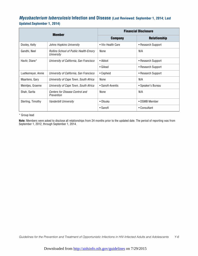
Downloaded from http://aidsinfo.nih.gov/guidelines on 7/29/2015
Guidelines for the Prevention and Treatment of Opportunistic Infections in HIV-Infected Adults and Adolescents Y-6
* Group lead
Note: Members were asked to disclose all relationships from 24 months prior to the updated date. The period of reporting was fromSeptember 1, 2012, through September 1, 2014.
Mycobacterium tuberculosis Infection and Disease (Last Reviewed: September 1, 2014; Last
Updated:September 1, 2014)
MemberFinancial Disclosure
Company Relationship
Dooley, Kelly Johns Hopkins University • Viiv Health Care • Research Support
Gandhi, Neel Rollins School of Public Health-EmoryUniversity
None N/A
Havlir, Diane* University of California, San Francisco • Abbot • Research Support
• Gilead • Research Support
Luetkemeyer, Annie University of California, San Francisco • Cepheid • Research Support
Maartens, Gary University of Cape Town, South Africa None N/A
Meintjes, Graeme University of Cape Town, South Africa • Sanofi-Aventis • Speaker’s Bureau
Shah, Sarita Centers for Disease Control andPrevention
None N/A
Sterling, Timothy Vanderbilt University • Otsuka • DSMB Member
• Sanofi • Consultant
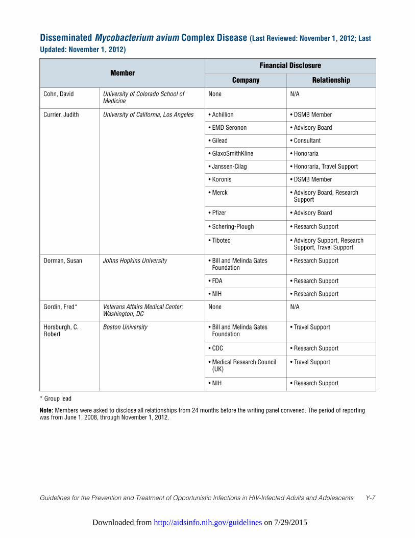
Downloaded from http://aidsinfo.nih.gov/guidelines on 7/29/2015
Guidelines for the Prevention and Treatment of Opportunistic Infections in HIV-Infected Adults and Adolescents Y-7
* Group lead
Note: Members were asked to disclose all relationships from 24 months before the writing panel convened. The period of reportingwas from June 1, 2008, through November 1, 2012.
Disseminated Mycobacterium avium Complex Disease (Last Reviewed: November 1, 2012; Last
Updated: November 1, 2012)
MemberFinancial Disclosure
Company Relationship
Cohn, David University of Colorado School ofMedicine
None N/A
Currier, Judith University of California, Los Angeles • Achillion • DSMB Member
• EMD Seronon • Advisory Board
• Gilead • Consultant
• GlaxoSmithKline • Honoraria
• Janssen-Cilag • Honoraria, Travel Support
• Koronis • DSMB Member
• Merck • Advisory Board, ResearchSupport
• Pfizer • Advisory Board
• Schering-Plough • Research Support
• Tibotec • Advisory Support, ResearchSupport, Travel Support
Dorman, Susan Johns Hopkins University • Bill and Melinda GatesFoundation
• Research Support
• FDA • Research Support
• NIH • Research Support
Gordin, Fred* Veterans Affairs Medical Center;Washington, DC
None N/A
Horsburgh, C.Robert
Boston University • Bill and Melinda GatesFoundation
• Travel Support
• CDC • Research Support
• Medical Research Council(UK)
• Travel Support
• NIH • Research Support
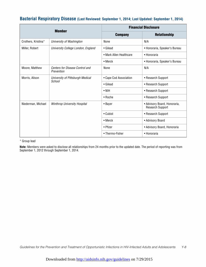
Downloaded from http://aidsinfo.nih.gov/guidelines on 7/29/2015
Guidelines for the Prevention and Treatment of Opportunistic Infections in HIV-Infected Adults and Adolescents Y-8
* Group lead
Note: Members were asked to disclose all relationships from 24 months prior to the updated date. The period of reporting was fromSeptember 1, 2012 through September 1, 2014.
Bacterial Respiratory Disease (Last Reviewed: September 1, 2014; Last Updated: September 1, 2014)
MemberFinancial Disclosure
Company Relationship
Crothers, Kristina* University of Washington None N/A
Miller, Robert University College London, England • Gilead • Honoraria, Speaker’s Bureau
• Mark Allen Healthcare • Honoraria
• Merck • Honoraria, Speaker’s Bureau
Moore, Matthew Centers for Disease Control andPrevention
None N/A
Morris, Alison University of Pittsburgh MedicalSchool
• Cape Cod Association • Research Support
• Gilead • Research Support
• NIH • Research Support
• Roche • Research Support
Niederman, Michael Winthrop University Hospital • Bayer • Advisory Board, Honoraria,Research Support
• Cubist • Research Support
• Merck • Advisory Board
• Pfizer • Advisory Board, Honoraria
• Thermo-Fisher • Honoraria
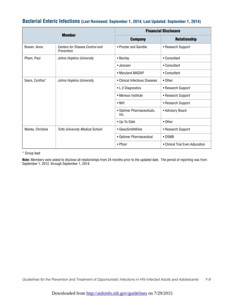
Downloaded from http://aidsinfo.nih.gov/guidelines on 7/29/2015
Guidelines for the Prevention and Treatment of Opportunistic Infections in HIV-Infected Adults and Adolescents Y-9
* Group lead
Note: Members were asked to disclose all relationships from 24 months prior to the updated date. The period of reporting was fromSeptember 1, 2012, through September 1, 2014.
Bacterial Enteric Infections (Last Reviewed: September 1, 2014; Last Updated: September 1, 2014)
MemberFinancial Disclosure
Company Relationship
Bowen, Anna Centers for Disease Control andPrevention
• Procter and Gamble • Research Support
Pham, Paul Johns Hopkins University • Barclay • Consultant
• Janssen • Consultant
• Maryland MADAP • Consultant
Sears, Cynthia* Johns Hopkins University • Clinical Infectious Diseases • Other
• L-2 Diagnostics • Research Support
• Merieux Institute • Research Support
• NIH • Research Support
• Optimer Pharmaceuticals,Inc.
• Advisory Board
• Up-To-Date • Other
Wanke, Christine Tufts University Medical School • GlaxoSmithKline • Research Support
• Optimer Pharmaceutical • DSMB
• Pfizer • Clinical Trial Even Adjucation
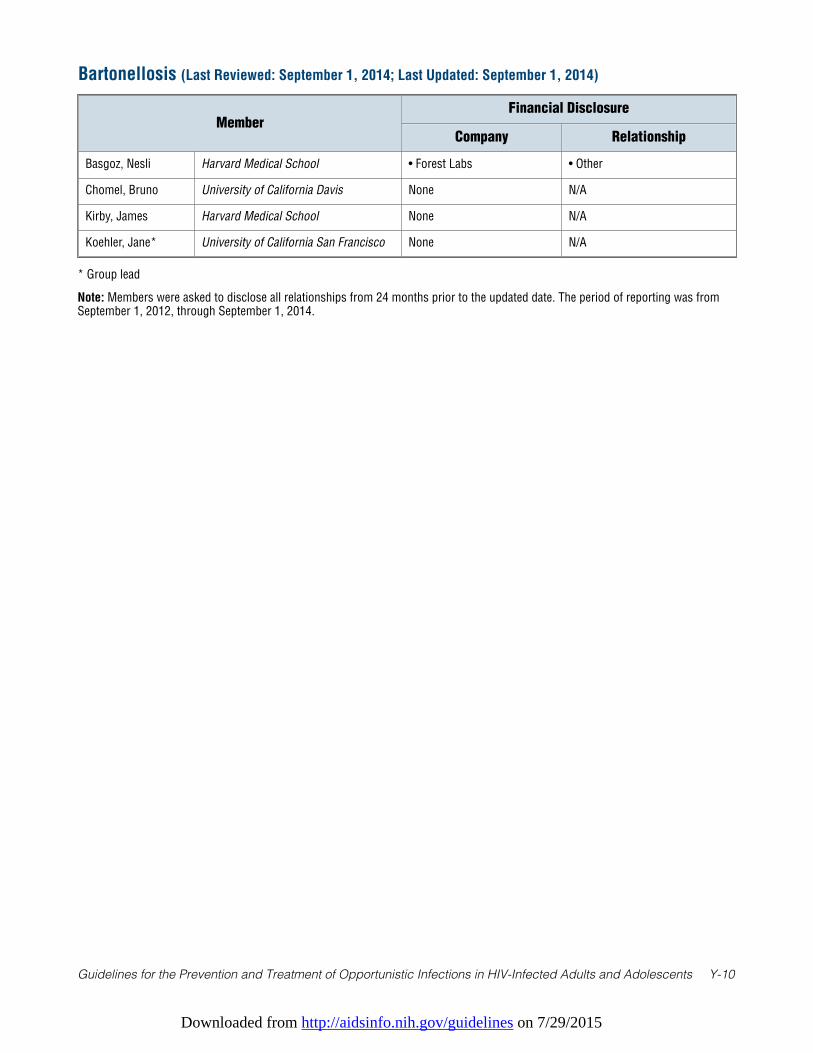
Downloaded from http://aidsinfo.nih.gov/guidelines on 7/29/2015
Guidelines for the Prevention and Treatment of Opportunistic Infections in HIV-Infected Adults and Adolescents Y-10
* Group lead
Note: Members were asked to disclose all relationships from 24 months prior to the updated date. The period of reporting was fromSeptember 1, 2012, through September 1, 2014.
Bartonellosis (Last Reviewed: September 1, 2014; Last Updated: September 1, 2014)
MemberFinancial Disclosure
Company Relationship
Basgoz, Nesli Harvard Medical School • Forest Labs • Other
Chomel, Bruno University of California Davis None N/A
Kirby, James Harvard Medical School None N/A
Koehler, Jane* University of California San Francisco None N/A
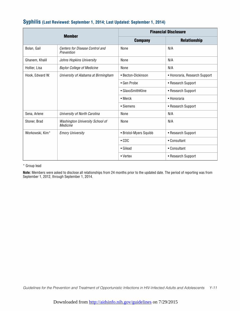
Downloaded from http://aidsinfo.nih.gov/guidelines on 7/29/2015
Guidelines for the Prevention and Treatment of Opportunistic Infections in HIV-Infected Adults and Adolescents Y-11
* Group lead
Note: Members were asked to disclose all relationships from 24 months prior to the updated date. The period of reporting was fromSeptember 1, 2012, through September 1, 2014.
Syphilis (Last Reviewed: September 1, 2014; Last Updated: September 1, 2014)
MemberFinancial Disclosure
Company Relationship
Bolan, Gail Centers for Disease Control andPrevention
None N/A
Ghanem, Khalil Johns Hopkins University None N/A
Hollier, Lisa Baylor College of Medicine None N/A
Hook, Edward W. University of Alabama at Birmingham • Becton-Dickinson • Honoraria, Research Support
• Gen Probe • Research Support
• GlaxoSmithKline • Research Support
• Merck • Honoraria
• Siemens • Research Support
Sena, Arlene University of North Carolina None N/A
Stoner, Brad Washington University School ofMedicine
None N/A
Workowski, Kim* Emory University • Bristol-Myers Squibb • Research Support
• CDC • Consultant
• Gilead • Consultant
• Vertex • Research Support
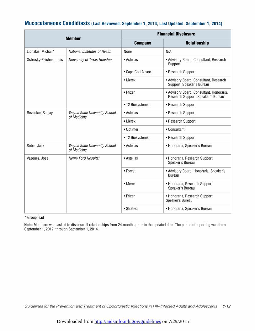
Downloaded from http://aidsinfo.nih.gov/guidelines on 7/29/2015
Guidelines for the Prevention and Treatment of Opportunistic Infections in HIV-Infected Adults and Adolescents Y-12
* Group lead
Note: Members were asked to disclose all relationships from 24 months prior to the updated date. The period of reporting was fromSeptember 1, 2012, through September 1, 2014.
Mucocutaneous Candidiasis (Last Reviewed: September 1, 2014; Last Updated: September 1, 2014)
MemberFinancial Disclosure
Company Relationship
Lionakis, Michail* National Institutes of Health None N/A
Ostrosky-Zeichner, Luis University of Texas Houston • Astellas • Advisory Board, Consultant, ResearchSupport
• Cape Cod Assoc. • Research Support
• Merck • Advisory Board, Consultant, ResearchSupport, Speaker’s Bureau
• Pfizer • Advisory Board, Consultant, Honoraria,Research Support, Speaker’s Bureau
• T2 Biosystems • Research Support
Revankar, Sanjay Wayne State University Schoolof Medicine
• Astellas • Research Support
• Merck • Research Support
• Optimer • Consultant
• T2 Biosystems • Research Support
Sobel, Jack Wayne State University Schoolof Medicine
• Astellas • Honoraria, Speaker’s Bureau
Vazquez, Jose Henry Ford Hospital • Astellas • Honoraria, Research Support,Speaker’s Bureau
• Forest • Advisory Board, Honoraria, Speaker’sBureau
• Merck • Honoraria, Research Support,Speaker’s Bureau
• Pfizer • Honoraria, Research Support,Speaker’s Bureau
• Strativa • Honoraria, Speaker’s Bureau
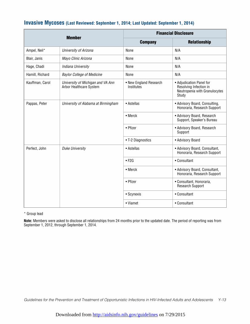
Downloaded from http://aidsinfo.nih.gov/guidelines on 7/29/2015
Guidelines for the Prevention and Treatment of Opportunistic Infections in HIV-Infected Adults and Adolescents Y-13
* Group lead
Note: Members were asked to disclose all relationships from 24 months prior to the updated date. The period of reporting was fromSeptember 1, 2012, through September 1, 2014.
Invasive Mycoses (Last Reviewed: September 1, 2014; Last Updated: September 1, 2014)
MemberFinancial Disclosure
Company Relationship
Ampel, Neil* University of Arizona None N/A
Blair, Janis Mayo Clinic Arizona None N/A
Hage, Chadi Indiana University None N/A
Hamill, Richard Baylor College of Medicine None N/A
Kauffman, Carol University of Michigan and VA AnnArbor Healthcare System
• New England ResearchInstitutes
• Adjudication Panel forResolving Infection inNeutropenia with GranulocytesStudy
Pappas, Peter University of Alabama at Birmingham • Astellas • Advisory Board, Consulting,Honoraria, Research Support
• Merck • Advisory Board, ResearchSupport, Speaker’s Bureau
• Pfizer • Advisory Board, ResearchSupport
• T-2 Diagnostics • Advisory Board
Perfect, John Duke University • Astellas • Advisory Board, Consultant,Honoraria, Research Support
• F2G • Consultant
• Merck • Advisory Board, Consultant,Honoraria, Research Support
• Pfizer • Consultant, Honoraria,Research Support
• Scynexis • Consultant
• Viamet • Consultant
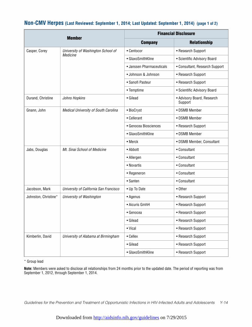
Downloaded from http://aidsinfo.nih.gov/guidelines on 7/29/2015
Guidelines for the Prevention and Treatment of Opportunistic Infections in HIV-Infected Adults and Adolescents Y-14
* Group lead
Note: Members were asked to disclose all relationships from 24 months prior to the updated date. The period of reporting was fromSeptember 1, 2012, through September 1, 2014.
Non-CMV Herpes (Last Reviewed: September 1, 2014; Last Updated: September 1, 2014) (page 1 of 2)
MemberFinancial Disclosure
Company Relationship
Casper, Corey University of Washington School ofMedicine
• Centocor • Research Support
• GlaxoSmithKline • Scientific Advisory Board
• Janssen Pharmaceuticals • Consultant, Research Support
• Johnson & Johnson • Research Support
• Sanofi Pasteur • Research Support
• Temptime • Scientific Advisory Board
Durand, Christine Johns Hopkins • Gilead • Advisory Board, ResearchSupport
Gnann, John Medical University of South Carolina • BioCryst • DSMB Member
• Cellerant • DSMB Member
• Genocea Biosciences • Research Support
• GlaxoSmithKline • DSMB Member
• Merck • DSMB Member, Consultant
Jabs, Douglas Mt. Sinai School of Medicine • Abbott • Consultant
• Allergen • Consultant
• Novartis • Consultant
• Regeneron • Consultant
• Santen • Consultant
Jacobson, Mark University of California San Francisco • Up To Date • Other
Johnston, Christine* University of Washington • Agenus • Research Support
• Aicuris GmhH • Research Support
• Genocea • Research Support
• Gilead • Research Support
• Vical • Research Support
Kimberlin, David University of Alabama at Birmingham • Cellex • Research Support
• Gilead • Research Support
• GlaxoSmithKline • Research Support
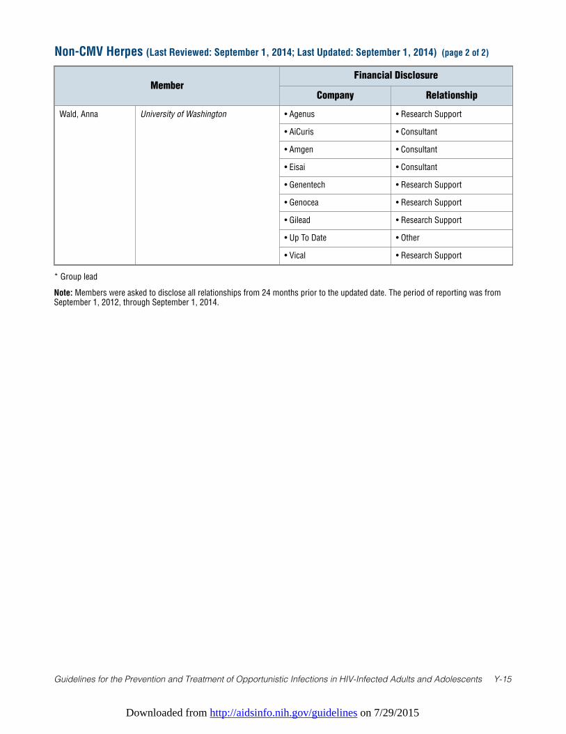
Downloaded from http://aidsinfo.nih.gov/guidelines on 7/29/2015
Guidelines for the Prevention and Treatment of Opportunistic Infections in HIV-Infected Adults and Adolescents Y-15
Non-CMV Herpes (Last Reviewed: September 1, 2014; Last Updated: September 1, 2014) (page 2 of 2)
MemberFinancial Disclosure
Company Relationship
Wald, Anna University of Washington • Agenus • Research Support
• AiCuris • Consultant
• Amgen • Consultant
• Eisai • Consultant
• Genentech • Research Support
• Genocea • Research Support
• Gilead • Research Support
• Up To Date • Other
• Vical • Research Support
* Group lead
Note: Members were asked to disclose all relationships from 24 months prior to the updated date. The period of reporting was fromSeptember 1, 2012, through September 1, 2014.
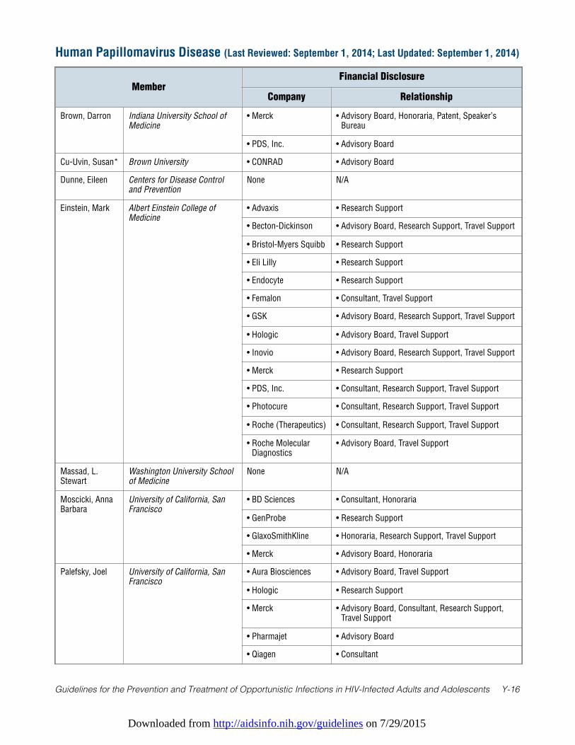
Downloaded from http://aidsinfo.nih.gov/guidelines on 7/29/2015
Guidelines for the Prevention and Treatment of Opportunistic Infections in HIV-Infected Adults and Adolescents Y-16
Human Papillomavirus Disease (Last Reviewed: September 1, 2014; Last Updated: September 1, 2014)
MemberFinancial Disclosure
Company Relationship
Brown, Darron Indiana University School ofMedicine
• Merck • Advisory Board, Honoraria, Patent, Speaker’sBureau
• PDS, Inc. • Advisory Board
Cu-Uvin, Susan* Brown University • CONRAD • Advisory Board
Dunne, Eileen Centers for Disease Controland Prevention
None N/A
Einstein, Mark Albert Einstein College ofMedicine
• Advaxis • Research Support
• Becton-Dickinson • Advisory Board, Research Support, Travel Support
• Bristol-Myers Squibb • Research Support
• Eli Lilly • Research Support
• Endocyte • Research Support
• Femalon • Consultant, Travel Support
• GSK • Advisory Board, Research Support, Travel Support
• Hologic • Advisory Board, Travel Support
• Inovio • Advisory Board, Research Support, Travel Support
• Merck • Research Support
• PDS, Inc. • Consultant, Research Support, Travel Support
• Photocure • Consultant, Research Support, Travel Support
• Roche (Therapeutics) • Consultant, Research Support, Travel Support
• Roche MolecularDiagnostics
• Advisory Board, Travel Support
Massad, L.Stewart
Washington University Schoolof Medicine
None N/A
Moscicki, AnnaBarbara
University of California, SanFrancisco
• BD Sciences • Consultant, Honoraria
• GenProbe • Research Support
• GlaxoSmithKline • Honoraria, Research Support, Travel Support
• Merck • Advisory Board, Honoraria
Palefsky, Joel University of California, SanFrancisco
• Aura Biosciences • Advisory Board, Travel Support
• Hologic • Research Support
• Merck • Advisory Board, Consultant, Research Support,Travel Support
• Pharmajet • Advisory Board
• Qiagen • Consultant
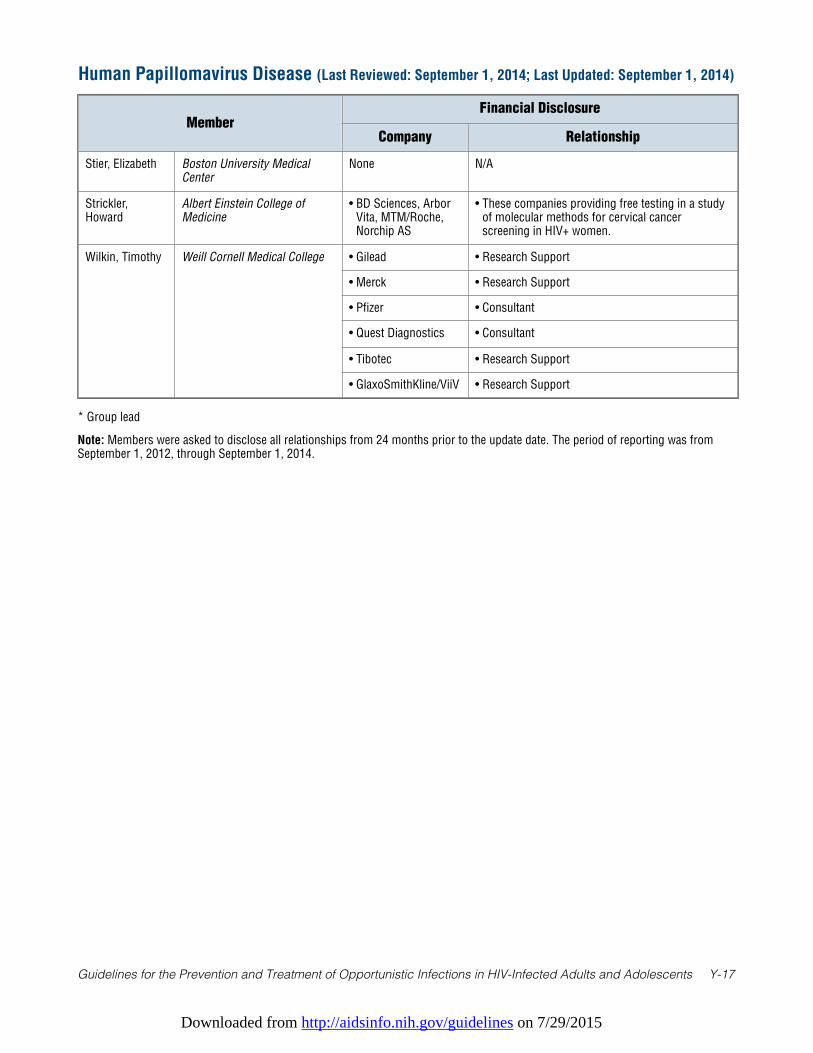
Downloaded from http://aidsinfo.nih.gov/guidelines on 7/29/2015
Guidelines for the Prevention and Treatment of Opportunistic Infections in HIV-Infected Adults and Adolescents Y-17
* Group lead
Note: Members were asked to disclose all relationships from 24 months prior to the update date. The period of reporting was fromSeptember 1, 2012, through September 1, 2014.
Human Papillomavirus Disease (Last Reviewed: September 1, 2014; Last Updated: September 1, 2014)
MemberFinancial Disclosure
Company Relationship
Stier, Elizabeth Boston University MedicalCenter
None N/A
Strickler,Howard
Albert Einstein College ofMedicine
• BD Sciences, ArborVita, MTM/Roche,Norchip AS
• These companies providing free testing in a studyof molecular methods for cervical cancerscreening in HIV+ women.
Wilkin, Timothy Weill Cornell Medical College • Gilead • Research Support
• Merck • Research Support
• Pfizer • Consultant
• Quest Diagnostics • Consultant
• Tibotec • Research Support
• GlaxoSmithKline/ViiV • Research Support
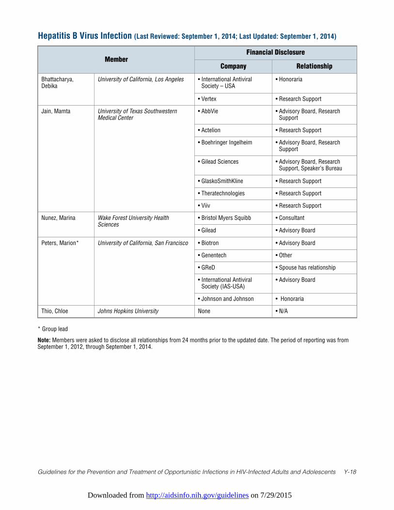
Downloaded from http://aidsinfo.nih.gov/guidelines on 7/29/2015
Guidelines for the Prevention and Treatment of Opportunistic Infections in HIV-Infected Adults and Adolescents Y-18
Hepatitis B Virus Infection (Last Reviewed: September 1, 2014; Last Updated: September 1, 2014)
MemberFinancial Disclosure
Company Relationship
Bhattacharya,Debika
University of California, Los Angeles • International AntiviralSociety – USA
• Honoraria
• Vertex • Research Support
Jain, Mamta University of Texas SouthwesternMedical Center
• AbbVie • Advisory Board, ResearchSupport
• Actelion • Research Support
• Boehringer Ingelheim • Advisory Board, ResearchSupport
• Gilead Sciences • Advisory Board, ResearchSupport, Speaker’s Bureau
• GlaskoSmithKline • Research Support
• Theratechnologies • Research Support
• Viiv • Research Support
Nunez, Marina Wake Forest University HealthSciences
• Bristol Myers Squibb • Consultant
• Gilead • Advisory Board
Peters, Marion* University of California, San Francisco • Biotron • Advisory Board
• Genentech • Other
• GReD • Spouse has relationship
• International AntiviralSociety (IAS-USA)
• Advisory Board
• Johnson and Johnson • Honoraria
Thio, Chloe Johns Hopkins University None • N/A
* Group lead
Note: Members were asked to disclose all relationships from 24 months prior to the updated date. The period of reporting was fromSeptember 1, 2012, through September 1, 2014.
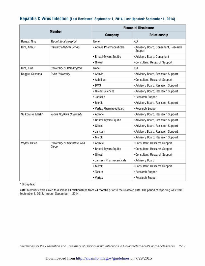
Downloaded from http://aidsinfo.nih.gov/guidelines on 7/29/2015
Guidelines for the Prevention and Treatment of Opportunistic Infections in HIV-Infected Adults and Adolescents Y-19
* Group lead
Note: Members were asked to disclose all relationships from 24 months prior to the reviewed date. The period of reporting was fromSeptember 1, 2012, through September 1, 2014.
Hepatitis C Virus Infection (Last Reviewed: September 1, 2014; Last Updated: September 1, 2014)
MemberFinancial Disclosure
Company Relationship
Bansal, Nina Mount Sinai Hospital None N/A
Kim, Arthur Harvard Medical School • Abbvie Pharmaceuticals • Advisory Board, Consultant, ResearchSupport
• Bristol-Myers Squibb • Advisory Board, Consultant
• Gilead • Consultant, Research Support
Kim, Nina University of Washington None N/A
Naggie, Susanna Duke University • Abbvie • Advisory Board, Research Support
• Achillion • Consultant, Research Support
• BMS • Advisory Board, Research Support
• Gilead Sciences • Advisory Board, Research Support
• Janssen • Research Support
• Merck • Advisory Board, Research Support
• Vertex Pharmaceuticals • Research Support
Sulkowski, Mark* Johns Hopkins University • AbbVie • Advisory Board, Research Support
• Bristol-Myers Squibb • Advisory Board, Research Support
• Gilead • Advisory Board, Research Support
• Janssen • Advisory Board, Research Support
• Merck • Advisory Board, Research Support
Wyles, David University of California, SanDiego
• AbbVie • Consultant, Research Support
• Bristol-Myers Squibb • Consultant, Research Support
• Gilead • Consultant, Research Support
• Janssen Pharmaceuticals • Advisory Board
• Merck • Consultant, Research Support
• Tacere • Research Support
• Vertex • Research Support
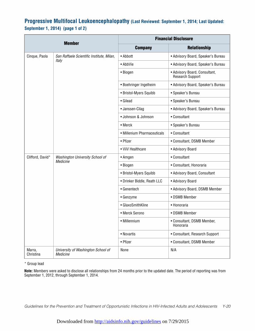
Downloaded from http://aidsinfo.nih.gov/guidelines on 7/29/2015
Guidelines for the Prevention and Treatment of Opportunistic Infections in HIV-Infected Adults and Adolescents Y-20
Progressive Multifocal Leukoencephalopathy (Last Reviewed: September 1, 2014; Last Updated:
September 1, 2014) (page 1 of 2)
MemberFinancial Disclosure
Company Relationship
Cinque, Paola San Raffaele Scientific Institute, Milan,Italy
• Abbott • Advisory Board, Speaker’s Bureau
• AbbVie • Advisory Board, Speaker’s Bureau
• Biogen • Advisory Board, Consultant,Research Support
• Boehringer Ingelheim • Advisory Board, Speaker’s Bureau
• Bristol-Myers Squibb • Speaker’s Bureau
• Gilead • Speaker’s Bureau
• Janssen-Cilag • Advisory Board, Speaker’s Bureau
• Johnson & Johnson • Consultant
• Merck • Speaker’s Bureau
• Millenium Pharmaceuticals • Consultant
• Pfizer • Consultant, DSMB Member
• ViiV Healthcare • Advisory Board
Clifford, David* Washington University School ofMedicine
• Amgen • Consultant
• Biogen • Consultant, Honoraria
• Bristol-Myers Squibb • Advisory Board, Consultant
• Drinker Biddle, Reath LLC • Advisory Board
• Genentech • Advisory Board, DSMB Member
• Genzyme • DSMB Member
• GlaxoSmithKline • Honoraria
• Merck Serono • DSMB Member
• Millennium • Consultant, DSMB Member,Honoraria
• Novartis • Consultant, Research Support
• Pfizer • Consultant, DSMB Member
Marra,Christina
University of Washington School ofMedicine
None N/A
* Group lead
Note: Members were asked to disclose all relationships from 24 months prior to the updated date. The period of reporting was fromSeptember 1, 2012, through September 1, 2014.
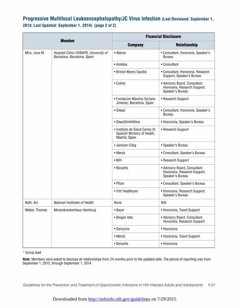
Downloaded from http://aidsinfo.nih.gov/guidelines on 7/29/2015
Guidelines for the Prevention and Treatment of Opportunistic Infections in HIV-Infected Adults and Adolescents Y-21
MemberFinancial Disclosure
Company Relationship
Miro, Jose M. Hospital Clinic-IDIBAPS, University ofBarcelona, Barcelona, Spain
• Abbvie • Consultant, Honoraria, Speaker’sBureau
• Astellas • Consultant
• Bristol-Myers Squibb • Consultant, Honoraria, ResearchSupport, Speaker’s Bureau
• Cubist • Advisory Board, Consultant,Honoraria, Research Support,Speaker’s Bureau
• Fundacion Maximo SorianoJimenez, Barcelona, Spain
• Research Support
• Gilead • Consultant, Honoraria, Speaker’sBureau
• GlaxoSmithKline • Honoraria, Speaker’s Bureau
• Instituto de Salud Carlos III,Spanish Ministry of Health,Madrid, Spain
• Research Support
• Janssen-Cilag • Speaker’s Bureau
• Merck • Consultant, Speaker’s Bureau
• NIH • Research Support
• Novartis • Advisory Board, Consultant,Honoraria, Research Support,Speaker’s Bureau
• Pfizer • Consultant, Speaker’s Bureau
• ViiV Healthcare • Honoraria, Research Support,Speaker’s Bureau
Nath, Avi National Institutes of Health None N/A
Weber, Thomas Marienkrankenhaus Hamburg • Bayer • Honoraria, Travel Support
• Biogen Idec • Advisory Board, Consultant,Honoraria, Research Support
• Genzyme • Honoraria
• Merck • Honoraria, Travel Support
• Novartis • Honoraria
Progressive Multifocal Leukoencephalopathy/JC Virus Infection (Last Reviewed: September 1,
2014; Last Updated: September 1, 2014) (page 2 of 2)
* Group lead
Note: Members were asked to disclose all relationships from 24 months prior to the updated date. The period of reporting was fromSeptember 1, 2012, through September 1, 2014.
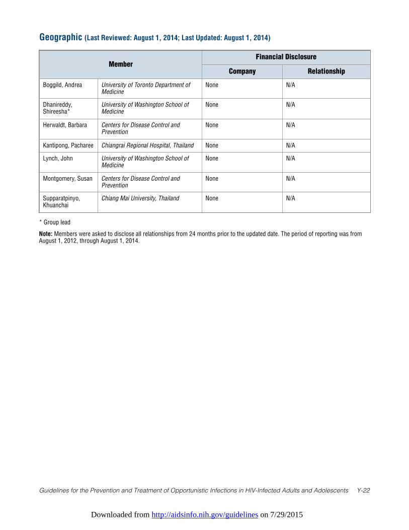
Downloaded from http://aidsinfo.nih.gov/guidelines on 7/29/2015
Guidelines for the Prevention and Treatment of Opportunistic Infections in HIV-Infected Adults and Adolescents Y-22
* Group lead
Note: Members were asked to disclose all relationships from 24 months prior to the updated date. The period of reporting was fromAugust 1, 2012, through August 1, 2014.
Geographic (Last Reviewed: August 1, 2014; Last Updated: August 1, 2014)
MemberFinancial Disclosure
Company Relationship
Boggild, Andrea University of Toronto Department ofMedicine
None N/A
Dhanireddy,Shireesha*
University of Washington School ofMedicine
None N/A
Herwaldt, Barbara Centers for Disease Control andPrevention
None N/A
Kantipong, Pacharee Chiangrai Regional Hospital, Thailand None N/A
Lynch, John University of Washington School ofMedicine
None N/A
Montgomery, Susan Centers for Disease Control andPrevention
None N/A
Supparatpinyo,Khuanchai
Chiang Mai University, Thailand None N/A
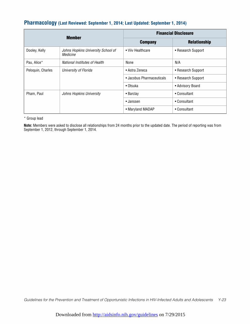
Downloaded from http://aidsinfo.nih.gov/guidelines on 7/29/2015
Guidelines for the Prevention and Treatment of Opportunistic Infections in HIV-Infected Adults and Adolescents Y-23
* Group lead
Note: Members were asked to disclose all relationships from 24 months prior to the updated date. The period of reporting was fromSeptember 1, 2012, through September 1, 2014.
Pharmacology (Last Reviewed: September 1, 2014; Last Updated: September 1, 2014)
MemberFinancial Disclosure
Company Relationship
Dooley, Kelly Johns Hopkins University School ofMedicine
• Viiv Healthcare • Research Support
Pau, Alice* National Institutes of Health None N/A
Peloquin, Charles University of Florida • Astra Zeneca • Research Support
• Jacobus Pharmaceuticals • Research Support
• Otsuka • Advisory Board
Pham, Paul Johns Hopkins University • Barclay • Consultant
• Janssen • Consultant
• Maryland MADAP • Consultant
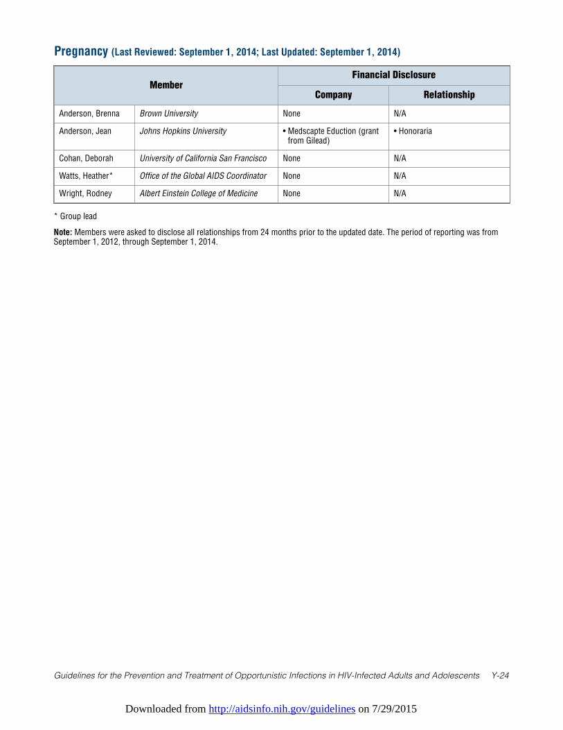
Downloaded from http://aidsinfo.nih.gov/guidelines on 7/29/2015
Guidelines for the Prevention and Treatment of Opportunistic Infections in HIV-Infected Adults and Adolescents Y-24
* Group lead
Note: Members were asked to disclose all relationships from 24 months prior to the updated date. The period of reporting was fromSeptember 1, 2012, through September 1, 2014.
Pregnancy (Last Reviewed: September 1, 2014; Last Updated: September 1, 2014)
MemberFinancial Disclosure
Company Relationship
Anderson, Brenna Brown University None N/A
Anderson, Jean Johns Hopkins University • Medscapte Eduction (grantfrom Gilead)
• Honoraria
Cohan, Deborah University of California San Francisco None N/A
Watts, Heather* Office of the Global AIDS Coordinator None N/A
Wright, Rodney Albert Einstein College of Medicine None N/A
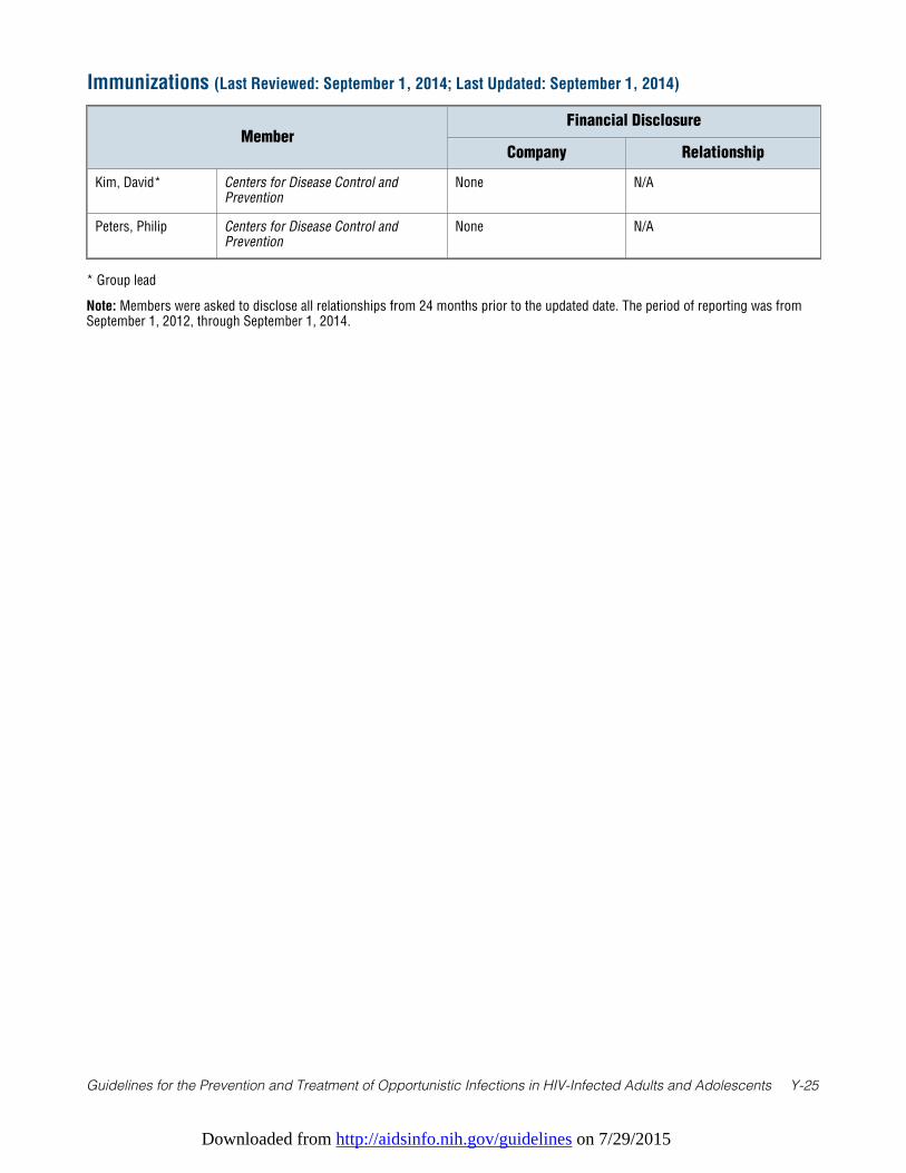
Downloaded from http://aidsinfo.nih.gov/guidelines on 7/29/2015
Guidelines for the Prevention and Treatment of Opportunistic Infections in HIV-Infected Adults and Adolescents Y-25
* Group lead
Note: Members were asked to disclose all relationships from 24 months prior to the updated date. The period of reporting was fromSeptember 1, 2012, through September 1, 2014.
Immunizations (Last Reviewed: September 1, 2014; Last Updated: September 1, 2014)
MemberFinancial Disclosure
Company Relationship
Kim, David* Centers for Disease Control andPrevention
None N/A
Peters, Philip Centers for Disease Control andPrevention
None N/A
Downloaded from http://aidsinfo.nih.gov/guidelines on 7/29/2015
Guidelines for the Prevention and Treatment of Opportunistic Infections in HIV-Infected Adults and Adolescents Z-1
Appendix D. ContributorsAs part of the revision process, a Clinical-Community Panel was convened to review these guidelines andadvise the author panel as to their usefulness for practicing clinicians with regard to content and format. Themembers of the Clinical Community Panel are as follows:
• Roberto Arduino; Thomas Street Health Center—Houston, Texas• Mark Baker; MedStar Washington Hospital Center—Washington, DC• Lisa Fitzpatrick; Howard University—Washington, DC• C. Bradley Hare; San Francisco General Hospital and University of California, San Francisco—
San Francisco, California• Robert Harrington; University of Washington—Seattle, Washington• E. Turner Overton; Washington University—St. Louis, Missouri• David Rimland; Emory University—Atlanta, Georgia• Martin Rodriguez, University of Alabama at Birmingham—Birmingham, Alabama• Peter Shalit; Swedish Hospital Medical Center HIV Program—Seattle, Washington• Tracy Swan; Treatment Action Group—New York, New York• Zelalem Temesgen; Mayo Clinic—Rochester, Minnesota• Mary Vogler; Weill Cornell—New York, New York• Dan Wlodarczyk; University of California, San Francisco—San Francisco, California
The panel would like to acknowledge Judith Welsh, Clinical Informationist at the National Institutes ofHealth Library, for performing comprehensive literature searches to identify the evidence used to supportrecommendations in these guidelines.