Embed Size (px)
Citation preview


Abnormal, heaped placenta
Normally, would expect placenta to lie flat
against contour of uterus
Complete breach in myometrial wall with
fetal parts in maternal peritoneal cavity
Uterine Rupture

Placental abruption
Complete breach in myometrial wall with
fetal parts in maternal peritoneal cavity
Uterine Rupture
Uterine Rupture

Uterine Rupture
Uterine Rupture

Myometrial defect
Uterine Rupture

Liver with ascites axial T2
Complete breach in myometrial wall with
fetal parts in maternal peritoneal cavity
Uterine Rupture

Myometrial defect and ascites
coronal T2
Complete breach in myometrial wall with
fetal parts in maternal peritoneal cavity
Uterine Rupture

MRI: Oligohydramnios coronal T2
myometrail silhoutte
Complete breach in myometrial wall with
fetal parts in maternal peritoneal cavity
Uterine Rupture

Myometrial defect
Coronal T2 Additional Defect in Myometrium
Axiaal T2
Complete breach in myometrial wall with
fetal parts in maternal peritoneal cavity
Uterine Rupture

Shortened Cervix
Sagittal T2
Free Fluid in Pelvis
Sagittal T2
Complete breach in myometrial wall with
fetal parts in maternal peritoneal cavity
Uterine Rupture

Umbilical cord in myometrial
defect
Sagittal T2
Complete breach in myometrial wall with
fetal parts in maternal peritoneal cavity
Uterine Rupture

Fetal parts abutting maternal body wall
AXIAL T2
Complete breach in myometrial wall with
fetal parts in maternal peritoneal cavity
Uterine Rupture


3 weeks later decrease in size of the retroplacental clot that has become hypoechoic over time without internal color flow.
retroplacental abruption posttrauma 24 weeks pregnant
shows thickening of the placenta with clot of heterogeneous echogenicity between
the placenta and uterine wall without internal flow to color Doppler assessment

Normal CT of gravid uterus
33-year-old woman who is 15 weeks pregnant. Normal CT image of gravid uterus shows early ossification of fetal parts.
Placenta (P) is forming on right side of uterus with heterogeneous enhancement. There are enhancing engorged
veins deep in relation to placenta representing retroplacental venous plexus (arrowheads). There is marked enlargement of
pelvic veins bilaterally (arrows)

35 weeks pregnant.
Placenta is posterior with heterogeneously
enhancing cotyledons (asterisk).
well-defined cotyledons characterized by central areas
of low attenuation with intervening rings of higher
attenuation (asterisks).
Right ovarian vein is enlarged to one half size of inferior
vena cava (arrow
Normal CT of gravid uterus
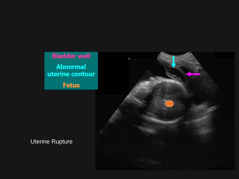
0–2 weeks Preimplantation Prenatal death (all or none)
Risk at > 50–100 mGy
2–8 weeks Major organogenesis Organ malformations
Risk threshold > 100 mGy (but not observed in humans at diagnostic levels)
2–15 weeks Organogenesis and rapid neuronal development and migration
Small head size
Risk threshold > 100 mGy
Severe mental retardation
Risk threshold > 100 mGy
2 weeks to term Postimplantation Childhood cancer (< 15 years old)
Risk childhood cancer death 0.06% per 10 mGy (or 1/1700)a
Lifetime cancer risk 0.4% per 10 mGyb
In Utero Effects of Diagnostic Levels of Ionizing Radiation

Recommendations by the ACOG and the ACR on Use of CT in Pregnancy
ACOG Recommendations ACR Recommendations
Perform necessary examinations only after clinical work-up
Keep radiation levels as low as reasonably achievable
Iodinated contrast material is safe in pregnancy
Iodinated contrast material is likely safe in pregnancy
Counsel for radiation exposure

21 weeks pregnant. Normal placenta
A, right retroperitoneal hemorrhage (asterisk) centered on enlarged right ovarian vein (arrowhead).
B, CT image through gravid uterus shows normal anterior placenta. Arrowheads show
nonenhancing chorionic villous plate indentations that should not be confused with abnormality.
There are enhancing engorged veins in right adnexa (asterisk).

33-year-old woman who is 33 weeks pregnant and sustained 20-foot fall from
balcony after using alcohol and cocaine.
Contrast-enhanced CT image of abdomen and pelvis shows anterior placenta
(asterisk) that appears normal.
33 weeks pregnant and sustained 20-foot fall from balcony after using alcohol and
cocaine

grade 2 splenic laceration (short arrow) and marginal placental abruption with
infarction (long arrow). There is diminished enhancement in the left side of the
placenta, extending from the placental base to the placental surface. Maternal
hydronephrosis of pregnancy can be noticed on the right
34 weeks pregnant

18-year-old woman who is 14 weeks pregnant
and was restrained passenger in T-bone high-speed motor vehicle crash
A, CT image through gravid uterus
is normal with anterior placenta
(arrow) and early ossification of
fetal parts (arrowhead). There is
small hematoma in left pelvic
sidewall (asterisk) due to pelvic
fracture.
B, Limited obstetric ultrasound image
obtained next day shows fetal death with
intrauterine gestation with no fetal cardiac
activity. Placenta was normal on
ultrasound. Patient

38 weeks pregnant normal placenta
Contrast-enhanced CT image of abdomen and pelvis shows areas of nonenhancement
in posterior placenta (asterisk) concerning for ischemia and areas of contrast
blush (arrow) concerning for active bleeding. Venous lakes and chorionic villous plate
indentations provide alternate explanation

23-year-old woman who is 20 weeks pregnant and was pedestrian hit by car on right
side of her body
CT image through gravid uterus shows posterior placenta with marginal placental
abruption on left with intermediate-density clot elevating placenta from uterine
wall (asterisk). Engorged right ovarian vein (arrow) and dilated ureters (arrowheads) are
normal in pregnancy

39 weeks pregnant with accident
Contrast-enhanced CT image of abdomen and pelvis shows
placenta on right with near-complete devascularization indicative of
severe abruption (asterisk). There are only small areas of
enhancing placenta (arrowhead). Patient was in right posterior
oblique position rather than left, and there is compression of inferior
vena cava by gravid uterus (arrow).
CT image obtained lower through gravid uterus shows cephalic
presentation of fetus. There is displaced fracture of right fetal parietal
bone (arrow) with intracranial blood (asterisks).

31 weeks pregnant + CT contrast
shows several areas of nonenhancing placenta (asterisk) infarction

Contrast-enhanced CT images of abdomen
and pelvis show uterine rupture with free
floating fetus
empty uterus
36 weeks pregnant and was restrained passenger in motor vehicle crash

bulky empty uterus with linear tear (arrow)
through the anterior myometrial wall
associated increased peritoneal fluid.
foetal head closely apposed to the liver
and gall bladder (GB)
Intrapartum Rupture of the Uterus Diagnosed by Ultrasound

Appendicitis in pregnant patient. (a) Acute appendicitis in a 35-year-old patient in the
third trimester with a history of nausea, vomiting, fever, and abdominal pain. US image
shows an enlarged dilated tubular structure measuring 10 mm in diameter in the right
lower quadrant (arrow), a finding suggestive of acute appendicitis. (b) Acute appendicitis
in a 28-year-old patient in the third trimester with a history of acute pain in the right lower
quadrant. Co-ronal contrast-enhanced reformatted CT image shows an enlarged
appendix measuring 9 mm in diameter (arrow).

retroplacental abruption posttrauma . 19-year-old female and is 24 weeks pregnant.
A-thickening of the placenta with clot of heterogeneous echogenicity
between the placenta and uterine wall without internal flow to color Doppler assessment
3 weeks later shows an interval decrease in size of the retroplacental clot that has
become hypoechoic over time without internal color flow

Hematoma in a patient with vaginal
bleeding who had undergone cesarean
section 14 days earlier.
(a) Sagittal contrast material–enhanced CT
scan shows a hematoma in the
endocervical canal (arrow)
(b, c) Axial (b) and coronal short-axis (c)
reformatted CT images show the cervical
hematoma (arrow) and parametrial vessels
(arrowheads) without active bleeding.

CT images show the normal
appearance of the placenta
(arrows) at different patient.

Placental abruption is best characterized
on CT images as a contiguous
retroplacental or full-thickness area of
decreased enhancement that forms acute
angles with the myometrium
The small amount of hyperattenuating
material seen in the placental tissue
could represent blood products or
minimally enhancing residual placental
tissue. High-attenuating material seen
inferiorly in the amniotic sac and uterine
cavity (arrow in b) represents blood
products

MR imaging findings of placental
abruption. (a) Sagittal T2-weighted
HASTE MR image shows a complex
collection (arrow) with a fluid-blood
level abutting the anterior aspect of
the placenta (*).
(b) Axial T1-weighted MR image shows
fluid with high signal intensity (arrow), a
finding consistent with hemorrhage.

fetal head in the uterine cavity (arrow in a), while the body is seen outside the uterus,
uncovered by myometrium (arrow in b).
the body of the fetus (*) extending beyond
the margin of the uterus (arrows)
Uterine rupture in a 28-week-pregnant woman

21 year old female patient with uterine perforation and abdominal expulsion of fetal
parts
two parallel echogenic foci in the left iliac
fossa (arrows) representing the extruded fetal
spine fetal skull bones in the left iliac fossa
(arrows)
multiple bony fetal parts comprising mainly the spine
and limb bones in the left iliac fossa (arrows).

Dose Reduction Techniques for CT of Pregnant Patients
One size does not fit all: do not use standard protocols
Decrease kilovoltage for small patients
Decrease milliamperage and use automatic tube current modulation
Increase pitch to >1
Obtain a single scout view and avoid directly imaging the fetus for planning purposes
Limit the field of view
Avoid imaging in multiple phases
Use more recently available novel reconstruction algorithms to reduce noise in images,
thus allowing reduction of milliamperage or increase in noise level requirements during
scanning
Lead shielding of the mother; most pronounced effect with circumferential shielding
Internal barium shielding with use of oral 30% barium sulfate solution
Local quality assurance program to monitor CT protocols and the resulting dose