Embed Size (px)
Citation preview
Radiology Chest assessment
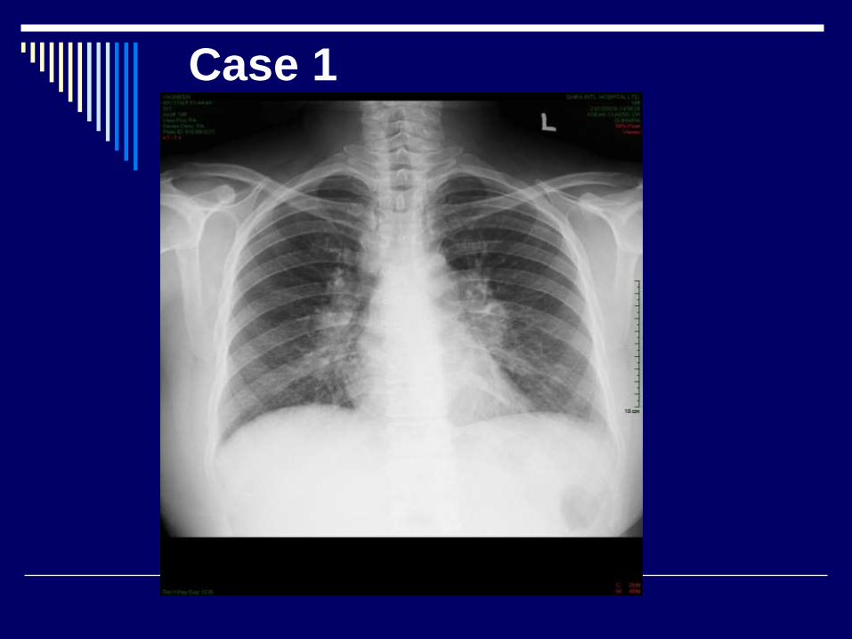
Case 1
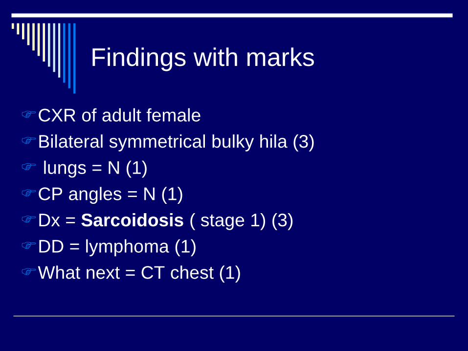
Findings with marks
CXR of adult female
Bilateral symmetrical bulky hila (3)
lungs = N (1)
CP angles = N (1)
Dx = Sarcoidosis ( stage 1) (3)
DD = lymphoma (1)
What next = CT chest (1)
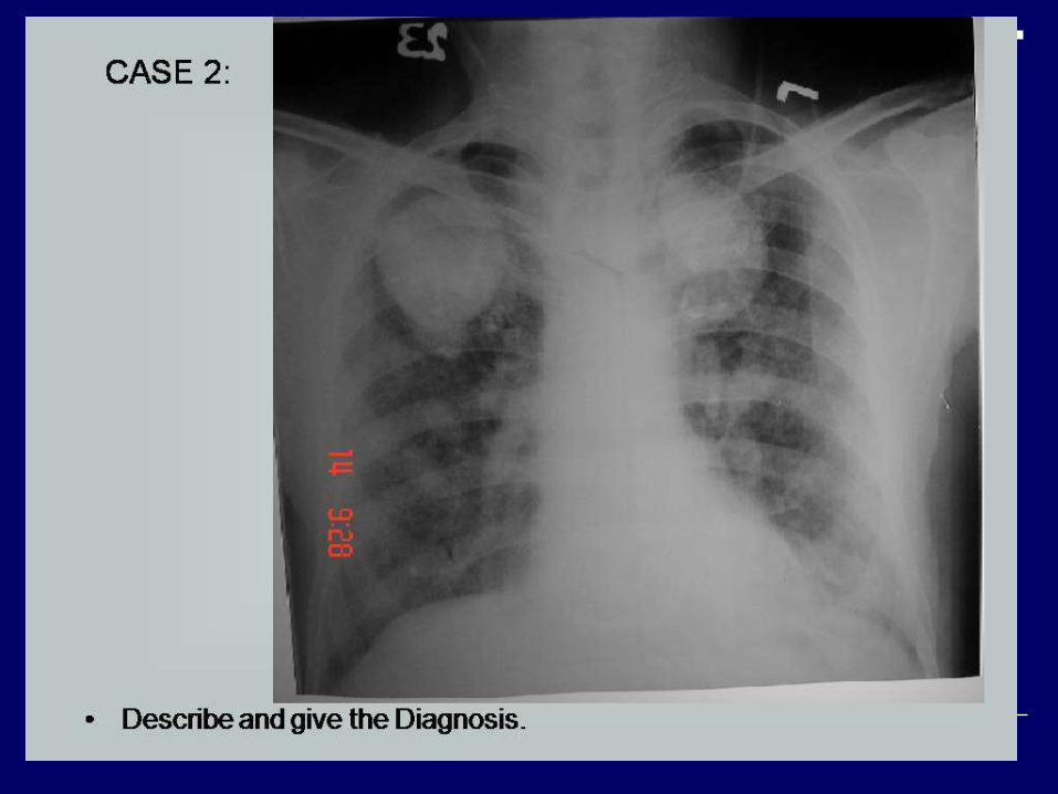
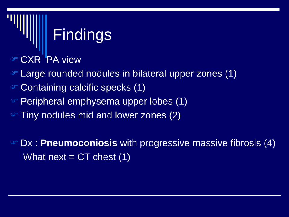
CXR PA view
Large rounded nodules in bilateral upper zones (1)
Containing calcific specks (1)
Peripheral emphysema upper lobes (1)
Tiny nodules mid and lower zones (2)
Dx : Pneumoconiosis with progressive massive fibrosis (4)
What next = CT chest (1)
Findings
Findings
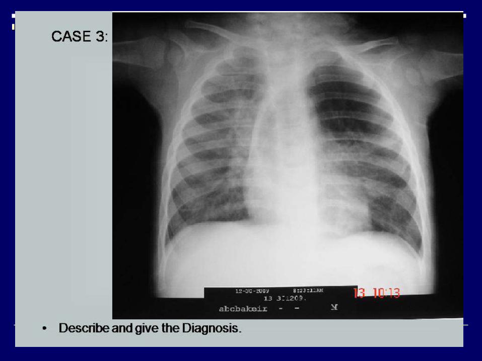
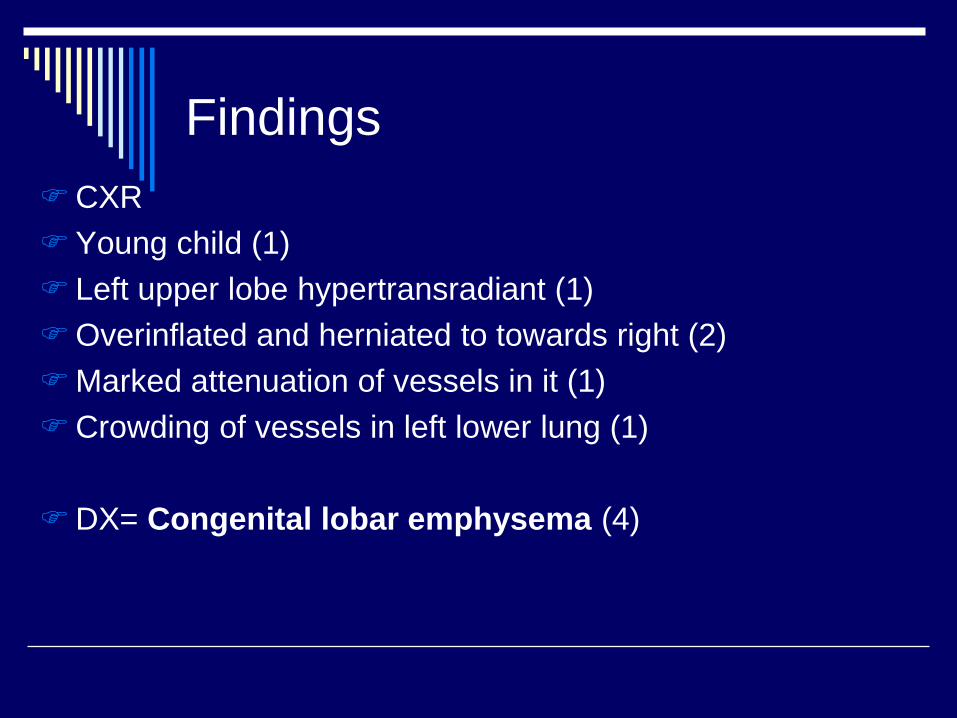
CXR
Young child (1)
Left upper lobe hypertransradiant (1)
Overinflated and herniated to towards right (2)
Marked attenuation of vessels in it (1)
Crowding of vessels in left lower lung (1)
DX= Congenital lobar emphysema (4)
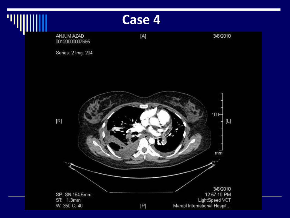
Case 4
Findings
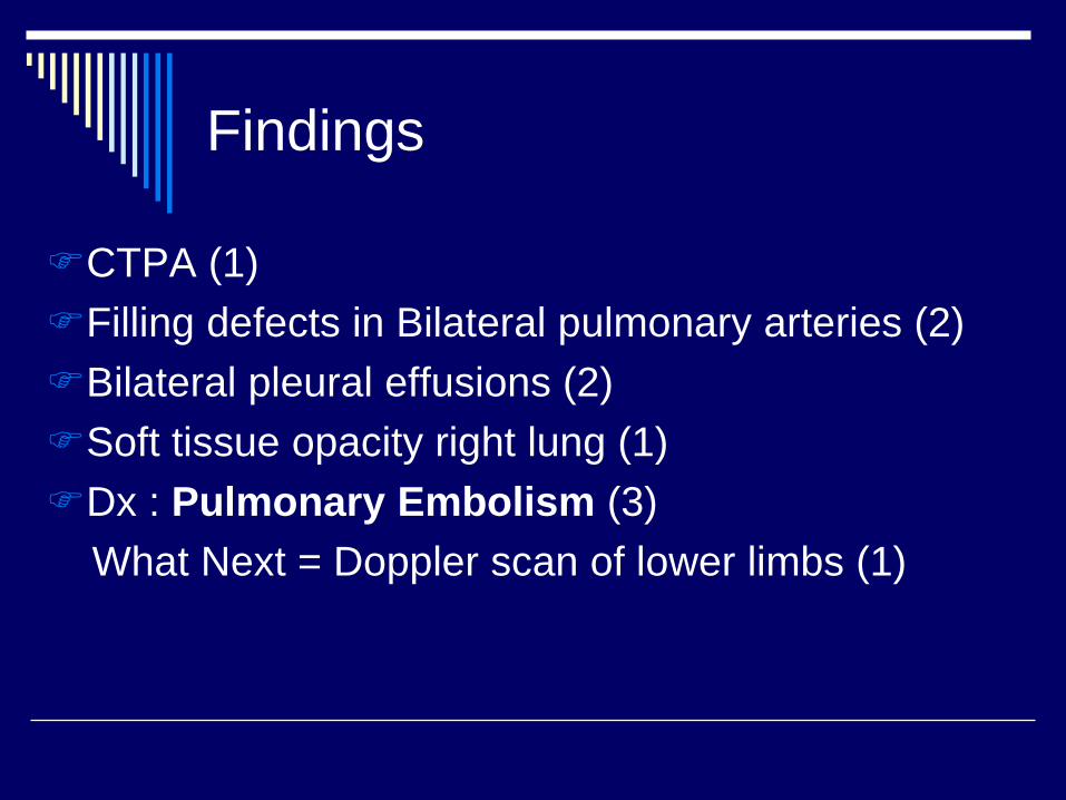
CTPA (1)
Filling defects in Bilateral pulmonary arteries (2)
Bilateral pleural effusions (2)
Soft tissue opacity right lung (1)
Dx : Pulmonary Embolism (3)
What Next = Doppler scan of lower limbs (1)
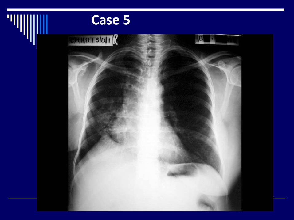
Case 5
Findings
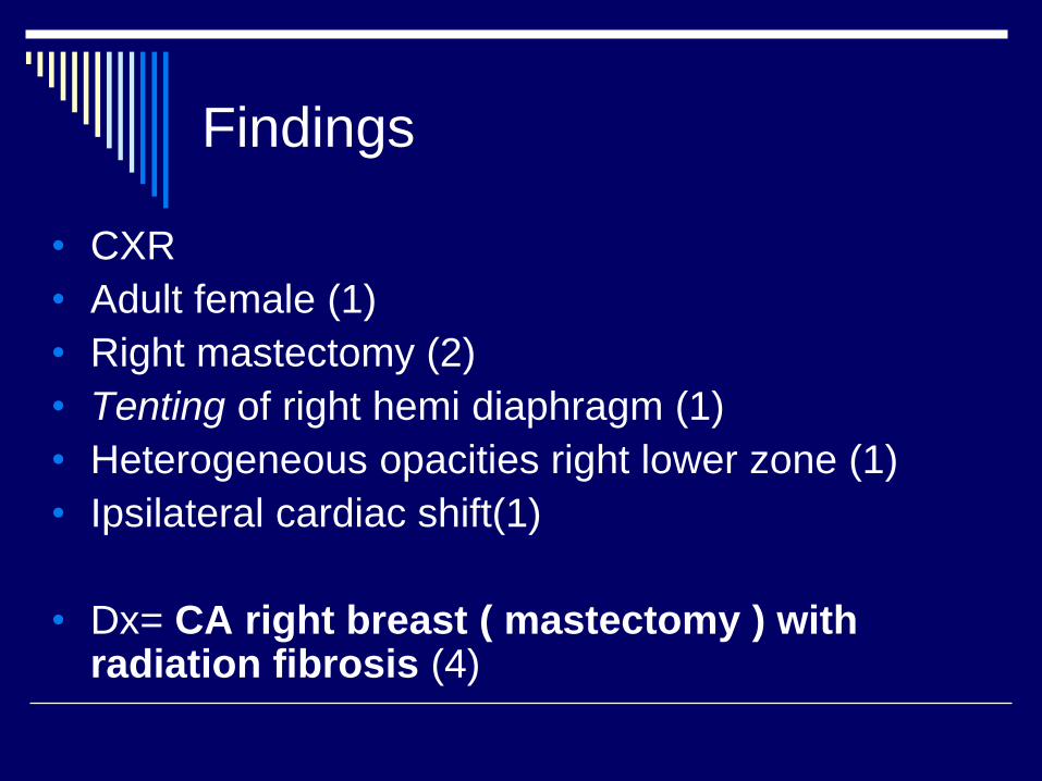
• CXR
• Adult female (1)
• Right mastectomy (2)
• Tenting of right hemi diaphragm (1)
• Heterogeneous opacities right lower zone (1)
• Ipsilateral cardiac shift(1)
• Dx= CA right breast ( mastectomy ) with radiation fibrosis (4)
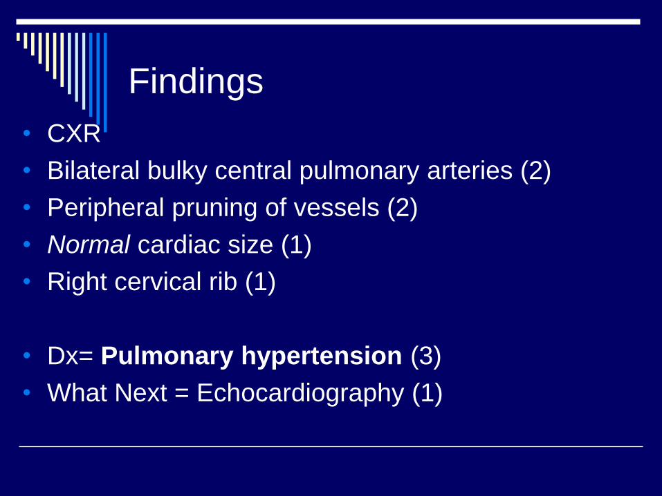
Findings
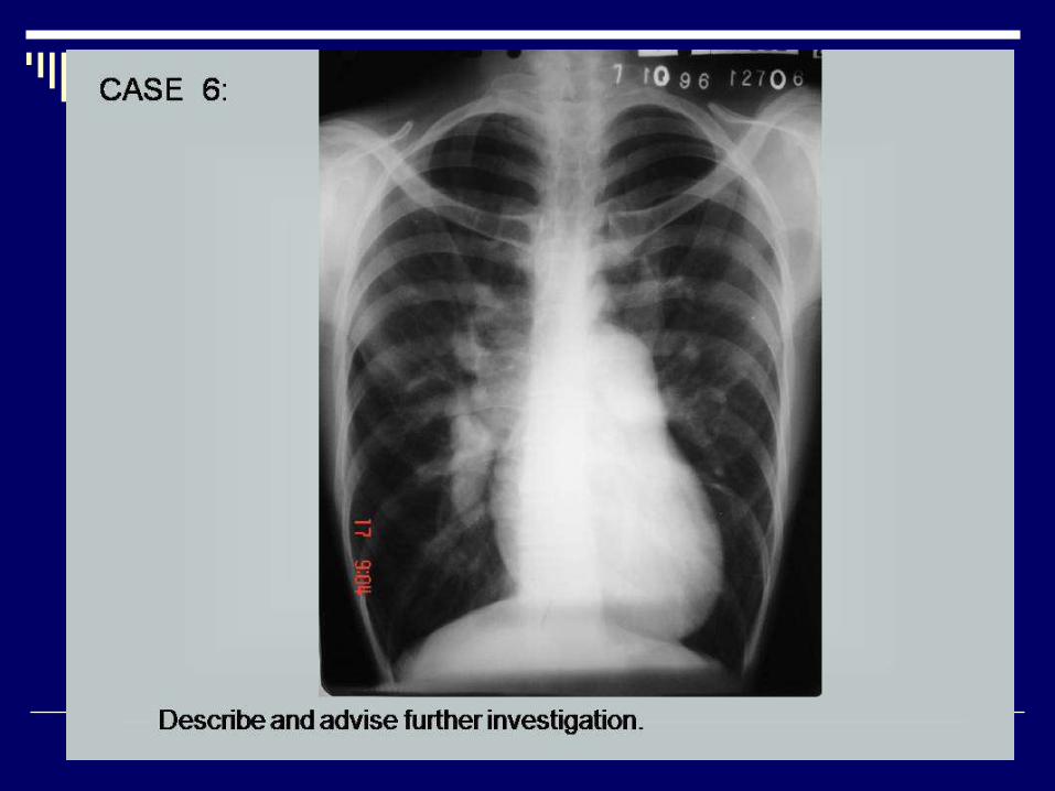
• CXR
• Bilateral bulky central pulmonary arteries (2)
• Peripheral pruning of vessels (2)
• Normal cardiac size (1)
• Right cervical rib (1)
• Dx= Pulmonary hypertension (3)
• What Next = Echocardiography (1)
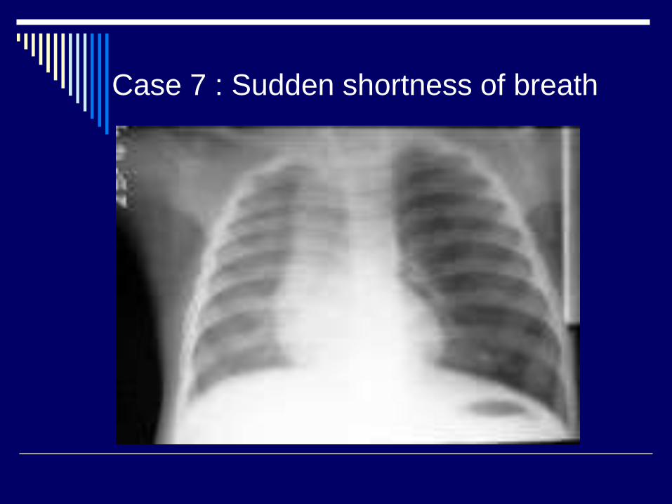
Case 7 : Sudden shortness of breath
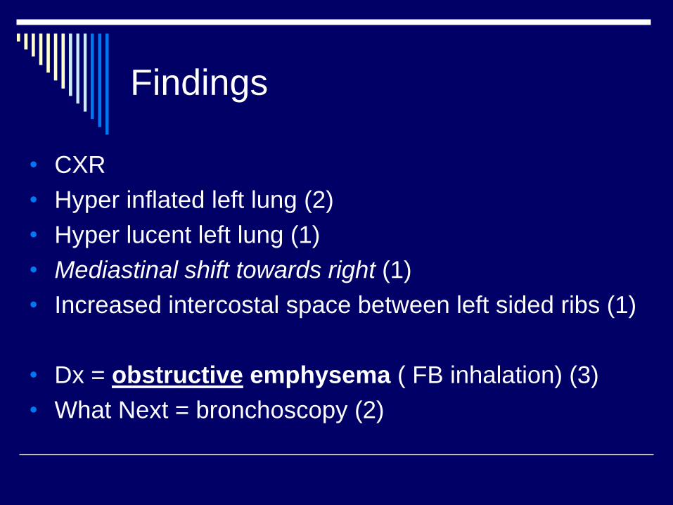
Findings
• CXR
• Hyper inflated left lung (2)
• Hyper lucent left lung (1)
• Mediastinal shift towards right (1)
• Increased intercostal space between left sided ribs (1)
• Dx = obstructive emphysema ( FB inhalation) (3)
• What Next = bronchoscopy (2)
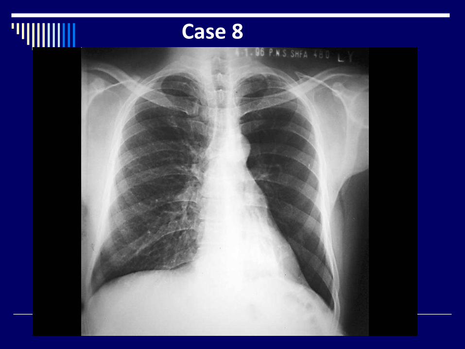
Case 8
Findings
CXR
Hypertransradiant left lung (2)
Oligemic left lung (1)
Increase bronchovascular markings in left retrocardiac
region (2)
Right lung = N (1)
Dx = Left lower lobe collapse with compensatory
hyperinflation of upper lobe (3)
What Next = Lateral CXR, CT (1)
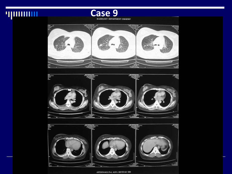
Case 9
• CT chest with contrast ( lung + mediastinal window) (1)
• Large, heterogeneous mass right breast (1)
• Skin thickening + nipple retraction (1)
• Nodules right upper lobe (1)
• Reticulations right upper lobe (1)
• Bilateral pleural effusions (1)
• Hypodense area in liver (1)
• Dx = CA breast with lymphangitis carcinomatosa and
liver metastasis (2)
What Next = FNAC breast (1)
Findings
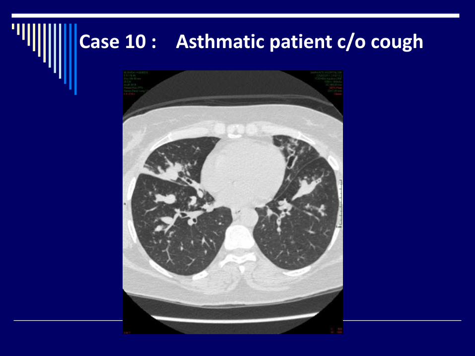
Case 10 : Asthmatic patient c/o cough
Findings
CT chest axial image ( lung window) (1)
Bilateral bronchiectasis (2)
Mucus plugs ( glove finger ) (3)
Dx = ABPA (4)
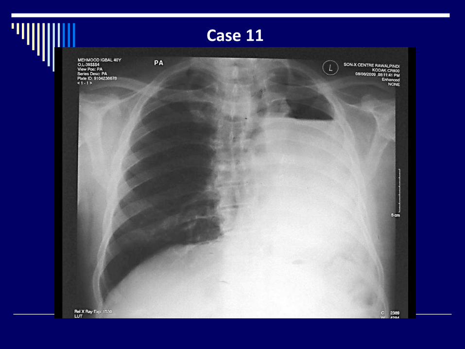
Case 11
Findings
CXR
Opaque left hemi thorax with air fluid level (2)
Ipsilateral mediastinal shift (2)
Overinflated right lung (2)
Dx =Left pneumonectomy (4)
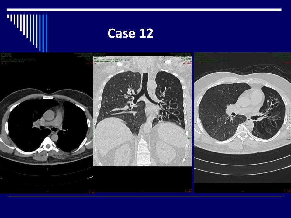
Case 12

Findings
• CT chest lung and mediastinal windows axial + coronal
views (1)
• Small, lucent left lung (2)
• ipsilateral mediastinal shift (1)
• Left sided bronchiectasis (1)
• Small left pulmonary artery (2)
Dx: Macleod's syndrome (3)
Findings
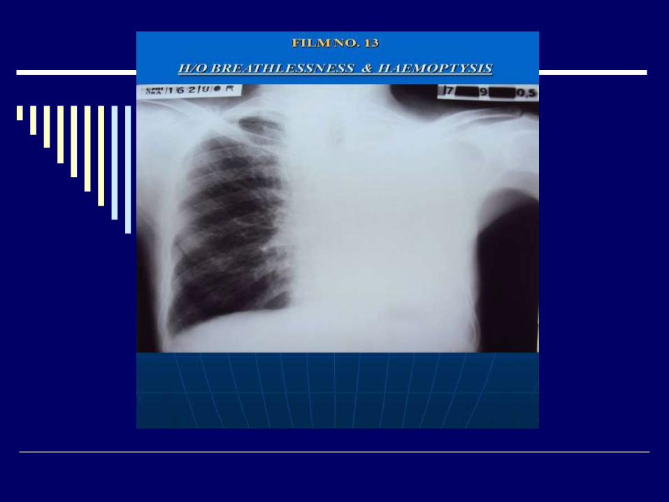
X-ray chest PA view of adult male (01)
Opaque left hemi thorax (01)
Mediastinal shift toward left (01)
No air-bronchogram, cavitations or bony lesion (02)
Normal right lung (01)
Diagnosis: -
Collapse left lung (due to hilar mass ?) (03)
Advised: -
CECT chest (01)
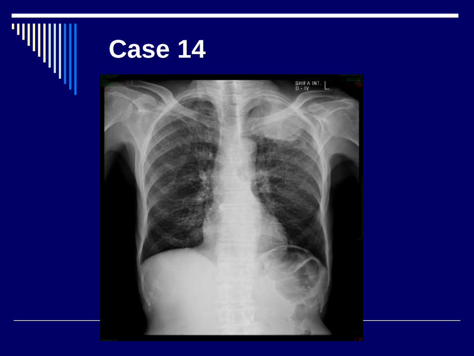
Case 14
Findings
• CXR
• Lung apical lung mass (1)
• Destruction of left 4th rib (2)
• CP angles = clear (1)
• Right lung = normal (1)
• Dx : pancoast tumor (4)
• What Next = CT chest (1)
Findings
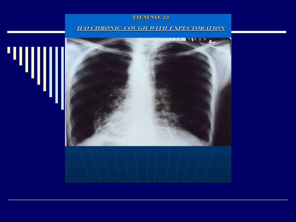
X-ray chest PA view of adult male (01)
Cystic shadows in both lower zones (02)
Dextrocardia with blurring of cardiac outline (02)
Diagnosis: - Dextrocardia with bilateral bronchiectasis (02)
(Kartagener's syndrome ) (02)
Advised: -X-ray PNS (01)