Embed Size (px)
Citation preview

Imagerie des lésions localement avancées
Advanced breast lesion imaging.
S. Taïeb, A.Mailliez, H.Gauthier, S.Giard, L.Ceugnart
Hanoi, 6 nov 2015

Advanced breast lesions : > 5 cm


1st step : Tumor Size -‐ > 5cm
CorrelaEng sonography, mammography, and pathology in the assessment of breast cancer size. § Hieken et al. Am J Surg. 2001 -‐
ü 180 à 146 invasive tumors mammo & US ü 69% size US > mammo with equivalent underesEmaEon / pathology ü Maximal tumor dimension accurate 75% US / 65% mammo
§ Bosch et al. Eur J Radiol 2003 (+ Physical examinaEon) ü 105 à 73 invasive tumors mammo & US ü CorrelaEon coefficient : US 0,68; Mammo 0,44; PE 0,42 ü PE : overesEmaEon; US : underesEmaEon
US > Mammographie CI 95% : +/-‐ 11 mm

48 ans, PE : Mass 5cm,

48 ans, PE : Mass 5cm, Mammo : 7cm

US Biopsy 11h, 4cm : CCI
48 ans, PE : Mass 5cm, Mammo : 7cm US 21 mm

Macrobiopsy 12 h : CCIS grade 3. Macrobiopsy 2 h : CCIS grade 2 & 3
48 ans, PE : Mass 5cm, Mammo : 7cm US 21 mm

§ Totale Mastectomy : CCI 15 mm + CCIS 7 cm Ki67 : 10 % RO+ (70 % Score = 5) RP-‐ (2 % Score = 1) HER2 = 3+, fort § SLN : 10N-‐ / 10N

56 y-‐o, PE : Mass 4 cm,

56 y-‐o, PE : Mass 4 cm, Mammo ?

56 y-‐o, PE : Mass 4 cm, Mammo ? US : 32 mm

56 y-‐o, PE : Mass 4 cm, Mammo ? US : 32 mm MRI : 20 x 19 mm CLI : 28 x 22 mm

33 y-‐o, PE : Mass 4 cm, Mammo 2 cm

33 y-‐o, PE : Mass 4 cm, Mammo 2 cm US : 4 cm

33 y-‐o, PE : Mass 4 cm, Mammo 2 cm US : 4 cm

33 y-‐o, PE : Mass 3 cm, Mammo 3 cm US : 40 mm MRI : 35 / 49 ?

33 y-‐o, PE : Mass 3 cm, Mammo 3 cm US : 40 mm MRI : 35 / 49 ? CCI grade II, 37 mm ER-‐, ErP -‐, Her2 -‐, KI67 20%

1st step : Tumor Size -‐ > 5cm
§ Luparia et al. Radiol Med. 2013 : CorrelaEng Sonography, Mammography, Tomosynthesis, MRI and pathology in the assessment of breast cancer size. ü 149 lesions / 110 paEents – 22,3mm ü CorrelaEon with pathological tumor size
• MRI : 0,92 -‐ TS : 0,89 • DM : 0,83 -‐ US : 0,77
§ Jethava et al. Conn Med 2015 : MRI and histological size ü 147 lesions / 124 paEents – 81 IDC, 35 DCIS, 15 ILC, 16 others ü Concordance 5 mm 56% ü OveresEmaEon : 32% -‐ High grade tumor and CCIS ü UnderesEmaEon : 12%
MRI > US > Mammography > PE

§ 33 pts, 37 lesions : 27 DIC, 10 LIC. § MRI and Gross specimen / Microscopic size
Behjatnia b et al. Int J Clin Exp Pathol 2010

§ 33 pts, 37 lesions : 27 DIC, 10 LIC. § MRI and Gross specimen / Microscopic size
Behjatnia b et al. Int J Clin Exp Pathol 2010

§ 33 pts, 37 lesions : 27 DIC, 10 LIC. § MRI and Gross specimen / Microscopic size
Behjatnia b et al. Int J Clin Exp Pathol 2010
Although MRI is the most accurate modality we have to image the breast so far, it remains imperfect : § OveresEmaEng tumor size in 11% to 70% of paEents, § UnderesEmaEng it in 10% to 56% of paEents.

Tumor Size -‐ > 5cm but not only
§ MRI à
§ US à
§ Mammo à
§ ER, PgR ?
§ HER 2 ?
§ Ki67 ?

St Gallen Conference 2015 & ESMO 2015
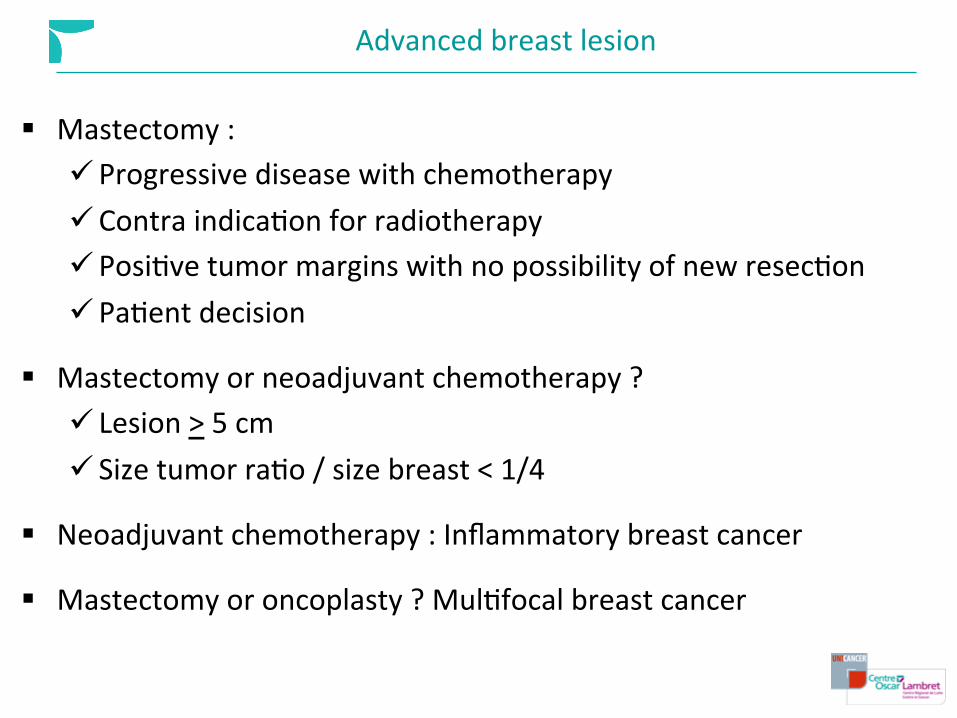
Advanced breast lesion
§ Mastectomy : ü Progressive disease with chemotherapy ü Contra indicaEon for radiotherapy ü PosiEve tumor margins with no possibility of new resecEon ü PaEent decision
§ Mastectomy or neoadjuvant chemotherapy ? ü Lesion > 5 cm ü Size tumor raEo / size breast < 1/4
§ Neoadjuvant chemotherapy : Inflammatory breast cancer
§ Mastectomy or oncoplasty ? MulEfocal breast cancer

Inflammatory breast cancer
§ 1-‐5%
§ PEV 0 : same volume § PEV 1 : x2 in 6 months § PEV 2 : focal form § PEV3 : Diffuse form (masEEs carcinomatosa)
§ Very poor prognosis § Neoadjuvant chemotherapy always

DescripEon
45 – 55 y-‐o Few weeks : § Redness, edema > 1/3 of the breast § Nipple retracEon § Orange-‐peel appearance of the skin § Pain
PE : § Mass 60%, § Axillary LN > 50% § No fever

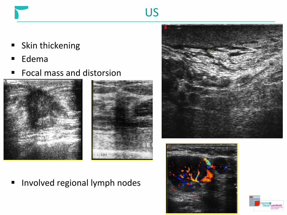
US
§ Skin thickening § Edema § Focal mass and distorsion
§ Involved regional lymph nodes

MasEEs Carcinomatosa

42 y-‐o 3 weeks

US : no mass à MRI

MRI

Inflammatory breast cancer : diagnosis
§ Skin punch biopsy : lymphovascular embols § US biopsy if mass § Vacuum-‐Assisted core biopsy if microcalcificaEon
Courtesy S.Morand

MulEfocal breast cancer 42 y-‐o, PE : leq axillary lymph node N+ Courtesy I.Thomassin


US : 2 lesions + LN

MRI : 3 lesions / 5 cm

2nd step : Lymph nodes
Houssami N et al. PreoperaEve ultrasound-‐guided needle biopsy of axillary nodes in invasive breast cancer: meta-‐analysis of its accuracy and uElity in staging the axilla. Ann Surg. 2011 ü 1965-‐2011 : 125 studies , 2874 US guided biopsy / 6166 femmes ü Se : 80%, Sp : 98% -‐ palpable or not
-‐ FNB / CNB -‐ Prevalence N+ in studies


3rd step : M ?
ESMO 2015 : addiEonal invesEgaEons should be considered : § N+ § T > 5 cm § Agressive biology § Symptoms or laboratory values suggesEng M+
§ CT : Chest + Abdomen + Pelvis with bone analysis § FDG-‐PET/CT may be useful
ü Inconclusive CT ü Follow-‐up neoadjuvant chemotherapy


§ N3 in 32 paEents § M1 in 43 paEents / PET-‐ CT; 28 / ConvenEonal Imaging
§ PET-‐CT > CT : bone metastasis, extra-‐axillary lymph nodes and liver metastasis
§ CT (98,3%) > PET-‐CT (97,4%) for lung metastasis
§ PET-‐CT (98,3%) > Bone scan (89,7%)
Conclusion : Whole body staging in one step !!
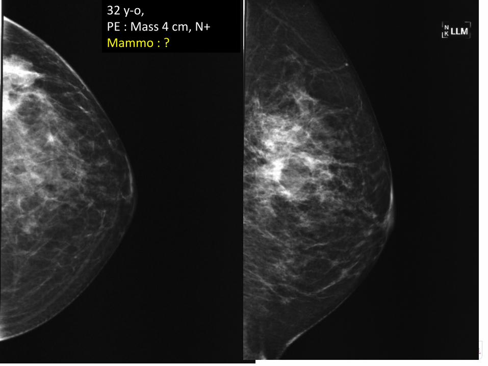
32 y-‐o, PE : Mass 4 cm, N+ Mammo : ?

32 y-‐o, PE : Mass 4 cm, N+ Mammo : ? US : 31 mm, N+

32 y-‐o, PE : Mass 4 cm, N+ Mammo : ? US : 31 mm, N+ MRI : 6 cm
CCI, ER-‐, ErP-‐, Her2-‐ KI67 90% N+


Complete response Breast conservatory therapy

61 y-‐o Nipple retracEon CCI : TN : KI67 > 95%

61 y-‐o Nipple retracEon CCI : TN : KI67 > 95%


61 y-‐o Nipple retracEon CCI : TN : KI67 > 95% Aqer 3 FEC 100

Take Home message
§ Tumor dimensions : MRI > US > Mammography MRI : First substracEon
ü Over esEmaEon 15-‐70% ü Under esEmaEon 20-‐50%
§ Lymph node : US + Biopsy
§ Stagging : PET-‐CT / CT (thorax, abdomen, pelvis and bone)
§ Neoadjuvant chemotherapy follow up : MRI, PET-‐CT
§ MulEple Tumors : MRI assessment à Oncoplasty ?

cảm ơn bạn