Embed Size (px)
Citation preview
+
3 Nov – 6 Nov 2016 Trident Nariman Point - Oberoi Mumbai
Vascular oncology emergencies management
Hemorrhage in oncologic interventional radiology
+
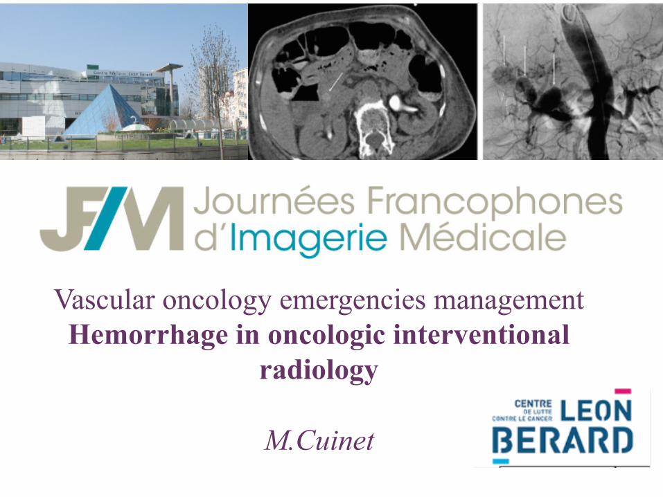
Vascular oncology emergencies management Hemorrhage in oncologic interventional
radiology
M.Cuinet
+ Intro
n Hemorrhage is the 3rd cause of death in neoplastic disease (after tumor invasion on organ function and infection). Bleeding occurs in up to 10 % of patients with advanced cancer and can take different forms.
n Apart from interventional procedures, the endovascular hemostatic embolisation is performed in emergency and can be practice: - to treat hemorrhage uncontrolled by medical treatment in a cancer kwowned
- after diagnostic and therapeutic procedures .
- to reduce morbidity and mortality compared to surgery procedures
+ Hemorrhage causes
n Blood vessels (capillaries or bigger vessels) affected by cancer
n Thrombocytopenia (being or not iatrogenic) leading to bleeding, especially when an infection occurs.
n Abnormal low level of coagulation factors (intravascular coagulation)

+ Goal and principle
n Goal of embolization: to reduce/stop the blood flow into the vessel to stop haemorrhage
n Occlusion site: proximal distal capillary
n Material: catheter and microcatheter
n Choice of embolisation: agent depends largely regarding the indication .
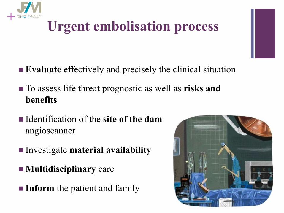
+ Urgent embolisation process
n Evaluate effectively and precisely the clinical situation
n To assess life threat prognostic as well as risks and benefits
n Identification of the site of the damage using an angioscanner
n Investigate material availability
n Multidisciplinary care
n Inform the patient and family
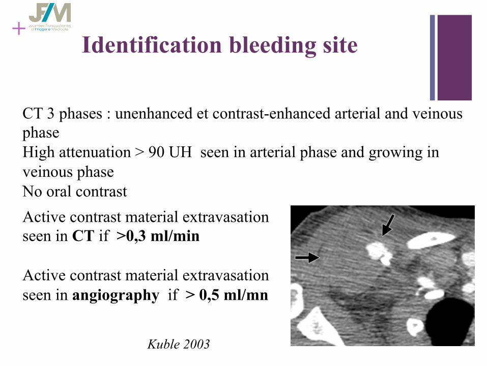
+ Identification bleeding site
CT 3 phases : unenhanced et contrast-enhanced arterial and veinous phase High attenuation > 90 UH seen in arterial phase and growing in veinous phase No oral contrast Active contrast material extravasation seen in CT if >0,3 ml/min Active contrast material extravasation seen in angiography if > 0,5 ml/mn
Kuble 2003
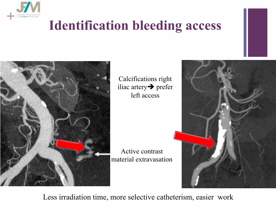
+ Identification bleeding access
Less irradiation time, more selective catheterism, easier work
Active contrast material extravasation
Calcifications right iliac arteryè prefer
left access
+
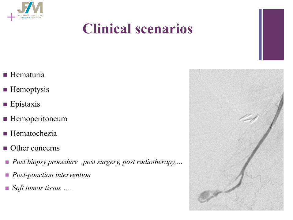
n Hematuria
n Hemoptysis
n Epistaxis
n Hemoperitoneum
n Hematochezia
n Other concerns
n Post biopsy procedure ,post surgery, post radiotherapy,…
n Post-ponction intervention
n Soft tumor tissus …..
Clinical scenarios

+ Embolisation agents 3 types
§ Mecanical occlusion devices: Coils/Plugs - Equivalent of surgical ligation
- Mechanic occlusion and fibres attached to them causes thrombosis - Occludes arteries proximally while preserves distal flow via
collaterals vessels - Don‘t cause complete organ infarction
§ Particulate embolic agents: PVA/microspheres/Gelfoam Inflammatory reaction involving vessel wall Flow downstream until they blocked in vessel
§ Liquid embolic agents: Nbutyl2 cyanoacrylate, isobutyl2cyanoacrylate - Rapidly hardening adhesives
- Necrosis and thrombosis - Rapid polymerization in contact with ionic media

+ Embolisation agents
§ Temporary – Gelfoam / Spongel
§ Permanent
– PVA / Microspheres – Sclerosing agents (Absolute ethanol) – Coils
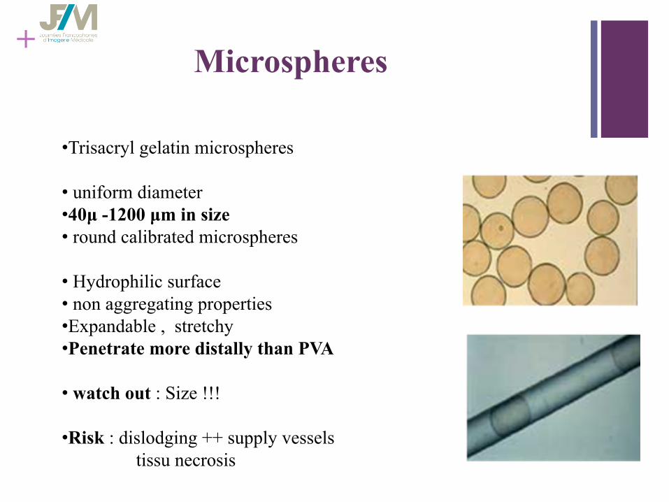
+ Microspheres
• Trisacryl gelatin microspheres
• uniform diameter • 40µ -1200 µm in size • round calibrated microspheres
• Hydrophilic surface • non aggregating properties • Expandable , stretchy • Penetrate more distally than PVA
• watch out : Size !!!
• Risk : dislodging ++ supply vessels tissu necrosis
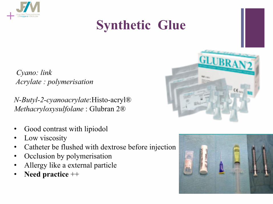
+ Synthetic Glue
Cyano: link Acrylate : polymerisation N-Butyl-2-cyanoacrylate:Histo-acryl® Methacryloxysulfolane : Glubran 2® • Good contrast with lipiodol • Low viscosity • Catheter be flushed with dextrose before injection • Occlusion by polymerisation • Allergy like a external particle • Need practice ++
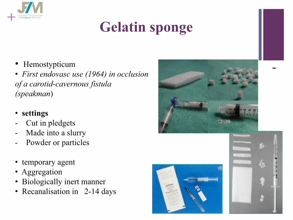
+ Gelatin sponge
• Hemostypticum • First endovasc use (1964) in occlusion of a carotid-cavernous fistula (speakman)
• settings - Cut in pledgets - Made into a slurry - Powder or particles
• temporary agent • Aggregation • Biologically inert manner • Recanalisation in 2-14 days
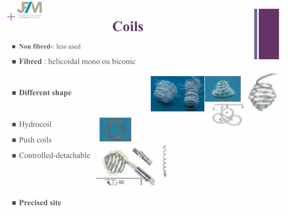
+ Coils
n Non fibred-: less used
n Fibred : helicoidal mono ou biconic
n Different shape
n Hydrocoil
n Push coils
n Controlled-detachable
n Precised site
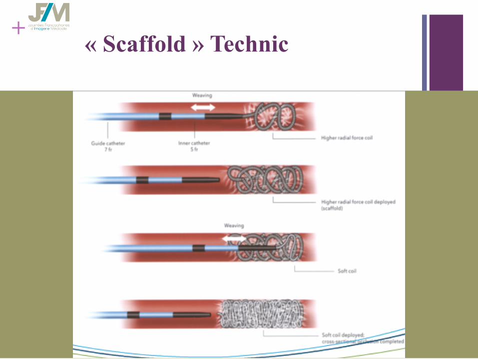
+ « Scaffold » Technic
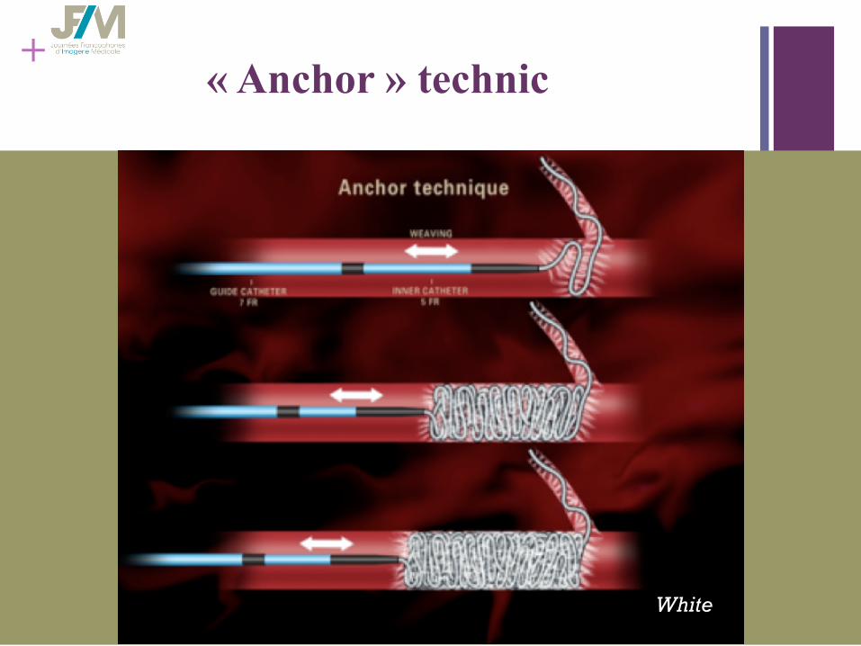
+ « Anchor » technic
White
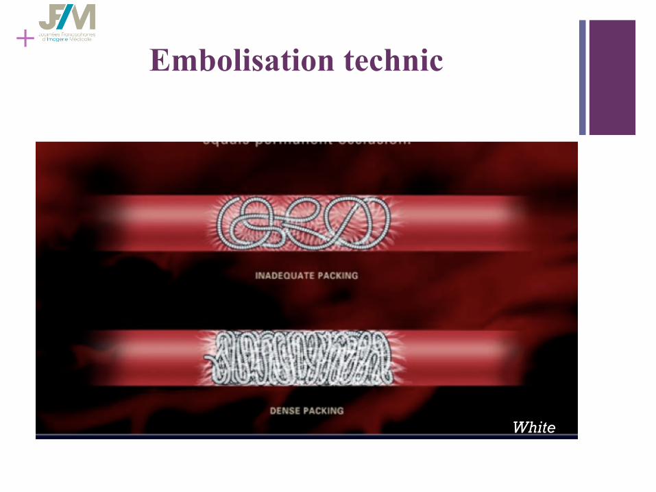
+ Embolisation technic
White
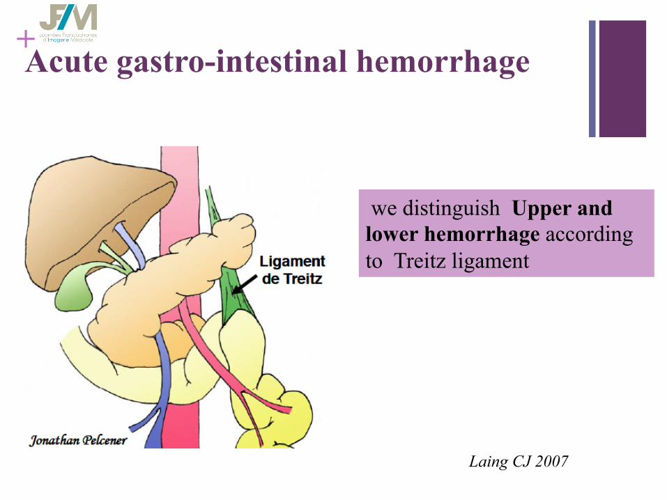
+ Acute gastro-intestinal hemorrhage
we distinguish Upper and lower hemorrhage according to Treitz ligament
Laing CJ 2007
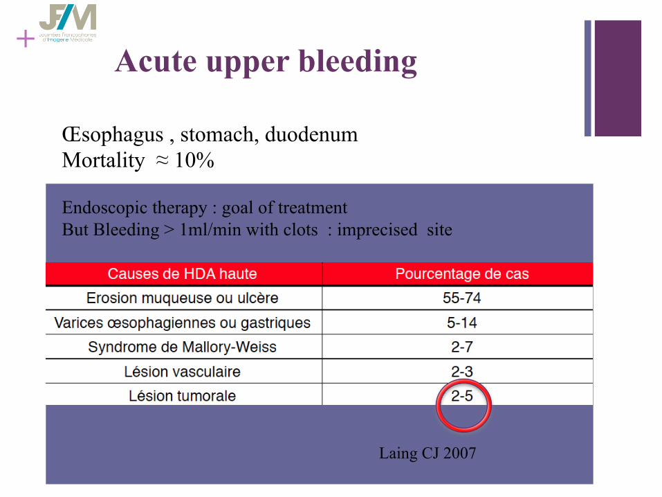
+ Acute upper bleeding
Laing CJ 2007
Œsophagus , stomach, duodenum Mortality ≈ 10%
Endoscopic therapy : goal of treatment But Bleeding > 1ml/min with clots : imprecised site
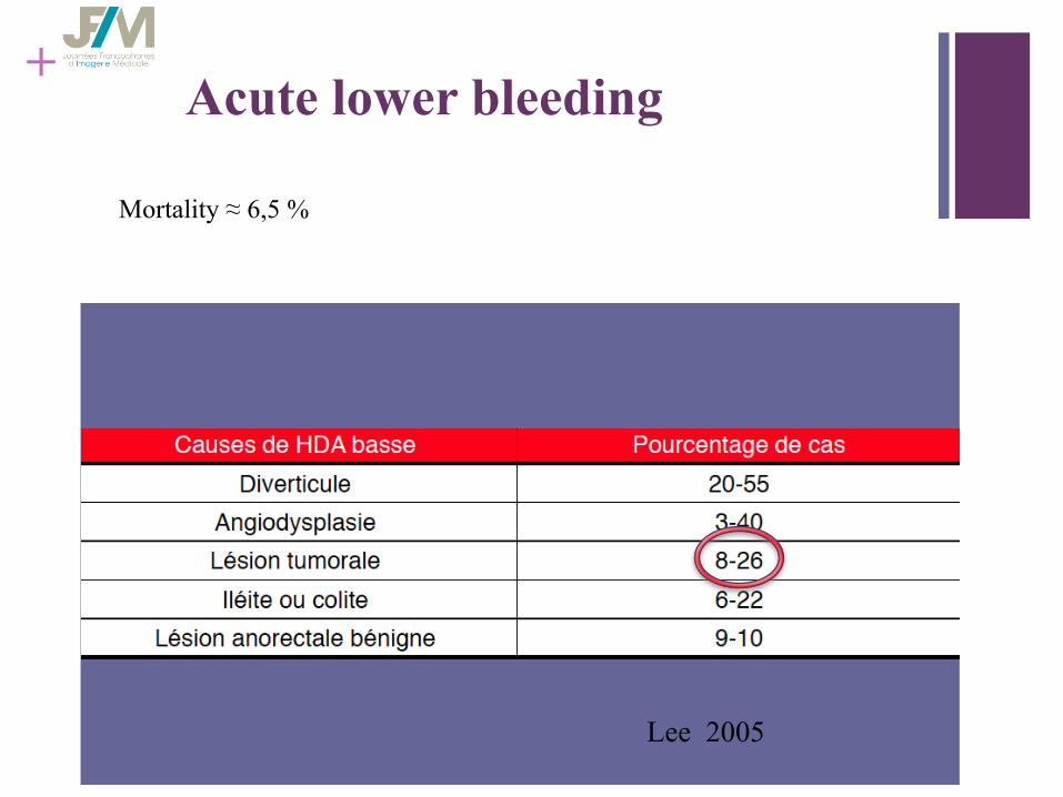
+ Acute lower bleeding
Lee 2005
Mortality ≈ 6,5 %
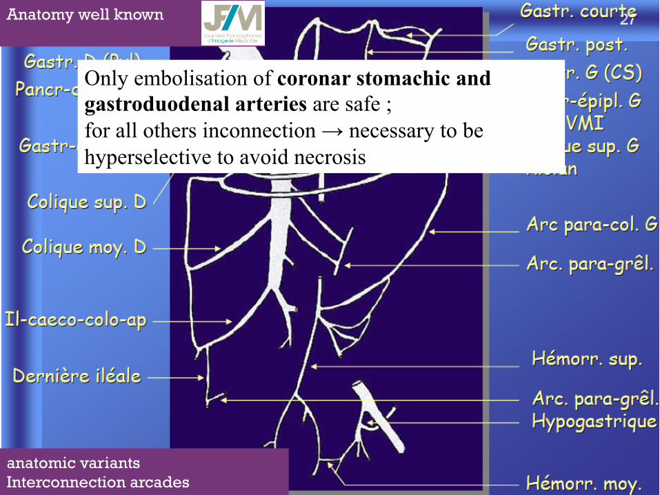
+ Anatomy well known
anatomic variants Interconnection arcades
Only embolisation of coronar stomachic and gastroduodenal arteries are safe ; for all others inconnection → necessary to be hyperselective to avoid necrosis

+ n Mr X., 69 years old , Jehovah’s witness
n adressed in emergency for management aggressive gastric lymphoma
Endoscopy important ulcer and necrotic lesion in the bulb
+
n Massive upper abdominal hemorrhage ( >1/2 l )
n blood pressure 47, violent abdominal pain .
n cannulation with macromolecules, perfusion, anxiolytic, painkillers
n Risk of upcoming death by hemorrhage without transfusion.
n Refusal of blood transfusion.
n Hemostasis embolization
Mr X., 69 years old , Jehovah’s witness
+
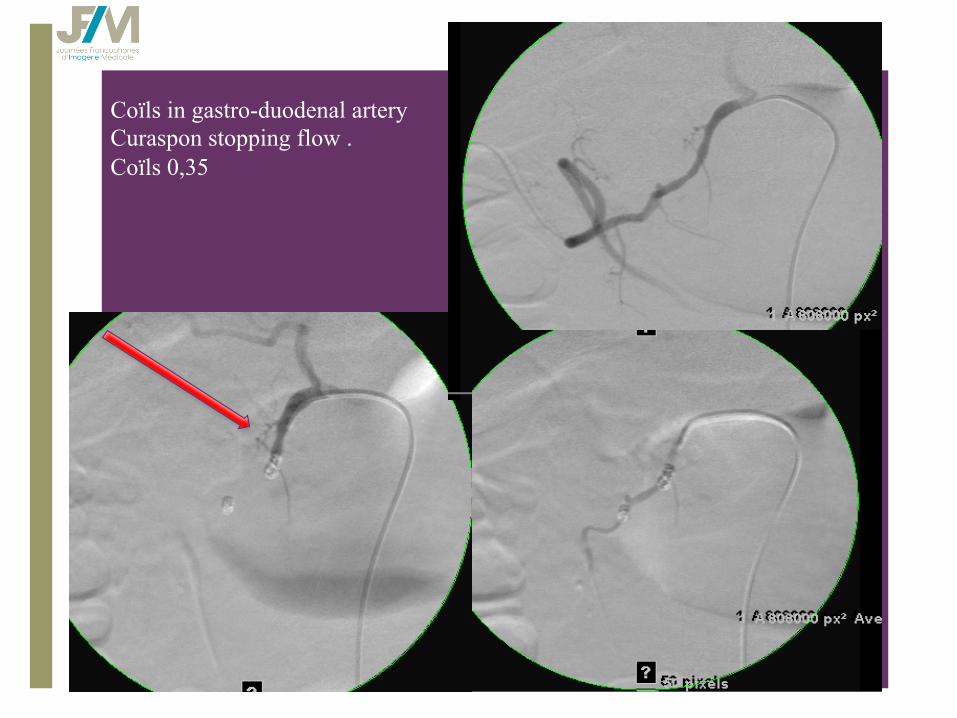
Coïls in gastro-duodenal artery Curaspon stopping flow . Coïls 0,35

+ Hemorrhage post pancreatic surgery
n erosion by fistula
n 10% of cases
n delay 20 days
n mortality 30-70%
Sato et all JVIR 2011

+ Mrs D, 75 years old
• Cephalic pancreatectomy : D 6 • Several episode of hematemesis • Hypovolaemic shock

+
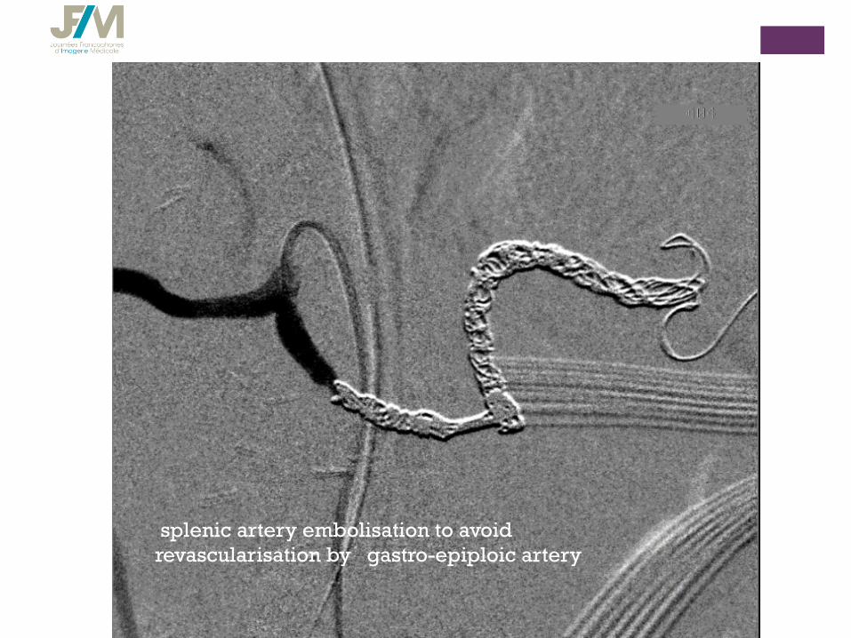
splenic artery embolisation to avoid revascularisation by gastro-epiploic artery
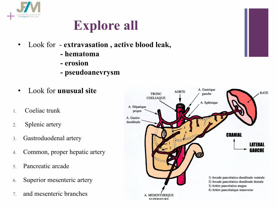
+ Explore all
1. Coeliac trunk
2. Splenic artery
3. Gastroduodenal artery
4. Common, proper hepatic artery
5. Pancreatic arcade
6. Superior mesenteric artery
7. and mesenteric branches
• Look for - extravasation , active blood leak, - hematoma - erosion - pseudoanevrysm • Look for unusual site

+ Hemorrhage post pancreatic surgery
n gastroduodenal strump embolization
n Sometimes use of cover stent
n If no lesion : discussion with surgeon to decide a systematic embolisation
n Not targeted embolisation make risk of recurrence ( Hur et all JVIR 11 )
Stump gastroduodenal artery
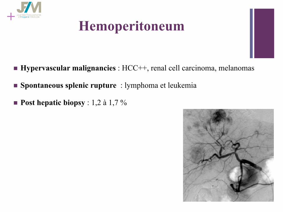
+ Hemoperitoneum
n Hypervascular malignancies : HCC++, renal cell carcinoma, melanomas
n Spontaneous splenic rupture : lymphoma et leukemia
n Post hepatic biopsy : 1,2 à 1,7 %

HCC complicated by rupture in 10-15 % High mortality rate within 30 days
Active bleeding
HCC bleeding
Portal thrombosis
+
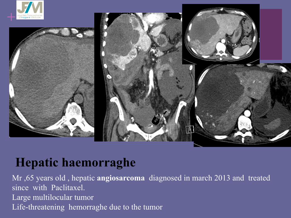
Mr ,65 years old , hepatic angiosarcoma diagnosed in march 2013 and treated since with Paclitaxel. Large multilocular tumor Life-threatening hemorraghe due to the tumor
Hepatic haemorraghe
+
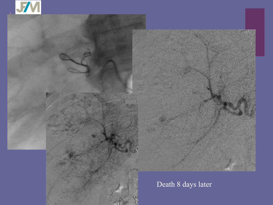
Death 8 days later

+
Most often iatrogenic injury cause massive hemorraghe First choice embolization
Epigastric artery
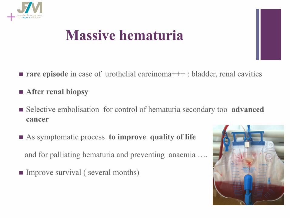
+ Massive hematuria
n rare episode in case of urothelial carcinoma+++ : bladder, renal cavities
n After renal biopsy
n Selective embolisation for control of hematuria secondary too advanced cancer
n As symptomatic process to improve quality of life
and for palliating hematuria and preventing anaemia ….
n Improve survival ( several months)
+
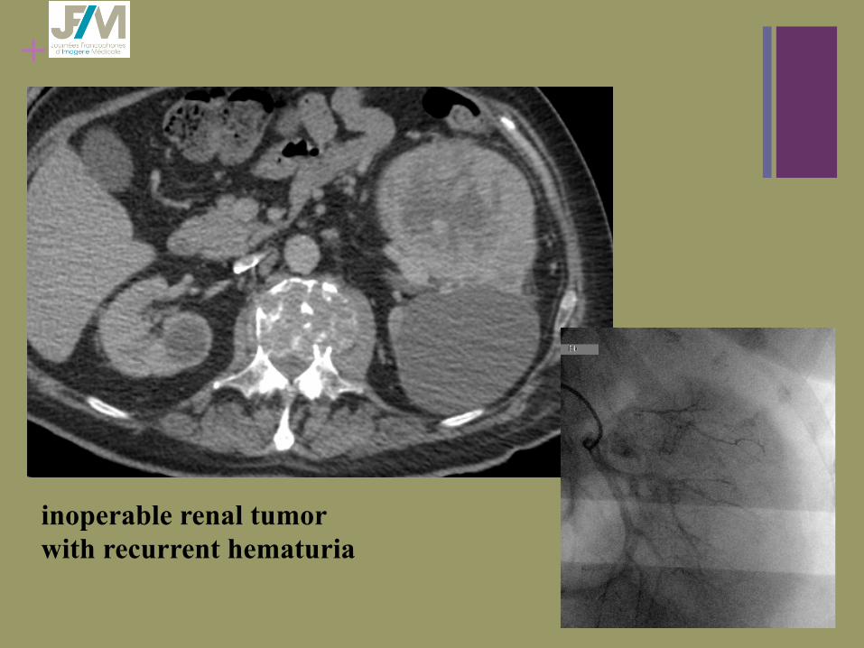
inoperable renal tumor with recurrent hematuria
+
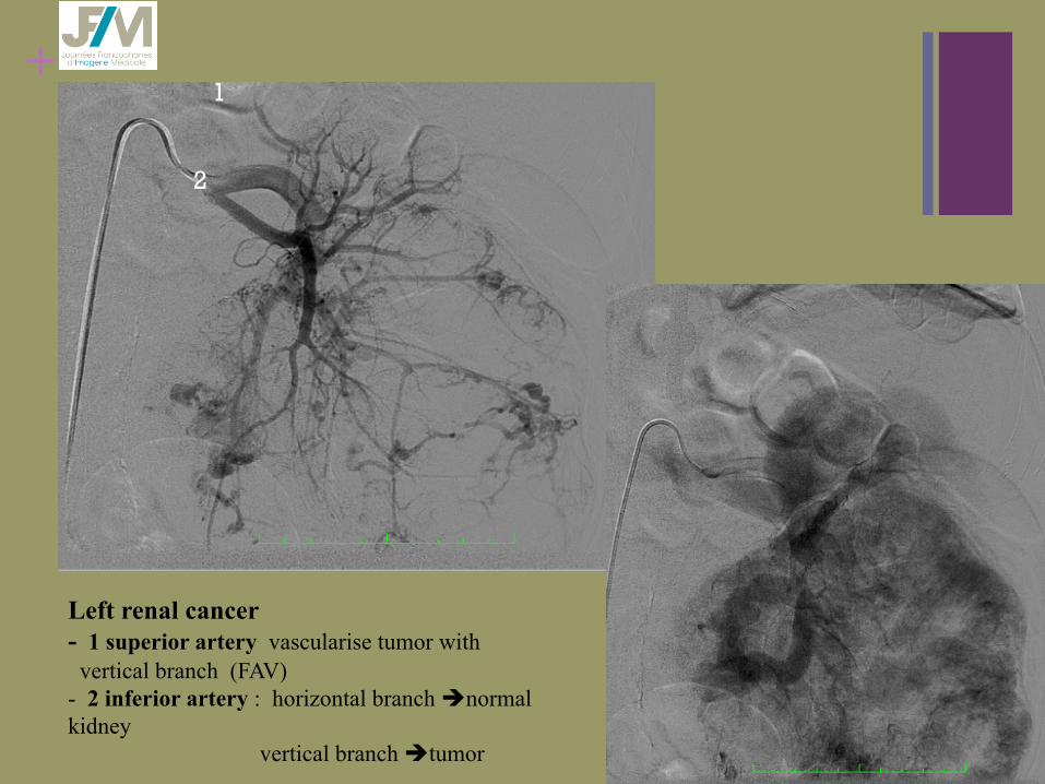
Left renal cancer - 1 superior artery vascularise tumor with vertical branch (FAV) - 2 inferior artery : horizontal branch ènormal kidney vertical branch ètumor
1
2
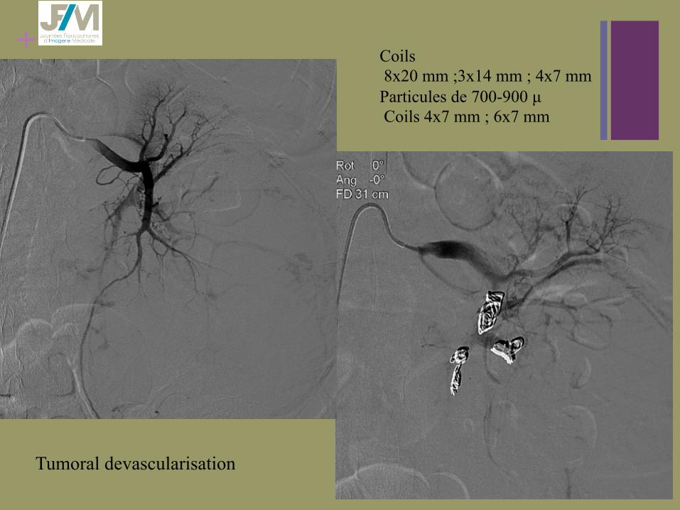
+ Coils 8x20 mm ;3x14 mm ; 4x7 mm Particules de 700-900 µ Coils 4x7 mm ; 6x7 mm
Tumoral devascularisation
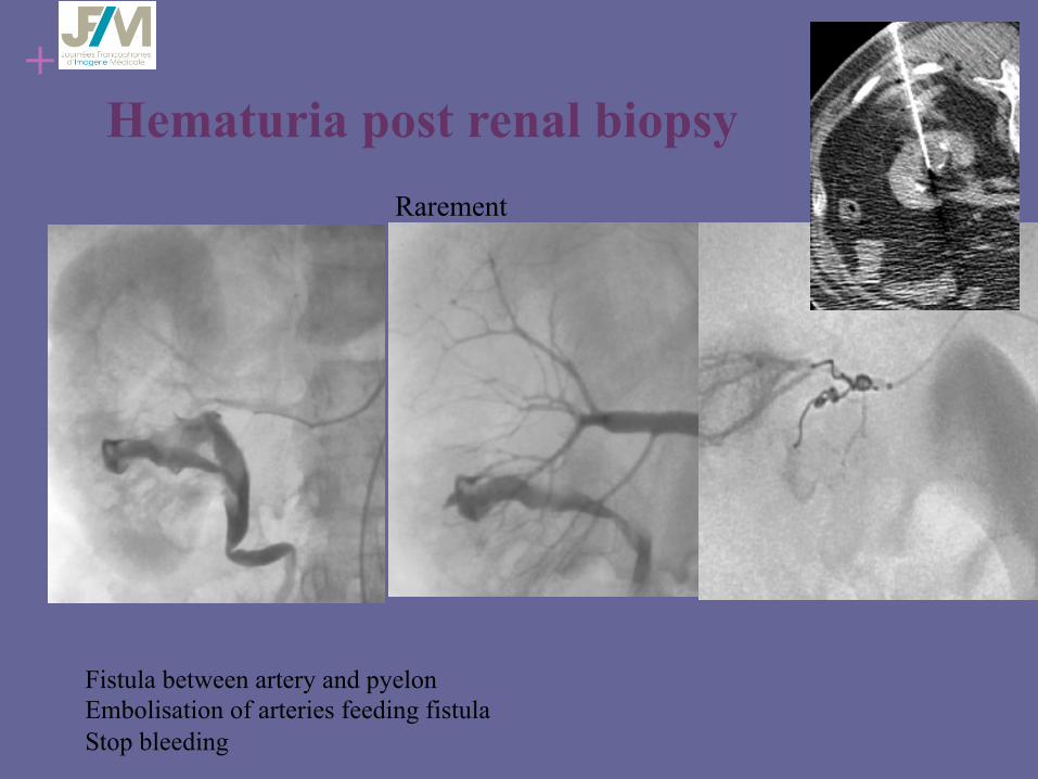
+ Hematuria post renal biopsy
Fistula between artery and pyelon Embolisation of arteries feeding fistula Stop bleeding
Rarement
+ Massive hemoptysis n 300–600 ml blood loss per day
n Mortality rate ≈ 64 % at one year ( Garcia 2014)
n Airway maintenance is vital more important than blood loss
n Causes : bronchogenic carcinoma (superficial mucosal invasion erosion into blood vessels and necrosis)
n Others : lung carcinoid tumor , metastasis from breast, renal ,and colon cancers
n Other ++: aspergillus , mucormycosis in patients with immunodeficiency (after chemotherapy)
n More rarely , hemoptysis after treatment (laser, curietherapy)
+
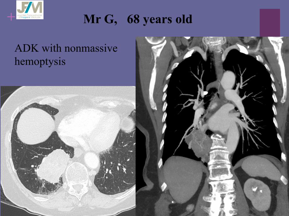
ADK with nonmassive hemoptysis
Mr G, 68 years old
+
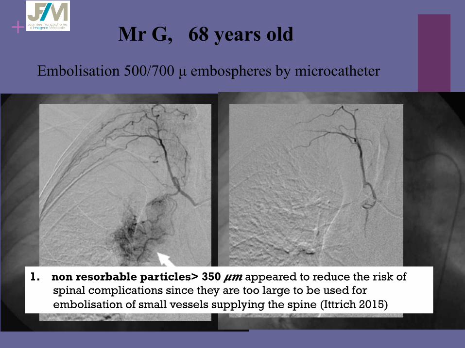
Embolisation 500/700 µ embospheres by microcatheter
Mr G, 68 years old
1. non resorbable particles> 350 𝝁𝒎 appeared to reduce the risk of spinal complications since they are too large to be used for embolisation of small vessels supplying the spine (Ittrich 2015)

+ Massive hemoptysis n Knowledge of the bronchial artery anatomy, variabilite, ectopic arteries ,
dangerous anastomoses
n Before : chest CT angiography look for supply arterial systemic anastomoses (Adamkiewicz ) embolisation
n Meanwhile : control lake of reflux
n After : control stop arteriel flow in treated artery
• Survival better if hemoptysis non correlated to tumor • Prognosis remains poor
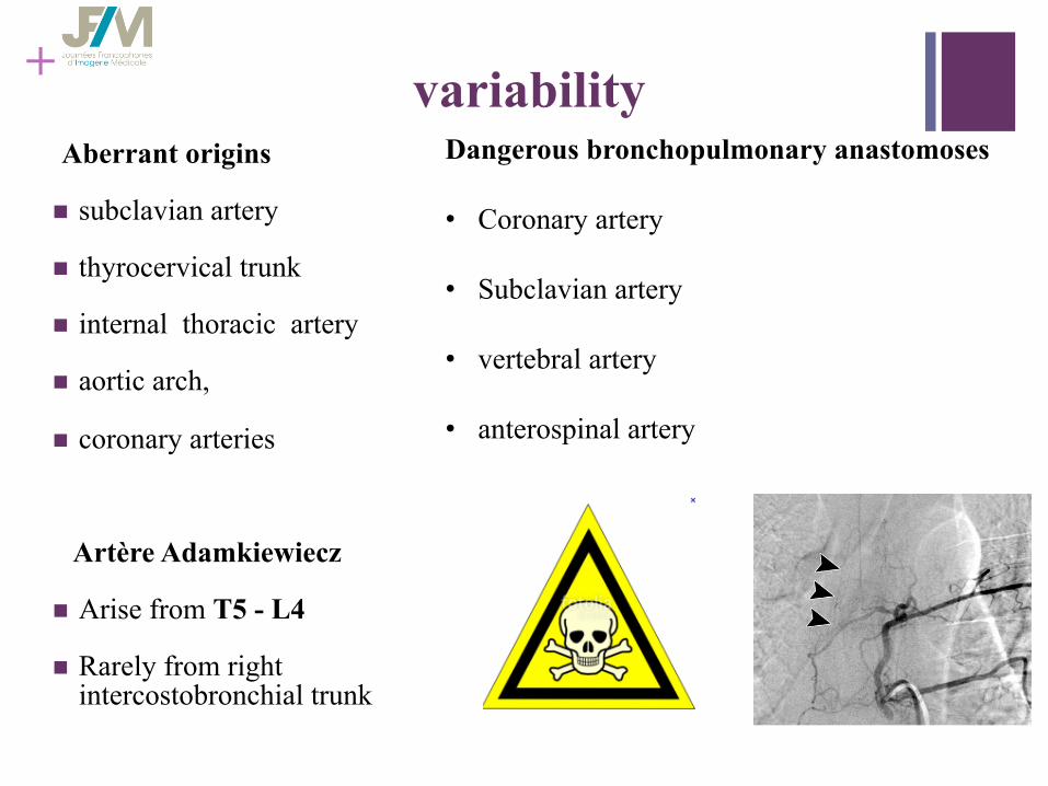
+ variability Aberrant origins
n subclavian artery
n thyrocervical trunk
n internal thoracic artery
n aortic arch,
n coronary arteries
Artère Adamkiewiecz
n Arise from T5 - L4
n Rarely from right intercostobronchial trunk
Dangerous bronchopulmonary anastomoses • Coronary artery
• Subclavian artery
• vertebral artery
• anterospinal artery
+
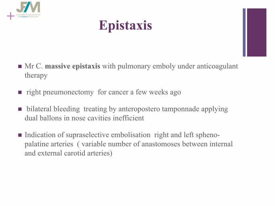
n Mr C. massive epistaxis with pulmonary emboly under anticoagulant therapy
n right pneumonectomy for cancer a few weeks ago
n bilateral bleeding treating by anteropostero tamponnade applying dual ballons in nose cavities inefficient
n Indication of supraselective embolisation right and left spheno-palatine arteries ( variable number of anastomoses between internal and external carotid arteries)
Epistaxis

superselective embolisation with microsphere 200 to 400 microns in both spheno-palatine arteries massive bilateral epitaxis with risk of death.
Left
Right
+ Epistaxis n Frequent event during chimiotherapy responsible myelosuppression
for acute leukemia , rarely dangerous for life , stopping spontaneously or manual bidigital compression anterior meshing or endoscopic electrocoagulation
n Posterieur epistaxis posterior tamponnage in pharyngoscopy.
n ethmoïde tumor bleeding: surgery or embolisation.
+ Coils: tips and tricks
n Choice coil : fonction of vessel size to exclude (oversizing)
: long coil for high volume
n Embolisation with 4,5 F (0,0038) : use coils 0,035 ou 0,038
n Embolisation with microcatheters 0,24 max use coils 0,018
n Always rinse microcatheter or catheter 5F before entering coil into them
precautionary injection to control ,upstream embolisation

+ Results
n Success or control in 80 à 100 % cases.
n Authors point at the need to act rapidly and to prevent any further delays in treating by surgery if embolisation procedure is too difficult or too long or insure of bleeding site
n Recurrence : 0-33 % (abdominal )
n Many material used alone or in association
n Coils : recanalisation described as classic process ( Sigler et all, Salamat M. et all)
n Spongel low clinical success < 65 %
n glue : high clinical sucess > 90 % ( Lee 2007 , Jae 2007)
n High sucess rate
n Recurrence more likely
+ Evolution ?
n Variety of embolisation agents
n Variety of catheters and microcatheters
n Technology evolution
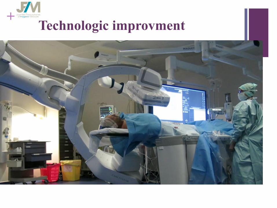
+ Technologic improvment

+ technologic improvment
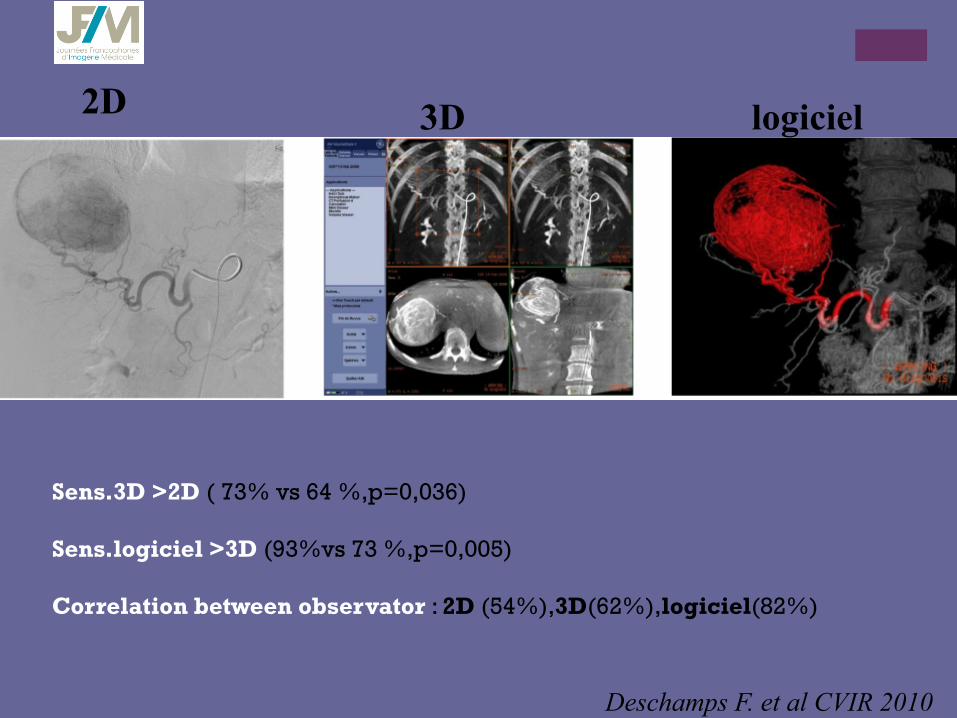
2D 3D logiciel
Deschamps F. et al CVIR 2010
Sens.3D >2D ( 73% vs 64 %,p=0,036) Sens.logiciel >3D (93%vs 73 %,p=0,005) Correlation between observator : 2D (54%),3D(62%),logiciel(82%)
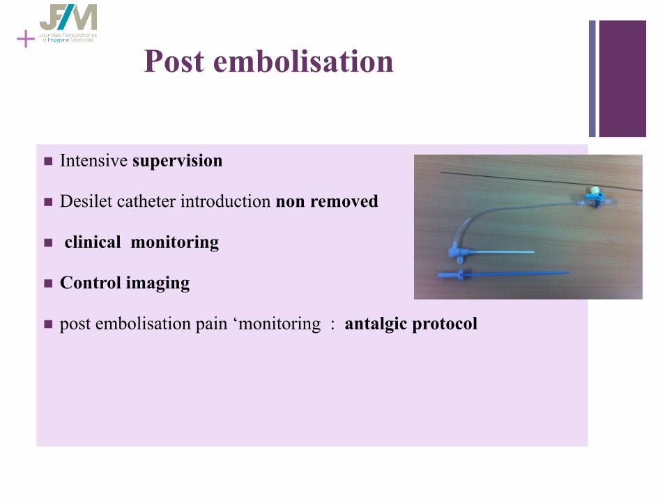
+ Post embolisation
n Intensive supervision
n Desilet catheter introduction non removed
n clinical monitoring
n Control imaging
n post embolisation pain ‘monitoring : antalgic protocol
+ Conclusion
n Interventionnel radiologist may be able to answer to emergencies
n To know embolisation technics and different agents
n Don’t run but think carefully before you act
n Effective tools
n Non exhaustive equipment
n pluridisciplinary management
+ Controlled robotization !!
Thank you for your attention

+
Thank you for your attention