Embed Size (px)
Citation preview
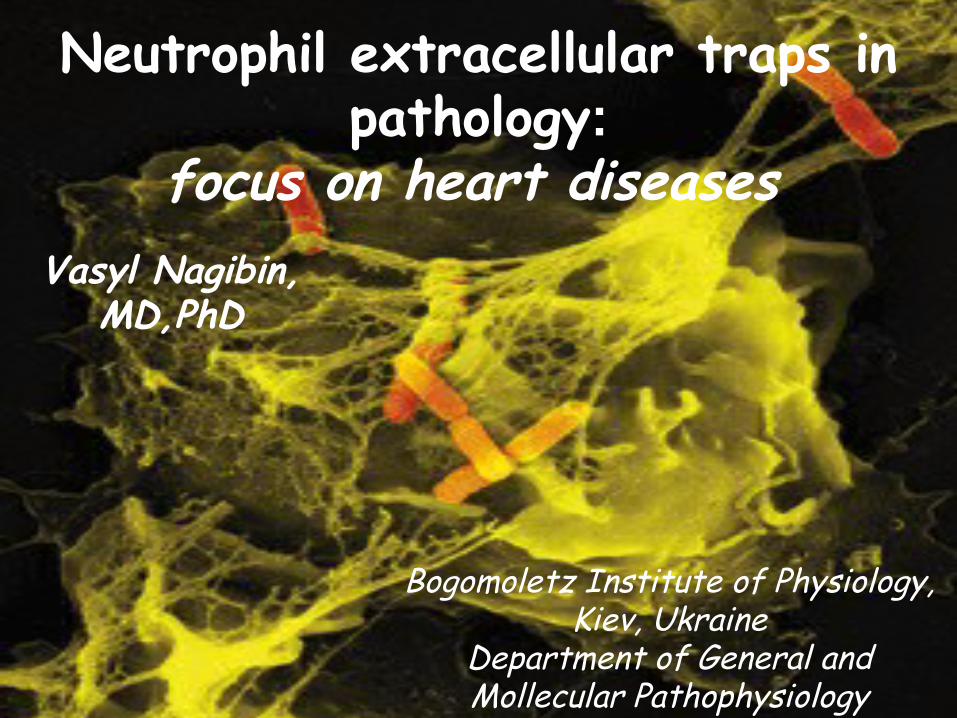
Neutrophil extracellular traps in pathology:
focus on heart diseases
Bogomoletz Institute of Physiology, Kiev, Ukraine
Department of General and Mollecular Pathophysiology
Vasyl Nagibin, MD,PhD
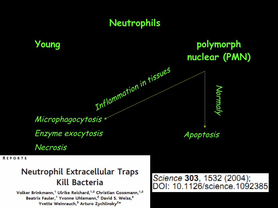
Neutrophils
Young polymorph nuclear (PMN)
MicrophagocytosisEnzyme exocytosisNecrosis
Apoptosis
Inflammation in tissues
Norm
a ly
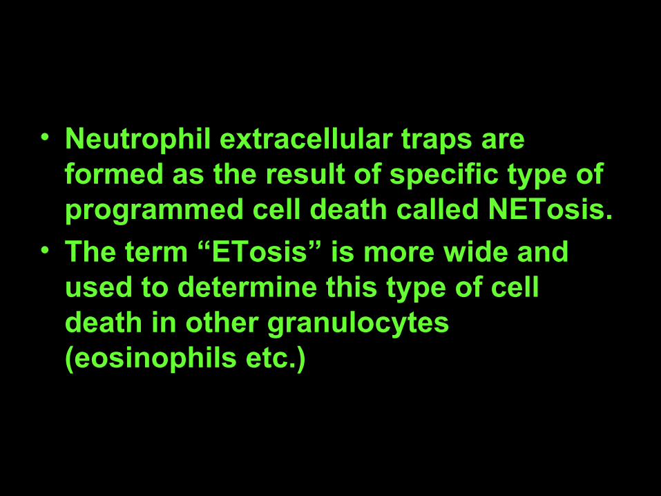
• Neutrophil extracellular traps are formed as the result of specific type of programmed cell death called NETosis.
• The term “ETosis” is more wide and used to determine this type of cell death in other granulocytes (eosinophils etc.)
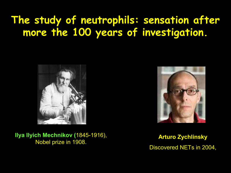
Ilya Ilyich Mechnikov (1845-1916), Nobel prize in 1908.
Arturo Zychlinsky
Discovered NETs in 2004,
The study of neutrophils: sensation after more the 100 years of investigation.
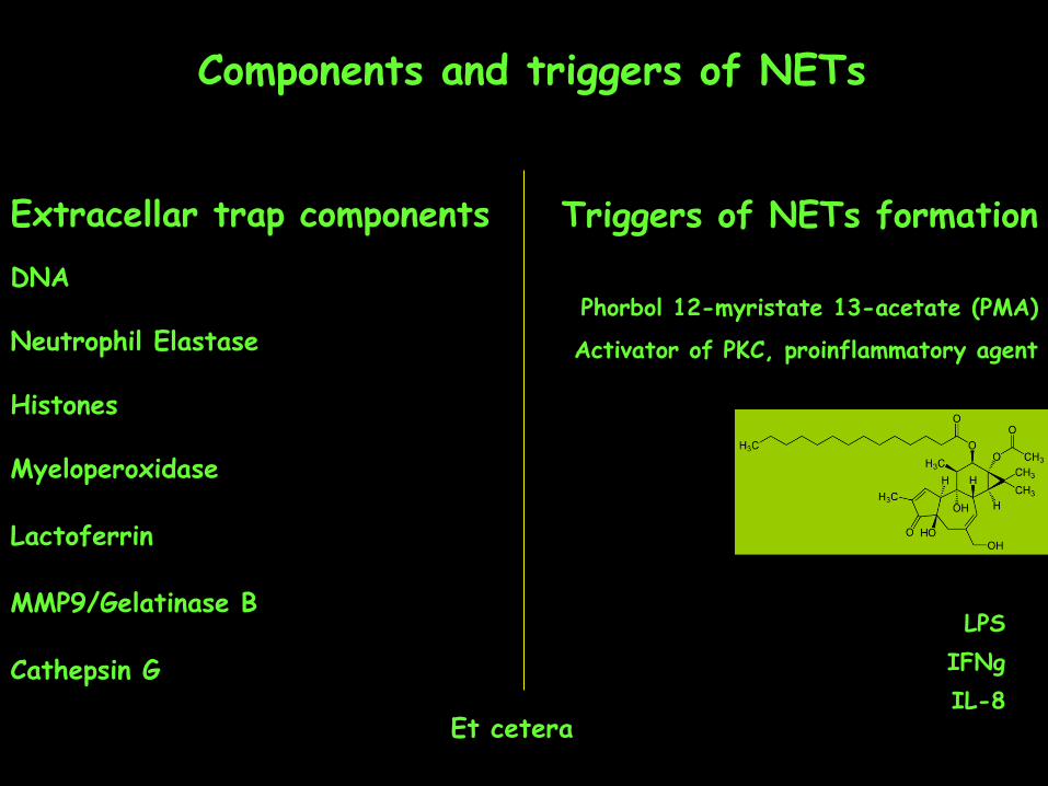
Extracellar trap componentsDNA
Neutrophil Elastase
Histones
Myeloperoxidase
Lactoferrin
MMP9/Gelatinase B
Cathepsin G
Components and triggers of NETs
Et cetera
Рhorbol 12-myristate 13-acetate (PMA)
Activator of PKC, proinflammatory agent
Triggers of NETs formation
LPSIFNgIL-8
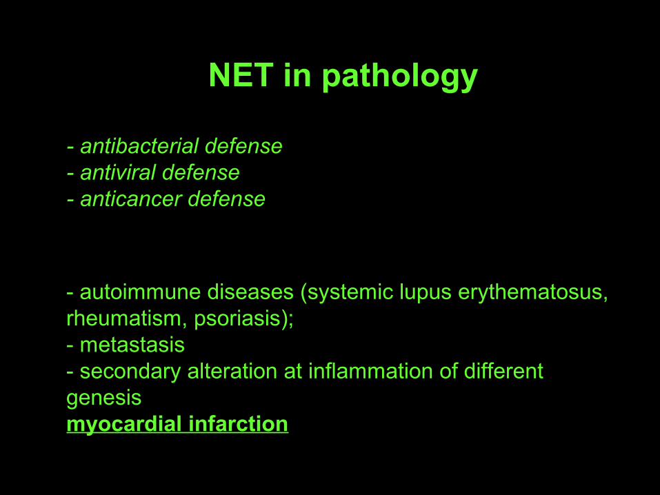
- antibacterial defense- antiviral defense- anticancer defense
- autoimmune diseases (systemic lupus erythematosus, rheumatism, psoriasis);- metastasis- secondary alteration at inflammation of different genesismyocardial infarction
NET in pathology
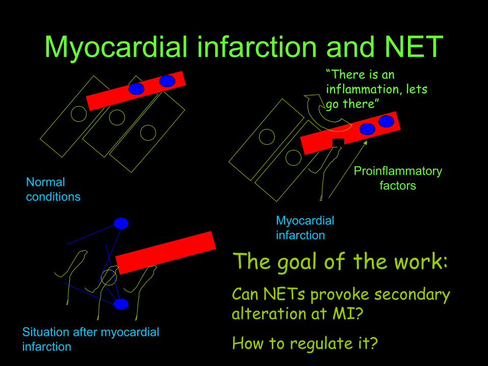
Myocardial infarction and NET“There is an inflammation, lets go there”
Proinflammatory factorsNormal
conditions
Myocardial infarction
Situation after myocardial infarction
The goal of the work:Can NETs provoke secondary alteration at MI?
How to regulate it?
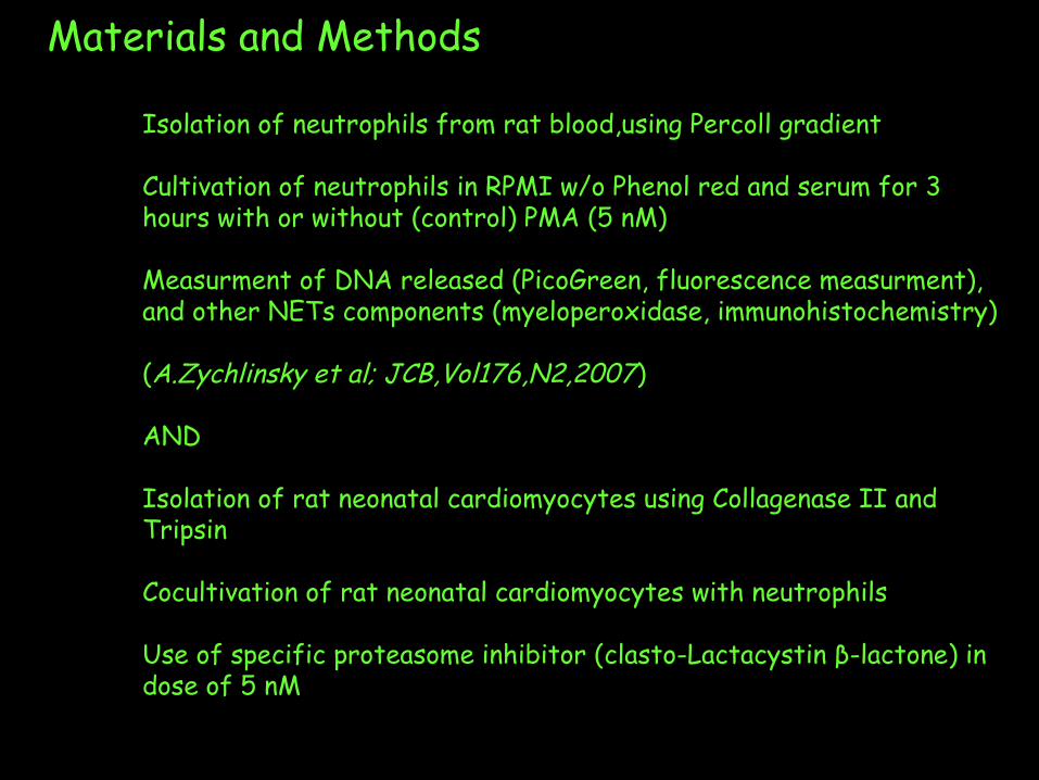
Materials and Methods
Isolation of neutrophils from rat blood,using Percoll gradient
Cultivation of neutrophils in RPMI w/o Phenol red and serum for 3 hours with or without (control) PMA (5 nM)
Measurment of DNA released (PicoGreen, fluorescence measurment), and other NETs components (myeloperoxidase, immunohistochemistry)
(A.Zychlinsky et al; JCB,Vol176,N2,2007)
AND
Isolation of rat neonatal cardiomyocytes using Collagenase II and Tripsin
Cocultivation of rat neonatal cardiomyocytes with neutrophils
Use of specific proteasome inhibitor (clasto-Lactacystin β-lactone) in dose of 5 nM
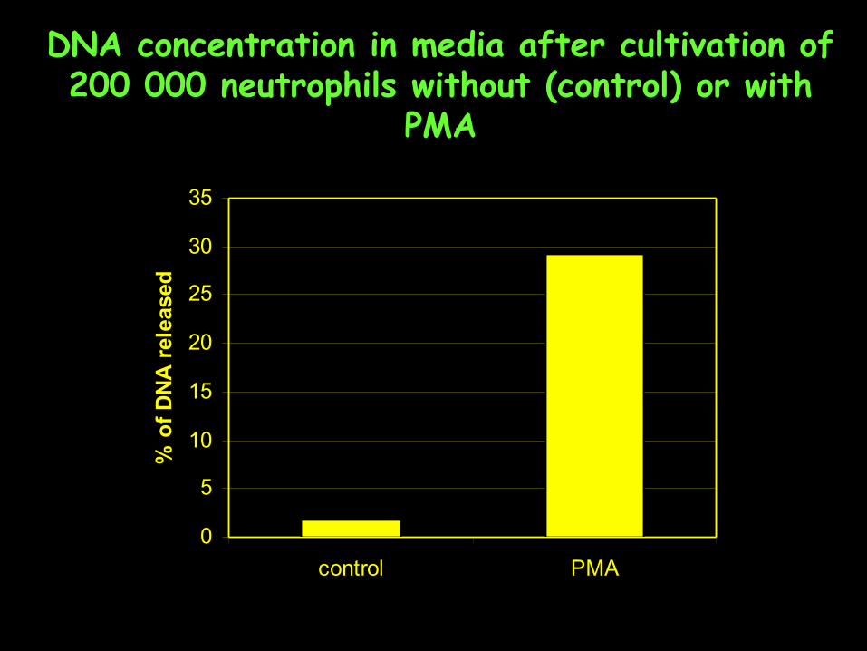
DNA concentration in media after cultivation of 200 000 neutrophils without (control) or with
PMA
0
5
10
15
20
25
30
35
control PMA
% o
f D
NA
rel
ease
d
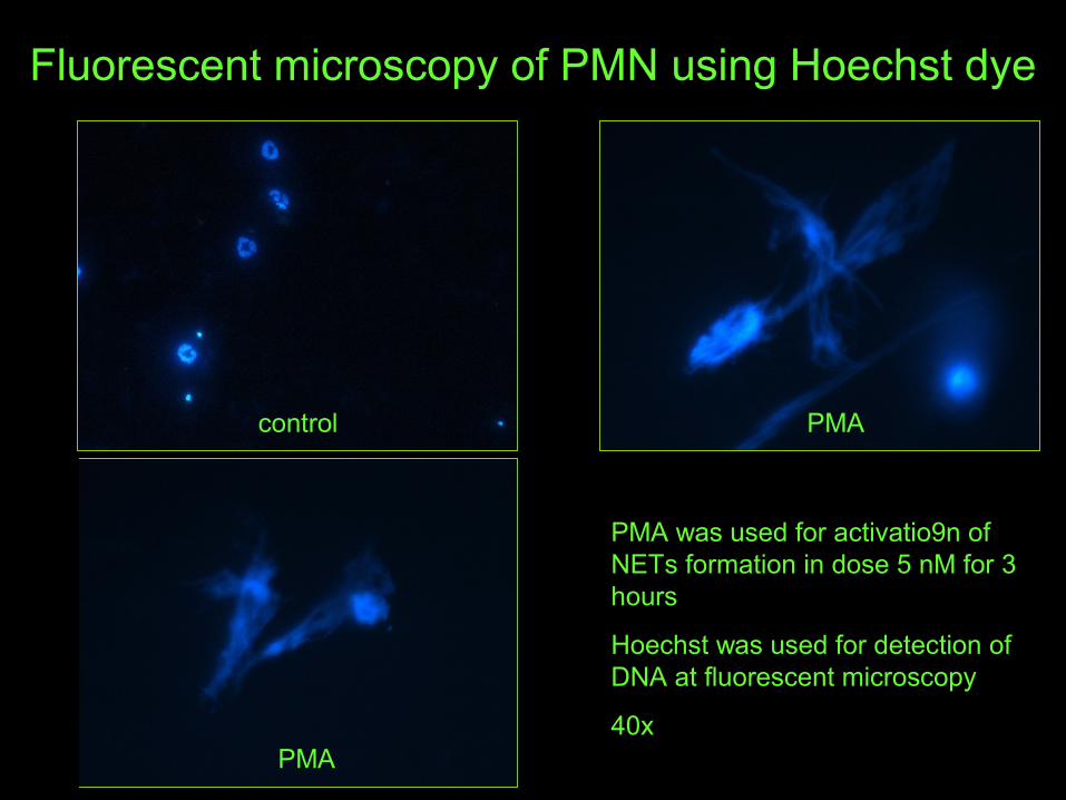
Fluorescent microscopy of PMN using Hoechst dye
control PMA
PMA
PMA was used for activatio9n of NETs formation in dose 5 nM for 3 hours
Hoechst was used for detection of DNA at fluorescent microscopy
40x
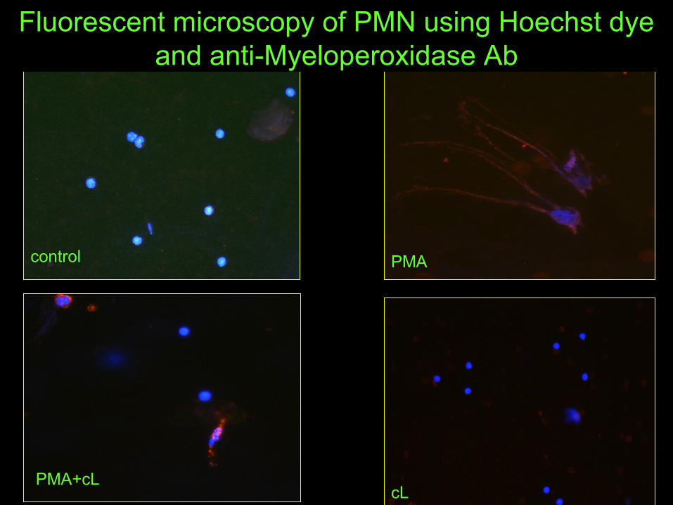
control PMA
cLPMA+cL
Fluorescent microscopy of PMN using Hoechst dye and anti-Myeloperoxidase Ab
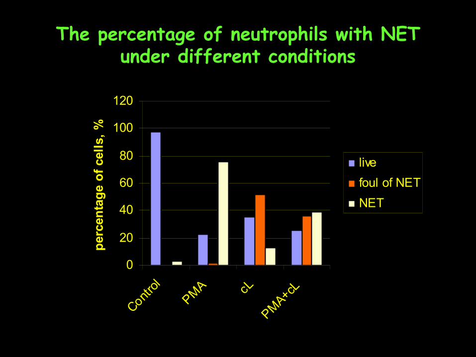
The percentage of neutrophils with NET under different conditions
0
20
40
60
80
100
120
Contro
l
PMA cL
PMA+cL
per
cen
tag
e o
f ce
lls,
%
live
foul of NET
NET
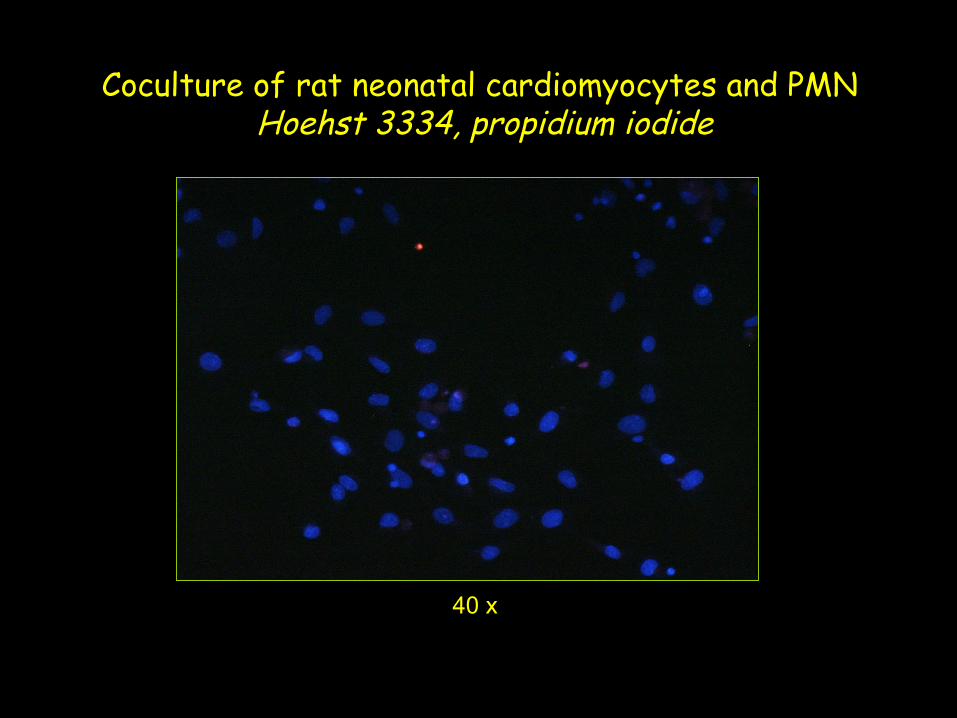
Coculture of rat neonatal cardiomyocytes and PMN Hoehst 3334, propidium iodide
40 x
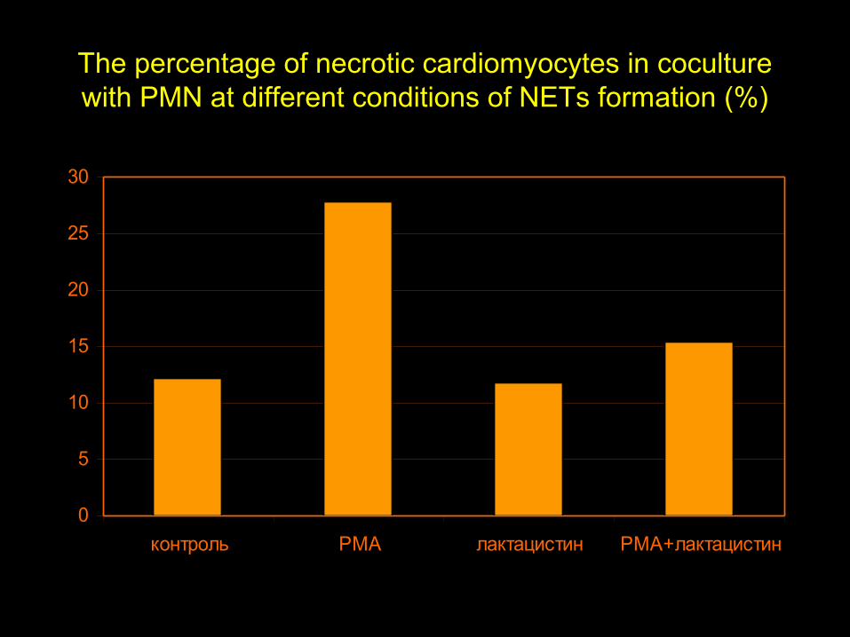
The percentage of necrotic cardiomyocytes in coculture with PMN at different conditions of NETs formation (%)
0
5
10
15
20
25
30
контроль PMA лактацистин PMA+лактацистин
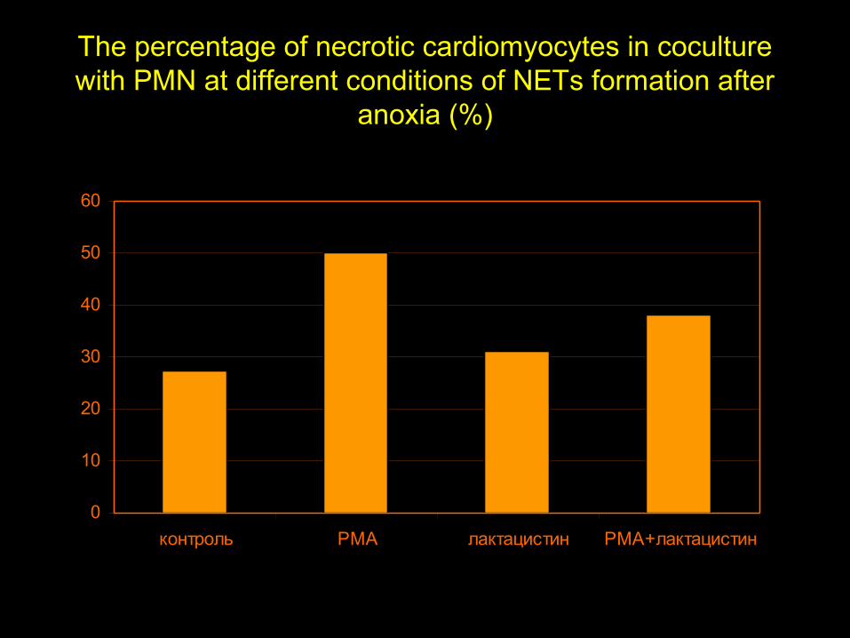
The percentage of necrotic cardiomyocytes in coculture with PMN at different conditions of NETs formation after
anoxia (%)
0
10
20
30
40
50
60
контроль PMA лактацистин PMA+лактацистин
Thanks for some of my colleagues, participating in this project
Prof. Moibenko O.O.
Prof. Dosenko V.E.
MD. PhD. Pashevin D.O.
BD. Tumanovska L.V.