Embed Size (px)
Citation preview
The Journal of Neuroscience, February 1994, 14(2): 897-913
A Functional Anatomical Analysis of Central Pathways Subserving the Effects of Interleukin- 1 on Stress-related Neuroendocrine Neurons
Anders Ericsson,a Krisztina J. Kovacs,b and Paul E. Sawchenko
Laboratory of Neuronal Structure and Function, The Salk Institute for Biological Studies, and The Foundation for Medical Research,- La Jolla, California 92037
Systemic administration of the cytokine interleukin-1 (IL-l) results in increased secretion of ACTH and corticosterone in rats. The available evidence suggests that the acute ef- fects of IL-1 are exerted ultimately at the level of the hy- pothalamus to increase corticotropin-releasing factor (CRF) secretion into the hypophyseal portal circulation, and hence the central drive on the pituitary-adrenal system. However, the route(s) and mechanism(s) by which circulating IL-l gains access to central mechanisms governing pituitary-adrenal output remain poorly understood. In this study, we show that intravenous injection of IL-lfi provokes time- and dose-de- pendent increases in the expression of the immediate-early gene c-fos, in identified CRF and oxytocin-producing cells of the paraventricular nucleus of the hypothalamus (PVH). Several cell groups known to be involved in central viscer- omotor regulation also displayed comparable time- and dose- related activation to systemic IL-l, including the bed nucleus of the stria terminalis, the central nucleus of the amygdala, the lateral parabrachial nucleus, and cell groups of the dor- somedial and ventrolateral medulla. Activation of circum- ventricular organs, which have been hypothesized to serve as central monitors of circulating IL-l, required doses rough- ly an order of magnitude above those required to activate CRF neurons in the PVH. Combined immunohistochemical and retrograde tracing experiments revealed many IL- 1 -re- sponsive cells in the nucleus of the solitary tract and the ventrolateral medulla to be catecholaminergic and to project to the region of the PVH. Discrete and unilateral interruption of ascending catecholaminergic projections from the me- dulla attenuated IL-l-stimulated increases in Fos immuno- reactivity and CRF mRNA in the PVH on the ipsilateral side. Disruption of descending projections from circumventricular structures associated with the lamina terminalis did not af-
Received June 1, 1993; revised July 19, 1993; accepted Aug. 13, 1993. This work was supported by NIH Grant NS-2 1182, and was conducted in part
by the Foundation for Medical Research. P.E.S. is an Investigator of the Foun- dation for Medical Research. Fellowship support was provided by the Fogarty Foundation (TW-04658), the Swedish Medical Research Council, The Foundation Blanceflor Boncompagni-Ludovisi, nee Bildt, Svenska tikaresiUskapet, Tore Nilsson Foundation for Medical Research, and the Swedish Royal Academy of Sciences (A.E.) and IREX (K.J.K). We are grateful to Belle Wamsley and Kris Trulock for excellent editorial and photography/graphics assistance, respectively.
Correspondence should be addressed to Dr. Paul E. Sawchenko, The Salk In- stitute, P.O. Box 85800, San Diego, CA 92186-5800.
‘Permanent address: Department of Rheumatology, The Karolinska Hospital, S- 17 176 Stockholm, Sweden.
hPermanent address: Institute of Experimental Medicine, Hungarian Academy of Sciences, H-1450 Budapest, Hungary. Copyright 0 1994 Society for Neuroscience 0270-6474/94/140897-17$05.00/O
feet IL-l-mediated Fos induction in the PVH. We conclude that medullary catecholaminergic projections to the PVH play either a mediating or a permissive role in the IL-l-induced activation of the central limb of the hypothalamo-pituitary- adrenal axis.
[Key words: catecholamines, c-fos, corticotropin-releas- ing factor, cytokine, hypothalamo-pituitary-adrenal axis, in- terleukin- 1, neorosecretory neurons, oxytocin, paraventric- ular nucleus, transcription factors]
Infectious and inflammatory processes evoke a broad spectrum of centrally mediated adaptive responses, including pyresis, somnolence, anorexia, and altered metabolic activities (Kus- chner, 1982). Interleukin-1 (IL-l), a 17.5 kDa protein that is rapidly secreted from immunologically challenged cells pri- marily of macrophage lineage, appears to play a pivotal role in mediating such host responses in that most are mimicked by exogenous administration of the cytokine (for review, see Di- narello, 1991). Important components of the acute phase re- sponse are marked perturbations in neuroendocrine functions, paradigmatic of which is the increased activity of the hypoth- alamo-pituitary-adrenal (HPA) axis seen following central or peripheral administration of IL- 1 (Besedovsky et al., 1986; Ber- kenbosch et al., 1987; Sapolsky et al., 1987; Harbuz et al., 1992). Glucocorticoids, the end product of the HPA cascade, are pow- erful immunosuppressants (Cupps and Fauci, 1982) and be- cause various immune functions are rapidly inhibited in re- sponse to IL-1 in vivo (Sundar et al., 1990; Brown et al., 1991; Hori et al., 1991), these hormonal events have been suggested as comprising a substrate for negative feedback control of the immune by the neuroendocrine system (Besedovsky et al., 1986). Dysfunction of this posited neuroimmune regulatory network has been implicated as contributing to certain autoimmune dis- orders (Kroemer et al., 1988; Sternberg et al., 1989).
The mechanisms by which circulating IL- 1 comes to modify pituitary-adrenal output are poorly understood, and potential sites of action at the levels of the hypothalamus (Berkenbosch et al., 1987; Sapolsky et al., 1987; Rivest and Rivier, 1991) pituitary (Bernton et al., 1987; Cambronero et al., 1992) and adrenal (Winter et al., 1990; Tominaga et al., 1991) have all been explored. The weight of the available evidence now tends to favor the hypothesis that the acute effects of IL- 1 are exerted principally at the level of the hypothalamus to increase corti- cotropin-releasing factor (CRF) secretion into the hypophyseal portal circulation, and hence the central drive on the pituitary- adrenal system.
The existence of a blood-brain barrier to macromolecules poses a formidable question as to how circulating IL-1 gains
898 Ericsson et al. * IL-l-Responsive Pathways in the CNS
access to the brain parenchyma to exert its effects, directly or indirectly, on hypothalamic neurosecretory neurons. A fre- quently advocated hypothesis is that signaling by IL-l may be transduced by one or more of the brain’s circumventricular organs (Katsuura et al., 1990; Stitt, 1990), which lack a func- tional barrier, and are closely interconnected with the endocrine hypothalamus (Johnson and Loewy, 1990). Evidence suggestive of an involvement of central catecholaminergic neurons in the HPA response to exogenous IL- 1 (Weidenfeld et al., 1989; Matta et al., 1990; Chuluyan et al., 1992) raises the possibility of peripheral neural transduction, since these pathways have been broadly implicated in the central processing of interoceptive information (Sawchenko and Swanson, 1982). Alternatively, it has been suggested that IL- 1 may be subject to active transport across the barrier (Banks et al., 1989), thereby affording it direct and privileged access to intraparenchymal neuronal populations that express IL-I receptors (Ban et al., 1991; Cunningham et al., 1992). The possibility also exists that IL-1 may interact with its own receptor on endothelial cells of the intracerebral vas- culature (Cunningham et al., 1992) to allow penetration into the brain parenchyma of blood-borne substances that are capable of stimulating the synthesis and secretion of secondary signaling molecules (Katsuura et al., 1990; Kilbourn and Belloni, 1990; Watanabe et al., 1990), which may, in turn, trigger or modify local neuronal responses.
To begin to sort among these disparate mechanistic possi- bilities, we employed a functional anatomical approach that revolves in its initial phases about use of the immediate-early gene c-&s as a marker for functionally activated neurons. The general utility, sensitivity, and resolving power of c-fos as a marker for synaptically and/or transcriptionally activated cells have been widely discussed (Sagar et al., 1988; Morgan and Curran, 199 1) and need not be reiterated here. We used this technique first to identify stress-related hypothalamic neurose- cretory neurons that respond to intravenous injection of IL- lp by increased expression of c-fis mRNA and its protein product, Fos. Comparison of dose-response relationships and the time course of Fos induction in hypothalamic neuroendocrine target neurons and in potentially relevant afferents were used to iden- tify candidate mediators/modulators of the IL-l effect. This information was taken as an entry point from which to assess the connectivity and neurotransmitter specificity of select pools of IL-l-responsive neurons regarding their potential for regu- lating HPA axis activity. Finally, discrete surgical ablation of implicated afferents was carried out to directly assess their con- tribution to IL-l-induced Fos and CRF expression in hypotha- lamic neuroendocrine effector neurons.
Portions of these data have been presented in abstract form (Ericsson et al., 1992a, b).
Materials and Methods Animals Germ-free male adult Sprague-Dawley albino rats (280-320 gm; Harlan Sprague Dawley Inc, Indianapolis, IN) were housed individually under temperature- and light-controlled conditions (12: 12 hr 1ight:dark cycle with lights on at 06:00), with food and water available ad libitum. Rats were handled daily for a week prior to the onset of experimentation. All protocols were approved by the Institutional Animal Care and Use Committee.
Systemic administration of IL- l/3 Recombinant human IL-I@ was kindly provided by Dr. S Gillis, Im- munex Research and Development Corporation (Seattle, WA). This
protein corresponds to the 152 residue mature form of IL- 10, and had an original specific biological activity in excess of 1 x IOX units/pg protein [A375 assay (Nakano et al., 1988); 17 pg endotoxin/Mg protein]. Upon receipt, the IL-10 protein was thawed on ice, diluted i:l in 200 mM Tris-HCI buffer. DH 7.4 at 25°C. containing 0.2% BSA. and then aliquoted in 1.5 pg batches and refrozen at - 70°C. Thawed aliquots of IL-10 were stored at 4°C for no longer than 7 d.
Our procedure for systemic administration of IL-ID to rats has pre- viously been described in detail (Ericsson and Sawchenko, 1993). Brief- ly, methoxyflurane-anesthetized rats were implanted with indwelling catheters (PE-50) containing sterile, pyrogen-free heparin-saline (500 U/ml) into the jugular vein; the sealed catheter was positioned with its internal Silastic tip at the atrium and was exteriorized at an interscapular position. After 2 d recovery, each experimental animal was connected, in its home cage, to a remote saline-filled catheter between 06:OO and 07:00, and either IL-l@ (doses ranging between 0.21 and 3.58 pg/kg), 1.87 &kg of heat-inactivated IL-l/3 (70°C for 30 min), or vehicle alone was subsequently remotely delivered to freely moving rats in a total volume of 300 bl over 3 min at l-4 hr prior to anesthetization and perfusion of the rats. The final composition of the vehicle was kept consistent at 0.01% BSA. 0.01% ascorbic acid. 10 mM Tris-HCI. 36 mM sodium-phosphate buffer, pH 7.4, at 25°C. The animals were routinely harvested by perfusion fixation between 12:30 and 14:OO to minimize the effects of diurnal variation in secretory and biosynthetic activities of the HPA axis.
Tissue processing and histology Animals were deeply anesthetized with chloral hydrate (35 mg/kg, i.p.) and perfused via the ascending aorta with saline followed by 500-700 ml of 4% paraformaldehyde in 0.1 M borate buffer (pH 9.5 at 10°C). Brains were posttixed for 3-4 hr, and then cryoprotected in 10% sucrose in 0.1 M phosphate buffer overnight at 4°C. Frozen frontal sections were cut at 30 pm. Six l-in-6 series through regions of interest, typically hypothalamus and medulla or whole brain, were collected in cold cryo- protectant (0.05 M sodium phosphate buffer, 30% ethylene glycol, 20% glycerol) and stored at -20°C until histochemical processing. Series of sections destined for in situ hybridization histochemistry were further postfixed in phosphate-buffered 4% paraformaldehyde overnight at 4°C before being transferred to cryoprotectant for storage.
Immunohistochemical labeling for Fos-like immunoreactivity (Fos- ir) was performed using an affinity-purified polyclonal antisera raised in rabbit against a synthetic peptide corresponding to residues 4-l 7 in the N-terminal portion of the human Fos protein (Oncogene Sciences). This antiserum displays no known cross-reactivity with any identified Fos-related antigen (Fra). Analvsis for Fos-ir in IL-la-iniected and ve- hicle-injected ra?s was performed on free-floating sections using a con- ventional avidin-biotin-immunoperoxidase technique (Sawchenko et al., 1990). This included pretreating sections for 10 min each in 0.3% (v/v) hydrogen peroxide and in 1 .O% (w/v) borohydride, with interposed rinses. Sections were incubated with the primary antiserum at dilutions ranging between 1:lOOO and 1:7500, depending on the batch. The pri- mary antiserum was localized using Vectastain Elite reagents, and the reaction product developed on ice using a nickel-enhanced glucose ox- idase method (Shu et al., 1988). The specificity of the primary antisera was analyzed in separate control experiments by preabsorbing the an- tiserum overnight at 4°C with 50 FM of the corresponding immunogenic peptide.
c-fis mRNA was detected using a YS-labeled antisense cRNA probe transcribed from a 2.4 kilobase (kb) cDNA encoding the full-length rat clfos mRNA (Curran et al., 1987). Control sections were hybridized with sense strand runoffs generated from the same cDNA clone. YS- labeled antisense and sense (control) cRNA probes transcribed from a 1.2 kb cDNA encoding the full-length rat prepro-CRF mRNA (kindly nrovided bv Dr. K. Mavo: cf. Jinaami et al.. 1985) were used to localize neuronal CRF mRNA for qualitative and quantitative analyses.
Hybridization histochemical localization of c-fis and CRF mRNA was performed as previously described (Simmons et al., 1989). Briefly, sections destined for in situ hybridization were subjected to additional postfixation in neutral-buffered 4% paraformaldehyde overnight at 4°C mounted on gelatin and poly-L-lysine-coated slides, and desiccated un- der vacuum overnight. Sections were then postlixed with neutral-buf- fered 4% paraformaldehyde for 30 min, digested in 10 fig/ml proteinase K at 37°C for 30 min, acetylated for 10 min, and then dehydrated and vacuum dried overnight. Hybridization ofthe 3SS-labeled antisense cRNA probes (1 O6 cpm/ 100 pi/slide) was carried out in 247 mM NaCl, 8.2 mM
The Journal of Neuroscience, February 1994, 74(2) 999
Tris-HCI (pH 8.0 at 25°C) 41% (v/v) formamide, 0.82~ Denhardt’s (1 x Denhardt’s is 0.002 % of BSA, Ficoll 400, and polyvinylpyrroli- done), 8.2 % (w/v) dextran sulphate, 411 mg/ml yeast tRNA, and 8.2 mM dithiothreitol (DTT) on coverslipped slides at 60°C for I6 hr. After initial rinses in 4 x saline-sodium citrate (SSC), slides were incubated in RNase A (20 fig/ml, 37”C, 30 min), washed in 0.1 x SSC, 10 mM DTT at 75°C for 30 min. and finallv dehvdrated and dried. Slides were exposed to Amersham p:max autoradiography film for l-4 d, defatted in graded ethanols and xylene, and dipped in Kodak NTB-2 nuclear track emulsion. Slides were exposed for 7-l 4 d and developed in D- 19 developer (Kodak) for 5 min at 14°C. Sections were counterstained with thionin, dehydrated, and coverslipped.
Procedures and analysis
Time course. To establish a time course of c-fis induction in the hy- pothalamus, 59 rats were injected with a moderate dose (1.87 wg/kg) of IL- lp or an equivalent volume of vehicle and perfused at hourly inter- vals for up to 4 hr. Immunohistochemical localization of Fos-ir and hybridization histochemical localization of C-$X mRNA were carried out on series of sections through the hypothalamus from each animal.
Histochemical identification of IL- I-responsive neuroendocrine neu- rons. To determine the neuropeptide phenotype of IL- l-responsive cells, immunoperoxidase localization ofFos-ir was combined with concurrent immunofluorescence staining for oxytocin (OT) or vasopressin, the prin- cipal peptide products of the magnocellular neurosecretory system, or with hybridization histochemical localization of CRF mRNA, in series of hypothalamic sections obtained from four rats injected with 1.87 pg/ kg of IL-I/3 and killed at the time of maximal Fos protein induction (3 hr). Dual staining for Fos-ir and OT-ir or arginine vasopressin-ir (AVP- ir) was performed by initially preparing the free-floating sections for immunoperoxidase localization of Fos-ir as outlined above, except that the hydrogen peroxide pretreatment was omitted. Sections were sub- sequently incubated in rabbit anti-OT serum (Incstar, 1:2000) or rabbit anti-AVP serum (Chemicon, 1:8000) for 48 hr and then in affinity- purified, FITC-labeled goat anti-rabbit IgG for I hr at room temperature (1:200; Tago, Inc.). Specific staining with the primary antisera was abol- ished by preincubation with 50 KM of the homologous, but not the heterologous, synthetic nonapeptide. Cells displaying nuclear immu- noperoxidase staining for Fos and cytoplasmic immunofluorescence staining for OT-ir or AVP-ir were identified.
The lesser relative abundance of CRF peptide in the paraventricular nucleus of the hypothalamus (PVH) precluded using a similar approach to determine the extent to which IL-l-stimulated Fos induction might be localized to CRF-expressing neurons. Instead, a combined immu- noperoxidase (Fos) and in situ hybridization (CRF mRNA) protocol, detailed elsewhere (Watts and Swanson, 1989a), was employed. In brief, immunolocalization of Fos-ir was carried out as described above, save that the tissue was not pretreated in borohydride or hydrogen peroxide. In addition, the primary antibody reaction was performed in buffer with 2% (v/v) BSA and 5 mg/ml heparin as blocking agents (in place of nonimmune goat serum) and the reaction product was developed with- out nickel enhancement. Subsequent localization of CRF mRNA by in situ hybridization was then carried out as described above.
Dose-response relationships. A comparison of the minimum effective doses at which IL-1 induces Fos-ir in the PVH and extrahypothalamic cell groups was undertaken in order to identify candidate afferent me- diators of cytokine effects on hypophysiotropic CRF neurons. Groups of three to eight animals each were implanted with jugular cannulas and injected with either 0.2 1,0.46,0.87, 1.87, or 3.58 pLg/kg IL- 10 or vehicle, anesthetized 3 hr later (the time point at which IL-l-stimulated Fos-ir was found to be maximal in the PVH), perfused, and prepared for demonstration of Fos-ir throughout the brain. Effective doses of IL-l were determined by comparing counts of Fos-ir nuclei in the PVH and cell groups either that are known to provide afferents to it or that have been implicated as potential mediators of the IL-1 effect on the HPA axis in control and cytokine-treated animals. We adopted the arbitrary criterion of considering as an effective dose one that resulted in Fos induction in cells whose number equalled or exceeded the mean + 2 SD of vehicle-injected control values in a majority of animals that received a particular dose of cytokine.
Characterization of candidate a&en& To determine whether cells displaying Fos induction in response to intravenous IL-l are anatom- ically related to the PVH, and to determine their neurotransmitter phe- notype, a combined retrograde transport-multiple immunohistochem- ical labeling paradigm was used. Under anesthesia (10 mg/kg xylazine,
50 mg/kg ketamine, 2 mg/kg acepromazine), seven rats received ste- reotaxically guided crystalline implants of the retrogradely transported Ruorochrome true blue aimed at the PVH (Lind, 1986). Four weeks later, tracer-injected rats were implanted with jugular cannulas and injected with I .87 fig/kg IL-16 as described above. Complete and reg- ularly spaced series of sections throughout the brains of animals with well placed injections were processed for immunoperoxidase detection of Fos-ir as described above, except that hydrogen peroxide pretreat- ment was omitted. The extent to which individual neurons manifested both IL-l-induced Fos-ir and retrograde labeling was determined. Be- cause such doubly labeled cells were seen consistently in the dorsomedial and ventrolateral parts of the medulla, regions known to provide cat- echolaminergic projections to the PVH (Cunningham and Sawchenko, 1988; Cunningham et al., 1990), brainstem sections were treated as above (i.e., for demonstration of Fos-ir and retrogradely transported true blue) and further incubated in a cocktail of antisera raised against dopamine-P-hydroxylase (DBH; a marker for adrenergic and noradren- ergic neurons) and phenylethanolamine-Nmethyltransferase (PNMT, a specific marker for adrenergic neurons). The anti-DBH serum was a mouse-derived monoclonal antibody (Chemicon; diluted 1: 1000) raised against rat adrenal DBH. The anti-PNMT serum (diluted at 1:2000), generously provided by Dr. M. C. Bohn (Univ. Rochester), was raised in rabbits against purified rat adrenal PNMT (Bohn et al., 1987). Pri- mary antisera were localized using a mixture of FITC-labeled goat anti- rabbit IgG (1:200; Tago, Inc.) and rhodamine-labeled goat anti-mouse IgG (1: 100; American Qualex). This procedure allowed determination of the extent to which medullary cells that are responsive to IL-l and that project to the region of the PVH displayed the adrenergic or nor- adrenergic phenotype.
Experimental manipulation of candidate afferents. Ablation methods were used to evaluate the dependency of IL- l-stimulated increases in the expression of Fos and/or CRF mRNA on afferents implicated in the analyses outlined above. A group of rats received discrete, stereo- taxically guided, unilateral transections, designed to interrupt ascending catecholamine projections near their origins in the medulla. Knife cuts were made in the coronal plane, using a retracting wire knife fashioned from a microliter syringe (Gold et al., 1973) as previously described (Sawchenko, 1988). Twelve days after surgery, the animals were im- planted with jugular cannulas, randomly injected 2 d later with 1.87 Kg/kg IL- lb or vehicle, and anesthetized and perfused 3 hr after injec- tion. Adjacent series of horizontal sections though the brainstem were stained with DBH and thionin to aid in evaluation of the placement and effectiveness of knife cuts. Separate series of coronal sections through the hypothalamus were prepared for immunohistochemical demon- stration of DBH-ir, Fos-ir, and hybridization histochemical demon- stration of CRF mRNA. Knife cuts were judged to be effective if they resulted in a pileup of DBH-ir in fibers immediately proximal to the lesion site, and a significant (>60%) depletion of DBH-ir varicosities in the ipsilateral PVH, as judged by semiquantitative (rating scale) com- parison of the strength of the DBH innervation of the PVH on the ipsilateral and contralateral sides by two independent observers (Saw- chenko, 1988).
Nuclear staining for Fos-ir was compared qualitatively in the PVH on the ipsilateral and contralateral sides of animals bearing cuts judged to be effective. In addition, relative levels of CRF mRNA were assessed densitometrically from emulsion-dipped slides using a Leitz optical sys- tem coupled to Macintosh-based IMAGE software (version 1.42, W. Ras- band, NIH). Optical density over the medial parvocellular subdivision of the PVH (PVHmpd), defined by redirected sampling from the cor- responding bright-field images of thionin-stained sections, was deter- mined under dark-field illumination on both sides of the brain. Optical densities were corrected for background, and calibrated to emulsion- dipped sections of brain paste standards containing serial dilutions of ?S-UTP that were processed in parallel with the experimental material. Data were normalized to the contralateral side in lesioned and saline- injected rats and statistically analyzed by two-way ANOVA as a mixed design with one within (ipsi- vs contralateral sides) and one between (IL- I vs saline) group variable. Duncan’s multiple range test was used for post-hoc comparisons between individual group means.
As an additional control, a separate group of rats received unilateral transections designed to disrupt descending projections from lamina terminalis-associated circumventricular structures using a similar knife construction, with cuts 2 mm wide x 3 mm high placed in the coronal plane at a locus, extending from the midline just caudal to the vascular organ of the lamina terminalis (OVLT), which has been shown to be
900 Ericsson et al. * IL-l-Responsive Pathways in the CNS
Figure 1. Time course of IL-l-stimulated c-fis mRNA induction in the PVH: dark-field photomicrographs of hybridization histochemical localization of a 35S-labeled antisense c-fos probe at a comparable level through the PVH of rats injected 1, 2, or 3 hr previously with 1.87 &kg of IL-l& control is representative of the low level of hybridization seen in rats that received injection of vehicle alone, 1 hr prior to perfusion fixation. Rapid induction of c-fos transcripts are evident at 1 hr after treatment, and localized predominantly to the dorsal aspect of the medial parvocellular subdivision (mp,,). Less widespread induction is also evident at the periphery of the magnocellular division (pm) of the nucleus, and in autonomic-related parts of the parvocellular division (dp, mpv). IL-l-stimulated c-fos induction is transient in neurosecretory populations, and more persistent in autonomic-related subdivisions. The third ventricle is at the Left of all micrographs. mp,, ventral aspect of the medial parvocellular subdivision. Magnification (all micrographs), 75 x
effective in disrupting osmotically driven alterations in CRF mRNA expression in both the magnocellular and parvocellular neurosecretory systems (Kovics and Sawchenko, 1993). In the absence of reliable his- tochemical markers for lamina terminals-PVH projections, the location of these lesions was evaluated histologically from thionin-stained sec- tions. After implantation of venous cannulas, injection of IL-I or ve- hicle, perfusion-fixation, and histology, the number of nuclei displaying Fos-ir and the relative strength of Fos-ir were compared on the sides ipsilateral and contralateral to the cuts.
Results Time course of IL- I-stimulated expression of c-b in the PVH Our initial experiment sought to determine whether, and over what time frame, transcription factor Fos may serve as a useful marker for identifying neuroendocrine neurons that are affected by systemic administration of IL- l/3. Tissue harvested from rats injected with a 1.87 pg/kg dose of IL- l/3 via an indwelling jugular catheter, a paradigm previously shown to stimulate secretory activity of the HPA axis (Sapolsky et al., 1987; Rivier et al., 1989) reliably displayed an orderly sequence and topography of c-fos mRNA and cognate Fos protein induction. Hybridiza- tion histochemical localization of a ?S-labeled rat c-fos ribo- probe in complete series of sections through the hypothalamus revealed significant labeling for c-f0.s mRNA over cells primarily localized in the dorsal aspect of the PVHmpd at 1 hr after administration of IL- l/3 (Fig. 1). Labeling in this region, which is recognized as the seat of parvocellular neurosecretory CRF- expressing neurons, was diminished by 2 hr, and not discernably distinct from that seen in controls by 3 hr after injection. IL- 1 p injection also reliably provoked c-fos mRNA expression that followed a similar time course in topographically distinctive aspects of the magnocellular division of the PVH, and a more persistent induction in aspects of the PVH that project to au- tonomic centers in the brainstem and spinal cord. No specific labeling over the PVH was observed in adjacent series of sec- tions exposed to a YS-labeled c-fos riboprobe transcribed in the sense direction, or using the antisense probe in tissue obtained from vehicle-injected controls (Fig. 1).
Series of sections from IL-lb-injected and vehicle-injected
rats were also prepared for immunohistochemical localization of Fos protein. In cytokine-treated rats, nuclear staining for Fos- ir in the PVNmpd was clearly detectable at 1 hr, augmented at 2 hr, and maximal, in terms of both cell number and staining intensity, at 3 hr after injection (Fig. 2). The temporal expression of Fos-ir in the magnocellular division of the PVH was similar to that of the PVHmpd, though less robust than was evident in hybridization material. Interestingly, and in line with the hy- bridization data, strong labeling for Fos protein was detected in the (autonomic-related) dorsal parvocellular part of the PVH that persisted throughout the 4 hr time period examined. Pread- sorption of Fos antiserum with synthetic immunogen abolished staining in the PVH of IL- lp-injected rats, and animals receiv- ing vehicle alone displayed little or no specific labeling in the PVH (Fig. 2). Furthermore, rats injected with IL-10 that had been previously preheated to 70°C for 30 min, a procedure that will not inactivate potentially contaminating bacterial endotox- ins (Bernton et al., 1987) failed to result in Fos-ir induction in the PVH.
To determine whether IL- 1 P-induced c-fos expression may be associated with an index of increased CRF biosynthesis, rel- ative levels of CRF mRNA content in the PVH were compared densitometrically in animals killed 3 hr after systemic admin- istration of IL-l@ at 1.87 fig/kg or vehicle; the 3 hr time point was chosen as it represented the time of maximal Fos protein induction in this region. This analysis revealed a reliable, 47.6 + 13.1% increase in CRF mRNA content in the PVHmpd at 3 hr after injection of IL-lfl as compared to vehicle-injected control rats (p < 0.02, n = 5, two-tailed t test).
Histochemical identification of IL-l-responsive neuroendocrine neurons
The topography of cells displaying IL-l-stimulated c-fos in- duction in the PVH corresponded principally to the recognized distribution of parvocellular CRF-synthesizing and magnocel- lular OT-synthesizing neurosecretory neurons. To confirm the biochemical identity of IL- l/3-responsive neurons in the PVH, we carried out combined immunohistochemical and in situ hy- bridization analysis for Fos-ir and CRF mRNA in the PVH of
The Journal of Neuroscience, February 1994, 14(2) 901
. . ’ *
‘. ’
.
5 I . q
.* . . ‘ . . FOSA L-l .
Figure 2. IL-l-stimulated patterns of c-fos mRNA and Fos protein induction, and their relationship to CRF-expressing neurons in the PVH: dark-field photomicrographs of hybridization histochemical localization of CRF and c-fis mRNAs and bright-field photomicrographs of immu- noperoxidase localization of Fos protein in IL-l-injected or vehicle-injected (control) animals. Hypophysiotropic cells expressing CRF mRNA are predominantly localized to the dorsal aspect of the medial parvocellular part (mp) of the PVH. Systemic administration of IL- l/3 results in specific and comparable patterns of c-fis mRNA and Fos-ir induction in the PVH, whose topography overlaps extensively with that of cells expressing CRF mRNA. Rats were injected with 1.87 pg IL-lb/kg or vehicle (Control). IL-l-stimulated c-fis mRNA and Fos-ir are illustrated in animals killed at time points optimal for detection (1 and 3 hr postinjection, respectively). Control animal was also killed at 3 hr after injection of saline. pm, periphery of the magnocellular division. Magnification, 60 X.
rats that had received 1.87 &kg of IL- l@ 3 hr prior to perfusion. The results confirmed that the majority of Fos-ir cells in the PVHmpd of IL-l-injected rats express CRF mRNA (Fig. 3). Dual immunostaining methods revealed that the great majority of Fos-ir cells in the magnocellular division of the PVH dis- played OT-ir; few, if any, Fos-ir neurons stained positively for vasopressin, the other major secretory product of the magno- cellular neurosecretory system (Fig. 3). Corrected (Abercrombie, 1946) counts in one representative animal indicated that 7 1% of 1134 Fos-ir cells in the PVHmpd displayed a positive hy- bridization signal for CRF mRNA, defined as a grain density more than five time background overlying a cell-sized area cen- tered about a Fos-positive nucleus. Counts in adjacent series from the same animal indicated that of 163 Fos-ir cells in the magnocellular division of the nucleus, 69% also stained posi- tively for OT, while 7% were immunoreactive for vasopressin.
Extrahypothalamic sites of IL-l-induced Fos expression
Cell groups outside the PVH that are known to be involved in neuroendocrine regulation and/or to provide afferent projec- tions to the PVH also displayed prominent and specific Fos
induction following intravenous injection of IL- l/3. Rapid and robust induction of c-fos mRNA (not shown) and Fos-ir were observed in the oval subnucleus of bed nucleus of the stria terminalis (Fig. 4A; Ju and Swanson, 1989) the lateral division of the central nucleus of the amygdala (Fig. 4B), and specific subdivisions of the lateral parabrachial nucleus (Fig. 4C) after systemic administration of 1.87 FLg IL- lo/kg. Likewise, specific IL- lb-mediated Fos induction was detected throughout much of the rostrocaudal extent of the medial and commissural sub- nuclei of the nucleus of the solitary tract (NTS; Fig. 5) as well as in the rostra1 ventrolateral medulla (Fig. 40). A few cells in the dorsal motor nucleus of the vagus nerve also displayed IL- l-stimulated c-fos mRNA and Fos-ir.
Significantly, little or no specific staining for c-fos mRNA or Fos protein was observed in circumventricular organs at the dose of IL-l@ utilized over 1-4 hr after injection. Aspects of these structures, which include the OVLT, subfornical organ, median eminence, and area postrema, lie outside the blood- brain barrier, and have been suggested as likely CNS monitors of circulating IL- 1 (Katsuura et al., 1990; Stitt, 1990). In ad- dition, we reliably failed to detect IL- l-stimulated Fos induction
902 Ericsson et al. * IL-l-Responsive Pathways in the CNS
Figure 3. Biochemical characterization of IL-l-responsive neurose- cretory neurons in the PVH: bright-field (fop) or fluorescence (middle, bottom) photomicrographs through a comparable level of the PVH showing combined immunoperoxidase localization of IL- l-stimulated nuclear Fos-ir with markers for major neurosecretory cell types. Animals were killed at 3 hr after iniection of 1.87 ue. IL-la/kg. The too vane/
. I . . shows a section cohybridized for CRF mRNA. Exteisive association of reduced silver grains indicative of CRF mRNA signal and Fos-ir neurons is apparent in the parvocellular division of the PVH. See inset for higher magnification of boxed region; arrowheads indicate examples of doubly labeled cells. The middle and bottom panels show combined immunoperoxidase localization of Fos-ir and immunofluorescence staining for oxytocin (07’) or vasopressin (AW’), respectively. OT-ir and A VP-ir are localized to topographically distinct aspects of the more laterally disposed magnocellular division of the nucleus. In the mag- nocellular compartment, IL- l-stimulated Fos-ir is preferentially asso- ciated with OT-ir neurons. Magnification, 150 x (inset, 360 x).
at intra- and extraparenchymal loci known to harbor IL-l re- ceptors and/or ligand binding sites (Ban et al., 199 1; Cunning- ham et al., 1992), including the dentate gyrus (Fig. 4E), choroid plexus (Fig. 4F), and the mesencephalic raphe nuclei. One ex- ception was a weak induction of c-fos mRNA throughout much of meninges at 1 hr after injection; Fos-ir was detected incon- sistently in these membranes at 2-3 hr after injection, suggesting a low and variable level of Fos induction in these membranes. No specific Fos induction was detected at any time point in the locus coeruleus, a structure that might be taken to serve as an indicator of generalized stress or arousal (Foote et al., 1983).
Finally, several structures within the CNS displayed c-fos mRNA and Fos-ir in both IL- I/3-injected and vehicle-injected animals. These areas included aspects of the cingulate and pir- iform cortices, several midline thalamic nuclei, including the paraventricular nucleus (Fig. 4fl, the habenula, and scattered cells in the preoptic region and in the anterior and lateral hy- pothalamic areas. Labeling for C-$X mRNA and Fos-ir was also observed in the suprachiasmatic nucleus, a structure known to contain a pacemaker that drives circadian rhythms in HPA axis activity (Watts and Swanson, 1989b). To avoid potential com- plications of oscillatory influences on IL- I@-responsive neu- rons, animals were killed consistently over a 90 min portion of the 1ight:dark cycle.
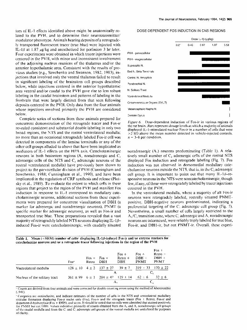
Dose-response relationships
Structures listed above as displaying specific IL- l-induced c-fos expression represent potential mediators of cytokine-induced activation of the HPA axis. To pare the list of candidates, we compared Fos induction in extrahypothalamic sites and the PVHmpd over a range of IL- 16 doses, reasoning that cell groups displaying a sensitivity similar to that of CRF target neurons in the PVH would be most apt to be mechanistically involved in the hypothalamic response. Animals were killed, again at 3 hr, following injection of IL-10 at doses ranging from 0.21 to 3.58 &kg (corresponding roughly to administration of 60-l 000 ng of cytokine to a 300 gm rat). The arbitrarily defined (see Ma- terials and Methods) effective dose for eliciting Fos induction differed markedly among the regions studied (Figs. 5, 6). Thus, few if any Fos-ir cells were detected in the PVNmpd in response to the lowest (0.2 1 pg/kg) dose tested (Fig. 6). However, begin- ning at 0.47 pg/kg (Figs. 5, 6), above criterion labeling was detected in this cell group, which progressively increased in number and staining intensity at higher dosage levels. At the highest dose (3.58 pg/kg), very strong labeling was observed throughout the PVHmpd, as well as in OT-rich aspects of the magnocellular division of the PVH (Figs. 5, 6). Among extra- hypothalamic regions, the central nucleus of the amygdala, the bed nucleus of the stria terminalis, the lateral parabrachial nu- cleus, as well as the NTS, displayed above criterion labeling even at the lowest dose of IL-l@ given (Fig. 6). The intensity and number of Fos-labeled cells increased progressively to a point where a large number of cells in these structures were strongly labeled for Fos-ir at the highest dose of IL- lfi studied, 3.58 pg/kg (Fig. 5). Reliable labeling in ventrolateral medulla required roughly 0.47 pg/kg IL-lb (Fig. 6), and the supraoptic nucleus, a pure magnocellular neurosecretory cell group, re- quired at least 0.87 Kg/kg IL-l@ to obtain a clear induction of Fos-ir (Fig. 6).
Interestingly, no reliable induction of Fos-ir was observed in the circumventricular organs at the minimum dose of IL-ID
The Journal of Neuroscience, February 1994, 14(2) 903
Figure 4. Extrahypothalamic sites of IL- l-stimulated Fos induction: bright-field photomicrographs of immunoperoxidase preparations, showing nuclear Fos induction in various rat CNS regions following intravenous administration of 1.87 pg IL- 1 P/kg 3 hr prior to perfusion fixation. Specific induction of Fos-ir was detected in the bed nucleus of the stria terminalis, particularly in the oval subnucleus (A), the lateral division of the central nucleus of amygdala (B), the lateral division of the parabrachial nucleus (C), and the rostra1 ventrolateral medulla, in the vicinity of the C, and A, catecholamine cell groups (D). No specific Fos induction was detected in the dentate gyrus of the hippocampal formation (E) or the choroid plexus (F,l in IL-l-injected rats as compared to vehicle controls. Constitutively high levels of Fos-ir was observed in the paraventricular nucleus of the thalamus in both IL-l-injected and vehicle-injected controls (F). ac, anterior commissure; bc, brachium conjunctivum; BST, bed nucleus of the stria terminalis; CA4, pyramidal cell layer of hippocampus; CeA,,,,,, central nucleus of the amygdala, medial, lateral parts; cp, choroid plexus; DG, dentate gyrus; ic, internal capsule; LRN, lateral reticular nucleus; LSv, ventral nucleus of the lateral septum; LV, lateral ventricle; P&,, parabrachial nuclei, medial, lateral; PT, parataenial nucleus; PVT, paraventricular nucleus (thalamus); SNV, spinal trigeminal nucleus; st, stria terminalis. Magnification, 75 x.
(0.47 pg/kg) required to activate neurons in the PVNmpd (Figs. observed at this and higher doses at the ventral and lateral 5,6). Extensive Fos induction was detected in these regions only margins ofthe OVLT region, at the rostralmost level ofthe optic at the upper extreme of the dosages tested (Figs. 5, 6). It may chiasm (Fig. 5). Based on cell size and contiguity with the me- be significant, however, that small Fos-ir cells were occasionally ninges, we interpret these as indicative of Fos induction in mem-
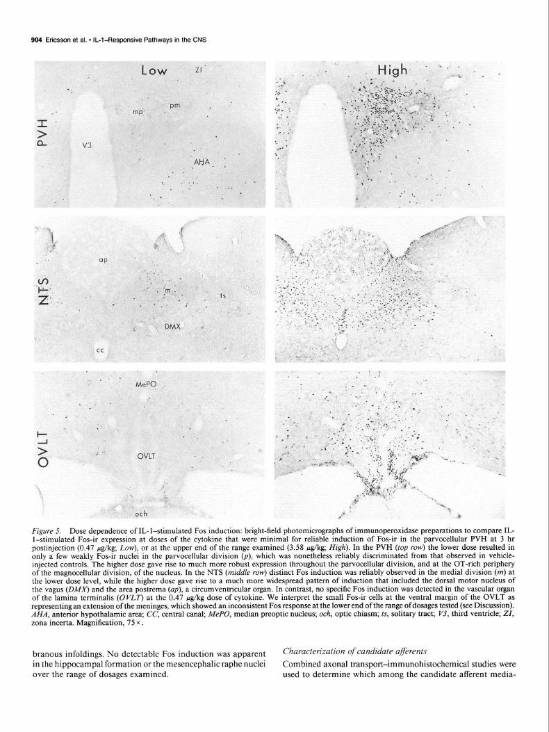
904 Ericsson et al. l IL-l-Responsive Pathways in the CNS
. .
__.
MeP$ _. .
Figure 5. Dose dependence of IL- l-stimulated Fos induction: bright-field photomicrographs of immunoperoxidase preparations to compare IL- l-stimulated Fos-ir expression at doses of the cytokine that were minimal for reliable induction of Fos-ir in the parvocellular PVH at 3 hr postinjection (0.47 &kg; Low), or at the upper end of the range examined (3.58 &kg; High). In the PVH (top row) the lower dose resulted in only a few weakly Fos-ir nuclei in the parvocellular division (p), which was nonetheless reliably discriminated from that observed in vehicle- injected controls. The higher dose gave rise to much more robust expression throughout the parvocellular division, and at the OT-rich periphery of the magnocellular division, of the nucleus. In the NTS (middle row) distinct Fos induction was reliably observed in the medial division (m) at the lower dose level, while the higher dose gave rise to a much more widespread pattern of induction that included the dorsal motor nucleus of the vagus @MA’) and the area postrema (up), a circumventricular organ. In contrast, no specific Fos induction was detected in the vascular organ of the lamina terminalis (OVLT) at the 0.47 &kg dose of cytokine. We interpret the small Fos-ir cells at the ventral margin of the OVLT as representing an extension of the meninges, which showed an inconsistent Fos response at the lower end of the range of dosages tested (see Discussion). AHA, anterior hypothalamic area; CC, central canal; MePO, median preoptic nucleus; och, optic chiasm; ts, solitary tract; Pj, third ventricle; ZI, zona incerta. Magnification, 75 x .
branous infoldings. No detectable Fos induction was apparent Characterization of candidate qflkrents
in the hippocampal formation or the mesencephalic raphe nuclei over the range of dosages examined.
Combined axonal transport-immunohistochemical studies were used to determine which among the candidate afferent media-
tors of IL-l effects identified above might be anatomically re- lated to the PVH, and to determine their neurotransmitter/ modulator phenotype. Animals bearing deposits ofa retrograde- ly transported fluorescent tracer (true blue) were injected with IL-ID at 1.87 fig/kg and anesthetized for perfusion 3 hr later. Four experiments were obtained in which tracer injections were centered in the PVH, with minor and inconsistent involvement of the adjoining nucleus reuniens of the thalamus and/or the anterior hypothalamic area. Consistent with the results of pre- vious studies (e.g., Sawchenko and Swanson, 1982, 1983), in- jections that involved only the ventral thalamus failed to result in significant labeling of the brainstem cell groups described below, while injections centered in the anterior hypothalamic area ventral and/or caudal to the PVH gave rise to less robust labeling in the caudal brainstem and patterns of labeling in the forebrain that were largely distinct from that seen following deposits centered in the PVH. Only data from the four animals whose injections involved primarily the PVH are considered below.
Complete series of sections from these animals prepared for concurrent demonstration of the retrograde tracer and Fos-ir revealed consistent and substantial double labeling in only two broad regions, the NTS and the rostra1 ventrolateral medulla; no more than an occasional retrogradely labeled Fos-ir cell was detected in components of the lamina terminalis or any of the other cell groups alluded to above that have been implicated as mediators of IL-l effects on the HPA axis. Catecholaminergic neurons in both brainstem regions (A, noradrenergic and C, adrenergic cells of the NTS and C, adrenergic neurons of the rostra1 ventrolateral medulla) have previously been shown to project to the parvocellular division of PVH (Cunningham and Sawchenko, 1988; Cunningham et al., 1990), and have been implicated in the regulation of CRF synthesis and release (Plot- sky et al., 1989). To evaluate the extent to which cells in these regions that project to the region of the PVH and manifest Fos induction in response to IL-I correspond to medullary cate- cholaminergic neurons, additional sections from these experi- ments were prepared for concurrent visualization of DBH (a marker for adrenergic and noradrenergic neurons), PNMT (a specific marker for adrenergic neurons), as well as Fos-ir and transported true blue. These preparations revealed that a vast majority of retrogradely labeled NTS neurons displaying IL- lp- induced Fos-ir were catecholaminergic, with caudally situated
The Journal of Neuroscience, February 1994. 14(2) 905
DOSE-DEPENDENT FOS INDUCTION IN CNS REGIONS
Dose IL-lb (vgikg)
PVH - parvocellular
0.21 0.46 0.87 1 .a7 3.58
PVH - magnocellular
Supraoptic N.
Bed N. Stria Terminals
Central N. Amygdala
Parabrachial N.
N. Solitary Tract
Ventrolateral Medulla
Circumventricular Organs (OVLT)
Mesencephalic Raphe N. I
Dentate Gyrus I
Figure 6. Dose-dependent induction of Fos-ir in various regions of the rat brain. Bars represent dosage levels at which a majority ofanimals displayed IL- l-stimulated nuclear Fos-ir in a number of cells that were >2 SD above the mean number detected in vehicle-injected controls. n = 3-8 per condition.
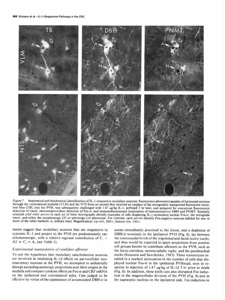
noradrenergic (A?) neurons predominating (Table 1). A rela- tively small number of Cz adrenergic cells of the rostra1 NTS displayed Fos induction and retrograde labeling (Fig. 7). Fos induction was not observed in dorsomedial medullary cate- cholamine neurons outside the NTS, that is, in the C, adrenergic cell group. It is important to point out that many IL-lb-re- sponsive neurons in the NTS were noncatecholaminergic, though few, ifany, ofthese were retrogradely labeled by tracer injections centered in the PVH.
In the ventrolateral medulla, where a majority of all Fos-ir neurons were retrogradely labeled, rostrally situated PNMT- positive, DBH-negative neurons predominated, indicating a preferential targeting of the C, adrenergic cell group (Fig. 7). Nevertheless, a small number of cells largely restricted to the A,!C, transition zone, where C, adrenergic and A, noradrenergic neurons are intermixed, were reliably triply labeled for true blue, Fos-ir, and DBH-ir, but not PNMT-ir. Overall, these experi-
Table 1. Mean (?SEM) number of cells” displaying IL-l&induced Fos-ir and/or enzyme markers for catecholamine neurons and/or a retrograde tracer following injections in the region of the PVH
Fos” Fos + Fos + Retro DBH
Fos + Fos + Fos + Retro + Retro + DBH + DBH + DBH PNMT PNMT
Ventrolateral medulla 128klO 4+-2 131 f 21 39 f I 319 i 33 170 +- 22
A, C,
Nucleus of the solitary tract 361 f 99 6 * 1 204 + 41 129 + 14 62 f 6 32 f 6
Al Cl
Li Counts are derived from four animals and were corrected for double-counting errors using the method of Abercrombie (1941). ’ Categories are noninclusive, and indicate estimates of the number of cells in the NTS and ventrolateral medullary reticular formation displaying Fos-ir nuclei only (Fos), Fos-ir and the retrograde tracer (Fos + Retro), Fos-ir and dopamine-fi-hydroxylase (Fos + DBH), and so on. It should be noted that no cells were identified that stained positively for PNMT but not DBH. Values indicative primarily of counts obtained from the A, and A2 noradrenergic cell groups of the caudal medulla and from the C, and C? adrenergic cell groups of the rostra1 medulla are underlined for purposes of clarity.
906 Ericsson et al. l IL-l-Responsive Pathways in the CNS
Figure 7. Anatomical and biochemical identification of IL- l-responsive medullary neurons: fluorescence photomicrographs of horizontal sections through the ventrolateral medulla (l&W) and the NTS from an animal that received an implant of the retrogradely transported fluorescent tracer, true blue (TB), into the PVH, was subsequently challenged with 1.87 &kg IL-I, perfused 3 hr later, and prepared for concurrent fluorescence detection of tracer, immunoperoxidase detection of Fos-ir, and immunofluorescence localization of immunoreactive DBH and PNMT. Similarly oriented solid white arrows in each set of three micrographs identify examples of cells displaying IL-l-stimulated nuclear Fos-ir, the retrograde tracer, and either the noradrenergic (N) or adrenergic (A) phenotype. For contrast, open arrows identify Fos-negative neurons labeled for one or more of the other markers. ts, solitary tract. Magnification: top row, 300 x ; bottom row, 150 x .
ments suggest that medullary neurons that are responsive to systemic IL-l and project to the PVH are predominantly cat- echolaminergic, with a relative regional contribution of C, > A2 > C2 = A, (see Table 1).
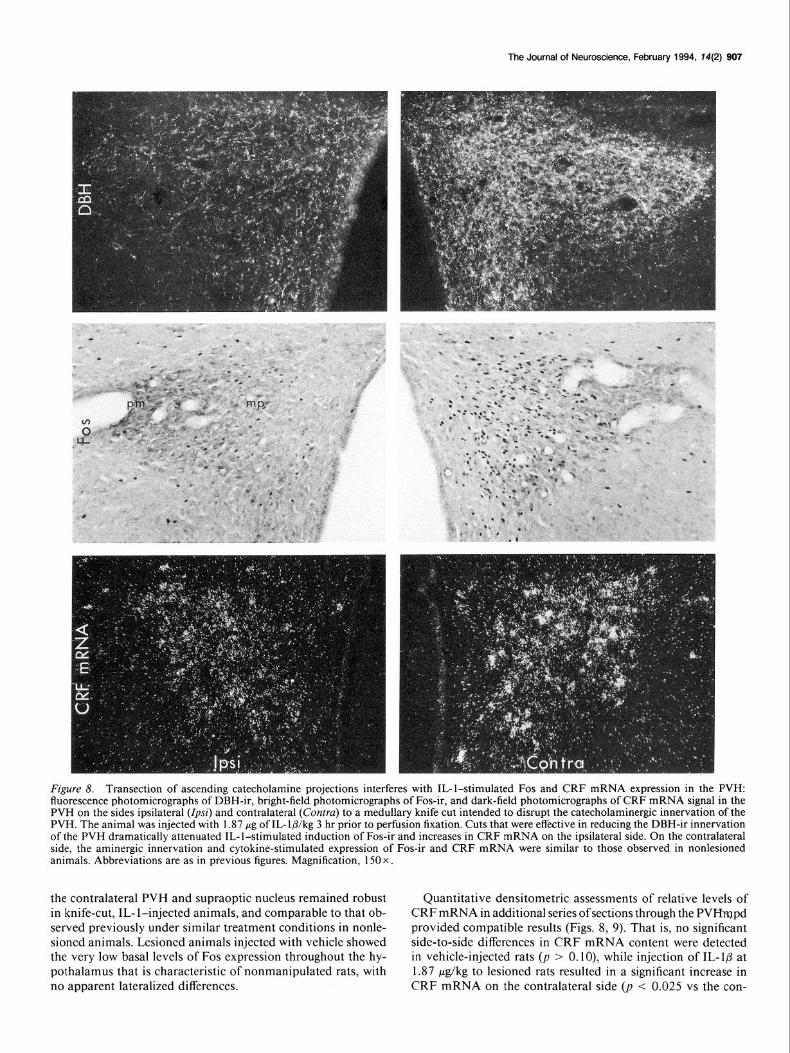
Experimental manipulation of candidate aferents To test the hypothesis that medullary catecholamine neurons are involved in mediating IL-16 effects on parvocellular neu- rosecretory neurons in the PVH, we attempted to unilaterally disrupt ascending aminergic projections near their origins in the medulla and compare cytokine effects on Fos-ir and CRF mRNA on the ipsilateral and contralateral’ sides. Cuts judged to be effective by virtue of the appearance of accumulated DBH-ir in
axons immediately proximal to the lesion, and a depletion of DBH-ir terminals in the ipsilateral PVH (Fig. 8) lay between the rostrocaudal levels of the trigeminal and facial motor nuclei, and thus would be expected to spare projections from pontine cell groups known to contribute afferents to the PVH, such as the locus coeruleus, mesencephalic raphe, and the parabrachial nuclei (Swanson and Sawchenko, 1983). These transections re- sulted in a marked attenuation in the number of cells that dis- played nuclear Fos-ir in the ipsilateral PVHmpd, seen in re- sponse to injection of 1.87 pg/kg of IL-1P 3 hr prior to death (Fig. 8). In addition, these knife cuts also disrupted Fos induc- tion in the magnocellular division of the PVH (Fig. 8) and in the supraoptic nucleus on the ipsilateral side. Fos induction in
The Journal of Neuroscience, February 1994. T4(2) 907
Fgure 8. Transection of ascending catecholamine projections interferes with IL-l-stimulated Fos and CRF mRNA expression in the PVH: fluorescence photomicrographs of DBH-ir, bright-field photomicrographs of Fos-ir, and dark-field photomicrographs of CRF mRNA signal in the PVH on the sides ipsilateral (Zp~z’) and contralateral (Contra) to a medullary knife cut intended to disrupt the catecholaminergic innervation of the PVH. The animal was injected with 1.87 wg of IL-lb/kg 3 hr prior to perfusion fixation. Cuts that were effective in reducing the DBH-ir innervation of the PVH dramatically attenuated IL-l-stimulated induction of Fos-ir and increases in CRF mRNA on the ipsilateral side. On the contralateral side, the aminergic innervation and cytokine-stimulated expression of Fos-ir and CRF mRNA were similar to those observed in nonlesioned animals. Abbreviations are as in previous figures. Magnification, 150 x.
the contralateral PVH and supraoptic nucleus remained robust Quantitative densitometric assessments of relative levels of in knife-cut, IL- l-injected animals, and comparable to that ob- CRF mRNA in additional series of sections through the PVlYmpd served previously under similar treatment conditions in nonle- provided compatible results (Figs. 8, 9). That is, no significant sioncd animals. Lcsioned animals injected with vehicle showed side-to-side differences in CRF mRNA content were detected the very low basal levels of Fos expression throughout the hy- in vehicle-injected rats (p > 0. lo), while injection of IL-l@ at pothalamus that is characteristic of nonmanipulated rats, with 1.87 &kg to lesioned rats resulted in a significant increase in no apparent lateralized differences. CRF mRNA on the contralateral side (p < 0.025 vs the con-
909 Ericsson et al. * IL-l-Responsive Pathways in the CNS
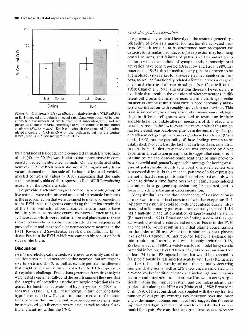
lpsi Contra lpsi Contra
Saline IL-l
Figure 9. Unilateral knife-cut effects on relative levels of CRF mRNA in IL- l-injected and vehicle-injected rats. Data were obtained by den- sitometric assessments of emulsion-dipped autoradiograms, and are presented as mean t SEM percentage of values obtained in the control condition (Saline, contra). Knife cuts abolish the expected IL-l-stim- ulated increase in CRF mRNA on the ipsilateral, but not the contra- lateral, side. n = 5 per group; *, p < 0.025.
tralateral side oflesioned, vehicle-injected animals), whose mag- nitude (40.1 + 20.5%) was similar to that noted above in com- parably treated nonlesioned animals. On the ipsilateral side, however, CRF mRNA levels did not differ significantly from values obtained on either side of the brain of lesioned, vehicle- injected controls (p values > 0. lo), suggesting that the knife cuts functionally ablated the response to IL- 1 of CRF-producing neurons on the ipsilateral side.
To provide a relevant surgical control, a separate group of five animals were administered unilateral stereotaxic knife cuts in the preoptic region that were designed to interrupt projections to the PVH from cell groups comprising the lamina terminalis of the third ventricle, whose circumventricular aspects have been implicated as possible central monitors of circulating IL- 1. These cuts, which were similar in size and placement to those shown previously to abolish osmotically driven responses of parvocellular and magnocellular neurosecretory neurons in the PVH (Kovacs and Sawchenko, 1993) did not affect IL-lb-in- duced Fos-ir in the PVH, which was comparably robust on both sides of the brain.
Discussion
In situ morphological methods were used to identify and char- acterize stress-related neuroendocrine neurons that are respon- sive to systemic IL-lb, as well as extrahypothalamic afferents that might be mechanistically involved in the HPA response to the cytokine challenge. Predictions generated from this analysis were tested experimentally, and the results support the view that the integrity of ascending catecholaminergic projections is re- quired for functional activation of hypophysiotropic CRF neu- rons by IL- 1 (see Fig. 10). These findings, in turn, define testable hypotheses as to how IL- 1, an important mediator of interac- tions between the immune and neuroendocrine systems, may be transduced to influence stress-related, as well as other, func- tional circuitries within the CNS.
Methodological considerations
The present analyses relied heavily on the assumed general ap- plicability of c-fos as a marker for functionally activated neu- rons. While it remains to be determined how widespread the capacity for stimulation-induced c-fis expression may be among central neurons, and failures of patterns of Fos induction to conform with other indices of synaptic and/or transcriptional activation have been reported (Dragunow and Faull, 1989; La- biner et al., 1993), this immediate-early gene has proven to be a reliable activity marker for stress-related neuroendocrine neu- rons, as well as functionally related afferents, across a range of acute and chronic challenge paradigms (see Ceccatelli et al., 1989; Chan et al., 1993, and citations therein). Fewer data are available that speak to the question of whether neurons in dif- ferent cell groups that may be recruited in a challenge-specific manner to comprise functional circuits need necessarily mani- fest c-fis induction with roughly equivalent sensitivities. This issue is important, as a comparison of dose-response relation- ships in different cell groups was used to restrict an initially sizeable list of candidate afferent mediators of IL-l effects to a testable subset. In the few relevant instances in which this notion has been tested, reasonable congruence in the sensitivity oftarget and afferent cell groups to express c-fos have been found (Chan et al., 1993), but the generality of these findings remain to be established. Nonetheless, the fact that an hypothesis generated, in part, from the dose-response data was supported by direct experimental evaluation prompts us to suggest that comparison of time course and dose-response relationships may prove to be a powerful and generally applicable strategy for honing anal- yses of polysynaptic circuits to a point where relatedness can be assessed directly. In this manner, patterns of c-fis expression are not utilized as end points unto themselves, but as tools with which to define a time frame over which stimulation-induced alterations in target gene expression may be expected, and to focus and refine subsequent experimentation.
Along similar lines, the dose dependency of Fos induction is also relevant to the critical question of whether exogenous IL- 1 injection may mimic cytokine levels encountered during infec- tious and inflammatory processes. Intravenously injected IL- 10 has a half-life in the rat circulation of approximately 2.9 min (Reimers et al., 199 1). Based on this finding, a dose of 0.47 pg/ kg, which provoked a reliable induction of Fos-ir in the PVH and the NTS, would result in an initial plasma concentration on the order of 20 nM. While this is similar to peak plasma levels of IL-l@ (about 30 nM) reported following systemic ad- ministration of bacterial cell wall lipopolysaccharide (LPS; Zuckerman et al., 1989) a widely employed model for systemic bacterial infection, elevated levels of cytokine are sustained for at least 24 hr in LPS-injected mice, but would be expected to fall precipitously in rats injected acutely with IL-l (Reimers et al., 1991). It is also worthy of note that naturally occurring immune challenges, as well as LPS injection, are associated with elevated levels ofadditional cytokines, including tumor necrosis factor-a and interleukin-6, that are well known to act synergis- tically within the immune system, and are independently ca- pable ofstimulating the HPA axis (Naito et al., 1988; Bernardini et al., 1990). These considerations, coupled with the very limited number of cell groups evincing Fos induction over the lower end of the range of dosages employed here, suggest that the acute injection paradigm is relevant at least to an established animal model for sepsis. We consider it an open question as to whether
The Journal of Neuroscience, February 1994, 14(2) 909
l?
4 ACTH
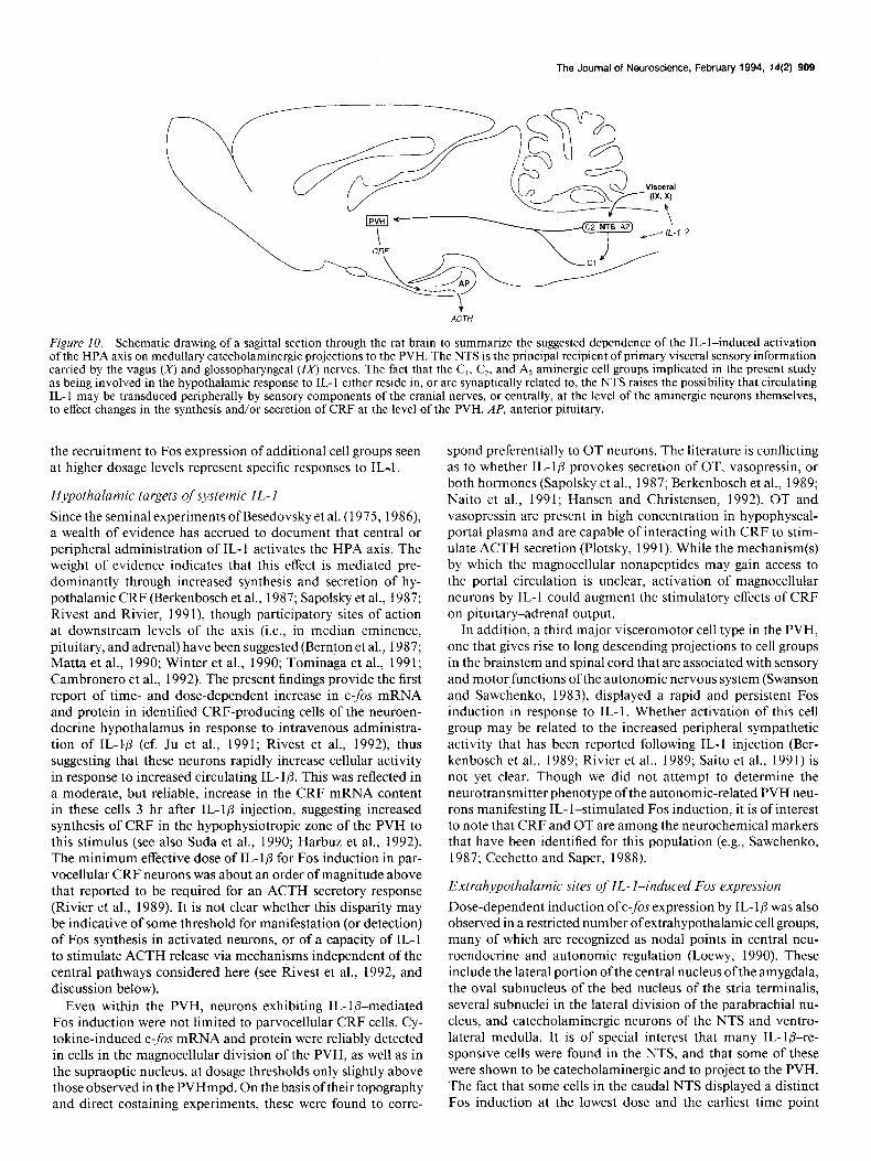
Figure 10. Schematic drawing of a sagittal section through the rat brain to summarize the suggested dependence of the IL- l-induced activation of the HPA axis on medullary catecholaminergic projections to the PVH. The NTS is the principal recipient of primary visceral sensory information carried by the vagus (x) and glossopharyngeal (1Xj nerves. The fact that the C,, C,, and A, aminergic cell groups implicated in the present study as being involved in the hypothalamic response to IL- 1 either reside in, or are synaptically related to, the NTS raises the possibility that circulating IL- 1 may be transduced peripherally by sensory components of the cranial nerves, or centrally, at the level of the aminergic neurons themselves, to effect changes in the synthesis and/or secretion of CRF at the level of the PVH. AP, anterior pituitary.
the recruitment to Fos expression of additional cell groups seen at higher dosage levels represent specific responses to IL-,1 .
H.vpothalamic targets of systemic IL- I
Since the seminal experiments of Besedovsky et al. (1975, 1986), a wealth of evidence has accrued to document that central or peripheral administration of IL- 1 activates the HPA axis. The weight of evidence indicates that this effect is mediated pre- dominantly through increased synthesis and secretion of hy- pothalamic CRF (Berkenbosch et al., 1987; Sapolsky et al., 1987; Rivest and Rivier, 199 l), though participatory sites of action at downstream levels of the axis (i.e., in median eminence, pituitary, and adrenal) have been suggested (Bernton et al., 1987; Matta et al., 1990; Winter et al., 1990; Tominaga et al., 1991; Cambronero et al., 1992). The present findings provide the first report of time- and dose-dependent increase in c-fos mRNA and protein in identified CRF-producing cells of the neuroen- docrine hypothalamus in response to intravenous administra- tion of IL-l/3 (cf. Ju et al., 199 1; Rivest et al., 1992), thus suggesting that these neurons rapidly increase cellular activity in response to increased circulating IL- l& This was reflected in a moderate, but reliable, increase in the CRF mRNA content in these cells 3 hr after IL-l@ injection, suggesting increased synthesis of CRF in the hypophysiotropic zone of the PVH to this stimulus (see also Suda et al., 1990; Harbuz et al., 1992). The minimum effective dose of IL- l/3 for Fos induction in par- vocellular CRF neurons was about an order of magnitude above that reported to be required for an ACTH secretory response (Rivier et al., 1989). It is not clear whether this disparity may be indicative of some threshold for manifestation (or detection) of Fos synthesis in activated neurons, or of a capacity of IL-l to stimulate ACTH release via mechanisms independent of the central pathways considered here (see Rivest et al., 1992, and discussion below).
Even within the PVH, neurons exhibiting IL-lP-mediated Fos induction were not limited to parvocellular CRF cells. Cy- tokine-induced c-fos mRNA and protein were reliably detected in cells in the magnocellular division of the PVH, as well as in the supraoptic nucleus, at dosage thresholds only slightly above those observed in the PVHmpd. On the basis oftheir topography and direct costaining experiments, these were found to corre-
spond preferentially to OT neurons. The literature is conflicting as to whether IL- lp provokes secretion of OT, vasopressin, or both hormones (Sapolsky et al., 1987; Berkenbosch et al., 1989; Naito et al., 199 1; Hansen and Christensen, 1992). OT and vasopressin are present in high concentration in hypophyseal- portal plasma and are capable of interacting with CRF to stim- ulate ACTH secretion (Plotsky, 199 1). While the mechanism(s) by which the magnocellular nonapeptides may gain access to the portal circulation is unclear, activation of magnocellular neurons by IL-l could augment the stimulatory effects of CRF on pituitary-adrenal output.
In addition, a third major visceromotor cell type in the PVH, one that gives rise to long descending projections to cell groups in the brainstem and spinal cord that are associated with sensory and motor functions of the autonomic nervous system (Swanson and Sawchenko, 1983), displayed a rapid and persistent Fos induction in response to IL-l. Whether activation of this cell group may be related to the increased peripheral sympathetic activity that has been reported following IL-l injection (Ber- kenbosch et al., 1989; Rivier et al., 1989; Saito et al., 1991) is not yet clear. Though we did not attempt to determine the neurotransmitter phenotype ofthe autonomic-related PVH neu- rons manifesting IL- l-stimulated Fos induction, it is of interest to note that CRF and OT are among the neurochemical markers that have been identified for this population (e.g., Sawchenko, 1987; Cechetto and Saper, 1988).
Extrahypothalamic sites of IL- l-induced Fos expression
Dose-dependent induction of c-fos expression by IL-l@ was also observed in a restricted number of extrahypothalamic cell groups, many of which are recognized as nodal points in central neu- roendocrine and autonomic regulation (Loewy, 1990). These include the lateral portion of the central nucleus of the amygdala, the oval subnucleus of the bed nucleus of the stria terminalis, several subnuclei in the lateral division of the parabrachial nu- cleus, and catecholaminergic neurons of the NTS and ventro- lateral medulla. It is of special interest that many IL-lb-re- sponsive cells were found in the NTS, and that some of these were shown to be catecholaminergic and to project to the PVH. The fact that some cells in the caudal NTS displayed a distinct Fos induction at the lowest dose and the earliest time point
910 Ericsson et al. * IL-l-Responsive Pathways in the CNS
examined suggests that this cell group may play a pivotal role in the coordinate regulation ofIL- l-mediated activation ofCRF secretory activity in the endocrine hypothalamus. The NTS is the principal central terminus of primary visceral sensory in- formation conveyed by the vagus and glossopharyngeal nerves, and projects to each of the major sites of IL-l-stimulated Fos induction described above (Loewy, 1990). Despite the fact that neurons in discrete aspects of amygdala, bed nucleus of the stria terminalis, and the parabrachial complex displayed among the lowest thresholds to IL- l-stimulated Fos induction of the cell groups that we examined, both the topography of Fos-ir neurons and the results of combined retrograde tracing and immuno- histochemical analyses suggested that the vast majority of IL-l- activated neurons in these regions are not anatomically related to the PVH. By contrast, IL- l-responsive catecholaminergic neurons of the NTS (A1 and CZ cell groups) and ventrolateral medulla (C, cell group) were found to project to the region of the PVH. This is consistent with previous findings that each of these cell groups projects preferentially to the parvocellular di- vision of the PVH (Cunningham and Sawchenko, 1988; Cun- ningham et al., 1990) where they have been implicated in reg- ulating CRF secretion principally via 011 -adrenoceptor mechanisms (Plotsky, 1987). By contrast, the A, cell group of the caudal ventrolateral medulla, which provides the bulk of the aminergic innervation of vasopressinergic magnocellular neurosecretory neurons (Cunningham et al., 1988) was rela- tively unresponsive to systemic IL- 1, an observation consistent with the paucity of magnocellular vasopressin neurons in the PVH that displayed Fos induction to IL- 1.
Some of our negative results warrant comment. Induction of Fos-ir in the circumventricular organs, which have been im- plicated as central transducers of circulating IL-l, required a dose of IL- l@ at the upper end of the range tested, approximately 5-10 times higher than the minimum needed to induce reliably Fos-ir in CRF neurons of the PVH. However, nuclear labeling for Fos-ir was sometimes observed at lower doses of IL-ID in small cells at the margins of the OVLT, where it adjoins the optic chiasm. Although this labeling was inconsistent at the lower doses administered, it is potentially relevant to IL- 1 effects on the HPA axis. However, these cells, which appear to be meningeal, are not situated within the OVLT proper and were not retrogradely labeled following tracer deposits in the PVH region. Moreover, unilateral transections designed to disrupt descending projections from the lamina terminalis failed to af- fect the IL-lp-mediated Fos induction in the PVH. Thus, our results fail to support an involvement of cells in the lamina terminalis region in driving IL-l-induced activation (i.e., Fos induction) of hypothalamic neurosecretory neurons.
In addition, we failed to detect IL- lb-mediated induction of C-$X expression in the hippocampal formation, the mesence- phalic raphe nuclei, the chorioid plexus, and postcapillary ven- ules, all structures in the CNS that have been shown to express high levels of the type 1 IL-l receptor and/or to be capable of IL- 1 ligand binding (Ban et al., 199 1; Cunningham et al., 1992). This would suggest that these cells are unresponsive to systemic IL- 1 fl injection, at least over the range of doses and time points examined here. It is important to bear in mind, however, that the lack of a Fos response does not necessarily preclude in- volvement of a cell group in a functional circuit. In addition to the above-mentioned caveats as to the uncertain generality and sensitivity for recruitment to Fos induction among central neu- rons, we are particularly concerned that neither Fos, nor any
other immediate-early gene yet examined as a potential activity index in the CNS, provides a marker for cells presented with net inhibitory synaptic and/or transcriptional drives (Chan et al., 1993). Thus, while the failures to detect IL-l-stimulated c-fis expression in candidate mediators of cytokine effects on the HPA axis are suggestive, they are tempered by demonstrated or potential limitations of Fos-based activity mapping ap- proaches.
Mechanisms underlying IL- 1 effects on the HPA axis In support of the hypothesized involvement of brainstem cat- echolamine neurons in the hypothalamic response to IL-l@, histochemically verified unilateral ablations of these ascending projections near their origins in the medulla were found to sig- nificantly attenuate cytokine-stimulated Fos induction, and to abolish the concomitant increase in CRF mRNA, in the ipsi- lateral PVH. Aminergic projections from the medulla are known to be predominantly ipsilateral, with a minor crossed compo- nent ofsimilar topography (e.g., Sawchenko and Swanson, 1982; Palkovits et al., 1992). Transections like those employed here have been shown to exert lateralized effects on aspects of par- vocellular neurosecretory function, while leaving intact the same parameters on the contralateral side (Sawchenko, 1988). Thus, the residual aminergic innervation on the side ipsilateral to such lesions seems insufficient to support normal function, while the minor denervation of the contralateral side is insufficient to disrupt them. It is unlikely that interference with IL-l effects were due to nonspecific sequelae of the lesions because the mag- nitude of the CRF mRNA response to IL- 1 on the contralateral side was similar to that observed in nonlesioned animals. More- over, surgical controls that involved unilateral disruption of lamina terminalis projections to the hypothalamus had no im- pact on IL-l-induced Fos expression in the PVH. It is also worthy of note that with the exception of a relatively small population of peptidergic neurons centered in the caudal part of the NTS (Sawchenko et al., 1992), medullary catecholamine neurons constitute the only known source of afferents to the PVH that arise distal to the level of the transections employed here. The fact that our combined axonal transport-multiple immunohistochemical labeling experiments revealed most Fos- ir neurons in the NTS that were retrogradely labeled from the PVH to be catecholaminergic supports the view that aminergic neurons comprised the principal substrate whose disruption in- terfered with the hypothalamic responses to IL- 1.
In assaying for the dependence of Fos and CRF mRNA re- sponses on the integrity ofparticular afferents, the present results do not speak directly to the substrates underlying cytokine effects on secretory responses of hypothalamic neuroendocrine neu- rons. An involvement of catecholaminergic afferents in the hy- pothalamic response to IL- 1 is supported by the results of pre- vious studies where systemic IL-l@ was shown to stimulate norepinephrine metabolism in the hypothalamus (Dunn, 1988; Kabiersch et al., 1988). Furthermore, complete deafferentation of the mediobasal hypothalamus (Ovadia et al., 1989) or neu- rotoxin-induced depletion of hypothalamic catecholamines (Weidenfeld et al., 1989; Matta et al., 1990; Chuluyan et al., 1992) has been shown to block or significantly attenuate ACTH and corticosterone secretory responses to central or peripheral injection of IL- 1. Although studies that have examined the ex- tent to which adrenoceptor antagonists may interfere with the secretory responses of the HPA axis to IL- 1 have failed to reach a consensus (Rivier et al., 1989; Weidenfeld et al., 1989), it is
The Journal of Neuroscience, February 1994, 74(2) 911
worthy of note that medullary catecholaminergic afferents to that IL-l may exert actions directly within the brain parenchy- the PVH are known to express a number of additional synap- ma. This is indicated by the fact that IL-l is a more potent tically active molecules (Sawchenko et al., 1992) any of which stimulant of the HPA axis when administered intracerebrov- could serve as cotransmitters or modulators in these projections. entricularly than systemically (Rivier et al., 1989). In addition, Collectively, these data support the view that the integrity of immunoreactive IL-l has been detected in astrocytes, microglia, medullary aminergic afferents is required for the IL-l stimu- and neuronal elements within the CNS (Breder et al., 1988; van lation of CRF synthesis in the endocrine hypothalamus; the Dam et al., 1992) and, as noted above, IL- 1 receptors are broad- likelihood that this dependence may generalize, at least in part, ly expressed in brain (Ban et al., 199 1; Cunningham et al., 1992). to cytokine-stimulated CRF secretion seems high, but remains The question of whether the cytokine can traverse the blood- to be evaluated directly. While the present data provide no brain barrier remains unresolved, with some reports claiming insight as to whether this involvement may be mediating or that IL- 1 is unable to enter the brain from the circulation (Co- merely permissive, support for the former is found in the ob- ceani et al., 1988; Ferreira et al., 1988) and others providing servation that hypothalamic amine depletion blocks pituitary- support for facilitated transport across the barrier (Banks et al., adrenal secretory responses to IL- 1, but not other stressors (Chu- 1989). An emerging alternative hypothesis holds that circulating luyan et al., 1992). IL- 1 may interact with IL- 1 receptors expressed on endothelial
Systemic administration of IL-l@ has, at doses similar to those cells of the cerebral vasculature, thereby stimulating synthesis employed here, been shown to provoke a number of systemic of secondary signaling molecules, such as IL- 1 itself (Schindler effects, including transient increases in body temperature (A. et al., 1990) nitric oxide (Kilbourn and Belloni, 1990) and/or Morimoto et al., 1990) heart rate (K. Morimoto et al., 1990) prostaglandins (McCarron, 1992) which may, in turn, be re- serum triglycerides (Feingold et al. 1991) and blood flow to leased locally to influence the activity of intraparenchymal neu- several vascular beds (Dascombe et al., 1989). This raises the rons. In line with this notion are reports that IL-l-mediated question of whether the central neuronal responses observed activation of the HPA axis may be attenuated by pretreatment following peripheral administration of IL- 1 reflect responses with prostaglandin synthesis inhibitors (Katsuura et al., 1990; targeted directly to specific neuroendocrine, among other, con- Watanabe et al., 1990). It remains to be determined whether trol systems, or are merely an indirect result of nonspecific stress transmission through medullary catecholaminergic neurons may and/or sympathetic activation. The weight of the available ev- be affected, directly or transneuronally, by such IL-l-stimulated idence argues for a substantial degree of targeting. For example, paracrine mechanisms. IL- 1 affects minimally, if at all, secretory responses of neuroen- In conclusion, by combining powerful c-&-based activity docrine axes controlling growth hormone, prolactin, and me- mapping with histochemical characterization in an experimen- lanotropin secretion (Berkenbosch et al., 1989). The sympatho- tal setting, the present analysis has served to better define hy- mimetic effects ofthe cytokine are generally modest (Berkenbosch pothalamic stress-related neurosecretory neuron pools that are et al., 1989; Rivier et al., 1989; Saito et al., 199 l), and can be affected by systemic IL-lb, and to identify specific medullary blocked by doses of systemic adrenergic antagonists that leave catecholamine cell groups as candidate afferent mediators ofthe intact the secretory responses of the HPA axis (Rivier et al., hypothalamic response (Fig. 10). Experimental evaluation of 1989). Regarding pyretic effects, analogs of IL- 1 have been iden- predictions generated from hodological analyses revealed that tified that do not affect core temperature, but that do elicit the integrity of these ascending afferents is at least required, and ACTH and corticosterone secretion (Naito et al., 1990). Finally, may play a mediating role, in the cytokine-stimulated cellular the restricted number of cell groups displaying IL-l-induced activation, and concomitant increase in CRF synthetic machin- Fos expression over at least the lower end ofthe range ofdosages ery, within hypophysiotropic neurons of the PVH. These data we employed and the absence of Fos induction in structures like provide a focus for further study of the site(s) of transduction the locus coeruleus, which might serve as generic indices of of circulating cytokine, and of the manner in which they evoke activation/arousal (Foote et al., 1983), lend additional support integrated neuroendocrine and autonomic activity to maintain to the notion that the central effects of the cytokine are not homeostasis following acute and chronic immunologic chal- simply a result of nonspecific stress. lenges.
While the data presented here identify a potential afferent mediator of IL-l effects on the central limb of the HPA axis, References the question remains as to how and where the circulating cy- tokine gains initial access to the CNS. In view of the strong evidence implicating aminergic neurons in the NTS and ven- trolateral medulla as important stations in visceral sensory in- formation processing (Loewy, 1990; Sawchenko et al., 1992) the possibility must be entertained that levels of circulating IL- 1 may be recorded by the PNS and conveyed synaptically to the CNS. This hypothesis finds some support in preliminary evi- dence to suggest that infusion of picomolar quantities of IL-l into the hepatic portal vein provokes increased firing in sensory fibers of the hepatic branch of the vagus nerve (Nijima, 1992). We are currently attempting to determine whether transection of various branches of the abdominal vagus nerve can mitigate IL-l&mediated cellular activation in the brainstem and hy- pothalamus.
Alternatively, a more substantial body of evidence suggests
Abercrombie M (1946) Estimation of nuclear population from mi- crotome sections. Anat Ret 94:239-247.
Ban E, Milon G, Prudhomme N, Haour F (1991) Receptors for in- terleukin (a and p) in mouse brain: mapping and neuronal localization in hippocampus. Neuroscience 43:2 l-30.
Banks WA, Kastin AJ, Durham DA (1989) Bidirectional transport of interleukin- 1 alpha across the blood-brain barrier. Brain Res Bull 23: 433-437.
Berkenbosch F, van Oers J, del Rey A, Tilders F, Besedovsky H (1987) Corticotropin-releasing factor-producing neurons in the rat activated by interleukin-1. Science 238524-526.
Berkenbosch F, de Goeij D, de1 Rey A, Besedovsky HO (1989) Neu- roendocrine, sympathetic and metabolic responses induced by inter- leukin- 1. Neuroendocrinology 50:570-576.
Bernardini R, Kamilaris TC, Calogero AE, Johnson EO, Gomez MT, Gold PW, Chrousos GP (1990) Interactions between tumor necrosis factor-q hypothalamic corticotropin-releasing hormone, and adren- ocorticotropin secretion in the rat. Endocrinology 126:2876-288 1.
Bemton EW, Beach JE, Holaday JW, Smallridge RC, Fein HG (1987)
912 Ericsson et al. * IL-l-Responsive Pathways in the CNS
Release of multiple hormones by a direct action of interleukin- 1 on pituitary cells. Science 238:5 19-52 1.
Besedovsky H, Sorkin E, Keller M, Miiller J (1975) Changes in blood hormone levels during the immune response. Proc Sot Exp Biol Med 150:466-470.
Besedovsky H, del Rey A, Sorkin E, Dinarello CA (1986) Immunor- egulatory feedback between interleukin-1 and glucocorticoid hor- mones. Science 233:652-654.
Bohn MC, Dreyfus CF, Friedman WJ, Markey KA (1987) Glucocor- ticoid effects on phenylethanolamine-Nmethyltransferase (PNMT) in explants of embryonic rat medulla oblongata. Dev Brain Res 37: 257-266.
Breder CD, Dinarello CA, Saper CB (1988) Interleukin-1 immuno- reactive innervation of the human hypothalamus. Science 240:32 l- 324.
Brown R, Li Z, Vriend CY, Nirula R, Janz L, Falk J, Nance DM, Dyck DC, Greenberg AH (199 1) Suppression of splenic macrophage interleukin-1 secretion following intracerebroventricular injection of interleukin-1 beta: evidence for pituitary-adrenal and sympathetic control. Cell Immunol 132:84-93.
Cambronero JC, Rivas FJ, Borrell J, Guaza C (1992) Interleukin-l- beta induces pituitary adrenocorticotropin secretion: evidence for glu- cocorticoid modulation. Neuroendocrinology 55:648-654.
Ceccatelli S, Villar MJ, Goldstein M, HGkfelt T (1989) Expression of c-Fos immunoreactivity in transmitter-characterized neurons after stress. Proc Nat1 Acad Sci USA 86:9569-9573.
Cechetto DF, Saper CB (1988) Neurochemical organization of the hypothalamic projection to the spinal cord in the rat. J Comp Neurol 2721579-604.
Chan RKW, Brown ER, Ericsson A, Kovacs KJ, Sawchenko PE (1993) A comparison of two immediate early genes, c-fos and NGFI-B, as markers for functional activation in stress-related neuroendocrine circuitry. J Neurosci, in press.
Chuluyan HE, Saphier D, Rohn WM, Dunn AJ (1992) Noradrenergic innervation of the hypothalamus participates in adrenocortical re- sponses to interleukin- 1. Neuroendocrinoiogy 56: 106-l 11.
Coceani F, Lees J, Dinarello CA (1988) Occurrence of interleukin-1 in cerebrospinal fluid of the conscious cat. Brain Res 446:245-250.
Cunningham ET Jr, Sawchenko PE (1988) Anatomical specificity of noradrenergic inputs to the paraventricular and supraoptic nuclei of the rat hypothalamus. J Comp Neurol 274:60-76.
Cunningham ET Jr, Bohn MC, Sawchenko PE (1990) Organization of adrenergic inputs to the paraventricular and supraoptic nuclei of the hypothalamus in the rat. J Comp Neurol 292:65 l-667.
Cunningham ET Jr, Wada E, Carter DB, Tracey DE, Battey JF, De Souza EB (1992) In situ histochemical localization of type 1 interleukin-1 receptor RNA in the central nervous system, pituitary, and the adrenal gland of the mouse. J Neurosci 12: 110 l-l 114.
Cupps TR, Fauci AS (1982) Corticosteroid-mediated immunoregu- lation in man. Immunol Rev 65: 133-l 55.
Curran T, Gordon MB, Rubino KL, Sambucetti LC (1987) Isolation and characterization of the c-f& (rat) cDNA and the analysis of post- translational modification in vitro. Oncogene 2:79-84.
Dascombe MJ, Rothwell NJ, Sagay BO, Stock MJ (1989) Pyrogenic and thermogenic effects of interleukin 1 beta in the rat. Am J Physiol 256:E7-Eli.
Dinarello CA (199 1) Interleukin- 1 and interleukin- 1 antagonism. Blood 77: 1627-1652.
Dragunow M, Faull R (1989) The use of c-fos as a metabolic marker in neuronal pathwav tracing. J Neurosci Methods 29:26 l-265.
Dunn AJ (1988) Systemic interleukin- 1 administration stimulates hy- pothalamic norepinephrine metabolism parallelling the increased plasma corticosterone. Life Sci 43:429-435.
Ericsson A, Sawchenko PE (1993) c-fos-based functional mapping of central pathways subserving the effects of interleukin-1 on the hy- pothalamo-pituitary-adrenal axis. In: Methods in neurosciences. The neurobiology of cytokines Vol 16 (Conn PM, ed), pp 153-172. New York: Academic.
Ericsson A, Amaral DC, Sawchenko PE (1992a) Systemic adminis- tration of interleukin- 10 results in increased levels ofNFGI-B mRNA in the endocrine hypothalamus and its medullary afferents. Sot Neu- rosci Abstr 18:1013.
Ericsson A, Kovacs K, Sawchenko PE (1992b) Systemic interleukin- 10 stimulates Fos expression in the paraventricular nucleus and its medullary afferents. Proc 74th Ann Meet Endocr Sot, p 44 1.
Feingold KR, Soued M, Adi S, Staprans I, Neese R, Shigenaga J, Do- errler W, Moser A, Dinarello CA, Grunfeld C (1991) Effect of interleukin-I on lipid metabolism in the rat. Similarities to and dif- ferences from tumor necrosis factor. Arterioscler Thromb 11:495- 500.
Ferreira SH, Lorenzetti BB, Bristow AF, Poole S (1988) Interleukin- I@ as a potent hyperalgesic agent antagonized by a tripeptide analogue. Nature 334:698-700.
Foote SL, Bloom FE, Aston-Jones G (1983) Nucleus locus ceruleus: new evidence ofanatomical and physiological specificity. Physiol Rev 63:844-914.
Gold RM, Kapatos G, Carey RJ (1973) A retractable wire knife for stereotaxic surgery made from a microliter syringe. Physiol Behav 10: 813-815.
Hansen EW, Christensen JD (1992) Endotoxin and interleukin- 1 beta induces fever and increased plasma oxytocin in rabbits. Pharmacol Toxic01 70:389-39 I.
Harbuz MS, Stephanou A, Sarlis N, Lightman SL (1992) The effects of recombinant human interleukin (IL)-lo(, IL-10 or IL-6 on hypoth- alamo-pituitarv-adrenal axis activation. J Endocrinol 133:349-355.
Hori T, Nakashima T, Take S, Kaizuka Y, Mori T, Katafuchi T (199 1) Immune cytokines and regulation of body temperature, food intake and cellular immunity. Brain Res Bull 27:309-3 13.
Jingami H, Mizuno N, Takahashi H, Shibahara S, Furutani Y, Imura H, Numa S (1985) Cloning and sequence analysis of cDNA for rat corticotropin-releasing factor precursor. FEBS Lett 19 1:63-66.
Johnson AK, Loewy AD (1990) Circumventricular organs and their role in visceral functions. In: Central regulations of autonomic func- tions (Loewy AD, Spyer KM, eds), pp 247-267. New York: Oxford UP.
Ju G, Swanson LW (1989) Studies on the cellular architecture of the bed nuclei of the stria terminalis in the rat: I. Cytoarchitecture. J Comp Neurol 280:587-602.
Ju G, Zhang X, Jin B-Q, Huang C-S (199 1) Activation ofcorticotropin- releasing factor-containing neurons in the paraventricular nucleus of the hypothalamus by interleukin- 1. Neurosci Lett 132: 15 l-l 54.
Kabiersch A, del RA, Honegger CC, Besedovsky HO (1988) Interleukin-1 induces changes in norepinephrine metabolism in the rat brain. Brain Behav Immunol 2:267-274.
Katsuura G, Arimura A, Koves K, Gottschall PE (1990) Involvement of organum vasculosum of lamina terminalis and preoptic area in interleukin 1 beta-induced ACTH release. Am J Physiol 258:E163- El71.
Kilboum RG, Belloni P (1990) Endothelial cell production of nitrogen oxides in response to interferon gamma in combination with tumor necrosis factor, interleukin-1, or endotoxin. J Nat1 Cancer Inst 82: 772-776.
Kovacs KJ, Sawchenko PE (1993) Osmoregulatory influences on neu- roendocrine corticotropin-releasing factor expression are mediated via the ventral lamina terminalis. Proc Nat1 Acad Sci USA 90:7681- 7685.
Kroemer G, Brezinschek HP, Faessler R, Schauenstein K, Wick G (1988) Physiology and pathology of an immunoendocrine feedback loop. Immunol Today 9:163-165.
Kuschner I (1982) The phenomenon of the acute-phase response. Ann NY Acad Sci 289:39-48.
Labiner DJ, Butler LS, Zhen C, Hosford DA, Shin C, McNamara JO (1993) Induction of c-fos mRNA by kindled seizures: complex re- lationship with neuronal burst firing: J Neurosci 13:744-75 1:
Lind RW (1986) Bi-directional. chemicallv snecified neural connec- tions between the subfornical organ and the midbrain raphe system. Brain Res 384:250-261.
Loewy AD (1990) Central autonomic pathways. In: Central regulations of autonomic functions (Loewy AD, Spyer KM, eds), pp 88-103. New York: Oxford UP.
Matta S, Singh J, Newton R, Sharp BM (1990) The adrenocortico- tropin response to interleukin-1 beta instilled into the rat median eminence depends on the local release of catecholamines. Endocri- nology 127:2175-2182.
McCarron RM (1992) IL-l-induced prostacyclin production by ce- rebral vascular endothelial cells inhibits myelin basic protein-specific lymphocyte proliferation. Cellular Immunol 145:21-29.
Morgan JI, Curran T (1991) Stimulus-transcription coupling in the nervous system: involvement ofthe inducible proto-oncogenesfosand jun. Annu Rev Neurosci 14~42 l-45 1.
The Journal of Neuroscience, February 1994, 14(2) 913
Morimoto A, Murakami N, Sakata Y, Watanabe T, Yamaguchi K (1990) Functional and structural differences in febrile mechanism between rabbits and rats. J Physiol (Lond) 427:227-239.
Morimoto K, Morimoto A, Nakamori T, Tan N, Minagawa T, Mu- rakami N (1990) Cardiovascular responses induced in free-moving rats by immune cytokines. J Physiol (Lond) 448:307-320.
Naito Y, Fukata J, Tominaga T, Nakai Y, Tamai S, Mori K, Imura H (1988) Interleukin-6 stimulates the secretion of adrenocorticotropic hormone in conscious, freely-moving rats. Biochem Biophys Res Commun 155: 1459-1463.
Naito Y, Fukata J, Masui Y, Hirai Y, Murakami N, Tominaga T, Nakai Y, Tamai S, Mori K, Imura H (1990) Interleukin- 1 beta analogues with markedly reduced pyrogenic activity can stimulate secretion of adrenocorticotropic hormone in rats. Biochem Biophys Res Commun 167:103-109.
Naito Y, Fukata J, Shindo K, Ebisui 0, Murakami N, Tominaga T, Nakai Y, Mori K, Kasting NW, Imura H (199 I) Effects of interleu- kins on plasma arginine vasopressin and oxytocin levels in conscious, freely moving rats. Biochem Biophys Res Commun 174: 1 189-l 195.
Nakano K, Okugawa K, Hayashi H, Abe S, Sohmura Y, Tsuboi T (1988) Establishment of dye-uptake method (A375 assay) for quan- titative measurement of IL-l: correlation with LAF assay. Dev Biol Stand 69:93-IO 1.
Nijima A (1992) The afferent discharges from sensors for interleukin l/3 in the hepato-portal system in the rat. J Physiol (Lond) 446:236P.
Ovadia H, Abramsky 0, Barak V, Conforti N, Saphier D, Weidenfeld J (1989) Effect of interleukin-1 on adrenocortical activity in intact and hypothalamic deafferented male rats. Exp Brain Res 76:246-249.
Palkovits M, Mezey E, Skirboll LR, Hijkfelt T (1992) Adrenergic projections from the lower brain-stem to the hypothalamic paraven- tricular nucleus, the lateral hypothalamic area and the central nucleus of the amygdala in rats. J Chem Neuroanat 5:407-415.
Plotsky PM (I 987) Facilitation of immunoreactive corticotropin re- leasing factor secretion into the hypophyseal-portal circulation after activation of catecholaminergic pathways or central norepinephrine injection. Endocrinology 121:924-930.
Plotsky PM (199 1) Pathways to the secretion of adrenocorticotropin: a view from the portal. J Neuroendocrinol 3: l-9.
Plotsky PM, Cunningham ET Jr, Widmaier EP (1989) Catecholamin- ergic modulation of corticotropin-releasing factor and adrenocorti- cotropin secretion. Endocrine Rev 10:437-458.
Reimers J, Wogensen LD, Welinder B, Hejnzs KR, Poulsen SS, Nilsson P, Nerup J (1991) The pharmacokinetics, distribution and degra- dation of human recombinant interleukin-10 in normal rats. Stand J Immunol 34:597-6 17.
Rivest S, Rivier C (199 I) Influence of the paraventricular nucleus of the hypothalamus in the alteration of neuroendocrine functions in- duced by intermittent footshock or interleukin. Endocrinology 129: 2049-2057.
Rivest S, Torres G, Rivier C (1992) Differential effects of central and peripheral injection of interleukin-10 on brain clfos expression and neuroendocrine functions. Brain Res 587: 13-23.
Rivier C, Vale W, Brown M (1989) In the rat, interleukin-lol and -p stimulate adrenocorticotropin and catecholamine release. Endocri- nology 125:3096-3 102.
Sagar SM, Sharp FR, Curran T (1988) Expression of c-fis protein in brain: metabolic mauning at the cellular level. Science 240:1328- __ _ 1331.
Saito M, Akiyoshi M, Shimizu Y (199 1) Possible role of the sympa- thetic nervous system in response to interleukin- I. Brain Res Bull 27: 305-308.
Sapolsky R, Rivier C, Yamamoto G, Plotsky P, Vale W (1987) Interleukin- 1 stimulates the secretion of hypothalamic corticotropin- releasing factor. Science 238:522-524.
Sawchenko P (1987) Evidence for differential regulation of cortico- tropin-releasing factor and vasopressin immunoreactivities in par- vocellular neurosecretory and autonomic-related projections of the paraventricular nucleus..Brain Res 437:253-263.
Sawchenko PE (1988) Effects of catecholamine-denletina medullarv knife cuts on dorticotropin-releasing factor and vasopreisin immu- noreactivity in the hypothalamus of normal and steroid-manipulated rats. Neuroendocrinology 48:459-470.
Sawchenko PE, Swanson LW (1982) The organization of noradren- ergic projections from the brain stem to the paraventricular and su- praoptic nuclei in the rat. Brain Res Rev 4:275-325.
Sawchenko PE, Swanson LW (1983) The organization of forebrain afferents to the paraventricular and supraoptic nuclei of the rat. J Comp Neural 218:121-144.
Sawchenko PE, Cunningham ET Jr, Mortrud MT, Pfeiffer SW, Gerfen CR (1990) Phaseolus vulgaris leucoagglutinin anterograde axonal transport technique. In: Methods in neurosciences, Vol 3 (Conn PM, ed), pp 247-260. New York: Academic.
Sawchenko PE, Cunningham ET Jr, Bittencourt JC (1992) Aminergic and peptidergic pathways subserving the stress response. In: Stress: neuroendocrine and molecular approaches (Kvetnansky R, McCarty R, Axelrod J, eds), pp 15-27. New York: Gordon and Breach.
Schindler R. Ghezzi P. Dinarello CA (1990) IL-1 induces IL-l. IV. IFN-gamma suppresses IL- I but not lipopolysaccharide-induced transcription of IL- 1. J Immunol 144:22 16-2222.
Shu S. Ju G. Fan L f 1988) The nlucose oxidase-DAB-nickel method in peroxidase histochem’istry of the nervous system. Neurosci Lett 89:169-171.
Simmons DM, Arriza JL, Swanson LW (1989) A complete protocol for in situ hybridization of messenger RNAs in brain and other tissues with radiolabeled single-stranded RNA probes. J Histotechnol 12: 169-181.
Sternberg EM, Hill JM, Chrousos GP, Kamilaris T, Listwak SJ, Gold PW, Wilder RL (1989) Inflammatory mediator-induced hypotha- lamic-pituitary-adrenal axis activation is defective in streptococcal cell wall arthritis-susceutible Lewis rats. Proc Nat1 Acad Sci USA 86: 2374-2378.
Stitt JT (1990) Passage of immunomodulators across the blood-brain barrier. Yale J Biol Med 63:121-131.
Suda T, Tozawa F, Ushiyama T, Sumitomo T, Yamada M, Demura H (1990) Interleukin- 1 stimulates corticotropin-releasing factor gene exnression in rat hvpothalamus. Endocrinolonv 126: 1223-l 228.
Sundar SK, Cierpial MA, Kilts C, Ritchie JC, Weiss JM (1990) Brain IL- I-induced immunosuppression occurs through activation of both pituitary-adrenal axis and sympathetic nervous system by cortico- tropin-releasing factor. J Neurosci 10:3701-3706.
Swanson LW, Sawchenko PE (1983) Hypothalamic integration: or- ganization of the paraventricular and supraoptic nuclei. In: Annual review of neuroscience, Vol 6 (Cowan WM, ed), pp 269-324. Palo Alto, CA: Annual Reviews.
Tominaea T. Fukata .I. Naito Y. Usui T. Murakami N. Fukushima M, NakarY, Hirai Y, Imura H (i99 1) Prostaglandin-dependent in vitro stimulation of adrenocortical steroidogenesis by interleukins. Endo- crinology 128:526-53 1.
van Dam A-M, Brouns M, Louisse S, Berkenbosch F (1992) Appear- ance of interleukin-I in macrophages and in ramified microglia in the brain of endotoxin-treated rats: a pathway for the induction of non- specific symptoms of sickness? Brain Res 588:29 l-296.
Watanabe T, Morimoto A, Sakata Y, Murakami N (1990) ACTH response induced by interleukin- 1 is mediated by CRF secretion stim- ulated by hypothalamic PGE. Experientia 46:48 1484.
Watts AC, Swanson LW (1989a) The combination of in situ hybrid- ization with immunohistochemistry and retrograde tract-tracing. In: Methods in neurosciences. Vol 1 (Conn PM, ed), pp 127-136. New York: Academic.
Watts AC, Swanson LW (1989b) Diurnal variations in the content of preprocorticotropin-releasing hormone messenger ribonucleic acids in the hypothalamic paraventricular nucleus of rats of both sexes as measured by in situ hybridization. Endocrinology 125: 1734-1738.
Weidenfeld J. Abramskv 0. Ovadia H (1989) Evidence for the in- volvement ‘of the cential adrenergic system in interleukin l-induced adrenocortical response. Neuropharmacology 28: 14 11-14 14.
Winter JS, Cow KW, Perry YS, Greenberg AH (1990) A stimulatory effect of interleukin- I on adrenocortical cortisol secretion mediated by prostaglandins. Endocrinology 127: 1904-l 909.
Zuckerman SH. Shellhaas J. Butler LD (1989) Differential regulation of lipopolysaccharide-induced interleukin 1 and tumor necrosis factor synthesis: effects of endogenous and exogenous glucocorticoids and the role of the pituitary-adrenal axis. Eur J Immunol 19:301-305.