Embed Size (px)
Citation preview

The Rockefeller University Press, 0021-9525/2001/08/549/23 $5.00The Journal of Cell Biology, Volume 154, Number 3, August 6, 2001 549–571http://www.jcb.org/cgi/doi/10.1083/jcb.200104057
JCB
Article
549
A protein interaction map for cell polarity development
Becky L. Drees,
2
Bryan Sundin,
4
Elizabeth Brazeau,
2
Juliane P. Caviston,
5
Guang-Chao Chen,
8
Wei Guo,
12
Keith G. Kozminski,
1
Michelle W. Lau,
1
John J. Moskow,
9
Amy Tong,
6
Laura R. Schenkman,
7
Amos McKenzie III,
7
Patrick Brennwald,
10
Mark Longtine,
11
Erfei Bi,
5
Clarence Chan,
8
Peter Novick,
13
Charles Boone,
6
John R. Pringle,
7
Trisha N. Davis,
4
Stanley Fields,
3
and David G. Drubin
1
1
Department of Molecular and Cell Biology, University of California, Berkeley, CA 94720
2
Departments of Genetics and Medicine,
3
Howard Hughes Medical Institute and Departments of Genetics and Medicine, and
4
Department of Biochemistry, University of Washington, Seattle, WA 98195
5
Department of Cell and Developmental Biology, University of Pennsylvania School of Medicine, Philadelphia, PA 19104
6
Banting and Best Department of Medical Research, University of Toronto, Toronto, Ontario M5G 1L6, Canada
7
Department of Biology, University of North Carolina, Chapel Hill, NC 27599
8
Section of Molecular Genetics and Microbiology, Institute for Cellular and Molecular Biology, University of Texas, Austin, TX 78712
9
Department of Pharmacology and Cancer Biology, Duke University Medical Center, Durham, NC 27710
10
Department of Cell Biology and Genetics, Cornell University, New York, NY 10021
11
Department of Biochemistry and Molecular Biology, Oklahoma State University, Stillwater, OK 74078
12
Biology Department, University of Pennsylvania, Philadelphia, PA 19104
13
Department of Cell Biology, Yale University School of Medicine, New Haven, CT 06520
any genes required for cell polarity developmentin budding yeast have been identified and ar-ranged into a functional hierarchy. Core elements
of the hierarchy are widely conserved, underlying cell po-larity development in diverse eukaryotes. To enumeratemore fully the protein–protein interactions that mediate cellpolarity development, and to uncover novel mechanismsthat coordinate the numerous events involved, we carriedout a large-scale two-hybrid experiment. 68 Gal4 DNAbinding domain fusions of yeast proteins associated withthe actin cytoskeleton, septins, the secretory apparatus, andRho-type GTPases were used to screen an array of yeast
transformants that express
�
90% of the predicted
Saccha-romyces cerevisiae
open reading frames as Gal4 activationdomain fusions. 191 protein–protein interactions were de-
tected, of which 128
had not
been described previously. 44
M
interactions implicated 20 previously uncharacterized pro-teins in cell polarity development. Further insights into pos-sible roles of 13 of these proteins were revealed by theirmultiple two-hybrid interactions and by subcellular local-ization. Included in the interaction network were associa-tions of Cdc42 and Rho1 pathways with proteins involvedin exocytosis, septin organization, actin assembly, microtu-bule organization, autophagy, cytokinesis, and cell wallsynthesis. Other interactions suggested direct connectionsbetween Rho1- and Cdc42-regulated pathways; the secre-tory apparatus and regulators of polarity establishment; ac-tin assembly and the morphogenesis checkpoint; and theexocytic and endocytic machinery. In total, a network of in-teractions that provide an integrated response of signalingproteins, the cytoskeleton, and organelles to the spatialcues that direct polarity development was revealed.
Introduction
Cell polarity is an essential characteristic of virtually everycell type (Drubin, 2000). In response to a cue acting at aspecific site on the cell cortex, a cascade of events involvingreceptors, signaling proteins, the cytoskeleton, and organelles
results in an asymmetric distribution of cellular components
(Drubin and Nelson, 1996). The budding yeast
Saccharomy-ces cerevisiae
has been critical for elucidation of proteins andmechanisms that underlie cell polarity development.Growth of the yeast cell is polarized to direct budding dur-ing cell division and projection formation during mating. Asin other eukaryotic cells, polarized growth is mediated by aseries of steps involving cortical landmarks, Rho GTPases,
and
a polarized actin cytoskeleton. Secretion is targeted tothe bud or mating projection, allowing selective growth in
Address correspondence to David Drubin, Department of Molecular andCell Biology, 401 Barker Hall, University of California, Berkeley, CA94720-3202. Tel.: (510) 642-3692. Fax: (510) 643-0062. E-mail:[email protected]
Key words: cytoskeleton; Rho proteins; secretion; cell polarity; endocytosis

550 The Journal of Cell Biology
|
Volume 154, 2001
that area (for reviews see Drubin and Nelson, 1996; Chant,1999; Pruyne and Bretscher, 2000a,b).
Several Rho type GTPases function in the establishmentand maintenance of cell polarity (Bender and Pringle, 1989;Johnson and Pringle, 1990; Matsui and Toh-e, 1992;Drgonová et al., 1996; Imai et al., 1996; Kamada et al.,1996; Robinson et al., 1999). One of these, Cdc42, is a cru-cial factor in the switch from isotropic to polarized growththat occurs when the cyclin-dependent protein kinase Cdc28is activated by G1 cyclins (Adams et al., 1990; Ayscough etal., 1997). A decisive event for the establishment and mainte-nance of cell polarity is the recruitment of Cdc42 to the cellsurface and its activation in response to positional cues andcell cycle signals (Chant, 1999). In budding cells, spatialmarkers left by previous cell divisions stimulate the local acti-vation of the Ras-related Rsr1/Bud1 GTPase, which recruitsand activates Cdc42 via interaction with the guanidine nu-cleotide exchange factor Cdc24 (Ruggieri et al., 1992;Bender, 1993; Zheng et al., 1995; Park et al., 1999). In hap-loid cells exposed to mating pheromone, the protein Far1 in-teracts with Cdc24 and recruits Cdc42 to the tip of themating projection (Butty et al., 1998). The activated GTP-bound form of Cdc42 interacts with several proteins that arepresumed to be effectors that transduce its signal to bringabout polarization of the actin cytoskeleton (Cvrcková et al.,1995; Brown et al., 1997; Chen et al., 1997; Evangelista etal., 1997; Bi et al., 2000). Actin cables are proposed to serveas tracks for vesicle, organelle, and mRNA transport, whereascortical actin patches are important for endocytosis (Pruyneand Bretscher, 2000a,b). Largely unknown are how corticalcues lead to localized activation of Rho GTPases, how theiractivation polarizes the spatial distribution of cytoskeletalproteins, the secretory apparatus, and other cellular constitu-ents, and what mechanisms coordinate the many events thatunderlie cell polarity development. For example, at the siteof bud formation, several Rho proteins function togetherwith associated protein kinases and other effector proteins,and the cytoskeleton and secretory apparatus become orga-nized around these signaling proteins. Bud morphogenesisrequires spatial and temporal coordination of these events,but little is known of the coordinating mechanisms.
The yeast two-hybrid system (Fields and Song, 1989) is apowerful method for identifying pairs of proteins that asso-ciate with each other, and it can be used in a high-through-put manner (Uetz et al., 2000; Ito et al., 2001). Uetz et al.(2000) constructed an array of yeast transformants, each ofwhich expresses one of the
�
6,000 predicted yeast ORFs asa fusion to an activation domain (Hudson et al., 1997). Thisarray was screened by a simple automated procedure inwhich protein–protein interactions responsible for positiveresponses were identified by the position within the array. Asimilar strategy was used for analysis of protein–protein in-teractions of vaccinia virus (McCraith et al., 2000). One ad-vantage of the array-based approach is that each individualassay is compared with multiple identical assays, making iteasier to distinguish bona fide interactions from backgrounddue to nonspecific activation of the reporter gene. Here wepresent the results of an array-based two-hybrid experimentdesigned to systematically detect protein–protein interac-tions involved in yeast cell polarity development. The pro-
teins screened included Cdc42 and other Rho-type GTP-ases, their regulators and effectors, actin cytoskeleton–associ-ated proteins, septin-associated proteins, and proteins in-volved in secretion. Our aims were to identify new links inthe network of protein–protein associations controlling po-larized growth and to provide biological context for ORFs ofunknown functions, with the goal of understanding theirfunctional roles. Owing to high conservation of cell polaritydevelopment pathways, this information should be usefulfor developing a deeper understanding of cell polarity devel-opment in all types of eukaryotic cells (Drubin and Nelson,1996; Pruyne and Bretscher, 2000a,b).
Results and discussion
Overview and general considerations
68 proteins with various functions in cell polarity develop-ment were used as DNA binding domain hybrids for two-hybrid screens. These included Rho-type GTPases and theirregulators and effectors, actin cortical patch components,septin-associated proteins, and proteins involved in secretion(Table I). The yeast ORF-Gal4 activation domain fusion ar-ray used in our experiments expresses
�
85–90% of the pre-dicted ORFs of
S. cerevisiae
(Hudson et al., 1997; Uetz etal., 2000). 14 proteins, Aip2, Bud5, Bud6, Bud7, Bud9,Cap2, Cdc3, Cdc10, Iqg1, Kin1, Msb1, Sec9, Snc1, andSnc2, showed no reproducible two-hybrid interactions whenused as baits in our screens. Screens of the other 54 baitsfound from 1 to 13 interactions each. Overall, 196 repro-ducible two-hybrid positives were detected that describe 191putative protein–protein interactions involving 110 proteins(Table I and Figs. 1–3). 128 interactions had not been de-scribed previously and 44 involve 20 proteins of unknownfunction. The results of this study clearly do not represent allof the detectable or probable interactions between the pro-teins examined. The lack of an interaction detected in thisanalysis is not necessarily meaningful, as some constructs inthe array might not express the expected fusion proteins ormight express them in a nonfunctional form due to the Gal4fusion. Differences in fusion construction, construct expres-sion, strain background, and selection stringency are alsofactors that may account for discrepancies between the set ofinteractions seen here and those found in other studies.
To observe the subcellular localization of the proteins ofunknown function, we expressed 13 of them in yeast undercontrol of their own promoters as fusions with yellow fluo-rescent protein, a variant of the
A. victoria
green fluorescentprotein (Niedenthal et al., 1996; Miller et al., 1999). Resultsof the localization experiments are shown in Table II andFigs. 4–6. Of the 128 new interactions, many appear plausi-ble on the basis of genetic or localization criteria. The signif-icance of others remains unclear. The two-hybrid results de-rived from these screens should be considered as a set ofputative interactions requiring further verification. It is alsoimportant to note that an interaction might be direct, ormight be bridged by a protein or proteins that bind to boththe bait and the prey protein.
As shown in Fig. 1, two-hybrid interactions were observednot only between proteins involved in the same polarity-related process, but also between proteins involved in distinct

Cell polarity interaction map |
Drees et al. 551
Table I.
Summary of protein–protein interactions detected intwo-hybrid screens
Protein Interacting protein(s)
Abp1Actin binding protein involved in cortical actin assembly, has SH3 domain
Cla4
a
(AD)PAK kinase required for cytokinesis
Rvs167 (BD)
Protein that affects actin distribution and bipolar budding, has an SH3 domain
Srv2(AD)
Adenylate cyclase–associated protein (CAP) that may provide a link between growth signals and the cytoskeleton
Ynl094w
a
(AD) Protein of unknown function
Yor284w
a
(AD) Protein of unknown function
Acf2 Protein involved in cortical actin assembly
Rvs167
a
(BD)
Protein that affects actin distribution and bipolar budding, has an SH3 domain
Act1Actin, involved in cell polarization, endocytosis, cytoskeletal function
Cof1(BD)
Cofilin, actin binding and severing protein
Las17(BD)
WASP homologue involved in cortical actin assembly
Pfy1(BD)
Profilin, can act to prevent actin polymerization and to complex with monomeric actin
Rvs167 (BD)
Protein that affects actin distribution and bipolar budding, has an SH3 domain
Srv2 (AD,BD)
Adenylate cyclase–associated protein (CAP) that may provide a link between growth signals and the cytoskeleton
Aip1Actin interacting protein involved in disassembly of actin filaments
Srv2 (BD)
Adenylate cyclase–associated protein (CAP) that may provide a link between growth signals and the cytoskeleton
Air1 RING finger protein that affects RNA processing
Cdc24
a
(BD)
GEF for Cdc42, involved in bud emergence, bud site selection, growth of mating projection
Apg17 Protein essential for autophagy
Apg17
a
(AD,BD) Protein essential for autophagy
Exo84
a
(BD)
Subunit of the exocyst complex, required for exocytosis
Myo1
a
(BD)
Myosin heavy chain (myosin II), involved in septation and cell wall organization
Nip100
a
(BD)
Mitotic spindle positioning protein, dynactin complex protein associated with the spindle
Rho1–GTP
a
(BD)
GTP-binding protein required to activate the PKC1 pathway and
�
-1,3-glucan synthase, member of the rho subfamily of ras-like proteins
Rho2–GTP
a
(BD)
GTP-binding protein, member of the rho subfamily of ras-like proteins
Sro77
a
(BD)
Protein that functions together with Sec9p in exocytosis downstream of the Rho3p GTPase
Ykr083c
a
(AD) Protein of unknown function
Apg7 Conjugation protein essential for autophagy
Sso2
a
(BD)
Syntaxin homologue (t-SNARE) involved in vesicle transport from Golgi to plasma membrane
Arp1
Actin-related protein of the dynactin complex, required for mitotic spindle orientation and nuclear migration
Nip100(BD)
Mitotic spindle positioning protein, dynactin complex protein associated with the spindle
Bcy1Regulatory subunit of cAMP-dependent protein kinases
Sro77
a
(BD)
Protein that functions together with Sec9p in exocytosis downstream of the Rho3p GTPase
Bem1
Protein required for cell polarization and bud formation, has two SH3 domains
Boi2 (BD, AD)
Bem1p-binding protein, involved in bud formation, has an SH3 domain, Boi1 homologue
Cdc24 (AD,BD)
GEF for Cdc42, involved in bud emergence, bud site selection, growth of mating projection
Cdc42–GDP (AD,BD)
Rho type GTPase involved in bud site assembly and cell polarity
Cdc42–GTP (BD)
Rho type GTPase involved in bud site assembly and cell polarity
Bem1(continued)
Protein required for cell polarization and bud formation, has two SH3 domains
Far1(AD)
Inhibitor of CDK–cyclin complexes involved in cell cycle arrest for mating and polarized growth of mating projection
Sec15
a
(AD)Component of exocyst complex required for exocytosis
Ste20(AD)
PAK kinase of the pheromone pathway; also regulates polarized growth
Swe1
a
(AD)
Serine/tyrosine dual-specificity protein kinase; able to phosphorylate Cdc28p on tyrosine and inhibit its activity
Bem3 GAP for Cdc42p and Rho1p
Bem4
a
(AD)
Bud emergence protein, interacts with Rho type GTPases
Cla4
a
(AD)PAK kinase required for cytokinesis
Zds1
a
(AD)
Protein with effects on cell polarity and transcriptional silencing, homologue of Zds2
Zds2
a
(AD)
Protein with effects on cell polarity and transcriptional silencing, homologue of Zds1
Bem4Bud emergence protein, interacts with Rho type GTPases
Bem3
a
(BD) GAP for Cdc42p and Rho1p
Cdc11
a
(AD) Septin, involved in cytokinesis
Cdc12
a
(AD) Septin, involved in cytokinesis
Cdc24
a
(BD)
GEF for Cdc42, involved in bud emergence, bud site selection, growth of mating projection
Cdc42–GDP (BD)
Rho type GTPase involved in bud site assembly and cell polarity
Cdc42–GTP (BD)
Rho type GTPase involved in bud site assembly and cell polarity
Gic1
a
(BD)
Effector of Cdc42p, important for bud emergence, Gic2 homologue
Nfi1
a
(AD) Septin-interacting protein
Rho1–GTP(BD)
GTP-binding protein required to activate the PKC1 pathway and
�
-1,3-glucan synthase, member of the rho subfamily of ras-like proteins
Rho2–GTP (BD)
GTP-binding protein, member of the rho subfamily of ras-like proteins
Rho4–GTP (BD)
GTP-binding protein of the rho subfamily of ras-like proteins
Rsr1–GDP
a
(BD)
GTP-binding protein of the ras superfamily involved in bud site selection
Rsr1–GTP
a
(BD)
GTP-binding protein of the ras superfamily involved in bud site selection
Spr28
a
(AD)Septin-related protein expressed during sporulation
Bni1Formin protein involved in cytoskeletal polarization and cytokinesis
Msb3
a
(BD)Protein involved in bud emergence
Pfy1(BD)
Profilin, can act to prevent actin polymerization and to complex with monomeric actin
Rho1–GTP (BD)
GTP-binding protein required to activate the PKC1 pathway and
�
-1,3-glucan synthase, member of the rho subfamily of ras-like proteins
Zds2
a
(BD)
Protein with effects on cell polarity and transcriptional silencing, homologue of Zds1
Bni4Protein linking chitin synthase III to septins of the neck filaments
Msb2
a
(BD)Protein involved in bud emergence
Bnr1 Bni1p-related formin protein
Pfy1 (BD)
Profilin, can act to prevent actin polymerization and to complex with monomeric actin
Boi1
Bem1p-binding protein, involved in bud formation, has an SH3 domain, Boi2 homologue
Ste20(AD)
PAK kinase of the pheromone pathway; also regulates polarized growth
Yer124c
a
(AD) Protein of unknown function
Zds2
a
(AD)
Protein with effects on cell polarity and transcriptional silencing, homologue of Zds1
Table I.
Summary of protein–protein interactions detected intwo-hybrid screens
(Continued)
Protein Interacting protein(s)

552 The Journal of Cell Biology
|
Volume 154, 2001
Cdc24(continued)
GEF for Cdc42, involved in bud emergence, bud site selection, growth of mating projection
Cdc42-GDP(BD)
Rho-type GTPase involved in bud site assembly and cell polarity
Ent2
a
(AD)Epsin homologue required for endocytosis
Far1(AD)
Inhibitor of CDK-cyclin complexes involved in cell cycle arrest for mating and polarized growth of mating projection
Rsr1-GDP(BD)
GTP-binding protein of the ras superfamily involved in bud site selection
Sec15
a
(AD)Component of exocyst complex required for exocytosis
Ste20(AD)
PAK kinase of the pheromone pathway; also regulates polarized growth
Swe1
a
(AD)
Serine/tyrosine dual-specificity protein kinase; able to phosphorylate Cdc28p on tyrosine and inhibit its activity
Ygr221c
a
(AD)Protein of unknown function
Cdc42–GDP
Rho type GTPase involved in bud site assembly and cell polarity, GDP-bound form
Bem1(AD)
Protein required for cell polarization and bud formation, has two SH3 domains
Bem4(AD, BD)
Bud emergence protein, interacts with Rho type GTPases
Cdc24(AD)
GEF for Cdc42, involved in bud emergence, bud site selection, growth of mating projection
Cdc42–GTP
Rho type GTPase involved in bud site assembly and cell polarity, GTP bound form
Bem1(AD)
Protein required for cell polarization and bud formation, has two SH3 domains
Bem4(AD)
Bud emergence protein, interacts with Rho type GTPases
Boi2(AD)
Bem1p-binding protein, in-volved in bud formation, has an SH3 domain, Boi1 homologue
Cla4(AD)
PAK kinase required for cytokinesis
Gic1(AD)
Effector of Cdc42p, important for bud emergence, Gic2 homologue
Gic2(AD)
Effector of Cdc42p, important for bud emergence, Gic1 homologue
Rgal(AD)
Rho-type GTPase activating protein (GAP) for Cdc42
Ste20(AD)
Serine/threonine protein kinase of the pheromone pathway; also participates in the pathway regulating filamentous growth
Chs4 Protein that stimulates chitin synthase III activity
Yil007c
a
(AD) Protein of unknown function
Cka1 Casein kinase II catalytic (
�
) subunit
Rho3–GTP
a
(BD)
GTP-binding protein involved in control of actin cytoskeleton and exocytosis, member of the rho subfamily of ras-like proteins
Cka2 Casein kinase II catalytic (
��
) subunitRho3–GTP
a
(BD)
GTP-binding protein involved in control of actin cytoskeleton and exocytosis, member of the rho subfamily of ras-like proteins
Ckb1 Casein kinase II regulatory (
�
) subunitRho3–GTP
a
(BD)
GTP-binding protein involved in control of actin cytoskeleton and exocytosis, member of the rho subfamily of ras-like proteins
Cla4 PAK kinase required for cytokinesis
Abp1
a
(BD)
Actin binding protein involved in cortical actin assembly, has SH3 domain
Bem3
a
(BD) GAP for Cdc42p and Rho1p
Boi2
a
(BD)
Bem1p-binding protein, involved in bud formation, has an SH3 domain, Boi1 homologue
Cdc12
a
(AD) Septin, involved in cytokinesis
Table I.
Summary of protein–protein interactions detected intwo-hybrid screens
(Continued)
Protein Interacting protein(s)
Boi2
Bem1p-binding protein, involved in bud formation, has an SH3 domain, Boi1 homologue
Bem1(BD, AD)
Protein required for cell polarization and bud formation, has two SH3 domains
Cdc42–GTP(BD)
Rho type GTPase involved in bud site assembly and cell polarity, GTP bound form
Cla4
a
(AD)PAK kinase required for cytokinesis
Mrs6
a
(AD)
Rab geranylgeranyltransferase regulatory component (component A) and rab GDI
Msb1
a
(AD)Protein involved in cell polarity and bud emergence
Ste20(AD)
PAK kinase of the pheromone pathway; also regulates polarized growth
Yer124c
a
(AD) Protein of unknown function
Zds2
a
(AD)
Protein with effects on cell polarity and transcriptional silencing, homologue of Zds1
Bub2Protein required for cell cycle arrest in response to loss of microtubule function
Gic1
a
(BD)
Effector of Cdc42p, important for bud emergence, Gic2 homologue
Bud2GTPase-activating protein for Rsr1, involved in bud site selection
Cln2
a
(AD) G1/S-specific cyclin
Bud8 Protein required for bipolar budding
Ste20
a
(AD)
PAK kinase of the pheromone pathway; also regulates polarized growth
Ykl082c
a
(AD) Protein of unknown function
Cap1Actin-capping protein,
�
subunit
Cap2(AD)
Actin-capping protein,
�
subunit
Gic2
a
(AD)
Effector of Cdc42p, important for bud emergence, Gic1 homologue
Ypr171w
a
(AD) Protein of unknown function
Cap2Actin-capping protein,
�
subunit
Cap1(BD)
Actin-capping protein,
�
subunit
Cdc11Septin, involved in cytokinesis
Bem4
a
(BD)
Bud emergence protein, interacts with Rho-type GTPases
Cdc12(AD,BD)
Septin, involved in cytokinesis
Nfi1(AD)
Septin-interacting protein
Spr28
a
(AD)Septin-related protein expressed during sporulation
Yor084w
a
(AD)Protein of unknown function
Zds2
a
(BD)
Protein with effects on cell polarity and transcriptional silencing, homologue of Zds1
Cdc12Septin, involved in cytokinesis
Bem4
a
(BD)
Bud emergence protein, interacts with Rho-type GTPases
Cdc11(AD,BD)
Septin, involved in cytokinesis
Cdc12(AD,BD)
Septin, involved in cytokinesis
Cla4
a
(BD)PAK kinase required for cytokinesis
Gic1
a
(BD)
Effector of Cdc42p, important for bud emergence, Gic2 homologue
Gic2
a
(BD)
Effector of Cdc42p, important for bud emergence, Gic1 homologue
She3
a
(AD)Protein required for mother cell-specific expression of HO
Cdc24
GEF for Cdc42, involved in bud emergence, bud site selection, growth of mating projection
Air1
a
(AD)RING finger protein that affects RNA processing
Bem1(AD,BD)
Protein required for cell polarization and bud formation, has two SH3 domains
Bem4
a
(AD)
Bud emergence protein, interacts with Rho-type GTPases
Table I.
Summary of protein–protein interactions detected intwo-hybrid screens
(Continued)
Protein Interacting protein(s)

Cell polarity interaction map |
Drees et al. 553
Gic1(continued)
Effector of Cdc42p, important for bud emergence, Gic2 homologue
Zds1
a
(AD)
Protein with effects on cell polarity and transcriptional silencing, homologue of Zds2
Zds2
a
(AD)
Protein with effects on cell polarity and transcriptional silencing, homologue of Zds1
Gic2
Effector of Cdc42p, important for bud emergence, Gic1 homologue
Cap1
a
(AD)Actin-capping protein,
�
subunit
Cdc12
a
(AD)Septin, involved in cytokinesis
Cdc42-GTP(BD)
Rho-type GTPase involved in bud site assembly and cell polarity
Cla4
a
(AD)PAK kinase required for cytokinesis
Gic1(AD,BD)
Effector of Cdc42p, important for bud emergence, Gic2 homologue
Rga1
a
(BD)Rho-type GTPase-activating protein (GAP) for Cdc42p
Ste50
a
(AD)
Protein required for feedback control of pheromone-induced signal transduction
Ycr086w
a
(AD)Protein of unknown function
Ykl082c
a
(AD)Protein of unknown function
Zds1
a
(AD)
Protein with effects on cell polarity and transcriptional silencing, homologue of Zds2
Zds2
a
(AD)
Protein with effects on cell polarity and transcriptional silencing, homologue of Zds1
Hof1Protein involved in cytokinesis, has an SH3 domain
Gic1
a
(BD)
Effector of Cdc42p, important for bud emergence, Gic2 homologue
Hsl7Negative regulatory protein of the Swe1p protein kinase
Ynl094w(AD)
Protein of unknown function
Kin3Serine/threonine protein kinase, unknown function
Sro77
a
(BD)
Protein that functions together with Sec9p in exocytosis downstream of the Rho3p GTPase
Las17 WASP homologue involved in cortical actin assembly
Act1(AD)
Actin, involved in cell polarization, endocytosis, cytoskeletal functions
Rvs167(AD, BD)
Protein that affects actin distribution and bipolar budding, has an SH3 domain
Sla1(BD)
Protein involved in assembly of cortical actin cytoskeleton, has three SH3 domains
Yhr133ca
(AD)Protein of unknown function
Ypl246ca
(AD)Protein of unknown function
Mrs6
Rab geranylgeranyltransferase regulatory subunit and rab GDI
Boi2a
(BD)
Bem1p-binding protein, involved in bud formation, has an SH3 domain, Boi1 homologue
Msb1 Protein involved in cell polarity and bud emergence
Boi2a
(BD)
Bem1p-binding protein, involved in bud formation, has an SH3 domain, Boi1 homologue
Msb2 Protein involved in bud emergence
Bni4a
(AD)
Protein linking chitin synthase III to septins of the neck filaments
Cla4a
(AD)PAK kinase required for cytokinesis
Msb3 Protein involved in bud emergence
Bni1a
(AD)
Formin protein involved in cytoskeletal polarization and cytokinesis
Spa2a
(AD)Protein involved in cell polarity and cell fusion during mating
Msb4 Protein involved in bud emergence
Spa2a
(AD)Protein involved in cell polarity and cell fusion during mating
Myo1
Myosin heavy chain (myosin II), involved in septation and cell wall organization
Apg17a
(AD)Protein essential for autophagy response to nutritional stress
Spr3a
(AD) Sporulation-specific septin
Ypr188ca
(AD) Protein of unknown function
Table I. Summary of protein–protein interactions detected intwo-hybrid screens (Continued)
Protein Interacting protein(s)
Cla4(continued)
PAK kinase required for cytokinesis
Cdc42–GTP (BD)
Rho type GTPase involved in bud site assembly and cell polarity
Gic1a
(BD)
Effector of Cdc42p, important for bud emergence, Gic2 homologue
Gic2a
(BD)
Effector of Cdc42p, important for bud emergence, Gic1 homologue
Msb2a
(BD)Protein involved in bud emergence
Rga1(BD)
Rho type GTPase-activating protein (GAP) for Cdc42p
Sla2a
(BD)
Talin-like protein involved in membrane cytoskeleton assembly and required for cell polarization; also required for the internalization phase of endocytosis
Zds2a
(BD)
Protein with effects on cell polarity and transcriptional silencing, homologue of Zds1
Cln2 G1/S-specific cyclin Bud2a
(BD)
GTPase-activating protein for Rsr1, involved in bud site selection
Cof1 Cofilin, actin binding and severing protein
Act1(AD)
Actin, involved in cell polarization, endocytosis, cytoskeletal functions
Crn1 Coronin, actin-bundling protein
Svl3a
(AD)
Protein involved in vacuolar uptake of endocytosed vital dyes
Ynl094wa
(AD) Protein of unknown function
Dfg5Protein required for cell polarity, apical growth, and pseudohyphal growth
Gic1a
(BD)
Effector of Cdc42p, important for bud emergence, Gic2 homologue
Ent2 Epsin homologue required for endocytosis
Cdc24a
(BD)
GEF for Cdc42, involved in bud emergence, bud site selection, growth of mating projection
Sro77a
(BD)
Protein that functions together with Sec9p in exocytosis downstream of the Rho3p GTPase
Exo84Subunit of the exocyst complex, required for exocytosis
Apg17a
(AD)Protein essential for autophagy response to nutritional stress
Sec15(AD)
Component of exocyst complex required for exocytosis
Far1
Inhibitor of CDK-cyclin complexes involved in cell cycle arrest for mating and polarized growth of mating projection
Bem1(BD)
Protein required for cell polarization and bud formation, has two SH3 domains
Cdc24(BD)
GEF for Cdc42, involved in bud emergence, bud site selection, growth of mating projection
Gic1
Effector of Cdc42p, important for bud emergence, Gic2 homologue
Bem4a
(AD)
Bud emergence protein, interacts with Rho-type GTPases
Bub2a
(AD)
Protein required for cell cycle arrest in response to loss of microtubule function
Cdc12a
(AD)Septin, involved in cytokinesis
Cdc42-GTP(BD)
Rho-type GTPase involved in bud site assembly and cell polarity
Cla4a
(AD)PAK kinase required for cytokinesis
Dfg5a
(AD)
Protein required for cell polarity, apical growth, and pseudohyphal growth
Gic1(AD,BD)
Effector of Cdc42p, important for bud emergence, Gic2 homologue
Gic2(AD,BD)
Effector of Cdc42p, important for bud emergence, Gic1 homologue
Hof1a
(AD)Protein involved in cytokinesis, has an SH3 domain
Ste50a
(AD)
Protein required for feedback control of pheromone-induced signal transduction
Ykl082ca
(AD)Protein of unknown function
Ycr086wa
(AD)Protein of unknown function
Table I. Summary of protein–protein interactions detected intwo-hybrid screens (Continued)
Protein Interacting protein(s)

554 The Journal of Cell Biology | Volume 154, 2001
Rho1–GTP(continued)
GTP-binding protein required to activate the PKC1 pathway and �-1,3-glucan synthase, member of the rho subfamily of ras-like proteins, GTP-bound form
Nip100a (AD)
Mitotic spindle positioning protein, dynactin complex protein associated with the spindle
Pkc1(AD)
Protein kinase C, regulates MAP kinase cascade involved in regulating cell wall metabolism
Shc1a
(AD)Protein involved in cell wall chitin synthesis or deposition
Yil007ca
(AD) Protein of unknown function
Zds2a
(AD)
Protein with effects on cell polarity and transcriptional silencing, homologue of Zds1
Rho2–GTP
GTP-binding protein, member of the rho subfamily of ras-like proteins, GTP bound form
Apg17a
(AD)Protein essential for autophagy response to nutritional stress
Bem4(AD)
Bud emergence protein, interacts with Rho type GTPases
Nip100a
(AD)
Mitotic spindle positioning protein, dynactin complex protein associated with the spindle
Rho3–GTP
GTP-binding protein, member of the rho subfamily of ras-like proteins, GTP bound form
Cka1a (AD)
Casein kinase II catalytic (�) subunit
Cka2a
(AD)Casein kinase II catalytic (��) subunit
Ckb1a (AD)
Casein kinase II regulatory (�) subunit
Rho4–GTPGTP-binding protein of the rho subfamily of ras-like proteins, GTP bound form
Bem4(AD)
Bud emergence protein, interacts with Rho type GTPases
Yil007ca
(AD) Protein of unknown function
Rpn4 Subunit of the regulatory particle of the proteasome
Yil007ca
(BD) Protein of unknown function
Rsr1-GDP
GTP-binding protein of the ras superfamily involved in bud site selection, GDP-bound form
Bem4a
(AD)
Bud emergence protein, interacts with Rho type GTPases
Cdc24(AD)
GEF for Cdc42, involved in bud emergence, bud site selection, growth of mating projection
Sec15a
(AD)Component of exocyst complex required for exocytosis
Rsr1–GTP
GTP-binding protein of the ras superfamily involved in bud site selection, GTP-bound form
Bem4a
(AD)
Bud emergence protein, interacts with Rho type GTPases
Rvs161
Protein required for viability after N, C, or S starvation, for internalization step of endocytosis, and for cell fusion during mating; roles in endocytosis and in cell fusion are independent of one another
Rvs167(AD,BD)
Protein that affects actin distribution and bipolar budding, has an SH3 domain
Rvs167
Protein that affects actin distribution and bipolar budding, has an SH3 domain
Abp1(AD)
Actin binding protein involved in cortical actin assembly, has SH3 domain
Acf2a
(AD)Protein involved in cortical actin assembly
Act1(AD)
Actin, involved in cell polarization, endocytosis, cytoskeletal functions
Las17(AD, BD)
WASP homologue involved in cortical actin assembly
Rvs161(AD, BD)
Protein required for viability after N, C, or S starvation, for internalization step of endocytosis, and for cell fusion during mating; roles in endocytosis and in cell fusion are independent of one another
Rvs167(AD, BD)
Protein that affects actin distribution and bipolar budding, has an SH3 domain
Sla1(BD)
Protein involved in assembly of cortical actin cytoskeleton, has three SH3 domains
Sla2(AD)
Talin-like protein involved in membrane cytoskeleton assembly and required for cell polarization; also required for the internalization phase of endocytosis
Srv2(AD)
Adenylate cyclase–associated protein (CAP) that may provide a link between growth signals and the cytoskeleton
Table I. Summary of protein–protein interactions detected intwo-hybrid screens (Continued)
Protein Interacting protein(s)
Nfi1 Septin-interacting protein
Bem4a
(BD)
Bud emergence protein, interacts with Rho type GTPases
Cdc11a
(BD) Septin, involved in cytokinesis
Nip100a
(BD)
Mitotic spindle positioning protein, dynactin complex protein associated with the spindle
Nip100
Mitotic spindle positioning protein, dynactin complex protein associated with the spindle
Apg17a
(AD)Protein essential for autophagy response to nutritional stress
Arp1(AD)
Actin-related protein of the dynactin complex, required for mitotic spindle orientation and nuclear migration
Nfi1a
(AD) Septin-interacting protein
Nip100 (AD,BD)
Mitotic spindle positioning protein, dynactin complex protein associated with the spindle
Pac11(AD)
Protein with similarity to rat dynein intermediate chain; required in the absence of Cin8p, member of WD (WD-40) repeat family
Rho1–GTPa
(BD)
GTP-binding protein required to activate the PKC1 pathway and �-1,3-glucan synthase, member of the rho subfamily of ras-like proteins
Rho2–GTPa
(BD)
GTP-binding protein, member of the rho subfamily of ras-like proteins
Sro77a
(BD)
Protein that functions together with Sec9p in exocytosis downstream of the Rho3p GTPase
Pac11 Protein with similarity to rat dynein intermediate chain
Nip100 (BD)
Mitotic spindle positioning protein, dynactin complex protein associated with the spindle
Pfs1 Protein required for sporulation
Yil007ca
(BD) Protein of unknown function
Pfy1
Profilin, can act to prevent actin polymerization and to complex with monomeric actin
Act1(AD)
Actin, involved in cell polarization, endocytosis, cytoskeletal functions
Bni1(AD)
Formin protein involved in cytoskeletal polarization and cytokinesis
Bnr1(AD) Bni1p-related formin protein
Srv2(AD)
Adenylate cyclase–associated protein (CAP) that may provide a link between growth signals and the cytoskeleton
Pkc1
Protein kinase C, regulates MAP kinase cascade involved in regulating cell wall metabolism
Rho1–GTP(BD)
GTP-binding protein required to activate the PKC1 pathway and �-1,3-glucan synthase, member of the rho subfamily of ras-like proteins
Ygr221Ca
(BD) Protein of unknown function
Zds2a
(BD)
Protein with effects on cell polarity and transcriptional silencing, homologue of Zds1
Rga1 Rho type GTPase-activating protein (GAP) for Cdc42p
Cdc42(BD)
Rho-type GTPase involved in bud site assembly and cell polarity
Cla4a
(AD)PAK kinase required for cytokinesis
Gic2a
(AD)
Effector of Cdc42p, important for bud emergence, Gic1 homologue
Rho1(AD,BD)
GTP-binding protein required to activate the PKC1 pathway and �-1,3-glucan synthase, member of the rho subfamily of ras-like proteins
Rho1–GTP
GTP-binding protein required to activate the PKC1 pathway and �-1,3-glucan synthase, member of the rho subfamily of ras-like proteins, GTP-bound form
Rga1(AD,BD)
Rho type GTPase-activating protein (GAP) for Cdc42 and Rho1
Apg17a
(AD)Protein essential for autophagy response to nutritional stress
Bem4(AD)
Bud emergence protein, interacts with Rho type GTPases
Bni1(AD)
Formin protein involved in cytoskeletal polarization and cytokinesis
Table I. Summary of protein–protein interactions detected intwo-hybrid screens (Continued)
Protein Interacting protein(s)

Cell polarity interaction map | Drees et al. 555
Rvs167(continued)
Protein that affects actin distribution and bipolar budding, has an SH3 domain
Ybr108c(AD) Protein of unknown function
Yjr083ca
(AD) Protein of unknown function
Ygr268ca
(AD) Protein of unknown function
Ynl086wa
(AD) Protein of unknown function
Ynl094wa
(AD, BD) Protein of unknown function
Yor284wa
(AD) Protein of unknown function
Ypr171wa
(AD) Protein of unknown function
Ysc84a
(AD)Protein of unknown function, has an SH3 domain
Sec15Component of exocyst complex required for exocytosis
Bem1a
(BD)
Protein required for cell polarization and bud formation, has two SH3 domains
Cdc24a
(BD)
GEF for Cdc42, involved in bud emergence, bud site selection, growth of mating projection
Exo84(BD)
Subunit of the exocyst complex, required for exocytosis
Rsr1–GDPa
(BD)
GTP-binding protein of the ras superfamily involved in bud site selection
Shc1Protein involved in cell wall chitin synthesis or deposition
Rho1–GTPa
(BD)
GTP-binding protein required to activate the PKC1 pathway and �-1,3-glucan synthase, member of the rho subfamily of ras-like proteins
She3Protein required for mother cell-specific expression of HO
Cdc12a
(BD) Septin, involved in cytokinesis
Sla1
Protein involved in assembly of cortical actin cytoskeleton, has three SH3 domains
Las17(AD)
WASP homologue involved in cortical actin assembly
Rvs167(AD)
Protein that affects actin distribution and bipolar budding, has an SH3 domain
Srv2(AD)
Adenylate cyclase–associated protein (CAP) that may provide a link between growth signals and the cytoskeleton
Ygr268ca
(AD) Protein of unknown function
Ynl094wa
(AD) Protein of unknown function
Ypr171wa
(AD) Protein of unknown function
Ysc84a
(AD)Protein of unknown function, has an SH3 domain
Sla2
Talin-like protein involved in membrane cytoskeleton assembly and required for cell polarization; also required for the internalization phase of endocytosis
Cla4a
(AD)PAK kinase required for cytokinesis
Rvs167(BD)
Protein that affects actin distribution and bipolar budding, has an SH3 domain
Ynl094wa
(AD) Protein of unknown function
Yor284wa
(AD) Protein of unknown function
Ysc84a
(AD)Protein of unknown function, has an SH3 domain
Spa2Protein involved in cell polarity and cell fusion during mating
Msb3a
(BD)Protein involved in bud emergence
Msb4a
(BD)Protein involved in bud emergence
Spr28Septin-related protein expressed during sporulation
Bem4a
(BD)
Bud emergence protein, interacts with Rho type GTPases
Cdc11a
(BD) Septin, involved in cytokinesis
Spr3 Sporulation-specific septin Myo1a
(BD)
Myosin heavy chain (myosin II), involved in septation and cell wall organization
Spr6 Sporulation-specific protein Zds2a
(BD)
Protein with effects on cell polarity and transcriptional silencing, homologue of Zds1
Sro77
Protein that functions together with Sec9p in exocytosis downstream of the Rho3p GTPase
Apg17a
(AD)Protein essential for autophagy response to nutritional stress
Bcy1a
(AD)Regulatory subunit of cAMP-dependent protein kinases
Ent2a
(AD)Epsin homologue required for endocytosis
Kin3a
(AD)Serine/threonine protein kinase, unknown function
Table I. Summary of protein–protein interactions detected intwo-hybrid screens (Continued)
Protein Interacting protein(s)
Sro77(continued)
Protein that functions together with Sec9p in exocytosis downstream of the Rho3p GTPase
Nip100a
(AD)
Mitotic spindle positioning protein, dynactin complex protein associated with the spindle
Yap1801a
(AD)
Protein homologous to clathrin assembly polypeptide AP180; interacts with Pan1p
Yip1a
(AD)
Protein involved in vesicular transport; interacts with transport GTPases Ypt1p and Ypt31p at the Golgi membrane
Ynl094wa
(AD) Protein of unknown function
Yor197wa
(AD) Protein of unknown function
Srv2
Adenylate cyclase-associated protein (CAP) that may provide a link between growth signals and the cytoskeleton
Abp1(BD)Actin binding protein involved in cortical actin assembly, has SH3 domain
Act1(AD,BD)
Actin, involved in cell polarization, endocytosis, cytoskeletal functions
Aip1(AD)
Actin interacting protein involved in disassembly of actin filaments
Pfy1(BD)
Profilin, can act to prevent actin polymerization and to complex with monomeric actin
Rvs167(BD)
Protein that affects actin distribution and bipolar budding, has an SH3 domain
Sla1(BD)
Protein involved in assembly of cortical actin cytoskeleton, has three SH3 domains
Srv2(AD,BD)
Adenylate cyclase-associated protein (CAP) that may provide a link between growth signals and the cytoskeleton
Yhr070wa
(AD)Protein of unknown function
Sso1
Syntaxin homologue (t-SNARE) involved in vesicle transport from Golgi to plasma membrane
Yap1801(AD)
Protein homologous to clathrin assembly polypeptide AP180; interacts with Pan1p
Sso2
Syntaxin homologue (t-SNARE) involved in vesicle transport from Golgi to plasma membrane
Apg7a
(AD)Protein essential for autophagy response to nutritional stress
Yap1801(AD)
Protein homologous to clathrin assembly polypeptide AP180; interacts with Pan1p
Ste20
Serine/threonine protein kinase of the pheromone pathway; also participates in the pathway regulating filamentous growth
Bem1(AD)
Protein required for cell polarization and bud formation, has two SH3 domains
Bem4a
(AD)
Bud emergence protein, interacts with Rho-type GTPases
Boi1(AD)
Bem1p-binding protein, involved in bud formation, has an SH3 domain, Boi2 homologue
Boi2(AD)
Bem1p-binding protein, involved in bud formation, has an SH3 domain, Boi1 homologue
Bud8a
(BD)Protein required for bipolar budding
Cdc42-GTP(BD)
Rho-type GTPase involved in bud site assembly and cell polarity
Ste50
Protein required for feedback control of pheromone-induced signal transduction
Gic1(BD)
Effector of Cdc42p, important for bud emergence, Gic2 homologue
Gic2(BD)
Effector of Cdc42p, important for bud emergence, Gic1 homologue
Svl3Protein involved in vacuolar uptake of endocytosed vital dyes
Crn1a
(BD)Coronin, actin-bundling protein
Swe1
Serine/tyrosine dual-specificity protein kinase; able to phosphorylate Cdc28p on tyrosine and inhibit its activity
Bem1a
(BD)
Protein required for cell polarization and bud formation, has two SH3 domains
Table I. Summary of protein–protein interactions detected intwo-hybrid screens (Continued)
Protein Interacting protein(s)

556 The Journal of Cell Biology | Volume 154, 2001
Swe1(continued)
Serine/tyrosine dual-specificity protein kinase; able to phosphorylate Cdc28p on tyrosine and inhibit its activity
Cdc24a
(BD)
GEF for Cdc42, involved in bud emergence, bud site selection, growth of mating projection
Ynl094wa
(AD)Protein of unknown function
Yal004wProtein of unknown function
Zds2a
(BD)
Protein with effects on cell polarity and transcriptional silencing, homologue of Zds1
Yap1801
Protein homologous to clathrin assembly polypeptide AP180; interacts with Pan1p
Sro77a
(BD)
Protein that functions together with Sec9p in exocytosis downstream of the Rho3p GTPase
Sso1(BD)
Syntaxin homologue (t-SNARE) involved in vesicle transport from Golgi to plasma membrane
Sso2(BD)
Syntaxin homologue (t-SNARE) involved in vesicle transport from Golgi to plasma membrane
Ybr108cProtein of unknown function
Rvs167a
(BD)
Protein that affects actin distribution and bipolar budding, has an SH3 domain
Ycr086w Protein of unknown function
Gic1a
(BD)
Effector of Cdc42p, important for bud emergence, Gic2 homologue
Gic2a
(BD)
Effector of Cdc42p, important for bud emergence, Gic1 homologue
Yel023Protein of unknown function
Zds2a
(BD)
Protein with effects on cell polarity and transcriptional silencing, homologue of Zds1
Yer124cProtein of unknown function
Boi1a
(BD)
Bem1p-binding protein, involved in bud formation, has an SH3 domain, Boi1 homologue
Boi2a
(BD)
Bem1p-binding protein, in-volved in bud formation, has an SH3 domain, Boi1 homologue
Zds2a
(BD)
Protein with effects on cell polarity and transcriptional silencing, homologue of Zds1
Ygr221cProtein of unknown function
Cdc24a
(BD)
GEF for Cdc42, involved in bud emergence, bud site selection, growth of mating projection
Pkc1a
(AD)
Protein kinase C; regulates MAP kinase cascade involved in regulating cell wall metabolism
Ygr268c Protein of unknown function
Rvs167a
(BD)
Protein that affects actin distribution and bipolar budding, has an SH3 domain
Sla1a
(BD)
Protein involved in assembly of cortical actin cytoskeleton, has three SH3 domains
Yhr070w Protein of unknown function
Srv2a
(BD)
Adenylate cyclase-associated protein (CAP) that may provide a link between growth signals and the cytoskeleton
Yhr133c Protein of unknown function
Las17a
(BD)WASP homologue involved in cortical actin assembly
Yhr149C Protein of unknown function
Zds1a
(AD)
Protein with effects on cell polarity and transcriptional silencing, homologue of Zds2
Zds2a
(AD)
Protein with effects on cell polarity and transcriptional silencing, homologue of Zds1
Yil007C Protein of unknown function
Chs4a
(AD)Protein that stimulates chitin synthase III activity
Pfs1a
(AD) Protein required for sporulation
Rho1–GTPa
(BD)
GTP-binding protein required to activate the PKC1 pathway and �-1,3-glucan synthase, member of the rho subfamily of ras-like proteins
Rho4–GTPa
(BD)GTP-binding protein of the rho subfamily of ras-like proteins
Rpn4a
(AD)Subunit of the regulatory particle of the proteasome
Yip1
Protein involved in vesicular transport; interacts with transport GTPases Ypt1p and Ypt31p at the Golgi membrane
Sro77a
(BD)
Protein that functions together with Sec9p in exocytosis downstream of the Rho3p GTPase
Table I. Summary of protein–protein interactions detected intwo-hybrid screens (Continued)
Protein Interacting protein(s)
Yjr083c Protein of unknown function
Rvs167a
(BD)
Protein that affects actin distribution and bipolar budding, has an SH3 domain
Ykl082c Protein of unknown function
Bud8a
(BD)Protein required for bipolar budding
Gic1a
(BD)
Effector of Cdc42p, important for bud emergence, Gic2 homologue
Gic2a
(BD)
Effector of Cdc42p, important for bud emergence, Gic1 homologue
Zds2a
(BD)
Protein with effects on cell polarity and transcriptional silencing, homologue of Zds1
Ykr083c Protein of unknown function
Apg17a
(BD)Protein essential for autophagy response to nutritional stress
Ynl086w Protein of unknown function
Rvs167a
(BD)
Protein that affects actin distribution and bipolar budding, has an SH3 domain
Ynl094w Protein of unknown function
Abp1a
(BD)
Actin binding protein involved in cortical actin assembly, has SH3 domain
Crn1a
(BD)Coronin, actin-bundling protein
Hsl7(BD)
Negative regulatory protein of the Swe1p protein kinase
Rvs167a
(AD,BD)
Protein that affects actin distribution and bipolar budding, has an SH3 domain
Sla1a
(BD)
Protein involved in assembly of cortical actin cytoskeleton, has three SH3 domains
Sla2a
(BD)
Talin-like protein involved in membrane cytoskeleton assembly and required for cell polarization; also required for the internalization phase of endocytosis
Sro77a
(BD)
Protein that functions together with Sec9p in exocytosis downstream of the Rho3p GTPase
Swe1a
(BD)
Serine/tyrosine dual-specificity protein kinase; able to phosphorylate Cdc28p on tyrosine and inhibit its activity
Yor084w Protein of unknown function
Cdc11a
(BD) Septin, involved in cytokinesis
Yor197w Protein of unknown function
Sro77a
(BD)
Protein that functions together with Sec9p in exocytosis downstream of the Rho3p GTPase
Yor284w Protein of unknown function
Abp1a
(BD)
Actin binding protein involved in cortical actin assembly, has SH3 domain
Rvs167a
(BD)
Protein that affects actin distribution and bipolar budding, has an SH3 domain
Sla2a
(BD)
Talin-like protein involved in membrane cytoskeleton assembly and required for cell polarization; also required for the internalization phase of endocytosis
Ypl246c Protein of unknown function
Las17a
(BD)WASP homologue involved in cortical actin assembly
Ypr171w Protein of unknown function
Cap1a
(BD)Actin-capping protein, � subunit
Rvs167a
(BD)
Protein that affects actin distribution and bipolar budding, has an SH3 domain
Sla1a
(BD)
Protein involved in assembly of cortical actin cytoskeleton, has three SH3 domains
Ypr188c Protein of unknown function
Myo1a
(BD)
Myosin heavy chain (myosin II), involved in septation and cell wall organization
Ysc84Protein of unknown function, has an SH3 domain
Rvs167a
(BD)
Protein that affects actin distribution and bipolar budding, has an SH3 domain
Sla1a
(BD)
Protein involved in assembly of cortical actin cytoskeleton, has three SH3 domains
Sla2a
(BD)
Talin-like protein involved in membrane cytoskeleton assembly and required for cell polarization; also required for the internalization phase of endocytosis
Table I. Summary of protein–protein interactions detected intwo-hybrid screens (Continued)
Protein Interacting protein(s)

Cell polarity interaction map | Drees et al. 557
Figure 1. Schematic overview of connections between processes involved in cell polarity development. Major processes are color coded in this and the following figures: blue, Cdc42-signaling path-ways; purple, Rho1-signaling pathways; green, septin organization; red, actin organization and endocytosis; yellow, exocytosis; brown, cell wall synthesis; turquoise, cytokinesis. Only individual proteins that appear to be branchpoints or major nodal connections between different processes are depicted. Bem4, for example, shows interac-tions with both Rho1 and Cdc42 GTPase pathways and with the septins. Zds1 and Zds2 link Rho1 with Cdc42 effectors and down-stream processes. Ygr221c also shows interactions with both Cdc42 and Rho1 pathways. Apg17 shows interactions with proteins in-volved in cytokinesis, exocytosis, and Rho1 function.
of the interacting proteins has a previously unrecognizedfunction. Additionally, it is not possible to know the direc-tionality of the flow of information through the protein in-teraction network. Finally, further studies are required to de-termine when, where, and why two proteins interact. Herewe discuss some interactions that appear particularly signifi-cant or provocative.
Cdc42 effectorsActivation of the Cdc42 GTPase is a key event in establish-ment and maintenance of cell polarity. Yeast cells deficientin Cdc42 function grow isotropically and are unable to formbuds, mating projections, or pseudohyphae. They are unableto properly organize the actin cytoskeleton, septins, or thesecretory pathway. Cdc42 interacts with several effectorproteins that transduce its signal to bring about severalprocesses, including polarization of the actin cytoskeleton(Cvrcková et al., 1995; Brown et al., 1997; Chen et al.,1997; Evangelista et al., 1997; Bi et al., 2000; Jaquenod andPeter, 2000). Protein–protein interactions detected in ourtwo-hybrid screens suggest connections between Cdc42, itsregulatory and effector proteins, and proteins involved inseveral different processes required for cell polarity develop-ment (Figs. 1–3 and Table I).
Screening with mutant Cdc42 baits locked in the GDP orGTP state, we found interactions between Cdc42 and sev-eral of its known regulators and effector proteins: Cdc24,Rga1, Bem1, Bem4, Cla4, Ste20, Gic1, and Gic2. Two-hybrid interactions were observed between Cdc42 GAPs andCdc42 effectors. The GAP Rga1 interacted with Gic2, andthe GAP Bem3 interacted with Cla4. It is possible that theseinteractions were bridged by the Cdc42 protein itself(Kozminski et al., 2000). However, if these interactions aredirect, they might reflect a feedback mechanism for Cdc42regulation.
Zds1
Bem3a
(BD)GAP for Cdc42p and Rho1p
Gic1a
(BD)
Effector of Cdc42p, important for bud emergence, Gic2 homologue
Gic2a
(BD)
Effector of Cdc42p, important for bud emergence, Gic1 homologue
Yhr149ca
(BD)Protein of unknown function
Zds2a
(AD,BD)
Protein with effects on cell polarity and transcriptional silencing, homologue of Zds1
Zds2
Protein with effects on cell polarity and transcriptional silencing, homologue of Zds1
Bem3a
(BD)GAP for Cdc42p and Rho1p
Bni1a
(AD)
Formin protein involved in cytoskeletal polarization and cytokinesis
Boi1a
(BD)
Bem1p-binding protein, in-volved in bud formation, has an SH3 domain, Boi2 homologue
Boi2a
(BD)
Bem1p-binding protein, in-volved in bud formation, has an SH3 domain, Boi1 homologue
Cdc11a
(AD)Septin, involved in cytokinesis
Cla4a
(AD)PAK kinase required for cytokinesis
Gic1a
(BD)
Effector of Cdc42p, important for bud emergence, Gic2 homologue
Gic2a
(BD)
Effector of Cdc42p, important for bud emergence, Gic1 homologue
Pkc1a
(AD)
Protein kinase C; regulates MAP kinase cascade involved in regulating cell wall metabolism
Rho1-GTPa
(BD)
GTP-binding protein required to activate the PKC1 pathway and beta-1,3-glucan synthase, member of the rho subfamily of ras-like proteins
Spr6aa
(AD)Sporulation-specific protein
Yal004wa
(AD)Protein of unknown function
Yel023ca
(AD)Protein of unknown function
Yer124ca
(AD)Protein of unknown function
Yhr149ca
(BD)Protein of unknown function
Ykl082ca
(AD)Protein of unknown function
Zds1a
(AD,BD)
Protein with effects on cell polarity and transcriptional silencing, homologue of Zds2
Zds2a
(AD,BD)
Protein with effects on cell polarity and transcriptional silencing, homologue of Zds1
A total of 191 reproducible two-hybrid interactions involving 110 proteinswere detected. Proteins are listed in alphabetical order. Each pairwiseinteraction appears twice in the table, once under the bait protein and onceunder the interacting prey protein. Entries in the second column are notedas BD (binding domain) or AD (activation domain) to signify the directionof the two-hybrid interaction.aInteractions not previously identified.
Table I. Summary of protein–protein interactions detected intwo-hybrid screens (Continued)
Protein Interacting protein(s)
processes. These two classes of interaction can, respectively,provide novel insights into the biochemical mechanisms re-sponsible for each process and into the regulatory mecha-nisms that coordinate the different processes spatially andtemporally within a cell. Interaction of a protein with othersinvolved in a process distinct from the one it was originallyimplicated in might reflect an underlying regulatory mecha-nism linking the two processes, or it might indicate that one

558 The Journal of Cell Biology | Volume 154, 2001
of the Cla4 kinase in unbudded cells, but not at later stages,leads to hyperpolarized bud growth and defects in cytokine-sis (Weiss et al., 2000). Perhaps Cla4 regulates the polarityof cortical patches via an interaction with Sla2. The NH2-terminal region of Cla4, which appears to have a function inmaintaining cell polarity (Bi et al., 2000), contains proline-rich motifs which might be binding sites for the SH3 do-main of Abp1 (Weiss et al., 2000). Both Sla2 and Abp1 havevertebrate homologues, and it will be important to test thesefor interactions with and regulation by PAK kinases (Eng-qvist-Goldstein et al., 1999; Kessels et al., 2000). Interest-ingly, PAK family protein kinase was implicated previouslyin the regulation of yeast class I myosins, and Abp1, Sla2,and class I myosins are each implicated in separate mecha-nisms to activate the Arp2/3 complex (Wu et al., 1996,1997; Evangelista et al., 2000; Lechler et al., 2000; Goode etal., 2001; M. Duncan, J. Cope, and D. Drubin, personalcommunication).
Multicopy expression of MSB2 suppresses the defects of acdc24 mutant (Bender and Pringle, 1992), but the functionof the Msb2 protein is unknown. A two-hybrid interactionbetween Cla4 and Msb2 suggests that Msb2 is also part ofthe Cdc42 regulatory pathway. We found that Msb2 inter-acts with Bni4, a protein that targets chitin deposition tosites of polarized growth by linking chitin synthase to septins(DeMarini et al., 1997). Msb2 might coordinate cell wall
Table II. Cellular localization of YFP-tagged proteins encoded by novel ORFs
ProteinNull mutantphenotype
Two-hybridinteractions Homologuesa
YFP fusionlocation Figure
Ykl082c LethalbBud8, Zds2, Gic1,Gic2
D. melanogaster CG13648 (21%); CG9274 (22%)C. elegans ZK354.3 (25%); C17F3.3 (27%)S. pombe Spac8c9.10cp (24%); Spbc1861.01cp (20%)
Nucleolus Fig. 4, A and B
Ycr086wViable, benomylsensitivec Gic1, Gic2 None known
Punctate localizationat nuclear periphery,nuclear envelope
Fig. 4, C and D
Ygr221cViable, sensitive tononhydrolyzableGDP analoguesc
Cdc24, Pkc1 S. cerevisiae Yhr149c (32%); Muc1(23%) Bud tip, bud neck Fig. 4, E and F
Yhr149c Viableb Zds1, Zds2 S. cerevisiae Ygr221c (32%) Bud tip, bud neck Fig. 4, O and P
Yil079c/Air1 Viableb Cdc24
H. sapiens ZNF9 (26%)M. musculus CNBP (26%)D. melanogaster CG9715 (28%); CG3800 (28%)C. elegans GLH-4 (25%)
Nucleolus Fig. 4, G and H
Yer124c Viableb Boi1, Boi2, Zds2 D. melanogaster DS02740.2:BG:DS02740.2 (32%) No detectable signal Data not shown
Yil007c Viableb Rho1, Rho4, Pfs1,Chs4, Rpn4
H. sapiens PSMD9 (35%)D. melanogaster CG9588 (28%)C. elegans C44B7.1 (31%); Y42H9AR.F (30%)S. pombe Spac2h10.02cp (27%)
Cytoplasm Data not shown
Ylr423c/Apg17 ViablebRho1, Rho2, Apg17,Exo84, Myo1, Nip100,Sro77, Ykr083c
S. cerevisiae Ynl047p (24%)Punctate localizationin cytoplasm
Fig. 4, I and J
Ypr171w Viableb Cap1, Rvs167, Sla1H. sapiens USP8 (23%)D. melanogaster CG13648 (22%)S. cerevisiae Crp1 (25%)
Actin patchesFigs. 4, K and L,and 5
Ygr268c Viableb Rvs167, Sla1 S. pombe Spac17a5.10p (31%) Cytoplasm Data not shown
Yor284w Viableb Abp1, Rvs167, Sla2 None known2–5 fast moving dotsaround cell periphery
Figs. 4, M and N,and 6
Yjr083c Viableb Rvs167 None known No detectable signal Data not shown
Ynl094w ViablebAbp1, Crn1, Hsl7,Rvs167, Sla1,Sla2, Sro77, Swe1
S. pombe Spbc29b5.04cp (26%) Actin patchesUnpublisheddata
Proteins are listed in the order in which they are discussed in the text. Percentages refer to amino acid identity between homologues.aInformation on homologues from other species is taken from the Yeast Proteome Database at http://www.proteome.com.bInformation on these phenotypes of null mutants is taken from Winzeler et al. (1999).cInformation on these phenotypes of null mutants is taken from Rieger et al. (1999).
The Cla4 p21-activated protein kinase (PAK)* showedtwo-hybrid interactions with several proteins. Its interactionwith the septin Cdc12 suggests that a direct interactionmight underlie the role of this PAK in regulation of septinfilament organization and cytokinesis (Benton et al., 1997;Weiss et al., 2000). The relevance of this interaction is sup-ported by the observation that a cla4 cdc12 double mutant isa synthetic lethal (Cvrcková et al., 1995). Two-hybrid inter-actions between Cla4 and the cortical patch proteins Sla2and Abp1 suggest a previously unrecognized regulatory roleassociated with cortical actin patches. Both Abp1 and Sla2have functions in cortical patch assembly and in endocytosis,a process that is intimately linked to cortical patches (Lilaand Drubin, 1997; Wesp et al., 1997). Sla2 is required tonucleate actin assembly in permeabilized yeast cells (Li et al.,1995; Ayscough et al., 1997) and it mediates polarization ofactin cortical patches in a Cdc42-dependent process (Yanget al., 1999). The cla4 null mutant, like an sla2 mutant, isdefective in actin nucleation in permeabilized yeast cells(Eby et al., 1998). Colocalization of Sla2 with actin is mostevident in unbudded and small-budded cells, suggesting thatits activity might be most important early in the cell cycle(Yang et al., 1999). The kinase activity of Cla4 also appearsto be required at an early stage of the cell cycle, as inhibition
*Abbreviation used in this paper: PAK, p21-activated protein kinase.

Cell polarity interaction map | Drees et al. 559
Figure 3. Protein interactions involved in actin assembly and actin functions in endocytosis, cytokinesis, and morphogenesis. Cdc42 effectors show interactions with proteins involved in endocy-tosis and cytokinesis. Interactions between Ynl094w, several actin cytoskeleton proteins, and Swe1 and Hsl7 may underlie the mor-phogenesis checkpoint that monitors actin assembly. Several inter-actions between SH3 domain–containing proteins (shaded rectan-gles) and proteins containing proline-rich putative SH3 binding sites (shaded ovals) are shown.
growth with other Cdc42-regulated processes. Cla4 alsoshowed two-hybrid interactions with the Cdc42 effectorsGic1 and Gic2 and with Zds2, a protein that might be a reg-ulator of Cdc42 and Rho1 (Bi and Pringle, 1996). As dis-cussed below, Zds2 and its homologue Zds1 showed interac-tions with Rho1 and several proteins downstream of Cdc42and may therefore connect the Rho1 and Cdc42 pathways.
The homologous Cdc42 effectors Gic1 and Gic2 (Brownet al., 1997; Chen et al., 1997; Jaquenod and Peter, 2000)also showed interactions with Ste50, a protein that positivelyregulates the Ste11 kinase in the pheromone response path-way (Xu et al., 1996), in the Hog1 osmotic stress path-way (Posas et al., 1998), and during pseudohyphal growth(Ramezani Rad et al., 1998). Ste50 and the pheromone re-sponse pathway have been implicated recently in mainte-nance of cell wall integrity in budding cells (Cullen et al.,2000). Dfg5, another protein required for polarized andpseudohyphal growth (Mösch and Fink, 1997), also inter-acts with Gic1. These interactions may be involved in main-taining polarized growth during budding and mating and inreestablishing polarity after osmotic stress (Brewster andGustin, 1994). An interaction between Gic2 and the Cap1subunit of the actin filament capping protein suggests a pos-sible role in regulating actin assembly and, therefore, a po-tential novel link between Cdc42 and the actin cytoskeleton.
Figure 2. Interaction map for proteins involved in Cdc42- and Rho1-regulated processes and in other cell polarity development pathways. Proteins that regulate Cdc42 function or that transduce signals from activated Cdc42 are shown in blue. Rho1 and its effectors are shown in purple. Cdc42-regulated pathways show interactions with proteins involved in septin organization via interactions with Bem4; with Rho1 via interactions with Zds2 and Ygr221c; and with proteins involved in cell cycle control, endocytosis, and polarized exocytosis. Cdc42 effector proteins show interactions with proteins involved in cytokinesis, microtubule stability, polarized growth, actin assembly, polarized secretion, and cell wall synthesis. Proteins involved in septin organization may interact with Rho1 via Bem4. Rho1 involvement in nuclear migration, actin/myosin ring contraction, and septum formation (exocytosis) during cytokinesis is suggested by Apg17-mediated connections between proteins involved in these processes. Other interactions suggest connections between late exocytic and early endocytic processes, between early and late steps in secretory pathways, and between exocytosis and autophagy.

560 The Journal of Cell Biology | Volume 154, 2001
Apical bud growth appears to stimulate establishment ofa distal bud site landmark that functions in the bipolarbudding pattern seen in diploids. Several interactions mayshed new light on this process. Gic1 and Gic2 both interactwith Zds2, and all three proteins showed interactions withYkl082c, an essential protein of unknown function (Winzeleret al., 1999). Ykl082c also showed interactions with Bud8, aprotein that appears to be a component of the distal bud sitetag (Zahner et al., 1996; Harkins et al., 2001). Recent find-ings also suggest that Ste20 PAK kinase, a Cdc42 effector,regulates the pattern of diploid bud site selection via Bud8(Sheu et al., 2000). Ste20 was found to affect bipolar bud siteselection through its regulation of apical growth in the bud.The decreased period of polarized bud growth seen in ste20mutants reduced the accuracy of bud site selection in diploidcells and produced a unipolar budding pattern like that of thebud8 mutant. Interestingly, we found that Bud8 interactswith Ste20. The Ykl082c protein may also be involved in po-larized growth and participate in this process. We found thathaploid cells containing the genomic Ykl082c-YFP fusionwere slow growing and temperature sensitive. Heterozygousdiploids appeared to have a cell cycle delay in late mitosis,suggesting a possible defect in nuclear migration (data notshown). Curiously, YFP-tagged Ykl082c localized to the nu-cleolus (Table II and Fig. 4, A and B). Nucleolar localizationmay be connected to its interaction with Zds2, which in ad-dition to its effects on cell polarity also has a role in gene si-lencing and interacts with the nucleolar protein Sir2 (Royand Runge, 2000). Nucleolar sequestration via associationwith a multiprotein complex containing Sir2 has been found
to control the functions of regulatory proteins, including theprotein phosphatase Cdc14 that regulates mitotic exit (Shouet al., 1999; Visintin and Amon, 2000).
The double mutant gic1 gic2 has depolarized microtubulesas well as a disorganized actin cytoskeleton (Brown et al.,1997). We found two novel interactions for Gic1 and Gic2that suggest that these proteins may directly affect microtu-bule polarization and nuclear migration during mitosis. Thefirst is an interaction between Gic1 and Bub2, which func-tions in the microtubule/spindle checkpoint (Hoyt et al.,1991). The second is with an uncharacterized protein,Ycr086w. The ycr086w null mutant is benomyl sensitive andhas impaired nuclear migration (Rieger et al., 1999). YFP-tagged Ycr086w localizes to the nuclear periphery in a punc-tate pattern (Table II and Fig. 4, C and D). Other interac-tions of Gic1 and Gic2 with the septin Cdc12 and withHof1/Cyk2, an SH3-domain containing protein involved incytokinesis (Kamei et al., 1998; Lippincott and Li, 1998a;Vallen et al., 2000), suggest that Gic1 and Gic2 might regu-late cytokinesis, particularly septum formation. In total, theseinteractions suggest that Gic1 and Gic2 have the potential toregulate microtubule polarity and to coordinate nuclear mi-gration and division with cytokinesis (Pereira et al., 2000).Gic1 also interacts with Bem4, which is interesting becauseBem4 also interacts with the septins Cdc11 and Cdc12 andwith several GTPases, including Cdc42 (and Rsr1, see below),and is thought to have a role in GTPase localization or regula-tion (Hirano et al., 1996; Mack et al., 1996). It is tempting tospeculate that these Bem4 interactions might target Cdc42and other GTPases to the bud neck to regulate septation.
Figure 4. Fluorescence micrographs of proteins tagged at the COOH terminus with YFP (A–N). YFP signal is shown in green. Cells were outlined by staining with Alexa fluor 633 conjugated to con-canavalin A (blue). (A and B) Ykl082c-YFP; (C and D) Ycr086w-YFP; (E and F) Ygr221c-YFP; (G and H) Yil079c/Air1-YFP; (I and J) Ylr423c/Apg17-YFP; (K and L) Ypr171w-YFP; (M and N) Yor284w-YFP. (O and P) Immunofluorescence micrographs of Yhr149c tagged with a 13Myc epitope. GFP-tagged Yhr149 ex-hibits the same localization although the GFP signal is extremely weak. Bar, 5 �m.
Cell polarity interaction map | Drees et al. 561
Cdc42 regulatorsThe guanidine nucleotide exchange factor Cdc24 is requiredfor activation of Cdc42 (Zheng et al., 1994). We foundnovel interactions between Cdc24 and several other proteins.One of the most interesting is Ygr221c, a protein of un-known function. Consistent with a function for Ygr221c as apositive GTPase regulator, the ygr221c null mutant is sensi-tive to a GDP analogue that inhibits G protein activation byGTP (Rieger et al., 1999). Ygr221c also interacted with theyeast protein kinase C (Pkc1), which functions downstreamof the Rho1 GTPase. Therefore, Ygr221c may provide a reg-ulatory connection between Rho1- and Cdc42-regulatedpathways. In support of the in vivo relevance of these two-hybrid interactions, YFP-tagged Ygr221c localizes to sites ofcell growth, including the nascent bud site, bud tips, andbud necks, similar to the pattern observed for Cdc42 (TableII and Fig. 4, E and F; S. Tcheperegine and E. Bi, personalcommunication). A homologue of Ygr221c, Yhr149c, wasidentified as a dosage suppressor of a cdc42-118 polarizedgrowth defect (M. Lau, S. Gadde, and K. Kozminski, per-sonal communication). Like Ygr221c, Yhr149c localizes tosites of cell growth (Fig. 4, Q and R; S. Tcheperegine and E.Bi, personal communication). Yhr149c was used as a bait ina two-hybrid screen and found to interact with Zds1 andZds2, suggesting that it may be involved in coordinatingRho1- and Cdc42-regulated pathways (see below).
Cdc24 localizes to the nucleus during the G1 phase of thecell cycle (Toenjes et al., 1999; Nern and Arkowitz, 2000;Shimada et al., 2000). We detected interactions betweenCdc24 and Yil079c. Yil079c was found recently to have arole in regulation of nuclear RNA processing and namedAir1 (Inoue et al., 2000). YFP-tagged Yil079c localized to
the nucleolus (Fig. 4, G and H). This result is particularlyintriguing in light of the fact that the human Cdc42 was re-cently found to stimulate RNA splicing (Wilson and Ceri-one, 2000; Wilson et al., 2000).
Cdc24 was also found to interact with the epsin Ent2(Wendland et al., 1999). Ent2 and other epsins are clathrin-binding proteins that function during the internalizationstep of endocytosis (Chen et al., 1998). They are also essen-tial for normal cortical actin patch assembly (Wendland etal., 1999; Tang et al., 2000). Cortical actin patch proteinsare essential for the internalization step of endocytosis(Kübler and Riezman, 1993), and the cortical patches areconcentrated proximal to sites of rapid exocytosis (Pruyneand Bretscher, 2000b). The interaction between Cdc24 andan epsin might target the endocytic pathway to bud tips,where it would be in proximity with the exocytic pathway.As each process retrieves components necessary for theother, both may be made more efficient by this proximity.
Zds1 and Zds2 are homologous proteins. The double mu-tant zds1 zds2 has abnormally elongated buds, abnormal septinlocalization, and a cytokinesis defect (Bi and Pringle, 1996).Zds1 localizes to bud tips in small- and medium-budded cells.Based on genetic interactions, Zds1 and Zds2 seem to be neg-ative regulators of the polarized growth and septation processesinitiated by Cdc42 activation (Bi and Pringle, 1996). Two-hybrid interactions of Zds1 and Zds2 with the Cdc42 effectorsGic1, Gic2, and Cla4, and with other proteins that are likelyto function downstream of these effectors (Table I and Figs. 1and 2), provide support for a role in regulation of Cdc42-dependent pathways. Intriguingly, Zds2 also showed inter-actions with Rho1 and its downstream effectors Pkc1 and Bni1,suggesting either a mechanism to coordinate Cdc42 and Rho1
Figure 5. Fluorescence micrographs of Ypr171w-YFP showing localization to actin cor-tical patches. Cells were outlined by staining with Alexa fluor 633 conjugated to concanavalin A (blue). (A and D) Ypr171w-YFP (green); (B and E) Abp1-CFP (red); (C and F) merged image. (G and H) Ypr171w-YFP (G) and Abp1-CFP (H) in an ark1 prk1 deletion strain. Bar, 5 �m.

562 The Journal of Cell Biology | Volume 154, 2001
pathways, or that Zds2 has distinct roles in the two pathways.Zds2 also interacts with the septin Cdc11, a sporulation-spe-cific protein, Spr6 (Kallal et al., 1990), and three proteins ofunknown function, Yer124c, Yal004w, and Yel023c.
Cell polarity develops in response to cortical cues. In bud-ding yeast, cortical markers left by previous cell divisions re-sult in recruitment and local activation of the Rsr1/Bud1GTPase. Rsr1 is linked to Cdc42 via interaction with Cdc24(Ruggieri et al., 1992; Bender, 1993; Zheng et al., 1995;Chant, 1999; Park et al., 1999) and via the scaffold proteinBem1, whose two-hybrid interactions are described in a subse-quent paragraph. We detected a novel interaction betweenRsr1 and Bem4, a protein mentioned above as interactingwith Gic1 and the septins Cdc11 and Cdc12. Bem4 interactswith both the GTP- and GDP-bound forms of Rsr1 (TableI), as it does in its interactions with Rho-type GTPases(Hirano et al., 1996; Mack et al., 1996). As suggested above,Bem4 might bring multiple GTPases to the bud neck.
The activity of the Cdc28 cyclin–dependent kinase is re-quired to recruit Cdc24 to the plasma membrane in G1 tocoordinate cell polarity development with the nuclear divi-sion cycle (Jaquenod and Peter, 2000). We found thatBud2, the GTPase-activating protein that regulates Rsr1
(Bender, 1993; Park et al., 1993, 1999), interacts with thecyclin Cln2, which activates Cdc28 in G1. Bud2 might be atarget of the Cln2-Cdc28 kinase to regulate polarity in G1.The observation that a bud2 cln2 double mutant is a syn-thetic lethal provides support for this possibility (Benton etal., 1993; Cvrcková and Nasmyth, 1993).
Bem1 interacts with several proteins involved in Cdc42 ac-tivation and is thought to act as a scaffold for proteins in-volved in cell polarity development (Chant, 1999; Moskowet al., 2000). Here, Bem1 and Cdc24 were found to interactwith Swe1. Swe1 is a protein kinase that phosphorylates andinhibits Cdc28 in a morphogenesis checkpoint response thatmonitors actin perturbation (McMillan et al., 1998) and sep-tin assembly (Barral et al., 1999; McMillan et al., 1999;Shulewitz et al., 1999; Lew, 2000; Longtine et al., 2000). In-teractions between Swe1, Bem1, and Cdc24 raise the possi-bility that Swe1 might also monitor assembly of the Bem1–Cdc24–Cdc42 complex at the bud site. Alternatively, theseinteractions may reflect a role suggested for Swe1 in adapta-tion to defects in polarity establishment (Weiss et al., 2000).
The Bem1 protein, via its two SH3 domains, interacts withBoi1 and Boi2 (Bender et al., 1996; Matsui et al., 1996). Boi1and Boi2 are homologous proteins and they themselves con-
Figure 6. Fluorescence micrographs of Yor284w-YFP showing the effect of actin depolarization (A–D), localization to the spindle pole body (E–J), and localization relative to the actin cytoskeleton (K–P). Cells were outlined by staining with Alexa fluor 633 conjugated to con-canavalin A (blue). (A and B) Yor284w-YFP; (C and D) Yor284w-YFP in an ark1 prk1 deletion strain. (E and H) Yor284w-YFP (green); (F and I) Spc29-CFP (red); (G and J) merged image. (K and N) Yor284w-YFP (green); (L and O) Abp1-CFP (red); (M and P) merged image. Bar, 5 �m.
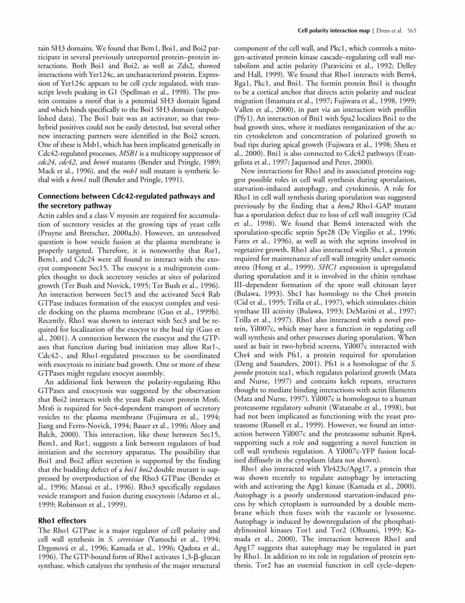
Cell polarity interaction map | Drees et al. 563
tain SH3 domains. We found that Bem1, Boi1, and Boi2 par-ticipate in several previously unreported protein–protein in-teractions. Both Boi1 and Boi2, as well as Zds2, showedinteractions with Yer124c, an uncharacterized protein. Expres-sion of Yer124c appears to be cell cycle regulated, with tran-script levels peaking in G1 (Spellman et al., 1998). The pro-tein contains a motif that is a potential SH3 domain ligandand which binds specifically to the Boi1 SH3 domain (unpub-lished data). The Boi1 bait was an activator, so that two-hybrid positives could not be easily detected, but several othernew interacting partners were identified in the Boi2 screen.One of these is Msb1, which has been implicated genetically inCdc42-regulated processes. MSB1 is a multicopy suppressor ofcdc24, cdc42, and bem4 mutants (Bender and Pringle, 1989;Mack et al., 1996), and the msb1 null mutant is synthetic le-thal with a bem1 null (Bender and Pringle, 1991).
Connections between Cdc42-regulated pathways and the secretory pathwayActin cables and a class V myosin are required for accumula-tion of secretory vesicles at the growing tips of yeast cells(Pruyne and Bretscher, 2000a,b). However, an unresolvedquestion is how vesicle fusion at the plasma membrane isproperly targeted. Therefore, it is noteworthy that Rsr1,Bem1, and Cdc24 were all found to interact with the exo-cyst component Sec15. The exocyst is a multiprotein com-plex thought to dock secretory vesicles at sites of polarizedgrowth (Ter Bush and Novick, 1995; Ter Bush et al., 1996).An interaction between Sec15 and the activated Sec4 RabGTPase induces formation of the exocyst complex and vesi-cle docking on the plasma membrane (Guo et al., 1999b).Recently, Rho1 was shown to interact with Sec3 and be re-quired for localization of the exocyst to the bud tip (Guo etal., 2001). A connection between the exocyst and the GTP-ases that function during bud initiation may allow Rsr1-,Cdc42-, and Rho1-regulated processes to be coordinatedwith exocytosis to initiate bud growth. One or more of theseGTPases might regulate exocyst assembly.
An additional link between the polarity-regulating RhoGTPases and exocytosis was suggested by the observationthat Boi2 interacts with the yeast Rab escort protein Mrs6.Mrs6 is required for Sec4-dependent transport of secretoryvesicles to the plasma membrane (Fujimura et al., 1994;Jiang and Ferro-Novick, 1994; Bauer et al., 1996; Alory andBalch, 2000). This interaction, like those between Sec15,Bem1, and Rsr1, suggests a link between regulators of budinitiation and the secretory apparatus. The possibility thatBoi1 and Boi2 affect secretion is supported by the findingthat the budding defect of a boi1 boi2 double mutant is sup-pressed by overproduction of the Rho3 GTPase (Bender etal., 1996; Matsui et al., 1996). Rho3 specifically regulatesvesicle transport and fusion during exocytosis (Adamo et al.,1999; Robinson et al., 1999).
Rho1 effectorsThe Rho1 GTPase is a major regulator of cell polarity andcell wall synthesis in S. cerevisiae (Yamochi et al., 1994;Drgonová et al., 1996; Kamada et al., 1996; Qadota et al.,1996). The GTP-bound form of Rho1 activates 1,3-�-glucansynthase, which catalyzes the synthesis of the major structural
component of the cell wall, and Pkc1, which controls a mito-gen-activated protein kinase cascade–regulating cell wall me-tabolism and actin polarity (Paravicini et al., 1992; Delleyand Hall, 1999). We found that Rho1 interacts with Bem4,Rga1, Pkc1, and Bni1. The formin protein Bni1 is thoughtto be a cortical anchor that directs actin polarity and nuclearmigration (Imamura et al., 1997; Fujiwara et al., 1998, 1999;Vallen et al., 2000), in part via an interaction with profilin(Pfy1). An interaction of Bni1 with Spa2 localizes Bni1 to thebud growth sites, where it mediates reorganization of the ac-tin cytoskeleton and concentration of polarized growth tobud tips during apical growth (Fujiwara et al., 1998; Sheu etal., 2000). Bni1 is also connected to Cdc42 pathways (Evan-gelista et al., 1997; Jaquenod and Peter, 2000).
New interactions for Rho1 and its associated proteins sug-gest possible roles in cell wall synthesis during sporulation,starvation-induced autophagy, and cytokinesis. A role forRho1 in cell wall synthesis during sporulation was suggestedpreviously by the finding that a bem2 Rho1-GAP mutanthas a sporulation defect due to loss of cell wall integrity (Cidet al., 1998). We found that Bem4 interacted with thesporulation-specific septin Spr28 (De Virgilio et al., 1996;Fares et al., 1996), as well as with the septins involved invegetative growth. Rho1 also interacted with Shc1, a proteinrequired for maintenance of cell wall integrity under osmoticstress (Hong et al., 1999). SHC1 expression is upregulatedduring sporulation and it is involved in the chitin synthaseIII–dependent formation of the spore wall chitosan layer(Bulawa, 1993). Shc1 has homology to the Chs4 protein(Cid et al., 1995; Trilla et al., 1997), which stimulates chitinsynthase III activity (Bulawa, 1993; DeMarini et al., 1997;Trilla et al., 1997). Rho1 also interacted with a novel pro-tein, Yil007c, which may have a function in regulating cellwall synthesis and other processes during sporulation. Whenused as bait in two-hybrid screens, Yil007c interacted withChs4 and with Pfs1, a protein required for sporulation(Deng and Saunders, 2001). Pfs1 is a homologue of the S.pombe protein tea1, which regulates polarized growth (Mataand Nurse, 1997) and contains kelch repeats, structuresthought to mediate binding interactions with actin filaments(Mata and Nurse, 1997). Yil007c is homologous to a humanproteasome regulatory subunit (Watanabe et al., 1998), buthad not been implicated as functioning with the yeast pro-teasome (Russell et al., 1999). However, we found an inter-action between Yil007c and the proteasome subunit Rpn4,supporting such a role and suggesting a novel function incell wall synthesis regulation. A Yil007c-YFP fusion local-ized diffusely in the cytoplasm (data not shown).
Rho1 also interacted with Ylr423c/Apg17, a protein thatwas shown recently to regulate autophagy by interactingwith and activating the Apg1 kinase (Kamada et al., 2000).Autophagy is a poorly understood starvation-induced pro-cess by which cytoplasm is surrounded by a double mem-brane which then fuses with the vacuole or lysosome.Autophagy is induced by downregulation of the phosphati-dylinositol kinases Tor1 and Tor2 (Ohsumi, 1999; Ka-mada et al., 2000). The interaction between Rho1 andApg17 suggests that autophagy may be regulated in partby Rho1. In addition to its role in regulation of protein syn-thesis, Tor2 has an essential function in cell cycle–depen-

564 The Journal of Cell Biology | Volume 154, 2001
dent organization of the actin cytoskeleton (Helliwell et al.,1998). Overexpression of PKC1, RHO1, or ROM2, a genethat encodes a Rho1 guanidine nucleotide exchange factor,suppresses the actin organization defect of a tor2 mutant.Perhaps Apg17 acts as an effector of Rho1 in a Tor2-depen-dent pathway that modulates cell polarity and autophagy.
Apg17 interacted with several proteins in addition toRho1, including Rho2, Myo1, Nip100, Exo84, and Sro77,and it had been reported previously to make additional twohybrid interactions (Ito et al., 2001). Interactions withExo84 and Sro77 suggest a connection to exocytosis. Exo84is an exocyst component (Guo et al., 1999a), and Sro77and its homologue Sro7 regulate vesicle docking and mem-brane fusion at the plasma membrane (Lehman et al.,1999). Interactions with Nip100 and Myo1 suggest thatApg17 may have a role in cytokinesis or that Myo1 andNip100 have roles in autophagy. Nip100 is a yeast dynactincomponent involved in nuclear division and migration (Ka-hana et al., 1998; Fujiwara et al., 1999). Myo1 is a type IImyosin that functions in the contractile ring (Bi et al.,1998; Lippincott and Li, 1998b). Sro77 and Sro7 havebeen found to form a complex with Myo1 (Kagami et al.,1998). The Drosophila homologue of Sro77 and Sro7, thelethal(2) giant larvae gene product, also interacts with myo-sins (Strand et al., 1994). During cytokinesis, targeted exo-cytosis at the site of cell division is coordinated with, andpossibly guided by, contraction of the actinomyosin ring(Hales et al., 1999; Vallen et al., 2000). Perhaps a Myo1–Sro77 complex couples septum formation to contraction ofthe actomyosin ring, with Myo1 playing a specialized rolein vesicle targeting to the bud neck (Schott et al., 1999). In-teractions between Apg17 and Nip100, Myo1, Sro77, andExo84 might be part of a mechanism coordinating nuclearmigration, actomyosin ring contraction, and exocytosis dur-ing cytokinesis or autophagy (Kahana et al., 1998; Fuji-wara et al., 1999; Hales et al., 1999). YFP-tagged Ylr423c/Apg17 localized in punctate patches in the cytoplasm (Fig.4, I and J). Perhaps these patches play a role in autophago-cytic vesicle formation.
Connections between Rho1 and Cdc42 pathwaysAs discussed above, Ygr221c interacts with both Cdc24 andthe Rho1 effector Pkc1. This interaction is quite interesting asa potential means for Pkc1 to regulate Cdc42 function andcell polarity. Cell wall stress induces hyperactivation of Rho1,which in turn results in a transient loss of actin polarity in or-der to depolarize cell wall synthesis and repair widespread cellwall damage (Delley and Hall, 1999). An undefined Pkc1-dependent pathway controls actin depolarization. Depolariza-tion is dependent on Pkc1 but not on the Pkc1-activated mi-togen-activated protein kinase cascade, which is necessary forrepolarization. The interactions between Pkc1, Ygr221c, andCdc24, together with the proposed function of Ygr221c as aGTPase regulatory protein (Rieger et al., 1999), suggest thatPkc1 could affect the actin cytoskeleton by inhibiting Cdc42function through an interaction with Ygr221c. Zds1 andZds2 might also monitor and regulate Cdc42 in response toRho1. Zds2 interacts with both Rho1 and the Rho1 effectorsPkc1 and Bni1, as well as with several proteins in Cdc42-regu-lated pathways. Interestingly, when Yhr149c, the homologue
of the Cdc24-interacting protein Ygr221c, was used as bait ina two-hybrid screen, Zds1 and Zds2 were both found to inter-act with Yhr149c. These interactions suggest that Yhr149cmay also be involved in coordinating Rho1- and Cdc42-regu-lated pathways.
Actin cortical patch assembly, the morphogenesis checkpoint, and endocytosisActin cortical patches are one of the major cytoskeletal struc-tures in yeast and are essential for normal endocytosis, cellgrowth, and morphology (Botstein et al., 1997; Pruyne andBretscher, 2000b). Patches are associated with invaginationsof the plasma membrane (Mulholland et al., 1994) and arefound in polarized clusters at regions of cell growth in bud-ding cells. Numerous cortical patch proteins have been iden-tified. How these proteins function in patch assembly andendocytosis are largely unknown. Patch assembly probablybegins with the association of assembly factors recruited tothe plasma membrane by Cdc42-associated proteins, and isthen followed by nucleation of actin filaments and actin-dependent association of proteins regulating filament assemblyand stability (Ayscough et al., 1997; Botstein et al., 1997;Pruyne and Bretscher, 2000b). Our results confirm severalinteractions between patch proteins, identify new interac-tions, and suggest roles for several uncharacterized proteinsin patch assembly or patch-mediated endocytosis (Fig. 3).
Sla2 (related to mammalian Hip1), Sla1, Las17/Bee1 (re-lated to mammalian WASp/SCAR), Rvs167 (related tomammalian amphiphysin), and Abp1 (related to mamma-lian Abp1) all function in actin nucleation and assembly(Holtzman et al., 1993; Amberg et al., 1995; Li et al., 1995;Li, 1997; Lila and Drubin, 1997; Wesp et al., 1997; Ay-scough et al., 1999; Balguerie et al., 1999; Yang et al., 1999;Goode et al., 2001). Las17/Bee1 and Abp1 both localizewith and activate the Arp2/3 complex to nucleate actin fila-ment assembly (Madania et al., 1999; Winter et al., 1999;Goode et al., 2001). Sla1, Rvs167 and Abp1 might sharefunctions in actin cortical patch assembly because they inter-act with overlapping sets of proteins and show synthetic–lethal genetic interactions (Holtzman et al., 1993; Lila andDrubin, 1997). The actin monomer binding adenylate cy-clase regulatory subunit Srv2 can, for example, apparentlyinteract with all three proteins to mediate the association ofmonomeric actin with the actin-nucleating complex (Free-man et al., 1996; Lila and Drubin, 1997).
Sla1, Rvs167, and Abp1 contain SH3 domains that areimportant for their function (Lila and Drubin, 1997; Ay-scough et al., 1999), and several of the proteins with whichthey interact contain proline-rich regions that are potentialSH3 domain binding sites. Sla1 and Rvs167 both showedinteractions with Las17/Bee1 (Li, 1997) and Ysc84 (Mada-nia et al., 1999). Ysc84 itself contains an SH3 domain thatmight make other protein contacts. Two proteins of un-known function, Ygr268c and Ypr171w, also interactedwith Sla1 and Rvs167. As noted in Fig. 3, Ygr268c andYpr171w contain proline-rich motifs that are potential SH3domain binding sites. Ypr171w also interacted with Cap1,the � subunit of the yeast actin filament capping protein,which regulates the growth of actin filaments (Amatrudaand Cooper, 1992; Amatruda et al., 1992). Thus, Ypr171w

Cell polarity interaction map | Drees et al. 565
may function as a link between actin-nucleating complexesand the actin-capping protein, perhaps coordinating thenucleation and elongation of actin filaments. YFP-taggedYpr171w was observed at growth sites in the bud in a punc-tate pattern and at the bud neck of large-budded cells (Fig.4, K and L). Coexpression of Ypr171w-YFP with CFP-tagged Abp1, an actin cortical patch component, demon-strated that Ypr171w colocalizes with some, but perhaps notall, Abp1-containing actin cortical patches (Fig. 5, A–F).The punctate localization pattern is, like that of Abp1, dis-turbed in the ark1 prk1 double deletion strain (Fig. 5, G andH), which has abnormal actin clumps (Cope et al., 1999).However, Ypr171w localization to the bud neck is still seenin the ark1 prk1 mutant (Fig. 5 G). A Ygr268c-YFP fusionlocalized diffusely in the cytoplasm (data not shown).
Another uncharacterized protein, Yor284w, interactedwith cortical patch components Sla2, Rvs167, and Abp1.Sla2 localizes to sites of polarized growth independently ofactin (Ayscough et al., 1997) and mediates an early step inthe polarization of actin cortical patches (Yang et al., 1999).Rvs167 also affects patch polarization (Balguerie et al., 1999),perhaps via the interaction observed here with Sla2. Colocal-ization of Sla2 with actin is most visible in unbudded andsmall-budded cells, implying that its patch assembly activityis most important early in the cell cycle (Yang et al., 1999).Expression of Yor284w peaks in the G1 phase of the cell cy-cle (Spellman et al., 1998), supporting the idea that it plays arole in this process. The Yor284w-YFP fusion localized to afew distinct, mobile punctate structures in the cell (Figs. 4, Mand N and 6, A and B). In many cases these dots were ob-served moving rapidly around the cell periphery. The rela-tionship of these structures to cortical patches is unclear.Yor284w appears in a stationary, diffuse cytoplasmic clumpin the ark1 prk1 double deletion strain (Fig. 6, C and D),suggesting that its localization is dependent on actin polariza-tion. However, coexpression of Yor284w-YFP with CFP-tagged Spc29 spindle pole body protein (Fig. 6, E–J) andCFP-tagged Abp1 (Fig. 6, K–P) showed that the Yor284w-containing dots do not localize to cortical patches, but that asubset of them colocalize with the spindle pole body.
Four other proteins that interact with Rvs167 containproline-rich potential SH3 domain binding sites. One isAcf2, a protein implicated in cortical actin assembly (Lechlerand Li, 1997), whereas the others, Ybr108w, Yjr083c, andYnl094w, are proteins of unknown function. Ybr108w hasbeen found to interact with both Rvs167 and Rvs161 (Bonet al., 2000), a protein that forms a complex with Rvs167(Navarro et al., 1997). A Yjr083c-YFP fusion localized dif-fusely in the cytoplasm (data not shown).
Ynl094w showed interactions with five cortical patch pro-teins. It interacted with Sla2 and with three SH3 domainproteins involved in actin nucleation, Sla1, Rvs167, andAbp1. Ynl094w also interacted with the actin-bundlingprotein Crn1 (Goode et al., 1999) and with Sro77, a pro-tein which functions in polarized secretion (Kagami et al.,1998; Lehman et al., 1999). These interactions suggest thatYnl094w may link actin nucleation to exocytosis. Other in-teractions suggest that Ynl094w might function in the mor-phogenesis checkpoint response. Because this response ispoorly understood, two-hybrid interactions involving the
checkpoint proteins might provide important insights intothe mechanisms for monitoring the cytoskeleton. Ynl094winteracted with two proteins involved in regulation of themorphogenesis checkpoint, Swe1 and Hsl7 (Shulewitz et al.,1999). In addition to monitoring the actin cytoskeleton(McMillan et al., 1998; Lew, 2000), Swe1 may monitor sep-tin organization via an interaction with Hsl7 and Hsl1 at thebud neck (Barral et al., 1999; McMillan et al., 1999; Shule-witz et al., 1999; Lew, 2000; Longtine et al., 2000). The factthat Ynl094w showed interactions with Swe1 and Hsl7 andwith five cortical patch proteins suggests that Swe1 and Hsl7might monitor actin assembly via an interaction withYnl094w. Ynl094w has been found to localize to actin corti-cal patches (L. Tseng, M. Schulewitz, and J. Thorner, per-sonal communication).
Interactions of the secretory apparatusPolarized growth and budding require the delivery of pro-teins and lipids to specific sites on the plasma membrane.Under cell cycle control, exocytosis first becomes localized toregions of cell growth at the presumptive bud site. As a budemerges, exocytosis initially localizes to a small region at thebud tip, then becomes delocalized in the bud, and finallyit becomes localized to the bud neck, mirroring cortical ac-tin cytoskeleton organization at each stage (Pruyne andBretscher, 2000b). The mechanisms that insure this contin-ual coordination between the exocytic machinery and the ac-tin cytoskeleton have yet to be fully elucidated. The Rho3GTPase interacts with elements of the exocytic machinery tocontrol transport of secretory vesicles and vesicle docking andfusion at the plasma membrane. Vesicle transport is depen-dent on function of the class V myosin Myo2 (Schott et al.,1999). Vesicle fusion occurs through an interaction withExo70 (Adamo et al., 1999; Robinson et al., 1999), a compo-nent of the exocyst complex. Rho3 can also affect organiza-tion of the actin cytoskeleton (Imai et al., 1996). We foundthat Rho3 interacts with three subunits of casein kinase II.This observation is interesting because one function of caseinkinase II is to maintain actin cytoskeleton polarity (Rethi-naswamy et al., 1998). Our results suggest that casein kinaseII might therefore function as an effector of Rho3 to regulateactin cytoskeleton organization, and that Rho3 might coordi-nate secretory and actin cytoskeletal organization.
The vesicle-docking protein Sro77, homologous to theDrosophila protein lethal(2) giant larvae gene product,showed numerous interactions implicating this protein inregulatory, cytoskeletal, and endocytic roles (Table I). Asmentioned above, Sro77 showed interactions with Apg17and Nip100. It also showed interactions with the epsin Ent2and with Yap1801, both clathrin-binding proteins that func-tion during the internalization step of endocytosis (Chen etal., 1998; Wendland and Emr, 1998; Wendland et al.,1999). Sro77 and the related protein Sro7 might thereforealso function in the early steps of endocytosis, or they maycoordinate endocytosis and exocytosis. The v-SNAREs Snc1and Snc2 appear to be involved in both exocytic and en-docytic transport at the plasma membrane. Snc1 and Snc2can interact with endosomal t-SNAREs and snc mutants aredefective in endocytosis as well as exocytosis (Gurunathan etal., 2000). Perhaps the plasma membrane t-SNAREs and

566 The Journal of Cell Biology | Volume 154, 2001
their regulators, such as Sro7/Sro77, are also able to partici-pate in both processes. Similiarly, an interaction we observedbetween Sro77 and Yip1, which recruits the Ypt1 and Ypt31transport GTPases to the Golgi apparatus, suggests similarpossible relationships between proteins involved in early andlate steps of exocytosis and/or endocytosis.
Sro77 also showed an interaction with Bcy1, the regula-tory subunit of cAMP-dependent protein kinases (Cannonet al., 1990). In epithelial cells, exocytosis is stimulated bythe cAMP-dependent protein kinase PKA (Takuma, 1990;Koh et al., 2000), which appears to regulate SNARE com-plex formation (Foster et al., 1998). Perhaps SNARE assem-bly in S. cerevisiae is also regulated in response to cAMP-dependent protein kinase activity.
Further Sro77 interactions were observed with several un-characterized proteins, including Ynl094w, which may be alink between the secretory apparatus and actin organization(see above), and Kin3, a protein kinase of unknown function.
Sso1 and Sso2 are syntaxin homologue t-SNAREs that me-diate vesicle targeting to the plasma membrane during exocy-tosis (Aalto et al., 1993). Sso2 interacted with Apg7, a regula-tor of autophagy, revealing another possible connectionbetween exocytic and autophagocytic processes. Apg7 has ho-mology to the E1 family of ubiquitin-activating enzymes andmediates a novel protein conjugation reaction required forautophagy (Mizushima et al., 1998; Kim et al., 1999; Tanidaet al., 1999). Perhaps Apg7-mediated modification of plasmamembrane t-SNAREs recruits them for formation of auto-phagocytic vesicles or for processes related to exocytosis.
MSB3 and MSB4 are multicopy suppressors of bud emer-gence mutations and appear to link Cdc42 to the actin cyto-skeleton (Bach et al., 2000; Bi et al., 2000). Surprisingly, Msb3and Msb4 were also found to have GAP activity towards theRab GTPases Sec4 and Ypt6, respectively (Albert and Gall-witz, 1999, 2000). Therefore, Msb3 and Msb4 may have rolesin exocytosis. Sec4 is required for formation of the exocystcomplex and for vesicle docking at the plasma membrane(Walworth et al., 1992; Guo et al., 1999b), whereas Ypt6 is re-quired for vesicle transport from the endoplasmic reticulum tothe Golgi apparatus (Lupashin et al., 1996). In our screens,both Msb3 and Msb4 interacted with Spa2, a protein thatconcentrates polarized growth to bud and mating projectiontips during apical growth, and to the septum during cytokine-sis (Fujiwara et al., 1998; Sheu et al., 2000). Sec4 is partiallydelocalized in spa2 mutants (Sheu et al., 2000), and Spa2 is re-quired for normal localization of secretory vesicles to the cellfusion zone during mating (Gammie et al., 1998). These ge-netic observations and the interactions with Msb3 and Msb4provide support for a role for Spa2 in polarized exocytosis.
ConclusionsWe have reported here the results of a two-hybrid study ofproteins involved in cell polarity development, a highlycomplex process involving cortical cues, signaling proteins,the cytoskeleton, and the secretory apparatus, each of whichis itself characterized by considerable complexity. Elucida-tion of the mechanisms that underlie cell polarity develop-ment is a daunting task, due to the vast number of proteinsinvolved. The result of our studies is a protein interactionmap that helps define the scope of the problem of under-
standing cell polarity development and can be used to guidefurther genetic and biochemical studies. Because most of theproteins and processes included in this study are highly con-served, this map should prove useful for studies of polaritydevelopment in diverse cell types. Among interactions iden-tified in this map are some that implicate new proteins inpolarity development, and others that suggest modes for co-ordinating distinct processes involved in polarity develop-ment. Our localization of previously uncharacterized pro-teins implicated here in polarity development is only thefirst step in verification of the biological relevance of each in-teraction. Future studies must address when, where, andwhy each interaction occurs and how these interactions areregulated. Despite years of genetic analysis of cell polaritydevelopment in budding yeast, we were able to implicate un-characterized proteins in this process, and to suggest novelfunctions for proteins studied previously. Genetic studiesmay have missed much of this information, due to factorsincluding redundancy, lack of appropriate alleles, homeosta-sis mechanisms, and checkpoints that mask the underlyingmutant defect by arresting a process before an informativephenotype develops. In total, the large number of interac-tions that we identified among proteins involved in diverseaspects of cell polarity development suggests a high level ofintegration in the functioning of these proteins.
Materials and methodsGeneration of DNA binding domain hybridsTransformants containing Gal4 DNA binding domain hybrids were con-structed in the � mating type of the yeast strain PJ694 (MAT� trp1-901leu2-3,112 ura3-52 his3-200 gal4� gal80� LYS2::GAL1-HIS3 GAL2-ADE2met2::GAL7-lacZ) (James et al., 1996) as described (McCraith et al., 2000;Uetz et al., 2000). Recombination (Ma et al., 1987) of the linearized vectorpOBD2 (McCraith et al., 2000) with PCR fragments corresponding to eachof the yeast ORFs was used to generate the hybrids (Hudson et al., 1997).Transformation was carried out using the lithium acetate procedure (Ito etal., 1983). After transformation, cells were plated on synthetic trp media.Yeast media were prepared as described (Sherman et al., 1986). Cdc3,Cdc11, Cdc12, and Bud8 baits were made in the vector pGDBU and trans-formants selected on ura media (James et al., 1996).
Several GTPase baits contained cysteine-to-serine amino acid substitu-tions to prevent prenylation and facilitate nuclear entry, and other substitu-tions that affect GTP binding and hydrolysis to favor either the GTP- orGDP-bound form: Rho1-GTP (Q68H, C206S), Rho2-GTP (Q 65H, C188S),Rho3-GTP (G25V, C228S), Rho4-GTP (Q70H, C228S), Cdc42-GTP(G12V, C188S), Cdc42-GDP (D57Y, C188S), Rsr1-GTP (G12V), and Rsr1-GDP (K16N). Gic1 and Gic2 fusions were truncations lacking 30 residuesat the COOH terminus because the full-length protein was an activator.The Sso1(1–515), Sso2(1–266), and fusions were truncations that removedpredicted transmembrane domains at their COOH termini. The Msb2 fu-sion contained a truncation, Msb2(1186–1306) that removed predictedtransmembrane domains at the NH2 and COOH termini. The Bud8 andBud9 fusions also contained truncations, Bud8(534–578) and Bud9(480–521), that removed predicted transmembrane domains.
Two-hybrid screensScreens of the yeast ORF activation domain fusion array were performed ina manner similar to that described previously (McCraith et al., 2000; Uetzet al., 2000). This array was expressed in the a mating type of strain PJ694(MATa trp1-901 leu2-3,112 ura3-52 his3-200 gal4� gal80� LYS2::GAL1-HIS3 GAL2-ADE2 met2::GAL7-lacZ) (James et al., 1996). To screen forprotein–protein interactions, we mated a transformant containing one ofthe DNA binding domain hybrids to all of the transformants of the array,selecting diploids using markers carried on the two-hybrid plasmids. Thediploids were then transferred to selective plates deficient in histidine, andcolonies positive for the two-hybrid reporter HIS3 gene were identified bytheir positions in the array.

Cell polarity interaction map | Drees et al. 567
Two-hybrid screens can generate significant numbers of false positivesthat are not reproducible in duplicate screens. Random generation of histi-dine-positive colonies can result from overexpression of a fusion proteinthat affects transcription or cell metabolism, rearrangements or deletions ofthe DNA-binding domain plasmid, recombinational events between plas-mids, or genomic rearrangements of the host strain. To identify reproduc-ible two-hybrid interactions rapidly, we duplicated the array so that eachORF-Gal4 activation domain fusion is represented twice on a microassayplate. The entire array is contained in duplicate on 16 microassay plates of768 colonies each. Only protein combinations that resulted in histidineprototrophy for both duplicate colonies of a given activation domain fu-sion were scored as two-hybrid interactions. We estimate that 10–20% ofthe yeast ORF-Gal4 activation domain fusions are not expressed due to er-rors in the PCR amplification of the ORFs. In addition, there are likely to beother constructs that are expressed poorly or in a nonfunctional form dueto improper folding.
All pinning steps were carried out using a Biomek 2000 robot (BeckmanCoulter) and a 768-pin replicating tool (UW Scientific Instruments, Ma-chine/Optical Division). Strains containing the Gal4-DNA binding-domainhybrids were first tested on plates of synthetic his media with different con-centrations of 3-aminotriazole to determine the level of stringency neededto eliminate background activation of the HIS3 reporter gene. Strains weregrown overnight at 30�C in 25 ml of trp synthetic medium, centrifuged at1,000 rpm for 2 min and resuspended in 5–7 ml of medium. The cell sus-pension was transferred to 16 plates of solid YEPD medium. The yeast ORFactivation domain fusion array was then replica-pinned onto the plates.Plates were incubated at 30�C for 2–4 d to allow mating, and then cellswere replica-pinned to synthetic trp leu medium. After 2–4 d, cells werereplica-pinned onto his synthetic media containing the appropriate con-centration of 3-aminotriazole. Plates were incubated at 30�C for 7–10 dand then scored for two-hybrid positives.
Generation of ORFs tagged with CFP or YFPA PCR-based method was used to integrate a gene encoding either YFP orCFP at the 3� end of the targeted yeast ORF such that each fusion protein isexpressed under the control of its native promoter (Wach et al., 1997). De-tailed protocols are described at http://depts.washington.edu/~yeastrc/fm_home3.htm. The template for integration of YFP was the plasmidpDH6, which contains the YFP ORF followed by the kanr gene. pDH6was made by replacing the AvaI–AscI fragment encoding GFP inpFA6a-GFP(S65T)KanMX6 (Wach et al., 1997) with the AvaI–AscI frag-ment encoding YFP from pDH5. pDH5 was derived from pFA6a-GFP(S65T)HIS3MX6 by site-directed mutagenesis using the QuikChangemethod as described by the supplier (Stratagene). The mutations in YFP ascompared with the original GFP are: S65G, V68L, Q69K, S72A, Q80R,and T203Y. The template for integration of CFP was plasmid pDH3, whichcontains the CFP ORF followed by the kanr gene. pDH3 was derivedfrom pFA6a-GFP(S65T)KanMX6 by site-directed mutagenesis using theQuikChange method. The mutations in CFP as compared with the originalGFP are: F64L, S65T, Y66W, Q80R, N146I, M153T, V163A, and N164H.Integrations were checked by PCR.
Fluorescence microscopyCells containing YFP or CFP fusion proteins were grown on solid YPD me-dium overnight at 30�C and then resuspended in PBS containing 1.3 �g/mlconcanavalin A tagged with Alexa 633 (Molecular Probes) and incubatedfor 30 min at 30�C. Cells were washed twice with PBS and resuspended inS medium or SD complete medium (1.7 g/liter Difco yeast nitrogen basewithout amino acids or ammonium sulfate, 5 g/liter ammonium sulfate,0.1% casamino acids, 25 �g/ml uracil, 50 �g/ml adenine, 100 �g/ml tryp-tophan, 2% glucose). Cells were mounted in one of two ways. An aliquotof cells (3 �l) was mixed on the slide with an equal volume of SD com-plete medium containing 1.2% SeaPlaque low melting temperature aga-rose (FMC BioProducts) at 40�C. A coverslip (No. 1.5) was quickly addedand pressed firmly. Alternatively, a cushion of 1.2% SeaKem LE agarose(FMC BioProducts) in SD complete medium was poured into 0.5-mm con-cavity slides (PGC Scientific) and pressed flat with another slide. Once theagar was solidified, an aliquot (15 �l) of cells was pipetted onto the cush-ion and covered with a coverslip (No. 1.5). The latter method is preferablefor examination of actin cytoskeletal components.
Fluorescence microscopy was performed on a DeltaVision microscopewith a Photometrics Quantix camera. The filter sets were from Omega. Theimages were sharpened by two dimensional deconvolution using the Soft-WorX software (Applied Precision, Inc.).
To localize Yhr149c, the COOH terminus was tagged with a 13Mycepitope using the PCR-based method of Longtine et al. (1998) in the strain
DDY1102 (MATa/MAT� ade2-1/ADE2 his3�200/his3�200 leu2-3,112/leu2-3,112 ura3-52/ura3-52 LYS2/lys2-801) (Kozminski et al., 2000). Logphase cells were processed for indirect immunofluorescence microscopyand images were acquired as described by Kozminski et al. (2000). Mouseanti-Myc antibody (9E10; Santa Cruz Biotechnology, Inc.) and FITC-conju-gated donkey anti–mouse antibody (Jackson ImmunoResearch Laborato-ries) were diluted 1:50 and 1:100, respectively.
Thanks to Daniel Lew, Avi Rodal, Eric Weiss, and Cammie Lesser for dis-cussion of the data. Sharat Gadde helped with plasmid construction.
S. Fields and T.N. Davis were supported by the National Center for Re-search Resources grant PHS P41 RR11823. S. Fields is an investigator of theHoward Hughes Medical Institute. D. Drubin was supported by the Na-tional Institutes of Health R01 grant GM50399. E. Brazeau was supportedby National Institutes of Health R01 grant GM59216. P. Brennwald wassupported by National Institutes of Health R01 grant GM54712. J.R. Pringlewas supported by National Institutes of Health R01 grant GM31006. K.G.Kozminski was funded by a fellowship from the Leukemia & LymphomaSociety. J.J. Moskow was supported by fellowship PF-98-008 from theAmerican Cancer Society. C. Chan was funded by the Texas Higher Educa-tion Coordinating Board, grant ARP-427. P. Novick was supported by Na-tional Institutes of Health R01 grant GM35370. C. Boone was supported bythe Natural Sciences and Engineering Council of Canada and by the Na-tional Cancer Institute of Canada. M. Longtine was supported by the Okla-homa Center for Advancement of Scientific Technology, grant HR00-32.
Submitted: 13 April 2001Revised: 18 June 2001Accepted: 3 July 2001
ReferencesAalto, M.K., H. Ronne, and S. Keranen. 1993. Yeast syntaxins Sso1p and Sso2p
belong to a family of related membrane proteins that function in vesiculartransport. EMBO J. 12:4095–4104.
Adamo, J.E., G. Rossi, and P. Brennwald. 1999. The Rho GTPase Rho3 has a di-rect role in exocytosis that is distinct from its role in actin polarity. Mol. Biol.Cell. 10:4121–4133.
Adams, A.E.M., D.I. Johnson, R.M. Longnecker, B.F. Sloat, and J.R. Pringle.1990. CDC42 and CDC43, two additional genes involved in budding andthe establishment of cell polarity in the yeast Saccharomyces cerevisiae. J. CellBiol. 111:131–142.
Albert, S., and D. Gallwitz. 1999. Two new members of a family of Ypt/Rab GTP-ase activating proteins. Promiscuity of substrate recognition. J. Biol. Chem.274:33186–33189.
Albert, S., and D. Gallwitz. 2000. Msb4p, a protein involved in Cdc42p-depen-dent organization of the actin cytoskeleton, is a Ypt/Rab-specific GAP. Biol.Chem. 381:453–456.
Alory, C., and W.E. Balch. 2000. Molecular basis for Rab prenylation. J. Cell Biol.150:89–103.
Amatruda, J.F., and J.A. Cooper. 1992. Purification, characterization, and immu-nofluorescence localization of Saccharomyces cerevisiae capping protein. J.Cell Biol. 117:1067–1076.
Amatruda, J.F., D.J. Gattermeir, T.S. Karpova, and J.A. Cooper. 1992. Effects ofnull mutations and overexpression of capping protein on morphogenesis, ac-tin distribution and polarized secretion in yeast. J. Cell Biol. 119:1151–1162.
Amberg, D.C., E. Basart, and D. Botstein. 1995. Defining protein interactionswith yeast actin in vivo. Nat. Struct. Biol. 2:28–35.
Ayscough, K.R., J. Stryker, N. Pokala, M. Sanders, P. Crews, and D.G. Drubin.1997. High rates of actin filament turnover in budding yeast and roles foractin in establishment and maintenance of cell polarity revealed using the ac-tin inhibitor latrunculin-A. J. Cell Biol. 137:399–416.
Ayscough, K.R., J.J. Eby, T. Lila, H. Dewar, K.G. Kozminski, and D.G. Drubin.1999. Sla1p is a functionally modular component of the yeast cortical actincytoskeleton required for correct localization of both Rho1p-GTPase andSla2p, a protein with talin homology. Mol. Biol. Cell. 10:1061–1075.
Bach, S., O. Bouchat, D. Portetelle, and M. Vandenbol. 2000. Co-deletion of theMSB3 and MSB4 coding regions affects bipolar budding and perturbs theorganization of the actin cytoskeleton. Yeast. 16:1015–1023.
Balguerie, A., P. Sivadon, M. Bonneu, and M. Aigle. 1999. Rvs167p, the buddingyeast homolog of amphiphysin, colocalizes with actin patches. J. Cell Sci.112:2529–2537.

568 The Journal of Cell Biology | Volume 154, 2001
Barral, Y., M. Parra, S. Bidlingmaier, and M. Snyder. 1999. Nim1-related kinasescoordinate cell cycle progression with the organization of the peripheral cy-toskeleton in yeast. Genes Dev. 13:176–187.
Bauer, B.E., S. Lorenzetti, M. Miaczynska, D.M. Bui, R.J. Schweyen, and A. Rag-nini. 1996. Amino- and carboxy-terminal domains of the yeast Rab escortprotein are both required for binding of Ypt small G proteins. Mol. Biol.Cell. 7:1521–1533.
Bender, A. 1993. Genetic evidence for the roles of the bud-site-selection genesBUD5 and BUD2 in control of the Rsr1p (Bud1p) GTPase in yeast. Proc.Natl. Acad. Sci. USA. 90:9926–9929.
Bender, A., and J.R. Pringle. 1989. Multicopy suppression of the cdc24 buddingdefect in yeast by CDC42 and three newly identified genes including the ras-related gene RSR1. Proc. Natl. Acad. Sci. USA. 86:9976–9980.
Bender, A., and J.R. Pringle. 1991. Use of a screen for synthetic lethal and multi-copy suppressee mutants to identify two new genes involved in morphogen-esis in Saccharomyces cerevisiae. Mol. Cell. Biol. 11:1295–1305.
Bender, A., and J.R. Pringle. 1992. A Ser/Thr-rich multicopy suppressor of a cdc24bud emergence defect. Yeast. 8:315–323.
Bender, L., H.S. Lo, H. Lee, V. Kokojan, J. Peterson, and A. Bender. 1996. Associ-ations among PH and SH3 domain-containing proteins and Rho-type GTP-ases in yeast. J. Cell Biol. 133:879–894.
Benton, B.K., A.H. Tinkelenberg, D. Jean, S.D. Plump, and F.R. Cross. 1993. Ge-netic analysis of Cln/Cdc28 regulation of cell morphogenesis in buddingyeast. EMBO J. 12:5267–5275.
Benton, B.K., A. Tinkelenberg, I. Gonzalez, and F.R. Cross. 1997. Cla4p, a Sac-charomyces cerevisiae Cdc42p-activated kinase involved in cytokinesis, is acti-vated at mitosis. Mol. Cell. Biol. 17:5067–5076.
Bi, E., and J.R. Pringle. 1996. ZDS1 and ZDS2, genes whose products may regu-late Cdc42p in Saccharomyces cerevisiae. Mol. Cell. Biol. 16:5264–5275.
Bi, E., P. Maddox, D.J. Lew, E.D. Salmon, J.N. McMillan, E. Yeh, and J.R. Prin-gle. 1998. Involvement of an actomyosin contractile ring in Saccharomycescerevisiae cytokinesis. J. Cell Biol. 142:1301–1312.
Bi, E., J.B. Chiavetta, H. Chen, G.-C. Chen, C.S.M. Chan, and J.R. Pringle.2000. Identification of novel, evolutionarily conserved Cdc42p-interactingproteins and of redundant pathways linking Cdc24p and Cdc42p to actinpolarization in yeast. Mol. Biol. Cell. 11:773–793.
Bon, E., P. Recordon-Navarro, P. Durrens, M. Iwase, A. Toh-e, and M. Aigle.2000. A network of proteins around Rvs167p and Rvs161p, two proteins re-lated to the yeast actin cytoskeleton. Yeast. 16:1229–1241.
Botstein, D., D. Amberg, J. Mulholland, T. Huffaker, A. Adams, D. Drubin, andT. Stearns. 1997. The yeast cytoskeleton. In The Molecular and Cellular Bi-ology of the Yeast Saccharomyces: Cell Cycle and Cell Biology. J.R. Pringle,J.R. Broach, and E.W. Jones, editors. Cold Spring Harbor Laboratory Press,Cold Spring Harbor, NY. 1–90.
Brewster, J.L., and M.C. Gustin. 1994. Positioning of cell growth and division af-ter osmotic stress requires a MAP kinase pathway. Yeast. 10:425–439.
Brown, J.L., M. Jaquenoud, M.-P. Gulli, J. Chant, and M. Peter. 1997. NovelCdc42-binding proteins Gic1 and Gic2 control cell polarity in yeast. GenesDev. 11:2972–2982.
Bulawa, C.E. 1993. Genetics and molecular biology of chitin synthesis in fungi.Annu. Rev. Microbiol. 47:505–534.
Butty, A.-C., P.M. Pryciak, L.S. Huang, I. Herskowitz, and M. Peter. 1998. Therole of Far1p in linking the heterotrimeric G protein to polarity establish-ment proteins during yeast mating. Science. 282:1511–1516.
Cannon, J.F., R. Gitan, and K. Tatchell. 1990. Yeast cAMP-dependent protein ki-nase regulatory subunit mutations display a variety of phenotypes. J. Biol.Chem. 265:11897–11904.
Chant, J. 1999. Cell polarity in yeast. Annu. Rev. Cell Dev. Biol. 15:365–391.Chen, G.-C., Y.-J. Kim, and C.S.M. Chan. 1997. The Cdc42 GTPase-associated
proteins Gic1 and Gic2 are required for polarized growth in Saccharomycescerevisiae. Genes Dev. 15:2958–2971.
Chen, H., S. Fre, V. Slepnev, M. Capua, K. Takei, M. Butler, P. Di Fiore, and P.De Camilli. 1998. Epsin is an EH-domain-binding protein implicated inclathrin-mediated endocytosis. Nature. 394:793–797.
Cid, V.J., A. Durán, F. del Rey, M.P. Snyder, C. Nombela, and M. Sánchez. 1995.Molecular basis of cell integrity and morphogenesis in Saccharomyces cerevi-siae. Microbiol. Rev. 59:345–386.
Cid, V.J., R. Cenamor, M. Sánchez, and C. Nombela. 1998. A mutation in theRho1-GAP-encoding gene BEM2 of Saccharomyces cerevisiae affects morpho-genesis and cell wall functionality. Microbiology. 144:25–36.
Cope, M.J.T.V., S. Yang, C. Shang, and D.G. Drubin. 1999. Novel protein ki-nases Ark1p and Prk1p associate with and regulate the cortical actin cyto-
skeleton in budding yeast. J. Cell Biol. 144:1203–1218.Cullen, P.J., J. Schultz, J. Horecka, B.J. Stevenson, Y. Jigami, and G.F. Sprague, Jr.
2000. Defects in protein glycosylation cause SHO1-dependent activation ofa STE12 signaling pathway in yeast. Genetics. 155:1005–1018.
Cvrcková, F., and K. Nasmyth. 1993. Yeast G1 cyclins CLN1 and CLN2 and aGAP-like protein have a role in bud formation. EMBO J. 12:5277–5286.
Cvrcková, F., C. De Virgilio, E. Manser, J.R. Pringle, and K. Nasmyth. 1995.Ste20-like protein kinases are required for normal localization of cell growthand for cytokinesis in budding yeast. Genes Dev. 9:1817–1830.
De Virgilio, C., D.J. DeMarini, and J.R. Pringle. 1996. SPR28, a sixth member ofthe septin gene family in Saccharomyces cerevisiae that is expressed specificallyin sporulating cells. Microbiology. 142:2897–2905.
Delley, P.-A., and M.N. Hall. 1999. Cell wall stress depolarizes cell growth via hy-peractivation of RHO1. J. Cell Biol. 147:163–174.
DeMarini, D.J., A.E.M. Adams, H. Fares, C. De Virgilio, G. Valle, J.S. Chuang,and J.R. Pringle. 1997. A septin-based hierarchy of proteins required for lo-calized deposition of chitin in the Saccharomyces cerevisiae cell wall. J. CellBiol. 139:75–93.
Deng, C., and W. Saunders. 2001. PFS1, a novel gene required for prospore mem-brane formation at selected spindle poles in Saccharomyces cerevisiae. Mol.Biol. Cell. In press.
Drgonová, J., T. Drgon, K. Tanaka, R. Kollár, G.-C. Chen, R.A. Ford, C.S.M.Chan, Y. Takai, and E. Cabib. 1996. Rho1p, a yeast protein at the interfacebetween cell polarization and morphogenesis. Science. 272:277–279.
Drubin, D.G., editor. 2000. Cell Polarity. Oxford University Press, Oxford. 313pp.
Drubin, D.G., and W.J. Nelson. 1996. Origins of cell polarity. Cell. 84:335–344.Eby, J.J., S.P. Holly, F. van Drogen, A.V. Grishin, M. Peter, D.G. Drubin, and
K.J. Blumer. 1998. Actin cytoskeleton organization regulated by the PAKfamily of protein kinases. Curr. Biol. 8:967–970.
Engqvist-Goldstein, Å.E., M.M. Kessels, V.S. Chopra, M.R. Hayden, and D.G.Drubin. 1999. An actin-binding protein of the Sla2/huntingtin interactingprotein 1 family is a novel component of clathrin-coated pits and vesicles. J.Cell Biol. 147:1503–1518.
Evangelista, M., K. Blundell, M.S. Longtine, C.J. Chow, N. Adames, J.R. Pringle,M. Peter, and C. Boone. 1997. Bni1p, a yeast formin linking Cdc42p andthe actin cytoskeleton during polarized morphogenesis. Science. 276:118–122.
Evangelista, M., B.M. Klebl, A.H.Y. Tong, B.A. Webb, T. Leeuw, E. Leberer, M.Whiteway, D.Y. Thomas, and C. Boone. 2000. A role for myosin-I in actinassembly through interactions with Vrp1p, Bee1p, and the Arp2/3 complex.J. Cell Biol. 148:353–362.
Fares, H., L. Goetsch, and J.R. Pringle. 1996. Identification of a developmentallyregulated septin and involvement of the septins in spore formation in Sac-charomyces cerevisiae. J. Cell Biol. 132:399–411.
Fields, S., and O. Song. 1989. A novel genetic system to detect protein-protein in-teractions. Nature. 340:245–246.
Foster, L., B. Yeung, M. Mohtashami, K. Ross, W.S. Trimble, and A. Klip. 1998.Binary interactions of the SNARE proteins syntaxin-4, SNAP23, andVAMP-2 and their regulation by phosphorylation. Biochemistry. 37:11089–11096.
Freeman, N.L., T. Lila, K.A. Mintzer, Z. Chen, A.J. Pahk, R. Ren, D.G. Drubin,and J. Field. 1996. A conserved proline-rich region of the Saccharomycescerevisiae cyclase-associated protein binds SH3 domains and modulates cyto-skeletal localization. Mol. Cell. Biol. 16:548–556.
Fujimura, K., K. Tanaka, A. Nakano, and A. Toh-e. 1994. The Saccharomyces cere-visiae MSI4 gene encodes the yeast counterpart of component A of Rab ger-anylgeranyltransferase. J. Biol. Chem. 269:9205–9212.
Fujiwara, T., K. Tanaka, A. Mino, M. Kikyo, K. Takahashi, K. Shimizu, and Y.Takai. 1998. Rho1p-Bni1p-Spa2p interactions: implication in localizationof Bni1p at the bud site and regulation of the actin cytoskeleton in Saccharo-myces cerevisiae. Mol. Biol. Cell. 9:1221–1233.
Fujiwara, T., K. Tanaka, E. Inoue, M. Kikyo, and Y. Takai. 1999. Bni1p regulatesmicrotubule-dependent nuclear migration through the actin cytoskeleton inSaccharomyces cerevisiae. Mol. Cell. Biol. 19:8016–8027.
Gammie, A.E., V. Brizzio, and M.D. Rose. 1998. Distinct morphological pheno-types of cell fusion mutants. Mol. Biol. Cell. 9:1395–1410.
Goode, B.L., J.J. Wong, A.-C. Butty, M. Peter, A.L. McCormack, J.R Yates, D.G.Drubin, and G. Barnes. 1999. Coronin promotes the rapid assembly andcross-linking of actin filaments and may link the actin and microtubule cy-toskeletons in yeast. J. Cell Biol. 144:83–98.
Goode, B.L., A.A. Rodal, G. Barnes, and D.G. Drubin. 2001. Activation of the

Cell polarity interaction map | Drees et al. 569
Arp2/3 complex by the actin filament binding protein Abp1p. J. Cell Biol.153:627–634.
Guo, W., A. Grant, and P. Novick. 1999a. Exo84p is an exocyst protein essentialfor secretion. J. Biol. Chem. 274:23558–23564.
Guo, W., D. Roth, C. Walch-Solimena, and P. Novick. 1999b. The exocyst is aneffector for Sec4p, targeting secretory vesicles to sites of exocytosis. EMBO J.18:1071–1080.
Guo, W., F. Tamanoi, and P. Novick. 2001. Spatial regulation of the exocyst com-plex by Rho1 GTPase. Nat. Cell Biol. 3:353–360.
Gurunathan, S., D. Chapman-Shimshoni, S. Trajkovic, and J.E. Gerst. 2000.Yeast exocytic v-SNAREs confer endocytosis. Mol. Biol. Cell. 11:3629–3643.
Hales, K.G., E. Bi, J.-Q. Wu, J.C. Adam, I.-C. Yu, and J.R. Pringle. 1999. Cyto-kinesis: an emerging unified theory for eukaryotes? Curr. Opin. Cell Biol. 11:717–725.
Harkins, H.A., N. Pagé, L.R. Schenkman, C. De Virgilio, S. Shaw, H. Bussey, andJ.R. Pringle. 2001. Bud8p and Bud9p, proteins that may mark the sites forbipolar budding in yeast. Mol. Biol. Cell. 12:2497–2518.
Helliwell, S.B., I. Howald, N. Barbet, and M.N. Hall. 1998. TOR2 is part of tworelated signaling pathways coordinating cell growth in Saccharomyces cerevi-siae. Genetics. 148:99–112.
Hirano, H., K. Tanaka, K. Ozaki, H. Imamura, H. Kohno, T. Hihara, T. Kam-eyama, K. Hotta, M. Arisawa, T. Watanabe, H. Qadota, Y. Ohya, and Y.Takai. 1996. ROM7/BEM4 encodes a novel protein that interacts with theRho1p small GTP-binding protein in Saccharomyces cerevisiae. Mol. Cell.Biol. 16:4396–4403.
Holtzman, D.A., S. Yang, and D.G. Drubin. 1993. Synthetic-lethal interactionsidentify two novel genes, SLA1 and SLA2, that control membrane cytoskele-ton assembly in Saccharomyces cerevisiae. J. Cell Biol. 122:635–644.
Hong, S.K., S.B. Han, M. Snyder, and E.Y. Choi. 1999. SHC1, a high pH induc-ible gene required for growth at alkaline pH in Saccharomyces cerevisiae. Bio-chem. Biophys. Res. Commun. 255:116–122.
Hoyt, M.A., L. Totis, and B.T. Roberts. 1991. S. cerevisiae genes required for cellcycle arrest in response to loss of microtubule function. Cell. 66:507–517.
Hudson, J.R., Jr., E.P. Dawson, K.L. Rushing, C.H. Jackson, D. Lockshon, D.Conover, C. Lanciault, J.R. Harris, S.J. Simmons, R. Rothstein, and S.Fields. 1997. The complete set of predicted genes from Saccharomyces cerevi-siae in a readily usable form. Genome Res. 7:1169–1173.
Imai, J., A. Toh-e, and Y. Matsui. 1996. Genetic analysis of the Saccharomyces cere-visiae RHO3 gene, encoding a Rho-type small GTPase, provides evidence fora role in bud formation. Genetics. 142:359–369.
Imamura, H., K. Tanaka, T. Hihara, M. Umikawa, T. Kamei, K. Takahashi, T.Sasaki, and Y. Takai. 1997. Bni1p and Bnr1p: downstream targets of theRho family small G-proteins which interact with profilin and regulate actincytoskeleton in Saccharomyces cerevisiae. EMBO J. 16:2745–2755.
Inoue, K., T. Mizuno, K. Wada, and M. Hagiwara. 2000. Novel RING finger pro-teins, Air1p and Air2p, interact with Hmt1p and inhibit the arginine meth-ylation of Npl3p. J. Biol. Chem. 275:32793–32799.
Ito, H., Y. Fukuda, K. Murata, and A. Kimura. 1983. Transformation of intactyeast cells treated with alkali cations. J. Bacteriol. 153:163–168.
Ito, T., T. Chiba, R. Ozawa, M. Yoshida, M. Hattori, and Y. Sakaki. 2001. A com-prehensive two-hybrid analysis to explore the yeast protein interactome.Proc. Natl. Acad. Sci. USA. 98:4569–4574.
James, P., J. Halladay, and E.A. Craig. 1996. Genomic libraries and a host straindesigned for highly efficient two-hybrid selection in yeast. Genetics. 144:1425–1436.
Jaquenod, M., and M. Peter. 2000. Gic2p may link activated Cdc42p to compo-nents involved in actin polarization, including Bni1p and Bud6p (Aip3p).Mol. Cell. Biol. 20:6244–6258.
Johnson, D.I., and J.R. Pringle. 1990. Molecular characterization of CDC42, aSaccharomyces cerevisiae gene involved in the development of cell polarity. J.Cell Biol. 111:143–152.
Jiang, Y., and S. Ferro-Novick. 1994. Identification of yeast component A: recon-stitution of the geranylgeranyltransferase that modifies Ypt1p and Sec4p.Proc. Natl. Acad. Sci. USA. 91:4377–4381.
Kagami, M., A. Toh-e, and Y. Matsui. 1998. Sro7p, a Saccharomyces cerevisiaecounterpart of the tumor suppressor l(2)gl protein, is related to myosins infunction. Genetics. 149:1717–1727.
Kahana, J.A., G. Schlenstedt, D.M. Evanchuk, J.R. Geiser, M.A. Hoyt, and P.A.Silver. 1998. The yeast dynactin complex is involved in partitioning the mi-totic spindle between mother and daughter cells during anaphase B. Mol.Biol. Cell. 9:1741–1756.
Kallal, L.A., M. Bhattacharyya, S.N. Grove, R.F. Iannacone, T.A. Pugh, D.A. Pri-merano, and M.J. Clancy. 1990. Functional analysis of the sporulation-spe-cific SPR6 gene of Saccharomyces cerevisiae. Curr. Genet. 18:293–301.
Kamada, Y., H. Qadota, C.P. Python, Y. Anraku, Y. Ohya, and D.E. Levin. 1996.Activation of protein kinase C by Rho1 GTPase. J. Biol. Chem. 271:9193–9196.
Kamada, Y., T. Funakoshi, T. Shintani, K. Nagano, M. Ohsumi, and Y. Ohsumi.2000. Tor-mediated induction of autophagy via an Apg1 protein kinasecomplex. J. Cell Biol. 150:1507–1513.
Kamei, T., K. Tanaka, T. Hihara, M. Umikawa, H. Imamura, M. Kikyo, K.Ozaki, and Y. Takai. 1998. Interaction of Bnr1p with a novel Src homology3 domain-containing Hof1p. Implication in cytokinesis in Saccharomycescerevisiae. J. Biol. Chem. 273:28341–28345.
Kessels, M.M., Å.E. Engqvist-Goldstein, and D.G. Drubin. 2000. Association ofmouse actin-binding protein 1 (mAbp1/SH3P7), a Src kinase target, withdynamic regions of the cortical actin cytoskeleton in response to Rac GTP-ase activators. Mol. Cell. Biol. 11:393–412.
Kim, J., V.M. Dalton, K.P. Eggerton, S.V. Scott, and D.J. Klionsky. 1999. Apg7p/Cvt2p is required for the cytoplasm-to-vacuole targeting, macroautophagy,and peroxisome degradation pathways. Mol. Biol. Cell. 10:1337–1351.
Koh, D.S., M.W. Moody, T.D. Nguyen, and B. Hille. 2000. Regulation of exocy-tosis by protein kinases and Ca(2�) in pancreatic duct epithelial cells. J.Gen. Physiol. 116:507–520.
Kozminski, K.G., A.J. Chen, A.A. Rodal, and D.G. Drubin. 2000. Functions andfunctional domains of the GTPase Cdc42p. Mol. Biol. Cell. 11:339–354.
Kübler, E., and H. Riezman. 1993. Actin and fimbrin are required for the internal-ization step of endocytosis in yeast. EMBO J. 12:2855–2862.
Lechler, T., and R. Li. 1997. In vitro reconstitution of cortical actin assembly sitesin budding yeast. J. Cell Biol. 138:95–103.
Lechler, T., A. Shevchenko, and R. Li. 2000. Direct involvement of yeast type Imyosins in Cdc42-dependent actin polymerization. J. Cell Biol. 148:363–373.
Lehman, K., G. Rossi, J.E. Adamo, and P. Brennwald. 1999. Yeast homologues oftomosyn and lethal giant larvae function in exocytosis and are associatedwith the plasma membrane SNARE, Sec9. J. Cell Biol. 146:125–140.
Lew, D.J. 2000. Cell-cycle checkpoints that ensure coordination between nuclearand cytoplasmic events in Saccharomyces cerevisiae. Curr. Opin. Genet. Dev.10:47–53.
Li, R. 1997. Bee1, a yeast protein with homology to Wiscott-Aldrich syndromeprotein, is critical for the assembly of cortical actin cytoskeleton. J. Cell Biol.136:649–658.
Li, R., Y. Zheng, and D.G. Drubin. 1995. Regulation of cortical actin cytoskeletonassembly during polarized cell growth in budding yeast. J. Cell Biol. 128:599–615.
Lila, T., and D.G. Drubin. 1997. Evidence for physical and functional interactionsamong two Saccharomyces cerevisiae SH3 domain proteins, an adenylyl cy-clase-associated protein and the actin cytoskeleton. J. Cell Biol. 136:649–658.
Lippincott, J., and R. Li. 1998a. Dual function of Cyk2, a Cdc15/PSTPIP familyprotein, in regulating actomyosin ring dynamics and septin distribution. J.Cell Biol. 143:1947–1960.
Lippincott, J., and R. Li. 1998b. Sequential assembly of myosin II, an IQGAP-likeprotein, and filamentous actin to a ring structure involved in budding yeastcytokinesis. J. Cell Biol. 140:355–366.
Longtine, M.S., A. McKenzie III, D.J. DeMarini, N.G. Shah, A. Wach, A.Brachat, P. Phillipsen, and J.R. Pringle. 1998. Additional modules for versa-tile and economical PCR-based gene deletion and modification in Saccharo-myces cerevisiae. Yeast. 14:953–961.
Longtine, M.S., C.L. Theesfeld, J.N. McMillan, E. Weaver, J.R. Pringle, and D.J.Lew. 2000. Septin-dependent assembly of a cell cycle-regulatory module inSaccharomyces cerevisiae. Mol. Cell. Biol. 20:4049–4061.
Lupashin, V.V., S. Hamamoto, and R.W. Schekman. 1996. Biochemical require-ments for the targeting and fusion of ER-derived transport vesicles with pu-rified yeast Golgi membranes. J. Cell Biol. 132:277–289.
Ma, H., S. Kunes, P.J. Schatz, and D. Botstein. 1987. Plasmid construction by ho-mologous recombination in yeast. Gene. 58:201–216.
Mack, D., K. Nishimura, B.K. Dennehey, T. Arbogast, J. Parkinson, A. Toh-e,J.R. Pringle, A. Bender, and Y. Matsui. 1996. Identification of the budemergence gene BEM4 and its interactions with rho-type GTPases in Sac-charomyces cerevisiae. Mol. Cell. Biol. 16:4387–4395.
Madania, A., P. Dumoulin, S. Grava, H. Kitamoto, C. Schärer-Brodbeck, A. Sou-lard, V. Moreau, and B. Winsor. 1999. The Saccharomyces cerevisiae homo-

570 The Journal of Cell Biology | Volume 154, 2001
logue of human Wiskott-Aldrich syndrome protein Las17p interacts withthe Arp2/3 complex. Mol. Biol. Cell. 10:3521–3538.
Mata, J., and P. Nurse. 1997. tea1 and the microtubular cytoskeleton are impor-tant for generating global spatial order within the fission yeast cell. Cell. 89:939–949.
Matsui, Y., and A. Toh-e. 1992. Yeast RHO3 and RHO4 ras superfamily genes arenecessary for bud growth, and their defect is suppressed by a high dose ofbud formation genes CDC42 and BEM1. Mol. Cell. Biol. 12:5690–5699.
Matsui, Y., R. Matsui, R. Akada, and A. Toh-e. 1996. Yeast src homology region 3domain-binding proteins involved in bud formation. J. Cell Biol. 133:865–878.
McCraith, S., T. Holtzman, B. Moss, and S. Fields. 2000. Genome-wide analysisof vaccinia virus protein-protein interactions. Proc. Natl. Acad. Sci. USA. 97:4879–4884.
McMillan, J.N., R.A.L. Sia, and D.J. Lew. 1998. A morphogenesis checkpointmonitors the actin cytoskeleton in yeast. J. Cell Biol. 142:1487–1499.
McMillan, J.N., M.S. Longtine, R.A.L. Sia, C.L. Theesfeld, E.S.G. Bardes, J.R.Pringle, and D.J. Lew. 1999. The morphogenesis checkpoint in Saccharomy-ces cerevisiae: cell cycle control of Swe1p degradation by Hsl1p and Hsl7p.Mol. Cell. Biol. 19:6929–6939.
Miller, D.M., N.S. Desai, D.C. Hardin, D.W. Piston, G.H. Patterson, J. Fleenor,S. Xu, and A. Fire. 1999. Two-color GFP expression system for C. elegans.Biotechniques. 26:914–918.
Mizushima, N., T. Noda, T. Yoshimori, Y. Tanaka, T. Ishii, M.D. George, D.J.Klionsky, M. Ohsumi, and Y. Ohsumi. 1998. A protein conjugation systemessential for autophagy. Nature. 395:395–398.
Mösch, H.-U., and G.R. Fink. 1997. Dissection of filamentous growth by transpo-son mutagenesis in Saccharomyces cerevisiae. Genetics. 145:671–684.
Moskow, J.J., A.S. Gladfelter, R.E. Lamson, P.M. Pryciak, and D.J. Lew. 2000.Role of Cdc42p in pheromone-stimulated signal transduction in Saccharo-myces cerevisiae. Mol. Cell. Biol. 20:7559–7571.
Mulholland, J., D. Preuss, A. Moon, A. Wong, D. Drubin, and D. Botstein. 1994.Ultrastructure of the yeast actin cytoskeleton and its association with theplasma membrane. J. Cell Biol. 125:381–391.
Navarro, P., P. Durrens, and M. Aigle. 1997. Protein-protein interaction betweenthe RVS161 and RVS167 gene products of Saccharomyces cerevisiae. Biochim.Biophys. Acta. 1343:187–192.
Nern, A., and R.A. Arkowitz. 2000. Nucleocytoplasmic shuttling of the Cdc42pexchange factor Cdc24p. J. Cell Biol. 148:1115–1122.
Niedenthal, R.K., L. Riles, M. Johnston, and J.H. Hegemann. 1996. Green fluo-rescent protein as a marker for gene expression and subcellular localization inbudding yeast. Yeast. 12:773–786.
Ohsumi, Y. 1999. Molecular mechanism of autophagy in yeast, Saccharomyces cere-visiae. Philos. Trans. R. Soc. Lond. B. Biol. Sci. 354:1577–1581.
Paravicini, G., M. Cooper, L. Friedli, D.J. Smith, J.-L. Carpentier, L.S. Klig, andM.A. Payton. 1992. The osmotic integrity of the yeast cell requires a func-tional PKC1 gene product. Mol. Cell. Biol. 12:4896–4905.
Park, H.-O., J. Chant, and I. Herskowitz. 1993. BUD2 encodes a GTPase-activat-ing protein for Bud1/Rsr1 necessary for proper bud-site selection in yeast.Nature. 365:269–274.
Park, H.-O., A. Sanson, and I. Herskowitz. 1999. Localization of Bud2p, a GTP-ase-activating protein necessary for programming cell polarity in yeast to thepresumptive bud site. Genes Dev. 13:1912–1917.
Pereira, G., T. Hofken, J. Grindlay, C. Manson, and E. Schiebel. 2000. TheBub2p spindle checkpoint links nuclear migration with mitotic exit. Mol.Cell. 6:1–10.
Posas, F., E.A. Witten, and H. Saito. 1998. Requirement of STE50 for osmostress-induced activation of the STE11 mitogen-activated protein kinase in thehigh-osmolarity glycerol response pathway. Mol. Cell. Biol. 18:5788–5796.
Pruyne, D., and A. Bretscher. 2000a. Polarization of cell growth in yeast. I. Estab-lishment and maintenance of polarity states. J. Cell Sci. 113:365–375.
Pruyne, D., and A. Bretscher. 2000b. Polarization of cell growth in yeast. II. Therole of the cortical actin cytoskeleton. J. Cell Sci. 113:571–585.
Qadota, H., C.P. Python, S.B. Inoue, M. Arisawa, Y. Anraku, Y. Zheng, T. Wa-tanabe, D.E. Levin, and Y. Ohya. 1996. Identification of yeast Rho1p GTP-ase as a regulatory subunit of 1,3-�-glucan synthase. Science. 272:279–281.
Ramezani Rad, M., G. Jansen, F. Buhring, and C.P. Hollenberg. 1998. Ste50p isinvolved in regulating filamentous growth in the yeast Saccharomyces cerevi-siae and associates with Ste11p. Mol. Gen. Genet. 259:29–38.
Rethinaswamy, A., M.J. Birnbaum, and C.V. Glover. 1998. Temperature-sensitivemutations of the CKA1 gene reveal a role for casein kinase II in maintenanceof cell polarity in Saccharomyces cerevisiae. J. Biol. Chem. 273:5869–5877.
Rieger, K.-J., M. El-Alama, G. Stein, C. Bradshaw, P.P. Slonimski, and K. Maun-drell. 1999. Chemotyping of yeast mutants using robotics. Yeast 15:973–986.
Robinson, N.G.G., L. Guo, J. Imai, A. Toh-e, Y. Matsui, and F. Tamanoi. 1999.Rho3 of Saccharomyces cerevisiae, which regulates the actin cytoskeleton andexocytosis, is a GTPase which interacts with Myo2 and Exo70. Mol. Cell.Biol. 19:3580–3587.
Roy, N., and K.W. Runge. 2000. Two paralogs involved in transcriptional silenc-ing that antagonistically control yeast life span. Curr. Biol. 10:111–114
Ruggieri, R., A. Bender, Y. Matsui, S. Powers, Y. Takai, J.R. Pringle, and K. Mat-sumoto. 1992. RSR1, a ras-like gene homologous to Krev-1 (smg21A/rap1A):role in the development of cell polarity and interactions with the Ras path-way in Saccharomyces cerevisiae. Mol. Cell. Biol. 12:758–766.
Russell, S.J., K.A. Steger, and S.A. Johnston. 1999. Subcellular localization, stoi-chiometry, and protein levels of 26 S proteasome subunits in yeast. J. Biol.Chem. 274:1943–1952.
Schott, D., J. Ho, D. Pruyne, and A. Bretscher. 1999. The COOH-terminal do-main of Myo2p, a yeast myosin V, has a direct role in secretory vesicle tar-geting. J. Cell Biol. 147:791–807.
Sherman, F., G.R. Fink, and J.B. Hicks. 1986. Methods in Yeast Genetics. ColdSpring Harbor Laboratory, Cold Spring Harbor, NY.
Sheu, Y.-J., Y. Barral, and M. Snyder. 2000. Polarized growth controls cell shapeand bipolar bud site selection in Saccharomyces cerevisiae. Mol. Cell. Biol. 20:5235–5247.
Shimada, Y., M.-P. Gulli, and M. Peter. 2000. Nuclear sequestration of the ex-change factor Cdc24 by Far1 regulates cell polarity during yeast mating.Nat. Cell Biol. 2:117–124.
Shou, W., J.H. Seol, A. Shevchenko, C. Baskerville, D. Moazed, Z.W. Chen, J.Jang, A. Shevchenko, H. Charbonneau, and R.J. Deshaies. 1999. Exit frommitosis is triggered by Tem1-dependent release of the protein phosphataseCdc14 from nucleolar RENT complex. Cell. 97:233–244.
Shulewitz, M.J., C.J. Inouye, and J. Thorner. 1999. Hsl7 localizes to a septin ringand serves as an adapter in a regulatory pathway that relieves tyrosine phos-phorylation of Cdc28 protein kinase in Saccharomyces cerevisiae. Mol. Cell.Biol. 19:7123–7137.
Spellman, P.T., G. Sherlock, M.Q. Zhang, V.R. Iyer, K. Anders, M.B. Eisen, P.O.Brown, D. Botstein, and B. Futcher. 1998. Comprehensive identification ofcell cycle-regulated genes of the yeast Saccharomyces cerevisiae by microarrayhybridization. Mol. Biol. Cell. 9:3273–3297.
Strand, D., R. Jakobs, G. Merdes, B. Neumann, A. Kalmes, H.W. Heid, I. Hus-mann, and B.M. Mechler. 1994. The Drosophila lethal(2)giant larvae tumorsuppressor protein forms homo-oligomers and is associated with nonmusclemyosin II heavy chain. J. Cell Biol. 127:1361–1373.
Takuma, T. 1990. Evidence for the involvement of cAMP-dependent protein ki-nase in the exocytosis of amylase from parotid acinar cells. J. Biochem. (To-kyo). 108:99–102.
Tang, H.-Y., J. Xu, and M. Cai. 2000. Pan1p, End3p, and S1a1p, three yeast pro-teins required for normal cortical actin cytoskeleton organization, associatewith each other and play essential roles in cell wall morphogenesis. Mol. Cell.Biol. 20:12–25.
Tanida, I., N. Mizushima, M. Kiyooka, M. Ohsumi, T. Ueno, Y. Ohsumi, and E.Kominami. 1999. Apg7p/Cvt2p: a novel protein-activating enzyme essentialfor autophagy. Mol. Biol. Cell. 10:1367–1379.
TerBush, D.R., and P. Novick. 1995. Sec6, Sec8, and Sec15 are components of amultisubunit complex which localizes to small bud tips in Saccharomycescerevisiae. J. Cell Biol. 130:299–312.
TerBush, D.R., T. Maurice, D. Roth, and P. Novick. 1996. The exocyst is a multi-protein complex required for exocytosis in Saccharomyces cerevisiae. EMBO J.15:6483–6494.
Toenjes, K.A., M.M. Sawyer, and D.I. Johnson. 1999. The guanine-nucleotide-exchange factor Cdc24p is targeted to the nucleus and polarized growthsites. Curr. Biol. 9:1183–1186.
Trilla, J.A., T. Cos, A. Durán, and C. Roncero. 1997. Characterization of CHS4(CAL2), a gene of Saccharomyces cerevisiae involved in chitin biosynthesisand allelic to SKT5 and CSD4. Yeast. 13:795–807.
Uetz, P., L. Giot, G. Cagney, T.A. Mansfield, R.S. Judson, J.R. Knight, D. Lock-shon, V. Narayan, M. Srinivasan, P. Pochart, A. Qureshi-Emili, Y. Li, B.Godwin, D. Conover, T. Kalbfleisch, G. Vijayadamodar, M. Yang, M.Johnston, S. Fields, and J.M. Rothberg. 2000. A comprehensive analysis ofprotein-protein interactions in Saccharomyces cerevisiae. Nature. 403:623–627.
Vallen, E.A., J. Caviston, and E. Bi. 2000. Roles of Hof1p, Bni1p, Bnr1p, and

Cell polarity interaction map | Drees et al. 571
Myo1p in cytokinesis in Saccharomyces cerevisiae. Mol. Biol. Cell. 11:593–611.
Visintin, R., and A. Amon. 2000. The nucleolus: the magician’s hat for cell cycletricks. Curr. Opin. Cell Biol. 12:372–377.
Wach, A., A. Brachat, C. Alberti-Segui, C. Rebischung, and P. Philippsen. 1997.Heterologous HIS3 marker and GFP reporter modules for PCR-targeting inSaccharomyces cerevisiae. Yeast. 13:1065–1075.
Walworth, N., P. Brennwald, A.K. Kabcenell, M. Garrett, and P. Novick. 1992.Hydrolysis of GTP by Sec4 protein plays an important role in vesiculartransport and is stimulated by a GTPase-activating protein in Saccharomycescerevisiae. Mol. Cell. Biol. 12:2017–2028.
Watanabe, T.K., A. Saito, M. Suzuki, T. Fujiwara, E. Takahashi, C.A. Slaughter,G.N. DeMartino, K.B. Hendil, C.H. Chung, N. Tanahashi, and K. Tanaka.1998. cDNA cloning and characterization of a human proteasomal modula-tor subunit, p27 (PSMD9). Genomics. 50:241–250.
Weiss, E.L., A.C. Bishop, K.M. Shokat, and D.G. Drubin. 2000. Chemical geneticanalysis of the budding-yeast p21-activated kinase Cla4p. Nat. Cell Biol.2:677–685.
Wendland, B., and S. Emr. 1998. Pan1p, yeast eps15, functions as a multivalentadaptor that coordinates protein-protein interactions essential for endocyto-sis. J. Cell Biol. 141:71–84.
Wendland, B., K.E. Steece, and S.D. Emr. 1999. Yeast epsins contain an essentialN-terminal ENTH domain, bind clathrin and are required for endocytosis.EMBO J. 18:4388–4393.
Wesp, A., L. Hicke, J. Palecek, R. Lombardi, T. Aust, A.L. Munn, and H. Riez-man. 1997. End4p/Sla2p interacts with actin-associated proteins for endocy-tosis in Saccharomyces cerevisiae. Mol. Biol. Cell. 8:2291–2306.
Wilson, K.F., and R.A. Cerione. 2000. Signal transduction and post-transcrip-tional gene expression. Biol. Chem. 381:357–365.
Wilson, K.F., W.J. Wu, and R.A. Cerione. 2000. Cdc42 stimulates RNA splicingvia the S6 kinase and a novel S6 kinase target, the nuclear cap-binding com-
plex. J. Biol. Chem. 275:37307–37310.Winter, D., T. Lechler, and R. Li. 1999. Activation of the yeast Arp2/3 complex by
Bee1p, a WASP-family protein. Curr. Biol. 9:501–504.Winzeler, E.A., D.D. Shoemaker, A. Astromoff, H. Liang, et al. 1999. Functional
characterization of the S. cerevisiae genome by gene deletion and parallelanalysis. Science. 285:901–906.
Wu, C., S.F. Lee, E. Furmaniak-Kazmierczack, G.P. Cote, D.Y. Thomas, and E.Leberer. 1996. Activation of myosin-I by members of the Ste20p protein ki-nase family. J. Biol. Chem. 271:31787–31790.
Wu, C., V. Lytvyn, D.Y. Thomas, and E. Leberer. 1997. The phosphorylation sitefor Ste20p-like protein kinases is essential for the function of myosin-I inyeast. J. Biol. Chem. 272:30623–30626.
Xu, G., G. Jansen, D.Y. Thomas, C.P. Hollenberg, and M. Ramezani Rad. 1996.Ste50p sustains mating pheromone-induced signal transduction in the yeastSaccharomyces cerevisiae. Mol. Microbiol. 20:773–783.
Yamochi, W., K. Tanaka, H. Nonaka, A. Maeda, T. Musha, and Y. Takai. 1994.Growth site localization of Rho1 small GTP-binding protein and its involve-ment in bud formation in Saccharomyces cerevisiae. J. Cell Biol. 125:1077–1093.
Yang, S., M.J.T.V. Cope, and D.G. Drubin. 1999. Sla2p is associated with theyeast cortical actin cytoskeleton via redundant localization signals. Mol. Biol.Cell. 10:2265–2283.
Zahner, J.E., H.A. Harkins, and J.R. Pringle. 1996. Genetic analysis of the bipolarpattern of bud site selection in the yeast Saccharomyces cerevisiae. Mol. Cell.Biol. 16:1857–1870.
Zheng, Y., R. Cerione, and A. Bender. 1994. Control of the yeast bud-site assem-bly GTPase Cdc42. Catalysis of guanine nucleotide exchange by Cdc24 andstimulation of GTPase activity by Bem3. J. Biol. Chem. 269:2369–2372.
Zheng, Y., A. Bender, and R.A. Cerione. 1995. Interactions among proteins in-volved in bud-site selection and bud-site assembly in Saccharomyces cerevi-siae. J. Biol. Chem. 270:626–630.