Embed Size (px)
Citation preview
30 April 2007
Polarity and intracellularcompartmentalization of Drosophila neurons
Melissa M Rolls et al.
Neural Development 2007, 2:7http://www.neuraldevelopment.com/content/2/1/7
www.neuraldevelopment.com
NEURAL DEVELOPMENT
BioMed CentralNeural Development
ss
Open AcceResearch articlePolarity and intracellular compartmentalization of Drosophila neuronsMelissa M Rolls*1,2, Daisuke Satoh3, Peter J Clyne4, Astra L Henner1, Tadashi Uemura5 and Chris Q Doe1Address: 1Institutes of Neuroscience and Molecular Biology, Howard Hughes Medical Institute, University of Oregon, Eugene, USA, 2Department of Biochemistry and Molecular Biology, Penn State, University Park, USA, 3Graduate School of Science, Kyoto University, Kyoto, Japan, 4Department of Biochemistry and Biophysics, University of California, San Francisco, USA and 5Graduate School of Biostudies, Kyoto University, Kyoto, Japan
Email: Melissa M Rolls* - [email protected]; Daisuke Satoh - [email protected]; Peter J Clyne - [email protected]; Astra L Henner - [email protected]; Tadashi Uemura - [email protected]; Chris Q Doe - [email protected]
* Corresponding author
AbstractBackground: Proper neuronal function depends on forming three primary subcellularcompartments: axons, dendrites, and soma. Each compartment has a specialized function (the axonto send information, dendrites to receive information, and the soma is where most cellularcomponents are produced). In mammalian neurons, each primary compartment has distinctivemolecular and morphological features, as well as smaller domains, such as the axon initial segment,that have more specialized functions. How neuronal subcellular compartments are established andmaintained is not well understood. Genetic studies in Drosophila have provided insight into otherareas of neurobiology, but it is not known whether flies are a good system in which to studyneuronal polarity as a comprehensive analysis of Drosophila neuronal subcellular organization hasnot been performed.
Results: Here we use new and previously characterized markers to examine Drosophila neuronalcompartments. We find that: axons and dendrites can accumulate different microtubule-bindingproteins; protein synthesis machinery is concentrated in the cell body; pre- and post-synaptic siteslocalize to distinct regions of the neuron; and specializations similar to the initial segment arepresent. In addition, we track EB1-GFP dynamics and determine microtubules in axons anddendrites have opposite polarity.
Conclusion: We conclude that Drosophila will be a powerful system to study the establishmentand maintenance of neuronal compartments.
BackgroundSince individual neurons were first observed, axons anddendrites have been recognized as distinct compartments.Dendrites were proposed to receive information, andaxons to transmit it to other sites. Some general morpho-
logical features distinguish axons and dendrites. Den-drites are typically shorter than axons, taper as they leavethe cell body, and decrease in size as they branch. Thediameter of axons is relatively constant, and does notdecrease with branching [1]. In the last several decades,
Published: 30 April 2007
Neural Development 2007, 2:7 doi:10.1186/1749-8104-2-7
Received: 6 December 2006Accepted: 30 April 2007
This article is available from: http://www.neuraldevelopment.com/content/2/1/7
© 2007 Rolls et al.; licensee BioMed Central Ltd. This is an open access article distributed under the terms of the Creative Commons Attribution License (http://creativecommons.org/licenses/by/2.0), which permits unrestricted use, distribution, and reproduction in any medium, provided the original work is properly cited.
Page 1 of 14(page number not for citation purposes)
Neural Development 2007, 2:7 http://www.neuraldevelopment.com/content/2/1/7
molecular differences, including the presence of differentmembrane and cytoskeletal proteins in neuron subre-gions, have been added to these original morphologicalobservations [1,2].
One type of molecular difference that is likely to be fun-damental to neuronal polarity is the distinction betweenthe axonal and dendritic microtubule cytoskeleton. Spe-cific sets of microtubule-binding proteins are found inaxons and dendrites. For example, the microtubule-bind-ing protein MAP2 is enriched in dendrites, while dephos-pho-tau is enriched in axons. The microtubulesthemselves are also organized differently in axons anddendrites. In axons, microtubules are oriented with theirplus-ends distal to the cell body, while in proximal den-drites microtubule polarity is mixed [3]. It is thought thatdifferences in the microtubule cytoskeleton contribute topolarized trafficking to axons and dendrites.
Membrane proteins, including neurotransmitter recep-tors, ion channels, and adhesion proteins, can also beselectively targeted to axons or dendrites [1,4]. To helpmaintain distinct axonal and somatodendritic plasmamembranes, a diffusion barrier is present in the initial seg-ment of the axon [5]. The implications of selective mem-brane protein targeting to axons and dendrites forneuronal function are profound. For example, the pres-ence of different receptors and adhesion molecules onaxons and dendrites means they can be guided by differ-ent external signals as they grow.
Another major type of neuronal compartmentalization islocalization of protein synthesis machinery. The bulk ofribosomes, RNA, and other proteins required for proteinsynthesis are localized to the cell body and proximal den-drites [1]. This type of specialization is easy to see by elec-tron microscopy, but its significance is not well-understood. Major advances in recent years have centeredon transport of specific RNAs, usually with associatedribosomes and other translation proteins, into dendrites[6]. In general, ribosomes and RNAs are rare in axons,although in specific circumstances, for example duringaxon pathfinding, axonal RNAs do have an important role[7]. Nothing is known about the mechanism that keepsmost RNAs and other protein synthesis machinery out ofaxons and dendrites.
In addition to the major division of a neuron into axons,dendrites and soma, further regional specialization canexist. For example, concentration of voltage-gated sodiumchannels in the beginning of the axon permits this regionto function as a decision point in action potential genera-tion [8].
The types of neuronal compartmentalization describedabove were identified primarily by analysis of mamma-lian neurons. In particular, much of the work on regionalmolecular differences in neurons has been carried out inprimary cultures of rodent neurons. While these culturesystems are extremely useful for studying neuronal polar-ity, it would be beneficial to have an alternative systemwith different strengths, for example, the ability to usegenetics and observe neurons in vivo.
Genetic analysis of neural development in both Drosophilaand Caenorhabditis elegans has already made profoundcontributions to studying axon pathfinding [9], and couldprove similarly useful for neuronal polarity. However,invertebrate neuronal organization has been viewed asfundamentally different from that of vertebrate neurons[1,10]. One of the most widely cited reviews on neuronalpolarity suggested of invertebrate neurons that "the organ-ization of their axonal and dendritic domains is suffi-ciently different from that in vertebrate neurons to suggestthat the details of molecular sorting may also differ" [1].In this study we assemble a set of new and previouslycharacterized markers to analyze the molecular compart-mentalization of two types of Drosophila neurons.Although some of the markers have been analyzed in iso-lation before, we believe that analyzing their localizationtogether provides a much more complete view of neuro-nal organization in Drosophila. We also use live imaging todirectly analyze axonal and dendritic microtubule orien-tation for the first time in an invertebrate.
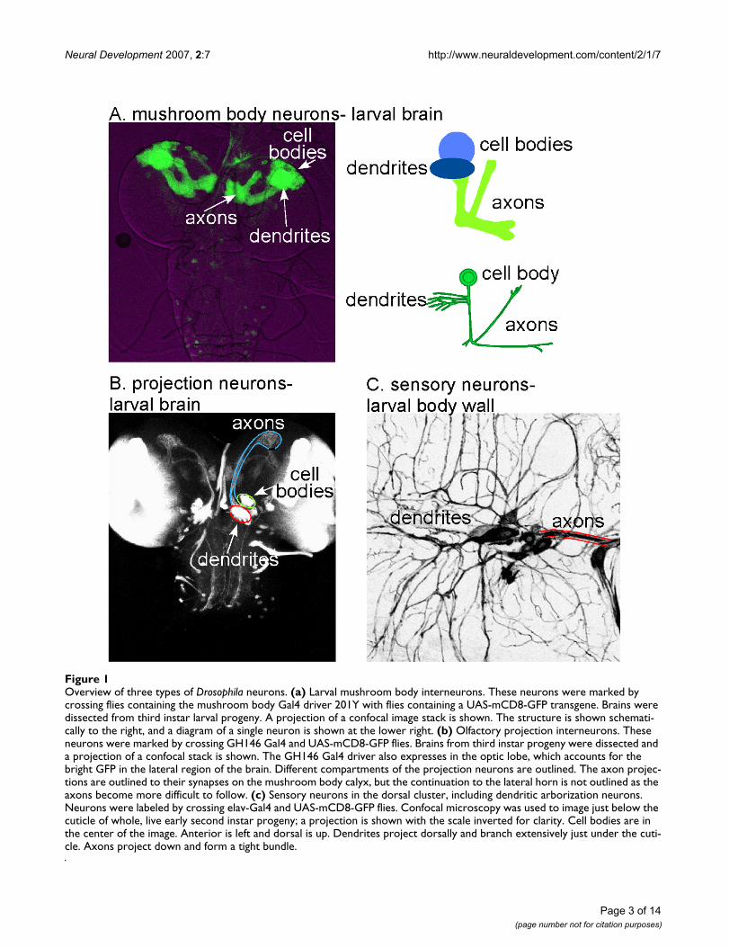
For analysis of neuronal compartmentalization, we stud-ied two types of larval interneurons: Kenyon cells of themushroom body, and projection neurons of the antennallobe. The development and morphology of both of thesetypes of neurons has been previously characterized[11,12], and both can be specifically labeled with theGal4-UAS binary expression system [13] (Figure 1a, b).The mushroom body is required for olfactory learningduring both larval and adult stages [14], and projectionneurons transmit signals from the antennal lobe glomer-uli to the mushroom body calyx (dendrites) and lateralhorn, where the information is further processed [15]. Vis-ualization of both types of neurons in the larval brainshows that they have axons and dendrites that occupy spe-cific regions in the brain (Figure 1a, b). We obtained sim-ilar results in both types of neurons, indicating that theyare likely to be broadly applicable to Drosophila neurons.For live imaging, we used the larval peripheral nervoussystem, as it lies close to the surface and can be easily vis-ualized in whole animals (Figure 1c).
We identified a set of markers that localize to differentcompartments in Drosophila neurons, and demonstratethat the major types of subcellular compartmentalization
Page 2 of 14(page number not for citation purposes)
Neural Development 2007, 2:7 http://www.neuraldevelopment.com/content/2/1/7
Page 3 of 14(page number not for citation purposes)
Overview of three types of Drosophila neuronsFigure 1Overview of three types of Drosophila neurons. (a) Larval mushroom body interneurons. These neurons were marked by crossing flies containing the mushroom body Gal4 driver 201Y with flies containing a UAS-mCD8-GFP transgene. Brains were dissected from third instar larval progeny. A projection of a confocal image stack is shown. The structure is shown schemati-cally to the right, and a diagram of a single neuron is shown at the lower right. (b) Olfactory projection interneurons. These neurons were marked by crossing GH146 Gal4 and UAS-mCD8-GFP flies. Brains from third instar progeny were dissected and a projection of a confocal stack is shown. The GH146 Gal4 driver also expresses in the optic lobe, which accounts for the bright GFP in the lateral region of the brain. Different compartments of the projection neurons are outlined. The axon projec-tions are outlined to their synapses on the mushroom body calyx, but the continuation to the lateral horn is not outlined as the axons become more difficult to follow. (c) Sensory neurons in the dorsal cluster, including dendritic arborization neurons. Neurons were labeled by crossing elav-Gal4 and UAS-mCD8-GFP flies. Confocal microscopy was used to image just below the cuticle of whole, live early second instar progeny; a projection is shown with the scale inverted for clarity. Cell bodies are in the center of the image. Anterior is left and dorsal is up. Dendrites project dorsally and branch extensively just under the cuti-cle. Axons project down and form a tight bundle.
Neural Development 2007, 2:7 http://www.neuraldevelopment.com/content/2/1/7
present in mammalian neurons can also be found in Dro-sophila neurons. In addition, we found that microtubulesare oriented differently in axons and dendrites. We con-clude that Drosophila will be a powerful system in whichto study neuronal compartmentalization.
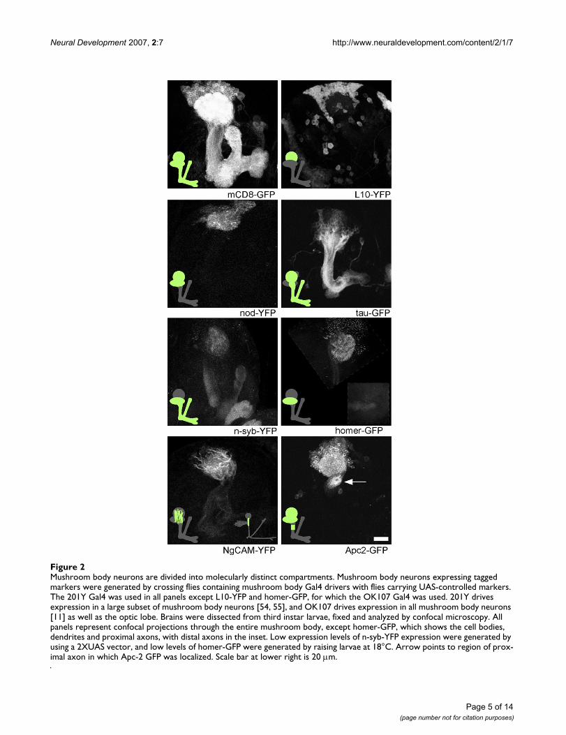
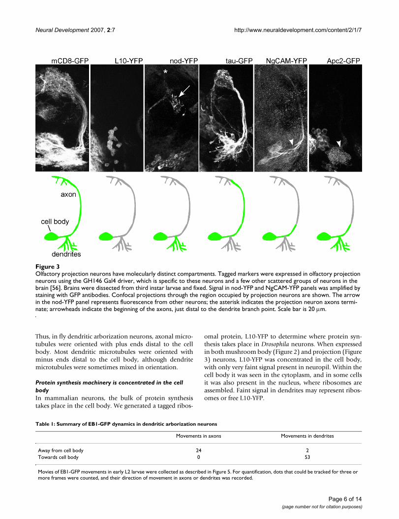
ResultsAxon and dendrite microtubules have different properties and orientationTo determine whether microtubule-binding proteins canbe preferentially localized to axons and dendrites in flies,we examined exogenous and endogenous microtubule-binding proteins in the Drosophila larval brain. The twoexogenous proteins we examined were: tau-green fluores-cent protein (tau-GFP) and nod-yellow fluorescent pro-tein (nod-YFP). Some reports have suggested that taggedversions of the microtubule binding domain of bovine taupreferentially label axons in flies [16], although othershave also reported dendrite localization [17]. We exam-ined the distribution of one of these tagged bovine tauproteins, tau-myc-GFP (which we call tau-GFP for sim-plicity) [17] in mushroom body and projection neurons(Figures 2 and 3). In both mushroom body and projectionneurons, tau-GFP was abundant in the main axon tracts.It was less abundant in distal axons and dendrites. Forcomparison, mCD8-GFP (Figures 2 and 3) is present in allneuronal compartments. Thus, tau-GFP preferentiallylabels proximal axons. Fusion proteins that consist of thenod motor domain, kinesin coiled-coil, and a tag havepreviously been localized to dendrites [16,18-21]. To con-firm that nod fusions label dendrites specifically, weexpressed nod-YFP in mushroom body and projectionneurons. In both cases nod-YFP localized clearly to den-drites but not axons (Figures 2 and 3). Thus, two exoge-nous microtubule-binding proteins, tau-GFP and nod-YFP, localized to different neuronal compartments, indi-cating that axonal and dendritic microtubules have dis-tinct features in Drosophila.
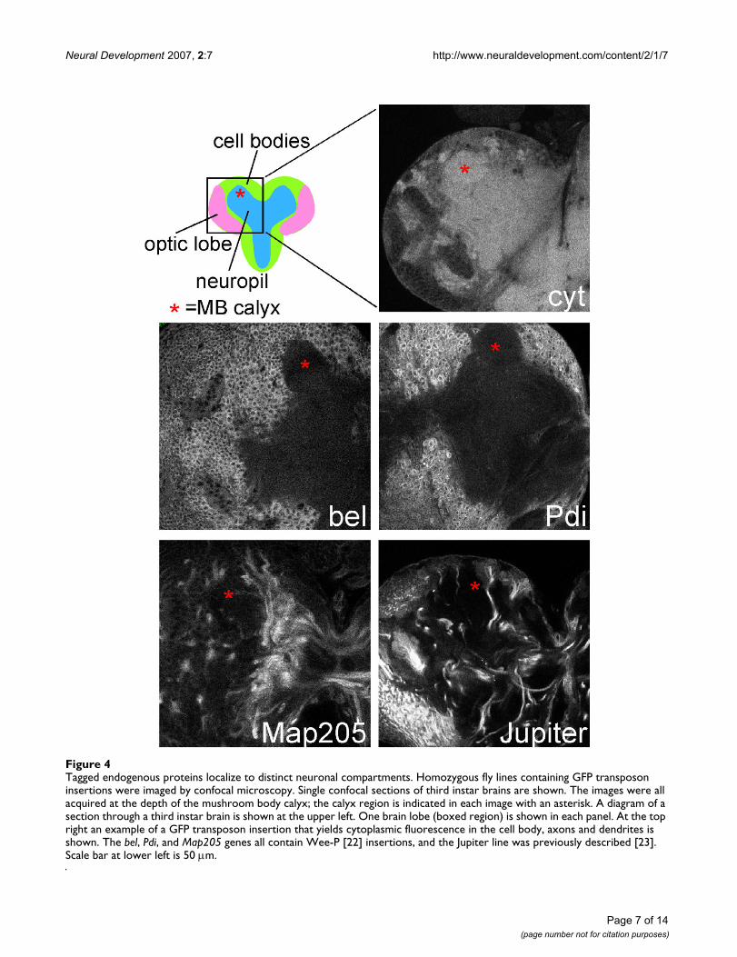
We also examined the localization of two Drosophilamicrotubule-binding proteins under control of their ownpromoters. We performed a protein trap screen asdescribed [22] to identify proteins targeted to specificregions of neurons. Fly lines isolated from this screen con-tain transposons with the GFP coding sequence insertedinto the genome. Insertions within genes result in the GFPcoding sequence being spliced into the mRNA, and gener-ate tagged proteins from the genomic locus. GFP-Map205was identified as a line with strong axonal GFP. Anothermicrotubule binding protein, GFP-Jupiter, was previouslyisolated [23]. Both protein trap lines exhibited strong GFPfluorescence in a subset of neurons. Like tau-GFP, bothDrosophila microtubule binding proteins were highly con-centrated in axons (Figure 4), although the relativeamounts of tagged protein in axons and dendrites were
harder to compare when expression was scatteredthroughout the brain. This preliminary localization datasuggest that both are good candidates for endogenousaxon-specific microtubule binding proteins.
Axon and dendrite microtubules have opposite orientationAs a difference in microtubule orientation in axons anddendrites is a fundamental aspect of vertebrate neuronalpolarity, we wished to determine exactly how microtu-bules are arranged in fly neurons. The dendritic localiza-tion of nod fusion proteins, which are believed to act asminus-end directed motors, has been used to argue thatDrosophila dendrites are likely to have minus-ends distalto the cell body like vertebrate dendrites [18]. However,direct analysis of the orientation of individual microtu-bules has not been performed in any invertebrate neuron.
Analysis of a microtubule plus-end tracking protein waspreviously used in mammalian neurons to confirm thataxon and dendrite microtubules have different orienta-tions [24]. As these proteins generally bind only to thegrowing plus ends of microtubules, microtubule orienta-tion can be inferred from the direction of movement of atagged plus-end binding protein. The peripheral nervoussystem of the Drosophila larva is well-suited to live imagingand has been previously used to study actin dynamics[20]. We therefore expressed the plus-end binding proteinEB1-GFP throughout the nervous system using an elav-Gal4 driver, and performed time lapse imaging of the dor-sal cluster of the peripheral nervous system (Figure 1c) inlive, whole, early L2 larvae. We primarily analyzed EB1-GFP dynamics in axons and dendrites of dendritic arbori-zation neurons, which have highly branched dendrites[25-27].
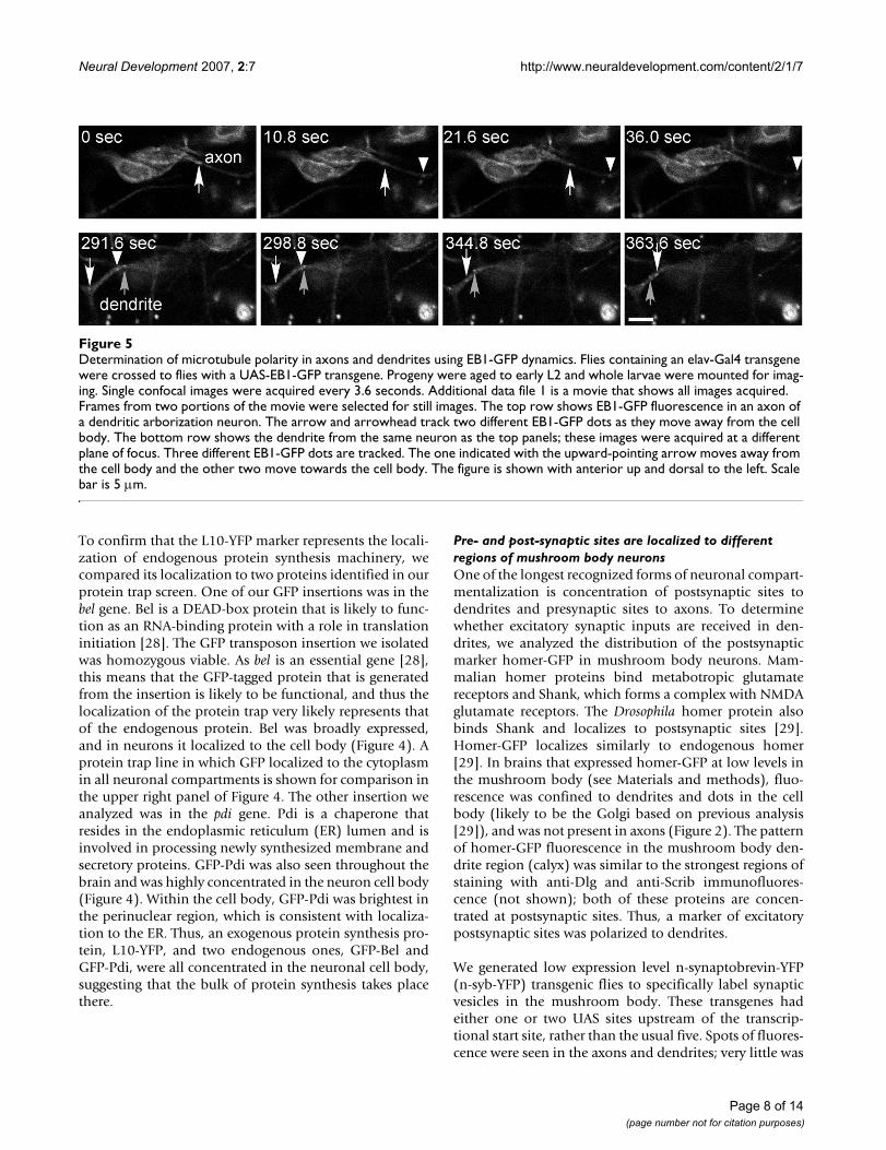
EB1-GFP dots were clearly seen moving in the cell body,axons, and dendrites (Figure 5 and Additional data file 1).Movements of EB1-GFP dots were consistent with thetagged protein only binding to growing microtubule plusends: an individual dot that could be followed throughmultiple frames never changed direction, and after a dottracked through a particular region of an axon or dendriteand disappeared, often a dot with a similar track appearedseveral frames later. All EB1-GFP dots in axons movedaway from the cell body (24 out of 24; only dots thatcould be followed in three consecutive frames werecounted; Table 1). An example of an axon with two differ-ent EB1-GFP dots is shown in the upper panels of Figure5. In dendrites the movements were very different. Thevast majority of dots moved toward the cell body (53 outof 55; dots present in 3+ frames). Occasionally, dendriteswere observed that had dots moving in opposite direc-tions (Figure 5, lower panels, and Additional data file 1).
Page 4 of 14(page number not for citation purposes)
Neural Development 2007, 2:7 http://www.neuraldevelopment.com/content/2/1/7
Page 5 of 14(page number not for citation purposes)
Mushroom body neurons are divided into molecularly distinct compartmentsFigure 2Mushroom body neurons are divided into molecularly distinct compartments. Mushroom body neurons expressing tagged markers were generated by crossing flies containing mushroom body Gal4 drivers with flies carrying UAS-controlled markers. The 201Y Gal4 was used in all panels except L10-YFP and homer-GFP, for which the OK107 Gal4 was used. 201Y drives expression in a large subset of mushroom body neurons [54, 55], and OK107 drives expression in all mushroom body neurons [11] as well as the optic lobe. Brains were dissected from third instar larvae, fixed and analyzed by confocal microscopy. All panels represent confocal projections through the entire mushroom body, except homer-GFP, which shows the cell bodies, dendrites and proximal axons, with distal axons in the inset. Low expression levels of n-syb-YFP expression were generated by using a 2XUAS vector, and low levels of homer-GFP were generated by raising larvae at 18°C. Arrow points to region of prox-imal axon in which Apc-2 GFP was localized. Scale bar at lower right is 20 μm.
Neural Development 2007, 2:7 http://www.neuraldevelopment.com/content/2/1/7
Thus, in fly dendritic arborization neurons, axonal micro-tubules were oriented with plus ends distal to the cellbody. Most dendritic microtubules were oriented withminus ends distal to the cell body, although dendritemicrotubules were sometimes mixed in orientation.
Protein synthesis machinery is concentrated in the cell bodyIn mammalian neurons, the bulk of protein synthesistakes place in the cell body. We generated a tagged ribos-
omal protein, L10-YFP to determine where protein syn-thesis takes place in Drosophila neurons. When expressedin both mushroom body (Figure 2) and projection (Figure3) neurons, L10-YFP was concentrated in the cell body,with only very faint signal present in neuropil. Within thecell body it was seen in the cytoplasm, and in some cellsit was also present in the nucleus, where ribosomes areassembled. Faint signal in dendrites may represent ribos-omes or free L10-YFP.
Olfactory projection neurons have molecularly distinct compartmentsFigure 3Olfactory projection neurons have molecularly distinct compartments. Tagged markers were expressed in olfactory projection neurons using the GH146 Gal4 driver, which is specific to these neurons and a few other scattered groups of neurons in the brain [56]. Brains were dissected from third instar larvae and fixed. Signal in nod-YFP and NgCAM-YFP panels was amplified by staining with GFP antibodies. Confocal projections through the region occupied by projection neurons are shown. The arrow in the nod-YFP panel represents fluorescence from other neurons; the asterisk indicates the projection neuron axons termi-nate; arrowheads indicate the beginning of the axons, just distal to the dendrite branch point. Scale bar is 20 μm.
Table 1: Summary of EB1-GFP dynamics in dendritic arborization neurons
Movements in axons Movements in dendrites
Away from cell body 24 2Towards cell body 0 53
Movies of EB1-GFP movements in early L2 larvae were collected as described in Figure 5. For quantification, dots that could be tracked for three or more frames were counted, and their direction of movement in axons or dendrites was recorded.
Page 6 of 14(page number not for citation purposes)
Neural Development 2007, 2:7 http://www.neuraldevelopment.com/content/2/1/7
Page 7 of 14(page number not for citation purposes)
Tagged endogenous proteins localize to distinct neuronal compartmentsFigure 4Tagged endogenous proteins localize to distinct neuronal compartments. Homozygous fly lines containing GFP transposon insertions were imaged by confocal microscopy. Single confocal sections of third instar brains are shown. The images were all acquired at the depth of the mushroom body calyx; the calyx region is indicated in each image with an asterisk. A diagram of a section through a third instar brain is shown at the upper left. One brain lobe (boxed region) is shown in each panel. At the top right an example of a GFP transposon insertion that yields cytoplasmic fluorescence in the cell body, axons and dendrites is shown. The bel, Pdi, and Map205 genes all contain Wee-P [22] insertions, and the Jupiter line was previously described [23]. Scale bar at lower left is 50 μm.
Neural Development 2007, 2:7 http://www.neuraldevelopment.com/content/2/1/7
To confirm that the L10-YFP marker represents the locali-zation of endogenous protein synthesis machinery, wecompared its localization to two proteins identified in ourprotein trap screen. One of our GFP insertions was in thebel gene. Bel is a DEAD-box protein that is likely to func-tion as an RNA-binding protein with a role in translationinitiation [28]. The GFP transposon insertion we isolatedwas homozygous viable. As bel is an essential gene [28],this means that the GFP-tagged protein that is generatedfrom the insertion is likely to be functional, and thus thelocalization of the protein trap very likely represents thatof the endogenous protein. Bel was broadly expressed,and in neurons it localized to the cell body (Figure 4). Aprotein trap line in which GFP localized to the cytoplasmin all neuronal compartments is shown for comparison inthe upper right panel of Figure 4. The other insertion weanalyzed was in the pdi gene. Pdi is a chaperone thatresides in the endoplasmic reticulum (ER) lumen and isinvolved in processing newly synthesized membrane andsecretory proteins. GFP-Pdi was also seen throughout thebrain and was highly concentrated in the neuron cell body(Figure 4). Within the cell body, GFP-Pdi was brightest inthe perinuclear region, which is consistent with localiza-tion to the ER. Thus, an exogenous protein synthesis pro-tein, L10-YFP, and two endogenous ones, GFP-Bel andGFP-Pdi, were all concentrated in the neuronal cell body,suggesting that the bulk of protein synthesis takes placethere.
Pre- and post-synaptic sites are localized to different regions of mushroom body neuronsOne of the longest recognized forms of neuronal compart-mentalization is concentration of postsynaptic sites todendrites and presynaptic sites to axons. To determinewhether excitatory synaptic inputs are received in den-drites, we analyzed the distribution of the postsynapticmarker homer-GFP in mushroom body neurons. Mam-malian homer proteins bind metabotropic glutamatereceptors and Shank, which forms a complex with NMDAglutamate receptors. The Drosophila homer protein alsobinds Shank and localizes to postsynaptic sites [29].Homer-GFP localizes similarly to endogenous homer[29]. In brains that expressed homer-GFP at low levels inthe mushroom body (see Materials and methods), fluo-rescence was confined to dendrites and dots in the cellbody (likely to be the Golgi based on previous analysis[29]), and was not present in axons (Figure 2). The patternof homer-GFP fluorescence in the mushroom body den-drite region (calyx) was similar to the strongest regions ofstaining with anti-Dlg and anti-Scrib immunofluores-cence (not shown); both of these proteins are concen-trated at postsynaptic sites. Thus, a marker of excitatorypostsynaptic sites was polarized to dendrites.
We generated low expression level n-synaptobrevin-YFP(n-syb-YFP) transgenic flies to specifically label synapticvesicles in the mushroom body. These transgenes hadeither one or two UAS sites upstream of the transcrip-tional start site, rather than the usual five. Spots of fluores-cence were seen in the axons and dendrites; very little was
Determination of microtubule polarity in axons and dendrites using EB1-GFP dynamicsFigure 5Determination of microtubule polarity in axons and dendrites using EB1-GFP dynamics. Flies containing an elav-Gal4 transgene were crossed to flies with a UAS-EB1-GFP transgene. Progeny were aged to early L2 and whole larvae were mounted for imag-ing. Single confocal images were acquired every 3.6 seconds. Additional data file 1 is a movie that shows all images acquired. Frames from two portions of the movie were selected for still images. The top row shows EB1-GFP fluorescence in an axon of a dendritic arborization neuron. The arrow and arrowhead track two different EB1-GFP dots as they move away from the cell body. The bottom row shows the dendrite from the same neuron as the top panels; these images were acquired at a different plane of focus. Three different EB1-GFP dots are tracked. The one indicated with the upward-pointing arrow moves away from the cell body and the other two move towards the cell body. The figure is shown with anterior up and dorsal to the left. Scale bar is 5 μm.
Page 8 of 14(page number not for citation purposes)
Neural Development 2007, 2:7 http://www.neuraldevelopment.com/content/2/1/7
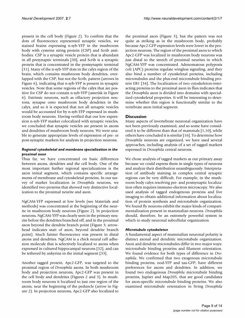
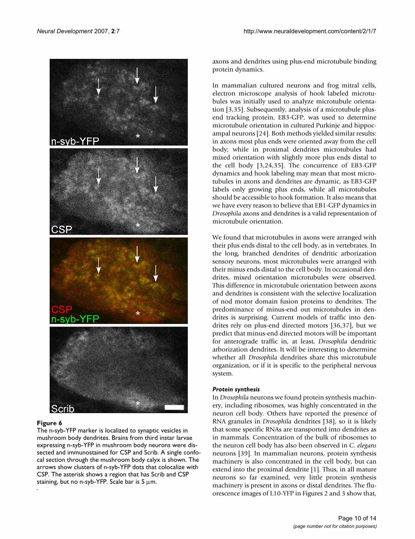
present in the cell body (Figure 2). To confirm that thedots of fluorescence represented synaptic vesicles, westained brains expressing n-syb-YFP in the mushroombody with cysteine string protein (CSP) and Scrib anti-bodies. CSP is a synaptic vesicle protein that is abundantin all presynaptic terminals [30], and Scrib is a synapticprotein that is concentrated in the postsynaptic terminal[31]. Many of the n-syb-YFP dots in the calyx region of thebrain, which contains mushroom body dendrites, over-lapped with the CSP, but not the Scrib, pattern (arrows inFigure 6), indicating that n-syb-YFP is present in synapticvesicles. Note that some regions of the calyx that are pos-itive for CSP do not contain n-syb-YFP (asterisk in Figure6). Extrinsic neurons, such as olfactory projection neu-rons, synapse onto mushroom body dendrites in thecalyx, and so it is expected that not all synaptic vesicleswould be accounted for by n-syb-YFP expressed in mush-room body neurons. Having verified that our low expres-sion n-syb-YFP marker colocalized with synaptic vesicles,we concluded that synaptic vesicles are present in axonsand dendrites of mushroom body neurons. We were una-ble to generate appropriate levels of expression of pre- orpost-synaptic markers for analysis in projection neurons.
Regional cytoskeletal and membrane specialization in the proximal axonThus far, we have concentrated on basic differencesbetween axons, dendrites and the cell body. One of themost important further regional specializations is theaxon initial segment, which contains specific arrange-ments of membrane and cytoskeletal proteins. In our sur-vey of marker localization in Drosophila neurons, weidentified two proteins that showed very distinctive local-ization to the proximal neurite and axon.
NgCAM-YFP expressed at low levels (see Materials andmethods) was concentrated at the beginning of the neur-ite in mushroom body neurons (Figure 2). In projectionneurons, NgCAM-YFP was clearly seen in the primary neu-rite before the dendrites branched off, and in the proximalaxon beyond the dendrite branch point (Figure 3; arrow-head indicates start of axon, beyond dendrite branchpoint). Much fainter fluorescence was present in distalaxons and dendrites. NgCAM is a chick neural cell adhe-sion molecule that is selectively localized to axons whenexpressed in cultured hippocampal neurons [32], and canbe tethered by ankyrins in the initial segment [33].
Another tagged protein, Apc2-GFP, was targeted to theproximal region of Drosophila axons. In both mushroombody and projection neurons, Apc2-GFP was present inthe cell body and dendrites (Figures 2 and 3). In mush-room body neurons it localized to just one region of theaxons, near the beginning of the peduncle (arrow in Fig-ure 2). In projection neurons, Apc2-GFP also localized to
the proximal axon (Figure 3), but the pattern was notquite as striking as in the mushroom body, probablybecause Apc2-GFP expression levels were lower in the pro-jection neurons. The region of the proximal axon to whichApc2-GFP was localized in mushroom body neurons wasjust distal to the stretch of proximal neurites in whichNgCAM-YFP was concentrated. Adenomatous polyposiscoli (APC) proteins regulate wingless signaling, and theyalso bind a number of cytoskeletal proteins, includingmicrotubules and the plus-end microtubule binding pro-tein EB1 [34]. The localization of two cytoskeleton-inter-acting proteins to the proximal axon in flies indicates thatthe Drosophila axon is divided into domains with special-ized cytoskeletal properties. It will be interesting to deter-mine whether this region is functionally similar to thevertebrate axon initial segment.
DiscussionMany aspects of invertebrate neuronal organization havenot been previously examined, and so some have consid-ered it to be different than that of mammals [1,10], whileothers have concluded it is similar [16]. To determine howDrosophila neurons are organized, we have used severalapproaches, including analysis of a set of tagged markersexpressed in Drosophila central neurons.
We chose analysis of tagged markers as our primary assaybecause we could express them in single types of neuronsand analyze their distribution unambiguously. Interpreta-tion of antibody staining in complex central synapticregions can be very difficult. For example, in the mush-room body calyx resolving pre- and postsynaptic localiza-tion often requires immuno-electron microscopy. We alsoused analysis of tagged endogenous proteins and liveimaging to obtain additional information about localiza-tion of protein synthesis and microtubule organization.We found fly neurons exhibit the major kinds of compart-mentalization present in mammalian neurons. Drosophilashould, therefore, be an extremely powerful system inwhich to study neuronal subcellular organization.
Microtubule cytoskeletonA fundamental aspect of mammalian neuronal polarity isdistinct axonal and dendritic microtubule organization.Axon and dendrite microtubules differ in two major ways:microtubule binding proteins and filament orientation.We found evidence for both types of difference in Dro-sophila. We confirmed that two exogenous microtubulebinding proteins, nod-YFP and tau-GFP, have differentpreferences for axons and dendrites. In addition, wefound two endogenous Drosophila microtubule bindingproteins, Jupiter and Map205, that are good candidatesfor axon-specific microtubule binding proteins. We alsoexamined microtubule orientation in living Drosophila
Page 9 of 14(page number not for citation purposes)
Neural Development 2007, 2:7 http://www.neuraldevelopment.com/content/2/1/7
axons and dendrites using plus-end microtubule bindingprotein dynamics.
In mammalian cultured neurons and frog mitral cells,electron microscope analysis of hook labeled microtu-bules was initially used to analyze microtubule orienta-tion [3,35]. Subsequently, analysis of a microtubule plus-end tracking protein, EB3-GFP, was used to determinemicrotubule orientation in cultured Purkinje and hippoc-ampal neurons [24]. Both methods yielded similar results:in axons most plus ends were oriented away from the cellbody; while in proximal dendrites microtubules hadmixed orientation with slightly more plus ends distal tothe cell body [3,24,35]. The concurrence of EB3-GFPdynamics and hook labeling may mean that most micro-tubules in axons and dendrites are dynamic, as EB3-GFPlabels only growing plus ends, while all microtubulesshould be accessible to hook formation. It also means thatwe have every reason to believe that EB1-GFP dynamics inDrosophila axons and dendrites is a valid representation ofmicrotubule orientation.
We found that microtubules in axons were arranged withtheir plus ends distal to the cell body, as in vertebrates. Inthe long, branched dendrites of dendritic arborizationsensory neurons, most microtubules were arranged withtheir minus ends distal to the cell body. In occasional den-drites, mixed orientation microtubules were observed.This difference in microtubule orientation between axonsand dendrites is consistent with the selective localizationof nod motor domain fusion proteins to dendrites. Thepredominance of minus-end out microtubules in den-drites is surprising. Current models of traffic into den-drites rely on plus-end directed motors [36,37], but wepredict that minus-end directed motors will be importantfor anterograde traffic in, at least, Drosophila dendriticarborization dendrites. It will be interesting to determinewhether all Drosophila dendrites share this microtubuleorganization, or if it is specific to the peripheral nervoussystem.
Protein synthesisIn Drosophila neurons we found protein synthesis machin-ery, including ribosomes, was highly concentrated in theneuron cell body. Others have reported the presence ofRNA granules in Drosophila dendrites [38], so it is likelythat some specific RNAs are transported into dendrites asin mammals. Concentration of the bulk of ribosomes tothe neuron cell body has also been observed in C. elegansneurons [39]. In mammalian neurons, protein synthesismachinery is also concentrated in the cell body, but canextend into the proximal dendrite [1]. Thus, in all matureneurons so far examined, very little protein synthesismachinery is present in axons or distal dendrites. The flu-orescence images of L10-YFP in Figures 2 and 3 show that,
The n-syb-YFP marker is localized to synaptic vesicles in mushroom body dendritesFigure 6The n-syb-YFP marker is localized to synaptic vesicles in mushroom body dendrites. Brains from third instar larvae expressing n-syb-YFP in mushroom body neurons were dis-sected and immunostained for CSP and Scrib. A single confo-cal section through the mushroom body calyx is shown. The arrows show clusters of n-syb-YFP dots that colocalize with CSP. The asterisk shows a region that has Scrib and CSP staining, but no n-syb-YFP. Scale bar is 5 μm.
Page 10 of 14(page number not for citation purposes)
Neural Development 2007, 2:7 http://www.neuraldevelopment.com/content/2/1/7
although this form of neuronal compartmentalization hasbeen studied very little, it is extremely striking. The strongconcentration difference of ribosomes between the cellbody and neuropil indicates that there may be an activemechanism that restricts the bulk of protein synthesismachinery to the cell body.
One of the endogenous proteins that we found highlyconcentrated in the cell body was Pdi. Pdi is localized tothe lumen of the ER. In all neurons previously examined,including Purkinje cells and C. elegans neurons, the ERextends from the cell body throughout axons and den-drites, and is believed to be continuous [39,40]. We there-fore also predict that a mechanism exists to concentrateproteins inside the ER to the neuron cell body.
Synaptic componentsIn mammalian motor and hippocampal neurons, presyn-aptic components are restricted to axons and postsynapticcomponents to dendrites, but in some mammalianinterneurons, dendrites can contain synaptic vesicles andbe presynaptic [10]. Drosophila embryonic motor neuronsare similar to mammalian motor neurons: a neurotrans-mitter receptor is exclusively localized to dendrites, and asynaptic vesicle marker is restricted to axons [16]. Wefound that a postsynaptic marker was exclusively targetedto dendrites in mushroom body neurons, while a synapticvesicle marker was present in dendrites and axons. Bothmammals and flies, therefore, have different classes ofneurons with distinct synaptic arrangements. Little isknown about how non-classic synapses, including den-dro-dendritic, are assembled. Drosophila has alreadyproven useful for studying 'traditional' synapses, like theneuromuscular junction [41], and could be useful forunderstanding other synaptic arrangements. One interest-ing question is: how are synaptic vesicles targeted to den-drites in mushroom bodies, but excluded from dendritesof motor neurons?
Plasma membrane proteinsIn mammalian neurons, different non-synaptic plasmamembrane proteins are localized to axons and dendrites.Examples include cell adhesion proteins and ion channels[1]. In Drosophila, non-synaptic plasma membrane pro-teins completely specific to axons or dendrites have notyet been identified. In part, this is because these experi-ments are easier to interpret using cultured neurons inwhich the plasma membranes are not contacting othercells, and in flies a fully polarized neuronal culture systemhas not been described. We localized one exogenoustransmembrane adhesion protein, NgCAM-YFP, to theprimary neurite and proximal axon, but did not identifymore broadly localized axonal or dendritic proteins.These are, however, likely to exist in flies. One good can-didate is the sodium channel pickpocket, which is present
in a subset of dendritic arborization neuron dendrites[42].
Do Drosophila neurons have an initial segment?The mammalian axon initial segment is a distinctiveregion of the axon that is important in generation ofaction potentials and protein sorting within the neuron.At the ultrastructural level, it can be recognized by an elec-tron-dense coating under the plasma membrane and bun-dled microtubules [10]. At the molecular level, the initialsegment contains a specialized spectrin and ankyrin Gsubmembrane skeleton that is important for localizingchannels and other plasma membrane proteins. Thissame network forms the basis of a diffusion barrier thatisolates the plasma membrane in the axon from that inthe soma [5]. The microtubule cytoskeleton in the initialsegment has also been suggested to be important for selec-tive sorting to axons and dendrites [43].
While concentration of cytoskeletal (see tau-GFP in Fig-ures 2 and 3) and membrane proteins [44] to unbranchedor fasciculated subregions of axons has been observed inflies, it has not been clear whether flies have an initial seg-ment similar to that in mammals. Many Drosophila neu-rons also generate action potentials, but the precise regionin which they are initiated has not been determined [45].Drosophila do possess a neuronal ankyrin that has axonaland somatic isoforms, but the axonal isoform has notbeen noted to be restricted to a particular domain [46,47].Nor have specific membrane proteins been previouslyidentified in the proximal axon, and the motif in ionchannels that binds to ankyrin in the vertebrate axon ini-tial segment is absent in insects [48].
Our data suggest that Drosophila neurons may have adomain similar to the mammalian axon initial segment.NgCAM-YFP localized to the proximal neurite and axon inmushroom body and projection neurons. In mammalianneurons, NgCAM seems to have two mechanisms of local-ization: an ankyrin-independent one that localizes italong the length of the axon, and an ankyrin-dependentone that anchors it at the initial segment [33]. One possi-ble explanation for the localization we see in flies is thatthe ankyrin-independent targeting signal is not recog-nized, but NgCAM can be concentrated in the proximalaxon by binding to a special submembrane skeleton anal-ogous to the ankyrin G domain in the initial segment. Wealso observed localization of Apc2-GFP to the proximalaxon. APC family members bind cytoskeletal proteins,including EB1. Overexpressed EB1-YFP localizes to theinitial segment in hippocampal neurons, and may recog-nize a local microtubule specialization important fordirectional transport within neurons [43]. The localiza-tion of overexpressed Apc2-GFP we observe may reflect asimilar microtubule organization in fly proximal axons.
Page 11 of 14(page number not for citation purposes)
Neural Development 2007, 2:7 http://www.neuraldevelopment.com/content/2/1/7
We therefore suggest that flies may have actin and micro-tubule cytoskeletal specializations in the proximal neuriteand axon that are similar to those in the mammalian axoninitial segment.
ConclusionWe have found evidence for many types of regional neu-ronal specialization in Drosophila, and conclude that Dro-sophila and mammalian neurons are compartmentalizedin the same ways. We have also generated a number ofuseful tools for following protein targeting in fly neurons,which, in conjunction with the sophisticated genetic tech-niques that can be used to manipulate Drosophila, meanthat this should be a powerful system for studying neuro-nal polarity. Questions that could be addressed include:how is protein synthesis machinery concentrated in thecell body? How are axon domains established? What reg-ulates the polarity of microtubules in axons and den-drites?
Materials and methodsFly stocksBloomington Stock Center provided the following stocks:OK107-Gal4, 201Y-Gal4, elav-Gal4, and UAS:Apc2-GFP.GH146-Gal4 was kindly provided by Reinhard Stocker,UAS:tau-myc-GFP by Shingo Yoshikawa, and UAS:homer-myc-GFP by Ulrich Thomas. P{PTT-GA}Jupiter [G147]was provided by Flytrap [49]. UAS:nod-YFP, 2XUAS:n-syb-YFP, UAS:NgCAM-YFP, UAS:EB1-GFP, P{Wee}bel[0–139], P{Wee}Pdi [1–149], and P{Wee}Map205 [382]were generated for this study.
P element constructionFor tagging proteins with three copies of YFP, a modifiedpUAST [13] vector was designed. In 3XYFP-CT apolylinker was placed before three copies of the YFP cod-ing sequence. UAS:nod-YFP was generated by PCR ampli-fying the nod motor domain and kinesin coiled coilsequence from pnod:lacZ [18], which was generously pro-vided by Ira Clark; this PCR product was cloned into3XYFP-CT. Tagged NgCAM was generated by PCR ampli-fying the NgCAM coding sequence from JPA7-NgCAMYFP, kindly provided by Gary Banker, and cloningit into 3XYFP-CT. A suboptimal ribosome binding site(TGAAAG) [50] was included in the upstream primer toreduce levels of translated protein. 2XUAS:n-syb-YFP wasgenerated using a modified version of 3XYFP-CT in whichthe five UAS sequences were removed and replaced withtwo copies of the UAS sequence. Full length n-syb wasPCR amplified from an expressed sequence tag clone(GH04664), and cloned into the 2XUAS-3XYFP-CT vec-tor. The UAS:EB1-GFP P element was created by insertingcDNA of enhanced GFP fused to the carboxy-terminal endof Drosophila EB1 [51] into pUAST.
Immunofluorescent staining of larval brainsThird instar larval brains were dissected in Schneider'smedium and fixed for 20 minutes with 4% paraformalde-hyde in phosphate-buffered saline (PBS). They were thenwashed several times with block buffer (PBS/1% bovineserum albumen/0.03% triton X-100/10 mM glycine).Brains were incubated overnight with primary antibodiesin block buffer at 4°C. They were then washed for severalhours in block buffer, incubated for one to two hours insecondary antibodies conjugated to rhodamine red-X orCy5 (Jackson ImmunoResearch, West Grove, PA, USA),and washed several more times. Brains were equilibratedovernight in 85% glycerol/50 mM Tris pH 8 beforemounting for microscopy. Primary antibodies used were:rabbit anti-GFP (Abcam, Cambridge, MA, USA), rabbitanti-Scrib [52], and mouse anti-CSP, kindly provided bySeymour Benzer.
Analysis of fluorescence in larval brainsLarval brains were dissected and fixed as above, and eitherwashed and transferred directly to glycerol, or processedfor immunofluorescence. Whole brains were mounted in85% glycerol/50 mM Tris pH8 under a coverslip andimaged on a BioRad (Hercules, CA, USA) Radiance 2100confocal microscope. Images were processed using ImageJ[53].
Generation of Wee-P insertionsWee-P GFP transposon insertions were generated essen-tially as described [22]. GFP-positive embryos were man-ually picked using a dissecting microscope equipped forfluorescence (Olympus, Center Valley, PA, USA). GFP-positive fly lines were then screened for localized fluores-cence either in the embryonic central nervous system orthird instar larval brain. Wee-P insertion sites in lines ofinterest were identified by inverse PCR.
Live imaging of the peripheral nervous systemLarvae with EB1-GFP or mCD8-GFP expressed in theperipheral nervous system were generated by crossinghomozygous GFP transgene-containing lines with elav-Gal4 flies. Early second instar larvae were picked andplaced on a slide with halocarbon 27 oil (Sigma, St Louis,MO, USA), then covered with a coverslip. Images wereonly collected for 10 minutes after mounting to ensurelarvae were healthy. Images were collected at 166 lines persecond, every 3.6 seconds, zoom 4 on a BioRad Radiance2100 confocal microscope. Movies were compiled inImageJ and Quicktime.
Competing interestsThe author(s) declare that they have no competing inter-ests.
Page 12 of 14(page number not for citation purposes)
Neural Development 2007, 2:7 http://www.neuraldevelopment.com/content/2/1/7
Authors' contributionsMMR performed analysis of markers in larvae presentedin the paper, and generated reagents needed unless speci-fied below. DS generated EB1-GFP flies under the supervi-sion of TU. PC designed the Wee-P screen and isolatedsome of the insertions shown. ALH oversaw the Wee-Pscreen performed in the Doe lab. CQD supervised andwas involved in planning experiments. All authors read,made suggestions, and approved the final manuscript.
Additional material
AcknowledgementsWe are extremely grateful to everyone who sent us reagents, including: Ira Clark, Gary Banker, Ulrich Thomas, Richard Stocker, Shingo Yoshikawa, Seymour Benzer, as well as the Flytrap project at Yale, the Bloomington Stock Center, and researchers who submitted their lines to the Stock Center. Amy Sheehan was instrumental in making the 3XYFP vectors. Lacey Brounstein, Ashley Harris-Deutch, Kendra Bolt and Evan Cope were very helpful with generating Wee-P lines, cloning and fly maintenance. We also thank Ryan Andersen for live imaging advice. This work was supported by the Howard Hughes Medical Institute (CQD), an American Heart Asso-ciation Postdoctoral Fellowship (MMR), a NARSAD Young Investigator Award (MMR), JSPS Research Fellowships for Young Scientists (DS) and MEXT of Japan (TU).
References1. Craig AM, Banker G: Neuronal polarity. Annu Rev Neurosci 1994,
17:267-310.2. Mattson MP: Establishment and plasticity of neuronal polarity.
J Neurosci Res 1999, 57:577-589.3. Baas PW, Deitch JS, Black MM, Banker GA: Polarity orientation of
microtubules in hippocampal neurons: uniformity in theaxon and nonuniformity in the dendrite. Proc Natl Acad Sci U SA 1988, 85:8335-8339.
4. Horton AC, Ehlers MD: Neuronal polarity and trafficking. Neu-ron 2003, 40:277-295.
5. Winckler B, Forscher P, Mellman I: A diffusion barrier maintainsdistribution of membrane proteins in polarized neurons.Nature 1999, 397:698-701.
6. Kiebler MA, Bassell GJ: Neuronal RNA granules: movers andmakers. Neuron 2006, 51:685-690.
7. Brittis PA, Lu Q, Flanagan JG: Axonal protein synthesis providesa mechanism for localized regulation at an intermediate tar-get. Cell 2002, 110:223-235.
8. Lai HC, Jan LY: The distribution and targeting of neuronal volt-age-gated ion channels. Nat Rev Neurosci 2006, 7:548-562.
9. Garrity PA: Signal transduction in axon guidance. CMLS 1999,55:1407-1415.
10. Peters A, Palay SL, Webster HD: The Fine Structure of the Nerv-ous System: Neurons and their Supporting Cells. 3rd edition.New York, Oxford University Press; 1991.
11. Lee T, Lee A, Luo L: Development of the Drosophila mush-room bodies: sequential generation of three distinct types ofneurons from a neuroblast. Development 1999, 126:4065-4076.
12. Marin EC, Watts RJ, Tanaka NK, Ito K, Luo L: Developmentallyprogrammed remodeling of the Drosophila olfactory circuit.Development 2005, 132:725-737.
13. Brand AH, Perrimon N: Targeted gene expression as a meansof altering cell fates and generating dominant phenotypes.Development 1993, 118:401-415.
14. Heisenberg M: Mushroom body memoir: from maps to mod-els. Nat Rev Neurosci 2003, 4:266-275.
15. Jefferis GS, Marin EC, Watts RJ, Luo L: Development of neuronalconnectivity in Drosophila antennal lobes and mushroombodies. Curr Opin Neurobiol 2002, 12:80-86.
16. Sanchez-Soriano N, Bottenberg W, Fiala A, Haessler U, Kerassoviti A,Knust E, Lohr R, Prokop A: Are dendrites in Drosophila homol-ogous to vertebrate dendrites? Dev Biol 2005, 288:126-138.
17. Callahan CA, Yoshikawa S, Thomas JB: Tracing axons. Curr OpinNeurobiol 1998, 8:582-586.
18. Clark IE, Jan LY, Jan YN: Reciprocal localization of Nod andkinesin fusion proteins indicates microtubule polarity in theDrosophila oocyte, epithelium, neuron and muscle. Develop-ment 1997, 124:461-470.
19. Lee T, Winter C, Marticke SS, Lee A, Luo L: Essential roles of Dro-sophila RhoA in the regulation of neuroblast proliferationand dendritic but not axonal morphogenesis. Neuron 2000,25:307-316.
20. Andersen R, Li Y, Resseguie M, Brenman JE: Calcium/calmodulin-dependent protein kinase II alters structural plasticity andcytoskeletal dynamics in Drosophila. J Neurosci 2005,25:8878-8888.
21. Yamamoto M, Ueda R, Takahashi K, Saigo K, Uemura T: Control ofaxonal sprouting and dendrite branching by the Nrg-Ankcomplex at the neuron-glia interface. Curr Biol 2006,16:1678-1683.
22. Clyne PJ, Brotman JS, Sweeney ST, Davis G: Green fluorescentprotein tagging Drosophila proteins at their native genomicloci with small P elements. Genetics 2003, 165:1433-1441.
23. Morin X, Daneman R, Zavortink M, Chia W: A protein trap strat-egy to detect GFP-tagged proteins expressed from theirendogenous loci in Drosophila. Proc Natl Acad Sci U S A 2001,98:15050-15055.
24. Stepanova T, Slemmer J, Hoogenraad CC, Lansbergen G, Dortland B,De Zeeuw CI, Grosveld F, van Cappellen G, Akhmanova A, Galjart N:Visualization of microtubule growth in cultured neurons viathe use of EB3-GFP (end-binding protein 3-green fluorescentprotein). J Neurosci 2003, 23:2655-2664.
25. Grueber WB, Jan LY, Jan YN: Tiling of the Drosophila epidermisby multidendritic sensory neurons. Development 2002,129:2867-2878.
26. Sugimura K, Yamamoto M, Niwa R, Satoh D, Goto S, Taniguchi M,Hayashi S, Uemura T: Distinct developmental modes andlesion-induced reactions of dendrites of two classes of Dro-sophila sensory neurons. J Neurosci 2003, 23:3752-3760.
27. Grueber WB, Ye B, Moore AW, Jan LY, Jan YN: Dendrites of dis-tinct classes of Drosophila sensory neurons show differentcapacities for homotypic repulsion. Curr Biol 2003, 13:618-626.
28. Johnstone O, Deuring R, Bock R, Linder P, Fuller MT, Lasko P: Belleis a Drosophila DEAD-box protein required for viability andin the germ line. Dev Biol 2005, 277:92-101.
29. Diagana TT, Thomas U, Prokopenko SN, Xiao B, Worley PF, ThomasJB: Mutation of Drosophila homer disrupts control of loco-motor activity and behavioral plasticity. J Neurosci 2002,22:428-436.
30. Eberle KK, Zinsmaier KE, Buchner S, Gruhn M, Jenni M, Arnold C,Leibold C, Reisch D, Walter N, Hafen E, Hofbauer A, Pflugfelder GO,Buchner E: Wide distribution of the cysteine string proteins inDrosophila tissues revealed by targeted mutagenesis. Cell Tis-sue Res 1998, 294:203-217.
31. Mathew D, Gramates LS, Packard M, Thomas U, Bilder D, PerrimonN, Gorczyca M, Budnik V: Recruitment of scribble to the synap-tic scaffolding complex requires GUK-holder, a novel DLGbinding protein. Curr Biol 2002, 12:531-539.
32. Jareb M, Banker G: The polarized sorting of membrane pro-teins expressed in cultured hippocampal neurons using viralvectors. Neuron 1998, 20:855-867.
Additional file 1Determination of microtubule polarity in axons and dendrites using EB1-GFP dynamics. Flies containing an elav-Gal4 transgene were crossed to flies with a UAS-EB1-GFP transgene. Progeny were aged to early L2 and whole larvae were mounted for imaging. Single confocal images were acquired every 3.6 seconds. The movie can be viewed with Quicktime.Click here for file[http://www.biomedcentral.com/content/supplementary/1749-8104-2-7-S1.mov]
Page 13 of 14(page number not for citation purposes)
Neural Development 2007, 2:7 http://www.neuraldevelopment.com/content/2/1/7
Publish with BioMed Central and every scientist can read your work free of charge
"BioMed Central will be the most significant development for disseminating the results of biomedical research in our lifetime."
Sir Paul Nurse, Cancer Research UK
Your research papers will be:
available free of charge to the entire biomedical community
peer reviewed and published immediately upon acceptance
cited in PubMed and archived on PubMed Central
yours — you keep the copyright
Submit your manuscript here:http://www.biomedcentral.com/info/publishing_adv.asp
BioMedcentral
33. Boiko T, Vakulenko M, Ewers H, Yap CC, Norden C, Winckler B:Ankyrin-dependent and -independent mechanisms orches-trate axonal compartmentalization of L1 family membersneurofascin and L1/neuron-glia cell adhesion molecule. J Neu-rosci 2007, 27:590-603.
34. Nathke I: APC at a glance. J Cell Sci 2004, 117:4873-4875.35. Burton PR: Dendrites of mitral cell neurons contain microtu-
bules of opposite polarity. Brain Res 1988, 473:107-115.36. Hirokawa N, Takemura R: Molecular motors and mechanisms
of directional transport in neurons. Nat Rev Neurosci 2005,6:201-214.
37. Kennedy MJ, Ehlers MD: Organelles and trafficking machineryfor postsynaptic plasticity. Annu Rev Neurosci 2006, 29:325-362.
38. Ye B, Petritsch C, Clark IE, Gavis ER, Jan LY, Jan YN: Nanos andPumilio are essential for dendrite morphogenesis in Dro-sophila peripheral neurons. Curr Biol 2004, 14:314-321.
39. Rolls MM, Hall DH, Victor M, Stelzer EH, Rapoport TA: Targetingof rough endoplasmic reticulum membrane proteins andribosomes in invertebrate neurons. Mol Biol Cell 2002,13:1778-1791.
40. Terasaki M, Slater NT, Fein A, Schmidek A, Reese TS: Continuousnetwork of endoplasmic reticulum in cerebellar Purkinjeneurons. Proc Natl Acad Sci U S A 1994, 91:7510-7514.
41. Prokop A, Meinertzhagen IA: Development and structure of syn-aptic contacts in Drosophila. Semin Cell Dev Biol 2006, 17:20-30.
42. Adams CM, Anderson MG, Motto DG, Price MP, Johnson WA,Welsh MJ: Ripped pocket and pickpocket, novel DrosophilaDEG/ENaC subunits expressed in early development and inmechanosensory neurons. J Cell Biol 1998, 140:143-152.
43. Nakata T, Hirokawa N: Microtubules provide directional cuesfor polarized axonal transport through interaction withkinesin motor head. J Cell Biol 2003, 162:1045-1055.
44. Bastiani MJ, Harrelson AL, Snow PM, Goodman CS: Expression offasciclin I and II glycoproteins on subsets of axon pathwaysduring neuronal development in the grasshopper. Cell 1987,48:745-755.
45. Wicher D, Walther C, Wicher C: Non-synaptic ion channels ininsects--basic properties of currents and their modulation inneurons and skeletal muscles. Prog Neurobiol 2001, 64:431-525.
46. Bouley M, Tian MZ, Paisley K, Shen YC, Malhotra JD, Hortsch M: TheL1-type cell adhesion molecule neuroglian influences the sta-bility of neural ankyrin in the Drosophila embryo but not itsaxonal localization. J Neurosci 2000, 20:4515-4523.
47. Hortsch M, Paisley KL, Tian MZ, Qian M, Bouley M, Chandler R: Theaxonal localization of large Drosophila ankyrin2 protein iso-forms is essential for neuronal functionality. Mol Cell Neurosci2002, 20:43-55.
48. Pan Z, Kao T, Horvath Z, Lemos J, Sul JY, Cranstoun SD, Bennett V,Scherer SS, Cooper EC: A common ankyrin-G-based mecha-nism retains KCNQ and NaV channels at electrically activedomains of the axon. J Neurosci 2006, 26:2599-2613.
49. Flytrap [http://flytrap.med.yale.edu/]50. Fujioka M, Lear BC, Landgraf M, Yusibova GL, Zhou J, Riley KM, Patel
NH, Jaynes JB: Even-skipped, acting as a repressor, regulatesaxonal projections in Drosophila. Development 2003,130:5385-5400.
51. Shimada Y, Yonemura S, Ohkura H, Strutt D, Uemura T: Polarizedtransport of Frizzled along the planar microtubule arrays inDrosophila wing epithelium. Dev Cell 2006, 10:209-222.
52. Albertson R, Doe CQ: Dlg, Scrib and Lgl regulate neuroblastcell size and mitotic spindle asymmetry. Nat Cell Biol 2003,5:166-170.
53. ImageJ [http://rsb.info.nih.gov/ij/]54. Yang MY, Armstrong JD, Vilinsky I, Strausfeld NJ, Kaiser K: Subdivi-
sion of the Drosophila mushroom bodies by enhancer-trapexpression patterns. Neuron 1995, 15:45-54.
55. Connolly JB, Roberts IJ, Armstrong JD, Kaiser K, Forte M, Tully T,O'Kane CJ: Associative learning disrupted by impaired Gs sig-naling in Drosophila mushroom bodies. Science 1996,274:2104-2107.
56. Stocker RF, Heimbeck G, Gendre N, de Belle JS: Neuroblast abla-tion in Drosophila P[GAL4] lines reveals origins of olfactoryinterneurons. J Neurobiol 1997, 32:443-456.
Page 14 of 14(page number not for citation purposes)


