Embed Size (px)
Citation preview
877
Braz J Med Biol Res 40(6) 2007
Topical laser irradiation and wound healing
www.bjournal.com.br
Brazilian Journal of Medical and Biological Research (2007) 40: 877-884ISSN 0100-879X
Influence of He-Ne laser therapy onthe dynamics of wound healing in micetreated with anti-inflammatory drugs
1Departamento de Ciências Fisiológicas, 2Departamento de Clínica Odontológica,Centro de Ciências da Saúde, Universidade Federal do Espírito Santo, Vitória,ES, Brasil
W.L.S. Gonçalves1,F.M. Souza1, C.L. Conti1,
J.P. Cirqueira1, W.A. Rocha1,J.G.P. Pires1, L.A.P. Barros2
and M.R. Moysés1
Abstract
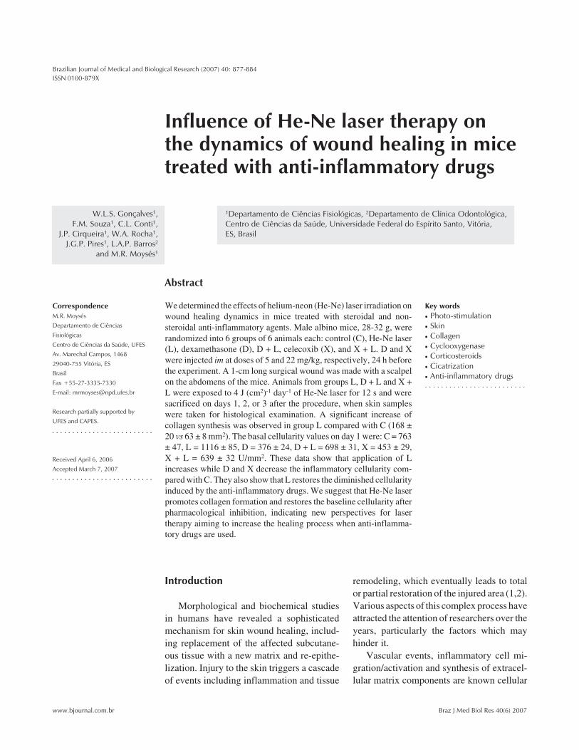
We determined the effects of helium-neon (He-Ne) laser irradiation onwound healing dynamics in mice treated with steroidal and non-steroidal anti-inflammatory agents. Male albino mice, 28-32 g, wererandomized into 6 groups of 6 animals each: control (C), He-Ne laser(L), dexamethasone (D), D + L, celecoxib (X), and X + L. D and Xwere injected im at doses of 5 and 22 mg/kg, respectively, 24 h beforethe experiment. A 1-cm long surgical wound was made with a scalpelon the abdomens of the mice. Animals from groups L, D + L and X +L were exposed to 4 J (cm2)-1 day-1 of He-Ne laser for 12 s and weresacrificed on days 1, 2, or 3 after the procedure, when skin sampleswere taken for histological examination. A significant increase ofcollagen synthesis was observed in group L compared with C (168 ±20 vs 63 ± 8 mm2). The basal cellularity values on day 1 were: C = 763± 47, L = 1116 ± 85, D = 376 ± 24, D + L = 698 ± 31, X = 453 ± 29,X + L = 639 ± 32 U/mm2. These data show that application of Lincreases while D and X decrease the inflammatory cellularity com-pared with C. They also show that L restores the diminished cellularityinduced by the anti-inflammatory drugs. We suggest that He-Ne laserpromotes collagen formation and restores the baseline cellularity afterpharmacological inhibition, indicating new perspectives for lasertherapy aiming to increase the healing process when anti-inflamma-tory drugs are used.
CorrespondenceM.R. MoysésDepartamento de CiênciasFisiológicasCentro de Ciências da Saúde, UFESAv. Marechal Campos, 146829040-755 Vitória, ESBrasilFax +55-27-3335-7330E-mail: [email protected]
Research partially supported byUFES and CAPES.
Received April 6, 2006Accepted March 7, 2007
Key words• Photo-stimulation• Skin• Collagen• Cyclooxygenase• Corticosteroids• Cicatrization• Anti-inflammatory drugs
Introduction
Morphological and biochemical studiesin humans have revealed a sophisticatedmechanism for skin wound healing, includ-ing replacement of the affected subcutane-ous tissue with a new matrix and re-epithe-lization. Injury to the skin triggers a cascadeof events including inflammation and tissue
remodeling, which eventually leads to totalor partial restoration of the injured area (1,2).Various aspects of this complex process haveattracted the attention of researchers over theyears, particularly the factors which mayhinder it.
Vascular events, inflammatory cell mi-gration/activation and synthesis of extracel-lular matrix components are known cellular
878
Braz J Med Biol Res 40(6) 2007
W.L.S. Gonçalves et al.
www.bjournal.com.br
processes intervening in the dynamics ofwound repair (3,4). The repairing processbegins immediately after the injury throughthe release of cytokines, growth factors, somehormones, and several low-molecular weightsubstances from plasma or activated plate-lets (1-4). The main endogenous hormoneinvolved in the dynamics of tissue repair iscortisol, which affects the metabolism ofcarbohydrates and proteins and exerts im-portant anti-inflammatory effects (5,6).
Recent studies have suggested that theearly events in the wound healing processare the most appropriate for useful therapeu-tic interventions, while the most importantrepair failures are those occurring in theinitial phase (3). In this respect, we shouldmention studies concerning the role of stressin the wound healing process, showing thatendogenous cortisol down-regulates pro-in-flammatory cytokine and chemokine expres-sion, which reduces the inflammation ede-ma, cellular recruitment and cell prolifera-tion at the injured site (7-9).
Several experimental and clinical studieshave evaluated the effects of helium-neon(He-Ne) laser therapy on the process of tis-sue regeneration in areas such as skin, bone,skeletal muscle, and the nervous system (10-13), with contradictory results. However,based on clinical experience, some investi-gators have proposed that photo-stimulationwith low-energy laser at certain wavelengthspromotes tissue repair by releasing growthfactors (2,10-15).
Taking into account these controversies,we decided to investigate the possible benefi-cial effects of He-Ne laser irradiation on theskin repair process in mice previously treatedwith steroidal or non-steroidal anti-inflamma-tory drugs, which possibly are able to affectthe release and/or the effects of factors in-volved in the tissue repair process (9,10).
Material and Methods
Experiments were performed on adult
male albino mice, weighing 28-32 g, fromour breeding stock. The animals, 6 to a group,were housed in 0.30-m2 cages under con-trolled conditions of 12-h light periods, tem-perature (~26ºC) and minimal noise. Ani-mals were allowed free access to filteredwater and standard rat chow. All the experi-mental procedures adopted were in accor-dance with the International Guidelines forAnimal Care.
Experimental design
The mice were randomly divided into 6groups of 6 mice each: 1) control (C), 2) He-Ne laser (L), 3) dexamethasone (D), 4) D +L, 5) celecoxib (X), and 6) X + L. Thesegroups were further divided into 24-, 48-and 72-h groups. D and X were injectedintramuscularly (im) at doses of 5 and 22mg/kg, respectively, 24 h before the begin-ning of the experiment.
A surgical skin wound was made with ascalpel on the abdomen of the mice whowere under general anesthesia with 10 mg/kg ketamine + 20 mg/kg xylazine, im. Theskin on the abdominal region was shavedbefore the procedure using an aseptic tech-nique. The surgical incision was standard-ized as follows: 1-cm length (xiphoid appen-dix as reference) and adequate depth to in-clude the epidermis, dermis and abdominalfascia. After the surgical procedure, laserirradiation was applied (see below) to thetreated groups. The animals were then re-turned to their home cages.
Laser treatment
A laser device (KLD-Biosystem®, SãoPaulo, SP, Brazil) containing a continuous-wave He-Ne light emitting at 633 nm and 5mW output was used. Power output wascalibrated by a power meter. In order todetermine the duration of cutaneous appli-cations, the spot size (1.5 cm) was measuredand the quantity of energy density and power
879
Braz J Med Biol Res 40(6) 2007
Topical laser irradiation and wound healing
www.bjournal.com.br
output were determined using the Tuner andHode equation (16). Each animal in the lasergroups received a fixed daily dose of He-Nelaser irradiation at an energy density of 4 J/cm2 applied over a period of 12 s. The laserprobe was positioned to contact the wound.Only one session of laser was applied imme-diately after surgery.
The animals were then submitted to eu-thanasia by ethyl ether inhalation at 24, 48,and 72 h after the procedures and skinsamples were removed for histological andmorphometric analysis.
Histological and morphometric analysis
All skin lesion samples obtained wereimmediately fixed in 10% buffered forma-lin, pH 7, for at least 24 h. After fixation, thesamples were gradually dehydrated in in-creasing ethanol concentrations (70 to 100%),cleared in xylene and embedded in paraffinaccording to routine histological methods.The paraffin-embedded fragments were cutwith an “820” Spence microtome (New York,NY, USA) and 6-µm thick sections wereobtained. Pairs of histological slides werekept in an incubator to dry and the sectionswere then stained with hematoxylin-eosin andMasson’s trichrome for histological analysis.
The histology scoring was based on thedegree of cellular invasion (cellularity),granulation tissue formation, vascularity, andre-epithelization. The code describing eachanimal’s treatment was broken after the pa-thologist completed the scoring and ranking(Table 1).
Histomorphometry was performed usingimages captured and evaluated by a compu-terized Sigma-pro®image (St. Louis, MO,USA) capture system. Images were capturedfrom five randomly chosen optical micro-scopic fields for each histological slide pairusing the digital camera (total magnification100 and 200X) from an Olympus®AX70Plus microscope (Tokyo, Japan). The im-ages were stored and submitted to a count of
inflammatory cells or cellularity (i.e., cellu-lar density) and analysis of collagen forma-tion and of re-epithelization of the ulceratedareas of the wounds at the end of the experi-ment using digital marking (color contrast)as the discriminating parameter.
Drugs
Dexamethasone phosphate salt (Dex-azona™, Bunker, São Paulo, SP, Brazil) andcelecoxib (COX-2 inhibitor; a kind donationfrom Pfizer, Guarulhos, SP, Brazil) wereused. Doses were chosen according to thoseusually reported in the literature. Ketaminehydrochloride (Ketamin™, 5% solution) waspurchased from Cristalia (São Paulo, SP,Brazil). A commercial veterinary prepara-tion of xylazine hydrochloride (Rompun™,Bayer, Rio de Janeiro, RJ, Brazil) was used.The two drugs were placed in the samesyringe for anesthesia.
Statistical analysis
The nature of the variables studied or thevariability of the means was considered us-ing the biostatistics software GraphPad Prism4. Unless otherwise stated, data were ana-lyzed statistically by ANOVA followed bythe Tukey test for multiple comparisons orthe Mann-Whitney U-test for independentsamples, as appropriate. The level of signif-icance was set at P < 0.05.
Table 1. Histological scoring.
Score Criteria
1-3 None to minimal cell accumulation. No granulation tissue or epithelialmigration.
4-6 Thin, immature granulation dominated by inflammatory cells but showingfibroblasts, capillaries, or collagen deposition. Minimal epithelial migration.
7-9 Moderately thick granulation tissue can range from being dominated byinflammation cells to more fibroblast and collagen deposition. Extensiveneovascularization. Epithelium can range from minimal to moderatemigration.
10-12 Thick, vascular granulation tissue dominated by fibroblasts and extensivecollagen deposition. Epithelium partially to completely lining the wound.
880
Braz J Med Biol Res 40(6) 2007
W.L.S. Gonçalves et al.
www.bjournal.com.br
Results
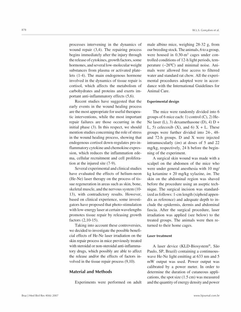
Clinical inspection of skin lesion samplesshowed a regular amount of humid clot onthe surface and the presence of many bloodvessels in the wound area in the C and Dgroups, but not in the other four groups (L, D+ L, X, X + L; data not shown).
The morphological criteria used for scor-ing the material are presented in Table 1.Histological analysis showed that specimensfrom the laser groups (L, D + L, X + L) hadfewer blood clots and displayed more accel-erated re-epithelization, granulation tissueformation and capillary proliferation than
the other groups, such as C (Figure 1). Basedon these morphological criteria (Table 1), itwas observed that the L group displayedimportant healing dynamics after 24 h oftreatment compared to the C group. Laser-treated animals showed significantly higherhistological scores than the respective con-trols, in the first 24 h (7.16 vs 1.38; P <0.0001, Mann-Whitney U-test).
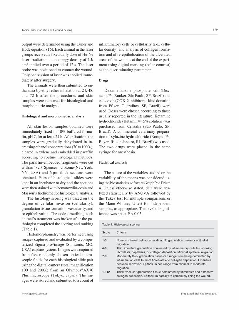
As shown in Figure 2A, the epitheliumsamples from the D (7.3 ± 0.7 mm2) and X(17.1 ± 0.8 mm2) groups showed expectedsignificant differences during the first 24 hwhen compared to the C group (12.6 ± 0.8mm2). D significantly reduced re-epitheliza-tion, while, surprisingly, X increased it. Inthe D + L group, laser irradiation restoredthe re-epithelization phenomenon (15.0 ±0.4 mm2), whereas in the X + L group, laserapplication further decreased the phenome-non (11.0 ± 0.4 mm2). In addition, the Lgroup showed that laser irradiation produceda significant increase (17.3 ± 0.8 mm2) in thewound re-epithelization (Figure 2A).
Collagen synthesis was significantlyhigher in the L (168 ± 20 mm2), D + L (189± 11 mm2), and X + L (185 ± 9 mm2) groups(Figure 2B) and slightly but significantlyattenuated in the D and X groups comparedwith C animals (56 ± 6, 55 ± 5, and 62 ± 8mm2, respectively).
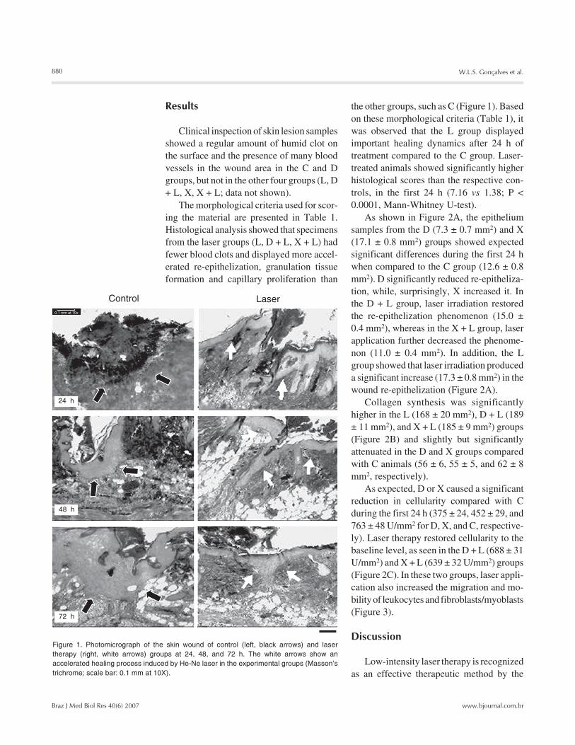
As expected, D or X caused a significantreduction in cellularity compared with Cduring the first 24 h (375 ± 24, 452 ± 29, and763 ± 48 U/mm2 for D, X, and C, respective-ly). Laser therapy restored cellularity to thebaseline level, as seen in the D + L (688 ± 31U/mm2) and X + L (639 ± 32 U/mm2) groups(Figure 2C). In these two groups, laser appli-cation also increased the migration and mo-bility of leukocytes and fibroblasts/myoblasts(Figure 3).
Discussion
Low-intensity laser therapy is recognizedas an effective therapeutic method by the
Figure 1. Photomicrograph of the skin wound of control (left, black arrows) and lasertherapy (right, white arrows) groups at 24, 48, and 72 h. The white arrows show anaccelerated healing process induced by He-Ne laser in the experimental groups (Masson’strichrome; scale bar: 0.1 mm at 10X).
Control Laser
24 h
48 h
72 h
0.1 mm at 10x
881
Braz J Med Biol Res 40(6) 2007
Topical laser irradiation and wound healing
www.bjournal.com.br
FDA, particularly to improve tissue healing(3-6). A large body of evidence from in vitroand in vivo studies has suggested that LILTenhances collagen synthesis (4-6,10), in-creases the motility of keratinocytes (17),releases growth factors (15,17), and pro-motes the transformation of fibroblasts intomyofibroblasts (4-6,15,17-19). On the otherhand, the idea of investigating the early phaseof the skin repair process under the influenceof anti-inflammatory agents (a corticoster-oid or a cyclooxygenase (COX-2) inhibitor)was based on the known pharmacologicalproperties of such drugs and on their broadclinical use and side-effects (20-24).
Studies have shown that during normalwound healing the inflammatory phase lastsup to two days (3,24-26). Cell proliferation,epithelization, granulation tissue formation,and wound contraction occur during the pro-liferative phase. Growth of new epithelialcells across the surface of the wound andcollagen remodeling occur during the matu-ration phase of wound healing, which lastsfor months or even longer. The present re-sults showed that He-Ne laser irradiation ofthe skin of mice produced beneficial effectson the macroscopic aspects of the surgicalwound, such as reduction of humidity, ap-parent vascular modulation and effectivecontrol of the amount of clot in the injuredarea, during the first 24 h. These data there-fore, suggest that laser therapy increased thehealing dynamics compared to control.
Werner and Grose (26) described threedifferent stages of wound repair: i) 12-24 hafter injury the wounded area is filled with ablood clot which is invaded by neutrophils;ii) at days 3-7 after injury, most neutrophilshave undergone apoptosis whereas macro-phages are abundant in the wound tissue atthis stage of repair. Endothelial cells migrateinto the clot, proliferate and form new bloodvessels. Fibroblasts migrate into the woundtissue, where they proliferate and depositextracellular matrix. The new tissue is calledgranulation tissue. Keratinocytes proliferate
Figure 2. Determinations of thevolumetric fractions of the epi-thelium (A), collagen (B), andcellularity (C; neutrophils andfibroblasts/myofibroblasts) ofmouse skin 24 h after treat-ments. C = control; L = He-Nelaser; D = steroidal anti-inflam-matory drug dexamethasone;D+L = dexamethasone plus la-ser; X = non-steroidal anti-in-flammatory drug celecoxib; X+L= celecoxib plus laser. Data arereported as mean ± SEM. *P <0.01 vs C; **P < 0.01 vs D+L; +P< 0.05 vs X+L (Tukey test).
Figure 3. Photomicrography of the skin wound in the dexamethasone (A) and dexametha-sone + laser groups (B) at 24 h. The white arrows point to increased cellularity with theintense presence of mononuclear cells in the infiltrate, predominantly fibroblasts andneutrophils in the skin wound margins (H&E; scale bar: 0.1 mm at 20X).
0.1 mm at 20x 0.1 mm at 20x
882
Braz J Med Biol Res 40(6) 2007
W.L.S. Gonçalves et al.
www.bjournal.com.br
at the wound edge and migrate down theinjured dermis and above the provisionalmatrix; iii) 1-2 weeks after injury the woundis completely filled with granulation tissue.Fibroblasts have transformed into myofi-broblasts, leading to wound contraction andcollagen deposition. The wound is then com-pletely covered with a neoepidermis.
The time course of the present resultsobtained from histological analysis of the Cgroup fit with the picture described by Wernerand Grose (26). However, morphologicalanalysis of the laser therapy groups indicatesthat significant differences do exist in thesegroups, suggesting an acceleration of thehealing dynamics during the first days causedby laser application.
Similar results were obtained by Maiyaet al. (15) in diabetic rats, showing that laser-treated animals healed faster and better thancontrols. Other studies (11,12,14,18,22) us-ing He-Ne low-energy laser have indicatedthat it is mainly the laser energy at 633 nmwavelength that affects the healing dynam-ics, producing changes in the early phase ofthe repair process, i.e., the inflammatoryphase.
In the first hours of healing, the repairevents are directed towards preventing sub-sequent blood loss (hemostasis) and towardsthe formation of a fibrin plug that suppliesthe preliminary matrix for the subsequentprocesses in which platelets adhere to thecollagen in the perivascular space. Such con-tact activates platelets, which releasesbioactive factors that accelerate the migra-tion and proliferation of fibroblasts, a keycell in this process (11,12).
The suggestion that laser affects the earlyevents in the dynamics of wound healingwas partially based on the observed low-intensity laser therapy-induced attenuationof reactive oxygen species production byneutrophils in inflammatory models. Thiswas initially suggested by the study ofFujimaki et al. (20), who described a dimin-ished oxidative stress-induced apoptosis of
neutrophils in acute inflammation (20,27-28). Nevertheless, the basic aspects of oxi-dative stress and the mechanisms by whichreactive oxygen species modulate physiologi-cal and pathological processes, with empha-sis on wound healing, are still motive ofdebate.
As stated before, the early events inwound healing, with special reference to theinflammatory phase, are crucial for the suc-cess of the process, as suggested by studieson the effects of anti-inflammatory drugs(3). It is well known that corticosteroidsdown-regulate pro-inflammatory proteinsand affect gene expression, interfering withalmost all phases of the inflammatory pro-cess (7-9). On the other hand, controlledlaboratory trials have reported that laserphotostimulation can reduce inflammationthrough inhibition of inducible COX-2, lead-ing to a reduction in prostanoid levels (28,29).Additionally, experiments involving vari-ous cell culture stages have shown that laserirradiation at early stages significantly stimu-lates cell proliferation, alkaline phosphataseactivity and osteocalcin gene expression,indicating that laser photostimulation en-hances bone formation in vitro (3,11,19).
The present data (Figure 2B) agree withthose reported by others (3,11-24), showingthat He-Ne (633 nm) laser application in-duces a more mature scar, displaying col-lagen fibers arranged in parallel. Our resultsalso show that the presence of an anti-in-flammatory drug, either a steroidal or a non-steroidal drug, does not inhibit the biostimu-latory effects of laser photostimulation. Itshould be mentioned that re-epithelizationincreased during the first 24 h in the He-Nelaser-treated animals, suggesting an enhance-ment of keratinocytes production, even inthe presence of dexamethasone. Interestingly,celecoxib (X; a COX-2 inhibitor) increasedthe re-epithelization phenomenon, which wasrestored to baseline levels after He-Ne laserirradiation (Figure 2A). Concomitantly, thecellularity counts showed important effects
883
Braz J Med Biol Res 40(6) 2007
Topical laser irradiation and wound healing
www.bjournal.com.br
produced by He-Ne irradiation in the pres-ence of D or X towards the maintenance ofinflammatory cells at the “baseline” level(Figure 2C). It should be mentioned thatrecent studies (30,31) have shown that He-Ne laser irradiation can effectively acceler-ate the expression of tumor growth factor ß1and facilitate changes in leukocyte activityand accumulation of lipids by oxidation prod-ucts. Furthermore, tumor growth factor ß1 isa chemoattractant for neutrophils, macro-phages and fibroblasts (18) and is also asso-ciated with the wound healing defect seen inglucocorticoid-treated animals (26).
Thus, the present findings suggest thatthe stimulatory effects of laser photostimu-lation are related to specific events duringthe first two phases of wound healing, i.e.,the inflammatory phase and the proliferativephase, indicating that the time of interven-tion may be critical and also suggesting thatsatellite cells are major irradiation-respon-sive candidates. The present results showthat He-Ne laser therapy can influence the
behavior of many inflammatory cell types,and that multiple effects can occur simulta-neously and accelerate healing dynamics inthe presence of corticosteroids or non-ste-roidal anti-inflammatory drugs. Addition-ally, our results suggest that He-Ne laserirradiation modulates the early phases of therepair process in vivo. We speculate that thiscould be due to biochemical events in themitochondrial oxidation process and/or toaccumulation of lipid by oxidation products.However, much more research using selec-tive inhibitors and markers will be need toelucidate the exact mechanisms of action oflaser photostimulation at the cellular andmolecular levels.
Acknowledgments
The authors thank Dr. C.A. Redins, Dr.M.A.S. Novaes and Mrs. L. Bressoni (UFES,Vitória) for help with the techniques andmorphological analysis.
References
1. Ishida Y, Watanabe H. Structural study of wound healing in mouseskin with special reference to quantitative changes of cellular con-stituents. Yamagata Med J 1999; 17: 332-342.
2. Werner S, Grose R. Regulation of wound healing by growth factorsand cytokines. Physiol Rev 2003; 83: 835-870.
3. Reddy GK. Photobiological basis and clinical role of low-intensitylasers in biology and medicine. J Clin Laser Med Surg 2004; 22:141-150.
4. Medrado AR, Pugliese LS, Reis SR, Andrade ZA. Influence of lowlevel laser therapy on wound healing and its biological action uponmyofibroblasts. Lasers Surg Med 2003; 32: 239-244.
5. Campana V, Moya M, Gavotto A, Juri H, Palma JA. Effects ofdiclofenac sodium and He:Ne laser irradiation on plasmatic fibrino-gen levels in inflammatory processes. J Clin Laser Med Surg 1998;16: 317-320.
6. Pessoa ES, Melhado RM, Theodoro LH, Garcia VG. A histologicassessment of the influence of low-intensity laser therapy on woundhealing in steroid-treated animals. Photomed Laser Surg 2004; 22:199-204.
7. Mercado AM, Padgett DA, Sheridan JF, Marucha PT. Altered kinet-ics of IL-1 alpha, IL-1 beta, and KGF-1 gene expression in earlywounds of restrained mice. Brain Behav Immun 2002; 16: 150-162.
8. Sheridan JF, Padgett DA, Avitsur R, Marucha PT. Experimental
models of stress and wound healing. World J Surg 2004; 28: 327-330.
9. Pruzanski W, Vadas P. Phospholipase A2 - a mediator betweenproximal and distal effectors of inflammation. Immunol Today 1991;12: 143-146.
10. Posten W, Wrone DA, Dover JS, Arndt KA, Silapunt S, Alam M. Low-level laser therapy for wound healing: mechanism and efficacy.Dermatol Surg 2005; 31: 334-340.
11. de Carvalho PT, Mazzer N, Barbieri CH, Siqueira JFR. Morphomet-ric analysis of the percentage of collagen and number of macro-phages highlighted by immunohistochemistry, in cutaneous woundin diabetic and non-diabetics rats treated through He-Ne laser.Lasers Med Sci 2003; 18 (Suppl 1): S0167 (Abstract).
12. de Carvalho PT, Mazzer N, dos Reis FA, Belchior AC, Silva IS.Analysis of the influence of low-power He-Ne laser on the healing ofskin wounds in diabetic and non-diabetic rats. Acta Cir Bras 2006;21: 177-183.
13. Amaral AC, Parizotto NA, Salvini TF. Dose-dependency of low-energy HeNe laser effect in regeneration of skeletal muscle in mice.Lasers Med Sci 2001; 16: 44-51.
14. Pugliese LS, Medrado AP, Reis SR, Andrade ZA. The influence oflow-level laser therapy on biomodulation of collagen and elasticfibers. Pesqui Odontol Bras 2003; 17: 307-313.
884
Braz J Med Biol Res 40(6) 2007
W.L.S. Gonçalves et al.
www.bjournal.com.br
15. Maiya GA, Kumar P, Rao L. Effect of low intensity helium-neon (He-Ne) laser irradiation on diabetic wound healing dynamics. PhotomedLaser Surg 2005; 23: 187-190.
16. Tuner J, Hode L. It’s all in the parameters: a critical analysis of somewell-known negative studies on low-level laser therapy. J Clin LaserMed Surg 1998; 16: 245-248.
17. Rood PA, Haas AF, Graves PJ, Wheeland RG, Isseroff RR. Low-energy helium neon laser irradiation does not alter human keratino-cyte differentiation. J Invest Dermatol 1992; 99: 445-448.
18. Rocha AM Jr, Oliveira RG, Farias RE, Andrade LCR, Aarestrup FM.Modulation of fibroblast proliferation and inflammatory response bylow-intensity laser in tissue repair process. An Bras Dermatol 2006;81: 150-156.
19. Hawkins DH, Abrahamse H. The role of laser fluence in cell viability,proliferation, and membrane integrity of wounded human skin fibro-blasts following helium-neon laser irradiation. Lasers Surg Med2006; 38: 74-83.
20. Fujimaki Y, Shimoyama T, Liu Q, Umeda T, Nakaji S, Sugawara K.Low-level laser irradiation attenuates production of reactive oxygenspecies by human neutrophils. J Clin Laser Med Surg 2003; 21:165-170.
21. Jiang Y. Thirty cases of sub-healthy state regulated by acupunctureand He-Ne laser vascular irradiation. J Tradit Chin Med 2006; 26:102-103.
22. Lan CC, Wu CS, Chiou MH, Hsieh PC, Yu HS. Low-energy helium-neon laser induces locomotion of the immature melanoblasts andpromotes melanogenesis of the more differentiated melanoblasts:recapitulation of vitiligo repigmentation in vitro. J Invest Dermatol
2006; 126: 2119-2126.23. Simunovic Z, Ivankovich AD, Depolo A. Wound healing of animal
and human body sport and traffic accident injuries using low-levellaser therapy treatment: a randomized clinical study of seventy-fourpatients with control group. J Clin Laser Med Surg 2000; 18: 67-73.
24. Limpanichkul W, Godfrey K, Srisuk N, Rattanayatikul C. Effects oflow-level laser therapy on the rate of orthodontic tooth movement.Orthod Craniofac Res 2006; 9: 38-43.
25. Singer AJ, Clark RA. Cutaneous wound healing. N Engl J Med 1999;341: 738-746.
26. Werner S, Grose R. Regulation of wound healing by growth factorsand cytokines. Physiol Rev 2003; 83: 835-870.
27. Rojkind M, Dominguez-Rosales JA, Nieto N, Greenwel P. Role ofhydrogen peroxide and oxidative stress in healing responses. CellMol Life Sci 2002; 59: 1872-1891.
28. Muller-Decker K. Cyclooxygenases in the skin. J Dtsch DermatolGes 2004; 2: 668-675.
29. Sakurai Y, Yamaguchi M, Abiko Y. Inhibitory effect of low-level laserirradiation on LPS-stimulated prostaglandin E2 production and cy-clooxygenase-2 in human gingival fibroblasts. Eur J Oral Sci 2000;108: 29-34.
30. Sun XH, Wang R, Zhang XY. Effects of He-Ne laser irradiation onthe expression of transforming growth factor beta1 during experi-mental tooth movement in rabbits. Shanghai Kou Qiang Yi Xue2006; 15: 52-57.
31. Klebanov GI, Chichuk TV, Osipov AN, Vladimirov I. The role of lipidperoxidation products in the effect of He-Ne laser on human bloodleukocytes. Biofizika 2005; 50: 862-866.