Embed Size (px)
Citation preview

AGE-RELATED FUNCTIONAL AND HISTOPATHOLOGICALCHANGES OF THE EAR IN THE MPS I MOUSE
Patricia A. Schachern1, Sebahattin Cureoglu1, Vladimir Tsuprun1, Michael M. Paparella1,2,and Chester Whitley3,41 Department of Otolaryngology, Otitis Media Research Center, University of Minnesota, Minneapolis, MN
2 Minnesota Ear Head and Neck Clinic, Minneapolis, MN
3 Department of Pediatrics University of Minnesota, Minneapolis, MN
4 Department of Human Genetics University of Minnesota, Minneapolis, MN
AbstractObjective— Mucopolysaccharidosis type I (MPS I) is an autosomal recessive disorder caused bya mutation in the gene encoding the enzyme α-L-iduronidase. This enzyme is responsible fordegradation of dermatan and heparan sulfates. Enzyme deficiency results in their accumulation inlysosomes of virtually all organs, resulting in severe somatic and neurological changes. Clinicalfindings of otitis media with mixed hearing loss are common. Cellular and molecular mechanismsof ear pathology and hearing loss are not understood. The purpose of this study is to describe theage-related audiologic and histopathologic changes of the ear in the mouse model of MPS I.
Methods— Auditory brainstemresponses (ABR) were obtained to clicks and tone bursts at 1-32kHz, and pathological changes to middle and inner ears were studied with light and electronmicroscopy in fifty-three mice that included: 1) wild type (+/+) - five at 2 months, five at 4 to 6months, and five at 13 to 19 months; 2) heterozygotes (+/−) - four at 2 months; five at 4 to 6 months;and eight at 13 to 19 months; and 3) homozygotes (−/−); five at 2 months; six at 4 to 6 months; andfive at 13 to 19 months. Histopathology was also done on five newborn −/− mice.
Results— In newborns no lysosomal storage was observed and the ear appeared age appropriatelynormal. In all other −/− mice, cells with lysosomal storage vacuoles were observed in spiral ligament,spiral prominence, spiral limbus, basilar membrane, epithelial and mesothelial cells of Reissner’smembrane, endothelial cells of vessels, and some ganglion cells; their number increased with aging.Hair cell loss was not observed at 2 or 6 months, but there was total loss of the organ of Corti in year-old mice. Hearing of −/− mice was significantly decreased at all ages compared to +/+ and +/−.Hearing loss progressed from mild to moderate loss at 2 months to profound at 6 months and totaldeafness by one year of age.
Conclusions— Progressive age-related changes suggest early therapeutic intervention to preventsensory cell damage and hearing loss.
KeywordsMPS I mouse; middle ear; cochlea; hearing loss; ear pathology; auditory brainstem response
Send Correspondence to: Patricia A. Schachern, Room 226 Lions Research Building, 2001 6th Street SE, Minneapolis, MN 55455Publisher's Disclaimer: This is a PDF file of an unedited manuscript that has been accepted for publication. As a service to our customerswe are providing this early version of the manuscript. The manuscript will undergo copyediting, typesetting, and review of the resultingproof before it is published in its final citable form. Please note that during the production process errors may be discovered which couldaffect the content, and all legal disclaimers that apply to the journal pertain.
NIH Public AccessAuthor ManuscriptInt J Pediatr Otorhinolaryngol. Author manuscript; available in PMC 2007 August 7.Published in final edited form as:
Int J Pediatr Otorhinolaryngol. 2007 February ; 71(2): 197–203.
NIH
-PA Author Manuscript
NIH
-PA Author Manuscript
NIH
-PA Author Manuscript

INTRODUCTIONMucopolysaccharidosis type I (MPS I) is one of several variants of lysosomal storage diseasesthat involve the accumulation of various mucopolysaccharides. It is an autosomal recessivelyinherited disorder caused by a mutation in the gene encoding the enzyme α-L-iduronidase[1]. This enzyme is responsible for the degradation of dermatan sulfate and heparan sulfateglycosaminoglycans (GAGs) and its deficiency results in accumulation of undegraded GAGsin lysosomes. Due to the wide distribution of GAGs in the body, virtually all organs are affectedresulting in severe somatic and neurologic changes. Otolaryngological manifestations includerecurrent middle ear effusions [2,3] and progressive mixed hearing loss [2]. Murine models ofMPS, deficient in specific enzymes are available. Functional [4] and histopathological changes[4,5] of the ear have been described for the murine model of MPS VII. Liu et al. [6] describedreduced hearing of the MPS I mouse at 8 months of age. To our knowledge, no one has studiedthe age-related functional and histopathological changes in the ears of a murine model of MPSI. The aim of this study was to determine functional and histopathological changes with ageing.
MATERIALS AND METHODSFifty-three mice, newborn to 19 months of age were included in this study. For +/+ mice, therewere five at 2 months, five at 4 to 6 months, and five at 13 to 19 months of age. For +/− mice,there were four at 2 months; five at 4 to 6 months; and eight at 13 to 19 months of age. For −/− mice, there were five newborn; five at 2 months; six at 4 to 6 months; and five at 13 to 19months of age. The care and use of animals used in this study were approved by the InstitutionalAnimal Care and Use Committee of the University of Minnesota. All animals were anesthetizedintraperitoneally with a combination of ketamine (40mg/kg) and acepromazine (0.5 mg/kg)prior to auditory brainstem response (ABR) recordings (supplemental doses, 30 % of the initialamount, were administered at intervals of 45 min). Animals were anesthetized again, prior tosacrifice. Newborn animals did not have auditory testing.
Auditory Brainstem ResponseBody temperature was actively maintained at 37° C by placing the animal on a thermal blanketand keeping the test room at a high ambient temperature. ABRs were collected by differentiallyamplifying voltages presented at subdermal needle electrodes using a standard vertex positive,ipsilateral mastoid-negative montage. ABR recordings, in response to calibrated acousticsignals, were obtained in mice to verify auditory sensitivity to rarefaction clicks and tone burstsover a 1–32 kHz range. Total tone durations were from 1 to 4 ms and consisted of Blackmanrise/fall times and no plateau. Five hundred twelve presentations were averaged at eachintensity. The gain of the physiologic amplifier was set to 100,000X for ABRs. Threshold wasdetermined in 5 dB steps of decreasing stimulus intensity, until waveforms lost reproduciblemorphology. Analysis of variance (ANOVA) was used to test any differences of ABRthresholds using SPSS software (SPSS Inc, IL). All data were expressed as the mean ± standarderror of mean. Differences in thresholds between IDUA −/−, +/− and +/+ mice were analyzedusing the non-parametric Wilcoxon rank sum test. Statistical significance was defined asP<0.05. Following ABR, mice were decapitated and the left and right bulla removed. One sidewas processed for light microscopy and the other for electron microscopy.
HistopathologyParaffin embedding—The bulla was opened and filled with 4% paraformaldehyde. Fixationwas continued by emersion for 2 hours. Samples were decalcified in 10% EDTA on a rotatorin a cold room for 3 days. EDTA was changed daily. Samples were dehydrated in a gradedseries of ethanol, followed by two changes in xylene, and embedded in paraffin. Sections werecut at a thickness of 5 μm and stained with hematoxylin and eosin.
Schachern et al. Page 2
Int J Pediatr Otorhinolaryngol. Author manuscript; available in PMC 2007 August 7.
NIH
-PA Author Manuscript
NIH
-PA Author Manuscript
NIH
-PA Author Manuscript

Epoxy embedding—The bulla was opened and the cochlea perfused via the oval and roundwindows with 2% glutaraldehyde in 0.1M phosphate buffer (pH 7.4). Cochleae were notperfused in newborn mice. Fixation was continued by emersion for 2 hours. Samples weredecalcified in 10% EDTA on a rotator in a cold room for 3 days. EDTA was changed daily.Samples were washed in phosphate buffer and post-fixed in 1% OsO4 in phosphate buffer (pH7.4) for 1 hour. They were washed again in buffer, dehydrated in a graded series of ethanol,followed by propylene oxide, and embedded in epoxy resin. Samples were cut at a thicknessof 1 μm and stained with Toluidine blue for light microscopic assessment. For electronmicroscopy, samples were cut at a thickness of 20 nm, stained with uranyl acetate and leadcitrate, and examined with a JEOL 100S electron microscope.
RESULTSAuditory Brainstem Response
Hearing thresholds of +/+, +/−, and −/− mice at 2 months of age were 30 dB (SE: 1.8), 31.8dB (SE: 3.5), and 55.8 dB (SE: 3.7), respectively (Fig. 1a). There was a significant differencebetween the −/− group and the other groups. Hearing thresholds of +/+, and +/− mice at 6months of age were 34 dB (SE: 2.3) and 37 dB (SE: 2.3) respectively. In −/− mice at 6 monthsof age, hearing threshold was elevated at frequencies of 12 kHz and 16 kHz; however, micewere totally deaf above and below these frequencies (Fig. 1b). Hearing thresholds of +/+ and+/− mice at one year of age were 54.3 dB (SE: 2.8) and 51.8 dB (SE: 1.9), respectively (Fig.1c). No hearing was detected at any frequency in the −/− group at one year of age. There wasa significant difference between the −/− group and the other groups at all ages and frequencies(Fig. 1a–c).
HistopathologyFindings in the +/+ and +/− animals were similar at all ages. Otitis media was not observed atany age. Organs of Corti and stria vascularis appeared normal in the 2 and 6-month animals.At one year of age, there were occasional missing outer hair cells. In the −/− mice, otitis mediawas observed at 2 months, 6 months and one year of age (Fig. 2). Cells containing lysosomalgranules and vacuoles were not observed in the inner ears of newborn mice. Cell containinglysosomal granules and vacuoles were observed in mice at two months of age in the area ofthe Type I, II, III and IV fibrocytes of the spiral ligament (Fig. 3). The area of the type IVfibrocytes was affected most and Type III least. Cells containing lysosomal storage were alsoseen in the spiral prominence, spiral limbus (Fig. 4), mesothelial cells lining the basilarmembrane (Fig. 5), both epithelial and mesothelial cells of Reisnner’s membrane (Fig. 6 a,b),endothelial cells of vessels, and some ganglion cells (Fig. 7). Cell with lysosomal storagegranules were not observed in the stria vascularis or hair cells (Fig. 8a,b) at any age. At twomonths of age, the hair cells and stria vascularis appeared normal. Findings in animals at sixmonths of age were similar to those at 2 months, but with an increase in lysosomal vacuoles.In all animals at one year of age or older, the organs of Corti and neurofilaments were missing,however, the morphology of the stria vascularis appeared normal (Fig. 9 a,b).
DISCUSSIONMucopolysaccharidosis type I is the most severe form of the family of lysosomal storagedisorders. Most organs of the body are affected with severe mental retardation and death inchildhood. Otitis media is a universal problem with this disease and is often a presentingsymptom [2,3]. Clinical [2,3] and histopathological findings [7–9] show a mixed hearing loss,however, mental retardation, due to changes of the cerebral cortex, often confounds hearingtesting, and the pathological component behind the sensorineural loss is not known. Mouse
Schachern et al. Page 3
Int J Pediatr Otorhinolaryngol. Author manuscript; available in PMC 2007 August 7.
NIH
-PA Author Manuscript
NIH
-PA Author Manuscript
NIH
-PA Author Manuscript

models of this disorder may give some insight into the histopathological correlates ofsensorineural hearing loss.
Functional and histopathological findings of the ear have been reported in a murine model ofMPS VII. Mucopolysaccharidosis VII is caused by a deficiency of β-glucoronidase [10]. It isa very rare form of mucopolysaccharidosis, which shares many of the same, but milder clinicalfeatures as MPS I including hearing impairment [11]. Ohlemiller, et al. [4] studied thefunctional and histopathological changes of the MPS VII murine model from 1.0 to 7.5 monthsof age. Although hearing thresholds were abnormal, they found no evidence of cell loss withinthe organ of Corti. Berry et al. [5] found some focal loss of hair-cell stereocilia in young adultswith damage increasing with age in older animals with MPS VII. In our study of the MPS Imouse, we observed aged-related pathological changes both in the middle and inner ears. Wefound no cochlear cell loss at 6 months of age, but total loss of the organ of Corti was seen inthe 12-month mice. The difference in our pathological findings of ageing animals comparedto the MPSV II model could be because MPS I is the most severe form of the MPS storagediseases. Another reason may be because we observed a progression of disease with aging, andthe oldest animals in our study were more aged than those in the MPS VII studies. Berry et al.[5] described lysosomal distension in inner hair cells; however, Ohlemiller et al. [4] did notobserve lysosomal granules in any hair cells in animals with MPS VII. We also did not findlysosomal granules in hair cells, indicating damage to the hair cells might be secondary to thelysosomal storage and degeneration of other cells.
Fibrocytes of the spiral limbus and spiral ligament have been reported to express carbonicanhydrase and Na,K-ATPase [12], connexins [13], Na-K-Cl co-transporter [14], and chloride-iodide transport protein [15] and are, thus, thought to play a significant role in maintaining theion homeostasis of the cochlea. In a study of C57 BL/6 mice, Hequembourg and Liberman[16] found that Type IV fibrocytes degeneration preceded loss of hair cells and neurons in bothspace and time. They found a hearing threshold shift in 7-month animals that did not have haircell loss and attributed this shift to reduction in the endolymphatic potential resulting from lossof Type IV fibrocytes. Delprat et al. [17] discovered otospiralin, an ear specific proteinproduced by fibrocytes of the spiral limbus and spiral ligament. They found that down-regulation of otospiralin resulted in rapid and severe degeneration of the organ of Corti leadingto irreversible deafness. In our 6-month −/− animals, there was a profound hearing loss withno loss of cochlear hair cells, but with damage and loss of fibrocytes in the lateral cochlearwall. This was followed at 12 months by total loss of the organ of Corti and neurons. Giventhe variety of important functions of these fibrocytes, related to the maintenance of the ioniccomposition of the endolymph and perilymph, it may be that damage of the hair cells in MPSI mice occurred secondary to the damage of these fibrocytes. The high level of lysosomalstorage granules in cells of Reissner’s membrane, which separates the endolymph from theperilymph, may also cause disturbance of ion and fluid homeostasis in the cochlea contributingto the loss of sensory cells.
In our study, the changes we observed in this model were progressive overtime and included:1) ears that appeared age appropriately normal at birth; 2) lysosomal storage with mild hearingloss and retention of sensory cells at 2 months; 3) lysosomal storage with a profound hearingloss and retention of sensory cells at 6 months; and 4) lysosomal storage with total loss of bothhearing and sensory cells at one year. Liu et al. [6] studied retroviral vector-based gene therapyof neonatal MPS I mice and found that those mice that received high dose therapy at birth hadnear-normal hearing at 8 months of age compared to non-treated mice who had profoundlyreduced hearing. We have observed therapeutic responses in other organs of newborn micetreated by AAV and lentiviral gene therapy (18,19). These findings suggest that early diagnosisand therapeutic intervention are critical to the prevention of sensory cell damage and hearingloss in patients with this disorder. Since newborn screening of MPS is currently not accepted
Schachern et al. Page 4
Int J Pediatr Otorhinolaryngol. Author manuscript; available in PMC 2007 August 7.
NIH
-PA Author Manuscript
NIH
-PA Author Manuscript
NIH
-PA Author Manuscript

practice, target screening of a high-risk population should identify many affected individuals(20). In our study of human temporal bones from patients with MPS I, we found residual fetalmesenchyme filling the middle ears of all temporal bones, even in patients as old as 6 years ofage [7]. This suggests that these children would likely not pass newborn hearing screening.We recommend testing every child who fails newborn hearing screening for MPS disease.
Acknowledgements
This work was supported in part by NIH-NIDCD Grant P30 DC04660, NIH-NICHD Grant P01 032652, theInternational Hearing Foundation, the Hubbard Foundation and the Starkey Foundation. We thank Carolyn Sutherlandfor her technical assistance and Brenda Konair for animal care and breeding.
References1. Neufeld, EF.; Muenzer, J. The mucopolysaccharidoses. In: Scriver, BA.; Sly, WS.; Vale, D., editors.
Metabolic and Molecular Basis of Inherited Disease. New York: McGraw-Hill; 2001. p. 3421-3452.2. Simmons MA, Bruce IA, Penny S, Wraith E, Rothera MP. Otorhinolaryngological manifestations of
the mucopolysaccharidoses. Int J Pediatr Otorhinolaryngol 2005;69:589–595. [PubMed: 15850680]3. Motamed M, Thorne S, Narula A. Treatment of otitis media with effusion in children with
mucopolysaccharidoses. Int J Pediatr Otorhinolaryngol 2000;53:121–124. [PubMed: 10906517]4. Ohlemiller KK, Hennig AK, Lett JM, Heidbreder AF, Sands MS. Inner ear pathology in the
mucopolysaccharidosis VII mouse. Hear Res 2002;16:969–84.5. Berry CL, Vogler C, Galvin NJ, Birdenmeier EH, Sly WS. Pathology of the ear in murine
mucopolysaccharidosis type VII. Morphologic correlates of hearing loss. Lab Invest 1994;71:438–444. [PubMed: 7933993]
6. Liu Y, Xu L, Hennig AK, Kovacs A, Fu A, Chung S, Lee D, Wang B, Herati RS, Olgilvie JM, Cai S,Ponder KP. Liver-directed neonatal gene therapy prevents cardiac, bone, ear, and eye disease inmucopolysaccharidosis I mice. Mol Ther 2005;11:35–47. [PubMed: 15585404]
7. Schachern PA, Shea DA, Paparella MM. Mucopolysaccharidosis I-H (Hurlers Syndrome) and humantemporal bone pathology. Ann Otol Rhinol Laryngol 1984;93:65–69. [PubMed: 6422830]
8. Komura Y, Kaga K, Ogawa Y, Yamaguchi T, Tsuzuku T, Suzuki JI. ABR and temporal bone pathologyin Hurler’s disease. Int J Pediatr Otorhinolaryngol 1998;43:179–188. [PubMed: 9578128]
9. Grocer C, Linthicum FH. Temporal bone histopathology case of the month – Hurler disease. OtolNeurotol 2004;25:81–82. [PubMed: 14724498]
10. Sly WS, Quinton BS, McAlister WH, Rimoin DL. Beta-glucuronidase deficiency: report of clinical,radiologic, and biochemical features of a new mucopolysaccharidosis. J Pediatr 1973;82:249–257.[PubMed: 4265197]
11. Simmons MA, Bruce IA, Penney S, Wraith E, Rothera MP. Otorhinolaryngological manifestationsof the mucopolysaccharidoses. J Pediatr Otorhinolaryngol 2005;69(5):589–595.
12. Spicer SS, Schulte BA. Differentiation of inner ear fibrocytes according to their ion transport relatedactivities. Hear Res 1991;56:53–64. [PubMed: 1663106]
13. Kikuchi T, Kimura R, Paul DL, Takasaka T, Adams JC. Gap junction systems in the mammaliancochlea. Brain Res Rev 2000;32:163–166. [PubMed: 10751665]
14. Crouch JJ, Sakaguchi N, Lytle C, Schulte BA. Immunohistochemical localization of the Na-K-Cl co-transporter (NKCC1) in the gerbil inner ear. J Histochem Cytochem 1991;45:773–778. [PubMed:9199662]
15. Scott DA, Wang R, Kreman TM, Sheffield VC, Karniski LP. The Pendred syndrome gene encodesa chloride-iodide transport protein. Nat Genet 1999;21:440–443. [PubMed: 10192399]
16. Hequembourg S, Liberman MC. Spiral ligament pathology: a major aspect of age- related cochleardegeneration in C57BL/6 mice. JARO 2001;2:118–129. [PubMed: 11550522]
17. Delprat B, Boulanger A, Wang J, Beaudoin V, Guitton MJ, Venteo S, Dechesne CJ, Pujol R, Lavigne-Rebillard M, Puel J, Hamer CP. Downregulation of otospiralin, a novel inner ear protein, causes hair-cell degeneration and deafness. J Neurosci 2002;22:1718–1725. [PubMed: 11880501]
18. Hartung SD, Frandsen JL, Pan D, Konair BL, Graupman P, Gunther R, Low WC, Whitley CB, McIvorRS. Correction of metabolic, craniofacial, and neurologic abnormalities in MPS I mice treated at
Schachern et al. Page 5
Int J Pediatr Otorhinolaryngol. Author manuscript; available in PMC 2007 August 7.
NIH
-PA Author Manuscript
NIH
-PA Author Manuscript
NIH
-PA Author Manuscript

birth with adeno-associated virus vector transducing the human alpha-L-iduronidase gene. Mol Ther2004;9(6):866–875. [PubMed: 15194053]
19. Pan D, Gunther R, Low W, Larson BL, Walkley SU, Frandsen JL, Kafri T, McIvor RS, Whitley CB.Metabolic and neuropathologic correction of Hurler syndrome by a single intravenous injection oflentiviral vector to newborn mice. Mol Ther 2003;7(5):S76.
20. Whitley CB, Spielmann RC, Herro G, Teragawa SS. Urinary glycosaminoglycan excretion quantifiedby an automated semimicro method in specimens conveniently transported from around the globe.Mol Genet Metab 2002;75:56–64. [PubMed: 11825064]
Schachern et al. Page 6
Int J Pediatr Otorhinolaryngol. Author manuscript; available in PMC 2007 August 7.
NIH
-PA Author Manuscript
NIH
-PA Author Manuscript
NIH
-PA Author Manuscript

Figure 1.Comparison of ABR thresholds in mice: a) 2 months of age; b) 6 months of age; and c) 12months of age. Hearing of −/− mice was significantly decreased at all ages compared to +/+and +/−. Hearing loss progressed from mild to moderate loss at 2 months to total deafness by12 months.
Schachern et al. Page 7
Int J Pediatr Otorhinolaryngol. Author manuscript; available in PMC 2007 August 7.
NIH
-PA Author Manuscript
NIH
-PA Author Manuscript
NIH
-PA Author Manuscript

Figure 2.Otitis media was seen in −/− mice at all time periods. In this animal at 1-year of age, middleear effusion (arrow) and granulation tissue (G) can be seen in the middle ear. Staining withHematoxylin and eosin. Original magnification X20.
Schachern et al. Page 8
Int J Pediatr Otorhinolaryngol. Author manuscript; available in PMC 2007 August 7.
NIH
-PA Author Manuscript
NIH
-PA Author Manuscript
NIH
-PA Author Manuscript

Figure 3.Lysosomal granules (arrowheads) and vacuoles (arrows) can be seen in the type IV fibroblastsof the spiral ligament (SL) in this one-year-old −/− mouse. Staining with Toluidine blue.Original magnification: X1,000.
Schachern et al. Page 9
Int J Pediatr Otorhinolaryngol. Author manuscript; available in PMC 2007 August 7.
NIH
-PA Author Manuscript
NIH
-PA Author Manuscript
NIH
-PA Author Manuscript

Figure 4.There are lysosomal granules (arrowheads) in the spiral limbus (SpL) in this −/− mouse at 1-year of age. Staining with Toluidine blue. Original magnification: X1,000.
Schachern et al. Page 10
Int J Pediatr Otorhinolaryngol. Author manuscript; available in PMC 2007 August 7.
NIH
-PA Author Manuscript
NIH
-PA Author Manuscript
NIH
-PA Author Manuscript

Figure 5.In this 2-month-old −/−, the mesothelial cells of the basilar membrane (BM) are distended withlysosomal vacuoles (arrows). Staining with Toluidine blue. Original magnification: X1,000.
Schachern et al. Page 11
Int J Pediatr Otorhinolaryngol. Author manuscript; available in PMC 2007 August 7.
NIH
-PA Author Manuscript
NIH
-PA Author Manuscript
NIH
-PA Author Manuscript

Figure 6.a) Both the mesothelial and epithelial cells of Reissner’s membrane contain lysosmal vacuoles(arrows) in this 2-month-old −/−. Staining with Toluidine blue. Original magnification:X1,000.; b) electron micrograph showing lysosomal storage vacuoles (arrows). Staining withuranyl acetate and lead citrate. Original magnification: X6,000.
Schachern et al. Page 12
Int J Pediatr Otorhinolaryngol. Author manuscript; available in PMC 2007 August 7.
NIH
-PA Author Manuscript
NIH
-PA Author Manuscript
NIH
-PA Author Manuscript

Figure 7.In this 6-month-old −/−, lysosomal vacuoles (arrowheads) were observed in the spiral ganglioncells (SG). Staining with Toluidine blue. Original magnification: X600.
Schachern et al. Page 13
Int J Pediatr Otorhinolaryngol. Author manuscript; available in PMC 2007 August 7.
NIH
-PA Author Manuscript
NIH
-PA Author Manuscript
NIH
-PA Author Manuscript

Schachern et al. Page 14
Int J Pediatr Otorhinolaryngol. Author manuscript; available in PMC 2007 August 7.
NIH
-PA Author Manuscript
NIH
-PA Author Manuscript
NIH
-PA Author Manuscript
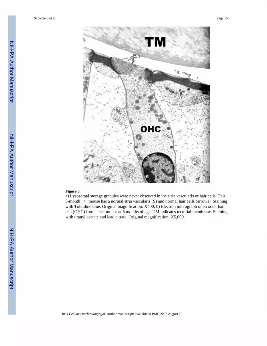
Figure 8.a) Lysosomal storage granules were never observed in the stria vascularis or hair cells. This6-month −/− mouse has a normal stria vascularis (S) and normal hair cells (arrows). Stainingwith Toluidine blue. Original magnification: X400; b) Electron micrograph of an outer haircell (OHC) from a −/− mouse at 6 months of age. TM indicates tectorial membrane. Stainingwith uranyl acetate and lead citrate. Original magnification: X5,000.
Schachern et al. Page 15
Int J Pediatr Otorhinolaryngol. Author manuscript; available in PMC 2007 August 7.
NIH
-PA Author Manuscript
NIH
-PA Author Manuscript
NIH
-PA Author Manuscript
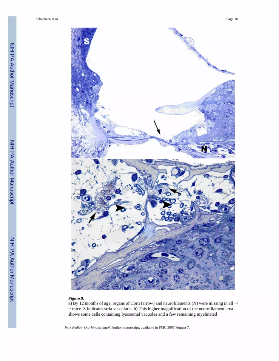
Figure 9.a) By 12 months of age, organs of Corti (arrow) and neurofilaments (N) were missing in all −/− mice. S indicates stria vascularis. b) This higher magnification of the neurofilament areashows some cells containing lysosomal vacuoles and a few remaining myelinated
Schachern et al. Page 16
Int J Pediatr Otorhinolaryngol. Author manuscript; available in PMC 2007 August 7.
NIH
-PA Author Manuscript
NIH
-PA Author Manuscript
NIH
-PA Author Manuscript

neurofilaments. Staining with Toluidine blue. Original magnification: X400 (a) and X1,000(b).
Schachern et al. Page 17
Int J Pediatr Otorhinolaryngol. Author manuscript; available in PMC 2007 August 7.
NIH
-PA Author Manuscript
NIH
-PA Author Manuscript
NIH
-PA Author Manuscript


