Embed Size (px)
Citation preview
Proteomics 2013, 13, 3117–3123 3117DOI 10.1002/pmic.201300108
RESEARCH ARTICLE
Amino-functionalized macroporous silica for efficient
tryptic digestion in acidic solutions
Jinrui Gan1, Kun Qian1, Jingjing Wan1, Liang Qiao2, Weichao Guo1, Pengyuan Yang1,Hubert H. Girault2 and Baohong Liu1
1 Department of Chemistry, Institute of Biomedical Sciences and State Key Lab of Molecular Engineering ofPolymers, Fudan University, Shanghai, China
2 Laboratoire d’Electrochimie Physique et Analytique, Ecole Polytechnique Federale de Lausanne, Lausanne,Switzerland
Amino-functionalized macroporous silica foam (NH2-MOSF) has been developed as a hostreactor to realize highly efficient proteolysis in acidic solutions where normal tryptic reactionscannot occur. The digestion protocol consists simply of adding the functionalized NH2-MOSFinto the protein and trypsin solutions without altering the bulk pH or preloading the enzymeson the materials. With this protocol, digestion of sample fractions from LC can be efficientlyrealized in the acidic solutions directly. Digestion of a protein fraction extracted from rat livertissue after LC separation was performed to illustrate this principle, where 103 proteins weresuccessfully identified at pH 3 after 1.5 h of tryptic digestion.
Keywords:
Acidic solutions / Amino-functionalized macroporous silica / Mass spectrometry /Nanoproteomics / Peptide mass fingerprinting
Received: March 18, 2013Revised: June 23, 2013
Accepted: August 28, 2013
� Additional supporting information may be found in the online version of this article atthe publisher’s web-site
1 Introduction
Modern proteome research at the levels of organelles, cells,tissues, organs, and organisms relies on a wide variety oftechniques to detect proteins and elucidate their functionseither in a healthy or disease state [1, 2]. Among all thesetechniques, MS-based analyses are extensively used both inlaboratory-based and clinical experiments for the discoveryof diagnostic, prognostic, and therapeutic protein biomark-ers [3, 4]. To date, comprehensive proteome information canbe obtained by analyzing fragmental peptides from protein di-gests, making proteolysis an essential procedure in many MS-based proteomics methods [5, 6]. Among various proteases,
Correspondence: Professor Baohong Liu, Department of Chem-istry, Fudan University, Shanghai 200433, ChinaE-mail: [email protected]: +86-21-65641740
Abbreviations: APTS, aminopropyltriethoxysilane; SEM, scan-ning electron microscopy; TEM, transmission electron micro-graphs
trypsin is most widely used, which cleaves peptide chainsmainly at the carboxyl side of the amino acids lysine or argi-nine. However, trypsin shows the highest biological activityat pH 8 and 37�C, and the tryptic digestions take place ratherslowly in neutral solutions and hardly in acidic conditions.This is a limitation for high-throughput analyses that involvecoupling proteolysis with separation techniques.
LC-MS/MS is universally used to perform high-throughput surveys of proteomes from a biological samplethrough MS/MS sequencing [7–9]. Basically, the complexbiological samples are separated by LC, where fractions arecollected, digested, and detected by LC-MS/MS. Consideringthe optimum conditions for proteolysis, the acidic LCfractions have to be lyophilized and redissolved in a weakbasic buffer (pH 8), which requires several steps in theworkflow. To circumvent the multistep pretreatment foradjusting pH, the development of novel methods capableof retaining enzymatic activity in different pH conditionsespecially in acids and increasing proteolytic efficiency arevital for large-scale proteome and peptidome research [5, 6].In order to retain enzymatic activity and accelerate enzymatic
C© 2013 WILEY-VCH Verlag GmbH & Co. KGaA, Weinheim www.proteomics-journal.com
3118 J. Gan et al. Proteomics 2013, 13, 3117–3123
reactions, various microfluidic reactors and nanoreactorshave been developed in recent proteomic protocols [10–21].Benefiting from the ordered pore structure, high surfacearea and high pore volumes, porous materials have beendemonstrated to be good candidates for nanoreactors [16–21].Additionally, with the surface modification of silica materi-als, selective enrichment and isolation of posttranslationalmodified peptides or proteins can be obtained [22–24].
In this study, we have developed a nanoreactor for trypticdigestion of proteins in acidic solutions, based on the newlydesigned amino-functionalized macroporous silica foams(MOSFs) [25], ∼100 nm in pore diameters, denoted NH2-MOSF. NH2-MOSF was added into the fractions of LC sys-tems to carry out digestions directly following separation,even when the bulk solutions are acidic. This NH2-MOSF-based protocol was applied to the digestion of biological sam-ples separated by RPLC. Successful digestion of one proteinfraction from rat liver was achieved in a solution at pH 3. Thepresent approach greatly simplifies the digestion step duringanalysis of proteomes.
2 Materials and methods
2.1 Chemicals
Triblock copolymer EO20PO70EO20 (denoted P123, whereEO is poly(ethylene oxide) and PO is poly(propylene ox-ide)), potassium chloride (KCl, 99.5%, AnalaR, Australia),fuming hydrochloric acid (37%, Lab-Scan, AnalyticalScience, Thailand), ethanol (99.5%, Asia Pacific Spe-cialty, Australia), ammonium bicarbonate, dry toluene,3-aminopropyltriethoxysilane (APTS), and tetramethyl-orthosilicate (99%) were purchased from Aldrich. Trypsin(from bovine pancreas) and myoglobin (horse heart) wereobtained from Sigma. 2,5-DHB (99.9%), CHCA (99%), ACN(99.9%), and TFA (99.8%) were purchased from Merck. Thenormal rat liver cytoplasm sample was obtained from theLiver Cancer Institute of Zhongshan Hospital, Fudan Univer-sity. Deionized water (18.4 M�·cm) used for all experimentswas obtained from a Milli-Q system (Millipore, Bedford, MA,USA).
2.2 Synthesis and characterization of macroporous
silica and the amino-functionalized materials
The macroporous silica (MOSF) used in this study were pre-pared according to the method previously reported [25]. Thesynthesis of MOSF was conducted at 35�C in pH 5.0 NaAc-HAc buffer solution, using tetramethylorthosilicate as a silicasource and P123 as a template agent. For standard modi-fication [26], APTS was used as the coupling agent. MOSFmaterials were firstly dried and degassed at 110�C, and thendispersed in dry toluene (0.1 g MOSF in 30 g toluene). Anexcess of APTS (3 mL) was added under stirring and the
mixture was stirred and refluxed for 24 h at 110�C. The re-sulting solid was filtered and washed sequentially by toluene,dichloromethane, and ethanol three times. The final NH2-MOSF products were obtained after drying at 70�C overnight.A zeta-potential meter (Malvern Zetasizer Nano) was used tomeasure the zeta potentials of the materials in an ammo-nium bicarbonate buffer solution (pH ∼ 8) at 25�C. The in-frared spectra were obtained with a FT-IR360 (Nicolet). Nitro-gen sorption isotherms were obtained using Quantachrome’sQuadrasorb SI analyzer at 77 K. Prior to measurement, thesamples were degassed at 120�C for at least 8 h under vacuum.Scanning electron microscopy (SEM) images were recordedon a JEOL Philips XL30 microscope operating at 20 kV. Thesamples were coated with gold before observation. Transmis-sion electron microscopy (TEM) images were obtained on aJEOL 2011 microscope operated at 200 kV.
2.3 Digestion of standard proteins in solutions
of different pH values
In-solution digestion was performed according to the stan-dard procedures but under different pH conditions. Proteinswere dissolved (20 ng/�L) in various solutions (pH ∼2.5–8,different concentrations of TFA were applied to achieve so-lutions with pH ∼ 2.5–6 and ammonium bicarbonate buffer(25 mM) was used to obtain solution with pH 8) and incu-bated at 37�C with trypsin. Considering that the trypsin usedin this work is not sequencing grade, an enzyme/substrateratio was kept at 1:30 w/w for digestion as normally usedcases. For NH2-MOSF-mediated digestion, the microparti-cles were directly dispersed in various solutions (pH ∼2.5–8,different concentrations of TFA were applied to achieve so-lutions with pH ∼ 2.5–6 and ammonium bicarbonate buffer(25 mM) was used to obtain solution with pH 8) with a finalconcentration of 0.4 mg/mL before addition of the enzyme(except that 2 mg/mL NH2-MOSF was used when the pHvalue of the solution is 2.5–3). The digestion products wereanalyzed after 10 min of proteolysis by an Applied Biosystems5800 proteomics analyzer (MALDI-TOF MS), using CHCA(99%) as matrix. All mass spectra were obtained in the pos-itive ion reflection mode. Mass spectrometric data analysiswas performed with the GPS Explorer software from AppliedBiosystems with Mascot as a search engine and NCBInr as adatabase with one missed cleavage site accepted and peptidemass tolerance of 80 ppm.
2.4 Analysis of biological samples
The samples were prepared according to published litera-ture [19]. Rat liver tissues were obtained from the Liver Can-cer Institute in Zhongshan Hospital, Fudan University. Thesamples were dissolved in buffer containing a urea (7 M)and sulfourea (2 M) mixture with protease inhibitors andphosphatase inhibitors (1 mM PMSF, 0.2 mM Na2VO3, and
C© 2013 WILEY-VCH Verlag GmbH & Co. KGaA, Weinheim www.proteomics-journal.com
Proteomics 2013, 13, 3117–3123 3119
1 mM NaF). The tissue samples were subsequently homog-enized (Handheld rotor–stator homogenizer, TissueRuptor,QIAGEN) in an ice bath and vortexed for 30 min. The sus-pensions were centrifuged at 18 000 × g for 1 h at 4�C, afterwhich the supernatants were collected. The protein concen-trations of the extracted samples were determined accordingto the modified Bradford method [27]. The extracted proteinswere reduced with DTT (20 mM) at 37�C for 30 min and thenalkylated with iodoacetamide (25 mM) for another 30 minat room temperature in darkness. The extracted and dena-tured sample from rat liver was taken for RPLC separationon a Shimadzu LC-20AD capillary pumping system by usinga column of Agilent ZORBAX SB-C8 (4.6 × 25 mm, 5 �m,300 A, C8, Hypersil, EliteHPLC, China) for 90 min at 25�C.A total of 0.05% v/v TFA in water was used as mobile phaseA and 0.05% v/v TFA in ACN was used as mobile phase B.
A fraction of the separated rat liver samples (retentiontime from 35 to 38 min) in the chromatographic solutionwas divided into three equal parts. Two fractions, with afinal protein concentration of 150 �g/mL at pH 3, weredirectly used for digestion with or without NH2-MOSF atan enzyme/substrate ratio of 1:30 w/w for 1.5 h. The thirdpart, initially lyophilized and redissolved in the ammoniumbicarbonate buffer (25 mM, pH ∼8) with a final proteinconcentration of 150 �g/mL, was then digested by trypsinunder the optimized conditions at 37�C for 12 h with anenzyme/substrate ratio of 1:30 w/w.
The digested peptides were analyzed by RPLC/ESI-MS/MSusing the Thermo-Fisher LTQ Orbitrap mass spectrometer.The mass spectra were analyzed using the Bioworks software(Version 3.3.1, Thermo Scientific) based on the SEQUESTalgorithm. The database used was the rat UniProtKB/Swiss-Prot database. To reduce false-positive identification ratios,a decoy database containing the reverse sequences of theproteins in the rat UniProtKB/Swiss-Prot database was ap-pended. The searching parameters were setup as follows:partial trypsin (KR) cleavage with two missed cleavages; fixedcarbamidomethyl modification of 57.02 Da for cysteine; oxi-dation of methionine as a variable modification; the peptidemass tolerance of 50 ppm; and the fragment ion tolerance of1.0 Da. The peptide identification criteria were setup based on�CN (≥0.1) and Xcorr (double charges, ≥2.3; triple charges,≥2.8) with false discovery rate less than 1.0%. Proteins thatidentified at least from two effective peptides were reservedas validated identifications.
3 Results and discussion
3.1 Characterization of NH2-functionalized
macroporous silica
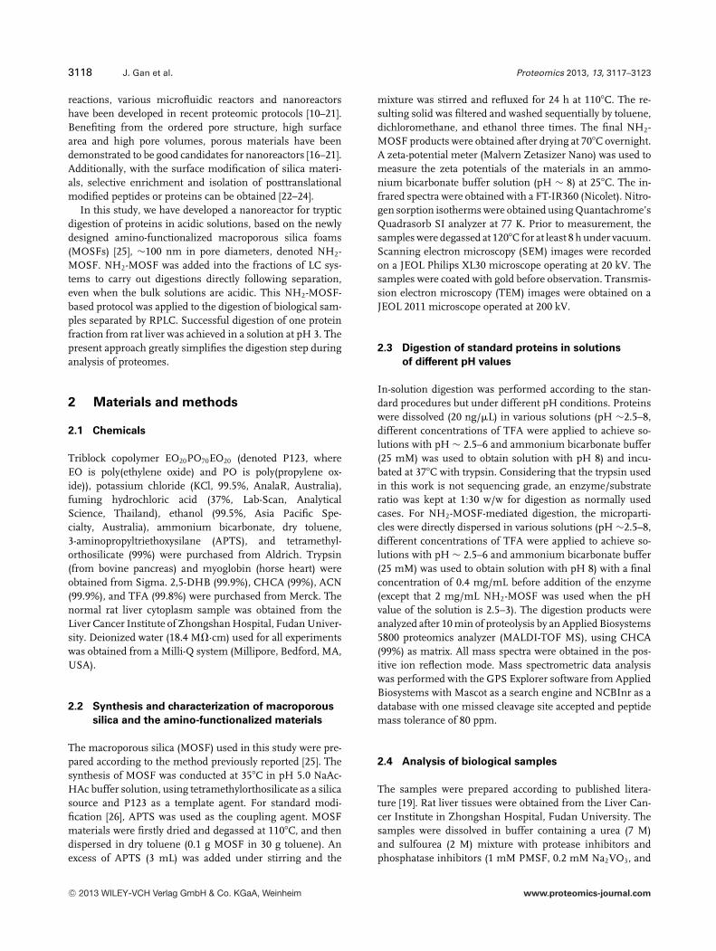
NH2-MOSF has been successfully prepared by modifying thesurfaces of macroporous silica with amino groups. Fouriertransform infrared spectroscopy was utilized to characterizethe surface modification, shown in Fig. 1A. The dominant
Figure 1. (A) Fourier transform infrared spectra and (B) zeta po-tential distributions of MOSF and NH2-MOSF.
multipeaks at 1000–1100 cm−1 are typically assigned to the Si-O bond and Si-OH groups in the bare MOSF material. In thecase of NH2-MOSF, an obvious additional single peak risesaround 1580 cm−1. This peak may be attributed to grafted-NH2 groups demonstrating the successful modification ofMOSF with amino groups. The -CH2- groups of the silicacoupling agent (APTS) result in the double peaks around2920 cm−1 [28]. Zeta potentials of MOSF and NH2-MOSFwere also examined as shown in Supporting Information Ta-ble 1. Figure 1B displays the zeta potential distributions ofthe two materials. MOSF has a zeta potential of −39 mVdue to the presence of silanol groups in abundance on thesurface [29]. In comparison, for NH2-MOSF a new peak ap-pears at +18 mV while the former recognized peak is absent,indicating modification of the surfaces by amino groups.
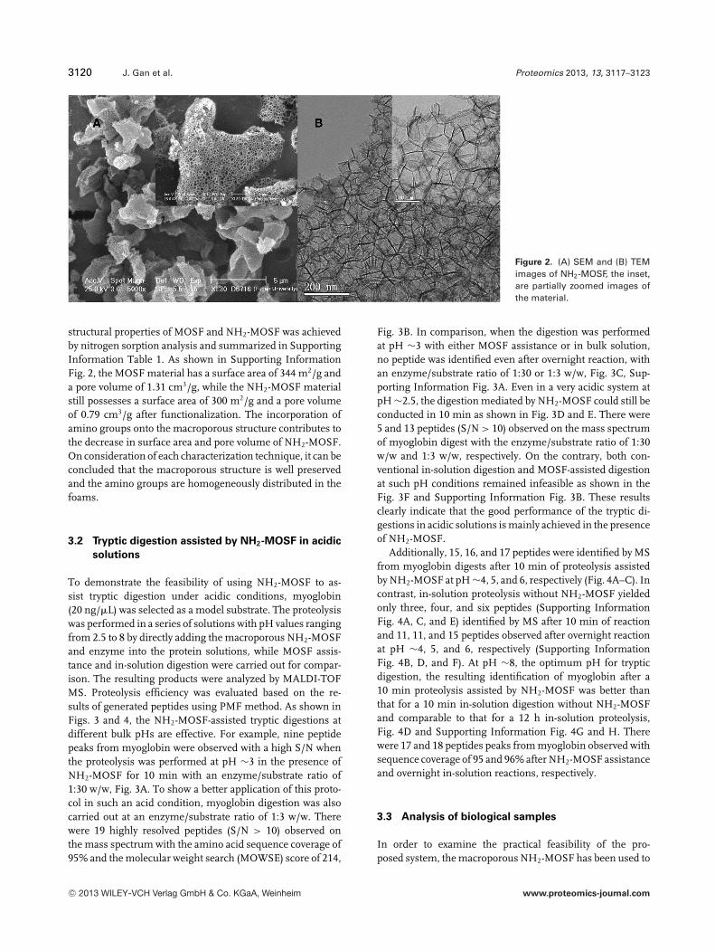
SEM and TEM were carried out to further investigate themorphologies and structures of the materials before/afterfunctionalization (Fig. 2). Compared to the bare siliceousfoams as shown in Supporting Information Fig. 1, the ar-rays of macropores (∼100 nm in diameter) are well retainedin NH2-MOSF. The TEM images also reveal the foam struc-ture of the NH2-MOSF with hexagonal column assemblies,Fig. 2B. From both SEM and TEM images, no large aggregatewas observed for NH2-MOSF, suggesting the well-dispersedstate of the amino species. In-depth characterization of the
C© 2013 WILEY-VCH Verlag GmbH & Co. KGaA, Weinheim www.proteomics-journal.com
3120 J. Gan et al. Proteomics 2013, 13, 3117–3123
Figure 2. (A) SEM and (B) TEMimages of NH2-MOSF, the inset,are partially zoomed images ofthe material.
structural properties of MOSF and NH2-MOSF was achievedby nitrogen sorption analysis and summarized in SupportingInformation Table 1. As shown in Supporting InformationFig. 2, the MOSF material has a surface area of 344 m2/g anda pore volume of 1.31 cm3/g, while the NH2-MOSF materialstill possesses a surface area of 300 m2/g and a pore volumeof 0.79 cm3/g after functionalization. The incorporation ofamino groups onto the macroporous structure contributes tothe decrease in surface area and pore volume of NH2-MOSF.On consideration of each characterization technique, it can beconcluded that the macroporous structure is well preservedand the amino groups are homogeneously distributed in thefoams.
3.2 Tryptic digestion assisted by NH2-MOSF in acidic
solutions
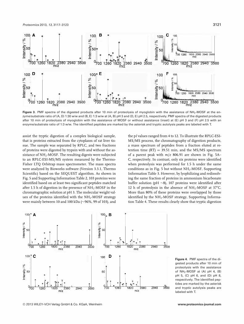
To demonstrate the feasibility of using NH2-MOSF to as-sist tryptic digestion under acidic conditions, myoglobin(20 ng/�L) was selected as a model substrate. The proteolysiswas performed in a series of solutions with pH values rangingfrom 2.5 to 8 by directly adding the macroporous NH2-MOSFand enzyme into the protein solutions, while MOSF assis-tance and in-solution digestion were carried out for compar-ison. The resulting products were analyzed by MALDI-TOFMS. Proteolysis efficiency was evaluated based on the re-sults of generated peptides using PMF method. As shown inFigs. 3 and 4, the NH2-MOSF-assisted tryptic digestions atdifferent bulk pHs are effective. For example, nine peptidepeaks from myoglobin were observed with a high S/N whenthe proteolysis was performed at pH ∼3 in the presence ofNH2-MOSF for 10 min with an enzyme/substrate ratio of1:30 w/w, Fig. 3A. To show a better application of this proto-col in such an acid condition, myoglobin digestion was alsocarried out at an enzyme/substrate ratio of 1:3 w/w. Therewere 19 highly resolved peptides (S/N > 10) observed onthe mass spectrum with the amino acid sequence coverage of95% and the molecular weight search (MOWSE) score of 214,
Fig. 3B. In comparison, when the digestion was performedat pH ∼3 with either MOSF assistance or in bulk solution,no peptide was identified even after overnight reaction, withan enzyme/substrate ratio of 1:30 or 1:3 w/w, Fig. 3C, Sup-porting Information Fig. 3A. Even in a very acidic system atpH ∼2.5, the digestion mediated by NH2-MOSF could still beconducted in 10 min as shown in Fig. 3D and E. There were5 and 13 peptides (S/N > 10) observed on the mass spectrumof myoglobin digest with the enzyme/substrate ratio of 1:30w/w and 1:3 w/w, respectively. On the contrary, both con-ventional in-solution digestion and MOSF-assisted digestionat such pH conditions remained infeasible as shown in theFig. 3F and Supporting Information Fig. 3B. These resultsclearly indicate that the good performance of the tryptic di-gestions in acidic solutions is mainly achieved in the presenceof NH2-MOSF.
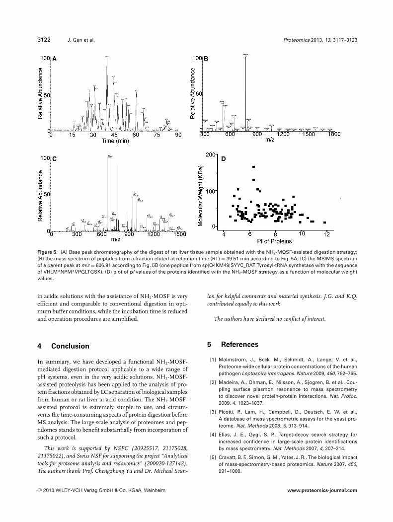
Additionally, 15, 16, and 17 peptides were identified by MSfrom myoglobin digests after 10 min of proteolysis assistedby NH2-MOSF at pH ∼4, 5, and 6, respectively (Fig. 4A–C). Incontrast, in-solution proteolysis without NH2-MOSF yieldedonly three, four, and six peptides (Supporting InformationFig. 4A, C, and E) identified by MS after 10 min of reactionand 11, 11, and 15 peptides observed after overnight reactionat pH ∼4, 5, and 6, respectively (Supporting InformationFig. 4B, D, and F). At pH ∼8, the optimum pH for trypticdigestion, the resulting identification of myoglobin after a10 min proteolysis assisted by NH2-MOSF was better thanthat for a 10 min in-solution digestion without NH2-MOSFand comparable to that for a 12 h in-solution proteolysis,Fig. 4D and Supporting Information Fig. 4G and H. Therewere 17 and 18 peptides peaks from myoglobin observed withsequence coverage of 95 and 96% after NH2-MOSF assistanceand overnight in-solution reactions, respectively.
3.3 Analysis of biological samples
In order to examine the practical feasibility of the pro-posed system, the macroporous NH2-MOSF has been used to
C© 2013 WILEY-VCH Verlag GmbH & Co. KGaA, Weinheim www.proteomics-journal.com
Proteomics 2013, 13, 3117–3123 3121
Figure 3. PMF spectra of the digested products after 10 min of proteolysis of myoglobin with the assistance of NH2-MOSF at the en-zyme/substrate ratio of (A, D) 1:30 w/w and (B, E) 1:3 w/w at (A, B) pH 3 and (D, E) pH 2.5, respectively. PMF spectra of the digested productsafter 10 min of proteolysis of myoglobin with the assistance of MOSF or without assistance (inset) at (E) pH 3 and (F) pH 2.5 with anenzyme/substrate ratio of 1:3 w/w. The identified peptides are marked by the asterisk and tryptic autolysis peaks are labeled with T.
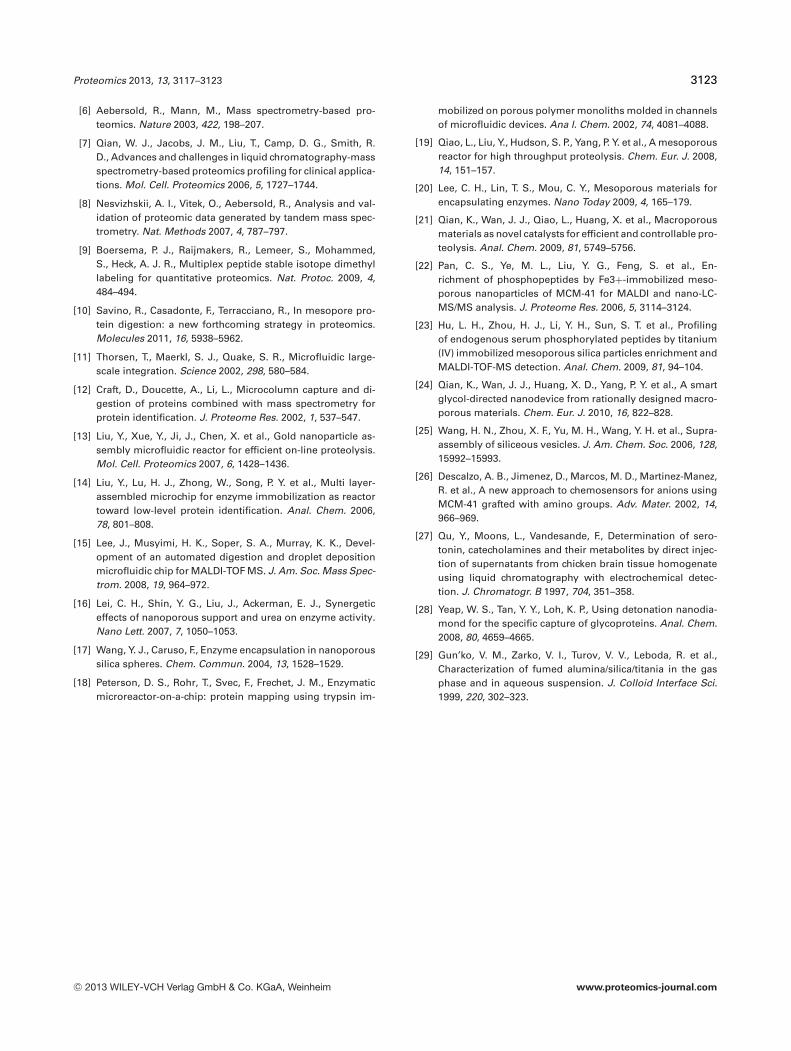
assist the tryptic digestion of a complex biological sample,that is proteins extracted from the cytoplasm of rat liver tis-sue. The sample was separated by RPLC, and two fractionsof proteins were digested by trypsin with and without the as-sistance of NH2-MOSF. The resulting digests were subjectedto an RPLC-ESI-MS/MS system measured by the Thermo-Fisher LTQ Orbitrap mass spectrometer. The mass spectrawere analyzed by Bioworks software (Version 3.3.1, ThermoScientific) based on the SEQUEST algorithm. As shown inFig. 5 and Supporting Information Table 2, 103 proteins wereidentified based on at least two significant peptides matchedafter 1.5 h of digestion in the presence of NH2-MOSF in thechromatographic solution at pH 3. The molecular weight val-ues of the proteins identified with the NH2-MOSF strategywere mainly between 10 and 100 kDa (∼96%, 99 of 103), and
the pI values ranged from 4 to 12. To illustrate the RPLC-ESI-MS/MS process, the chromatography of digestion products,a mass spectrum of peptides from a fraction eluted at re-tention time (RT) = 39.51 min, and the MS/MS spectrumof a parent peak with m/z 806.91 are shown in Fig. 5A–C, respectively. In contrast, only six proteins were identifiedwhen proteolysis was performed for 1.5 h under the sameconditions as in Fig. 5 but without NH2-MOSF, SupportingInformation Table 3. However, by lyophilizing and redissolv-ing the same fraction of proteins in ammonium bicarbonatebuffer solution (pH ∼8), 107 proteins were identified after12 h of proteolysis in the absence of NH2-MOSF at 37�C.More than 80% of these proteins were overlapped by thoseidentified by the NH2-MOSF strategy, Supporting Informa-tion Table 4. These results clearly show that tryptic digestion
Figure 4. PMF spectra of the di-gested products after 10 min ofproteolysis with the assistanceof NH2-MOSF at (A) pH 4, (B)pH 5, (C) pH 6, and (D) pH 8,respectively. The identified pep-tides are marked by the asteriskand tryptic autolysis peaks arelabeled with T.
C© 2013 WILEY-VCH Verlag GmbH & Co. KGaA, Weinheim www.proteomics-journal.com
3122 J. Gan et al. Proteomics 2013, 13, 3117–3123
Figure 5. (A) Base peak chromatography of the digest of rat liver tissue sample obtained with the NH2-MOSF-assisted digestion strategy;(B) the mass spectrum of peptides from a fraction eluted at retention time (RT) = 39.51 min according to Fig. 5A; (C) the MS/MS spectrumof a parent peak at m/z = 806.91 according to Fig. 5B (one peptide from sp|Q4KM49|SYYC_RAT Tyrosyl-tRNA synthetase with the sequenceof VHLM*NPM*VPGLTGSK); (D) plot of pI values of the proteins identified with the NH2-MOSF strategy as a function of molecular weightvalues.
in acidic solutions with the assistance of NH2-MOSF is veryefficient and comparable to conventional digestion in opti-mum buffer conditions, while the incubation time is reducedand operation procedures are simplified.
4 Conclusion
In summary, we have developed a functional NH2-MOSF-mediated digestion protocol applicable to a wide range ofpH systems, even in the very acidic solutions. NH2-MOSF-assisted proteolysis has been applied to the analysis of pro-tein fractions obtained by LC separation of biological samplesfrom human or rat liver at acid condition. The NH2-MOSF-assisted protocol is extremely simple to use, and circum-vents the time-consuming aspects of protein digestion beforeMS analysis. The large-scale analysis of proteomes and pep-tidomes stands to benefit substantially from incorporation ofsuch a protocol.
This work is supported by NSFC (20925517, 21175028,21375022), and Swiss NSF for supporting the project “Analyticaltools for proteome analysis and redoxomics” (200020-127142).The authors thank Prof. Chengzhong Yu and Dr. Micheal Scan-
lon for helpful comments and material synthesis. J.G. and K.Q.contributed equally to this work.
The authors have declared no conflict of interest.
5 References
[1] Malmstrom, J., Beck, M., Schmidt, A., Lange, V. et al.,Proteome-wide cellular protein concentrations of the humanpathogen Leptospira interrogans. Nature 2009, 460, 762–765.
[2] Madeira, A., Ohman, E., Nilsson, A., Sjogren, B. et al., Cou-pling surface plasmon resonance to mass spectrometryto discover novel protein-protein interactions. Nat. Protoc.2009, 4, 1023–1037.
[3] Picotti, P., Lam, H., Campbell, D., Deutsch, E. W. et al.,A database of mass spectrometric assays for the yeast pro-teome. Nat. Methods 2008, 5, 913–914.
[4] Elias, J. E., Gygi, S. P., Target-decoy search strategy forincreased confidence in large-scale protein identificationsby mass spectrometry. Nat. Methods 2007, 4, 207–214.
[5] Cravatt, B. F., Simon, G. M., Yates, J. R., The biological impactof mass-spectrometry-based proteomics. Nature 2007, 450,991–1000.
C© 2013 WILEY-VCH Verlag GmbH & Co. KGaA, Weinheim www.proteomics-journal.com
Proteomics 2013, 13, 3117–3123 3123
[6] Aebersold, R., Mann, M., Mass spectrometry-based pro-teomics. Nature 2003, 422, 198–207.
[7] Qian, W. J., Jacobs, J. M., Liu, T., Camp, D. G., Smith, R.D., Advances and challenges in liquid chromatography-massspectrometry-based proteomics profiling for clinical applica-tions. Mol. Cell. Proteomics 2006, 5, 1727–1744.
[8] Nesvizhskii, A. I., Vitek, O., Aebersold, R., Analysis and val-idation of proteomic data generated by tandem mass spec-trometry. Nat. Methods 2007, 4, 787–797.
[9] Boersema, P. J., Raijmakers, R., Lemeer, S., Mohammed,S., Heck, A. J. R., Multiplex peptide stable isotope dimethyllabeling for quantitative proteomics. Nat. Protoc. 2009, 4,484–494.
[10] Savino, R., Casadonte, F., Terracciano, R., In mesopore pro-tein digestion: a new forthcoming strategy in proteomics.Molecules 2011, 16, 5938–5962.
[11] Thorsen, T., Maerkl, S. J., Quake, S. R., Microfluidic large-scale integration. Science 2002, 298, 580–584.
[12] Craft, D., Doucette, A., Li, L., Microcolumn capture and di-gestion of proteins combined with mass spectrometry forprotein identification. J. Proteome Res. 2002, 1, 537–547.
[13] Liu, Y., Xue, Y., Ji, J., Chen, X. et al., Gold nanoparticle as-sembly microfluidic reactor for efficient on-line proteolysis.Mol. Cell. Proteomics 2007, 6, 1428–1436.
[14] Liu, Y., Lu, H. J., Zhong, W., Song, P. Y. et al., Multi layer-assembled microchip for enzyme immobilization as reactortoward low-level protein identification. Anal. Chem. 2006,78, 801–808.
[15] Lee, J., Musyimi, H. K., Soper, S. A., Murray, K. K., Devel-opment of an automated digestion and droplet depositionmicrofluidic chip for MALDI-TOF MS. J. Am. Soc. Mass Spec-trom. 2008, 19, 964–972.
[16] Lei, C. H., Shin, Y. G., Liu, J., Ackerman, E. J., Synergeticeffects of nanoporous support and urea on enzyme activity.Nano Lett. 2007, 7, 1050–1053.
[17] Wang, Y. J., Caruso, F., Enzyme encapsulation in nanoporoussilica spheres. Chem. Commun. 2004, 13, 1528–1529.
[18] Peterson, D. S., Rohr, T., Svec, F., Frechet, J. M., Enzymaticmicroreactor-on-a-chip: protein mapping using trypsin im-
mobilized on porous polymer monoliths molded in channelsof microfluidic devices. Ana l. Chem. 2002, 74, 4081–4088.
[19] Qiao, L., Liu, Y., Hudson, S. P., Yang, P. Y. et al., A mesoporousreactor for high throughput proteolysis. Chem. Eur. J. 2008,14, 151–157.
[20] Lee, C. H., Lin, T. S., Mou, C. Y., Mesoporous materials forencapsulating enzymes. Nano Today 2009, 4, 165–179.
[21] Qian, K., Wan, J. J., Qiao, L., Huang, X. et al., Macroporousmaterials as novel catalysts for efficient and controllable pro-teolysis. Anal. Chem. 2009, 81, 5749–5756.
[22] Pan, C. S., Ye, M. L., Liu, Y. G., Feng, S. et al., En-richment of phosphopeptides by Fe3+-immobilized meso-porous nanoparticles of MCM-41 for MALDI and nano-LC-MS/MS analysis. J. Proteome Res. 2006, 5, 3114–3124.
[23] Hu, L. H., Zhou, H. J., Li, Y. H., Sun, S. T. et al., Profilingof endogenous serum phosphorylated peptides by titanium(IV) immobilized mesoporous silica particles enrichment andMALDI-TOF-MS detection. Anal. Chem. 2009, 81, 94–104.
[24] Qian, K., Wan, J. J., Huang, X. D., Yang, P. Y. et al., A smartglycol-directed nanodevice from rationally designed macro-porous materials. Chem. Eur. J. 2010, 16, 822–828.
[25] Wang, H. N., Zhou, X. F., Yu, M. H., Wang, Y. H. et al., Supra-assembly of siliceous vesicles. J. Am. Chem. Soc. 2006, 128,15992–15993.
[26] Descalzo, A. B., Jimenez, D., Marcos, M. D., Martinez-Manez,R. et al., A new approach to chemosensors for anions usingMCM-41 grafted with amino groups. Adv. Mater. 2002, 14,966–969.
[27] Qu, Y., Moons, L., Vandesande, F., Determination of sero-tonin, catecholamines and their metabolites by direct injec-tion of supernatants from chicken brain tissue homogenateusing liquid chromatography with electrochemical detec-tion. J. Chromatogr. B 1997, 704, 351–358.
[28] Yeap, W. S., Tan, Y. Y., Loh, K. P., Using detonation nanodia-mond for the specific capture of glycoproteins. Anal. Chem.2008, 80, 4659–4665.
[29] Gun’ko, V. M., Zarko, V. I., Turov, V. V., Leboda, R. et al.,Characterization of fumed alumina/silica/titania in the gasphase and in aqueous suspension. J. Colloid Interface Sci.1999, 220, 302–323.
C© 2013 WILEY-VCH Verlag GmbH & Co. KGaA, Weinheim www.proteomics-journal.com