Embed Size (px)
Citation preview

ORIGINAL PAPER
Apoptosis of Purkinje and Granular Cells of the CerebellumFollowing Chronic Ethanol Intake
Suelen A. Oliveira & Luiz Gustavo A. Chuffa & Beatriz Aparecida Fioruci-Fontanelli &Fermino Sanches Lizarte Neto & Paulo Cezar Novais & Luiz Fernando Tirapelli &Jorge Camargo Oishi & Luiz Fernando Takase & Maira Aparecida Stefanini &Marcelo Martinez & Francisco Eduardo Martinez
# Springer Science+Business Media New York 2014
Abstract Ethanol alters motricity, learning, cognition, andcellular metabolism in the cerebellum.We evaluated the effectof ethanol on apoptosis in Golgi, Purkinje, and granule cells ofthe cerebellum in adult rats. There were two groups of 20 rats:a control group that did not consume ethanol and an experi-mental group of UChA rats that consumed ethanol at 10 %(<2 g ethanol/kg body weight/day). At 120 days old, rats wereanesthetized and decapitated, and their cerebella were collect-ed and fixed. Cerebellar sections were subjected to immuno-histochemistry for terminal deoxynucleotide transferase dUTPnick end labeling (TUNEL), caspase-3, X-linked inhibitor ofapoptosis protein (XIAP), and insulin-like growth factor 1-receptor (IGF-1R); real-time PCR (RT-PCR) to determinecaspase-3, XIAP, and IGF-1R gene expression; and transmis-sion electron microscopy (TEM). We identified fragmentationof DNA and an increase in caspase-3 protein and XIAP in
Purkinje cells, whereas granule cells exhibited increasedcaspase-3 and XIAP. IGF-1R expression was unchanged.There was no significant difference in gene expression ofcaspase-3, XIAP, and IGF-1R. There were an increase in lipiddroplets, a reduction in the cellular cytoplasm in electron-dense nuclei, and changes in the myelin sheath in the cerebel-lar cortex. In conclusion, our data demonstrated that ethanolinduced apoptosis in the Purkinje and granule cells of thecerebellum of adult UChA rats.
Keywords Apoptosis . XIAP . IGF-1R . Caspase-3 .
Cerebellum . UChA rats
Introduction
Alcoholism affects millions of people worldwide. It is char-acterized by the excessive consumption of ethanol and isassociated with various health risks. It is a multifactorialsyndrome that leads to physical, mental, and social impair-ments [1, 2]; alters the structure and function of the brain; andin some cases, can lead to neurodegeneration [3].
Prenatal exposure to ethanol decreases cerebral volume. Inthe cerebellum, where a stronger reduction occurs, prenatalexposure leads to the degeneration of cognitive and behavioralfunctions [4]. Throughout its development, the cerebellum isextremely sensitive to high exposure to ethanol, which causesimpairment in Purkinje and granule cells and in glia [5, 6].Purkinje cells are the dominant elements involved in theprocessing of cerebellar information [7]. Golgi cells contributeto information processing during information input to thecerebellar cortex. Purkinje cell dysfunction can damage motorcoordination [8, 9], and Purkinje cells are susceptible to dam-age from ethanol [10–12].
S. A. OliveiraGraduate Program in General and Applied Biology, Institute ofBioscience, Univ. Estadual Paulista (UNESP), Botucatu,SP 18618-970, Brazil
L. G. A. Chuffa : B. A. Fioruci-Fontanelli : F. E. Martinez (*)Department of Anatomy, Institute of Biosciences, Univ. EstadualPaulista (UNESP), P.O. Box 510, Rubião Júnior, s/n, Botucatu,SP 18618-970, Brazile-mail: [email protected]
F. S. L. Neto : P. C. Novais : L. F. TirapelliDepartment of Surgery and Anatomy, Faculty of Medicine, Univ.São Paulo (USP), Ribeirão Preto, SP 14049-900, Brazil
J. C. OishiDepartment of Physiology, Universidade Federal de São Carlos,São Carlos, SP 13565-905, Brazil
L. F. Takase :M. A. Stefanini :M. MartinezDepartment of Morphology and Pathology, Universidade Federal deSão Carlos, São Carlos, SP 13565-905, Brazil
CerebellumDOI 10.1007/s12311-014-0591-2

There are two main pathways leading to apoptosis: anextrinsic pathway, which activates caspase-8, and an intrinsicpathway, which culminates in the cleavage and activation ofprocaspase-3. Activation of caspase-3 produces morphologi-cal changes typical of apoptosis [13, 14]. Although the apo-ptotic pathway has been extensively studied in the cerebellum,no studies have identified alterations in apoptosis in ethanol-preferring rats treated with different volumes of ethanol ordurations of exposure.
Insulin-like growth factors (IGFs) are a family of mitogenicproteins involved in the regulation of cell growth and differ-entiation, and insulin-like growth factor 1-receptor (IGF-1R)plays a critical role in the regulation of cognitive and motorfunctions for various cell types in the central nervous system(CNS) [15]. Caspase-3 and X-linked inhibitor of apoptosisprotein (XIAP) are involved in ethanol-induced apoptosis.Caspase-3 is linked to the effector phase of programmed celldeath, promoting the expression of restriction endonucleasesthat cleave DNA. XIAP plays an antiapoptotic role byinhibiting the effector caspases, caspase-3 and -7, and theinitiator caspase, caspase-9, which modulates the transcriptionfactor—nuclear factor-kappa β (NF-kB). Such proteins aretherefore markers of the signaling mechanisms that, oncechanged, can lead to neurological deficits [16].
UChA andUChB rats are voluntary ethanol consumers andoriginate from a colony of Wistar rats developed at the Uni-versity of Chile (UCh) 70 generations ago [17–19]. These ratscomprise one of the best models for studying the effects ofethanol consumption on several physiological systems and,furthermore, represent the context of alcoholism-related pa-thologies (e.g., dependence or abstinence syndrome) such asthose found in human diseases. Our aim was to evaluateapoptosis in Golgi, Purkinje, and granule cells of the cerebel-lum of adult UChA rats.
Materials and Methods
Animals
Sixty adult male rats were provided by the Department ofAnatomy (Biosciences Institute, Univ. Estadual Paulista(UNESP), Botucatu, SP, Brazil). They were individuallyhoused in polypropylene cages with laboratory-grade pineshavings as bedding and were kept under controlled roomtemperature (23±1 °C) and lighting conditions (12-h light/dark photoperiod, lights on at 6 a.m.). The UChA rats (amodel of ethanol-preferring rats, developed by selectivebreeding) have a spontaneous genetic propensity to voluntar-ily consume 10 % (v/v) ethanol (<2 g ethanol/kg body weight/day). At 15 days old, 40 UChA rats were provided two bottlesfor consumption over a 15-day period: one bottle containingwater ad libitum (1) and another containing a 10 % (v/v)
ethanol solution (2). After this 15-day period, 20 rats fromeach group showing ethanol consumption lower than 2.0 gethanol/kg of body weight/day were selected, according to theprotocol of Mardones and Segovia-Riquelme [20]. In the ratsselected for this study, the preference ratio associated withethanol-seeking behavior was approximately 65 %. The ratswere then divided into two groups: the UChA group (n=20),consisting of rats provided a 10 % (v/v) ethanol solution adlibitum (free choice for water or ethanol), drinking between0.1 to 2 g ethanol/kg body weight/day, and a control group(n=20) consisting of rats without access to ethanol. From thebeginning of the experiment (65 days old) to euthanasia(120 days old), the UChA rats consumed 10 % (v/v) ethanolover the course of 55 days (Fig. 1). All animal procedureswere approved by the Biosciences Institute, Univ. EstadualPaulista (UNESP), Botucatu, Institutional Animal Care andUse Committee.
Electron Microscopy
Four rat cerebellar samples were collected from each groupusing a 21-gauge needle, following an intracardiac perfusionwith 2.5 % glutaraldehyde in 0.10 M sodium phosphatebuffer. Samples were immersed in the same fixative for aminimum of 3 h, postfixed in 1 % osmium tetroxide in0.10 M phosphate buffer for 1 h, and stained en bloc in 3 %uranyl acetate for 1 h. Samples were dehydrated in ethanol,embedded in epoxy resin (Sigma-Aldrich), sectioned at 50 to60 nm on a Leica UCT ultramicrotome, and picked up oncarbon-coated copper grids. Sections were stained with 3 %uranyl acetate for 5 min and Sato’s lead stains for 1 min. Gridswere viewed using a Philips CM 100 transmission electronmicroscope (TEM).
Immunohistochemistry of Terminal DeoxynucleotideTransferase dUTP Nick End Labeling (TUNEL)
Apoptotic cells were analyzed in samples from five rats fromeach group using a FragELTMDNA kit (Calbiochem, La Jolla,CA, USA), according to the manufacturer’s instructions. Theslides were counterstained with Harris hematoxylin and as-sembled with Permount. Negative controls were treated in thesame way but were incubated without the TdT enzyme.
Immunohistochemistry of Caspase-3, XIAP, and IGF-1R
Sections of the cerebellum of the rats were fixed by immersionin 4 % paraformaldehyde and embedded in paraffin. Cuts at athickness of 3 μm were obtained using a Reichert Jung 2040microtome. Antigen recovery was performed in citrate bufferat 10 mM, pH 6.0 for 40 min, after being incubated in 3 %hydrogen peroxide in order to block endogenous peroxidaseactivities. The cuts underwent preincubation for 30 min in
Cerebellum

serum-blocking solution (1 % bovine serum albumin), and toblock nonspecific binding, Background Sniper, Biocare Med-ical (Polymer Kit: Mach 4 Universal HRP) was used.
To mark caspase-3, we used caspase-3 polyclonal activatedantibodies (CPP32, monoclonal lyophilized, Novocastra®)and XIAP (SC-11426 polyclonal, Santa Cruz®) diluted at1/200 in 0.01 M phosphate-buffered saline (PBS). For incu-bation of IGF-1R, we used N-20 rabbit primary polyclonalantibody (SC-720, polyclonal, Santa Cruz®) at 1/50 dilutionin 0.01 M PBS solution with 3 % bovine serum albumin(BSA). The reactions were detected using Betazoid DAB(BDB900G5) chromogen diluted in Betazoid DAB substratebuffer (DS900L) from the Mach 4 kit. Samples used aspositive controls were the encephala of rats subjected toischemia. The sections were stained with diaminobenzidinefor 10 min at room temperature and then counterstained withhematoxylin-eosin and assembled in resin.
The slides were analyzed using a Zeiss® microscope,model Axioskop 2 plus, with a camera (Axio Cam Hrc®)attached. Axio Vision 4.6® software with ×400 magnificationwas used for the analysis. The number of positive cells in eachgroup was counted using ten nonoverlapping high randomfields and counts are reported as group averages.
Real-Time PCR for Caspase-3, XIAP, and IGF-1R GeneExpression
Cerebellum samples were frozen at −80 °C. After extractingthe RNA using the reactant Trizol® (Invitrogen, EUA), RNAwas separated, quantified, and analyzed for integrity in aga-rose gel. To synthesize complementary DNA (cDNA), weperformed a reverse transcription using a commercial HighCapacity cDNA Reverse Transcription (Applied Biosystems,Foster City, CA) kit, according to the manufacturer’s instruc-tions. Real-time quantitative PCR was performed using anABI PRISM 7500 Fast Real Time PCR System and FAMTM
dyes (Applied Biosystems, Contraboeuf, France), followingthe manufacturer’s protocols, in our genomics facility. Thecaspase-3 (Rn00563902_m1), IGF-1R (Rn00583837_m1),and XIAP (Rn00573706_m1) genes used were from theTaqMan Assay-on-Demand system, which is comprised ofoligonucleotides and probes acquired from AppliedBiosystems. Amplification was carried out in a final volumeof 10 μL, using 5 μL of the specific reactant TaqMan Master
Mix (Applied Biosystems), 0.5 μL of each specific probe, and4.5 μL of cDNA. All reactions were performed in duplicateand analyzed using a real-time PCR detection device (7500Real Time PCR System, Applied Biosystems). Data werecollected constantly during PCR and analyzed using theABI-7500 SDS software package.
Statistical Analysis
Data are presented asmeans±SD. Differences between groupswere compared by unpaired t test and Mann-Whitney test.Differences were considered to be statistically significant atP<0.05 (P value: two-tailed). Statistical analysis was per-formed using GraphPad Prism 6 software.
Results
Electron Microscopy
The images of cerebella displayed dilation of the endoplasmicreticulum, an increase in lipid droplets, and electron density inthe nuclei of UChA rats. Furthermore, there was a severereduction in the medullary body of the cerebellum indicatingdisruption of myelin in UChA rats (Fig. 2d–i).
Immunohistochemistry Using TUNEL, Caspase-3, XIAP,and IGF-1R
All of the Purkinje, granular, and Golgi cells exhibited apo-ptosis in both UChA (Fig. 3b, d) and control rats (Fig. 3a, c),but were most evident in UChA rats. While a strong reactionto XIAPwas observed in the Purkinje and granular cells of theUChA rats (Fig. 3e), a positive reaction to IGF-1R was notobserved (Fig. 4a, b).
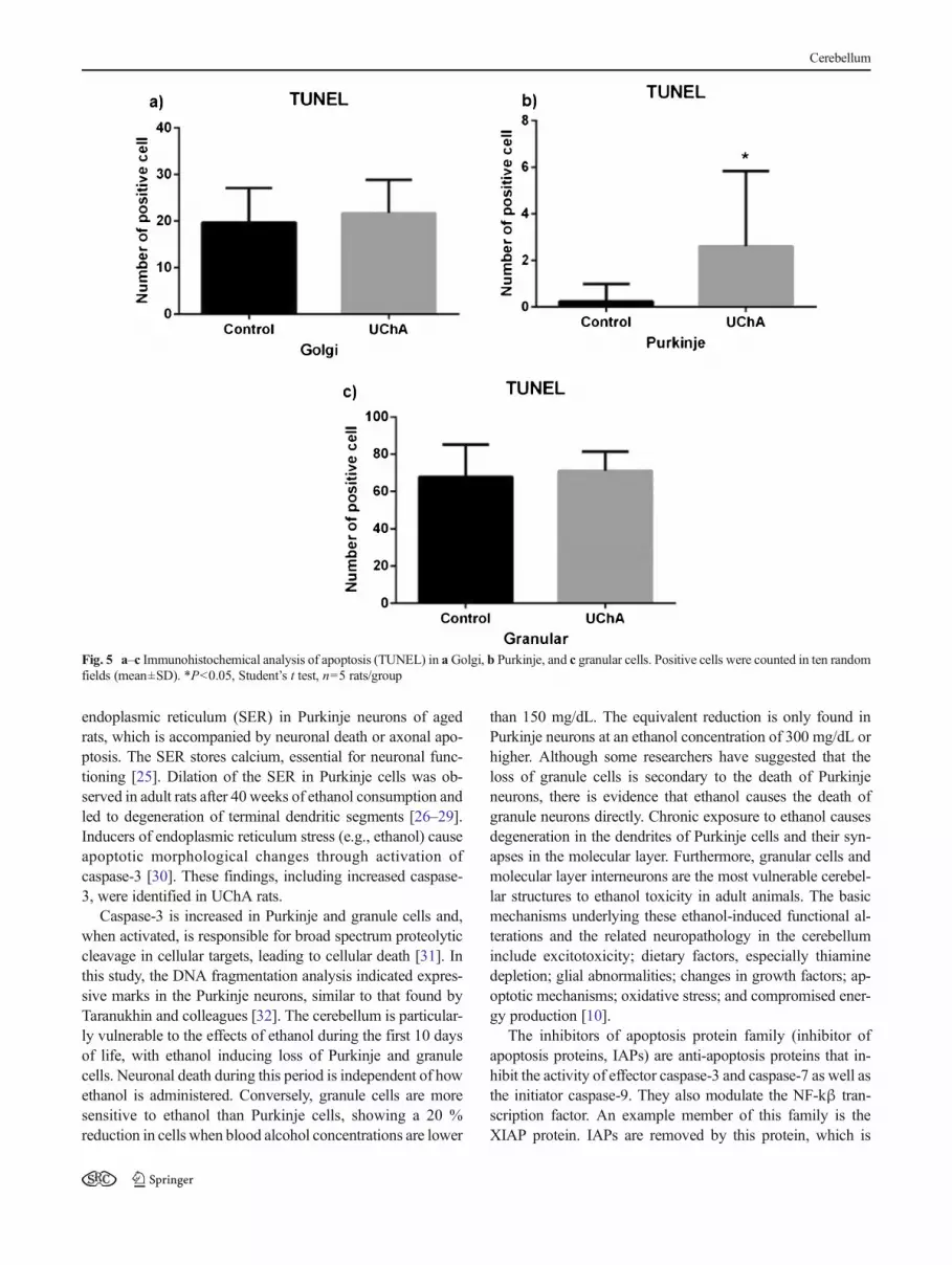
The count for Golgi cells positive for TUNEL and caspase-3 did not differ significantly (Figs. 5a and 6a), but the numberof XIAP-positive cells was lower in the UChA rats than incontrols (Fig. 7a). Purkinje cells positive for TUNEL, caspase-3, and XIAP were more frequent in the UChA rats (Figs. 5b,6b, and 7b). Granular cells positive for caspase-3 and XIAPwere more frequent in the UChA rats (Figs. 6c and 7c), butthere was no difference in TUNEL (Fig. 5c).
Fig. 1 Schematic representationof the overall experimental periodfrom birth until the selectionperiod for ethanol preference
Cerebellum

Expression of Caspase-3, XIAP, and IGF-1R
Gene expression of caspase-3, XIAP, and IGF-1R was un-changed in the presence of ethanol, suggesting posttransla-tional modifications to these target proteins (Fig. 8a–c).
Discussion
Apoptosis in the cerebellar cortex of rats was observed usingelectron microscopy, immunohistochemistry to detectcaspase-3 and XIAP, and the TUNEL method. Gene expres-sion of caspase-3 and XIAP, as well as of IGF-1R, underwentchanges upon ethanol use, which led to the damage of struc-tures necessary for normal cerebellar function [21]. Changesin the chain of apoptotic events can lead to malformations andpathological conditions, including cancer, autoimmune disor-ders, and neurodegenerative diseases [15, 22].
The ultrastructure of the cerebellum in the UChA ratsshowed signs of cellular death typical of ethanol toxicity. A
previous work carried out with UChA rats also demonstratedalterations in cerebellar structure, arising from changes to thePurkinje neurons [23]. For example, Lewandowska and col-leagues [24] showed similar findings during pregnancy, ob-serving degenerative changes in Purkinje and granule cells.Jaatinen and Rintali [10] reported that in adult rats, chronicexposure to ethanol leads to cerebellar atrophy. The mainneuropathological features of this atrophy included loss ofPurkinje cells and a decrease in the volume of the Purkinjecell dendritic network in the molecular layer. Ethanol leads tostress as well as persistent hepatic lesions and hepaticsteatosis, promoting lipolysis and increasing toxic lipids. Ofthese toxic lipids, ceramides exacerbate hepatic lesions, caus-ing insulin resistance, oxidative stress, and activation of pro-inflammatory cytokines. Once they are released to the periph-eral blood, they cross the blood-brain barrier and initiate acomplex sequence of neurodegeneration. Insulin resistanceleads to neural impairments and damages the function ofoligodendrocytes, leading to myelin degradation, which thencauses an increase in the production of toxic lipids in the CNS.In addition, the direct neurotoxic effects caused by ethanol and
Fig. 2 a–i TEM micrographs of the cerebellum of control (a–c) andUChA (d–i) rats. a Normal Golgi (Go) and granular (G) cells. bGranular(G) and Purkinje (P) cells. Note the Purkinje cell with normal cisterns ofthe endoplasmic reticulum (arrows). c Molecular layer with a typicalarrangement (M). dGolgi with cytoplasmic edema (Go) and lipid droplets(asterisks) and granular cells (G) with pyknotic nuclei (arrows). e Mag-nified view of Golgi cells (Go) with intense accumulation of lipid droplets
(asterisks) and the granular layer (G) with pyknotic nuclei (arrow). fPurkinje cell with electron-dense nuclei (P) and lipid droplets (asterisks).Note dilated cisterns of the endoplasmic reticulum (arrows). g Granularlayer (G) with intense lipid droplets (asterisks) and dilated cisterns of theendoplasmic reticulum (arrow). h Severe lipid droplet accumulation(asterisks) in the molecular layer (M). i Disorganization of the medullarybody of the cerebellum with changes in the myelin sheath (arrow)
Cerebellum

its metabolites impair mitochondrial function and reduce theintegrity of the membrane. This diminishes messenger RNAassociated with myelin, which responds to insulin and in-creases the apoptosis index, oxidative stress, lipid
preoxidation, damage to DNA, and disruption of acetylcho-line homeostasis, contributing to neurodegeneration [15].Chronic ethanol consumption causes imbalances in neuronalcalcium homeostasis resulting in dilation of the smooth
Fig. 3 a–f Representative immunohistochemistry of the cerebellar cor-tex: molecular layer (M), Purkinje cells (P), and granular cells (G) at ×400magnification. a Purkinje marked and unmarked (P) in control TUNEL. bPurkinje cells (P) in TUNEL UChA. cGolgi neurons (Go) and caspase-3
in the control group. dGranular (G) and Golgi neurons (Go) in caspase-3UChA. eGolgi neurons (Go) in control XIAP. f Purkinje (P) and granular(G) cells in XIAP UChA
Fig. 4 a, bImmunohistochemical images at×400 magnification of thecerebellum of a control and bUChA rats. Arrows show a strongpositive reaction to IGF-1R inglomerular areas
Cerebellum
endoplasmic reticulum (SER) in Purkinje neurons of agedrats, which is accompanied by neuronal death or axonal apo-ptosis. The SER stores calcium, essential for neuronal func-tioning [25]. Dilation of the SER in Purkinje cells was ob-served in adult rats after 40 weeks of ethanol consumption andled to degeneration of terminal dendritic segments [26–29].Inducers of endoplasmic reticulum stress (e.g., ethanol) causeapoptotic morphological changes through activation ofcaspase-3 [30]. These findings, including increased caspase-3, were identified in UChA rats.
Caspase-3 is increased in Purkinje and granule cells and,when activated, is responsible for broad spectrum proteolyticcleavage in cellular targets, leading to cellular death [31]. Inthis study, the DNA fragmentation analysis indicated expres-sive marks in the Purkinje neurons, similar to that found byTaranukhin and colleagues [32]. The cerebellum is particular-ly vulnerable to the effects of ethanol during the first 10 daysof life, with ethanol inducing loss of Purkinje and granulecells. Neuronal death during this period is independent of howethanol is administered. Conversely, granule cells are moresensitive to ethanol than Purkinje cells, showing a 20 %reduction in cells when blood alcohol concentrations are lower
than 150 mg/dL. The equivalent reduction is only found inPurkinje neurons at an ethanol concentration of 300 mg/dL orhigher. Although some researchers have suggested that theloss of granule cells is secondary to the death of Purkinjeneurons, there is evidence that ethanol causes the death ofgranule neurons directly. Chronic exposure to ethanol causesdegeneration in the dendrites of Purkinje cells and their syn-apses in the molecular layer. Furthermore, granular cells andmolecular layer interneurons are the most vulnerable cerebel-lar structures to ethanol toxicity in adult animals. The basicmechanisms underlying these ethanol-induced functional al-terations and the related neuropathology in the cerebelluminclude excitotoxicity; dietary factors, especially thiaminedepletion; glial abnormalities; changes in growth factors; ap-optotic mechanisms; oxidative stress; and compromised ener-gy production [10].
The inhibitors of apoptosis protein family (inhibitor ofapoptosis proteins, IAPs) are anti-apoptosis proteins that in-hibit the activity of effector caspase-3 and caspase-7 as well asthe initiator caspase-9. They also modulate the NF-kβ tran-scription factor. An example member of this family is theXIAP protein. IAPs are removed by this protein, which is
Fig. 5 a–c Immunohistochemical analysis of apoptosis (TUNEL) in aGolgi, b Purkinje, and c granular cells. Positive cells were counted in ten randomfields (mean±SD). *P<0.05, Student’s t test, n=5 rats/group
Cerebellum

released by the mitochondrial protein second mitochondria-derived activator of caspase/direct IAP-binding protein withlow pI (Smac/DIABLO). After mitochondrial damage,Smac/DIABLO is released from the intermembrane space intothe cytoplasm, along with cytochrome c, promoting apoptosis.Cytochrome c links itself to the apoptotic protease activatingfactor 1 (APAF-1) and directly activates caspase-9, whereasSmac/DIABLO removes the IAPs from their inhibitory inter-actions with the caspase [16]. Our results demonstrated thatXIAP acts as an antiapoptotic element in Purkinje and granuleneurons of UChA rats. In control rats, XIAP expression wasonly high in Golgi neurons displaying no apoptosis. This canbe explained by the fact that this rat model has developed newstrategies to avoid neuronal cell death in the presence of aminimum amount of ethanol during the period for ethanolpreference. Lee and colleagues [33] found similar resultswhere ethanol intake promoted high expression of cellsurvivor-related genes such as XIAP, BCl-xL, and catalase.Although in the liver of mice, alcohol intake increased ex-pression of p53, Omi/HtrA2, Bcl-2, and Bax without affectingXIAP expression or the Bcl-2/Bax ratio [34], Antonio andcolleagues [35] demonstrated that ethanol-treated fetalrhombencephalic neurons showed reduced XIAP and Bcl-2
gene expression (prosurvival genes) with high caspase-3 ac-tivity and reactive oxygen species formation. Notably, therapywith lipoic acid exerted a neuroprotective effect. To date, themechanism by which ethanol modulates XIAP expressionremains unclear.
There were no markers identified for IGF-1R in the cere-bellar cortex, which may be due to the age of the rats [36].Bondy [37] reported that IGF-1 is abundant in Purkinje cellsand develops synaptic stations. It may play an important rolein both the formation and myelinization of specific synapticconnections.
In the long term, ethanol could change gene expressionrelated to myelinization, apoptosis, cellular adhesion,neurogenesis, and neural disease [38]. Genes that are frequent-ly super-expressed by ethanol actions codify heat-shock pro-teins (HSPs), which are related to synaptic transmission andplasticity. Ethanol diminishes gene expression related tomyelinization and to protein synthesis [39], thus corroboratingour findings for changes to the myelin sheath in the medullarybody of the cerebellum.
Despite not reaching a significant level, there was an ap-parent change in gene expression that regulated caspase-3 inthe UChA group and XIAP in the control group. An increase
Fig. 6 a–c Immunohistochemical analysis of caspase-3 in a Golgi, b Purkinje, and c granular cells. Positive cells were counted in 10 random fields(mean±SD). *P<0.05, Student’s t test, n=5 rats/group
Cerebellum

in cellular reactive oxygen species (ROS) can activate severalclasses of genes, including those that are dependent on NF-kβ, that bind to the antioxidant response element (ARE).Because XIAP, Bcl-XL, or catalase can be regulated by NF-kβ-dependent mechanisms and because catalase also has anantioxidant response element that can bind to Nrf2, it is likelythat this initial ethanol-associated augmentation of gene ex-pression is mediated by the effects of increased ROS on ROS-sensitive transcription factors [33]. Homeostasis is maintainedby controlling the balance between antiapoptotic andproapoptotic proteins. Stimuli such as DNA damage triggeredby chronic ethanol consumption can lead to an increase in theexpression of proapoptotic proteins, and this imbalance in-duces apoptosis [16]. This was supported by the observationof caspase-3 markers in the Purkinje and granule neurons ofthe UChA group.
IGF-1R regulator of gene expression was more evident inthe control group. Ethanol decreased the production of IGF inrats and in cerebellar neural cell cultures, and we measuredsigns of insulin-stimulated cerebellar hypoplasia. In addition,
Purkinje and granule cell populations in the cerebellum werereduced by ethanol exposure during development. Growthfactors regulate the cellular cycle. It has been shown thatethanol increases time in G1, delaying the cellular cycle anddiminishing the amount of proliferating cells [40]. Our resultsshowed a decrease in the regulator gene of the IGF-1R in theUChA, corroborating findings previously published byEwenczyk and colleagues [21].
The clinical symptoms of ethanol abuse can be confusedwith age-related neuropathy in human patients. However,prolonged ethanol consumption significantly decreases thenumber of synaptic connections of Purkinje neurons [41] aswell as the activity of granule cells by increasing inhibitoryGABAergic transmission from Golgi cells [42].
The cerebellum controls motor coordination, balance, mus-cle tone, motor learning, and cognition. These functions arepartly mediated by neurons in the cerebellar cortex, whichreceives excitatory input from the somatosensory system andthe cerebral cortex. These excitatory inputs are relayed byglutamatergic mossy fibers originating in the brain stem and
Fig. 7 a–c Immunohistochemical analysis of XIAP in aGolgi, b Purkinje, and c granular cells. Positive cells were counted in 10 random fields (mean±SD). *P<0.05, Student’s t test, n=5 rats/group
Cerebellum

spinal cord. A single mossy fiber makes synaptic connectionswith hundreds of granule cells, and thousands of these cellsprovide excitatory input to Purkinje neurons. The activity ofgranule cells is regulated by GABAergic inhibitory inputprovided by a specialized interneuron, the Golgi cell. TheGolgi cell axon forms a complex plexus that connects withthousands of granule cell dendrites in glial-ensheathed glo-meruli also containing mossy fibers. Golgi cells receive excit-atory inputs from mossy fibers, granule cell axons (parallelfibers and ascending axons), and climbing fibers. Inhibitoryinputs are provided by molecular layer interneurons and per-haps Purkinje cells [43]. Furthermore, Botta and colleagues[44] identified a novel effect of ethanol on Golgi cellphysiology that involves reduced activity of a feedbackmechanism that normally results in a transient decreasein GABAA receptor-mediated inhibition of granulecells. This effect may contribute to the decrease ingranule cell responsiveness to mossy fiber input that isobserved during acute ethanol exposure. Although etha-nol intoxication is associated with cellular damage ofcerebellar cortical circuits, which results in complexmotor and cognitive alterations, our results demonstratethat these alterations may be caused, in part, by ethanol-induced apoptotic cell death.
Thus, we evaluated the ethanol injury to the cerebel-lum of animals and humans at different ages and indifferent forms of exposure. The action of neuroprotec-tive substances (e.g., taurine, vitamin E, beta-carotene,
resveratrol) can minimize the impact of ethanol in thecerebellum [32, 45–47].
Conclusion
Ethanol causes apoptosis of Purkinje and granule cells in thecerebellum of adult rats by modulating the expression ofproteins associated with the apoptotic pathway. Further inves-tigations are required to investigate how neural lesions in-duced by ethanol consumption interfere with cerebellar con-nections to other cortical regions during the performance ofmotor, behavioral, and cognitive functions.
Acknowledgments We would like to thank Mr. Wanderley Thiago daSilva for animal care, Mr. Gelson Rodrigues for technical support and thefunding agency (FAPESP 2011/50466-0).
Conflict of Interest The authors declare no conflicts of interest.
References
1. Edwards G, GrossMM. Alcohol dependence: provisional descriptionof a clinical syndrome. BMJ. 1976;1:1058–61.
2. World Health Organization. Global status report on alcohol andhealth. Geneva: WHO Press; 2011. p. 1–85.
Fig. 8 a–cGene expression analysis of a caspase-3, bXIAP, and c IGF-1R in the cerebellar cortex.Mean±SD, *P<0.05, Student’s t test, n=6 rats/group
Cerebellum
3. Alfonso-Loeches S, Guerri C. Molecular and behavioral aspects ofthe actions of alcohol on the adult and developing brain. Crit RevClin Lab Sci. 2011;48:19–47.
4. De Smet HJ, Parquier PF, De Deyn PP, Marie P. The cerebellum andneurocognition: a review of clinical and neuroimaging studies.AJCN. 2010;4:1.
5. Maier SE, West JR. Regional differences in cell loss associate withbinge-like alcohol exposure during the first two trimesters equivalentin the rat. Alcohol. 2001;23:49–57.
6. Sakata-Haga H, Sawada K, Hisano S, Fukui Y. Abnormalities ofcerebellar foliation in rats prenatally exposed to ethanol. ActaNeuropathol. 2001;102(1):36–40.
7. Apfel MI, Esberard CA, Rodrigues FK, Bahamad Jr FM, Sillero RO.Stereological study of the cerebellar Purkinje cells submitted to alco-holic intoxication inWistar rats. ArqNeuropsiquiatr. 2002;60:258–63.
8. ItoM. The cerebellum and neural control. NewYork: Raven; 1984. p.121–30.
9. Huang JJ, Yen CT, Tsao HW, Tsai ML, Huang C. Neuronal oscilla-tions in Golgi cells and purkinje cells are accompanied by decreasesin Shannon information entropy. Cerebellum. 2013;13:523–6.
10. Jaatinen P, Rintala J. Mechanisms of ethanol-induced degeneration inthe developing, mature, and aging cerebellum. Cerebellum. 2008;7:332–47.
11. Luo J. Mechanisms of ethanol-induced death of cerebellar granulecells. Cerebellum. 2012;11:145–54.
12. Sarna JR, Hawkes R. Patterned Purkinje cell death in the cerebellum.Prog Neurobiol. 2003;70:473–507.
13. Young C, Klocke BJ, Tenkova T, Choi J, Labruyere J, Qin Y-Q, et al.Ethanol-induced neuronal apoptosis in vivo requires BAX in thedeveloping mouse brain. Cell Death Differ. 2003;10:1148–55.
14. Olney JW, Tenkova T, Dikranian K, Labruyere J, Qin YQ,Ikonomidou C. Ethanol-induced apoptotic neurodegeneration in thedeveloping C57BL/6 mouse brain. Dev Brain Res. 2002;133:115–26.
15. De La Monte SM, Longato L, Tong M, DeNucci S, Wands JR. Theliver-brain axis of alcohol-mediated neurodegeneration: role of toxiclipids. Int J Environ Res Public Health. 2009;6:2055–75.
16. Grivicich I, Regner A, Da Rocha AB. Morte celular por apoptoseapoptosis: programmed cell death. Rev Bras Cancerol. 2007;53:335–43.
17. QuintanillaME, Israel Y, Sapag A, Tampier L. The UChA and UChBrat lines: metabolic and genetic differences influencing ethanol in-take. Addict Biol. 2006;11:310–23.
18. Chuffa LG, Amorim JP, Teixeira GR, Mendes LO, Fioruci BA,Pinheiro PF, et al. Long-term exogenous melatonin treatment modu-lates overall feed efficiency and protects ovarian tissue against inju-ries caused by ethanol-induced oxidative stress in adult UChB rats.Alcohol Clin Exp Res. 2011;35:1498–508.
19. Chuffa LG, Seiva FR, Fávaro WJ, Amorim JP, Teixeira GR, MendesLO, et al. Melatonin and ethanol intake exert opposite effects oncirculating estradiol and progesterone and differentially regulate sexsteroid receptors in the ovaries, oviducts, and uteri of adult rats.Reprod Toxicol. 2013;39:40–9.
20. Mardones J, Segovia-Riquelme N. Thirty-two years of selection ofrats by ethanol preference: UChA and UChB strains. NeurobehavToxicol Teratol. 1983;5:171–8.
21. Ewenczyk A, Ziplow J, Tong M, De La Monte SM. Sustainedimpairments in brain insulin/IGF signaling in adolescent rats subject-ed to binge alcohol exposures during development. J Clin ExpPathol. 2012;2:106. doi:10.4172/2161-0681.1000106.
22. Maycotte P, Blancas S, Morán J. Role of inhibitor of apoptosisproteins and Smac/DIABLO in staurosporine-induced cerebellargranule neurons death. Neurochem Res. 2008;33(8):1534–40.
23. Oliveira SA, Fontanelli BAF, Stefanini MA, Chuffa LGA, TeixeiraGR, Lizarte FSN, et al. Interaction of maternal separation on the UChrat cerebellum. Microsc Res Tech. 2014;77:44–51.
24. Lewandowska E, Stepien T, Wierzba-Bobrowicz T, Felczak P, SzpakGM, Pasennik E. Alcohol-induced changes in the developing cere-bellum. Ultrastructural and quantitative analysis of neurons in thecerebellar cortex. Folia Neuropathol. 2012;50(4):397–406.
25. Dlugos CA, Pentney RJ. Effects of chronic ethanol consumption onSER of Purkinje neurons in old F344 rats. Alcohol. 2000;20:125–32.
26. Pentney RJ. Measurements of dendritic path lengths provide evi-dence that ethanol-induced lengthening of terminal dendritic seg-ments may result from dendritic regression. Alcohol Alcohol.1995;30:87–96.
27. Pentney RJ, Dlugos CA. Cerebellar Purkinje neurons with alteredterminal dendritic segments are present in all lobules of the cerebellarvermis of ageing, ethanol-treated F344 rats. Alcohol Alcohol.2000;35:35–43.
28. Pentney RJ, Quackenbush LJ. Dendritic hypertrophy in Purkinjeneurons of old Fischer 344 rats after long-term ethanol treatment.Alcohol Clin Exp Res. 1990;14:878–86.
29. Pentney RJ, Quackenbush LJ. Effects of long durations of ethanoltreatment during aging on dendritic plasticity in Fischer 344 rats.Alcohol Clin Exp Res. 1991;15:1024–30.
30. Hitomi J, Katayama T, Taniguchi M, Honda A, Imaizumi K,Tohyama M. Apoptosis induced by endoplasmic reticulum stressdepends on activation of caspase-3 via caspase-12. Neurosci Lett.2003;357:127–30.
31. Shi Y. Mechanical aspects of apoptosome assembly. Curr Opin CellBiol. 2006;18:677–84.
32. Taranukhin AG, Taranukhina EY, Saransaari P, Podkletnova IM,Pelto-Huikko M, Oja SS. Neuroprotection by taurine in ethanol-induced apoptosis in the developing cerebellum. J Biomed Sci2010; 17. doi: 10.1186/1423-0127-17-S1-S12.
33. Lee JH, Tajuddin NF, Druse AJ. Effects of ethanol and ipsapirone onthe expression of genes encoding anti-apoptotic proteins and anantioxidant enzyme in ethanol-treated neurons. Brain Res.2009;1249:54–60.
34. Guo R, Zhong L, Ren J. Overexpression of aldehyde dehydrogenase-2 attenuates chronic alcohol exposure-induced apoptosis, change inAkt and Pim signalling in liver. Clin Exp Pharmacol Physiol.2009;36(5–6):463–8.
35. Antonio AM, Gillespie RA, Druse-Manteuffel MJ. Effects of lipoicacid on antiapoptotic genes in control and ethanol-treated fetalrhombencephalic neurons. Brain Res. 2011;1383:13–21.
36. Marks JL, Porte Jr D, Baskin DG. Localization of type I insulin-likegrowth factor receptor messenger RNA in the adult rat brain by in situhybridization. Mol Endocrinol. 1991;5(8):1158–68.
37. Bondy CA. Transient IGF-I gene expression during the maturation offunctionally related central projection neurons. J Neurosci. 1991;11:3442–55.
38. Liu J, Lewohl JM, Harris RA, Iyer VR, Dodd PR, Randall PK, et al.Patterns of gene expression in the frontal cortex discriminate alco-holic from nonalcoholic individuals. Neuropsychopharmacology.2006;31:1574–82.
39. Pignataro L, Varodayan FP, Tannenholz LE, Harrison NL. The reg-ulation of neuronal gene expression by alcohol. Pharmacol Ther.2009;124:324–35.
40. Cassini C, Linden R. Prenatal exposure to ethanol: toxicity, bio-markers and detection methods. Rev Psiq Clin. 2011;38:116–21.
41. Dlugos CA, Pentney RJ. Morphometric evidence that the total num-ber of synapses on Purkinje neurons of old F344 rats is reduced afterlong-term ethanol treatment and restored to control levels after re-covery. Alcohol Alcohol. 1997;32:161–72.
42. Botta P, Souza FMS, Sangrey T, Schutter E, Valenzuela F. Alcoholexcites cerebellar Golgi cells by inhibiting the Na+/K+ ATPase.Neuropsychopharmacology. 2010;35:1984–96.
43. Botta P, Souza FMS, Sangrey T, Schutter E, Valenzuela F. Excitationof rat cerebellar Golgi cells by ethanol: further characterization of themechanism alcohol. Clin Exp Res. 2012;36(4):616–24.
Cerebellum

44. Botta P, Zucca A, Valenzuela F. Acute ethanol exposure inhibitssilencing of cerebellar Golgi cell firing induced by granule cell axoninput. Front Integr Neurosci. 2014;8:10. doi:10.3389/fnint.2014.00010.
45. Wang GH, Jiang ZL, Li YC, Li X, Shi H, Gao YQ, et al.Free-radical scavenger edaravone treatment confers neuropro-tection against traumatic brain injury in rats. J Neurotrauma.2011;28(10):2123–34.
46. Oomen CA, Farkas E, Roman V, Van Der Beek EM, Luiten PGM.Resveratrol preserves cerebrovascular density and cognitive functionin aging mice. Front Aging Neurosci. 2009;1:4. doi:10.3389/neuro.24.004.
47. Shirpoor A, Minassian S, Salami S, Khadem-Ansari MH, Ghaderi-Pakdel F, Yeghiazaryan M. Vitamin E protects developing rat hippo-campus and cerebellum against ethanol-induced oxidative stress andapoptosis. Food Chem. 2009;113:115–20.
Cerebellum


