Embed Size (px)
Citation preview

Assembly and Routing of von WiUebrand Factor Variants: The Requirements for Disulfide-linked Dimerization Reside within the Carboxy-Terminal 151 Amino Acids J an Voorberg, R u u d Fonti jn, Jero Calafat,* Hans Janssen,* Jan A. van Mour ik , and H a n s Pannekoek
Departments of Molecular Biology and Blood Coagulation, Central Laboratory of the Netherlands Red Cross Blood Transfusion Service, Amsterdam, The Netherlands; * Division of Cell Biology, The Netherlands Cancer Institute, Amsterdam, The Netherlands
Abstract. The precursor protein of von Willebrand factor (pro-vWF) consists of four different repeated do- mains, denoted D1-D2-D'-D3-A1-A2-A3-D4-B1-B2-B3- C1-C2, followed by a carboxy-terminal region of 151 amino acids without obvious internal homology. Previ- ously, we have shown the requirement of the domains D1, D2, D', and D3 of pro-vWF in the assembly of pro-vWF dimers into multimers. Here, we define the domains of vWF involved in dimerization, using dele- tion mutants of full-length vWF cDNA transiently ex- pressed in monkey kidney COS-1 cells. It is shown that only the carboxy-terminal 151 amino acid residues of vWF are required for dimerization. In addition, by analyzing a construct, encoding only the carboxy- terminal 151 amino acids of vWF, we find that the for-
mation Of dimers is an event independent of other do- mains present on pro-vWF, such as the domains C1 and C2 previously suggested to be involved in dimer- ization. Furthermore, it is shown that a deletion mu- tant of vWF, lacking the carboxy-terminal 151 amino ~tcid residues and thus unable todimerize, is proteo- lytically degraded in the ER. In contrast, a mutant protein, composed only of the carboxy-terminal 151 amino acids of vWF, and able to dimerize, is trans- ported from the ER in a similar fashion as wild-type vWF. The role of the ER in the assembly of vWF is discussed with regard to the data presented in this pa- per on the intracellular fate of several vWF mutant proteins.
T HE routing of secretory and membrane proteins through the cell depends both on information contained in the poly-peptide chain and on the ability of the cell to re-
spond to this information (Munro and Pelham, 1987; Gould et al., 1987; Medda et al., 1989). Besides the information contained by the primary amino acid sequence of a protein, other parameters are of importance for correct routing of secretory and membrane proteins (for review see Lodish, 1988; Rose and Doms, 1988; Hurtley and Helenius, 1989). Using the influenza virus hemagglutinin, it has been shown that correct folding upon translation in the ER is a prerequi- site for transport through the cell (Gething et al., 1986; Copeland et al., 1988). In addition, for oligomeric proteins, as for example immunoglobulins and the T cell receptor, correct assembly appears to be an absolute prerequisite for normal routing (Bole et al., 1986; Gething et al., 1986; Copeland et al., 1988; Lippincott-Schwartz et al., 1988). The assembly of oligomers occurs in the ER where appar- ently a discrimination is made between correctly assembled and unassembled proteins. Recently, a group of ER-resident proteins, termed polypeptide chain binding (PCB) 1 proteins,
1. Abbreviations used in this paper: IPB, immunopreeipitation buffer; PCB, polypeptide chain binding; vWF, yon WiUebrand factor.
have been implicated in this discriminatory process (for re- view see Rothman, 1989).
von Willebrand factor (vWF), a large, adhesive glycopro- tein involved in the adhesion of platelets to a damaged vessel wall, is yet another protein assembled in the ER into dimers (Wagner and Marder, 1984). The vWF protein is synthesized in megakaryocytes and endothelial cells, and its biosynthesis has been studied extensively (for review see Verweij, 1988a). A unique property of the vWF protein is that, in addition to dimer formation, it is able to assemble into a heterogeneous series of multimeric structures with molecular weights rang- ing from 400,000 to 20,000,000 (Van Mourik and Bolhuis, 1978; Hoyer and Shainoff, 1980; Ruggeri and Zimmerman, 1981). Assembly of dimers into multimeric vWF has been localized to the Golgi apparatus (Wagner and Marder, 1984). In this compartment, part of the multimers is packaged into specialized organdies, the so-called Weibel-Palade bodies and another part of the vWF protein, consisting of relatively small multimers, is secreted constitutively (Loesberg et al., 1983; Sporn et al., 1985). Proteolytic processing of the pro- vWF protein, as evidenced by release of the pro-polypeptide of the pro-vWF precursor, occurs at the arginine (763)-serine (764) peptide bond (Verweij et al., 1988). Reeently, we have shown that furin, an endopeptidase with a specificity for
© The Rockefeller University Press, 0021-9525/91/04/195/11 $2.00 The Journal of Cell Biology, Volume 113, Number I, April 1991 195-205 195

dibasic amino acid residues, is able to specifically cleave pro-vWF at the indicated peptide bond (Van der Ven et al., 1990). As yet, the intracellular location of furin in the en- dothelial cell is unknown, vWF secreted by the constitutive pathway is encountered as partially processed molecules. In contrast, vWF secreted upon stimulation with endothelial cell agonists is released by the Weibel-Palade bodies in a fully processed form.
The determination of the primary amino acid sequence of vWF, both by molecular cloning of the full-length cDNA and by direct amino acid sequencing, has revealed a pronounced structure of homologous domains, denoted A, B, C, and D, respectively (Sadler et al., 1985; Verweij et al., 1986; Shel- ton-Inloes et al., 1986; Bonthron et al., 1986; Titani et al., 1986). The propolypeptide of the vWF, constituting the do- mains D1 and D2, is preceded by a signal peptide of 22 ami- no acids. The remainder of the vWF molecule encodes ma- ture vWF, consisting of the domains D', D3, A1, A2, A3, D4, B1, B2, B3, C1, C2 and, finally, a carboxy-terminal stretch of amino acids exhibiting no internal homology. Recently, we and others have defined the domains involved in multimer assembly of vWF by expressing full-length vWF cDNA and mutants derived thereof in heterologous cells (monkey kid= ney COS-I) (Verweij et al., 1987, 1988; Wise et ai., 1988; Voorberg et al., 1990). Besides the involvement of the amino- terminal D domains in multimer assembly, we were able to show that multimer assembly can proceed independently of dimerization. Electron microscopy has implicated the car- boxy-terminal part of vWF in dimerization, an observation further substantiated by Marti and co-workers who showed that, in vWF multimers derived from plasma, intermolecular disulfide bridges are located in the extreme carboxy-termi- nus beyond domain C2 (Fretto et al., 1986; Marti et al., 1987). However, in those studies the involvement in dimer- ization of other domains at the carboxy-terminus of vWF has not been excluded. In this respect, it should be noted that the domains C1 and C2 may participate in this process as well, since the localization of cysteine residues corresponds with those of homologous domains on the oligomeric proteins, thrombospondin and procollagen (Hunt and Baker, 1987). To obtain conclusive evidence on the domains of vWF in- volved in the assembly of dimers, we constructed a set of mu- tant vWF cDNAs and expressed these derivatives in COS-1 cells. The properties of the resulting mutant proteins with re- gard to dimerization are described. Furthermore, we have analyzed the routing of both wild-type and the mutant vWF proteins. Finally, the relationship between dimer- and mul- timer assembly and the routing of the different (mutant) vWF proteins through the cell will be discussed.
Materials and Methods
Materials
Restriction enzymes and DNA modifying enzymes were obtained from Bethesda Research Laboratories (Gaithersburg, MD).-The Sequenase kit was from the U.S. Biochemical Corp. (Cleveland, OH). Immunoreagents were either from the Central Laboratory of the Netherlands Red Cross Blood Transfusion Service (Amsterdam, The Netherlands) or from Dako- patts (Glostrup, Denmark). Radioactive chemicals were purchased from Amersham Radiochemical (Amershum, UK). Synthetic oligonuclcotides were prepared with an automated DNA synthesizer (model 381A; Applied Biosystems, Foster City, CA). Culture media and antibiotics were pur-
chased from Gibco Laboratories (Paisley, UK) and from Flow Laboratories (Irvine, UK). Gelatin-Sepharose, Protein A-Sepharose, cyanogen bromide- activated Sepharose-4B, and plasmid pSVL were obtained from Pharmacia LKB (Uppsala, Sweden). Endoglycosidase H was obtained from Boehringer (Mannheim, FRG).
Plasmid Constructions The plasmids pSVLvWF and pSVLE have been described previously (Voor- berg et al., 1990). A partial Eco [ ] digestion of pSVLE was performed to insert an Eco [ ] fragment containing the previously described mutant vWF cDNA that lacks the propolypeptide (Verweij et al., 1987). The resulting plasmid was designated pSVLvWFdelpro. To construct a mutant eDNA lacking the domains C1 and C2 of vWE an 1,841-bp Bgl II-Eco [ ] frag- ment (position: 7065-8804) was subcloned into double-stranded M13mp9 phage DNA, digested both with Barn HI and Eco [ ] (Messing and Vieira, 1982). The deletion of the domains C1 and C2 was made by using the M13- gapped duplex mutagenesis procedure (Kramer et al., 1984). As an out- loop-primer in this procedure, a synthetic oligonucleotide (36-met) was used (5'CTC.GAG.CGT.CTC. ATC. AC G.GAC. ACA .GTT.GCA .GC~. ACA. 3'). A clone that contained the desired deletion was selected and the complete se- quence of a Sst I-Xho II fragment that contained the mutation was deter- mined by dideoxy-sequencing using the Sequenase protocol (Vabor and Richardson, 1987). The Sst I-Xho II fragment was inserted into the plasmid pSP8800vWF (Verweij et ai., 1986) and, subsequently, the mutant eDNA (lacking the domains C1 and C2) was inserted into the plasmid pSVLE yielding plasmid pSVLvWFdelC1C2. Plasmid pSVLvWFter2663, was con- structed essentially according to the same protocol. As a template for mnta- genesis, the M13mp9 phage with the 1,841-bp Bgl II-Eco [ ] insert described above, was used. The introduction of two point mutations, substituting the C for T at nucleotide 8216 and the T for A at nucleotide 8218, replaced the amino acid residue arginine at position 2663 by a translation termination codon. For that purpose an oligonueleotide primer (21-mer) (5'CGT.CTC. ATC.T*CA*.CTT.CAG.TGT3') was used that contained two substitutions (in- dicated with *) with regard to the wild-type sequence. The complete se- quence ofa Sph I-Eco [ ] fragment containing the mutation was determined and, subsequently, the fragment was inserted into pSP8800vWE Finally, the vWF eDNA containing the mutation was inserted into plasmid pSVLE, yielding pSVLvWFter2663. The plasmid pSVLdelD1-C2 was constructed as follows. The M13mp9 clone that contained the 1,841-bp Bgl II-Eco [ ] insert described above was digested with Sal I. An Xho I fragment, ranging from the Xho I site in the poly-linker of pSVLvWF till the Xho I site at nucleotide position 1320 of the vWF cDNA, was inserted into the Sal I site. An M13 clone with the described Xho I fragment in the correct orientation was selected and the resulting construct, containing both the 5' end as well as the 3' end of the vWF eDNA, was used as a template for mutagenesis. An oligonucleotide primer (36-mer) (5'CTG.GAG.CGT.CTC.ATC.ACG.- "IV_~.ACA.AAG.GGT.CCC.TGG.3') was used to fuse nueleotide 298 of vWF cDNA to nucleotide 8215 ofvWF eDNA. A selected clone, containing the desired mutation, was picked and the complete sequence of the vWF cDNA insert was determined. Next, an Eco [ ] fragment containing the en- tire mutated vWF eDNA was isolated and cloned into pSVLE, yielding pSVLvWFdelD1-D2. In this construct, the signal peptide ofvWF including the alanine-residue immediately beyond the signal peptide cleavage site is linked to the arginine at position 2663. DNA preparations, used for transfec: tion of monkey kidney cells (COS-l), were purified by cesium chloride/ ethidium-bromide equilibrium centrifugation.
1Issue Culture and Transfection Monkey kidney COS-1 ceils were maintained in Iscove's modified minimal medium, supplemented with 100 U/ml penicillin, 100/~g/ml streptomycin, and 10% (vol/vol) FCS. 24 h after seeding, the semiconlluent cells were transfected with 40/~g of plasmid DNA in 4 ml of Iscove's modified minimal medium, supplemented with 200 #g/ml DEAE dextran. This incubation was followed by a chloroquine shock (Luthman and Magnusson, 1983). Af- ter the transfection, cells were maintained in Iscove's minimal medium. As a control, cells transfected with pSV'2/t-PA DNA were used (Van Zonneveld et al., 1986).
Metabolic Labeling of Transfected Cells Transfected cells were maintained in serum-free medium for 70 h after transfection. At this time, the cells were starved for 1 h in RPMI medium, lacking either methionine or cysteine. The cell were labeled for 4 h in the
The Journal of Cell Biology, Volume 113, 1991 196

same medium supplemented either with [~S]methionine (50 /~Ci/ml, specific radioactivity >800 Ci/mmol) or [35S]cysteine (50 t~Ci/ml, specific radioactivity >480 Ci/mmol), followed by a chase period of 16 h with nonla- beled methionine (30 #g/ml). After radiolabeliug, the medium was col- lected and centrifuged for 5 rain at 1,500 rpm. The calls were washed twice with 10 mbl sodium phosphate (pH Z4), 0.14 M NaCI (PBS) and lysed in immunoprecipitation buffer 0PB), consisting of 10 mM Tris-HC1 (pH 7.8), 150 mM NaCI, 5 mM EDTA, 1% (vol/vol) NP-40, 10 mM benzamidine, 5 mM N-ethy!maleimide and 1 mM PMSE Conditioned media and cell extracts obtained from metabolically labeled ceils were stored at -80"C or used immediately for immunoprecipitation. Pulse-chase experiments, spanning a shorter period, were performed essentially according to the above protocol.
Immunoprecipitation ~
Preclearing of media and cell extracts was performed by two successive in- cubations at room temperature with gelatin-Sepharose and, subsequently, with preformed complexes of rabbit preimmune serum coupled to protein A-Sepharose. Immunoprecipitation of radiolabeled vWF was carried out ei- ther by preformed complexes of an IgG preparation, derived from rabbit anti-vWF serum (Dakopatts) coupled to protein A-Sepharose or by using this anti-vWF serum coupled to cyanogen bromide-activated Sepharose. Immunoprecipitates were extensively washed with immunoprecipitation buffer (IPB), pelleted through a discontinuous 10-20% (wt/vol) sucrose gradient and dissolved in IPB supplemented with 0.5 % desoxycholate and 10 mM Tris-HC1 (pH 7.8), respectively. After a final wash with 10 mM Tris-HC1 (pH ZS), the immunoprecipitates were analyzed by SDS-PAGE (Laemmli, 1970).
vWF Multimer Analysis
Conditioned media of transfected cells was harvested three days after trans- fection. After centrifugation for 5 min at 1,500 rpm, the conditioned medium was adjusted to Ix IPB with 5 times concentrated IPB and then concentrated by Centricon-30 (Amicon Corp., Danvers, MA) filtration un- til a final concentration of 0.25 ng vWF/#I. Cell extracts of transfected cells were collected three days post transfection. An 80-cm 2 flask with trans- fected COS-1 cells was washed twice with PBS. Subsequently, the cells were scraped in PBS, collected by centrifugation and lysed in 250/~1 of IPB. Cel- lular debris was removed by centrifugation for 30 rain at 15,000 rpm and the resulting superrmtant was analyzed. Discontinuous SDS-agarose gel electrophoresis of 10-#1 samples of both medium and cell extract on a 0.8 % (wt/vol) agarose stacking gel and 2 % (wt/vol) running gel was performed as described previously (Ruggeri and Zimmerman, 1981). After electropho- resis, the gel was fixed, dried and incubated with afffinity-purified []~I]- labeled rabbit anti-vWF IgG. The multimeric composition of the samples were visualized by autoradiugraphy. Partially cross-linked fibrinugen was used as a molecular weight marker (Verweij et al., 1987). Multimeric analy- sis of the mutant protein vWFdelD1-C2 was performed by analyzing im- munoprecipitates of metabolically labeled COS-I cells transfected with pSVLvWF'delDI-C2 under non-reducing conditions on a 15 % (wt/vol) SDS- polyacrylamide gel.
Endoglycosidase H Digestions
Endoglycosidase H (endo H) digestions were performed as follows. Cell ex- tracts of transfected and metabolically labeled COS-I cells were immuno- precipitated as described. Sepharose beads, containing the immunopurified vWF, were incubated with ehition buffer (20 mM Tris-HC1 (pH 7.3), 1 mM PMSF, 10 mM EDTA, 10 mM benzamidine, 2 mM N-ethylmaleimide, 1% (wt/vol) SDS, 20 mM dithiothreitol) for 20 min at 600C. The resulting elu- ate was then diluted 10 times in incubation buffer (100 mM sodium acetate [pH 5.5], 1 mM PMSF, 10 mM EDTA, 10 mM benzamidine, 2 mM N-ethyl- maleimide, 20 t~g/ml soybean trypsin inhibitor [SBTI], 0.1% [wt/vol] BSA) and digested with 50 mU/ml endo H for 16 h at 37"C. Endo H-digested samples were analyzed on a 5 % (wt/vol) SDS-polyacrylamide gel, in paral- lel with nondigested material and a mixture of digested and nondigested material.
Immunoelectronmicroscopy
Transfected COS-1 cells were maintained in serum-containing medium for 48 h after transfection. At this time the cells were fixed in a mixture of 4% paraformaldehyde and 0.5% ghitaraldehyde in 0.1 M phosphate buffer (pH
D~ D2 D' D3 A~ A 2 A 3 D4 BC~C2 vWF FL ~ HI I I I
t t
D~ D= D, D3 A1 A 2 A 3 o4 .
t
D1 D2 D' D3 A1 A2 A3 D4 [3C1 C2
vWFter2663 "1 I H I I I I t
vWFdelD 1 - C2
Figure 1. Domain structure of the vWF protein and mutant proteins used in this study. The nomenclature of the repeated domains is ac- cording to Verweij et al. (1986). The sites of the vWF protein that are cleaved during biosynthesis, behind the signal peptide and be- tween the pro-polypeptide and the mature vWF subunit, respec- tively, are indicated by vertical arrows. Mutant proteins lacking specific domains or, alternatively, containing point mutations were constructed as indicated in Materials and Methods.
7.2). The cells were scraped from the dishes and embedded in 10% gelatin. Ultrathin frozen sections were prepared from the blocks and incubated with rabbit anti-vWF antiserum (Dakopatts; dilution 1:100) and gold-conjugated goat anti-rabbit IgG (Janssen Pharmaceutica, Beerse, Belgium; dilution 1:40, particle size 10 nm). Both incubations were done for ! h at room tem- perature. The sections were stained with uranyl acetate and embedded in methylcellulose. Other gelatin blocks were embedded at low temperature with Lowicryl K4M (Bio-Rad Laboratories, Veenendaal, The Nether- lands). Thin sections were incubated with anti-vWF antiserum and gold conjugate as described above and stained with uranyl acetate and lead ci- trate. All the preparations were examined with a Philips CM 10 electron microscope.
Antigen Determination
The determination of the amount of vWF antigen in conditioned media was carried out by an ELISA, using rabbit anti-vWF IgG (Dakopatts) as a solid- phase and a murine mAb against vWF (CLB RAg35), in conjunction with peroxidase-conjugated goat anti-mouse IgG, as a indicator (Reinders et al., 1985).
Results
Expression and Processing of vWF Mutant Proteins It has been shown previously that the carboxy-terminal part of vWF mediates the formation of intermolecular disulfide bonds which results in the formation of vWF dimers (Fretto et al., 1986; Marti et al., 1987). Here, we have investigated the involvement of the different domains present on the vWF molecule in this process. Therefore, we constructed a set of mutant vWF cDNAs that were expressed in COS-1 cells and compared the properties of the resultingmutant proteins with those of the wild-type protein. Full-length vWF qDNA served as a starting material for the preparation of the de- sired deletion mutants. Fig. 1 shows a schematic representa- tion of the mutant vWF cDNAs used in this study. The borders of the deleted domains were based on the amino acid homol- ogy reported for the domains C1 and C2 (Sadler et al., 1985; Verweij et al., 1986). Since the region beyond domain C2 does not display internal homology nor homology with other proteins, it was regarded as a single domain and was entire- ly "deleted" by the introduction of a translation termination codon immediately downstream of domain C2. The resulting mutant cDNAs were expressed in COS-1 cells under control
Voorberg et al. Dimer Assembly of yon Willebrand Factor 197

Figure 2. Subunit composition of wild-type and mutant proteins ex- pressed in COS-1 ceils. Transfected COS-1 cells were metabolically labeled and both conditioned media and cell extracts were immuno- precipitated and analyzed under reducing conditions. COS-1 cells transfeeted with pSV2/t-PA were treated identically and used as controls. (A) Analysis of vWF proteins secreted into the medium: lane 1, wild-type vWF; lane 2, vWFdelC1C2; lane 3, vWFter2663; lane 4, pSV2/t-PA control (B) Analysis of cell extracts of trans- fected cells: lane 1, wild-type vWF; lane 2, vWFdelC1C2; lane 3, vWFter2663; lane 4, pSV21t-PA control. The position of the pro- vWF and mature vWF subunit of the wild-type protein are indicated by arrows. Molecular weight markers of 200,000 and 92,000 are indicated at the left of the figure.
cells transfected with pSVLvWFter2663 reveals a weak band with a molecular weight in between that of the wild-type pro- tein and that of the mutant protein vWFdelC1C2. This obser- vation is in accordance with the length of the deletion (151 amino acids), constituting the extreme carboxy-terminus of vWF. Together, our results show that both mutant protein vWFdelC1C2 as well as mutant protein vWFter2663 are not secreted by COS-1 cells. Mutant protein vWFdelC1C2 is present at a level similar to that of wild-type vWF protein inside the cell, whereas the mutant protein vWFter2663 is present at a lower level inside the cell.
Multimer Analysis o f Mutant Proteins
The multimeric structure of the mutant proteins was exam- ini~d by performing an electrophoretic analysis on 2% (wt/ vol) SDS-agarose gels (Fig. 3). Staining of the gels was done by incubation with ~zSI-labeled anti-vWF antibodies. As shown before, wild-type vWF secreted by COS-1 cells is present as a multimeric structure (Verweij et al., 1987, 1988). Since both mutant protein vWFdelC1C2 and mutant protein vWFter- 2663 are not secreted, multimer analysis of conditioned me- dium of ceils transfected with pSVLvWFdelC1C2 and pSVL- vWFter2663 obviously did not reveal any signal (data not shown). Inspection of the multimeric pattern obtained for the wild-type protein from cell extracts also reveals the presence
of the SV-40 "late" promoter. Transfected cells were metabol- ically labeled with [3sS]methionine and the secreted prod- ucts were immunoprecipitated with anti-vWF antibodies and analyzed under reducing conditions by 5 % (wt/vol) SDS- PAGE to determine whether the level of expression and the pattern of proteolyfic processing was identical to that of wild- type vWF (Fig. 2 A). As has been shown before, transfection of COS-1 cells with full-length vWF eDNA yields a mixture of pro-vWF- and mature vWF subunits (Bonthron et al., 1986; Verweij et al., 1987). Surprisingly, however, no vWF protein could be immunoprecipitated from the conditioned media upon expression in COS-1 cells of the mutant cDNAs pSVL- vWFdelC1C2 and pSVLvWFter2663. Subsequently, we ana- lyzed cell extracts of metabolically labeled COS-1 cells trans- fected with the full-length and mutant cDNAs (Fig. 2 B). As shown previously, in transfected COS-1 cells the wild-type protein is present in the nonproeessed form (Bonthron et al., 1986; Verweij et al., 1988). Upon analysis of cell extracts of metabolically labeled COS-1 cells transfected with pSVL- vWFdelC1C2, a single protein is immunoprecipitated which migrates slightly faster than the wild-type protein. The amount of the mutant protein vWFdelC1C2 encountered within the cell is similar to that of wild-type vWE The apparent molec- ular weight of this mutant protein coincides with the deletion of the domains C1 and C2 (i.e., 263 amino acids). Further- more, like the wild-type protein, the mutant protein vWFdel- C1C2 is present intracellularly in the nonprocessed form. Since we were not able to detect the mutant protein vWF- delC1C2 in the conditioned medium of transfected COS-1 cells, we conclude that this mutant protein is defective in its secretion.
Analysis of cell extracts of metabolically labeled COS-1
Figure 3. Multimeric analysis of wild-type vWF and mutant pro- teins expressed in COS4 cells media and cell extracts of cells trans- fected with wild-type and mutant cDNAs was analyzed with regard to multimeric composition under non-reducing conditions on 2% (wt/vol) SDS-agarose gels (Rnggeri and Zimmerman, 1981). (A) Multimer composition of conditioned medium of transfected cells; lane I, wild-type vWE The position of the dimer consisting of two pro-vWF subunits and the dimer consisting of two mature vWF subunits is indicated. The large multimers of wild-type vWF do not enter the running gel and are found on top of it. (B) Multimer com- position of cell extracts of cells transfected with wild-type and mu- tant cDNAs; lane 1, wild-type vWF; lane 2, vWFdelC1C2; lane 3, vWFter2663; lane 4, pSV2/t-PA control. Partially cross-linked fibrinogen was used as a molecular weight marker. The position of the dimer consisting of two pro-vWF subunits is indicated for the wild-type protein. The monomeric form of mutant protein vWFter- 2663 is indicated by an arrowhead. Dimeric and multimeric forms of wild type-vWF and vWFdelC1C2 are also indicated (,).
The Journal of Cell Biology, Volume 113, 1991 198

Figure 4. Analysis of subunit and multimeric composition of vWFdelDI-C2. (A) Subunit composition of vWFdelD1-C2. COS-1 cells transfected with pSVLvWFdelD1-C2 were metabolically la- beled, immunoprecipitated and analyzed on a 15 % (wt/vol) SDS- polyacrylamide gel under reducing conditions. As a control for the specificity of the obtained signal, COS-1 cells transfected with pSV2/t-PA were used; lane 1, vWFdelDI-C2; lane 2, pSV2/t-PA control. Molecular weight markers are indicated at the right of the figure. (B) Multimeric composition of vWFdelD1-C2; COS-1 cells transfected with pSVLvWFdelD1-C2 were metabolically labeled, immunoprecipitated and analyzed on a 15% (wt/vol) SDS-poly- acrylamide gel under nonreducing conditions; lane 1, vWFdelD1- C2; lane 2, pSV2/t-PA control. Molecular weight markers are indi- cated at the right of the figure.
of multimers. Since wild-type vWF is present in the cell ex- tract exclusively in the nonprocessed form, only pro-vWF multimers are observed. The main form encountered for wild-type vWF is the dimer, whereas only a small portion is organized in multimers, in agreement with our previous observations (Verweij et al., 1987). Several bands can be ob- served with a higher mobility than the wild-type pro-vWF dimer, most likely representing monomeric forms of wild- type vWF. The intraceUular, multimeric pattern obtained for the mutant protein vWFdelC1C2 is similar to that of the wild- type protein. As anticipated, the prominent vWFdelC1C2 dimer migrates slightly faster than the wild-type vWF dimer. These observations indicate that this mutant protein can di- merize and, consequently, is also able to multimerize, since the area involved in the latter process has been assigned to the distant amino-terminal D domains of the vWF molecule (Voorberg et al., 1990). Hence, we conclude that the do- mains C1 and C2 do not participate in the formation of vWF dimers. Mnltimer analysis of cell extracts of COSA cells transfected with pSVLvWFter2663, reveals only one band which migrates significantly faster than the wild-type vWF and vWFdelC1C2 dimer. Clearly, the mutant protein vWF- ter2663 is not able to form dimers and is encountered within the cell as a monomer. This observation suggests that the re- gion beyond domain C2 would be solely required for the generation of dimers.
Expression and Analysis of a Mutant vWF Protein Containing Only the Region beyond Domain C2
To demonstrate unambiguously that the region beyond C2 contains all the requirements for dimerization, a construct (pSVLvWFdelD1-C2) was made on which cDNAs are fused which encode the signal peptide and the utmost 151 car- boxy-terminal amino acids of vWE Transfected COS-1 cells were metabolically labeled with [35S]cysteine and the se- creted products were immunoprecipitated and analyzed un- der reducing conditions by 15 % (wt/vol) SDS-PAGE (Fig. 4 A). A single protein is secreted and immunoprecipitated with the expected, apparent molecular weight of 20,000. Analysis of the secreted product under nonreducing conditions by 15 % (wt/vol) SDS-PAGE, yielded a protein with an apparent molecular weight of 40,000 (Fig. 4 B), indicating the forma- tion of a dimeric product linked by intermolecular disulfide bonds. Evidently, the region beyond C2 is capable of dimer- ization independent of the presence of other domains of the vWF molecule. This finding enables us to conclude that only the region beyond domain C2 of vWF is required for dimer- ization of vWE
A Defect in Assembly Results in Aberrant Transport of vWF Mutants through the Cell In previous paragraphs, we have shown that the mutant pro- tein vWFter2663 is unable to dimerize and present at a low level inside the cell. We investigated the fate of this protein inside the cell more closely, to obtain insight into routing of the protein as compared to wild-type vWF and vWFdelC1C2. Therefore, COS-1 cells transfected with pSVLvWF, pSVL- vWFdelC1C2 and pSVLvWFter2663, were metabolically la- beled for 1 h and followed by chase periods of different lengths. Both cell extracts and media, obtained after the different chase periods were irnmunoprecipitated and ana- lyzed under reducing conditions by 5 % (wt/vol) SDS-PAGE (Fig. 5). Autoradiography revealed a gradual decrease of wild-type vWF in the cell, accompanied by a concomitant increase of secreted vWF, initiating after a chase of 4 h. After this period, a sharp decrease of the amount of wild-type pro- tein inside the cell is observed. The fate of the mutant protein vWFdelC1C2 inside the cell exactly mimics that of the wild- type protein; again, the intracellular amount of this vWF protein sharply decreases after a 4-h chase. However, in con- trast to the wild-type protein, for the mutant protein vWF- delC1C2 no concomitant increase of secreted vWF is ob- served. Even after a chase of 24 h, no secreted material can be detected for the mutant protein vWFdelC1C2, in accor- dance with the data presented in Fig. 2. Clearly, mutant vWFdelC1C2 is defective in secretion and, consequently, the synthesized protein must be degraded inside the cell. Inspec- tion of the pattern obtained for mutant protein vWFter2663, lacking the utmost 151 amino acids of vWF, reveals that until 4 h after synthesis this protein behaves identically to wild- type vWF. However, at chase periods longer than 4 h, the amount of vWFter2663 found in the cell deviates from the amount of wild-type protein. After 8 h, a significant reduc- tion in the intraceUular amount of the mutant protein vWF- ter2663 is observed, compared with the wild-type protein, whereas 24 h after synthesis hardly any protein can be de- tected inside the cell (see Fig. 2). Combined with the obser- vation that the mutant protein vWFter2663 is not secreted,
Voorberg et al. Dimer Assembly of von Willebrand Factor 199
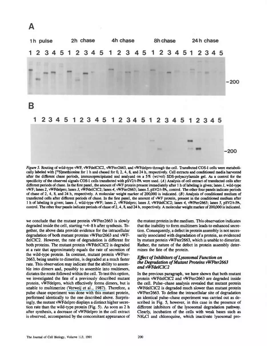
Figure 5. Routing of wild-type vWF, vWFdelC1C2, vWFter2663, and vWFdelpro through the cell. Transfeeted COS-1 cells were metaboli- cally labeled with [3SS]methionine for 1 h and chased for 0, 2, 4, 8, and 24 h, respectively. Cell extracts and conditioned media harvested after the different chase periods, immunoprecipitated and analyzed on a 5% (wt/vol) SDS-polyacrylamide gel. As a control for the specificity of the observed signals COS-1 cells transfected with pSV2/t-PA were used. (A) Analysis of cell extract of transfeeted cells after different periods of chase. In the first panel, the amount of vWF protein present immediately after 1 h of labeling is given; lanes 1, wild-type vWF; lanes 2, vWFdelpro; lanes 3, vWFdelC1C2; lanes 4, vWFter2663; lanes 5, pSV2/t-PA, control. The other four panels indicate periods of chase of 2, 4, 8, and 24 h, respectively. A molecular weight marker of 200,000 is indicated. (B) Analysis of conditioned medium of transfected cells after different periods of chase. In the first panel, the amount of vWF protein, present in the conditioned medium after 1 h of labeling is given; lanes 1, wild-type vWF; lanes 2, vWFdelpro; lanes 3, vWFdelC1C2; lanes 4, vWFter2663; lanes 5, pSV2/t-PA, control. The other four panels indicate periods of chase of 2, 4, 8, and 24 h, respectively. A molecular weight marker of 200,000 is indicated.
we conclude that the mutant protein vWFter2663 is slowly degraded inside the cell, starting ,,~4-8 h after synthesis. To- gether, the above data provide evidence for the intracellular degradation of both mutant proteins vWFter2663 and vWF- delC1C2. However, the rate of degradation is different for both proteins. The mutant protein vWFdelC1C2 is degraded at a rate that approximately equals the rate of secretion of the wild-type protein. In contrast, mutant protein vWFter- 2663, being unable to dimerize, is degraded at a much faster rate. This observation may indicate that the ability to assem- ble into dimers and, possibly to assemble into multimers, dictates the route followed within the cell. To test this option, we investigated the fate of a previously described mutant protein, vWFdelpro, which effectively forms dimers, but is unable to multimerize (Verweij et al., 1987). Therefore, a pulse chase experiment was done with this mutant protein, performed identically to the one described above. Surpris- ingly, the mutant vWFdelpro displays a distinct higher secre- tion rate than the wild-type protein (Fig. 5). As soon as 2 h after synthesis, a decrease of vWFdelpro in the cell extract is observed, accompanied by the concomitant appearance of
the mutant protein in the medium. This observation indicates that the inability to form multimers leads to enhanced secre- tion. Consequently, a defect in protein assembly is not neces- sarily associated with degradation of a protein, as evidenced by mutant protein vWFter2663, which is unable to dimerize. Rather, the nature of the defect in protein assembly deter- mines the fate of the protein.
Effect of lnhibitors of Lysosomal Function on the Degradation of Mutant Proteins vWFter2663 and vWFdelCIC2 In the previous paragraph, we have shown that both mutant protein vWFdelCIC2 and vWFter2663 are degraded inside the cell. Pulse-chase analysis revealed that mutant protein vWFdelC1C2 is degraded much slower than mutant protein vWFter2663. To define the intracellular site of degradation an identical pulse-chase experiment was carried out as de- scribed in Fig. 5, however, in this case in the presence of different inhibitors of the lysosomal degradation pathway. Clearly, incubation of the cells with weak bases such as NI-LC1 and chloroquine, which inactivate lysosomal pro-
The Journal of Cell Biology, Volume 113, 1991 200
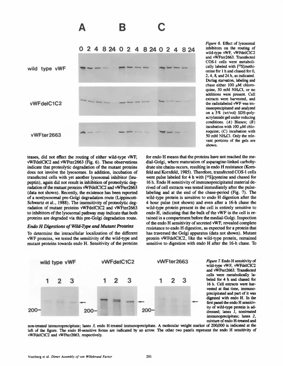
Figure 6. Effect of lysosomal inhibitors on the routing of wild-type vWF, vWFdelC1C2 and vWFter2663. Transfected COS-1 cells were metaboli- cally labeled with 13SS]methi - onine for 1 h and chased for 0, 2, 4, 8, and 24 h, as indicated. During starvation, labeling and chase either 100/zM chloro- quine, 50 mM NI-hCl, or no additions were present. Cell extracts were harvested, and the radiolabeled vWF was im- munoprecipitated and analyzed on a 5% (wt/vol) SDS-poly- acrylamide gel under reducing conditions. (A)Blanco; (B) incubation with 100 ~M chlo- roquine; (C) incubation with 50 mM NI-hC1. Only the rele- vant portions of the gels are shown.
teases, did not effect the routing of either wild-type vWF, vWFdelC1C2 and vWFter2663 (Fig. 6). These observations indicate that proteolytic degradation of the mutant proteins does not involve the lysosomes. In addition, incubation of transfected cells with yet another lysosomal inhibitor (leu- peptin), again did not result in inhibition of proteolytic deg- radation of the mutant proteins vWFdelC1C2 and vWFter2663 (data not shown). Recently, the existence has been reported of a nonlysosomal pre-Golgi degradation route (Lippincott- Schwartz et al., 1988). The insensitivity of proteolytic deg- radation of mutant proteins vWFdelC1C2 and vWFter2663 to inhibitors of the lysosornal pathway may indicate that both proteins are degraded via this pre-Golgi degradation route.
Endo H Digestions o f Wild-1)~e and Mutan t Proteins
To determine the intraceUular localization of the different vWF proteins, we tested the sensitivity of the wild-type and mutant proteins towards endo H. Sensitivity of the proteins
for endo H means that the proteins have not reached the me- dial-Golgi, where maturation of asparagine-linked carbohy- drate site chains occurs, resulting in endo H resistance (Kom- feld and Kornfeld, 1985). Therefore, transfected COS-1 cells were pulse labeled for 4 h with [3~Slcysteine and chased for 16 h. Endo H sensitivity of immunoprecipitated material de- rived of cell extracts was tested immediately after the pulse- labeling and at the end of the chase-period (Fig. 7). The wild-type protein is sensitive to endo H digestion after the 4 hour pulse (not shown) and even after a 16-h chase the wild-type protein present in the cell is entirely sensitive to endo H, indicating that the bulk of the vWF in the cell is re- tained in a compartment before the medial-Golgi. Inspection of the endo H sensitivity of secreted vWE revealed complete resistance to endo H digestion, as expected for a protein that has traversed the Golgi apparatus (data not shown). Mutant protein vWFdelC1C2, like the wild-type protein, remained sensitive to digestion with endo H after the 16-h chase. To
Figure 7. Endo H sensitivity of wild-type vWF, vWFdelC1C2 and vWFter2663. Transfected cells were metabolically la- beled for 4 h and chased for 16 h. Cell extracts were har- vested at that time, immuno- precipitated and part of it yeas digested with endo H. In the first panel the endo H sensitiv- ity of wild-ty~ protein is ad- dressed; lanes 1, nontreated immunoprecipitate; lanes 2, mixture of endo H-treated and
non-treated immunoprecipitate; lanes 3, endo H-treated immunoprecipitate. A molecular weight marker of 200,000 is indicated at the left of the figure. The endo H-sensitive forms are indicated by an arrow. The other two panels represent the endo H sensitivity of vWFdelC1C2 and vWFter2663, respectively.
Voorberg et al. Dimer Assembly of yon WiUebrand Factor 201

Figure 8. Immunolocalization of wild-type protein in COS-1 cells transfected with vWFcDNA. (A) Lowicryl K4M thin section showing labeling in the cisternae of ribosome-studded ER (arrows), a lysosome (ly) is unlabeled. (B and C) Ultrathin frozen sections, with high density of labeling in RER (arrows), in B the cisternae of the RER have been distended, mitochondria (m) and plasma membrane were not labeled. Bar, 0.5 ~m.
analyze the mutant protein vWFter2663, we adjusted the chase period to 4 h, since the amount of this mutant protein in the cell rapidly decreases upon longer chase periods. Again, only endo H sensitive forms of the mutant protein vWFter2663 are encountered in the cell. In conclusion, the major part both of wild-type vWF and of the mutant proteins vWFdelC1C2 and vWFter2663, are retained in a compart- ment prior to the medial-Golgi.
Immunoelectron Microscopy of Valid-Type vWF and Mutant Proteins in Transfected COS-1 Cells In the previous section, we have shown that both wild-type vWF and mutant vWF proteins are present in a compartment before the medial Golgi. To determine the localization of the wild-type and mutant proteins in the cell in more detail, im- munoelectronmicroscopy was performed on ultrathin frozen sections and Lowicryl K4M sections of transfected COS-1 cells. Analysis of COS-1 cells, transfected with wild-type
vWF cDNA, showed labeling of the cisternae of the RER (Fig. 8). In Lowicryl K4M, thin sections (Fig. 8 A), the reac- tion with ribosome-studded ER is clearly visible, although the intensity of labeling was weaker than on thin frozen sec- tions (Fig. g, B and C). No labeling was observed on the lysosomes, mitochondria, cytoplasm or the plasma mem- brane. Together with our biochemical data, this indicates that wild-type vWF is predominantly located in the RER. The mutant protein vWFdelC1C2 displayed a pattern identi- cal to that of the wild-type protein. Also for this mutant pro- tein label was exclusively associated with the RER (data not shown). Analysis of the localization of mutant protein vWFter2663, revealed that like the wild-type protein and vWFdelC1C2 this protein resides in the RER (Fig. 9, A and B). Again, no labeling of ly~osomes was observed, in agree- ment with the inability of lysosomal inhibitors to prevent degradation of the mutant proteins vWFdelC1C2 and vWFter- 2663. Apparently, exit from the ER is a rate-limiting step
The Journal of Cell Biology, Volume 113, 1991 202

Figure 9. Localization of the mutant protein vWFter2663 on ultrathin frozen sections of COS-1 cells transfected with pSVLvWFter2663 DNA. (A) Area of a cell with profiles of labeled RER (arrows). (B) High magnification of labeled RER (arrows). (C) Area of a cell showing autophagic vacuoles (Av) containing segments of labeled ER. Inset, higher magnification of marked area showing two autophagosomes. The membranes of the tubular (t) and vesicular (v) ER are clearly visible, labeling is in the lumen. These structures are comparable to the labeled RER shown A. Bars: (A and C) 0.5 #m; (B and C, inset) 0.1 #m.
both for the ultimate assembly of wild-type vWF multimers and for the degradative events associated with the mutant proteins vWFdelC1C2 and vWFter2663. During our mor- phological studies, on cells transfected with vWFter2663 cDNA, autophagosomes were detected, specifically labeled with anti-vWF antiserum (Fig. 9 C). These structures con- sist of bodies that contained segregated portions of cytoplasm surrounded by a membrane. Vesicular and tubular elements (Fig. 9 C, inset), reminiscent of segments of ER, labeled on their "lumenai" aspect, could be recognized in these auto- phagosomes.
In all experiments described, ,-5 % of the cells were trans- fected with plasmid DNA. The untransfected cells in the sec- tions were observed as negative control for the specificity of the reaction. None of these ceils showed labeling with anti- vWF antiserum.
D i s c u s s i o n
Dimerization of vWF involves the formation of intermolecu-
lar disulfide-bridges at the carboxy-terminus of the vWF molecule (Fretto et ai., 1986). A detailed inspection of the disulfide bridge organization of vWF derived with plasma re- veaied that intermolecular disulfide bridges are located be- tween amino acid residues 2689 and amino acid 2813 ofvWF (Marti et al., 1987). The aforementioned study, however, did not exclude the presence of intermolecular disulfide bridges in the domains C1 and C2, since this region of the vWF was not covered in their studies. Recently, these domains have been implicated in oligomerization, based on the homology of this part of the vWF with the oligomeric proteins throm- bospondin and procollagen (Hunt and Barker, 1987). In this paper, we clearly show that a mutant protein, lacking the do- mains C1 and C2, is able to form dirners and conclude that this part of the molecule is not involved in dimerization of vWF. Both our results with the mutant protein vWFter2663, lacking the extreme carboxy-terminus of vWF, and vWF- delD1-C2 show that solely the utmost 151 carboxy-terminai amino acid residues of vWF are responsible for dimeriza- tion. Previously, we have shown that multimer assembly, in-
Voorberg et al. Dimer Assembly of yon Willebrand Factor 203

volving intermolecular disulfide bonding at the amino termi- nus of vWF, requires only the amino-terminal D domains of vWF (Voorberg et al., 1990). Together with our current ob- servation on the involvement of the region beyond domain C2 in dimerization, the areas of vWF involved in the multimer assembly of vWF have now definitely been assigned.
The time course of vWF biosynthesis, dimerization, car- bohydrate maturation and multimer assembly reveals remark- able features. The majority of vWF present in the cell is en- countered in the dimeric form (Fig. 3), indicating that the rate-limiting step in assembly is before or at the level of mul- timerization. In accord with this assumption are the data on the routing of vWFdelpro, a mutant unable to multimerize, and secreted significantly faster than wild-type vWF (Fig. 5). In addition, both biochemical and morphological data of both wild-type vWF and the vWF mutant proteins demon- strate an accumulation of material in the ER. Since relatively few wild-type multimers are detected in cell extracts, we as- sume that upon a time-requiring initial multimerization pro- cess, these proteins are then rapidly, secreted. It should be noted that similar observations have been made on synthesis and assembly ofvWF in cultured vascular endothelial cells (Wagner and Marder, 1984). Also in these cells, part of the vWF is encountered as endo H-sensitive material that is mainly present in the dimeric form. Hence, transfected COS-1 ceils serve as a suitable model system to study the constitutive secretory pathway for vWF-1 of endothelial cells, without interference of a regulated secretory pathway.
Our observations on the routing and the fate of the mutant protein vWFdelC1C2 are puzzling. Its properties to assemble into dimers and, subsequently, into multimers are similar to those exhibited by the wild-type protein. Despite its correct assembly, the mutant protein vWFdelC1C2 is not secreted, but instead degraded inside the cell. As yet we cannot present an explanation for the degradation of this mutant protein. It could be argued that the mutant protein vWFdelC1C2 is partly malfolded, and thus retained. Although we cannot fully exclude this possibility, the fact that this mutant protein is able to assemble into dimers and multimers, in our view argues against malfolding. Our morphological data clearly show that, like the wild-type protein, vWFdelCIC2 is pre- dominantly present in the ER. After exit from the ER, the wild-type protein is transported rapidly through the Golgi- apparatus to the outside of the cell. The proteolytic degrada- tion of mutant protein vWFdelC1C2 is insensitive to inhibi- tor's of the lysosomal pathwhy (Fig. 5), in accord with our morphological data which dirt not reveal specific staining of lysosomes with polyclonal anti-vWF antibodies. Recently, a degradation route from the ER has been described for the a-chain of the T cell receptor, the H2 subunlt of the asialo- glycoprotein receptor and a mutant of the serpin cxl-antitryp- sin (Lippincott-Schwartz et al., 1988; Amara et al., 1990; Le et al., 1990). In view of the properties of vWFdelC1C2, it is conceivable that this degradative route is pursued by this mutant protein. Numerous studies with oligomeric proteins, e.g., the asialoglycoprotein receptor, inununoglobulins, the T cell receptor and the influenza virus hemagglutinin, have indicated a close correlation between the assembly of a pro- tein mad its routing through the cell (Bole et al., 1986; Cope- land et al., 1988; LippincoWSchwartz et al., 1988; Amara et al., 1989; Doms et al., 1987). These studies demonstrate that a defect in the oligomer assembly of a protein results
in retention or proteolytic degradation of the nonassembled product at the site of oligomer-assembly, i.e., the ER (Lod- ish, 1988). The behavior of the mutant protein vWFter2663 strictly follows the pattern outlined above. The inability of this protein to undergo dimerization in the ER, its sensitivity to endo H and its relatively fast rate of degradation are com- patible with the view that this protein is degraded from the ER. This assumption is further substantiated by insensitivity of the proteolytic degradation of vWFter2663 to inhibitors of lysosomal degradation. The linkage between assembly and routing is further illustrated by the properties of the mu- tant protein vWFdelD1-C2, containing only the region be- yond domain C2. This mutant protein is able to assemble into dimers and, subsequently, readily transported from the ER, through the secretory pathway as evidenced by its ap- pearance in the conditioned medium.
In this paper, we describe two mutant vWF proteins which conceivably are degraded from the ER. It has been suggested that autophagosomes may be the site of degradation from the ER (Lippincott-Schwartz et al., 1988). Here, we report that in COS-1 cells transfected with pSVLvWFter2663, but not in COS-1 cells transfected with wild-type vWF, autophago- somes were detected which contain vWF (Fig. 9 C). These observations are suggestive for a role of the autophagosome in degradation of this mutant protein from the ER. The properties of mutant protein vWFdelC1C2 indicate that this mutant protein is also degraded from the ER. However, in COS-1 cells transfected with vWFdelC1C2 no autophago- somes could be detected. Since the degradation of vWFdel- C1C2 proceeds much slower than that of vWFter2663, the number of autophagosomes present in the transfected COS-1 cell may significantly differ for these two mutant proteins. So far, morphological examinations have been limited by the frequency of vWF-transfected cells among the population of untransfected COS-1 cells. Currently, we are establishing stable cell lines expressing either wild-type vWF, vWFdel- C1C2 or vWFter2663, to enable a more thorough analysis of the relation between protein degradation from the ER and the occurrence of autophagosomes.
We thank A. Sormenberg, A. Leyte, and members of the Department of Molecular Biology for critically reading the manuscript.
This study was supported by the Netherlands Organization for Scientific Research (N.W.O.) (grant no. 900-526-075).
Received for publication 25 June 1990 and in revised form 11 October 1990.
References
Amara, J. F., G. Lederkremer, and H. F. Lodish. 1989. Intracellular degrada- tion of unassembled Asialoglycoprotein receptor subunits: a pre-Golgi, non- lysosomal endopmteolytic cleavage. J. Cell Biol. 109:3315-3324.
Bole, D. G., L. M. Headerflaot, and J. F. Kearney. 1986. Posttranslational as- sociation of immunoglobulin heavy chain binding protein with nascent heavy chains in nonsecrvting and secreting hybridomas. J. Cell Biol. 102:1558- 1566.
Bonthron, D. T., R. I. Handin, R. J. Kaufman, L. C. Wasley, E. C. Orr, L. M. Mitsoek, B. Ewenstein, J. Loscalzo, D. Ginsburg, and S. H. Orkin. 1986. Structure of pre-pro-von Wfllebrand factor and its expression in heterolo- gous cells. Nature (Lond.). 324:270-273.
Copetand, C. S., K. -P. Zimmers, K. R. Wagner, G. A. Healy, I. Mellman, and A. Helenius. 1988. Folding, tfimerization, and transport aresequential events in the biogenesis of influenza virus hemagglutirtin. Cell. 53:197-209.
Doms, R. W., A. Ruusala, C. Machamer, J. Helenius, A. Helenius, andJ. K. Rose. 1988. Differential effects of mutations in three domains on folding, quaternary structure, intracellular transport of vesicular stomatitus virus G protein. J. Cell Biol. 107:89-99.
Fretto, L. J., W. E. Fowler, D. R. McCaslin, H. P. Edckson, and P. A.
The Journal of Cell Biology, Volume 113, 1991 204

McKee. 1986. Substructure of human yon Willebrand factor. J. Biol. Chem. 261:15679-15689.
Gething, M.-J., K. McCammon, and J. Sambrook. 1986. Expression of wild- type and mutant forms of influenza haemagglutinin: The role of folding in intracellular transport. Cell. 46:939-950.
Gould, S. J., G.-A. Keller, and S. Subramani. 1987. Identification of a perox- isomal targeting signal at the carboxyl terminus of firefly luciferase. J. Cell Biol. 105:2923-2931.
Hoyer, L. W., and J. R. Shalnoff. 1980. Factor VIII-related protein circulates in normal human plasma as high molecular weight multimers. Blood. 55: 1056-1059.
Hunt, L. T., and W. C. Baker. 1987. Von Willebrand factor shares a distinctive cysteine-rich domain with thrombospondin and procollagun. Biochim. Bio- phys. Res. Commun. 144:876-882.
Hurtley, S. M., and A. Helenius. 1989. Protein oligumerization in the endo- plasmic reticulum. Annu. Rev. Cell Biol. 5:277-307.
Kornfeld, R., and S. Kornfeld. 1985. Assembly of asparagine-linked oligosac- charides. Annu. Rev. Biochem. 54:631-664.
Kramer, W., V. Drutsa, H.-W. Jansen, B. Kramer, M. Plugfelder, and H. J. Fritz. 1984. The gapped duplex DNA approach to oligonucleotide-directed mutation construction. Nucleic. Acids Res. 12:9441-9456.
Laemmli, U. K. 1970. Cleavage of structural proteins during the assembly of the head of bacteriophage T4. Nature (Lond.). 227:680-685.
Le, A., K. S. Graham, and R. N. Sifers. 1990. Intracellular degradation of the transport-impaired human PiZ ct 1-antitrypsin variant. Biochemical mapping of the degradative event among compartments of the secretory pathway. J. Biol. Chem. 265:14001-14007,
Lippincott-Schwartz, J., J. S. Bonifacino, L. C. Yuan, and R. D. Klausner. 1988. Degradation from the endoplasmic reticulum: disposing of newly syn- thesized proteins. Cell. 54:209-220.
Lodish, H. F. 1988. Transport of secretory and membrane glycoproteins from the rough endoplasmic reticuinm to the Golgi: a rate-limiting step in protein maturation and secretion. J. Biol. Chem. 263:2107-2110.
Loesborg, C., M. D. Gonsalves, J. Zandbergen, Cb. Wlllems, W. G. van Aken, H. V. Stel, J. A. van Mourik, and P. G. de Groot. 1983. The effect of calcium on the secretion of Factor VIII-related antigen by cultured en- dothelial cells. Biochim. Biophys. Acta. 763:160-168.
Luthman, H., and G. Magnusson. 1983. High efl]ciency polyoma DNA trans- fection of chloroquine treated cells. Nucleic Acids Res. 11:1295-1308.
Marti, T., S. J. Rosselet, K. Titani, and K. A. Walsh. 1987. Identification of disulfide-bridged structures within human yon Willebrand factor. Biochem- istry. 26:8099-8109.
Medda, S., R. M. Chemelli, J. L. Martin, L. R. Pohl, and R. T. Swank. 1989. Involvement of the carboxyl-terminal propeptide of/~-glucuronidase in its compartmentalization within the endoplasmic rcticulum as determined by a synthetic peptide approach. J. Biol. Chem. 264:15824-15828.
Messing, J., and J. Vieira. 1982. A new pair of M13 vectors for selecting either strand of double-digest restriction fragments. Gene (Amst.). 19:269-276.
Munro, S., and H. R. B. Pelham. 1987. A C-terminal signal prevents secretion of luminal ER proteins. Cell. 48:899-907.
Reinders, J. H., Ph. G. de Groot, J. Dawes, M. R. Hunter, H. A. A. van Heug- ten, J. Zandbergen, M. D. Gonsalves, and J. A. van Mourik. 1985. Compar- ison of secretion and subcellular localisation of yon Willebrand protein with that of thrombospondin and fibronectin in cultured human vascular en-
dothelial cells. Biochim. Biophys. Acta. 844:306-313. Rose J. K., and R. W. Doms. 1988. Regulation of protein export from the endo-
plasmic reticulum. Annu. Rev. Cell. Biol. 4:257-288. Rothman, J. E. 1989. Polypeptide chain binding proteins: catalysts of protein
folding and related processes in cells. Cell. 59:591-601. Rugguri, Z. M., and T. S. Zimmerman. 1981. The complex multimeric compo-
sition of Factor VHI/von Willebrand factor. B/ood. 57:1140-1143. Sadler, J. E., B. B. Shelton-Inlces, J. M. Sorace, J. M. Harlan, K. Titani, and
E. W. Davie. 1985. Cloning and characterization of two cDNAs coding for human yon WiUebrand factor. Proc. Natl. Acad. Sci. USA. 82:6394-6398.
Shelton-Inlces, B. B., K. Titani, and J. E. Sadler. 1986. cDNA sequences for human yon Willebrand factor reveal five types of repeated domains and five possible protein sequence polymorphisms. Biochemistry. 25:3164-3171.
Sporn, L. A., V. J. Marder, and D. D. Wagner. 1986. Inducible secretion of large, biologically potent yon Willebrand factor multimers. Cell. 46:185- 190.
Tabor, S., and C. C. Richardson. 1987. DNA sequence analysis with a modified bacteriophage T7 DNA polymerase. Proc. Natl. Acad. Sci. USA. 80:3963- 3965.
Titani, K., S. Kumar, K. Takio, L. H. Ericsson, R. D. Wade, K. Ashida, K. A. Walsh, M. W. Chopek, J. E. Sadler, and K. Fujikawa. 1986. Amino-acid sequence of human yon Willebrand factor. Biochemistry. 25:3171-3184.
Van der Van, W. J. M., J. Voorberg, R. Fontijn, H. Pannekcek, A. M. W. Van den Ouweland, H. L. P. Van Duynhoven, A. J. M. Roebroek, and R. J. Siezen. 1990. Furin is a subtilisin-like proprotein processing enzyme in higher eukaryotes. Mol. Biol. Rep. 14:265-275.
Van Mourik, J. A:, and P. A. Bolhuis. 1978. Dispersity of human Factor VIII- yon Willebrand factor. Thromb. Res. 13:15-24.
Van Zonneveld, A.-J., H. Veerman, and H. Pannekock. 1986. Autonomous functions of structural domains on human tissue-type plasminogcn activator. Proc. Natl. Acad. Sci. USA. 83:4670-4674.
Verwcij, C. L. 1988. Biosynthesis of human yon Willebrand factor. Haemostu- sis. 18:224-245.
Verweij, C. L., P. Diergaarde, M. Hart, and H. Pannckoek. 1986. Full-length yon Willebrand factor (vWF) eDNA encodes a highly repetitive protein, con- siderably larger that the mature vWF subunit. EMBO (Eur. Mol. Biol. Or- gan.) J. 5:1839-1847.
Verweij, C. L., M. Hart, and H. Pannekcek. 1987. Expression of a variant von WiUebrand factor vWF eDNA in heterologuus cells: requirement of the pro- polypeptide in vWF multimer assembly. EMBO (Fur. Mol. Biol. Organ.) J. 6:2885-2890.
Verwcij, C. L., M. Hart, and H. Pannekoek. 1988. Proteolytic cleavage of the precursor of yon Willebrand factor (pro-vWF) is not essential for multimer formation. J. Biol. Chem. 263:7921-7924.
Voorborg, J., R. Fontijn, J. A. yon Mourik, and H. Pannekoek. 1990. Domains involved in multimer assembly of yon Willebrand factor (vWF): multimer- ization is independent of dimcrization. EMBO (Fur. Mol. Biol. Organ.) J. 9:797-803.
Wagner, D. D., and V. J. Marder. 1984. Biosynthesis of yon Willebrand pro- tein by human endothelial cells: processing steps and their intracclhilar local- ization. J. Cell Biol. 99:2123-2130.
Wise, R. J., D. D. Pitmann, R. I. Handin, R. J. Kaufman, and S. H. Orkin. 1988. The propeptide of yon Willebrand factor independently mediates the assembly of yon Willebrand multimers. Cell. 52:229-236.
Voorberg et al. Dimer Assembly of yon Willebrand Factor 205


