Embed Size (px)
Citation preview

A B S T R A C T : We examined changes in membrane propertiesupon acidification of dioleoylphosphatidylethanolamine/cho-lesterylhemisuccinate liposomes and evaluated their potentialto deliver entrapped tracers in cultured macrophages. Mem-brane permeability was determined by the release of entrappedcalcein or hydroxypyrene-1,3,6-trisulfonic acid (HPTS)-p- x y-lene-bis-pyridinium bromide (DPX); membrane fusion, by mea-suring the change in size of the liposomes and the dequenchingof octadecylrhodamine-B fluorescence; and change in lipid or-ganization, by 3 1P nuclear magnetic resonance spectroscopy.Measurement of cell-associated fluorescence and confocal mi-croscopy examination were made on cells incubated with lipo-somes loaded with HPTS or HPTS-DPX. The biophysical stud-ies showed (i) a lipid reorganization from bilayer to hexagonalphase progressing from pH 8.0 to 5.0, (ii) a membrane perme-abilization for pH <6.5, (iii) an increase in the mean diameterof liposomes for pH <6.0, and (iv) a mixing of liposome mem-branes for pH <5.7. The cellular studies showed (i) an uptake ofthe liposomes that were brought from pH 7.5–7.0 to 6.5–6.0and (ii) a release of ~15% of the endocytosed marker associatedwith its partial release from the vesicles (diffuse localization).We conclude that the permeabilization and fusion of pH-sensi-tive liposomes occur as a consequence of a progressive lipid re-organization upon acidification. These changes may developintracellularly after phagocytosis and allow for the release ofthe liposome content in endosomes associated with a redistri-bution in the cytosol.
Paper no. L8278 in Lipids 35, 213–223 (February 2000).
Delivery of macromolecules and other nonpermeant con-stituents into the cytosol of target cells is essential for the de-velopment of many new therapeutic approaches. Conventionalliposomes are often ineffective in this context since they tendto bring their load to lysosomes (1). pH-sensitive liposomes(2) may represent a significant improvement for intracellular
drug unloading thanks to their progressive, acid-driven desta-bilization along the endocytic pathway (3). These liposomes,indeed, not only are susceptible to releasing their content inacid vacuoles, such as late endosomes and lysosomes, but alsoare capable of promoting the transfer of encapsulated tracersto the cytosol (4). Accordingly, they have been assessed in aseries of studies for potential therapeutic applications, includ-ing antitumor and antibacterial chemotherapy (5–6), and forthe delivery of proteins, oligonucleotides, or even DNA (7–9).
In the present study, we examined a typical type of pH-sensitive liposomes made of dioleoylphosphatidylethan-olamine and an anionic component, cholesterylhemisuccinate(DOPE/CHEMS). The small cross-sectional area of the head-group of phosphatidylethanolamine, relative to that of theacyl chains, confers to this molecule a propensity to sponta-neously form an inverted hexagonal phase HII structure, a par-ticular organization involved in membrane destabilization andfusion (10). At alkaline or neutral pH, however, the nega-tively charged CHEMS will reduce the intermolecular repul-sions of phosphatidylethanolamine head groups and stabilizethe structure in a bilayer organization. Yet, upon acidification,CHEMS will become less charged and will no longer hinderthe reorganization of phosphatidylethanolamine in an HI Istructure, ensuring the destabilization of the membrane struc-ture (3). This mechanism has been documented concerningthe increase in membrane permeability (11–16) and fuso-genicity (2,12,14,17) of liposomes, and to a lesser extent, thecellular delivery of the vesicles’ contents (1,4,18,19). Yet thechanges in polymorphic organization of the lipids (20) remainl a rgely undefined in this context. Moreover, most of thesestudies have used different types and compositions of lipo-somes, which make their conclusions difficult to generalizebecause lipid composition critically determines the pH sensi-tivity of acid-labile liposomes (4,11). In the present study, wehave therefore undertaken to characterize, in a more system-atic fashion, the biophysical phenomena (including changesin polymorphic organization) leading to an effective in vitrorelease of membrane-impermeant probes from one type of li-posomes of a fixed composition (DOPE/CHEMS, 7:3 molarratio), and have examined whether such release occurs incells. DOPE/CHEMS liposomes were systematically com-
Copyright © 2000 by AOCS Press 213 Lipids, Vol. 35, no. 2 (2000)
*To whom correspondence should be addressed at UCL 73.70 avenue E.Mounier 73, 1200 Brussels; Belgium. E-mail: [email protected]: CHEMS, cholesterylhemisuccinate; DOPC, dioleoylphos-phatidylcholine; DOPE, dioleoylphosphatidylethanolamine; DPX, p- x y l e n e -bis-pyridinium bromide; HPTS, hydroxypyrene-1,3,6-trisulfonic acid; LUV,large unilamellar vesicles; MLV, large multilamellar vesicles; NMR, nuclearmagnetic resonance; PBS, phosphate buffered saline; R18, octadecylrho-damine B; RPMI, Roswell Park Memorial Institute.
Biophysical Studies and Intracellular Destabilization of pH-sensitive Liposomes
Françoise Van Bambekea,*, Anne Kerkhofsa, André Schanckb, Claude Remaclec,Etienne Sonveauxd, Paul M. Tulkensa, and Marie-Paule Mingeot-Leclercqa
aUnité de Pharmacologie Cellulaire et Moléculaire, Brussels; bUnité de Chimie Physique Moléculaire et de Cristallographie,Louvain-La-Neuve; cLaboratoire de Biologie Cellulaire, Unité de Biologie Animale, Louvain-La-Neuve; and dUnité de Chimie
Pharmaceutique et de Radiopharmacie, Brussels; Université Catholique de Louvain, Belgium
L8278 7/19/04 4:00 PM Page 213

pared to vesicles made of dioleoylphosphatidylcholine andCHEMS (DOPC/CHEMS), since phosphatidylcholine doesnot spontaneously form an HII structure and is therefore non-fusogenic.
MATERIALS AND METHODS
Liposome pre p a r a t i o n . L a rge unilamellar vesicles (LUV) wereused throughout all experiments, but large multilamellar vesicles( M LV) were also included for 3 1P nuclear magnetic resonance(NMR) studies. Both DOPE/CHEMS and DOPC/CHEMS lipo-s o m e s were made at a phospholipid/CHEMS molar ratio of 7:3.Depending on the experiments and on the sensitivity of themethod used, the volume of buffer varied between 1 and 5 mL,and the concentration of lipids, between 1 and 50 mg/mL. M LVwere obtained by hydration of the dry lipid films for 1 h undernitrogen at 37°C in a 40 mM glycine-NaOH solution adjusted topH 11 and containing the fluorescent probes (see below). Theywere thereafter submitted to five successive cycles of freezing(at −80°C) and thawing (at 37°C). LUV were obtained by ex-truding the resulting suspension 10 times through two super-posed polycarbonate filters (pore size, 100 nm; NucleoporeCostar Corporation, Badhoevedorp, The Netherlands) under anitrogen pressure of 17 bars, in a 10-mL Thermobarrel Extruder(Lipex Biomembranes, Va n c o u v e r, Canada) (21,22). The actualphospholipid concentration of each preparation was determinedby phosphorus assay, as decribed previously (22), and adjustedto the desired value by dilution in the appropriate buffer just priorto each experiment. (All concentrations of liposomes are ex-pressed by reference to their total lipid concentration, based onphospholipid determination and on a molar ratio of phospho-lipid/CHEMS of 7:3. In preliminary experiments, we checkedthat the phospholipid/cholesterol molar ratio was effectively veryclose to 7:3 after the hydration and extrusion procedures.) Lipo-somes were stored under nitrogen at 4°C until the beginning ofthe experiment, to minimize the risk of extensive lipid hydroly-sis and oxidation (23), and were used within 24 h. The lack ofchemical hydrolysis of phosphatidylethanolamine in our experi-mental conditions was checked by preparing DOPE/CHEMS li-posomes with a trace amount of phosphatidylethanolamine 1-palmitoyl-2-(1-14C)-linoleoyl (100 µCi/mmol of DOPE), andletting them age at 4°C for 24 h. Samples were then analyzed by thin-layer chromatography using CHCl3/CH3OH/NH3( 2 5 % ) / H2O 24:16:2:1 (by vol) as mobile phase. The radioactiv-ity associated with free fatty acid and lysophosphatidylethan-olamine was then measured and found to be less than 2% of thetotal amount of radioactivity recovered, whereas approximately94% co-migrated with phosphatidylethanolamine.
Studies on liposomes. (i) Permeability studies. The releaseof calcein [entrapped at a self-quenching concentration(18,22,24)] and of HPTS [hydroxypyrene-1,3,6-trisulfonicacid (25)] coentrapped with and quenched by DPX (p- x y l e n e -bis-pyridinium bromide (11)] from the liposomes was fol-lowed by the increase of fluorescence upon dilution followingtheir leakage from the vesicles. Calcein was prepared in 6 NNaOH and purified by chromatography using a Sephadex®
LH-20 column, as described previously (22). The final solu-tion had a concentration of 74 mM calcein, a pH of 11, and anosmolarity of 448 mOsm/kg (measured by the freezing pointtechnique; model 3C2, Advanced Cryomatic Osmometer, Ad-vanced Instruments, Needham Heights, MA). HPTS and DPXwere dissolved in a 40 mM glycine-NaOH mixture adjusted topH 11 (393 mOsm/kg) at a concentration of 31.8 and 35 mM,respectively (4,11). In preliminary experiments, we checkedthat DPX effectively quenched the fluorescence of HPTS forthis concentration ratio. These solutions were then used forhydration of the lipid films as described above. After liposomepreparation at a lipid concentration of 30 mM, the unencapsu-lated dye was removed by minicolumn centrifugation (22).Before each experiment, liposomes were adjusted to a totallipid concentration of 7.14 µM and 2.16 µM for calcein- andHPTS-release studies, respectively, using isoosmotic phos-phate buffers of pH ranging from 8.0 to 5.0. Calcein fluores-cence was measured at room temperature on a PerkinElmer2000 Fluorescence Spectrophotometer [PerkinElmer, Bea-c o n s field, United Kingdom; λe x c, 490 nm; λe m, 516 nm (22)];the inner effect was checked and found to be negligible. Thefluorescence of free calcein in solution was found to be time-and pH-dependent, and our measurements were therefore sys-tematically corrected for by appropriate factors. These factorswere calculated by measuring the fluorescence of calcein so-lutions at all the pH values investigated as a function of thetime and by comparing these values to that a calcein solutionincubated for the same period of time and at pH 7.5. Fluores-cence of HPTS was recorded at room temperature, using a λe mof 520 nm and λexc of 390 and 450 nm (25), using a LS-30 Flu-orescence Spectrophotometer (PerkinElmer). The percentageof dye release at any given time was defined as the ratio be-tween the fluorescence measured at that time and that ob-served at the same time after disruption of the liposomes byexposure to 0.1% Triton X-100 in the same buff e r. For eachpH- and time-condition tested, the fluorescence valuesrecorded were corrected for the influence of Triton X-100 bycomparing the fluorescence value measured for a calcein solu-tion in the absence of Triton to that measured in the presenceof the detergent. A leakage efficiency factor, defined as theratio between the percentage of marker released at a given pHto that measured at pH 7.5, was then used to assess the labilityof the liposomes upon pH change.
(ii) Mixing of the liposome membranes: studies with oc -t a d e c y l rhodamine B (R18). Mixing of the liposome mem-branes was followed by measuring the fluorescence increaseof R18, a lipid-soluble probe, upon dilution in the membraneoccurring by fusion between labeled and unlabeled liposomes(26,27). Labeled liposomes were obtained by incorporatingR18 in the dry lipid film at a concentration of 5.7% of the totallipids. Labeled and unlabeled liposomes, prepared at 2 mM,were mixed at a ratio of 1:4 and diluted to a final concentra-tion of 5 µM by addition of appropriate buffers at the time ofthe experiment (40 mM cacodylate buffers from pH 7.4 to 5.0and 40 mM acetate buffer for pH 4.5). Fluorescence wasrecorded at room temperature (λexc, 560 nm; λem, 590 nm; the
214 F. VAN BAMBEKE ET AL.
Lipids, Vol. 35, no. 2 (2000)
L8278 7/19/04 4:00 PM Page 214

inner filter effect was checked and found to be negligible) onan LS-30 PerkinElmer fluorimeter. The percentage of fusionwas calculated by comparing the fluorescence values ob-served after liposome mixing to that measured for 25 µM li-posomes labeled by 1.16% of R18 (i.e., an identical totalamount of marker diluted five times in membranes).
(iii) Size studies. The apparent size of the liposomes di-luted to 50 µM in 40 mM cacodylate buffers of pH rangingfrom 8.0 to 5.0 was measured by quasi-elastic light scatteringspectroscopy (28) using a Coulter Nano-sizer N4 MD parti-cle Analyzer (Coulter Electronics Inc., Luton, England) at anangle of 90°, using both unimodal and size distribution analy-sis modes to determine the mean diameter and the full sizedistribution profile of each preparation, respectively.
(iv) Polymorphic behavior of the lipids as determined by3 1P NMR. The phase behavior of liposomes was determinedas a function of pH and temperature. Classically, three typesof phases can be distinguished on the basis of their 3 1P NMRsignals. The so-called bilayer signal is characterized by a high-field maximum and a low-field shoulder; the inverted hexago-nal phase, by an inverse symmetry and a twofold reducedwidth; and the isotropic signal, by a sharp symmetric signal.In practice, MLV give a bilayer signal due to their org a n i z a-tion in concentric bilayers, where rapid motion of phospho-lipid molecules along their long axis results in axial symmetryand partially averages the chemical shift anistropy (29). LUVgive an isotropic signal, because the 3 1P atoms are submittedto rapid isotropic motion owing to the rapid tumbling of thevesicles (30). As stated in the introduction, phosphatidyl-ethanolamine has a propensity to organize itself in hexagonalphase (especially upon warming), in which the molecules pro-ject radially from the center of a cylinder of very small radius.The rapid rotation of the cylinders causes further averaging ofchemical shift anistropy, explaining the reversed shape and thereduced width of the spectrum by comparison with the bilayerspectrum (29). To compare the appearance of this hexagonalphase starting from an isotropic or a bilayer phase, we per-formed NMR studies on both LUV and MLV. Liposome sus-pensions prepared at an initial lipid concentration of 75 mMwere diluted in the appropriate buffers (Na carbonate from pH10.0 to 9.2; Tris-HCl from pH 9.0 to 7.0; citric acid-NaOHfrom pH 6.2 to 5.2; and Na citrate-HCl from pH 5.0–3.0) andmixed with 0.2 mL D2O (for locking on the deuterium signal)in 10 mm RMN tubes (final lipid concentration, 12.5 mM).The actual pH value of each suspension was measured with aminielectrode and a 691 Metrohm pH meter (Herisau, Switzer-land), at both 30 and 70°C. All spectra were obtained at 101.3MHz with a WM 250 Bruker instrument. Typical Fouriertransform conditions were as follows: spectral width 20 kHz;8 K data acquisition points; flip angle 60° (17 µs); 1 s repeti-tion time. Five thousand free induction decays (FID) were ac-cumulated with broad-band proton powergated decoupling. Aline broadening of 50 Hz was applied to the FID beforeFourier transformation. Measurements were done as a func-tion of temperature in the range 30–70°C in the increasing ordecreasing mode. After each change of temperature, the sam-
ple was allowed to equilibrate during 30 min before accumu-lation. The lipid phase proportions were calculated as de-scribed earlier (31).
Studies on cultured cells. (i) Cell culture and incubationwith liposomes. J774 macrophages, a continuous reticulosar-coma cell line of murine origin (32), were grown as monolay-ers in 5% CO2/95% air, at 37°C in an RPMI (Rosewell ParkMemorial Institute) 1640 medium supplemented with 10%(vol/vol) fetal calf serum, 1.45% (wt/vol) NaHCO3, and an-tibiotics (streptomycin 50 µg/mL, penicillin 50 IU/mL). Con-fluent cells were suspended by incubation with trypsin( 0 . 5 % ) / E D TA (0.2%) in phosphate-buffered saline (PBS; 8g/L NaCl, 0.2 g/L KCl, 1.15 g/L Na2H P O4, 0.2 g/L KH2P O4;pH 7.4), centrifuged, and plated in six-well plates for 3 d inRPMI medium supplemented with fetal calf serum, NaHCO3,and antibiotics, as decribed above. At the time of the experi-ment, the cells were washed three times with PBS and reincu-bated during 15 min with 2 mL PBS containing liposomes (90µM total lipid concentration) loaded with HPTS or HPTS-DPX. In preliminary experiments, the total fluorescence of theliposome preparations was determined after disruption by ex-posure to Triton X-100 (0.1%) and corrected for Triton X-100influence as described above. These studies showed that theamount of HPTS or HPTS/DPX entrapped in DOPC/CHEMSwas slightly larger than that entrapped in DOPE/CHEMS li-posomes (30% in excess). After 15 min of uptake at 37°C, thecells were washed 10 times with ice-cold PBS and returned ingrowth medium at 37°C for different chase times.
(ii) Fluorescence assay. The cells were detached bytrypsinization, pelleted by centrifugation, washed twice withice-cold PBS, and gently resuspended in PBS. Viability andmembrane integrity were checked by measuring the releaseof the cytosolic enzyme lactate dehydrogenase. HPTS fluo-rescence was recorded at a λe m of 520 nm upon excitation at363, 390, 450, or 488 nm and the signal expressed by refer-ence to the protein cell content (33). The wavelengths of 390and 450 nm correspond to the maximum in the excitationspectrum of HPTS at acidic and neutral pH, respectively (25),whereas those at 363 and 488 nm were the λe x c used for con-focal microscopy studies (see below). The 450/390 fluores-cence ratio of HPTS was then used to evaluate the pH atwhich the probe was exposed in cells, based on calibrationcurves constructed by preparing the probe in phosphatebuffers of known pH (between 5 and 8).
(iii) Confocal micro s c o p y. Cells were treated as describedabove, except that they were cultivated in Lab-Tek Permanoxculture chambers (Nunc, Roskilde, Denmark) allowing for theobservation of living cells at high magnification under an in-verted microscope. Images were recorded during incubationat 37°C using a Bio-Rad MRC 1024-UV laser scanning con-focal microscope (Hemel Hempstead, United Kingdom) oper-ated under control of the Lasersharp 2.10 software. Conditionsof imaging were: filters E2 and UBHS, 63× oil-immersion ob-jective, zoom 3, PMT2 (photomultiplier) and PMT3, iris = 3.0,gain = 1300–1500, black level = 0, laser power 0.3–3%. Thefirst channel (PMT2) was set to collect the fluorescence signal
DOPE/CHEMS LIPOSOMES: IN VITRO AND CELLULAR STUDIES 215
Lipids, Vol. 35, no. 2 (2000)
L8278 7/19/04 4:00 PM Page 215

emitted upon excitation at 488 nm, and this signal was treatedto appear in green on the screen and micrographs. The secondchannel (PMT3) was set to read the fluorescence emitted uponexcitation at 363 nm and this signal appears in red on thescreen and micrographs. Although the excitation wavelengths,which were imposed by the equipment, are not optimal, theynevertheless allowed for a clear-cut analysis of the pH diff e r-ences in HPTS environment. The pH at which the flu o r e s c e n c esignals recorded upon excitation at 363 and 488 nm are equalwas determined to be ~7.15, setting therefore the value forshifting the appearance of the tracer from green to red.
M a t e r i a l s . DOPE and DOPC were obtained from Av a n t iPolar Lipids, Inc. (Alabaster, AL), and radiolabeled phos-phatidylethanolamine (specific radioactivity = 54 mCi/mmol),from Amersham International plc (Amersham, United King-dom). Calcein and CHEMS were purchased from SigmaChemical Co (St. Louis, MO). HPTS, DPX, and R18 camefrom Molecular Probes Inc. (Eugene, OR). Cell culture mediaand fetal calf serum were obtained from Gibco-Biocult (Peis-l e y, Scotland). Other products were of analytical grade and ob-tained from E. Merck (Darmstadt, Germany).
RESULTS
Studies with liposomes. (i) Permeability studies. Figure 1A showsthe release of calcein from LUV as a function of the time of incu-bation in buffers of different pH. DOPE/CHEMS liposomes re-leased a large proportion (~ 60%) of calcein almost instanta-neously when exposed to pH 5.0, with no further release after thisfirst burst. The effect was highly dependent on pH since less than10% release was observed at pH 7.5. DOPE/CHEMS liposomesexposed to pH 5.5 showed an intermediate behavior. In contrast,DOPC/CHEMS liposomes released a much lower proportion ofcalcein at pH 5.0, and this release proceeded slowly over the fir s t10 min of observation (Fig. 1B). Figure 1C shows the compara-tive release of calcein at 5 min over pH values from 8.0 to 5.0.The data confirm that acidity exerts a marked influence on cal-cein-release from DOPE/CHEMS liposomes, with a signific a n te ffect already observed at pH 6.0 (P < 0.05 by t-test). In contrast,DOPC/CHEMS liposomes appear considerably less sensitive tothe decrease of pH. In parallel experiments, we measured the re-lease of another marker (HPTS/DPX) from DOPE/CHEMS lipo-somes (Fig. 1C) with essentially similar results. HPTS/DPX didnot significantly leak out from DOPC/CHEMS liposomes underthe same conditions (data not shown).
(ii) Liposome membranes mixing studies. Figure 2A showsthe fluorescence of R18 upon mixing of labeled and unlabeledLUV made of DOPE/CHEMS at different pH values. Lipo-somes exposed to pH 5.0 quickly (<5 min) showed a markedsignal increase which amounted to approximately 10 timesthat recorded for the same liposomes exposed to pH 7.4. Lipo-somes exposed to pH 5.4 showed an intermediate behavior.(More time seems to be needed to reach a plateau value at pH5 than at pH 5.4; because no measurement was possible fortime periods shorter than 30 s, we assume that the dequench-ing proceeds very quickly during these first 30 s, and more
216 F. VAN BAMBEKE ET AL.
Lipids, Vol. 35, no. 2 (2000)
FIG. 1. Release of fluorescent tracers from large unilammelar vesicles(LUV) upon acidification. (A) Percentage of calcein released from dioleoylphosphatidylethanolamine/cholesterylhemisuccinate (DOPE/CHEMS) or(B) dioleoylphosphatidylcholine/CHEMS (DOPC/CHEMS) liposomes afterdilution and incubation at 37°C in isoosmotic phosphate buffers (closedsymbols, pH 5.0; grey symbols, pH 5.5; open symbols, pH 7.5). Datashown are the mean values of three independent measurements in singleliposome preparations. (C) Leakage efficiency factor (see Materials andMethods section) of DOPE/CHEMS (closed circles) and DOPC/CHEMS(open squares) liposomes loaded with calcein ( ) or with hydroxy-pyrene;1,3,6-trisulfonic acid–p-xylene-bis-pyridinium bromide (HPTS-DPX) (- - - - ; DOPE/CHEMS only) 5 min after dilution in isoosmoticbuffers of different pH. Results are the means ± SD of three indepen-dent measures on a single preparation of liposomes.
L8278 7/19/04 4:00 PM Page 216

slowly thereafter; thus time-dependence of the increase canonly be assessed at pH 5.0.) These experiments were then re-peated over a whole range of pH values, and DOPE/CHEMSliposomes were compared to DOPC/CHEMS liposomes. Fig-ure 2B shows that the increase of the signal became signifi-cant at pH ≤ 5.4. DOPC/CHEMS liposomes did not show sig-nificant fluorescence increase throughout the whole range ofpH values. The data of Figure 2 were also used to calculate thepercentage of liposome fusion and this is shown on the rightordinate. Finally, similar experiments were also run in phos-phate buffers (as used in permeability studies) to rule out anyi n fluence of the buffer system used. No difference were foundwhen comparing data at the same pH with the phosphate andcacodylate buffer systems.
(iii) Liposome size studies. Unimodal analysis of the parti-cle size showed that the mean diameter of DOPE/CHEMSLUV increased from 120 nm at pH 7.5 to 250 nm at pH 5.0,
whereas the mean diameter of DOPC/CHEMS liposomes re-mained nearly constant at 100 nm in the same range of pH.The size distribution analysis of the DOPE/CHEMS liposomesillustrated in Figure 3 showed a narrowly distributed popula-tion at pH 7.5, but a progressive and marked shift towardl a rge-diameter vesicles at acid pH. Titration back to pH 7.5 ofliposomes exposed to pH 5.0 did not reverse this effect, andliposomes still presented a larger diameter (mean = 295 nm).
(iv) Polymorphism studies. The polymorphic behavior ofDOPE/CHEMS LUV was studied upon temperature and pH
DOPE/CHEMS LIPOSOMES: IN VITRO AND CELLULAR STUDIES 217
Lipids, Vol. 35, no. 2 (2000)
FIG. 2. Octadecylrhodamine B fluorescence dequenching in mixturesof labeled and unlabeled LUV upon acidification. (A) fluorescencerecorded for DOPE/CHEMS liposomes after dilution and incubation ofthe mixed liposome population in 40 mM buffers (closed circles, pH5.0; grey circles, pH 5.4; open circles, pH 7.4). Each curve is the meanof three independent determinations in single liposome preparations.(B) fluorescence recorded 5 min after dilution in 40 mM buffers forDOPE/CHEMS liposomes (closed circles) and DOPC/CHEMS liposomes(open squares). The right ordinate shows the percentage of fusion. Re-sults are the mean ± SD of three independent measures on a singlepreparation of liposomes. For abbreviations see Figure 1.
FIG. 3. Size distribution analysis of DOPE/CHEMS LUV determined bylight-scattering spectroscopy 5 min after dilution and incubation at 37°Cin buffers of different pH. For samples exposed to (C) pH 5.0, one read-ing was made after acidification [diagram in solid line (a)] and one ad-ditional reading was made after titration back to pH 7.5 [diagram in dot-ted line (b)]. For abbreviations see Figure 1.
L8278 7/19/04 4:01 PM Page 217
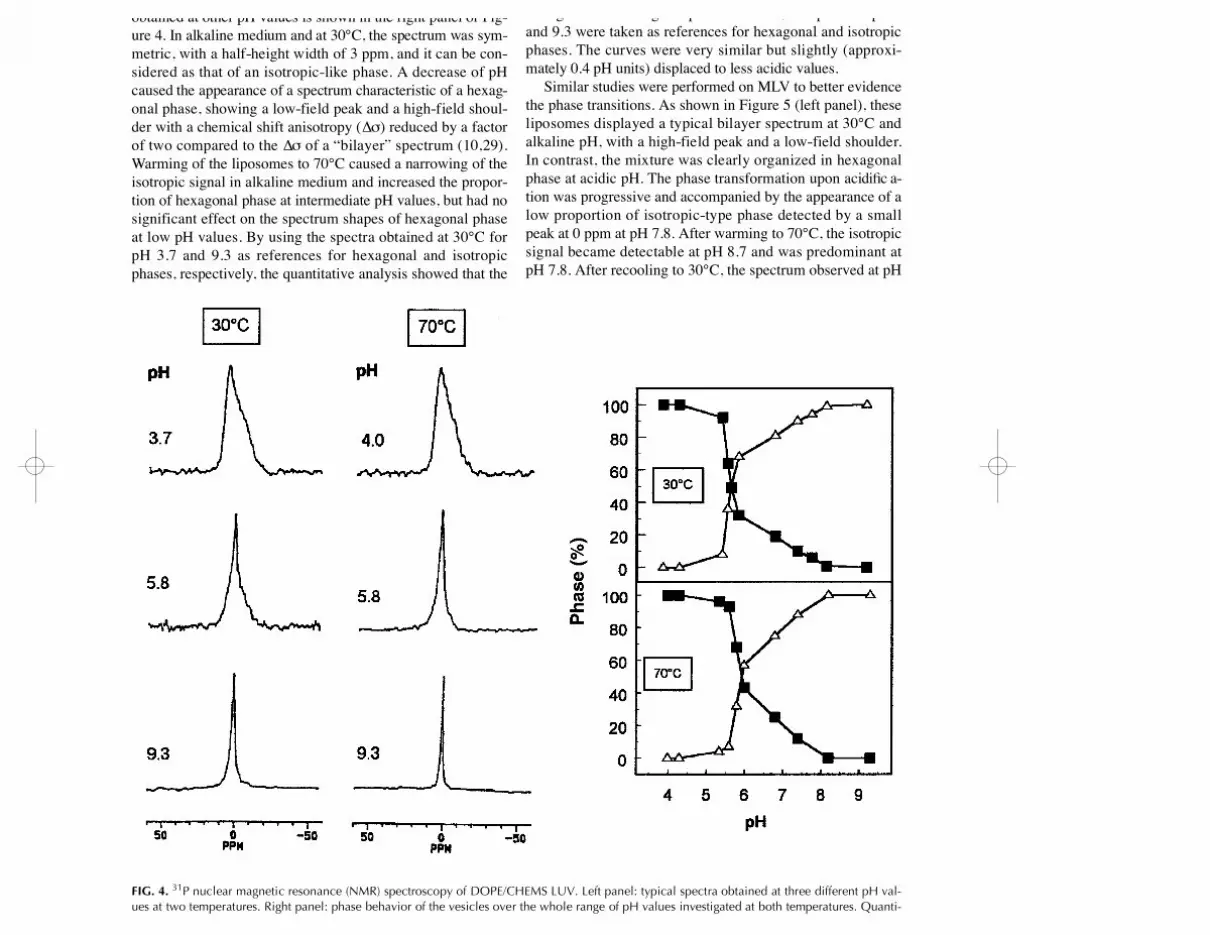
variations using 3 1P NMR spectroscopy. Figure 4 (left panel)shows the spectra obtained at three different pH values at 30and 70°C. Quantitative analysis of these spectra and of thoseobtained at other pH values is shown in the right panel of Fig-ure 4. In alkaline medium and at 30°C, the spectrum was sym-metric, with a half-height width of 3 ppm, and it can be con-sidered as that of an isotropic-like phase. A decrease of pHcaused the appearance of a spectrum characteristic of a hexag-onal phase, showing a low-field peak and a high-field shoul-der with a chemical shift anisotropy (∆σ) reduced by a factorof two compared to the ∆σ of a “bilayer” spectrum (10,29).Warming of the liposomes to 70°C caused a narrowing of theisotropic signal in alkaline medium and increased the propor-tion of hexagonal phase at intermediate pH values, but had nosignificant effect on the spectrum shapes of hexagonal phaseat low pH values. By using the spectra obtained at 30°C forpH 3.7 and 9.3 as references for hexagonal and isotropicphases, respectively, the quantitative analysis showed that the
lipidic phase reorganization occurring upon acidificationstarted, at 30°C, from a pH close to 8.0 but became prominentat pH ~ 5.6. Below this value, most of the lipids appeared tobe organized in hexagonal phase. At 70°C, the spectra at pH 4and 9.3 were taken as references for hexagonal and isotropicphases. The curves were very similar but slightly (approxi-mately 0.4 pH units) displaced to less acidic values.
Similar studies were performed on MLV to better evidencethe phase transitions. As shown in Figure 5 (left panel), theseliposomes displayed a typical bilayer spectrum at 30°C andalkaline pH, with a high-field peak and a low-field shoulder.In contrast, the mixture was clearly organized in hexagonalphase at acidic pH. The phase transformation upon acidific a-tion was progressive and accompanied by the appearance of alow proportion of isotropic-type phase detected by a smallpeak at 0 ppm at pH 7.8. After warming to 70°C, the isotropicsignal became detectable at pH 8.7 and was predominant atpH 7.8. After recooling to 30°C, the spectrum observed at pH
218 F. VAN BAMBEKE ET AL.
Lipids, Vol. 35, no. 2 (2000)
FIG. 4. 3 1P nuclear magnetic resonance (NMR) spectroscopy of DOPE/CHEMS LUV. Left panel: typical spectra obtained at three different pH val-ues at two temperatures. Right panel: phase behavior of the vesicles over the whole range of pH values investigated at both temperatures. Quanti-tative assignment to hexagonal (closed squares) or isotropic (open triangles) phases was made for data obtained at 30°C, by reference to the spectraobtained at pH 3.7 (hexagonal phase) or 9.3 (isotropic phase), respectively, and for data obtained at 70°C, by reference to the spectra obtained atpH 4.0 (hexagonal phase) or 9.3 (isotropic phase), as described in Reference 31. Data shown are the mean of successive determinations obtainedwith SD ~5%. For abbreviations see Figure 1.
L8278 7/19/04 4:01 PM Page 218

7.3 showed a prominent symmetrical and narrow signal witha half-height width (∆ν1/2) of 4 ppm. We then quantified thesechanges using the spectra at pH 8.7 and 30°C, at pH 7.3 and30°C after recooling (30°C final), and at pH 3.7 and 30°C asreference to a bilayer phase, an isotropic phase, and an hexag-onal phase, respectively. The results, presented in Figure 5(right panel) show that 50% of the phospholipids adopt ahexagonal phase organization at a pH close to neutrality. Thistransformation from bilayer to hexagonal phase involved apassage through isotropic structures, the proportion of whichdrastically increased after one cycle of heating and cooling.
Studies with cultured cells. (i) Fate of entrapped HPTS.Figure 6 (upper panel) shows the accumulation and cellularfate of HPTS encapsulated in DOPE/CHEMS andDOPC/CHEMS LUV, in J774 macrophages during a first ex-posure to the vesicles for a 15-min period (uptake) followed
by a 45-min washout (chase; total incubation time: 60 min).Panel A shows that HPTS, whether detected by excitation at450 or 390 nm, rapidly accumulates in cells exposed toDOPE/CHEMS liposomes. During the chase period, the sig-nal obtained by excitation at 450 nm declined while that ob-tained by excitation at 390 nm remained stable with a trendtoward an increase. HPTS encapsulated in DOPC/CHEMS li-posomes was accumulated to a considerably lesser extent, butits fate during the chase was qualitatively similar to that ofHPTS encapsulated in DOPE/CHEMS liposomes. To getmore insight on the intracellular fate of HPTS, we present inthe panel B the ratio of the signals recorded upon excitationat 450 and 390 nm. This shows that a steady and significantshift of pH occurs in the environment of the probe, both withDOPE/CHEMS and DOPC/CHEMS liposomes from earlyuptake (pH 7.0–7.5) to late chase phase (pH 6.0–6.5); some
DOPE/CHEMS LIPOSOMES: IN VITRO AND CELLULAR STUDIES 219
Lipids, Vol. 35, no. 2 (2000)
FIG. 5. 31P NMR spectroscopy of DOPE/CHEMS large multilamellar vesicles (MLV). Left panel: typical spectra obtained at four different pH valuesat three temperatures. Right panel: phase behavior of the vesicles over the whole range of pH values investigated at the three temperatures. Quan-titative assignment to hexagonal (closed squares), bilayer (grey circles), or isotropic (open triangles) phases was made by reference to the spectraobtained at 30°C and pH 3.7 (hexagonal phase), pH 8.7 (bilayer phase), or 30°C final and pH 7.3 (isotropic phase), respectively, as described inReference 31. Data shown are the mean of successive determinations obtained with SD ~5%. For abbreviations see Figures 1 and 4.
L8278 7/19/04 4:01 PM Page 219

d e l a y, however, was noted for DOPC/CHEMS liposomes (tomake the link with the confocal microscopy studies, all mea-surements were also made at 488 and 363 nm, with essentiallysimilar results). The confocal microscopic studies analysis (il-lustrated in the lower panel of Fig. 6) first confirmed that theaccumulation of HPTS in cells exposed to DOPE/CHEMS li-posomes was considerably larger than that of the same probe
entrapped in DOPC/CHEMS vesicles. With both types of li-posomes, the tracer initially appeared as tiny dots located atthe periphery of the cell (set to a green color). Wi t hDOPE/CHEMS liposomes, these structures became progres-sively enlarged and more centrally located in a perinuclearfashion, while also gradually emitting a larger proportion ofthe signal in the red channel, the yellow color resulting fromthe colocalization of green and red signals. Some of thesestructures were already visible at 5 min. After 45 min chase,most of the tracer was detected as large red/yellow patches,often with a diffuse appearance. With DOPC/CHEMS lipo-somes, the tracer still displayed a peripheral appearance after30 min of incubation. It eventually, but slowly, became asso-ciated with larger structures, some of which appeared asred/yellow dots. Yet it was never present in a diffuse fashion.
(ii) Release of HPTS. Figure 7 (upper panel) shows thatsuspensions of cells incubated with DOPE/CHEMS LUVloaded with HPTS and DPX display almost no signal whenexcited at 450 nm. At the end of the uptake period, however,they showed a minor signal when excited at 390 nm, the valueof which increased throughout the chase period. In confocalmicroscopy (lower panel), only a few red structures were seenat the end of the uptake period. During the chase, increasinglylarger yellow/red patches, with a diffuse appearance, were ob-served, sometimes surrounded by tiny green spots. No signal
220 F. VAN BAMBEKE ET AL.
Lipids, Vol. 35, no. 2 (2000)
FIG. 6. Accumulation and distribution of fluorescence in J774macrophages during incubation in the presence of HPTS-containingLUV (uptake) and subsequent transfer to liposome-free medium (chase).Circles, DOPE/CHEMS vesicles; squares, DOPC/CHEMS vesicles. (A)Readings obtained with suspensions of living cells upon excitation at450 nm (open symbols) or 390 nm (closed symbols); (B), ratio of therecordings made upon excitation at these two wavelengths. Results arethe mean of three independent experiments (±SD) (these ratios wereused to calculate the mean pH to which HPTS is exposed, and the cor-responding values are shown on the right ordinate). Bottom panel: con-focal microscopy of cells treated as indicated in the upper panels. Cellswere illuminated at 488 nm (green look-up table) and 363 nm (red look-up table). The yellow color results from the colocalization of green andred signals. Bar = 10 µm. For abbreviations see Figure 1.
FIG 7. Accumulation and distribution of fluorescence in J774macrophages incubated in the presence of DOPE/CHEMS LUV contain-ing HPTS together with DPX. The protocol and the experimental condi-tions were similar to those of Figure 6. Upper panel: readings obtainedwith cell suspensions (open symbols, λexc 450 nm; closed symbols, λexc390 nm). Lower panel: confocal microscopy observations. Bar = 10 µm.For abbreviations see Figure 1.
L8278 7/19/04 4:01 PM Page 220

was detected when macrophages were incubated withDOPC/CHEMS liposomes loaded by HPTS and DPX (resultsnot shown).
DISCUSSION
pH-sensitive liposomes have been developed for the purposeof enhancing the cytosolic delivery of drugs and other en-trapped solutes through destabilization in the acid milieu of theendosomal/phagosomal apparatus (2–4). We selected DOPE/CHEMS liposomes since vesicles of this composition show agreater stability than liposomes made of DOPE and oleic acid(34), and also because CHEMS induces destabilization throughtransition to inverted hexagonal (HI I) phase (20) in a physio-logical pH range [7 to 5 (35)]. DOPC/CHEMS liposomes wereused as controls since these vesicles cannot undergo reorg a n i-zation to HI I phase, which is considered essential for membranedestabilization and fusion (10). As structural modificationscould be the initiating factor of the destabilization of pH-sensi-tive liposomes upon acidification, we have focused our atten-tion on the changes in lipid organization as well as their rela-tion with membrane permeabilization and fusion.
A first observation in this respect is that change in poly-morphic organization of the lipids occurs over a wide pHrange and that the hexagonal-phase proportion increases frompH 8.0 down to ~6.0. At this pH value, it affects a large pro-portion of the lipids and probably reaches a critical thresholdto cause alterations of other membrane properties. Thisthreshold cannot, however, be more quantitatively defined inL U V. The broad component seen in the 3 1P NMR spectra atacid pH, which is typical of a hexagonal phase (10), may in-deed include a symmetrical broader component associatedwith fused liposomes of a still sufficiently small size (seebelow) to undergo motional averaging of ∆σ (30). The role ofstructures giving rise to an isotropic signal in the process ofphase transition is further substantiated by the 31P NMR stud-ies made on MLV. The highly mobile structures responsiblefor the isotropic signal may also be involved in membrane fu-sion (36). However, a quantitative relationship between theproportion of these structures in the membrane and the fusionprocess cannot be established since MLV are too heteroge-neous in size to be used in fusion assays. In addition, LUV,which were therefore used for fusion studies, reorganize inhexagonal phase at a lower pH than MLV. The endothermiccharacter of the transition clearly appears for LUV, since itoccurs at a higher pH at 70 than at 30°C. This effect of tem-perature is particularly dramatic for MLV, since a rise from30 to 70°C increases the percentage of isotropic-type struc-tures from ~10 to ~40%, and cooling down to 30°C causes acomplete transformation of the spectrum in a narrow symmet-rical signal. It therefore appears that heating and cooling in-duce the formation of isotropic-type structures in an irre-versible manner. The nature of this structural change is diff i-cult to interpret on the basis of 3 1P NMR results alone andfurther investigations, out of the main scope of this paper, areprobably needed to independently determine the influences of
the temperature and the duration of the cycle on membraneb e h a v i o r. Whatever the exact nature of these changes, themain conclusion is that both LUV and MLV undergo changesin lipid organization upon acidification susceptible to altercritical membrane properties such as permeability and fusioncapacity.
A second observation in this study is that DOPE/CHEMSliposomes require more membrane structure reorg a n i z a t i o nfor fusion than for permeabilization, since there is a diff e r-ence of approximately 0.5 pH units between the onset of eachphenomenon (see data on calcein release on the one side andlight-scattering spectroscopy and R18 dequenching on theother side). Our data therefore support and extend those ob-tained by Collins et al. (17), who also reported that leakageoccurs at a less acidic pH than membrane fusion. Quite sur-p r i s i n g l y, we found that a 100% leakage or fusion could notbe achieved, at least in the range of pH investigated, suggest-ing that a part of the liposome population is resistant to acidi-fication or that membrane perturbations are not sufficient tocause the complete release of the entrapped probe or the fu-sion of all the vesicles. Incomplete release of a tracer en-trapped in DOPE/CHEMS liposomes containing higher pro-portions of CHEMS was also noted by other investigators( 11). In the present study, the kinetic data unambiguouslyshow that a plateau has been reached. It has been proposed( 11) that destabilization of pH-sensitive liposomes is primar-ily mediated by bilayer contact. The degree of permeabiliza-tion may therefore be directly related to the lipid concentra-tion. However, in these studies, the authors (11) did notspecifically consider whether the aggregation process is fol-lowed by fusion of the liposomes. The mixing of lipid com-ponents we detected is actually highly suggestive of a true fu-sion process rather than a simple aggregation, since the in-crease in LUV size was moderate (only 2–3 times thediameter of control liposomes) and irreversible, and sinceR18 fluorescence increased very rapidly upon liposome mix-ing. The present data are therefore very similar to those wereported earlier for negatively charged liposomes incubatedwith typical fusogenic agents like melittin or (β- d i e t h y l -aminoethylether)hexestrol (these agents cause a two to fivetimes increase in liposome diameter and an immediate de-quenching of R18 fluorescence). They are also in sharp con-trast with what we found for aggregating agents like spermineand gentamicin, which induce an at least 10-fold increase inliposome diameter associated with a slow increase of R18 flu-orescence (26,37). Altogether, the biophysical studies dis-cussed so far strongly suggest that DOPE/CHEMS liposomesof the composition we selected may become destabilized andrelease their contents when pH falls from >7 to approximately6. This range corresponds to that encountered along the endo-cytic pathway (38). The results of the cellular studies withHPTS-entrapped liposomes (in which the tracer is used toquantify the liposome uptake and to estimate the pH at whichit becomes exposed) concur with this consideration. By as-suming a rapid equilibration of protons through the liposomemembrane (25), it clearly appears that HPTS entrapped in
DOPE/CHEMS LIPOSOMES: IN VITRO AND CELLULAR STUDIES 221
Lipids, Vol. 35, no. 2 (2000)
L8278 7/19/04 4:01 PM Page 221

DOPE/CHEMS or DOPC/CHEMS vesicles travels throughcompartments whose pH decreases progressively from 7.4 to~6. It must, however, be stressed that the pH values obtainedby the examination of cell suspensions (Fig. 6, upper panel)represent an average value for a material that, as evidencedby confocal microscopy (Fig. 6, lower panel), is spreadamong vacuoles whose pH values range from neutrality to alow value. Our studies also show that cells handleDOPE/CHEMS and DOPC/CHEMS liposomes very diff e r-e n t l y. First, the fluorescence signal recorded in cells wasmarkedly lower for DOPC/CHEMS liposomes than forDOPE/CHEMS liposomes. This cannot be attributed to a dif-ference in the amount of probe entrapped (see Materials andMethods section) and must therefore be ascribed to a low en-docytic rate of DOPC/CHEMS liposomes, as already ob-served in P388D1 macrophages (4). This clearly points to theimportance of liposome composition for uptake, beyond asimple variation in charge (39). Second, the confocal mi-croscopy studies show that HPTS entrapped inDOPC/CHEMS liposomes remains for about 30 min in theperiphery of the cell and at neutral pH before being trans-ferred to vesicles of lower pH. Because our calibration stud-ies indicate that the shift from the green to the red signalshould occur around pH 7.15, the data therefore suggest thatDOPC/CHEMS liposomes remain associated with the cellsurface, or with invaginations of the plasma membrane, forquite a time before being transferred to early endosomes andother acidic vacuoles. In contrast, HPTS entrapped inDOPE/CHEMS liposomes appears to move more quickly intothe cell where it progressively shows a diffuse appearance inparallel with a marked shift from green to yellow and red.Again, taking into account the pH at which this shift is ob-served (~7.15), we interpret these images as indicating a rapidinternalization into early endosomes followed by a partial re-lease of the tracer in the cytosol, the pH of which appears tobe precisely around this value in J774 macrophages (40). Ane ffective release of the content of DOPE/CHEMS liposomesinto the cytosol also is largely evidenced by the results of theexperiments using vesicles containing both HPTS and itsquenching agent DPX. Comparing the values of the fluores-cence signal obtained with these liposomes (Fig. 7) to that ob-tained with vesicles without the quenching agent (Fig. 6), wesuggest that approximately 15% of the HPTS has been madefree within 45 min after endocytosis, a figure close to thatfound by Chu et al. (4) for DOPE/CHEMS (3:2) liposomes.This value may, however, be underestimated, since a signifi-cant increase in HPTS fluorescence will be only observedupon high dilution of the dye/quencher mixture (41).
In conclusion, our data confirm the potential usefulness ofDOPE/CHEMS liposomes as pH-sensitive vehicles for the in-tracytosolic delivery of an entrapped tracer. This deliveryseems to occur without grossly affecting cell viability, sug-gesting interesting applications for therapeutics. Our biologi-cal observations are supported by biophysical data pointingto fruitful strategies for further improving the design and con-struction of this type of liposomes.
ACKNOWLEDGMENTS
F. V.B. and M.P.-M.L. are respectively Chargé de Recherchesand Chercheur Qualifié of the Belgian Fonds National de laRecherche Scientifique. Francine Renoird, Marie-ClaireC a m b i e r, and Christelle Flore provided expert technical as-sistance. This work was supported by the Belgian Fonds de laRecherche Scientifique Médicale (grants nos. 9.4541.95F,3.4516.94 and 9.451492), the Fonds National de la RechercheScientifique (grant no. 9.4546.94), the Actions de RecherchesConcertées 94/99-172 of the Direction Générale de laRecherche Scientifique—Communauté Française de Bel-gique, Belgium.
REFERENCES
1. Straubinger, R.M., Hong, K., Friend, D., and Papahadjopoulos,D. (1983) Endocytosis of Liposomes and Intracellular Fate ofEncapsulated Molecules: Encounter with a Low pH Compart-ment After Internalization in Coated Vesicles, C e l l 3 2,1069–1079.
2. Connor, J., Yatvin, M.B., and Huang, L. (1984) pH-SensitiveLiposomes: Acid-Induced Liposome Fusion, Proc. Natl. Acad.Sci. USA 81, 1715–1718.
3. Chu, C.J., and Szoka, F.C. (1994) pH-Sensitive Liposomes, J .Liposome Res. 4, 361–395.
4. Chu, C.J., Dijkstra, J., Lai, M.Z., Hong, K., and Szoka, F.C.(1990) Efficiency of Cytoplasmic Delivery by pH-Sensitive Li-posomes to Cells in Culture, Pharm. Res. 7, 824–834.
5. Connor, J., and Huang, L. (1986) pH-Sensitive Immunolipo-somes as an Efficient and Target-Specific Carrier for AntitumorDrugs, Cancer Res. 46, 3431–3435.
6. Lutwyche, P., Cordeiro, C., Wiserman, D.J., St. Louis, M., Uh,M., Hope, M.J., and Finlay, B.B. (1998) Intracellular Deliveryand Intracellular Activity of Gentamicin Encapsulated in pH-Sensitive Liposomes, Antimicrob. Agents Chemother. 42,2511–2520.
7. Couvreur, P., Fattal, E., Malvy, C., and Dubernet, C. (1997) pH-Sensitive Liposomes: an Intelligent System for the Delivery ofAntisense Oligonucleotides, J. Liposome Res. 7, 1–18.
8. Nair, S., Zhou, F., Reddy, R., Huang, L., and Rouse, B.T. (1992)Soluble Proteins Delivered to Dendritic Cells v i a p H - S e n s i t i v eLiposomes Induce Primary Cytotoxic T Lymphocyte Responsesin vitro, J. Exp. Med. 175, 609–612.
9. Wang, C.Y., and Huang, L. (1987) Plasmid DNA Adsorbed topH-Sensitive Liposomes Efficiently Transforms the TargetCells, Biochem. Biophys. Res. Commun. 147, 980–985.
10. Seddon, J.M. (1990) Structure of the Inverted Hexagonal (HI I)Phase, and Nonlamellar Phase Transitions of Lipids, B i o c h i m .Biophys. Acta 1031, 1–69.
11. Ellens, H., Bentz, J., and Szoka, F.C. (1984) pH-Induced Desta-bilization of Phosphatidylethanolamine-Containing Liposomes:Role of Bilayer Contact, Biochemistry 23, 1532–1538.
12. Düzgünes, N., Straubinger, R.M., Baldwin, P.A., Friend, D.S.,and Papahadjopoulos, D. (1985) Proton-Induced Fusion ofOleic-Phosphatidylethanolamine Liposomes, Biochemistry 24,3091–3098.
13. Liu, D., and Huang, L. (1989) Small, but Not Large, Unilamel-lar Liposomes Composed of Dioleoylphosphatidylethanolamineand Oleic Acid Can Be Stabilized by Human Plasma, B i o c h e m -istry 28, 7700–7707.
14. Collins, D., Litzinger, D.C., and Huang, L. (1990) Structural andFunctional Comparisons of pH-Sensitive Liposomes Composedof Phosphatidylethanolamine and Three Different Diacylsuc-cinylglycerols, Biochim. Biophys. Acta 1025, 234–242.
222 F. VAN BAMBEKE ET AL.
Lipids, Vol. 35, no. 2 (2000)
L8278 7/19/04 4:01 PM Page 222

15. Hazemoto, N., Harada, M., Suzuki, S., Kaiho, F., Haga, M., andKato, Y. (1993) Effect of Phosphatidylcholine and Cholesterolon pH-Sensitive Liposomes, Chem. Pharm. Bull. 41,1003–1006.
16. Tari, A.M., Fuller, N., Boni, L.T., Collins, D., Rand, P., andHuang, L. (1994) Interactions of Liposome Bilayers Composedof 1,2-Diacyl-3-succinylglycerol with Protons and DivalentCations, Biochim. Biophys. Acta 1192, 253–262.
17. Collins, D., Maxfield, F., and Huang, L. (1989) Immunolipo-somes with Different Acid Sensitivities as Probes for the Cellu-lar Endocytic Pathway, Biochim. Biophys. Acta 987, 47–55.
18. Straubinger, R.M., Düzgünes, N., and Papahadjopoulos, D.(1985) pH-Sensitive Liposomes Mediate Cytoplasmic Deliveryof Encapsulated Macromolecules, FEBS Lett. 179, 148–154.
19. Kono, K., Igawa, T., and Takagishi, T. (1997) Cytoplasmic De-livery of Calcein Mediated by Liposomes Modified with a pH-Sensitive Poly(ethylene Glycol) Derivative, Biochim. Biophys.Acta 1325, 143–154.
20. Lai, M.Z., Vail, W.J., and Szoka, F.C. (1985) Acid- and Cal-cium-Induced Structural Changes in PhosphatidylethanolamineMembranes Stabilized by Cholesterol Hemisuccinate, Biochem -istry 24, 1654–1661.
21. Hope, M.J., Bally, M.B., Webb, G., and Cullis, P.R. (1985) Pro-duction of Large Unilamellar Vesicles by a Rapid ExtrusionProcedure. Characterization of Size Distribution, Trapped Vol-ume and Ability to Maintain a Membrane Potential, B i o c h i m .Biophys. Acta 812, 55–65.
22. Van Bambeke, F., Mingeot-Leclercq, M.P., Schanck, A.,Brasseur, R., and Tulkens, P.M. (1993) Alterations in Mem-brane Permeability Induced by Aminoglycoside Antibiotics:Studies on Liposomes and Cultured Cells, Eur. J. Pharmacol.247, 155–168.
23. Grit, M., Zuidam, N.J., Underberg, W.J.M., and Crommelin,D.J.A. (1993) Hydrolysis of Partially Saturated Egg Phos-phatidylcholine in Aqueous Liposome Dispersions and the Ef-fect of Cholesterol Incorporation on Hydrolysis Kinetics, J .Pharm. Pharmacol. 45, 490–495.
24. Weinstein, J.N., Yoshikami, S., Henkart, P., Blumenthal, R., andHagins, W.A., (1977) Liposome–Cell Interaction: Transfer andIntracellular Release of a Trapped Fluorescent Marker, S c i e n c e195, 489–491.
25. Straubinger, R.M., Papahadjopoulos, D., and Hong, K. (1990)Endocytosis and Intracellular Fate of Liposomes Using Pyra-nine as a Probe, Biochemistry 29, 4929–4939.
26. Van Bambeke, F., Tulkens, P.M., Brasseur, R., and Mingeot-Leclercq, M.P. (1995) Aminoglycoside Antibiotics Induce Ag-gregation but Not Fusion of Negatively-Charged Liposomes,Eur. J. Pharmacol. 289, 321–333.
27. Hoekstra, D., de Boer, T., Klappe, K., and Wilschut, J. (1984)Fluorescence Method for Measuring the Kinetics of Fusion Be-tween Biological Membranes, Biochemistry 23, 5675–5681.
28. Mazer, N.A., Carey, M.C., Kwasnick, R.F., and Benedek, G.B.(1979) Quasi-elastic Light Scattering Studies of Aqueous Bil-iary Lipid Systems. Size, Shape and Thermodynamics of BileSalt Micelles, Biochemistry 18, 3064–3075.
29. Seelig, J. (1978) 31P Nuclear Magnetic Resonance and the HeadGroup Structure of Phospholipids in Membranes, Biochim. Bio -phys. Acta 515, 105–140.
30. Burnell, E.E., Cullis, P.R., and De Kruijff, B. (1980) Effects ofTumbling and Lateral Diffusion on Phosphatidylcholine ModelMembrane 3 1P-NMR Lineshapes, Biochim. Biophys. Acta 603,63–69.
31. Schanck, A. (1992) A Method for Determining the Proportionsof Different Phases in Hydrated Phospholipids by 3 1P NuclearMagnetic Resonance (NMR) Spectroscopy, Appl. Spectrosc. 46,1435–1437.
32. Snyderman, R., Pike, M.C., Fischer, D.G., and Koren, H.S.(1977) Biologic and Biochemical Activities of ContinuousMacrophage Cell Lines P 338 D1 and J 774 1, J. Immunol. 119,2060–2066.
33. Lowry, O.H., Rosebrough, N.J., Farr, A.L., and Randall, R.J.(1951) Protein Measurement with the Folin Phenol Reagent, J .Biol. Chem. 193, 265–275.
34. Straubinger, R.M. (1993) pH-Sensitive Liposomes for Deliveryof Macromolecules into Cytoplasm of Cultured Cells, M e t h o d sEnzymol. 194, 28–36.
35. Ellens, H., Bentz, J., and Szoka, F.C. (1985) H+ and Ca2 + F u-sion and Destabilization of Liposomes, Biochemistry 24,3099–3106.
36. Van Bambeke, F., Mingeot-Leclercq, M.P., Brasseur, R.,Tulkens, P.M., and Schanck, A. (1996) Aminoglycoside Antibi-otics Prevent the Formation of Non-Bilayer Structures in Nega-tively-Charged Membranes. Comparative Studies Using Fuso-genic [bis-(beta-diethylaminoethylether)hexestrol] and Aggre-gating (spermine) Agents, Chem. Phys. Lipids 79, 123–135.
37. Mingeot-Leclercq, M.P., Schanck, A., Ronveaux-Dupal, M.F.,Deleers, M., Brasseur, R., Ruysschaert, J.M., Laurent, G., andTulkens, P.M. (1989) Ultrastructural, Physico-Chemical andConformational Study of the Interactions of Gentamicin andbis(beta-diethylaminoethylether)hexestrol with Negatively-Charged Phospholipid Bilayers, Biochem. Pharmacol. 38,729–741.
38. Hubbard, A.L. (1989) Endocytosis, Curr. Opin. Cell Biol. 1,675–683.
39. Miller, C.R., Bondurant, B., McLean, S.D., McGovern, K.A.,and O’Brien, D.F. (1998) Liposome–Cell Interactions in vitro:Effect of Liposome Surface Charge on the Binding and Endocy-tosis of Conventional and Sterically Stabilized Liposomes, B i o -chemistry 37, 12875–12883.
40. Gan, B.S., Krump, E., Shrode, L.D., and Grinstein, S. (1998)Loading Pyranine v i a Purinergic Receptors or Hypotonic Stressfor Measurement of Cytosolic pH by Imaging, Am. J. Physiol.Cell Physiol. 44, C1158–C1166.
41. Daleke, D.L., Hong, K., and Papahadjopoulos, D. (1990) Endo-cytosis of Liposomes by Macrophages: Binding, Acidificationand Leakage of Liposomes Monitored by a New FluorescenceAssay, Biochim. Biophys. Acta 1024, 352–366.
[Received June 9, 1999, and in revised form November 18, 1999;revision accepted January 4, 2000]
DOPE/CHEMS LIPOSOMES: IN VITRO AND CELLULAR STUDIES 223
Lipids, Vol. 35, no. 2 (2000)
L8278 7/19/04 4:01 PM Page 223


