Embed Size (px)
Citation preview

Catalytic Properties of ADAM12 and Its Domain Deletion Mutants†
Jonas Jacobsen,*,‡,§ Robert Visse,§ Hans Peter Sørensen,‡ Jan J. Enghild,| Keith Brew,⊥ Ulla M. Wewer,‡ andHideaki Nagase§
Department of Biomedical Sciences and Biotech Research & InnoVation Centre (BRIC), UniVersity of Copenhagen, Ole MaaløesVej 5, DK-2200 Copenhagen, Denmark, Kennedy Institute of Rheumatology DiVision, Faculty of Medicine, Imperial College
London, 1 Aspenlea Road, London W6 8LH, United Kingdom, Department of Molecular Biology, UniVersity of Aarhus, Sciencepark, GustaV WiedsVej 10C, DK-8000 Aarhus C, Denmark, and Department of Biomedical Science, Florida Atlantic UniVersity,
Boca Raton, Florida 33431
ReceiVed August 13, 2007; ReVised Manuscript ReceiVed NoVember 9, 2007
ABSTRACT: Human ADAM12 (a disintegrin and metalloproteinase) is a multidomain zinc metalloproteinaseexpressed at high levels during development and in human tumors. ADAM12 exists as two splicevariants: a classical type 1 membrane-anchored form (ADAM12-L) and a secreted splice variant(ADAM12-S) consisting of pro, catalytic, disintegrin, cysteine-rich, and EGF domains. Here we presenta novel activity of recombinant ADAM12-S and its domain deletion mutants on S-carboxymethylatedtransferrin (Cm-Tf). Cleavage of Cm-Tf occurred at multiple sites, and N-terminal sequencing showedthat the enzyme exhibits restricted specificity but a consensus sequence could not be defined as its subsiterequirements are promiscuous. Kinetic analysis revealed that the noncatalytic C-terminal domains areimportant regulators of Cm-Tf activity and that ADAM12-PC consisting of the pro domain and catalyticdomain is the most active on this substrate. It was also observed that NaCl inhibits ADAM12. Among thetissue inhibitors of metalloproteinases (TIMP) examined, the N-terminal domain of TIMP-3 (N-TIMP-3)inhibits ADAM12-S and ADAM12-PC with low nanomolarKi(app) values while TIMP-2 inhibits themwith a slightly lower affinity (9-44 nM). However, TIMP-1 is a much weaker inhibitor. N-TIMP-3 variantsthat lack MMP inhibitory activity but retained the ability to inhibit ADAM17/TACE failed to inhibitADAM12. These results indicate unique enzymatic properties of ADAM12 among the members of theADAM family of metalloproteinases.
A disintegrin and metalloproteinase ADAM12,1 alsoknown as meltrinR, is a multifunctional zinc-dependentenzyme that belongs to a subfamily of reprolysin metal-lopeptidases (1-3). Human ADAM12 is expressed as aclassical type 1 membrane-anchored form (ADAM12-L) andas a secreted splice variant (ADAM12-S) (1). The latterconsists of a propeptide, a catalytic domain, and cell adhesivedisintegrin-, cysteine-rich-, and epidermal growth factor-like(EGF-like) domains (Figure 1A) but lacks the C-terminaltransmembrane and cytoplasmic domains. Both forms arestrongly expressed in placenta, and decreased serum levelsof ADAM12 in pregnant women are reliable biomarkers forpreeclampsia and Down’s syndrome in clinical prenatal
diagnostics (1, 4-7). Low expression levels of ADAM12are found in most normal adult tissues, but the level ofexpression is greatly increased in different types of cancers(8-11). In the polyoma middle T antigen (PymT) mousemodel of breast cancer, ADAM12 stimulated tumor progres-sion by increasing the tumor burden and decreasing the timeto tumor onset (12). Importantly, ADAM12 is readilydetected in urine of breast and bladder cancer patients andcorrelates with disease status and stage (13, 14).
The three-dimensional structure of ADAM12 has not yetbeen determined experimentally. Using electron microscopy,we modeled the overall domain organization of ADAM12-Scontaining the pro domain as a compact four-leaf clover-like molecule (15). This model was supported by the recentlydetermined structure of a snake venom homologue ofmammalian ADAMs, vascular apoptosis inducing protein 1(VAP1) (16) consisting of the catalytic domain, disintegrin,and cysteine-rich domains. Crystal structures for the catalyticdomains of homologues tumor necrosis factor-R-convertingenzyme (TACE or ADAM17) (17) and ADAM33 (18) havebeen determined. Both structures reveal a polypeptide foldconsisting of a five-strandedâ-sheet surrounded by fiveR-helices, resembling the folding of the catalytic domainsof the related zinc metalloproteinases collectively called themetzincins (19).
On the basis of its zinc-binding consensus sequence motifHEXXHXXGXXH, ADAM12 was predicted to be an active
† U.M.W. was supported by the Danish Cancer Society, the DanishMedical Council, Novo Nordic, the Lundbeck Foundation, and theMunksholm Foundation, and H.N. was supported by NIH Grant AR40994 and The Wellcome Trust Grant 07547.
* To whom correspondence should be addressed. Telephone:+45-35326081. Fax:+45-35325669. E-mail: [email protected].
‡ University of Copenhagen.§ Imperial College London.| University of Aarhus.⊥ Florida Atlantic University.1 Abbreviations: ADAM12, disintegrin and metalloprotease; EGF,
epidermal growth factor; PymT, polyoma middle T antigen; VAP1,vascular apoptosis-inducing proteins; TACE, tumour necrosis factor-R-converting enzyme;R2M, R2-macroglobulin; IGFBP, insulin-likegrowth factor binding protein; P-LAP, placental leucine aminopeptidase;HB-EGF, heparin binding EGF-like growth factor; TIMP, tissueinhibitor of metalloprotease; Cm-Tf, carboxymethylated transferrin.
537Biochemistry2008,47, 537-547
10.1021/bi701629c CCC: $40.75 © 2008 American Chemical SocietyPublished on Web 12/15/2007

metzincin metalloproteinase, and its catalytic activity wasoriginally verified using theR2-macroglobulin (R2M) entrap-ment assay (20, 21). Potential physiological substrates includeinsulin-like growth factor binding proteins IGFBP-3 and -5(4, 22), placental leucine aminopeptidase (P-LAP) (23),heparin binding EGF-like growth factor (HB-EGF) (24),EGF, and betacellulin (25). These substrates are alsosusceptible to other metalloproteases (reviewed in refs3, 26,and27), and the relative contribution of ADAM12-mediatedcleavage of these substrates in vivo is poorly understood.Previous studies have shown that ADAM12 latency can beexplained by the cysteine switch mechanism in whichCys179 in the pro domain coordinates the active site zincion (21, 28). The enzyme is processed in the trans-Golgi at
the proprotein convertase recognition motif RHKR, andfollowing translocation to the plasma membrane or extra-cellular space, ADAM12 is considered to be present in itsactive form (21, 28). However, the pro domain remainsnoncovalently associated with the mature enzyme followingcleavage (15, 22), indicating possible extracellular functionsof the pro domain. Our previous in vitro characterization ofthe catalytic activity of ADAM12 usingR2M reactivityindicated that this activity requires Cu2+, and we proposedthat this activity was revealed through binding of thetransition ion Cu2+ to the unpaired Cys273 (29). ADAM12is inhibited by the tissue inhibitor of metalloproteinases 3(TIMP-3) (22), and other inhibitors of ADAM12 include thesynthetic hydroxymate inhibitors GM6001 and its derivative
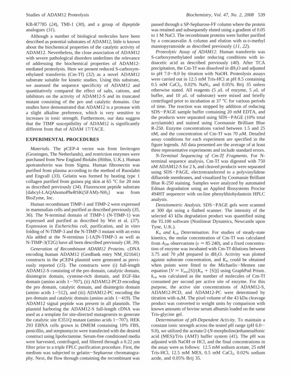
FIGURE 1: SDS-PAGE analysis of recombinant ADAM12 and detection of proteolytic activity. (A) Full-length ADAM12-S and its domaindeletion mutants are shown with their experimentally determined molecular masses. Pro stands for the pro domain, Cat for the catalyticdomain, Dis for the disintegrin domain, Cys for the cysteine-rich domain, and EGF for the EGF-like domain. (B) The purities of therecombinant proteins were analyzed by SDS-PAGE (12% acryl-amide), and the proteins were stained using Coomassie Brilliant BlueR-250. (C) The activity of ADAM12-S was assayed with gelatin, fibronectin, and Cm-Tf for 24 h in 50 mM Tris-HCl (pH 7.5), 0.05% Brij35, and 0.02% sodium azide: lane 1, substrate at 0 h; lane 2, substrate after 24 h; and lane 3, incubation with 100 nM enzyme. (D) All threevariants of ADAM12 were employed in a time course study of Cm-Tf degradation. Reaction buffer contained 0.5 mM CaCl2: lane 1,substrate at 0 h; lane 2, substrate after 18 h; lane 3, enzyme for 1 h; lane 4, enzyme for 8 h; lane 5, enzyme for 18 h; and lane 6, enzymeand 20 mM EDTA for 18 h.
538 Biochemistry, Vol. 47, No. 2, 2008 Jacobsen et al.
KB-R7785 (24), TMI-1 (30), and a group of dipeptideanalogues (31).
Although a number of biological molecules have beendescribed as potential substrates of ADAM12, little is knownabout the biochemical properties of the catalytic activity ofADAM12. Nevertheless, the close association of ADAM12with severe pathological disorders underlines the relevanceof addressing the biochemical properties of ADAM12-mediated proteolysis. Here we present reduced S-carboxym-ethylated transferrin (Cm-Tf) (32) as a novel ADAM12substrate suitable for kinetic studies. Using this substrate,we assessed the sequence specificity of ADAM12 andquantitatively compared the effect of salts, cations, andinhibitors on the activity of ADAM12-S and its truncatedmutant consisting of the pro and catalytic domains. Ourstudies have demonstrated that ADAM12 is a protease witha slight alkaline preference, which is very sensitive toincreases in ionic strength. Furthermore, our data suggestthat the TIMP susceptibility of ADAM12 is significantlydifferent from that of ADAM 17/TACE.
EXPERIMENTAL PROCEDURES
Materials. The pCEP-4 vector was from Invitrogen(Groningen, The Netherlands), and restriction enzymes werepurchased from New England Biolabs (Hithin, U.K.). Humanapotransferrin was from Sigma. Human fibronectin waspurified from plasma according to the method of Ruoslahtiand Engvall (33). Gelatin was formed by heating type 1collagen purified from guinea pig skin at 65°C for 20 minas described previously (34). Fluorescent peptide substrate[dabcyl-LAQAhomoPheRSK(5FAM)-NH2] was fromBioZyme, Inc.
Human recombinant TIMP-1 and TIMP-2 were expressedin mammalian cells and purified as described previously (35,36). The N-terminal domain of TIMP-1 (N-TIMP-1) wasexpressed and purified as described by Wei et al. (37).Expression inEscherichia coli, purification, and in vitrofolding of N-TIMP-3 and the N-TIMP-3 mutant with an extraAla added at the N-terminus [-1A]N-TIMP-3 as well asN-TIMP-3(T2G) have all been described previously (38, 39).
Generation of Recombinant ADAM12 Proteins.cDNAencoding human ADAM12 (GenBank entry NM_021641)constructs in the pCEP4 plasmid were generated as previ-ously reported (15). The constructs were (i) full-lengthADAM12-S consisting of the pro domain, catalytic domain,disintegrin domain, cysteine-rich domain, and EGF-likedomain (amino acids 1-707), (ii) ADAM12-PCD encodingthe pro domain, catalytic domain, and disintegrin domain(amino acids 1-512), and (iii) ADAM12-PC encoding thepro domain and catalytic domain (amino acids 1-419). TheADAM12 signal peptide was present in all plasmids. Theplasmid harboring the ADAM12-S full-length cDNA wasused as a template for site-directed mutagenesis to generatethe catalytic site E351Q mutant (amino acids 1-707). HEK293 EBNA cells grown in DMEM containing 10% FBS,penicillin, and streptomycin were transfected with the desiredconstruct using lipofectamine. Serum-free conditioned mediawere harvested, centrifuged, and filtered through a 0.22µmfilter prior to a triple FPLC purification procedure. First, themedium was subjected to gelatin-Sepharose chromatogra-phy. Next, the flow through containing the recombinant was
passed through a SP-Sepharose-FF column where the proteinwas retained and subsequently eluted using a gradient of 0.05to 1 M NaCl. The recombinant proteins were further purifiedby a concanavalin A column and elution withR-D-methylmannopyranoside as described previously (11, 22).
Proteolytic Assay of ADAM12.Human transferrin wasS-carboxymethylated under reducing conditions with io-doacetic acid as described previously (40). After TCAprecipitation, the Cm-Tf was dissolved in dH2O and adjustedto pH 7.0-8.0 by titration with NaOH. Proteolysis assayswere carried out in 12.5 mM Tris-HCl at pH 8.5 containing0.5 mM CaCl2, 0.02% NaN3, and 0.05% Brij 35 unlessotherwise stated. All reagents (5µL of enzyme, 5µL ofbuffer, and 10µL of substrate) were mixed and brieflycentrifuged prior to incubation at 37°C for various periodsof time. The reaction was stopped by addition of reducingSDS-PAGE sample buffer containing 20 mM EDTA, andthe products were separated using SDS-PAGE (10% totalacrylamide) and stained using Coomassie Brilliant BlueR-250. Enzyme concentrations varied between 1.5 and 25nM, and the concentration of Cm-Tf was 70µM. Detailedassay conditions for each experiment are specified in thefigure legends. All data presented are the average of at leastthree representative experiments and include standard errors.
N-Terminal Sequencing of Cm-Tf Fragments.For N-terminal sequence analysis, Cm-Tf was digested with 750nM ADAM12-S for 2 h, and cleaved products were separatedusing SDS-PAGE, electrotransferred to a polyvinylidenedifluoride membranes, and visualized by Coomassie BrilliantBlue R-250 staining. Samples were analyzed by automatedEdman degradation using an Applied Biosystems Procise494HT sequencer with on-line phenylthiohydantoin HPLCanalysis.
Densitometric Analysis.SDS-PAGE gels were scannedat 300 dpi using a flatbed scanner. The intensity of theselected 43 kDa degradation product was quantified usingthe TL100 software (Nonlinear Dynamics, Newcastle uponTyne, U.K.).
Km and kcat Determination.For studies of steady-statekinetics, the molar concentration of Cm-Tf was calculatedfrom A280 observations (ε ) 85 240), and a fixed concentra-tion of enzyme was incubated with Cm-Tf dilutions between3.75 and 70µM prepared in dH2O. Activity was plottedagainst substrate concentration, andKm could be obtainedwhen points were fitted to the Michaelis-Menten rateequation [V ) Vmax[S]/(Km + [S])] using GraphPad Prism.kcat was calculated as the number of molecules of Cm-Tfconsumed per second per active site of enzyme. For thispurpose, the active site concentrations of ADAM12-S,ADAM12-PCD, and ADAM12-PC were determined bytitration withR2M. The pixel volume of the 43 kDa cleavageproduct was converted to weight units by comparison withknown amounts of bovine serum albumin loaded on the sameTris-glycine gel.
Determination of pH-Dependent ActiVity. To maintain aconstant ionic strength across the tested pH range (pH 6.0-9.0), we utilized the acetate/2-(N-morpholino)ethanesulfonicacid (MES)/Tris (AMT) buffer system (41). The pH wasadjusted with NaOH or HCl, and the final concentrations inthe assay were as follows: 12.5 mM sodium acetate, 25 mMTris-HCl, 12.5 mM MES, 0.5 mM CaCl2, 0.02% sodiumazide, and 0.05% Brij 35.
Studies of ADAM12 Proteolysis Biochemistry, Vol. 47, No. 2, 2008539

Studies of ADAM12 Inhibition Kinetics.The concentrationsof TIMP-1, N-TIMP-1, TIMP-2, and N-TIMP-3 inhibitorswere determined by active site titration with a knownconcentration of MMP-1, and N-TIMP-3(2TG), N-TIMP-3,and [-1A]N-TIMP-3 were titrated with a known concentra-tion of ADAMTS-5. Various concentrations of TIMPs weremixed with 2 nM ADAM12-S or 1.5 nM ADAM12-PC in25 mM Tris-HCl (pH 8.0), 1.25 mM CaCl2, and 18.75 mMNaCl in a total volume of 15µL and incubated for 30 minat 37°C. The mixture was then incubated with 5µL of Cm-Tf (140 µM) at 37 °C for 9 h. Studies with N-TIMP-3 andN-TIMP-3 mutants included 2.5% glycerol to preventprecipitation. The reaction was stopped with reducing samplebuffer containing 20 mM EDTA. The products were sepa-rated using SDS-PAGE (10% total acrylamide) and stainedusing Coomassie Brilliant Blue R-250. IC50 values weredetermined by linear regression analysis of residual activityplotted against log values of inhibitor concentration. For tightbinding inhibitors at low enzyme concentrations (at∼Ki),this represents a good approximation of theKi(app) (42).
The Ki values of N-TIMP-3 against ADAM12-S andADAM12-PC were obtained using the fluorescent substrate[dabcyl-LAQAhomoPheRSK(5FAM)-NH2] and the Morrisonequation describing tight binding inhibition (30, 43). Bufferconditions were identical to those of inhibitor studies usingCm-Tf as a substrate, though 5% DMSO was included toincrease the solubility of the substrate; 85µL of assay buffer,5 µL of inhibitor, and 5 µL of enzyme (2 nM, finalconcentration) were preincubated at 37°C for 2 h prior tothe addition of 5µL of substrate (10µM, final concentration).A BMG Fluorostar Optima fluorometer (excitation at 485nm and emission at 530 nm) was used to monitor fluores-cence.
RESULTS
Purification and Detection of ADAM12-S and Its DomainDeletion Mutants. Full-length ADAM12-S and its C-termi-nally truncated domain deletion mutants as well as theADAM12-S(E351Q) mutant (Figure 1A) were expressed inHEK 293 EBNA cells and purified from the conditionedmedia. The recombinant enzyme preparations were free fromvisible contaminants as determined by SDS-polyacrylamidegel electrophoresis and staining with Coomassie BrilliantBlue R-250 (Figure 1B). Also, they reacted with ADAM12domain specific antisera (data not shown). ADAM12-PCDshows a double band around 45 kDa. Both bands react withantisera directed against the catalytic domain (data notshown), suggesting possible processing of the C-terminus(15). The experimentally determined sizes are bigger thanthe theoreticalMr calculated from the amino acid composi-tion. This can possibly be explained by predicted N-linkedglycosylations at two sites in the pro domain, and one sitein all other domains except the EGF-like domain. Thepresence of the copurified but noncovalently linked∼26 kDapropeptide has previously been observed (15, 22) and isconfirmed here.
Catalytic ActiVity of ADAM12 and Its Domain DeletionMutants.The catalytic properties of purified ADAM12-Swere first evaluated using three different substrates. Eachsubstrate was incubated with 100 nM ADAM12-S in 50 mMTris-HCl (pH 7.5) for 24 h at 37°C and subjected to SDS-
PAGE analysis, essentially as described by Roy et al. (13).As seen in Figure 1C, degradation of gelatin and fibronectin(FN) was not observed. However, when samples wereassayed using the general protease substrate carboxymethy-lated transferrin (Cm-Tf), several degradation products wereobserved (Figure 1C). Not only wild-type ADAM12-S butalso the domain deletion mutants ADAM12-PCD andADAM12-PC were shown to cleave Cm-Tf (Figure 1D). Cm-Tf cleavage patterns generated by the three isoforms ofADAM12 were identical, indicating that the lack of ancillaryC-terminal domains does not affect substrate specificity onCm-Tf (data not shown). Even at a concentration of 1µMand after incubation for 48 h, the catalytic site mutantADAM12-S(E351Q) did not degrade the substrate, confirm-ing the importance of the glutamic acid in catalysis andindicating that the purified enzymes were free of contaminat-ing proteases (data not shown). All ADAM12 isoforms wereinhibited by 20 mM EDTA (Figure 1D, lanes 6) and byTIMP-2 and the N-terminal domain of TIMP-3 (N-TIMP-3) (see Table 2).
N-Terminal Sequencing of Cm-Tf CleaVage Products. Thedegradation of Cm-Tf with ADAM12-S yielded severalproducts (Figure 2A). The restricted number of productssuggests that ADAM12 does not cleave Cm-Tf randomly.To analyze the sequence required for ADAM12 activity, weconducted N-terminal sequence analysis of Cm-Tf degrada-tion products. A total of 15 bands were sequenced, and eightof these contained more than one sequence in which therecovery of phenylthiohydantoin amino acids was of com-parable concentration. VPDKTV sequences represent themature N-termini of full-length transferrin and are thereforenot informative. On the basis of their size and rapidappearance, the two degradation products of 43 and 37 kDa(Figure 2A) are likely to represent the initial hydrolysis offull-length Cm-Tf into N- and C-terminal fragments. Allother Cm-Tf degradation bands appear later in time coursestudies (data not shown). The identified amino acid residueswith P1′ positions were Ala, Leu, Met, Val, Tyr, Ser, Asp,and Cm-Cys (Figure 2A,B). Cm-Cys (resembling glutamicacid) was detected only once in the P1′ position and threetimes in the P1 position. Aligning the cleavage sequencesfrom P18 to P18′ did not reveal any clear consensus sequencerequirement for ADAM12 cleavage (Figure 2B). To givefurther insight into the sequence requirement of ADAM12,the homologous N- and C-terminal domains of Cm-Tf werealigned and the sequence sites subsequently compared(Figure 2C), but once again, no common message could beextracted.
Enzyme Kinetics Using the Cm-Tf Substrate. To quantifyenzymatic activity against Cm-Tf, we first carried outexperiments to determine the time and dose dependency ofADAM12-S, ADAM12-PCD, and ADAM12-PC degradationof Cm-Tf. The digested samples were subjected to SDS-
Table 1: Substrate Affinities and Turnover Numbersa
Km kcat kcat/Km
ADAM12-S 14.5( 1.6 0.12( 0.01 8.3× 10³
ADAM12-PCD 19.2( 1.2 0.18( 0.02 9.4× 10³
ADAM12-PC 16.5( 1.3 0.47( 0.06 28.4× 10³
a Km, in micromolar, andkcat, in inverse seconds, were determinedfrom three replications as described in Experimental Procedures.
540 Biochemistry, Vol. 47, No. 2, 2008 Jacobsen et al.

PAGE, stained with Coomassie Brilliant Blue R-250, andanalyzed by densitometry (data not shown). We selected a43 kDa cleavage product for quantification, and the pixelvolumes were taken as a measure of activity. This band wasselected because it appeared rapidly and separated fairly wellfrom surrounding degradation products. From these studies,it was evident that the formation of the Cm-Tf 43 kDadegradation product was linear up to 80 000 pixels, corre-sponding to a substrate turnover of less than 10% (data notshown). As the substrate appeared to be homogeneous andconsist mainly of full-length Cm-Tf, theKm of the substratewith the enzyme was determined by incubating a fixedconcentration of enzyme with increasing amounts of sub-strate, which resulted in the classical hyperbolic-shaped curve(data not shown). Nonlinear regression using the Michaelis-Menten equation revealed that ADAM12-S, ADAM12-PCD,and ADAM12-PC had comparableKm values of 14.5( 1.6,19.2 ( 1.2, and 16.5( 1.3 µM, respectively (Table 1).Interestingly, calculation of the substrate specificity,kcat/Km,of the three isoforms (Table 1) showed a 4-fold increase inthe ratio in the absence of the noncatalytic disintegrin andcysteine-rich domains, primarily due to an increase inkcat
values (Table 1).pH Optimum of ADAM12-S and ADAM12-PC.An evalu-
ation of the effect of pH on enzymatic activity was carriedout with the Cm-Tf substrate using the AMT triple buffersystem to ensure a constant ionic strength over the entirepH range that was tested. Both ADAM12-S and ADAM12-PC exhibited optimal activities at a slightly alkaline pH of8.0-8.5 (Figure 3). The enzyme activity could not bedetected at pH values lower than 6.5, and∼50% enzymeactivity was detected at pH values of 7.5 and 9.
Calcium Requirement and Salt SensitiVity of ADAM12-Sand ADAM12-PC.When the sequence of the ADAM12catalytic domain is examined in relation to the crystalstructure of the catalytic domains of ADAM33, VAP1, andVAP2B, it strongly suggests the presence of a single fullyconserved structurally important Ca2+ coordination siteopposing the active site cleft (16, 18, 44). In fact, manymetalloproteinases such as MMPs (45) and ADAMTS-13(46) require calcium for activity. We found that ADAM12-Swas active when placed in a calcium-free buffer (Figure 4A).The activity was increased by only∼50% upon addition of250-500µM CaCl2, while higher concentrations were lesseffective. An apo-ADAM12-S generated by EDTA treatmentfollowed by dialysis showed no activity and required calciumin addition to zinc for reactivation (data not shown).Furthermore, we found that NaCl had an inhibitory effecton ADAM12-S with an IC50 value of 31( 3 mM, and evenaddition of 500µM CaCl2 was not able to produce any
significant recovery of activity (Figure 4B). ADAM12-PCwas also active in the calcium-depleted buffer, and a profileof the salt inhibitory effect comparable to that of ADAM12-Swas obtained (Figure 4C). The estimated IC50 value of NaClinhibition of ADAM12-PC was 49( 6 mM. The highsensitivity of both ADAM12-S and ADAM12-PC to in-creased ionic strength was also seen with potassium ions orTris-HCl (data not shown).
Effect of Other Metal Ions on ADAM12-S ActiVity. In aprevious qualitative study of ADAM12 proteolysis, Cu2+ wasimplicated in the activation of ADAM12 (29). We decidedto reassess this quantitatively to determine the requirementof ADAM12 catalytic activity for biologically relevant metalions. Addition of ZnCl2 at concentrations below 50µMincreased the activity of ADAM12-S by approximately 30%.However, when concentrations exceeded 50µM, activity wasnegatively affected, and at a concentration of 500µM ZnCl2,substrate degradation was only just detectable (Figure 5A).Besides Ca2+ and Zn2+, only Mg2+ had a marginal stimu-latory effect, while none of the other tested divalent cationswere capable of inducing activity (Figure 5B). In contrastto an earlier report that usedR2M as a substrate (29), Cu2+
was not an activator of ADAM12-S. Rather, data suggestedthat even low levels of CuCl2 had a negative effect on theADAM12-S-mediated hydrolysis of Cm-Tf. Hg2+ and Mn2+
inhibited activity, whereas Fe2+ had practically no effect.Notably, 10µM CoCl2 inhibited the activity by 80%, whileincreasing concentrations rescued the lost activity. Lowconcentrations of NiCl2 inhibited ADAM12-S drastically.The hydrolysis of Cm-Tf was inhibited by more than 50%in the presence of 1µM NiCl2 and was essentially absent atconcentrations above 10µM. Similar results were obtainedfor NiCl2 in the presence of Ca2+ (Figure 5C). This suggeststhat the Ni2+ binding site is unrelated to the Ca2+ bindingsite.
Inhibition of ADAM12 by TIMPs.The catalytic activitiesof MMPs, ADAMTSs, and ADAMs are believed to beregulated by endogenous TIMPs in vivo. To determine thecontributions of different TIMPs to ADAM12 regulation, weconducted studies of the inhibition of ADAM12-S andADAM12-PC by wild-type and mutant TIMPs. The calcu-latedKi values are summarized in Table 2. TIMP-1 inhibitedADAM12-S with aKi(app) of 454( 161 nM but exhibited a5-fold higher affinity toward the ADAM12-PC mutant.TIMP-2 was a more effective inhibitor of ADAM12, andsome differences were observed between ADAM12-S andADAM12-PC with theKi(app) values of 44( 16 and 9.3(2.5 nM, respectively. To test if the differences inKi(app) valuesbetween ADAM12-S and ADAM12-PC were related tointeractions between the C-terminal domains of enzyme and
Table 2: Apparent Inhibition Constants [Ki(app)] of ADAM12 Inhibitorsa
ADAM12-S ADAM12-PC MMP-3(∆C)b TACEb
TIMP-1 454( 161 110( 2N-TIMP-1 400( 98 506( 103TIMP-2 44( 16 9.3( 2.5N-TIMP-3 Ki ) 12.5( 1.1c Ki ) 3.2( 0.25c 67 ( 2.8 13.7( 0.2[-1A]N-TIMP-3 629 ( 147 816( 81 ∼3.3× 10³ 33.9( 2.8N-TIMP-3(T2G) 194( 48 307( 22 >1 × 10⁴ 35.6( 1.9
a Values were obtained from the average of three replications, calculated as described in Experimental Procedures and given in nanomolar.b Data obtained from ref37. c Due to limitations of the Cm-Tf assay, theKi(app) values of N-TIMP-3 inhibition could not be determined reliably.Instead, we used a fluorescent peptide substrate to determine substrate-independentKi values.
Studies of ADAM12 Proteolysis Biochemistry, Vol. 47, No. 2, 2008541

inhibitor, an N-TIMP-1 variant was included in the study.When assayed under identical conditions, N-TIMP-1 turnedout to have a lower affinity for ADAM12-PC with aKi(app)
value of 506( 103 nM. The inadequate sensitivity of theCm-Tf substrate did not allow for the correct determinationof Ki values for the potent inhibition of N-TIMP-3 towardADAM12. Since the ability of TIMP-3 to inhibit ADAM12potently indicates that TIMP-3 is the physiological inhibitorof ADAM12, we thought it was important to determine theexactKi value of this interaction. To do so, we utilized afluorescent TNF-R-derived fluorescent peptide substraterecently reported to be ADAM12 sensitive (30). Under bufferconditions identical to those used in the Cm-Tf assay,substrate-independentKi values of 12.5( 1.1 nM for
ADAM12-S and 3.2( 0.25 nM for ADAM12-PC wereobtained.
We then evaluated the inhibitory potency of N-TIMP-3reactive site mutants which lack inhibitory activity for MMPsbut retained activity against ADAM17 (39). These include[-1A]N-TIMP-3 which has an extra Ala at the N-terminusof N-TIMP-3 and N-TIMP-3(T2G) where Thr2 of TIMP-3is mutated to Gly. In contrast to ADAM17, these two mutantswere poor inhibitors for ADAM12-S and ADAM12-PC(Table 2). N-TIMP-3(T2G) inhibited ADAM12-S with aKi(app) value of 194( 48 nM and exhibited an even loweraffinity for ADAM12-PC [Ki(app) ) 307( 22 nM], whereas[-1A]N-TIMP-3 was an even less effective inhibitor of thetwo ADAM12 isoforms.
FIGURE 2: ADAM12-S cleavage sites in Cm-Tf. (A) Full-length Cm-Tf was digested with 750 nM ADAM12-S for 2 h. N-Terminal Edmandegradation yielded sequences from 15 bands represented by their apparent molecular masses in kilodaltons. Sequences preceded with asuperscript N refer to the N-terminus of full-length human transferrin. (B) The obtained sequences, 18 in total when N-terminal full-lengthCm-Tf was excluded, were aligned usingClustal Wand are color-coded. (C) The homologous N- and C-terminal domains were alignedusingClustal W, and cleavage sites are marked by arrows. Red arrows highlight the presence of a cleavage “pair”. N-Terminal numberingof amino acids was included, and Cm-Cys is highlighted in bold.
542 Biochemistry, Vol. 47, No. 2, 2008 Jacobsen et al.

DISCUSSION
This paper reports the catalytic properties of recombinanthuman ADAM12-S and its domain deletion mutantsADAM12-PCD and ADAM12-PC. We first assessed theactivity of ADAM12-S toward a number of potentialsubstrates. Gelatin and fibronectin were both reported to becleaved by ADAM12 (13), but our initial screen did notexhibit any detectable activities against these substrates.When redetermined under conditions optimized forADAM12-S activity (pH 8.5 and 0.5 mM CaCl2), activitywas not detected against gelatin or fibronectin (data notshown). The discrepancy may be due to some contaminationof other proteainases in those enzyme preparations as it wasreported that EDTA failed to inhibit those activities (13).On the other hand, the three isoenzymes readily digestedCm-Tf (Figure 1C), a substrate used to detect activity ofMMPs (40) and some ADAMTSs (32, 47). The presence ofcontaminating proteases was excluded on the basis ofTIMP-3 inhibition of the three enzymes and the inability ofthe ADAM12-S (E351Q) to cleave Cm-Tf.
Substrate Specificity.Even though ADAM12 has beenreported to cleave IGFBP-3, IGFBP-5, HB-EGF, and anumber of other substrates, its substrate specificity withregard to sequences cleaved has not been investigated. Wehave presented here the first data on the cleavage specificityof ADAM12. With regard to the S1′ specificity pocket,ADAM12 does not have a preference for specific amino acidsand can accept residues with widely different physicochem-ical properties, including Ala, Leu, Met, Val, Tyr, Ser, Asp,and Cm-Cys. When the sequences of all 18 identifiedcleavage sites are aligned, it becomes evident that no strictconsensus sequence can be recognized that is required forADAM12 cleavage (Figure 2B). A similar promiscuity wasreported for ADAMTS-4 (32) and ADAMTS-5 (47). Theclosest relatives of ADAM12, ADAM19, and ADAM33 wereboth reported to cleave an Ala-Leu bond in an insulinâ-chain-derived peptide (48), and ADAM19 has been foundto cleave between Lys and Ala in myelin basic protein (49).We observed that ADAM12 was capable of cleaving similarbonds in Cm-Tf (Figure 2). An advantage of using Cm-Tfdegradation for investigation of sequence specificity is thatthe C-terminal and N-terminal halves of human transferrinare homologous (Figure 2C) (50). Examination of the enzymecleavage sites in a sequence alignment of the N- andC-terminal domains of Cm-Tf can provide useful information
about the effects of amino acid substitutions around thecleavage site. One example of a cleavage “pair” in theN-terminal and C-terminal domains deduced from sites inthe present study is the G-Y bond highlighted with redarrows in Figure 2C. Unfortunately, the residues around thesetwo cleavage sites are similar, and therefore, this analysisdoes not give much insight into the specificity. The presenceof juxtaposed cleavage sites, e.g., such as the ones seen inthe 43 kDa band and the 28 kDa fragments, might suggestthat exosite binding or secondary structures of the substrateare key determinants, while some degrees of freedom areallowed at the cleavage site as originally suggested byKashiwagi et al. (32). Since clustered cleavage sites can befound only in a limited number of places and the residuescleaved are promiscuous, we propose that ADAM12 func-tions only as an endopeptidase rather than an endo- andexopeptidase to cleave the clustered sites.
For comparison, the pregnancy-associated plasma pro-tein-A (PAPP-A)-mediated cleavage of IGFBP-4 is highlydependent on conserved basic residues located up to 16amino acids N-terminal of the scissile bond (51). Anothersimilar example is the inability of ADAMTS aggrecanasesto cleave peptides shorter than 30 amino acids (52). Oncethe physiological substrate of ADAM12 has been determined,it will be of great interest to establish the biologically relevantcleavage site.
Catalytic Properties.Removal of C-terminal disintegrin,cysteine-rich, and EGF domains had little or no effect ontheKm, suggesting that the affinity for substrate is unchanged(Table 1); however, at the same time, thekcat for Cm-Tfcleavage was increased. ADAM12-PC was approximately4-fold more active, while ADAM-PCD exhibited a 1.5-foldincrease in activity. This indicates that the cysteine-rich and,in particular, the disintegrin domain reduce the activity ofthe enzyme possibly by affecting the environment of thecatalytic site or inducing conformational changes. Theseresults may be partially contrasted by ADAMTS-4 wherethe full-length enzyme has little activity on Cm-Tf, but whenthe C-terminal spacer domain is deleted, it exhibits activitywith Cm-Tf whereas the catalytic domain alone has littleactivity (32).
To extend our knowledge of ADAM12 proteolysis, wecharacterized the cleavage of Cm-Tf with regard to the saltdependency, pH, and effects of metal cations. Sequencealignment revealed that the catalytic domain of ADAM12
FIGURE 3: Dependency of ADAM12-S and ADAM12-PC activity on pH. The influence of pH on cleavage of Cm-Tf by ADAM12-S andADAM12-PC was tested in the AMT buffer system. Besides the AMT buffer components, the reaction mixture contained 0.5 mM CaCl2.Samples were incubated with 5 nM ADAM12-S (2) or ADAM12-PC (O) for 3 or 2 h, respectively.
Studies of ADAM12 Proteolysis Biochemistry, Vol. 47, No. 2, 2008543

contains an acidic calcium binding consensus motif (44)known from other proteases to be essential for the structuralintegrity of their catalytic domains (18, 26, 53). Furthermore,in a recent publication on the crystal structure of vascularapoptosis-inducing protein-1 (VAP1), a snake venom ho-mologue of ADAMs, it was shown that the disintegrindomain comprises two additional structural calcium bindingsites that are fully conserved in ADAM12 (16). ADAM12-S
and ADAM12-PC were active on Cm-Tf in a calcium-freebuffer, and an optimal concentration of 0.5 mM CaCl2
increased ADAM12-S activity by∼50% (Figure 4A). Apo-ADAM12-S was on the other hand not active without theaddition of both calcium and zinc to the buffer verifying theimportance of calcium. Since we observed a similar fold
FIGURE 4: Influence of CaCl2 and NaCl on the enzymatic activityof ADAM12-S and ADAM12-PC. (A) CaCl2- and NaCl-freesolutions of enzymes were prepared by dialysis against a buffercontaining 25 mM Tris-HCl (pH 8.5), 0.05% Brij 35, and 0.02%sodium azide. Then, CaCl2 (0-5 mM) was titrated into anADAM12-S (]) assay, and activity was expressed as a percentageof the control activity measured under CaCl2- and NaCl-depletedconditions. (B) Under identical conditions, the inhibitory effect ofNaCl was investigated for ADAM12-S in the absence (2) andpresence (4) of 0.5 mM CaCl2. (C) Similarly, the influence of NaClon ADAM12-PC in the absence (b) and presence (O) of 0.5 mMCaCl2 was determined. Samples were incubated with 5 nMADAM12-S or ADAM12-PC for 3 or 2 h, respectively.
FIGURE 5: Dependency of ADAM12-S on metal ions in the absenceof Ca2+. (A) To determine the influence of Zn2+ on ADAM12-Scleavage of Cm-Tf, increasing concentrations of ZnCl2 (0-0.5 mM)were added to an ADAM12-S (O) assay. (B) Six different divalentcations, Mg2+ ([), Fe2+ (0), Co2+ (O), Mn2+ (2), Hg2+ (×), andCu2+ (4), were in a similar manner added (0-0.25 mM) to anADAM12-S activity assay. For both panels A and B, activity wasexpressed as a percentage of the control activity measured in theabsence of externally added cations. (C) Activity of ADAM12-Swas assessed in the presence of NiCl2 (0-10 µM) with (O) orwithout (2) 0.5 mM CaCl2 in the buffer, and activity was expressedin the same manner described above. All samples were incubatedwith 5 nM ADAM12-S for 3 h.
544 Biochemistry, Vol. 47, No. 2, 2008 Jacobsen et al.

increase in activity upon the addition of CaCl2 to eitherADAM12-S or ADAM12-PC, we believe that calcium atleast binds the catalytic domain. In comparison, the activityof other metalloproteases, e.g., ADAMTS-13 (46) andMMP-3 (45), is much more dependent upon the addition ofCa2+ to the assay to yield optimal activity.
The sensitivity to increases in ionic strength displayed byADAM12 is intriguing (Figure 4B,C). Similar salt sensitivityhas been observed for other metalloproteinases, includingADAM17 (54), PAPP-A (51), ADAMTS-13 (55), andmitochondrial processing peptidase (MPP) (51, 54-56).Electrostatic interactions between charged surfaces on theenzyme and the substrate are known to be greatly importantfor substrate recognition in MPP and TACE (54, 57).Theoretically, this can be projected onto ADAM12 whereelectrostatic surface potential modeling predicted the surfacearea surrounding the catalytic cleft of ADAM12 to be mainlynegatively charged (44). If electrostatic interactions with thesubstrate are important in ADAM12, then certain cleavagesites in Cm-Tf could theoretically be favored over otherswhen the ionic strength is increased. However, there wasno difference in the Cm-Tf cleavage patterns in the presenceand absence of physiological NaCl concentrations (data notshown). Alternatively, increased hydrophobicity in the pres-ence of high salt may result in inappropriate interaction ofthe substrate with subsites in the substrate binding site, thusreducing the enzymatic activity.
The active site of ADAM12 is considered to contain acatalytic Zn2+ ion which when coordinated by a polarizedwater molecule assists in a nucleophilic attack of the carbonylcarbon of the scissile bond of the substrate. When ZnCl2
was added to the assay, we observed a small increase inactivity of approximately 30% at 25µM, and at higherconcentrations, hydrolysis was adversely affected (Figure5A). This inhibitory effect can potentially be explained bythe formation of harmful Zn(OH)+ that binds to glutamatein the active site (58). The activities of other metallopro-teinases, such as MMP-1 and MMP-3, that share metal ionbinding properties with ADAM12 are affected by otherdivalent cations besides Ca2+ and Zn2+ (59-61). Only Mg2+
enhanced ADAM12-S activity, whereas Ni2+, Hg2+, andCu2+ potently inhibited the enzyme activity (Figure 5B,C).These cation inhibition profiles closely resemble those ofastacin (62). Crystal structures of astacin complexed withNi2+ and Hg2+ showed less availability in terms of the watermolecule essential for catalysis. Similar steric changes maypotentially explain the diminished proteolytic activity thatwe observed for ADAM12. In contrast to an earlier reporton Cu2+ activation of ADAM12 measured byR2M entrap-ment (29), our experiments show that ADAM12-S isnegatively affected by CuCl2. Cu2+ may exhibit an opposingeffect on the activity of ADAM12 depending on the substrate.The dual role of CoCl2 as both an inhibitor and an activatorof ADAM12-S and the inhibitory effect of NiCl2 (IC50 < 1µM) are interesting, but they cannot be readily explained.
The activity of ADAM12-S and ADAM12-PC is abrogatedbelow pH 6.5 (Figure 3). This may be in part due toprotonation of the histidines (pKa ∼ 6.5-7), thereby affectingthe coordination of the active site zinc ion. Activity peaksin the pH 8-8.5 range and several other zinc peptidases haveshown a similar preference for a slightly alkaline environment(51, 53).
ProADAM12 is processed at the furin cleavage site,presumably in the trans-Golgi (28). Following cleavage, thepro domain remains noncovalently associated with the matureenzyme, yet the enzyme expresses proteolytic activity (29).It is not known how the pro domain contributes to thisactivity as we have not yet been able to successfully obtainthe catalytic domain without the pro domain. The recombi-nant form fails to be expressed without the pro domain inmammalian cells (our observations and ref21). A clearunderstanding of the function of the pro domain in thecatalytic activity of ADAM12 requires the resolution of thethree-dimensional structure of ADAM12.
TIMP Inhibition of ADAM12.It is generally thought thatthe inhibitory activity of TIMP-1 and TIMP-2 is restrictedto MMPs with a few exceptions such as the TIMP-1inhibition of ADAM-10 (63). We found full-length TIMP-2to be a potent inhibitor of both ADAM12-S and ADAM12-PC (Table 1). To the best of our knowledge, this is the firststudy to show potent inhibition of a member of the ADAMfamily by TIMP-2. The study also showed that N-TIMP-3is the strongest inhibitor of both ADAM12-PC and ADAM12-S. TheKi values of 12.5( 1.1 nM for the inhibition ofADAM12-S and 3.2 ( 0.25 nM for the inhibition ofADAM12-PC obtained using the fluorescent substrate indi-cated that TIMP-3 is the physiological inhibitor of ADAM12,perhaps together with TIMP-2. Overall, the tested TIMPsseem to have a higher affinity for the truncated ADAM12-PC than for full-length ADAM12-S. These results indicatethat the C-terminal domains in ADAM12-S weaken theinhibitory action of TIMPs as seen for TACE previously (64).
Recently, Wei et al. (39) reported that the substitution ofThr for Gly2 [N-TIMP-3(T2G)] or the addition of anN-terminal Ala extension ([-1A]N-TIMP-3) abrogated MMPinhibition, but their inhibitory activity toward ADAM17(TACE) was not affected significantly (39). We thereforetested these two mutants for their abilities to inhibitADAM12 and found that both failed to inhibit ADAM12potently. This observation is noteworthy, since it resembleswhat was seen for the MMPs but not for the more closelyrelated ADAM17/TACE (39).
One major challenge in the treatment of pathologicaldisorders such as arthritis and cancer with protease inhibitorsis nonspecific inhibition. In this light, it is encouraging tosee that the broad-range metalloprotease inhibitor TIMP-3can be designed to discriminate not only between ADAMsand MMPs but also within the ADAM family itself. Thisobservation is encouraging for the future design of specificTIMP-derived inhibitors suitable for treatment of pathologicaldisorders caused by altered metalloproteinase activities. Itwill be important to extend these studies to different TIMP-sensitive subfamilies of the metzincins such as the ADAMsand ADAMTSs.
ACKNOWLEDGMENT
We thank Ida B. Tøgersen for conducting N-terminalsequence analysis.
REFERENCES
1. Gilpin, B. J., Loechel, F., Mattei, M. G., Engvall, E., Albrechtsen,R., and Wewer, U. M. (1998) A novel, secreted form of humanADAM 12 (meltrin R) provokes myogenesis in vivo,J. Biol.Chem. 273, 157-166.
Studies of ADAM12 Proteolysis Biochemistry, Vol. 47, No. 2, 2008545

2. Yagami-Hiromasa, T., Sato, T., Kurisaki, T., Kamijo, K., Na-beshima, Y., and Fujisawa-Sehara, A. (1995) A metalloprotease-disintegrin participating in myoblast fusion,Nature 377, 652-656.
3. Seals, D. F., and Courtneidge, S. A. (2003) The ADAMs familyof metalloproteases: Multidomain proteins with multiple functions,Genes DeV. 17, 7-30.
4. Shi, Z. D., Xu, W. Z., Loechel, F., Wewer, U. M., and Murphy,L. J. (2000) ADAM 12, a disintegrin metalloprotease, interactswith insulin-like growth factor-binding protein-3,J. Biol. Chem.275, 18574-18580.
5. Laigaard, J., Sorensen, T., Frohlich, C., Pedersen, B. N., Chris-tiansen, M., Schiott, K., Uldbjerg, N., Albrechtsen, R., Clausen,H. V., Ottesen, B., and Wewer, U. M. (2003) ADAM12: A novelfirst-trimester maternal serum marker for Down syndrome,Prenatal Diagn. 23, 1086-1091.
6. Laigaard, J., Sorensen, T., Placing, S., Holck, P., Frohlich, C.,Wojdemann, K. R., Sundberg, K., Shalmi, A. C., Tabor, A.,Norgaard-Pedersen, B., Ottesen, B., Christiansen, M., and Wewer,U. M. (2005) Reduction of the disintegrin and metalloproteaseADAM12 in preeclampsia,Obstet. Gynecol. 106, 144-149.
7. Laigaard, J., Christiansen, M., Frohlich, C., Pedersen, B. N.,Ottesen, B., and Wewer, U. M. (2005) The level of ADAM12-Sin maternal serum is an early first-trimester marker of fetal trisomy18, Prenatal Diagn. 25, 45-46.
8. Iba, K., Albrechtsen, R., Gilpin, B. J., Loechel, F., and Wewer,U. M. (1999) Cysteine-rich domain of human ADAM 12 (meltrinR) supports tumor cell adhesion,Am. J. Pathol. 154, 1489-1501.
9. Kodama, T., Ikeda, E., Okada, A., Ohtsuka, T., Shimoda, M.,Shiomi, T., Yoshida, K., Nakada, M., Ohuchi, E., and Okada, Y.(2004) ADAM12 is selectively overexpressed in human glioblas-tomas and is associated with glioblastoma cell proliferation andshedding of heparin-binding epidermal growth factor,Am. J.Pathol. 165, 1743-1753.
10. Kveiborg, M., Frohlich, C., Albrechtsen, R., Tischler, V., Dietrich,N., Holck, P., Kronqvist, P., Rank, F., Mercurio, A. M., andWewer, U. M. (2005) A role for ADAM12 in breast tumorprogression and stromal cell apoptosis,Cancer Res. 65, 4754-4761.
11. Thodeti, C. K., Frohlich, C., Nielsen, C. K., Holck, P., Sundberg,C., Kveiborg, M., Mahalingam, Y., Albrechtsen, R., Couchman,J. R., and Wewer, U. M. (2005) Hierarchy of ADAM12 bindingto integrins in tumor cells,Exp. Cell Res. 309, 438-450.
12. Kveiborg, M., Frohlich, C., Albrechtsen, R., Tischler, V., Dietrich,N., Holck, P., Kronqvist, P., Rank, F., Mercurio, A. M., andWewer, U. M. (2005) A role for ADAM12 in breast tumorprogression and stromal cell apoptosis,Cancer Res. 65, 4754-4761.
13. Roy, R., Wewer, U. M., Zurakowski, D., Pories, S. E., and Moses,M. A. (2004) ADAM 12 cleaves extracellular matrix proteins andcorrelates with cancer status and stage,J. Biol. Chem. 279, 51323-51330.
14. Frohlich, C., Albrechtsen, R., Dyrskjot, L., Rudkjaer, L., Orntoft,T. F., and Wewer, U. M. (2006) Molecular profiling of ADAM12in human bladder cancer,Clin. Cancer Res. 12, 7359-7368.
15. Wewer, U. M., Morgelin, M., Holck, P., Jacobsen, J., Lydolph,M. C., Johnsen, A. H., Kveiborg, M., and Albrechtsen, R. (2006)ADAM12 is a four-leafed clover: The excised prodomain remainsbound to the mature enzyme,J. Biol. Chem. 281, 9418-9422.
16. Takeda, S., Igarashi, T., Mori, H., and Araki, S. (2006) Crystalstructures of VAP1 reveal ADAMs’ MDC domain architectureand its unique C-shaped scaffold,EMBO J. 25, 2388-2396.
17. Maskos, K., Fernandez-Catalan, C., Huber, R., Bourenkov, G. P.,Bartunik, H., Ellestad, G. A., Reddy, P., Wolfson, M. F., Rauch,C. T., Castner, B. J., Davis, R., Clarke, H. R. G., Petersen, M.,Fitzner, J. N., Cerretti, D. P., March, C. J., Paxton, R. J., Black,R. A., and Bode, W. (1998) Crystal structure of the catalyticdomain of human tumor necrosis factor-R-converting enzyme,Proc. Natl. Acad. Sci. U.S.A. 95, 3408-3412.
18. Orth, P., Reichert, P., Wang, W. Y., Prosise, W. W., Yarosh-Tomaine, T., Hammond, G., Ingram, R. N., Xiao, L., Mirza, U.A., Zou, J., Strickland, C., Taremi, S. S., Le, H. V., and Madison,V. (2004) Crystal structure of the catalytic domain of humanADAM33, J. Mol. Biol. 335, 129-137.
19. Gomis-Ruth, F. X. (2003) Structural aspects of the metzincin clanof metalloendopeptidases,Mol. Biotechnol. 24, 157-202.
20. Loechel, F., Gilpin, B. J., Engvall, E., Albrechtsen, R., and Wewer,U. M. (1998) Human ADAM 12 (meltrinR) is an activemetalloprotease,J. Biol. Chem. 273, 16993-16997.
21. Loechel, F., Overgaard, M. T., Oxvig, C., Albrechtsen, R., andWewer, U. M. (1999) Regulation of human ADAM 12 proteaseby the prodomain: Evidence for a functional, cysteine switch,J.Biol. Chem. 274, 13427-13433.
22. Loechel, F., Fox, J. W., Murphy, G., Albrechtsen, R., and Wewer,U. M. (2000) ADAM 12-S cleaves IGFBP-3 and IGFBP-5 and isinhibited by TIMP-3,Biochem. Biophys. Res. Commun. 278, 511-515.
23. Ito, N., Nomura, S., Iwase, A., Ito, T., Kikkawa, F., Tsujimoto,M., Ishiura, S., and Mizutani, S. (2004) ADAMs, a disintegrinand metalloproteinases, mediate shedding of oxytocinase,Biochem.Biophys. Res. Commun. 314, 1008-1013.
24. Asakura, M., Kitakaze, M., Takashima, S., Liao, Y., Ishikura, F.,Yoshinaka, T., Ohmoto, H., Node, K., Yoshino, K., Ishiguro, H.,Asanuma, H., Sanada, S., Matsumura, Y., Takeda, H., Beppu, S.,Tada, M., Hori, M., and Higashiyama, S. (2002) Cardiac hyper-trophy is inhibited by antagonism of ADAM12 processing of HB-EGF: Metalloproteinase inhibitors as a new therapy,Nat. Med.8, 35-40.
25. Horiuchi, K., Le Gall, S., Schulte, M., Yamaguchi, T., Reiss, K.,Murphy, G., Toyama, Y., Hartmann, D., Saftig, P., and Blobel,C. P. (2007) Substrate selectivity of epidermal growth factor-receptor ligand sheddases and their regulation by phorbol estersand calcium influx,Mol. Biol. Cell 18, 176-188.
26. Nagase, H., Visse, R., and Murphy, G. (2006) Structure andfunction of matrix metalloproteinases and TIMPs,CardioVasc.Res. 69, 562-573.
27. Becherer, J. D., and Blobel, C. P. (2003) Biochemical propertiesand functions of membrane-anchored metalloprotease-disintegrinproteins (ADAMs),Curr. Top. DeV. Biol. 54, 101-123.
28. Cao, Y., Kang, Q., Zhao, Z. F., and Zolkiewska, A. (2002)Intracellular processing of metalloprotease disintegrin ADAM12,J. Biol. Chem. 277, 26403-26411.
29. Loechel, F., and Wewer, U. M. (2001) Activation of ADAM 12protease by copper,FEBS Lett. 506, 65-68.
30. Moss, M. L., and Rasmussen, F. H. (2007) Fluorescent substratesfor the proteinases ADAM17, ADAM10, ADAM8, and ADAM12useful for high-throughput inhibitor screening,Anal. Biochem. 366,144-148.
31. Oh, M., Im, I., Lee, Y. J., Kim, Y. H., Yoon, J. H., Park, H. G.,Higashiyama, S., Kim, Y. C., and Park, W. J. (2004) Structure-based virtual screening and biological evaluation of potent andselective ADAM12 inhibitors,Bioorg. Med. Chem. Lett. 14,6071-6074.
32. Kashiwagi, M., Enghild, J. J., Gendron, C., Hughes, C., Caterson,B., Itoh, Y., and Nagase, H. (2004) Altered proteolytic activitiesof ADAMTS-4 expressed by C-terminal processing,J. Biol. Chem.279, 10109-10119.
33. Engvall, E., and Ruoslahti, E. (1978) Immunochemical andcollagen-binding properties of fibronectin,Ann. N.Y. Acad. Sci.312, 178-191.
34. Itoh, Y., Binner, S., and Nagase, H. (1995) Steps Involved inActivation of the Complex of Pro-Matrix Metalloproteinase-2(Progelatinase-A) and Tissue Inhibitor of Metalloproteinases(Timp)-2 by 4-Aminophenylmercuric Acetate,Biochem. J. 308,645-651.
35. Huang, W., Suzuki, K., Nagase, H., Arumugam, S., VanDoren,S. R., and Brew, K. (1996) Folding and characterization of theamino-terminal domain of human tissue inhibitor of metallopro-teinases-1 (TIMP-1) expressed at high yield inE. coli, FEBS Lett.384, 155-161.
36. Troeberg, L., Tanaka, M., Wait, R., Shi, Y. E., Brew, K., andNagase, H. (2002)E. coli expression of TIMP-4 and comparativekinetic studies with TIMP-1 and TIMP-2: Insights into theinteractions of TIMPs and matrix metalloproteinase 2 (gelatinaseA), Biochemistry 41, 15025-15035.
37. Wei, S., Chen, Y., Chung, L., Nagase, H., and Brew, K. (2003)Protein engineering of the tissue inhibitor of metalloproteinase 1(TIMP-1) inhibitory domain: In search of selective matrixmetalloproteinase inhibitors,J. Biol. Chem. 278, 9831-9834.
38. Kashiwagi, M., Tortorella, M., Nagase, H., and Brew, K. (2001)TIMP-3 is a potent inhibitor of aggrecanase 1 (ADAM-TS4) andaggrecanase 2 (ADAM-TS5),J. Biol. Chem. 276, 12501-12504.
39. Wei, S., Kashiwagi, M., Kota, S., Xie, Z. H., Nagase, H., andBrew, K. (2005) Reactive site mutations in tissue inhibitor ofmetalloproteinase-3 disrupt inhibition of matrix metalloproteinasesbut not tumor necrosis factor-R-converting enzyme,J. Biol. Chem.280, 32877-32882.
546 Biochemistry, Vol. 47, No. 2, 2008 Jacobsen et al.

40. Nagase, H. (1995) Human stromelysins 1 and 2,Methods Enzymol.248, 449-470.
41. Ellis, K. J., and Morrison, J. F. (1982) Buffers of Constant IonicStrength for Studying pH-Dependent Processes,Methods Enzymol.87, 405-426.
42. Bieth, J. G. (1995) Theoretical and practical aspects of proteinaseinhibition kinetics,Methods Enzymol 248, 59-84.
43. Williams, J. W., and Morrison, J. F. (1979) The kinetics ofreversible tight-binding inhibition,Methods Enzymol. 63, 437-467.
44. Andreini, C., Banci, L., Bertini, I., Elmi, S., and Rosato, A. (2005)Comparative analysis of the ADAM and ADAMTS families,J.Proteome Res. 4, 881-888.
45. Housley, T. J., Baumann, A. P., Braun, I. D., Davis, G., Seperack,P. K., and Wilhelm, S. M. (1993) Recombinant Chinese-HamsterOvary Cell Matrix Metalloprotease-3 (Mmp-3, Stromelysin-1):Role of Calcium in Promatrix Metalloprotease-3 (Pro-Mmp-3,Prostromelysin-1) Activation and Thermostability of the Low MassCatalytic Domain of Mmp-3,J. Biol. Chem. 268, 4481-4487.
46. Anderson, P. J., Kokame, K., and Sadler, J. E. (2006) Zinc andCalcium Ions Cooperatively Modulate ADAMTS13 Activity,J.Biol. Chem. 281, 850-857.
47. Gendron, C., Kashiwagi, M., Lim, N. H., Enghild, J. J., Thogersen,I. B., Hughes, C., Caterson, B., and Nagase, H. (2007) Proteolyticactivities of human ADAMTS-5: Comparative studies with humanADAMTS-4, J. Biol. Chem.(in press).
48. Zou, J., Zhu, F., Liu, J. J., Wang, W. Y., Zhang, R. M., Garlisi,C. G., Liu, Y. H., Wang, S. H., Shah, H., Wan, Y. T., and Umland,S. P. (2004) Catalytic activity of human ADAM33,J. Biol. Chem.279, 9818-9830.
49. Chesneau, V., Becherer, J. D., Zheng, Y. F., Erdjument-Bromage,H., Tempst, P., and Blobel, C. P. (2003) Catalytic properties ofADAM19, J. Biol. Chem. 278, 22331-22340.
50. Macgillivray, R. T. A., Mendez, E., Sinha, S. K., Sutton, M. R.,Linebackzins, J., and Brew, K. (1982) The Complete Amino-Acid-Sequence of Human-Serum Transferrin,Proc. Natl. Acad. Sci.U.S.A. 79, 2504-2508.
51. Laursen, L. S., Overgaard, M. T., Nielsen, C. G., Boldt, H. B.,Hopmann, K. H., Conover, C. A., Sottrup-Jensen, L., Giudice, L.C., and Oxvig, C. (2002) Substrate specificity of the metallopro-teinase pregnancy-associated plasma protein-A (PAPP-A) assessedby mutagenesis and analysis of synthetic peptides: Substrateresidues distant from the scissile bond are critical for proteolysis,Biochem. J. 367, 31-40.
52. Miller, J. A., Liu, R. Q., Davis, G. L., Pratta, M. A., Trzaskos, J.M., and Copeland, R. A. (2003) A microplate assay specific forthe enzyme aggrecanase,Anal. Biochem. 314, 260-265.
53. Furlan, M., Robles, R., and Lammle, B. (1996) Partial purificationand characterization of a protease from human plasma cleaving
von Willebrand factor to fragments produced by in vivo proteoly-sis,Blood 87, 4223-4234.
54. Milla, M. E., Leesnitzer, M. A., Moss, M. L., Clay, W. C., Carter,H. L., Miller, A. B., Su, J. L., Lambert, M. H., Willard, D. H.,Sheeley, D. M., Kost, T. A., Burkhart, W., Moyer, M., Blackburn,R. K., Pahel, G. L., Mitchell, J. L., Hoffman, C. R., and Becherer,J. D. (1999) Specific sequence elements are required for theexpression of functional tumor necrosis factor-R-convertingenzyme (TACE),J. Biol. Chem. 274, 30563-30570.
55. Majerus, E. M., Anderson, P. J., and Sadler, J. E. (2004)Characterization of the binding interaction between ADAMTS13and von Willebrand factor (VWF),Blood 104, 150A.
56. Kitada, S., and Ito, A. (2001) Electrostatic recognition of matrixtargeting signal by mitochondrial processing peptidase,J. Biochem.129, 155-161.
57. Taylor, A. B., Smith, B. S., Kitada, S., Kojima, K., Miyaura, H.,Otwinowski, Z., Ito, A., and Deisenhofer, J. (2001) Crystalstructures of mitochondrial processing peptidase reveal the modefor specific cleavage of import signal sequences,Structure 9, 615-625.
58. Auld, D. S. (1995)Methods Enzymol. 248, 228-242.59. Salowe, S. P., Marcy, A. I., Cuca, G. C., Smith, C. K., Kopka, I.
E., Hagmann, W. K., and Hermes, J. D. (1992) Characterizationof Zinc-Binding Sites in Human Stromelysin-1: Stoichiometryof the Catalytic Domain and Identification of a Cysteine Ligandin the Proenzyme,Biochemistry 31, 4535-4540.
60. Hojima, Y., Behta, B., Romanic, A. M., and Prockop, D. J. (1994)Cadmium Ions Inhibit Procollagen C-Proteinase and Cupric IonsInhibit Procollagen N-Proteinase,Matrix Biol. 14, 113-120.
61. Okada, Y., Nagase, H., and Harris, E. D. (1986) A Metallopro-teinase from Human Rheumatoid Synovial Fibroblasts ThatDigests Connective-Tissue Matrix Components: Purification andCharacterization,J. Biol. Chem. 261, 4245-4255.
62. Gomisruth, F. X., Grams, F., Yiallouros, I., Nar, H., Kusthardt,U., Zwilling, R., Bode, W., and Stocker, W. (1994) CrystalStructures, Spectroscopic Features, and Catalytic Properties ofCobalt(II), Copper(II), Nickel(II), and Mercury(II) Derivatives ofthe Zinc Endopeptidase Astacin: A Correlation of Structure andProteolytic Activity,J. Biol. Chem. 269, 17111-17117.
63. Amour, A., Knight, C. G., Webster, A., Slocombe, P. M., Stephens,P. E., Knauper, V., Docherty, A. J. P., and Murphy, G. (2000)The in vitro activity of ADAM-10 is inhibited by TIMP-1 andTIMP-3, FEBS Lett. 473, 275-279.
64. Lee, M. H., Verma, V., Maskos, K., Becherer, J. D., Knauper,V., Dodds, P., Amour, A., and Murphy, G. (2002) The C-terminaldomains of TACE weaken the inhibitory action of N-TIMP-3,FEBS Lett. 520, 102-106.
BI701629C
Studies of ADAM12 Proteolysis Biochemistry, Vol. 47, No. 2, 2008547


