Embed Size (px)
Citation preview
1
2
3
4Q1
5
6
7891011
12
131415161718192021222324252627
44
45
46
47
48
49
50
51
52
Journal of Molecular and Cellular Cardiology xxx (2013) xxx–xxx
Q2
YJMCC-07688; No. of pages: 11; 4C:
Contents lists available at ScienceDirect
Journal of Molecular and Cellular Cardiology
j ourna l homepage: www.e lsev ie r .com/ locate /y jmcc
Original article
Caveolin-3 regulates compartmentation of cardiomyocytebeta2-adrenergic receptor-mediated cAMP signaling☆
RO
OFPeter T. Wright a,1, Viacheslav O. Nikolaev a,b,1, Thomas O'Hara e, Ivan Diakonov a, Anamika Bhargava a,
Sergiy Tokar a, Sophie Schobesberger a, Andrew I. Shevchuk c, Markus B. Sikkel a, Ross Wilkinson a,Natalia A. Trayanova e, Alexander R. Lyon a,d, Sian E. Harding a, Julia Gorelik a,⁎a Department of Cardiovascular Sciences, National Heart and Lung Institute, Imperial College, London, UKb Emmy Noether Group of the DFG, Department of Cardiology and Pneumology, Heart Research Center Göttingen, Georg August University, Göttingen, Germanyc Department of Medicine, Imperial College London, London, UKd Cardiovascular Biomedical Research Unit, Royal Brompton Hospital, London, UKe Department of Biomedical Engineering and Institute for Computational Medicine, Johns Hopkins University, Baltimore, MD, USA
UN
Abbreviations: β-AR, β-adrenergic receptor; ARVM,Cav3, caveolin-3; Cav3DN, caveolin-3 dominant negativeonance energy transfer; HF, heart failure; MβCD, methyl-ion conductance microscopy.☆ This is an open-access article distributed under the tAttribution-NonCommercial-No Derivative Works Lcommercial use, distribution, and reproduction in any methor and source are credited.⁎ Corresponding author at: Imperial College London, Na
4th floor, Imperial Centre for Translational and ExperimCampus, Du Cane Road, London W12 0NN, UK. Tel.: +4594 3653.
E-mail address: [email protected] (J. Gorelik).1 Both authors contributed equally to this work.
0022-2828/$ – see front matter © 2013 The Authors. Pubhttp://dx.doi.org/10.1016/j.yjmcc.2013.12.003
Please cite this article as: Wright PT, et al, Casignaling, J Mol Cell Cardiol (2013), http://dx
Pa b s t r a c t
a r t i c l e i n f o28
29
30
31
32
33
34
35
36
37
38
Article history:Received 2 August 2013Received in revised form 28 November 2013Accepted 6 December 2013Available online xxxx
Keywords:Beta-adrenergic receptorsCardiomyocytesT-tubulesFRETSICM
39
40
41
ECTED
The purpose of this study was to investigate whether caveolin-3 (Cav3) regulates localization of β2-adrenergicreceptor (β2AR) and its cAMP signaling in healthy or failing cardiomyocytes. We co-expressed wildtype Cav3or its dominant-negative mutant (Cav3DN) together with the Förster resonance energy transfer (FRET)-basedcAMP sensor Epac2-camps in adult rat ventricular myocytes (ARVMs). FRET and scanning ion conductance mi-croscopy were used to locally stimulate β2AR and to measure cytosolic cAMP. Cav3 overexpression increasedthe number of caveolae and decreased the magnitude of β2AR-cAMP signal. Conversely, Cav3DN expression re-sulted in an increased β2AR-cAMP response without altering the whole-cell L-type calcium current. Followinglocal stimulation of Cav3DN-expressing ARVMs, β2AR response could only be generated in T-tubules. However,the normally compartmentalized β2AR-cAMP signal became diffuse, similar to the situation observed inheart failure. Finally, overexpression of Cav3 in failing myocytes led to partial β2AR redistribution back intothe T-tubules. In conclusion, Cav3 plays a crucial role for the localization of β2AR and compartmentation ofβ2AR-cAMP signaling to the T-tubules of healthy ARVMs, and overexpression of Cav3 in failingmyocytes can par-tially restore the disrupted localization of these receptors.
© 2013 The Authors. Published by Elsevier Ltd. All rights reserved.
4243
R53
54
55
56
57
58
59
60
CO
R1. Introduction
The beta-adrenergic receptors (βAR) [1] are the main G-proteincoupled receptors which mediate the functional effects of catechol-amines in the heart. There are two main βAR subtypes (β1and β2AR)which are expressed in human and rodent cardiomyocytes [1–3]. Selec-tive acute or chronic stimulation of β1 or β2AR elicits different cellularresponses with respect to contractility, calcium cycling, hypertrophy
61
62
63
64
65
66
67
68
69
70
71
72
73
adult rat ventricular myocytes;mutant; FRET, fluorescence res-β-cyclodextrin; SICM, scanning
erms of the Creative Commonsicense, which permits non-dium, provided the original au-
tional Heart and Lung Institute,ental Medicine, Hammersmith4 207 5942736; fax: +44 207
lished by Elsevier Ltd. All rights reser
veolin-3 regulates compartm.doi.org/10.1016/j.yjmcc.201
and apoptosis.While chronicβ1AR stimulation usually promotes hyper-trophy and apoptosis, β2AR elicits rather protective effects unlessoverexpressed at very high levels [4–7].
Highly localized activation of signaling pathways within differentsubcellular microdomains may be the key reason for these differences.β1AR has been shown to act exclusively by increasing cAMP levels andstimulating CAMKII via the stimulatory G-proteins (Gs), while β2ARcan also activate inhibitory G-proteins (Gi), thereby inhibiting cAMPproduction, activating anti-apoptotic pathways and altering target pro-tein phosphorylation through actions onphosphatases [5,8,9]. Previous-ly, we used Förster resonance energy transfer (FRET)-based imaging toinvestigate subtype-specific cAMP signaling by β1AR and β2AR. Using atransgenically expressed FRET sensor in mouse cardiomyocytes, wedemonstrated that β1AR-mediated cAMP signals diffused over long dis-tances throughout the cell, whereas β2AR-receptor signals were locallyconfined [10].
Such differences in spatial cAMPdynamicsmight result fromdifferen-tial localization of both receptor subtypes in caveolae micro-domains,whileβ1AR has been found in both caveolar and non-caveolarmembranefractions of neonatal and adult cardiomyocytes; β2AR was shown to ex-clusively localize in caveolae [11–15]. Caveolae are invaginations of the
ved.
entation of cardiomyocyte beta2-adrenergic receptor-mediated cAMP3.12.003

T
74
75
76
77
78
79
80
81
82
83
84
85
86
87
88
89
90
91
92
93
94
95
96
97
98
99
100
101
102
103
104
105
106
107
108
109
110
111
112
113
114
115
116
117
118
119
120
121
122
123
124
125
126
127
128
129
130
131
132
133
134
135
136
137
138
139
140
141
142
143
144
145
146
147
148
149
150
151
152
153
154
155
156
157
158
159
160
161
162
163
164
165
166
167
168
169
170
171
172
173
174
175
176
177
178
179
180
181
182
183
184
185
186
187
188
189
190
191
192
193
194
2 P.T. Wright et al. / Journal of Molecular and Cellular Cardiology xxx (2013) xxx–xxx
UNCO
RREC
plasma membrane enriched in cholesterol, glycophospholipids, andglycosylphosphatidylinositol-anchored proteins [16–20]. Caveolin, themain protein component of caveolae, recruits components of varioussignaling pathways, including Gi proteins [21], endothelial nitric oxidesynthase [22,23] and several protein kinases.
Neonatal cardiomyocytes lack transverse tubules (T-tubules), buthave an increased caveolae density. Caveolae may be developmentalprecursors of T-tubules and share some of their functions [24]. T-tubular development in striated muscle depends on cholesterol andcaveolin-3 (Cav3), the principal protein component of cardiac caveolae[17]. Several studies using transgenic mice overexpressing Cav3revealed increased number of sarcolemmal muscle cell caveolae [25].Overexpression of Cav3 in Duchene muscular dystrophy skeletalmyofibers increased caveolae number [26]. In contrast, Cav3 knockoutmice showed complete loss of cardiomyocyte caveolae, with associatedT-tubular disorganization and dramatic cardiomyopathy [27,28]. Exper-iments in adult rat ventricularmyocytes (ARVMs) indicated that choles-terol depletion disrupted β2AR coupling to inhibitory G-proteins, andacute chemical caveolae disruption using methyl-β-cyclodextrin(MβCD) led to an increase in β2ARmediated cAMP and cell contractility[29]. This agrees with experimental evidence placing the β2AR in afunctional relationship with the L-type Ca2+ channel, both of whichare predominantly localized in the T-tubules and caveolae [30,31].
However, no studies have addressed subtype-specific βAR signalingand its regulation by caveolae in failing cardiomyocytes. Recently, weand others have shown that during the development of heart failure(HF) after myocardial infarction, a dramatic remodeling of intracellularT-tubular network occurs which is characterized by its disorganizationand dilation at earlier stages [32], as well as loss of cell surface T-tubular openings in severe chronic HF [33]. Importantly, the latter con-dition leads to a redistribution of β2AR from T-tubules to detubulatedmembrane areas. This triggers a loss of β2AR-cAMP signal confinement,the diffusion of which is normally restricted to this microdomain inhealthy cells, as revealed by a novel scanning ion conductance micros-copy (SICM)/FRET-based imaging technique [34].
In this study, we tested the hypothesis that Cav3 regulates T-tubularlocalization of β2AR and its signaling to cAMP which are altered in HF.Using SICM/FRET and adenovirus-mediated expression of Cav3 or thedominant-negative mutant of Cav3 (Cav3DN), we found that Cav3selectively modulates the spatial compartmentation of β2AR-cAMPresponses in the T-tubular compartment which is altered in HF. Cav3overexpression in failing cardiomyocytes reversed the pathologicalredistribution of β2AR-cAMP signaling.
2. Materials and methods
2.1. Heart failure model and cell isolation
All animal surgical procedures and perioperative management werecarried out in accordancewith the Guide for the Care andUse of Labora-tory Animals published by the U.S. National Institutes of Health underassurance number A5634-01. All animal surgical procedures andperioperative management conformed to the UK Animals (ScientificProcedures) Act 1986. Adult male Sprague–Dawley rats (250–300 g)underwent proximal coronary ligation to induce myocardial infarctionas described [33]. Anesthesia was induced by the administration of 5%isoflurane for induction and reduced to 2% isoflurane once intubatedand ventilated. It was ensured that pain reflexes were absent prior toan incision being made by testing pedal and palpebral reflexes. Pre-operatively, buprenorphine was administered subcutaneously ata dose of 0.05 mg/kg to ensure adequate analgesia in addition to anesthe-sia. Further doses of buprenorphine were administered as requiredpost-operatively if there was any sign of distress. In addition,enrofloxacin (5 mg/kg) and 0.9% saline (10 ml/kg) were adminis-tered pre-operatively. Cardiac failure was assessed via biometricand echocardiographic means. Heart weight corrected to tibia length
Please cite this article as: Wright PT, et al, Caveolin-3 regulates compartmsignaling, J Mol Cell Cardiol (2013), http://dx.doi.org/10.1016/j.yjmcc.201
ED P
RO
OF
provided a measure of hypertrophy. Echocardiography was performedunder anesthesia (2% isoflurane) in the week of sacrifice. B-Mode echo-cardiographic images were acquired in the parasternal long axis at70 Hz using a Visualsonics Vevo 770. This chronic HF model associatedwith massive hypertrophy and dilation has been previously well char-acterized by histological and functional analysis [33]. Isolated failingcardiomyocytes have been shown to have lower β1AR and unchangedβ2AR densities [34].
Hearts were harvested for cell isolation 16 weeks post myocardialinfarction, when a chronic HF phenotype has developed. Rats weresacrificed by cervical dislocation following brief exposure to 5%isoflurane until righting reflexwas lost.Myocytes fromhealthy or failinghearts were isolated by the Langendorff perfusion method [34], platedon laminin coated coverslips and infected for 48 h with Epac2-camps,Cav3 or Cav3DN adenoviral vectors [35]. For control experimentsshown in Figs. 3 and 4, cells were isolated from age-matched sham-operated control animals.
2.2. Electron microscopy
Cardiomyocytes were fixed with 2.5% glutaraldehyde for 2 to 4 hand then centrifuged at 500 g for 5 min and the pellet was left over-night. The pellet was washed three times in cacodylate buffer andfixed in 1% osmium-tetroxide, followed by a 5–10 min washing withpure water. A small amount (25 to 50 μL) of liquid 2% agar at 45 °Cwas added to the pellet. Drops were left to solidify on polythene, pro-viding agar blocks with evenly distributed cells. The blocks weredehydrated through a series of graded alcohols, propylene oxide, andembedded in araldite. For low power examination by light microscopybefore EM examination, 1 μm thick sections were cut and stainedwith 1% toluidine blue in 1% borax. For transmission electronmicrosco-py, ultra-thin sectionswere stainedwith uranyl acetate and lead citrate.The ultrastructural features of cardiac myocytes, especially the mem-brane area were examined.
2.3. Western blot analysis
ARVMswere transfectedwith the control (LacZ), Cav3 or Cav3DN ad-enoviruses (all atMOI 500). 48 h later cell was washed once and homog-enized in the lysis buffer containing: 300 mM sucrose, 150 mM NaCl,1 mM EGTA, 2 mM CaCl2, 1% Triton-X-100, 10 mM HEPES, pH = 7.4.5 μg protein samples were separated on a 15% SDS-polyacrylamide geland blotted onto nitrocellulose membrane (Millipore Corp., Bedford,MA, USA). Membranes were blocked for 1 h at room temperature with5% non-fat milk in PBS containing 0.05% Tween-20 and incubated withprimary monoclonal Cav3 antibodies (1:5000, Santa Cruz Biotech, USA)overnight at 4 °C. After washing, the blots were probedwith a 1:5000 di-lution of horseradish peroxidase (HRP)-conjugated anti-mouse IgG(Sigma, St Louis, MO, USA) and visualized using the ECL kit (AmershamBiosciences).
2.4. Radioligand binding studies
Radioligand binding studieswere performed as previously described[36]. Briefly, isolated cell membranes were incubated for 1 h at 30 °Cwith 60–100 pM 125I-cyanopindolol (125I-CYP) (PerkinElmer Life Sci-ences, Dreieich, Germany) and increasing concentrations of ICI118,551. Competition binding curves were fitted and analyzed with thePrism software (GraphPad, San Diego, CA).
2.5. FRET imaging of cAMP in living cardiac myocytes
FRETwas performed in cells infected for 48 hwith Epac2-camps ad-enovirus [37]. Cells were washed once and measured at room tempera-ture in the buffer containing 144 mM NaCl, 5.4 mM KCl, 1 mM MgCl2,2 mM CaCl2, and 10 mM HEPES, pH = 7.3. The imaging system was
entation of cardiomyocyte beta2-adrenergic receptor-mediated cAMP3.12.003

T
195
196
197
198
199
200
201
202
203
204
205
206
207
208
209
210
211
212
213
214
215
216
217
218
219
220
221
222
223
224
225
226
227
228
229
230
231
232
233
234
235
236
237
238
239
240
241
242
243
244
245
246
247
248
249
250
251
252
253
254
255
256
257
258
259
260
261
262
263
264
265
266
267
268
269
270
271
272
273
274
275
276
277
278
279
280
281
282
283
284
285
286
287
288
289
290
291
292
293
294
295
296
297
298
299
300
301
302
303
304
305
306
307
308
309
310
311
312
313
3P.T. Wright et al. / Journal of Molecular and Cellular Cardiology xxx (2013) xxx–xxx
UNCO
RREC
build around the Nikon TE2000 microscope equipped with halogenlamp illuminator pillar, EX436/20 excitation filter combined withDM455 dichroic mirror. Cell fluorescence was split into YFP and CFPchannels using the DualView (Optical Insights, equipped with 535/40and 480/30 emission filters) and monitored by the ORCA-ER CCD cam-era (Hamamatsu Photonics,WelwynGardenCity, UK). Cell imageswereanalyzed using SimplePCI software (Hamamatsu). FRET ratios werecorrected for the bleedthrough of CFP into the YFP channel and analyzedusing the Origin software (OriginLab Corporation, Northhampton, MA).
2.6. SICM and SICM/FRET experiments
SICM/FRETmeasurementswere performed exactly as previously de-scribed [34]. These included scanning of the living cell surface, local li-gand application into single T-tubules via the scanning pipette andsub-cellular analysis of the induced cAMP signal by FRET microscopy.For high-resolution SICM, resistance of the pipette tip was around200 MΩ. To calculate the Z-groove index, we measured the maximumlength of Z-grooves observed on single SICM images and divided thisvalue by the total estimated Z-groove length as previously described[33].
2.7. Electrophysiological recordings from adult cardiac myocytes
Macroscopic currents were recorded using the whole-cell patchclamp technique with the external recording solution containing1 mM CaCl2, 0.5 mM MgCl2, 5 mM HEPES, 140 mM choline chloride,5 mM CsCl, 5.5 mM glucose, pH 7.4 with CsOH, ~305 mOsm, and theinternal pipette solution containing 130 mM Cs-methanesulphonate,11 mM EGTA, 10 mM HEPES, 2 mM MgCl2, 5 mM Mg-ATP, 0.3 mMNa-GTP, pH 7.2 with CsOH, and ~290 mOsm/kg.
2.8. Statistical analysis
Data were analyzed using Origin Pro 8.6 software (OriginLab Corpo-ration, Northhampton, MA) and presented as means ± SE from the in-dicated number of independent experiments, animals or cells isolatedfrom several rats per condition, as indicated in thefigure legends. Differ-ences were tested using one-way ANOVAwith Bonferroni post-test andconsidered significant at P b 0.05.
2.9. Computer simulations
The β-adrenergic signaling formulation of Heijman et al. [39] wasmodified to represent one dimensional spatial distribution along thelong axis of a simulated ventricular myocyte. A brief summary of aspectsof the Heijman et al. model that are not directly germane to the simula-tions shownhere can be found in the Supplementary Text. Crucial aspectsare listed here together with modifications for the present study. Themodel includes distinct caveolar, extra-caveolar, and cytosolic signalingdomains (cav, ecav, and cyt, respectively) and separate representationsfor β1 and β2ARs and their associated G-proteins. Here, we distinguishT-tubular from non-T-tubular, or “crest” sarcolemma (named based ontopological appearance in SICM scans). In both membrane types, β2ARsreside primarily in cav domains (85%, with 15% in ecav in T-tubule;100% cav in crest). The cyt domain is without β2ARs.
The 100 μm, 38e-6 μL (volume) cell was divided into 120 evenlyspaced, equal volume segments. Segments alternated between T-tubule and crest type: 6 of each within 10 μm, as in SICM scans. Diffu-sion of cAMP and other molecules (i.e. PKA, phosphodiesterases, andPKI) between ecav and cyt domains in adjacent segments was at arate of 1.0e-7 μL/s. The specific choice of 1.0e-7 μL/s was similar to theinter-domain diffusion rates in the original formulation by Heijmanet al. and was supported by the empirical observation that either aten-fold increase (to 1.0e-6 μL/s) or a ten-fold decrease (1.0e-8 μL/s)prevented cAMP concentration gradients from forming in disease
Please cite this article as: Wright PT, et al, Caveolin-3 regulates compartmsignaling, J Mol Cell Cardiol (2013), http://dx.doi.org/10.1016/j.yjmcc.201
ED P
RO
OF
cases where a diffusion gradient was expected (e.g. Cav3DN, heartfailure).
Cav domains are discrete and isolated from one another. Therefore,there was no direct inter-segmental diffusion of cAMP emanatingfrom cav domains. However, indirect inter-segmental diffusion be-tween adjacent T-tubule and crest segments could occur via ecav andcyt domains, which were represented as connected and contiguous.That is, ecav was connected to same-segment cav and cyt domainsand to the neighboring segment ecav domain; cyt was connected tosame-segment cav and ecav domains and to the neighboring segmentcyt domain.
3. Results
3.1. Caveolin-3 regulates the number of caveolae in cardiomyocytes
To analyze the effect of the functional Cav3 protein on the amount ofcaveolae in adult cardiomyocytes,wemanipulated the function of endog-enous Cav3 in two ways. First, we overexpressed the c-myc-taggedhuman Cav3 via the previously described adenoviral vector which hasbeen shown to protect NRCM from hypertrophy [35]. Transduction ofARVMs for 48 h with this Cav3 adenovirus resulted in a 3-fold increasecaveolae number, as revealed by electron microscopy of longitudinalcardiomyocyte sections (Figs. 1A, B). We quantified the degree of Cav3overexpression in ARVMs by Western blot analysis with a monoclonalCav3 antibody, which detects both endogenous and the c-myc taggedCav3 bands, and observed a 3.8 ± 0.1 fold increase (mean ± SE,n = 3) in the amount of Cav3. In living Cav3 overexpressing ARVMs,cell-surface caveolae could be detected by high-resolution SICM(Fig. 1D). Second, we used an adenovirus which encodes an untaggedmutant of Cav3 lacking 3 amino acids (threonine, phenylalanine andthreonine) in the caveolin scaffolding domain and behaves in adominant-negative fashion (Cav3DN) [40], causing limb–girdlemusculardystrophy in humans [35]. Cav3DN could also be detected in Cav3 west-ern blot, at a 2.9 ± 0.4 fold (mean ± SE, n = 3) higher level than the en-dogenous Cav3 (Fig. 1C). In contrast to wildtype Cav3, expression of theCav3DN mutant led to a moderate but significant decrease in caveolaenumber (see Figs. 1A, B).
3.2. Caveolin-3 regulates βAR-cAMP signaling in a subtype-specific manner
We next investigated whether Cav3 might have an effect on βARsubtype-specific cAMP signaling. To test whether Cav3 overexpressionhad an effect on the total and relative numbers of β1 and β2ARs, weprepared cell membranes from LacZ or Cav3 transfected ARVMs andused them in total radioligand binding and competition displacementexperiments. There was no significant difference in total amounts ofβAR receptors (~0.5 fmol/μg membrane protein) and in the relativefractions of β1 and β2ARs as measured by radioligand displacementwith ICI 118551 (Supplementary Fig. 1A). To elucidate whether func-tional activity of β1 or β2AR is affected by Cav3 overexpression, we per-formed FRET-based imaging of cAMP levels in living ARVMs. 48 h afterco-transfection with adenoviruses for the Epac2-camps FRET biosensorand Cav3, we monitored intracellular cAMP levels upon selective stim-ulation of ARVMwith either β1 or β2AR ligands (Fig. 2). We stimulatedentire cardiomyocytes (bath application) with saturating concentra-tions of the βAR agonist isoproterenol (ISO, 100 nM) in the presenceof selective antagonists of either β2AR (ICI118551, 50 nM) or β1AR(CGP20712A, 100 nM). Subsequently, the adenylyl cyclase activatorforskolin, 10 μM, was applied to determine the maximal cAMP re-sponse. In control cardiomyocytes, cAMP signals from β2AR weresmaller than β1AR responses, consistent with our previous results inadult mouse and rat cardiomyocytes [10,34]. Selective stimulation ofβ1AR led to similar amounts of cAMP produced in case of control orCav3 expressing cells (Figs. 2A, B). In sharp contrast, the cAMP signalsupon selective β2AR stimulation were ~2-fold smaller when Cav3 was
entation of cardiomyocyte beta2-adrenergic receptor-mediated cAMP3.12.003

CO
RRECTED P
RO
OF
314
315
316
317
318
319
320
321
322
323
324
325
326
327
328
329
330
331
332
333
334
335
336
337
338
339
340
341
A
Control Cav3DN Cav3
B C
D
T-tubuleCaveolae
GAPDH
Cav3
Control Cav3 Cav3DN
T-tubule Caveolae
Fig. 1. Cav3 overexpression increases caveolae number, while the expression of the dominant-negative Cav3mutant slightly decreases it. A) Representative electronmicroscopy image oflongitudinal cardiomyocyte sections showing caveolae (marked with black arrows) in control cells (infected with LacZ adenovirus) as well as in cells overexpressing wildtype Cav3 orCav3DN mutant (all at MOI 500). B) Quantification of the number of caveolae per μm cross-section of the membrane from the electron microscopy images. Data are means ± SE,n = 4–5 per condition. *, **—Differences are significant at P b 0.05 or 0.01, respectively. C) Western blot analysis of the Cav3 overexpression 48 h after transduction with the control(LacZ), Cav3 or Cav3DN adenoviruses (all at MOI 500). D) High resolution SICM image of an ARVM overexpressing Cav3 (MOI 500) which shows clearly visible caveolae.
4 P.T. Wright et al. / Journal of Molecular and Cellular Cardiology xxx (2013) xxx–xxx
UNoverexpressed, without any change in the FRET-responses to the direct
adenylyl cyclase activation (Figs. 2C, D).
3.3. Cav3DN increases the levels and intracellular diffusion of β2AR-cAMP
We hypothesized that endogenous Cav3 restricts local cAMP gradi-ents generated by β2AR stimulation to the T-tubular compartment. Totest this, we studied whether the expression of the dominant negativeCav3 mutant affects the spatio-temporal characteristics of the β2AR-cAMP responses. In Cav3DN expressing cells, selective whole-cellβ2AR stimulation led to significantly higher total levels of intracellularcAMP (~2-fold increase in the FRET signal amplitude) (Fig. 3A). Tostudy whether this might have any effect on β2AR localization and thesub-cellular cAMP dynamics, we performed SICM/FRET experiments inCav3DN expressing cells. Previously, using this technique we showed
Please cite this article as: Wright PT, et al, Caveolin-3 regulates compartmsignaling, J Mol Cell Cardiol (2013), http://dx.doi.org/10.1016/j.yjmcc.201
that in healthy ARVMs, functional β2ARs are restricted to T-tubules,while in failing cardiomyocytes or in cells after chemical cholesteroldepletion using MβCD, β2AR was redistributed from the T-tubulesto non-tubular membrane areas where this receptor induced far-reaching cAMP signals [34]. Here we took advantage of Cav3DN as amore specific tool to address the role of Cav3 in β2AR localization andsignaling. In contrast to our previous MβCD results, Cav3DN expressingcells did not show any β2AR redistribution (see Fig. 3B). However, thespatial distribution of the β2AR-cAMP signals after local stimulation ofsingle T-tubules was dramatically changed. In contrast to control cellswhere these signals are stringently localized (Fig. 3C), Cav3DN expres-sion led to far-reaching cAMP signals (Fig. 3D) similar to those whichcan be observed in failing cardiomyocytes isolated from rats 16 weeksafter myocardial infarction (Fig. 3E). These results suggest that Cav3plays a crucial role in the confinement of β2AR-cAMP signaling to the
entation of cardiomyocyte beta2-adrenergic receptor-mediated cAMP3.12.003

CTED P
RO
OF
342
343
344
345
346
347
348
349
350
351
352
353
354
355
356
357
358
359
360
361
362
363
364
365
366
367
368
369
370
A
C
B
D2AR FRET
1AR FRET
Fig. 2. FRET-based cAMPmeasurements upon selective stimulation of β1 or β2AR in cardiomyocytes overexpressing Cav3. A) Cav3 overexpression does not affect β1AR responses.ARVMs were infected for 48 h with Epac2-camps cAMP sensor adenovirus in combination with LacZ or the Cav3 virus (all at MOI 500). cAMP levels are shown as % change of theYFP/CFP ratio. A decrease in this ratio corresponds to an increase in intracellular cAMP. Cells were first treated with 100 nM isoproterenol (ISO) plus 50 nM of the β2AR blockerICI118551 (ICI) and subsequently fully stimulated with 10 μM forskolin. Quantification of the FRET responses from β1AR is presented in B (means ± SE, n = 5–14cells). C) Cav3overexpression decreases cAMP production after β2AR stimulation. Cells were first treated with 100 nM ISO plus 100 nM of the β1AR blocker CGP20712A (CGP) and subsequent-ly fully stimulated with 10 μM forskolin. Quantification of the FRET responses from β2AR is shown in D (means ± SE, n = 7–12 cells). *—Differences between the control andCav3 groups are statistically significant at P b 0.05.
5P.T. Wright et al. / Journal of Molecular and Cellular Cardiology xxx (2013) xxx–xxx
RET-tubular compartment. Similar to the previously published results
withMβCD [41], the density of the whole-cell L-type Ca2+ channel cur-rents were not affected by Cav3DN (Supplementary Fig. 2), suggestingthat the basal activity of these channels is not altered by the disruptionof the T-tubular cAMP micro-domain.
R 371372
373
374
375
376
377
378
379
380
381
382
383
384
385
386
387
388
389
390
391
392
393
UNCO3.4. Overexpression of Cav3 partially restores the disrupted β2AR
localization in heart failure
Finally, we asked whether Cav3 overexpression might improve theabnormal localization of β2AR and β2AR-cAMP signals found in failingcardiomyocytes [34]. We isolated cells from the same rat chronicHF model used in this former study (Supplementary Fig. 3), andoverexpressed Cav3 together with Epac2-camps for 48 h to analysethe membrane structure and receptor localization by SICM/FRET. Cav3overexpression for this short period of time did not result in anysignificant increase in the membrane curvature or the appearanceof the T-tubular openings, as judged based on overall membranetopography and on the Z-groove index (Figs. 4A, B). However, cellsanalyzed by SICM/FRET showed redistribution of β2AR back intothe remaining T-tubules (Fig. 4C) in comparison to heart failurecells without Cav3 overexpression. This suggests that Cav3 is impor-tant for T-tubular localization of β2AR, and its overexpression can re-store the disrupted β2AR distribution in the failing cardiomyocytes.However, Cav3 overexpression also reduces the amplitude of β2ARresponses as demonstrated by the bath application experimentsdescribed above (Fig. 4D—representative trace).
Please cite this article as: Wright PT, et al, Caveolin-3 regulates compartmsignaling, J Mol Cell Cardiol (2013), http://dx.doi.org/10.1016/j.yjmcc.201
3.5. Mechanisms by which Cav3 regulates localization of the β2
ARs response
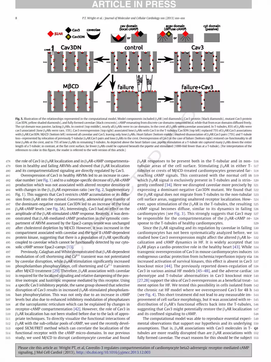
We developed a theoretical framework so that a hypothetical expla-nation for Cav3 interactions with β2ARs under the various conditionsconsidered here and in our previous related work [34] could be pro-posed. The conditions considered include control, Cav3 overexpression(Cav3), expression of dominant negativemutant Cav3 (Cav3DN), appli-cation of MβCD, heart failure, and failure with Cav3 overexpression(failure + Cav3). The framework is illustrated in Fig. 5.
Theworking components of themodel, which associate with β2ARs,include Cav3, Cav3DN, and fully formed caveolae; β2ARs can also be un-associated (lone). Pairings of β2AR with caveolae or Cav3 were consid-ered to be discrete “cav” signaling domains, where cAMP was localdue to spatial isolation (see Methods Section 2.9) and where relativelyhigh phosphodiesterase (PDE) activity partially muted signals (see Sup-plementary Text). Lone β2ARs or β2ARs paired with dysfunctionalCav3DN constituted ecav signaling domains. cAMP generated by ecavβ2ARs was readily diffusible because ecav elements were contiguous(not discrete like cav, c.f. Methods 2.9) and because PDE activity waslow enough to permit cAMP accumulation. Ecav lacks PDE type 3,which is found in cav, and ecav has a volume that is twice that of cav,causing relative dilution of PDE activity (see Supplementary Text). Thecytosol was devoid of β2ARs. Passive diffusion of cAMP in cyt occurred,but signals were greatly diminished by the fact that cyt volume wasroughly ten-fold larger than sub-membrane volume (cav + ecav).
Our electron micrographs showed fully formed, flask-shaped caveo-lae in crest regions, but they were not observed in T-tubules. Recently,
entation of cardiomyocyte beta2-adrenergic receptor-mediated cAMP3.12.003

RRECTED P
RO
OF
394
395
396
397
398
399
400
401
402
403
404
405
406
407
408
409
410
411
412
413
414
415
416
417
418
419
420
421
422
423
424
425
426
427
428
429
430
431
A B
C
D HFCav3DN
2AR SICM/FRET2AR FRET
E
Control
ForskolinISO+CGP
Fig. 3.Cav3modulates cAMP levels and its spatial distribution afterβ2AR stimulation. A) Expression of the dominant negative Cav3DN construct results in an increase in intracellular cAMPstimulated byβ2AR. Cells were transducedwith either LacZ (control) or Cav3DN together with the Epac2-camps sensor adenovirus (all atMOI 500) and stimulated as described in Figs. 2CandD. Data aremeans ± SE (n = 12–13 cells). *—Difference from control is significant at P b 0.05. B) SICM/FRET analysis ofβ2ARdistribution inCav3DNexpressing cells.β2ARwas locallystimulated with the scanning pipette in either single T-tubules or in cell crests located between the Z-lines. The experiment was performed exactly as previously described [34]. β2AR-cAMP signals can be detected only upon local receptor stimulation in the T-tubules, suggesting that β2AR localization is unaltered in Cav3DN expressing cardiomyocytes. Data are present-ed asmeans ± SE (n = 5–8 cells). **—Difference is significant at P b 0.01. C) Local β2AR stimulation in single T-tubules (white arrow denotes the position of the SICM pipette) of control(LacZ expressing) cardiomyocytes results in a highly localized cAMP signalwhich ismeasurable only in the subcellular region adjacent to the pipette. Data in the graph showYFP/CFP ratiosin different color-labeled regions across the cell cytosol. Representative experiment, n = N6 cells. D) Local β2AR stimulation in single T-tubules of Cav3DN expressing cardiomyocytes re-sults in a far-reaching cAMP gradientwhich diffuses across the entire cell. Data are presented as inC. Representative experiment, n = N8 cells. E) Local T-tubularβ2AR stimulation in failingcardiomyocytes leads to a similar far-reaching cAMP gradient. Representative experiment, n = 10 cells.
6 P.T. Wright et al. / Journal of Molecular and Cellular Cardiology xxx (2013) xxx–xxx
UNCO
Wong et al. presented a similar finding on the basis of a highly novelfluorescent microscopy technique coupled with electron microscopy(their Supplementary Fig. S4 [38]). Cav3 protein is apparently abundantin T-tubules but it is not clearwhether fully stabilized caveolae domainscan be formed in this region (e.g. [38]). Thus, to represent the controlcase, β2ARs were placed in caveolae in the crest and were Cav3-associated in T-tubules (exception: 15% of T-tubular β2ARs were lone;Fig. 5, top middle).
Cav3 overexpression (Fig. 5, top right) paired the few lone T-tubularβ2ARs with Cav3 and added nonfunctional Cav3 and caveolae lackingβ2ARs. We hypothesized that Cav3DN (Fig. 5, top left) affected T-tubular β2AR:Cav3 pairs, replacing 75% of native Cav3 with mutantCav3DN, and changing their designation from cav to ecav in themodel. Importantly, we hypothesized that β2AR:caveolae pairs at thecrest were immune to Cav3DN replacement. This concept is supportedfurther below. We hypothesized that formation of β2AR:caveolae andβ2AR:Cav3 complexes requires cholesterol. Therefore,MβCDwas repre-sented by strictly lone β2ARs in crest and T-tubules (Fig. 5, bottom left).Failure was represented by Cav3 loss (affecting 75% of β2AR:Cav3 pairs
Please cite this article as: Wright PT, et al, Caveolin-3 regulates compartmsignaling, J Mol Cell Cardiol (2013), http://dx.doi.org/10.1016/j.yjmcc.201
in intact T-tubules) and also loss of T-tubules (half of them, as indicatedby Z-groove index reduction shown here and previously [34]). Lost T-tubules purged their contents to the crest, dissociating Cav3 from 90%of β2ARs in the process (Fig. 5, bottom middle). Finally, overexpressingCav3 in heart failure was represented as pairing lone β2ARs with Cav3(1000% and 300% increase in β2ARs:Cav3 pairings in crest and T-tubules, respectively, Fig. 5, bottom right).
The central assumption of this theoretical framework is summarizedas follows (supported below).β2AR associationswith caveolae, found inthe crest, are stable in the face of Cav3DN expression, while associationswith Cav3 protein(s) in the T-tubules are not. This assumption can alter-natively be viewed as causing limited pools of Cav3 to prioritize thecrest over T-tubule when it comes to the localization of cAMP byforming stronger cAMP compartments. Other assumptions (e.g. the %changes above), though not incidental, were of quantitative but notqualitative consequence and needed to be made in order to realize acomputational model to test the validity of the framework in Fig. 5.
Fig. 6 presents simulation results. Plotted are sub-membrane cAMPconcentration landscapes ([cAMP] in μM) computed in space and time
entation of cardiomyocyte beta2-adrenergic receptor-mediated cAMP3.12.003

RECTED P
RO
OF
432
433
434
435
436
437
438
439
440
441
442
443
444
445
446
447
448
449
450
451
452
453
454
455
456
457
458
459
460
461
462
463
464
465
466
467
468
469
470
471
472
473
474
475
476
477
HF+Cav3
2AR SICM/FRET
A B
C D
0 50 100 150
0.96
0.98
1.00
FR
ET
Rat
io (
norm
.)
Time (s)
ISO+ CGP
Fig. 4.Cav3overexpression in failing cardiomyocytes partially restores the T-tubular localization ofβ2AR. A)Cav3 overexpression for 48 h (MOI 500) doesnot lead to a significant improve-ment of the cell surfacemorphology. Representative SICM image of a failing cardiomyocyte overexpressing Cav3 (n = 20 cells). B) Quantification of the Z-groove index in these cells com-pared to healthy (Control) and failing (HF) cardiomyocytes without Cav3 overexpression (LacZ transfected, MOI 500). Shown are means ± SE (n = 12–16 cells). **—Difference issignificant at P b 0.01. NS—no significant difference. C) SICM/FRET analysis of β2AR localization in Cav3 expressing cells, isolated from rats 16 weeks post-MI. FRET responses are signif-icantly higher when agonist is applied to the t-tubules compared to the cellular crests. Graphs show mean ± SE (n = 15–19 cells) *P b 0.05. This suggests that some restoration of thenormal healthy character of β2AR has been achieved by Cav3 overexpression; this is in contrast to cells derived from the same animal without Cav3 overexpression. E) A representativetrace of the β2AR response from a heart failure cell with Cav3 overexpressed.
7P.T. Wright et al. / Journal of Molecular and Cellular Cardiology xxx (2013) xxx–xxx
UNCO
Ralong a longitudinal line scan (100 μm) following β2AR stimulationat either a distal T-tubular site (panel A) or at a distal crest site(panel B). We chose to measure [cAMP] at the membrane wherethemajority of important signaling targets reside (rationale outlinedfurther in Supplementary Text and Supplementary Fig. 4). Pipetteplacement over a T-tubule stimulates many β2ARs, all along the en-tire depth of the structure, while stimulation at crest sites affectsonly the few β2ARs immediately beneath the pipette (e.g. pipettecartoon in Fig. 5, bottom middle). Simulations accounted for thiswith a 1000-fold T-tubule to crest β2AR capture ratio (results werenot different using a lower estimate of 100-fold).
T-tubular stimulation resulted in sharp local spikes in [cAMP] at thestimulation site in all cases. However, diffuse cAMP (i.e. smooth concen-tration gradient from the stimulus site) occurred only in Cav3DN, failure,andMβCD cases, but not in cases of control, Cav3 overexpression, or heartfailure with Cav3 overexpression. This matches experiments shown hereand in our previouswork: Cav3DN—Fig. 3D, heart failure—Fig. 3E,MβCD—Supplementary Fig. S8C of Nikolaev et al. [34], control—Fig. 3C, andfailure + Cav3—Fig. 4D. The cAMP signal was reduced by Cav3 overex-pression (integrated total cAMP was ~70% of control). This is in agree-ment with Fig. 2D. Cav3DN simulations showed an increase in overallcAMP relative to control (73% increase in integrated total cAMP). Al-though the amplitude is not different, more cAMP response is presentdue to propagation throughout the cell. This is in agreement with Fig. 3A.
Please cite this article as: Wright PT, et al, Caveolin-3 regulates compartmsignaling, J Mol Cell Cardiol (2013), http://dx.doi.org/10.1016/j.yjmcc.201
Simulation results showed that a response to crest stimulation wasonly registered for the MβCD and heart failure cases. This is in accordwith experiments (no response: control—Fig. 2G of Nikolaev et al.[34], Cav3DN—Fig. 3B, and failure + Cav3—Fig. 4C. Response: MβCD—Supplementary Fig. S8B of Nikolaev et al. [34], and failure—Fig. 4C). Im-portantly, when we relaxed the central assumption that Cav3DN doesnot disrupt β2AR associations with caveolae in the crest, there was a ro-bust crest response in Cav3DN simulations, in violation of Fig. 3B exper-imental results. Without this central assumption, the Cav3DN case wasnearly identical to the heart failure case. Though crest caveolaewere notdegraded in failure, T-tubule loss caused redistribution of newly loneβ2ARs to the crest. This distinguished the failure response from theCav3DN response.
Simulations predicted baseline cAMP elevation in pathological states(i.e. Cav3DN, failure andMβCD). Thiswas caused by the greater propor-tion of β2ARs in the ecav domains, where PDE activity was relativelylow. FRET experiments cannot support or refute this prediction.
4. Discussion
The β2AR has been shown to associate with caveolin and caveolae inneonatal and adult ventricular cardiomyocytes. However, functionalconsequences of the receptor–caveolin interactions remained poorlydefined, especially in failing cardiomyocytes. In this work, we analyzed
entation of cardiomyocyte beta2-adrenergic receptor-mediated cAMP3.12.003
T
PRO
OF
478
479
480
481
482
483
484
485
486
487
488
489
490
491
492
493
494
495
496
497
498
499
500
501
502
503
504
505
506
507
508
509
510
511
512
513
514
515
516
517
518
519
520
521
522
523
524
525
526
527
528
529
530
531
532
533
534
535
536
537
538
539
540
541
542
543
544
545
546
547
548
549
550
551Q3
552
553
Fig. 5. Illustration of the relationships represented in the computational model. Model components included β2AR (red diamonds), Cav3 protein (black diamonds), mutant Cav3 protein(Cav3DN, yellow shaded diamonds), and fully formed caveolae (black crescents). cAMPemanating fromdiscrete cav domains remained local,while that fromecav domains diffused freely.The cyt domain was passive, lacking β2ARs. In control (topmiddle), nearly all β2ARs were in cav domains. In the crest all β2ARs were caveolae associated. In T-tubules, 85% of β2ARs werecav3 associated (lone β2ARs were rare, 15%). Cav3 overexpression (top right) associated lone β2ARs with Cav3 in the T-tubules. Cav3DN (top left) replaced 75% of β2AR:Cav3 associationswith β2AR:Cav3DN.MβCD (bottom left) removed all caveolae and Cav3, leaving only lone β2ARs. Heart failure (bottommiddle) involved disassociation of β2AR:Cav3 pairs (75%) and T-tubuleloss—represented by relocation of previously T-tubular β2AR:Cav3 pairs and lone β2ARs to the crest. Overexpression of Cav3 in the case of failure (bottom right) restored cav functionality to alllone β2ARs at the crest, and to 75% of lone β2ARs in remaining T-tubules. As depicted above the heart failure case, pipette stimulation at a T-tubule site captured many β2ARs down the entirelength of a T-tubule; in contrast, at the flat crest surface, far fewer β2ARs could be captured beneath the pipette and stimulated (1000-fold fewer than at a T-tubule). (For interpretation of thereferences to color in this figure, the reader is referred to the web version of this article.)
8 P.T. Wright et al. / Journal of Molecular and Cellular Cardiology xxx (2013) xxx–xxx
UNCO
RREC
the role of Cav3 in β2AR localization and in β2AR-cAMP compartmenta-tion in healthy and failing ARVMs and showed that β2AR localizationand its compartmentalized signaling are directly regulated by Cav3.
Overexpression of Cav3 in healthy ARVMs led to an increase in cave-olae number (see Fig. 1) and to a subtype-specific decrease ofβ2AR-cAMPproduction which was not associated with altered receptor densities orwith changes in the β1/β2AR expression ratio (see Fig. 2, SupplementaryFig. 1). This suggests that Cav3 restricts cAMP accumulation or its diffu-sion from β2AR into the cytosol. Conversely, adenoviral gene transfer ofthe dominant-negative mutant Cav3DN led to an increase of the totalβ2AR-cAMP levels (see Fig. 3A). Interestingly, Cav3 had no effect on theamplitude of the β1AR-stimulated cAMP response. Recently, it was dem-onstrated that β1AR-mediated cAMP production in the cytosolic com-partment of ARVM assessed by the Epac2-camps sensor was unchangedafter cholesterol depletion by MβCD. However, it was increased in thecompartment associated with caveolae and the type II cAMP-dependentprotein kinase, suggesting that there is a population of β1AR specificallycoupled to caveolae which cannot be functionally detected by our cyto-solic cAMP sensor Epac2-camps [13].
Previously, Calaghan andWhite demonstrated thatβ1AR-dependentmodulation of cell shortening and Ca2+ transient was not potentiatedby caveolar disruption, while β2AR stimulation significantly increasedphospholamban phosphorylation, cell shortening and Ca2+ transientsafter MβCD treatment [29]. Therefore, β2AR association with caveolaeis required for the localized signaling and relative dampeningof thepos-itive inotropic and lusitropic response mediated by this receptor. Usinga specific Cav3 inhibitory peptide, the same group showed that selectivedisruption of Cav3 results in increased β2AR-stimulated phospholam-ban phosphorylation. This was not only due to an increase in cAMPlevels but also due to enhanced inhibitory modulation of phosphatasesat the sarcoplasmic reticulum which can be explained by changes inreceptor-cAMP compartmentation [42]. However, the role of Cav3 inβ2AR localization has not been studied before due to the lack of appro-priate techniques. To directly visualize the functional interactions ofβ2AR with the sub-cellular pools of cAMP, we used the recently devel-oped SICM/FRET method which can correlate the localization of thefunctional receptor with the cAMP micro-domains. In our previousstudy, we used MβCD to disrupt cardiomyocyte caveolae and found
Please cite this article as: Wright PT, et al, Caveolin-3 regulates compartmsignaling, J Mol Cell Cardiol (2013), http://dx.doi.org/10.1016/j.yjmcc.201
EDβ2AR responses to be present both in the T-tubular and in non-
tubular areas of the cell surface. Stimulating β2AR in either T-tubules or crests of MβCD-treated cardiomyocytes generated far-reaching cAMP signals. This contrasted with the normal cell inwhich β2AR signal is exclusively present in T-tubules and is strin-gently confined [34]. Here we disrupted caveolae more precisely byexpressing a dominant-negative Cav3DN mutant. We found thatthe β2AR signal does not migrate from T-tubules to the non-tubularcell surface areas, suggesting unaltered receptor localization. How-ever, upon stimulation of the β2AR in the T-tubules, the resultingcAMP signal becomes diffuse, similar to its dynamics in failingcardiomyocytes (see Fig. 3). This strongly suggests that Cav3 maybe responsible for the compartmentation of the β2AR-cAMP re-sponse to the T-tubules of healthy cardiomyocytes.
Since the β2AR signaling and its regulation by caveolae in failingcardiomyocytes has not been systematically analyzed before, westudied how Cav3 overexpression might affect the altered β2AR lo-calization and cAMP dynamics in HF. It is widely accepted thatβ2AR plays a cardio-protective role in the healthy heart [43]. Whiletransgenic overexpression of Cav3 in mouse cardiomyocytes inducesendogenous cardiac protection from ischemia/reperfusion injury viaincreased activation of survival kinases, this effect is absent in Cav3knockout mice [44]. The previously reported down-regulation ofCav3 in various animal HF models [45–48], and the adverse cardiacphenotype and T-tubular abnormalities in Cav3 knockout mice[27,28] support the idea of Cav3 overexpression as a beneficial treat-ment option for HF. We tested this possibility in cells isolated fromthe chronic rat HF model where we overexpressed Cav3 for 48 h(see Fig. 4). This short treatment did not lead to any measurable im-provement of cell surface morphology, but it was associated with re-distribution of β2AR's functional effects back into the T-tubules,suggesting that Cav3 might potentially restore the β2AR localizationand its confined signaling to cAMP.
The computational model was able to reproduce essential experi-mental observations that support our hypothesis and its underlyingassumptions. That is, β2AR associations with Cav3 molecules in T-tubules are more readily disrupted than are β2AR associations withfully formed caveolae. The exact reasons for this should be the subject
entation of cardiomyocyte beta2-adrenergic receptor-mediated cAMP3.12.003

CO
RRECTED P
RO
OF
554
555
556
557
558
559
560
561
562
563
564
565
566
567
568
569
570
571
572
573
574
575
576
577
578
579
580
581
582
583
Fig. 6. Simulations based on the relationships proposed in Fig. 5 qualitatively reproduced cAMP experimental measurements. Shown are surfacemaps of cAMP concentration at themem-brane ([cAMP], from 0.5 to 4 μM) along the cell length (100 μm) during the time just prior to andminutes followingβ2AR stimulation. Stimulationwas at distal T-tubule (panel A, top) ordistal crest sites (panel B, bottom)—indicated in space and time by black rectangles labeled “β2AR stim.”. The protocol was designed tomimic experiments shown in Figs. 3C, D, E and 4D.Results for control, Cav3 overexpression, Cav3DN, MβCD, heart failure, and heart failure with Cav3 overexpression are shown. Cases are arranged as in Fig. 5. A) Following T-tubular stim-ulation, cAMPwas robustly generated at the stimulus site in all cases. However, it did not diffuse in control, Cav3, and failure + Cav3 cases (flat [cAMP] landscapes, “no diffusion” straightarrow). Pathological diffusion was exhibited by Cav3DN, MβCD, and heart failure (contoured [cAMP] landscapes, “diffusion” curved arrow). B) Crest stimulation did not always generatecAMP: e.g. the non-disease cases of control andCav3, but also Cav3DNand failure + Cav3 cases. cAMPwas generated following crest stimulation forMβCDand failure cases, and it diffusedalong the cell. Color scales are the same for all cases. Z-axes scales are the same for each case.
9P.T. Wright et al. / Journal of Molecular and Cellular Cardiology xxx (2013) xxx–xxx
UNof future work. We speculate that binding among the 14–16 Cav3 mol-
ecules in the formation of a caveolae [17] is cooperative, and that thiscould be the stabilizing factor. The mechanism by which caveolae orCav3 association can confine cAMP response resulting from β2AR stim-ulation is likely to be indirect. We speculate that individual β2AR:Cav3and β2AR:caveolae are diffusively separated from the general milieu.Moreover, we speculate that relatively high phosphodiesterase activityin the cav domain (included in the Heijman et al. model, see Supple-mentary Text for more detail), can mute cAMP levels and therebylimit its reach.
Implications of this study extend beyond Cav3 mutations and heartfailure. A recent publication showed that a diet high in palmitatedisrupted Cav3 in ventricular myocytes [49]. High dietary palmitatemay lead to cellular behavior similar to Cav3DN case, where localcAMP responses to sympathetic β2AR stimulation were lost. This may
Please cite this article as: Wright PT, et al, Caveolin-3 regulates compartmsignaling, J Mol Cell Cardiol (2013), http://dx.doi.org/10.1016/j.yjmcc.201
have consequences for individuals' cardiovascular physiology. Finally,although the rat cardiomyocyte is the model presented in this paperthe authors are aware of the limitations of this species with regard tothe necessity of culture. The t-tubular network is observed to degradeduring the course of the culturing process. Equally, adenovirus appearsto be the only method of gene delivery robust enough for use with thiscell type. The physical and molecular changes caused by adenoviralentry and t-tubular disruption as well as the effects of unloading in cul-turemay reduce the relevance of data from studies using this cell to fail-ing cells in vivo.
Future studies should analyze β2AR localization and cAMP signalingby SICM/FRET in cardiomyocyte-specific Cav3 transgenic and knockoutmice where the differences in membrane morphology might be evenmore pronounced due to constitutive expression or deletion of Cav3.Transgenic mice are in existence, which constitutively express FRET
entation of cardiomyocyte beta2-adrenergic receptor-mediated cAMP3.12.003
T
584
585
586
587
588
589
590
591
592
593
594
595
596
597
598
599
600
601Q4
602
603
604
605
606
607
608
609
610
611
612
613614615616617618619620621622623624625626627628629630631632633634635636637638639640641642643644645
646Q6647648649650651652653654655656657658659660661662663664665666667668669670671672673674675676677678679680681682683684685686687688689690691692693694695696697698699700701702703704705706707708709710711712713714715716717718719720721722723724725726727728729730731
10 P.T. Wright et al. / Journal of Molecular and Cellular Cardiology xxx (2013) xxx–xxx
UNCO
RREC
sensors similar to the one employed by this study [34]. Crossing thesemice with Cav3 transgenics may provide an avenue to studying therole of Cav3 inmodulatingβ2AR function in freshly isolated cells. Finally,measurements in failing human myocytes with Cav3 re-expressioncould be performed when technically possible, as this would also re-quire culturing. These studies might pave the way towards a newgene therapy option for HF patients.
5. Conclusions
In healthy adult rat cardiomyocytes, Cav3 regulates β2AR-cAMP sig-naling, playing a crucial role for the compartmentation of cAMP signalsto the T-tubular compartment. Disruption of Cav3 function with adominant-negative mutant leads to far-reaching β2AR-cAMP signalssimilar to those observed in heart failure. In failing cardiomyocytes,Cav3 overexpression can partially restore the disrupted localization ofthese receptors.
Disclosures
None declared.
Acknowledgements
We thank Peter O'Gara for the cardiomyocyte isolation, ChristianDees for the radioligand binding studies and Karina Zimmermann forthe Western blot analysis. This work was supported by grants fromthe Wellcome Trust (WTN090594 to JG and 092852 to MS), the BritishHeart Foundation (NH/10/3/28574 to JG and SEH, and FS/11/67/28954to ARL) and the Deutsche Forschungsgemeinschaft (SFB 1002, TP A01to VON).
Appendix A. Supplementary data
Supplementary data to this article can be found online at http://dx.doi.org/10.1016/j.yjmcc.2013.12.003.
References
[1] Brodde OE, Michel MC. Adrenergic and muscarinic receptors in the human heart.Pharmacol Rev 1999;51:651–90.
[2] Xiang Y, Kobilka BK. Myocyte adrenoceptor signaling pathways. Science2003;300:1530–2.
[3] Lohse MJ, Engelhardt S, Eschenhagen T.What is the role of beta-adrenergic signalingin heart failure? Circ Res 2003;93:896–906.
[4] Schäfer M, Frischkopf K, Taimor G, Piper HM, Schlüter K-D. Hypertrophic effect ofselective β1-adrenoceptor stimulation on ventricular cardiomyocytes from adultrat. Am J Physiol Cell Physiol 2000;279:C495–503.
[5] ZhuW-Z, Zheng M, KochWJ, Lefkowitz RJ, Kobilka BK, Xiao R-P. Dual modulation ofcell survival and cell death by β2-adrenergic signaling in adult mouse cardiacmyocytes. Proc Natl Acad Sci 2001;98:1607–12.
[6] Engelhardt S, Hein L, Wiesmann F, Lohse MJ. Progressive hypertrophy and heartfailure in beta1-adrenergic receptor transgenic mice. Proc Natl Acad Sci U S A1999;96:7059–64.
[7] Dorn II GW, Tepe NM, Lorenz JN, Koch WJ, Liggett SB. Low- and high-leveltransgenic expression of beta2-adrenergic receptors differentially affect cardiachypertrophy and function in Galphaq-overexpressing mice. Proc Natl Acad Sci U S A1999;96:6400–5.
[8] Xiao R-P. {beta}-Adrenergic signaling in the heart: dual coupling of the {beta}2-ad-renergic receptor to Gs and Gi proteins. Sci Signal 2001;2001:re15.
[9] Xiao RP, Ji X, Lakatta EG. Functional coupling of the beta 2-adrenoceptor to a pertus-sis toxin-sensitive G protein in cardiac myocytes. Mol Pharmacol 1995;47:322–9.
[10] Nikolaev VO, Bünemann M, Schmitteckert E, Lohse MJ, Engelhardt S. Cyclic AMP im-aging in adult cardiac myocytes reveals far-reaching β1-adrenergic but locally con-fined β2-adrenergic receptor-mediated signaling. Circ Res 2006;99:1084–91.
[11] Head BP, Patel HH, Roth DM, Lai NC, Niesman IR, Farquhar MG, et al. G-protein-coupled receptor signaling components localize in both sarcolemmal and intracellu-lar caveolin-3-associated microdomains in adult cardiac myocytes. J Biol Chem2005;280:31036–44.
[12] Xiang Y, Rybin VO, Steinberg SF, Kobilka B. Caveolar localization dictates physi-ologic signaling of β2-adrenoceptors in neonatal cardiac myocytes. J Biol Chem2002;277:34280–6.
Please cite this article as: Wright PT, et al, Caveolin-3 regulates compartmsignaling, J Mol Cell Cardiol (2013), http://dx.doi.org/10.1016/j.yjmcc.201
ED P
RO
OF
[13] Agarwal SR, MacDougall DA, Tyser R, Pugh SD, Calaghan SC, Harvey RD. Effects ofcholesterol depletion on compartmentalized cAMP responses in adult cardiacmyocytes. J Mol Cell Cardiol 2011;50:500–9.
[14] Ostrom RS, Violin JD, Coleman S, Insel PA. Selective enhancement of β-adrenergic re-ceptor signaling by overexpression of adenylyl cyclase type 6: colocalization of re-ceptor and adenylyl cyclase in caveolae of cardiac myocytes. Mol Pharmacol2000;57:1075–9.
[15] Rybin VO, Xu X, Lisanti MP, Steinberg SF. Differential targeting of β-adrenergic re-ceptor subtypes and adenylyl cyclase to cardiomyocyte caveolae. J Biol Chem2000;275:41447–57.
[16] Cohen AW, Hnasko R, Schubert W, Lisanti MP. Role of caveolae and caveolins inhealth and disease. Physiol Rev 2004;84:1341–79.
[17] Razani B,Woodman SE, Lisanti MP. Caveolae: from cell biology to animal physiology.Pharmacol Rev 2002;54:431–67.
[18] Shaul PW, Anderson RGW. Role of plasmalemmal caveolae in signal transduction.Am J Physiol Lung Cell Mol Physiol 1998;275:L843–51.
[19] Harvey RD, Calaghan SC. Caveolae create local signalling domains through theirdistinct protein content, lipid profile and morphology. J Mol Cell Cardiol2012;52:366–75.
[20] Gratton JP, Bernatchez P, Sessa WC. Caveolae and caveolins in the cardiovascularsystem. Circ Res 2004;94:1408–17.
[21] Lisanti MP, Scherer PE, Vidugiriene J, Tang Z, Hermanowski-Vosatka A, Tu YH,et al. Characterization of caveolin-rich membrane domains isolated froman endothelial-rich source: implications for human disease. J Cell Biol1994;126:111–26.
[22] Feron O, Balligand JL. Caveolins and the regulation of endothelial nitric oxide syn-thase in the heart. Cardiovasc Res 2006;69:788–97.
[23] Barouch LA, Harrison RW, Skaf MW, Rosas GO, Cappola TP, Kobeissi ZA, et al. Nitricoxide regulates the heart by spatial confinement of nitric oxide synthase isoforms.Nature 2002;416:337–9.
[24] Parton RG,WayM, Zorzi N, Stang E. Caveolin-3 associates with developing t-tubulesduring muscle differentiation. J Cell Biol 1997;136:137–54.
[25] Galbiati F, Volonte D, Chu JB, Li M, Fine SW, Fu M, et al. Transgenic overexpression ofcaveolin-3 in skeletal muscle fibers induces a Duchenne-like muscular dystrophyphenotype. Proc Natl Acad Sci U S A 2000;97:9689–94.
[26] Repetto S, Bado M, Broda P, Lucania G, Masetti E, Sotgia F, et al. Increased number ofcaveolae and caveolin-3 overexpression in duchenne muscular dystrophy. BiochemBiophys Res Commun 1999;261:547–50.
[27] Woodman SE, Park DS, Cohen AW, Cheung MWC, Chandra M, Shirani J, et al.Caveolin-3 knock-out mice develop a progressive cardiomyopathy and show hyper-activation of the p42/44 MAPK cascade. J Biol Chem 2002;277:38988–97.
[28] Galbiati F, Engelman JA, Volonte D, Zhang XL, Minetti C, Li M, et al. Caveolin-3null mice show a loss of caveolae, changes in the microdomain distribution ofthe dystrophin–glycoprotein complex, and t-tubule abnormalities. J Biol Chem2001;276:21425–33.
[29] Calaghan S, White E. Caveolae modulate excitation–contraction coupling andβ2-adrenergic signalling in adult rat ventricular myocytes. Cardiovasc Res2006;69:816–24.
[30] Balijepalli RC, Foell JD, Hall DD, Hell JW, Kamp TJ. Localization of cardiac L-typeCa2+ channels to a caveolar macromolecular signaling complex is required forβ2-adrenergic regulation. Proc Natl Acad Sci 2006;103:7500–5.
[31] Chen-Izu Y, Xiao R-P, Izu LT, Cheng H, Kuschel M, Spurgeon H, et al. Gi-dependentlocalization of β2-adrenergic receptor signaling to L-type Ca2+ channels. BiophysJ 2000;79:2547–56.
[32] Wagner E, Lauterbach MA, Kohl T, Westphal V, Williams GS, Steinbrecher JH, et al.Stimulated emission depletion live-cell super-resolution imaging shows prolifera-tive remodeling of T-tubule membrane structures after myocardial infarction. CircRes 2012;111:402–14.
[33] Lyon AR, MacLeod KT, Zhang Y, Garcia E, Kanda GK, Lab MJ, et al. Loss of T-tubulesand other changes to surface topography in ventricular myocytes from failinghuman and rat heart. Proc Natl Acad Sci U S A 2009;106:6854–9.
[34] Nikolaev VO, Moshkov A, Lyon AR, Miragoli M, Novak P, Paur H, et al. Beta2-adrenergic receptor redistribution in heart failure changes cAMP compartmentation.Science 2010;327:1653–7.
[35] Koga A, Oka N, Kikuchi T, Miyazaki H, Kato S, Imaizumi T. Adenovirus-mediatedoverexpression of caveolin-3 inhibits rat cardiomyocyte hypertrophy. Hypertension2003;42:213–9.
[36] Hannawacker A, Krasel C, Lohse MJ. Mutation of Asn293 to Asp in transmembranehelix VI abolishes agonist-induced but not constitutive activity of the β2-adrenergic receptor. Mol Pharmacol 2002;62:1431–7.
[37] Nikolaev VO, Gambaryan S, Engelhardt S, Walter U, Lohse MJ. Real-time monitoringof the PDE2 activity of live cells. J Biol Chem 2005;280:1716–9.
[38] Wong J, Baddeley D, Bushong EA, Yu Z, Ellisman MH, Hoshijima M, et al. Nanoscaledistribution of ryanodine receptors and caveolin-3 in mouse ventricular myocytes:dilation of T-tubules near junctions. Biophys J 2013;104:L22–4.
[39] Heijman J, Volders PG, Westra RL, Rudy Y. Local control of beta-adrenergic stimula-tion: effects on ventricular myocyte electrophysiology and Ca(2+)-transient. J MolCell Cardiol 2011;50:863–71.
[40] Galbiati F, Volonté D, Minetti C, Chu JB, Lisanti MP. Phenotypic behavior of caveolin-3 mutations that cause autosomal dominant limb girdle muscular dystrophy(LGMD-1C). J Biol Chem 1999;274:25632–41.
[41] Agarwal SR, MacDougall DA, Tyser R, Pugh SD, Calaghan SC, Harvey RD. Effects ofcholesterol depletion on compartmentalized cAMP responses in adult cardiacmyocytes. J Mol Cell Cardiol 2011;50:500–9.
[42] Macdougall DA, Agarwal SR, Stopford EA, Chu H, Collins JA, Longster AL, et al. Cave-olae compartmentalise beta2-adrenoceptor signals by curtailing cAMP production
entation of cardiomyocyte beta2-adrenergic receptor-mediated cAMP3.12.003

732733734735736737738739740741742
743744745746747748749750751752753
755
11P.T. Wright et al. / Journal of Molecular and Cellular Cardiology xxx (2013) xxx–xxx
and maintaining phosphatase activity in the sarcoplasmic reticulum of the adultventricular myocyte. J Mol Cell Cardiol 2012;52:388–400.
[43] Communal C, Singh K, Sawyer DB, Colucci WS. Opposing effects of beta(1)- andbeta(2)-adrenergic receptors on cardiac myocyte apoptosis: role of a pertussistoxin-sensitive G protein. Circulation 1999;100:2210–2.
[44] Tsutsumi YM, Horikawa YT, Jennings MM, KiddMW, Niesman IR, Yokoyama U, et al.Cardiac-specific overexpression of caveolin-3 induces endogenous cardiac protec-tion by mimicking ischemic preconditioning. Circulation 2008;118:1979–88.
[45] Ratajczak P, Damy T, Heymes C, Oliviero P, Marotte F, Robidel E, et al. Caveolin-1 and-3 dissociations from caveolae to cytosol in the heart during aging and after myocar-dial infarction in rat. Cardiovasc Res 2003;57:358–69.
UNCO
RRECT
754
Please cite this article as: Wright PT, et al, Caveolin-3 regulates compartmsignaling, J Mol Cell Cardiol (2013), http://dx.doi.org/10.1016/j.yjmcc.201
[46] Oka N, Asai K, Kudej RK, Edwards JG, Toya Y, Schwencke C, et al. Downregulation ofcaveolin by chronic beta-adrenergic receptor stimulation in mice. Am J Physiol1997;273:C1957–62.
[47] Piech A,Massart PE, Dessy C, Feron O, Havaux X,Morel N, et al. Decreased expressionof myocardial eNOS and caveolin in dogs with hypertrophic cardiomyopathy. Am JPhysiol Heart Circ Physiol 2002;282:H219–31.
[48] Fujita T, Toya Y, Iwatsubo K, Onda T, Kimura K, Umemura S, et al. Accumulation ofmolecules involved in alpha1-adrenergic signal within caveolae: caveolin expres-sion and the development of cardiac hypertrophy. Cardiovasc Res 2001;51:709–16.
[49] Knowles CJ, Cebova M, Pinz IM. Palmitate diet-induced loss of cardiac caveolin-3: anovel mechanism for lipid-induced contractile dysfunction. PLoS One 2013;8:e61369.
ED P
RO
OF
entation of cardiomyocyte beta2-adrenergic receptor-mediated cAMP3.12.003


