Embed Size (px)
Citation preview
Changes in Histone H3 Lysine 36 Methylation in PorcineOocytes and Preimplantation EmbryosYun Fei Diao, Reza K. Oqani, Xiao Xia Li, Tao Lin, Jung Won Kang, Dong Il Jin*
Department of Animal Science & Biotechnology, Research Center for Transgenic Cloned Pigs, Chungnam National University, Daejeon, Korea
Abstract
Histone H3 lysine 36 (H3K36) methylation is known to be associated with transcriptionally active genes, and is considered agenomic marker of active loci. To investigate the changes in H3K36 methylation in pig, we determined the mono-, di-, andtri-methylations of H3K36 (H3K36me1, H3K36me2 and H3K36me3, respectively) in porcine fetal fibroblasts, oocytes andpreimplantation embryos by immunocytochemistry using specific antibodies and confocal microscopy. These analysesrevealed that only H3K36me3 in porcine fetal fibroblasts consistently colocalized with transcription sites identified asactively synthesizing RNA based on fluorouridine (FU) incorporation. Treatment of cells with flavopiridol, which blockstranscription elongation, completely abrogated both H3K36me3 signals and RNA synthesis. All three types of H3K36methylation were present and did not significantly differ during oocyte maturation. In parthenogenetic embryos,H3K36me1 and -me2 were detected in 1-cell through blastocyst-stage embryos. In contrast, H3K36me3 was not detected inmost 1-cell stage embryos. H3K36me3 signals became detectable in 2-cell stage embryos, peaked at the 4-cell stage,decreased at the 8-cell stage, and then became undetectable at blastocyst stages in both parthenogenetic and in vitro-fertilized (IVF) embryos. Unlike the case in IVF embryos, H3K36me3 could not be demethylated completely during the 1-cellstage in somatic cell nuclear transfer (SCNT) embryos. These results collectively indicate that H3K36me3, but not H3K36me1or -me2, is associated with transcription elongation in porcine fetal fibroblasts. H3K36me3 is developmentally regulated andmay be a histone mark of embryonic gene activation in pig. Aberrant H3K36 tri-methylation occurred during the nuclearreprogramming of SCNT embryos.
Citation: Diao YF, Oqani RK, Li XX, Lin T, Kang JW, et al. (2014) Changes in Histone H3 Lysine 36 Methylation in Porcine Oocytes and PreimplantationEmbryos. PLoS ONE 9(6): e100205. doi:10.1371/journal.pone.0100205
Editor: Qiang Wu, National University of Singapore, Singapore
Received January 6, 2014; Accepted May 23, 2014; Published June 13, 2014
Copyright: � 2014 Diao et al. This is an open-access article distributed under the terms of the Creative Commons Attribution License, which permitsunrestricted use, distribution, and reproduction in any medium, provided the original author and source are credited.
Funding: This work was supported by the BioGreen 21 Program of the Rural Development Administration (grant no. PJ009060) and the Bio-industry TechnologyDevelopment Program (grant no. IPET312060-5) of the Ministry for Food and Agriculture, Republic of Korea. The funders had no role in study design, datacollection and analysis, decision to publish, or preparation of the manuscript.
Competing Interests: The authors have declared that no competing interests exist.
* E-mail: [email protected]
Introduction
During mammalian fertilization, maternal and paternal chro-
matids are combined to form a fully totipotent embryo. In oocytes,
gene expression is maintained in a silent state during maturation
[1,2]. When oocytes are fertilized by sperm, the zygotes undergo
reprogramming and genome activation, followed by replacement
of maternal transcripts with embryonic transcripts that regulate
embryonic development [3,4,5].
The timing of genome activation is different among species.
Embryonic genome activation (EGA) in mice occurs at the 2-cell
stage [6], whereas porcine and bovine embryos initiate genome
transcription at the 4-cell and 8-cell stage, respectively [7,8].
Although the mechanisms regulating EGA are still not clear,
changes in chromatin structure in the early embryo may play an
important role. Chromatin compaction affects the accessibility of
proteins that regulate gene expression, such as transcription factors
and RNA polymerases [9,10]. The major events involved in this
process include changes in DNA methylation and histone
acetylation or methylation [11,12]. In particular, methylation of
histones at specific residues is an important epigenetic modifica-
tion, playing an essential role in both activating and repressing
transcription during embryonic development, depending on which
lysine residues are methylated [13,14,15,16,17]. For example,
histone H3 tri-methylated at lysine 4 (H3K4me3) is known to be
associated with gene activation [18,19,20,21], whereas histone H3
di-methylated and tri-methylated at lysine 9 (H3K9me2 and -me3)
and histone H3 tri-methylated at lysine 27 (H3K27me3) are
associated with gene silencing [22,23].
Histone H3 methylation at lysine 36 (H3K36) is another
important post-translational modification that is associated with
transcription elongation. In yeast, H3K36 methylation which is
mediated by Set2, is associated with transcribed genes and is
usually referred to as an activating histone mark [24]. Di-
methylation and tri-methylation of H3K36 (H3K36me2 and -
me3) are generally associated with actively transcribed genes,
whereas only H3K36me3 is positively correlated with transcrip-
tion rates [25,26]. H3K36me3 enrichment in the coding region of
transcribed genes is a mark of the actively transcribed chromatin
associated with transcription elongation [27]. During transcription
elongation, the chromodomain of Eaf3, a subunit of the Rpd3S
histone deacetylase complex, recognizes Set2-mediated H3K36
methylation; the resulting complex is then recruited in the wake of
the transcribing RNA polymerase II [28,29]. Accordingly, H3K36
methylation is also a mark for histone deacetylation [30]. Without
Set2 or Rpd3S, acetylated histones accumulate on open reading
frames (ORFs), which can lead to transcription initiation from
cryptic promoters within ORFs [31,32]. Thus, Set2 regulates the
PLOS ONE | www.plosone.org 1 June 2014 | Volume 9 | Issue 6 | e100205
methylation of histone H3K36, suppressing the incorporation of
acetylation and thereby decreasing the initiation of spurious
cryptic transcription from within ORFs; this pathway can
maintain the accuracy of transcription by RNA polymerase II
[33].
Although considerable research on H3K36 methylation has
been reported in yeast, there is limited information about H3K36
methylation in mammals. To date, H3K36 methylations in
porcine oocytes and preimplantation embryos have not been
reported. In the current study, we investigated the changes in
H3K36 methylation status in porcine oocytes, and parthenoge-
netic and in vitro-fertilized (IVF) embryos to determine whether
this epigenetic modification is related to genome activation. As in
somatic cell nuclear transfer (SCNT) embryos, abnormal epige-
netic modification is known to be a major reason for the low
efficiency; we also investigated the H3K36 methylation status in
SCNT embryos to determine whether these embryos have
abnormalities in this epigenetic modification compared with IVF
embryos.
Materials and Methods
Chemicals and EthicsUnless otherwise noted, chemicals were purchased from Sigma-
Aldrich Chemical Company (St. Louis, MO, USA). All animal
care and use procedures were approved by the Institutional
Animal Care and Use Committee of Chungnam National
University.
Porcine Oocyte Collection and in vitro MaturationPorcine ovaries were obtained from a local slaughterhouse (NH
Livestock Cooperation Association, Nonsan City, Chungnam
Province, Korea) where we had acquired permission to use
porcine ovaries, and transported to the laboratory within 2 h in
phosphate-buffered saline (PBS) solution supplemented with
100 IU/ml penicillin and 50 mg/ml streptomycin at 30 to 35uC.
Cumulus-oocyte complexes (COCs) were obtained from follicles
(2–6 mm in diameter) using a 10-ml syringe fixed with an 18-
gauge needle. The COCs were washed three times in TL-HEPES
containing 0.1% (w/v) polyvinyl alcohol (PVA). The oocytes were
then cultured in maturation medium (500 ml per well, see below
for details) in 4-well plates (Nunc, Roskilde, Denmark) and
incubated for 42 to 44 h at 38.5uC in humidified air containing
5% CO2. After 22 h of in vitro maturation, the oocytes were
washed three times and transferred to 500 ml of maturation
medium without hormones for an additional 20 to 22 h of culture.
The maturation medium consisted of TCM-199 (M-4530, Sigma)
supplemented with 10% (v/v) porcine follicular fluid, 3.05 mM D-
glucose, 0.91 mM sodium pyruvate, 0.57 mM L-cysteine, 0.5 mg/
ml LH (L-5269, Sigma), 0.5 mg/ml FSH (F-2293, Sigma), 10 ng/
ml epidermal growth factor (E-4127, Sigma), 75 mg/ml penicillin,
50 mg/ml streptomycin, and 0.05% (v/v) MEM vitamins (M-
6895, Sigma). Following in vitro maturation, the COCs were
transferred to 0.3% hyaluronidase in TL-HEPES-PVA and
pipetted repeatedly for 2 min to denude the oocytes of cumulus
cells.
Preparation of Porcine Fetal FibroblastsIn this manuscript, all animal procedures were approved by the
Institutional Animal Care and Use Committee of Chungnam
National University. The porcine fetal fibroblasts used in this study
were isolated from Korean native pig fetuses at day 35 of gestation.
The head and internal tissues were removed using fine scissors,
and soft tissues such as liver and intestine were discarded. The
remaining tissue was cut into small pieces with fine scissors, treated
with 0.05% trypsin and 0.5 mM EDTA (15050-065, Gibco), and
shaken for 10 min at 38.5uC. The resulting suspension was
centrifuged at 500 rpm for 10 min, and the pellet (containing
porcine fetal fibroblasts) was washed three times in DMEM. The
cells were resuspended in DMEM containing 75 mg/ml penicillin
G, 50 mg/ml streptomycin, 5% (v/v) fetal bovine serum (FBS;
16000-044, Gibco) and 5% (v/v) newborn calf serum (NCS;
26010-074, Gibco), and cultured at 38.5uC. Fetal fibroblasts from
passage 5 were used for experiments.
Parthenogenetic ActivationAfter maturation, cumulus-free oocytes were transferred to
activation solution (0.3 M mannitol, 1.0 mM CaCl2?H2O,
0.1 mM MgCl2?6H2O, and 0.5 mM HEPES) and activated with
one DC pulse of 1.1 kV/cm for 30 ms using a BTX Electro-Cell
Manipulator 2001 (BTX, San Diego, CA, USA). Activated
embryos were cultured in Porcine Zygote Medium (PZM-3)
supplemented with 0.3% bovine serum albumen (BSA) at 38.5uCin humidified air containing 5% CO2.
In vitro FertilizationIn vitro fertilization of oocytes were carried out as described
previously by Li et al. [34] After maturation, cumulus-free oocytes
with first polar body extruded were washed three times with
fertilization medium, a modified Tris-buffered medium (mTBM)
containing of 113 mM NaCl, 3 mM KCl, 7.5 mM CaCl2?2H2O,
5 mM sodium pyruvate, 11 mM glucose, 20 mM Tris, 1 mM
caffeine, 0.57 mM L-cysteine, and 0.1% wt/vol BSA. 10 ml fresh
semen was transferred into a 15-ml tube and centrifuged at 7006g
for 3 min. The pellet was then resuspended and washed once with
1 ml mTCM-199 medium consisting of TCM-199 medium
supplemented with 26.2 mM NaHCO3, 3.05 mM glucose,
0.91 mM Na-pyruvate, 2.92 mM, Ca-lactate?5H2O, 75 mg/l
kanamycin, and 10% (vol/vol) FBS (16000-044, Gibco) by
centrifugation at 7006g for 3 min; washed twice in 1 ml mTBM
at 7006g for 3 min respectively. After the last wash, the sperm
pellet was resuspended in mTBM medium and the sperm
concentration was adjusted to 16106 sperm/ml. Approximately
15 to 20 oocytes were transferred into 60 ml droplets of fertilization
medium covered with mineral oil at 38.5uC in 5% CO2 air for
30 min, to which 10 ml diluted sperm was then added. Oocytes
were coincubated with sperm for 6 h at 38.5uC in 5% CO2 air,
then removed the attached sperm by washing in PZM-3.
Thereafter, 10 to 15 zygotes were cultured in 40 ml of in vitro
culture medium PZM-3 supplemented with 0.3% BSA, and
maintained in a 5% CO2 atmosphere at 38.5uC.
Nuclear TransferNuclear transfer, fusion, and activation were carried out as
previously described by Diao et al. [35]. Cumulus-free oocytes in
PZM-3 medium containing 7.5 mg/ml cytochalasin B at 38uCwere enucleated by aspirating the first polar body and adjacent
cytoplasm with a fine glass pipette. A single donor cell was placed
in the perivitelline space of the enucleated each oocyte. SCNT
embryos were simultaneously fused and activated with two DC
pulses of 1.1 kV/cm for 30-ms each pulse using a BTX Electro-
Cell Manipulator 2001 in 0.3 M mannitol medium containing
1.0 mM CaCl2?H2O, 0.1 mM MgCl2?6H2O, and 0.5 mM
HEPES. Then 10 to 15 embryos were cultured in 40 ml of
PZM-3 medium supplemented with 0.3% BSA and maintained in
a 5% CO2 atmosphere at 38.5uC.
H3K36 Methylations in Porcine Oocyte and Embryo
PLOS ONE | www.plosone.org 2 June 2014 | Volume 9 | Issue 6 | e100205

Flavopiridol Treatment and Transcript LabelingFetal fibroblasts at passage 5 were seeded onto coverslips in the
wells of 6-well dishes containing 3 ml DMEM (supplemented with
10% FBS) per well and grown to 90% confluence for using in
experiments. Cells were treated with 200 nM flavopiridol (F3055,
Sigma) in DMEM (containing 10% FBS) for 30 min, followed by
combined treatment with 200 nM flavopiridol and 2.5 mM 5-
fluorouridine (F5130, Sigma) in DMEM (containing 10% FBS) for
an additional 30 min. After treatment, the cells were washed
briefly in PBS three times and fixed by incubating for 5 min in 2%
paraformaldehyde (PFA; 18814, Polysciences, Inc. PA, USA) in
PBS. The cells were then transferred to PBS containing 2% PFA
and 1% Triton X-100 (X100, Sigma) for 10 min to fix and
permeabilize. The cells were further permeabilized in PBS-PVA
containing 0.5% Triton X-100 and 100 mM glycine for 40 min,
and blocked for 30 min at room temperature in 3% BSA and
0.3% Triton X-100 in PBS. After washing in PBG [(PBS
containing 0.5% BSA and 0.1% gelatin from cold water fish skin
(G7041, Sigma)] for 10 min, the cells were incubated in PBG
containing 0.3% Triton X-100 and primary antibodies (1:100
dilution) against 5-bromo-deoxyuridine (BrdU; B2531, Sigma),
H3K36me1 (mono-methyl K36; ab9048, Abcam, Cambridge,
UK), H3K36me2 (di-methyl K36; ab9049, Abcam, Cambridge,
UK) or H3K36me3 (tri-methyl K36; 9763, Cell Signaling, MA,
USA) at 4uC overnight. Cells were then washed in PBG for 10 min
and incubated with fluorescein isothiocyanate (FITC)-conjugated
bovine anti-rabbit (1:100 dilution; sc-2365, Santa Cruz Biotech-
nology, CA, USA) or Texas Red (TR)-conjugated goat anti-mouse
(1:100 dilution; sc-2781, Santa Cruz Biotechnology, CA, USA)
secondary antibodies in the dark at room temperature for 1 h.
Finally, cells were washed twice with PBG (10 min each) and slide-
mounted and stained using mounting medium containing the
DNA-binding fluorescent dye, diamidino-2-phenylindol (DAPI;
H-1200, Vector Laboratories, Inc. Burlingame, CA). Fetal
fibroblasts were observed and imaged using a Zeiss laser-scanning
confocal microscope (LSM5 Live, Carl Zeiss, Germany) equipped
with 663 objectives and running Zeiss LSM Image Browser
software (Zeiss LSM5 Live Release ver. 4.2. SP1 Image Browser
software, Carl Zeiss, Germany).
Immunocytochemistry and Quantification AnalysisOocytes and embryos were washed in PBS-PVA (containing
0.1% PVA) for 10 min and fixed in 2% PFA in PBS-PVA for
5 min, followed by combined fixation and permeabilization in
PBS-PVA containing 2% PFA and 1% Triton X-100 for 10 min.
Oocytes and embryos were then sequentially permeabilized in
PBS-PVA containing 0.5% Triton X-100 and 100 mM glycine
(G7126, Sigma) for 40 min and blocked in 3% BSA and 0.3%
Triton x-100 in PBS for 30 min at room temperature. After
washing in PBG for 10 min, oocytes and embryos were incubated
in PBG containing 0.3% Triton X-100 and primary antibodies
(1:100 dilution) against H3K36me1, H3K36me2, or H3K36me3
at 4uC overnight. Oocytes and embryos were then washed in PBG
for 10 min and incubated with secondary antibodies (1:100
dilution) in the dark at room temperature for 1 h. Finally, oocytes
and embryos were washed twice with PBG (10 min each) and
slide-mounted and stained using DAPI-containing mounting
medium. Oocytes and embryos were observed and imaged using
a Zeiss laser-scanning confocal microscope equipped with 663
objectives and running Zeiss LSM Image Browser software.
Quantification of global H3K36 mono-, di- and tri-methylation of
nuclei or cytoplasmic areas was performed using Image J software
(National Institutes of Health, Bethesda, MD, USA). The border
around nuclei was manually delineated according to DNA
staining. In addition, at least two different cytoplasmic areas were
delineated for normalization to background. The average pixel
intensity of the nuclear areas were calculated by Image J, then
normalized by dividing by the average pixel intensity of the
background areas.
Statistical AnalysisAll experiments were replicated at least three times. Statistical
analysis was carried out using statistical product and service
solutions (SPSS) 17.0 software (Inc. 233 South Wacker Drive, 11th
Floor, Chicago). Global H3K36 mono-, di- and tri-methylation
were compared by one way analysis of variance (ANOVA). Bars
represent least-squares showed the standard error in each group.
P-values ,0.05 were considered statistically significant.
Results
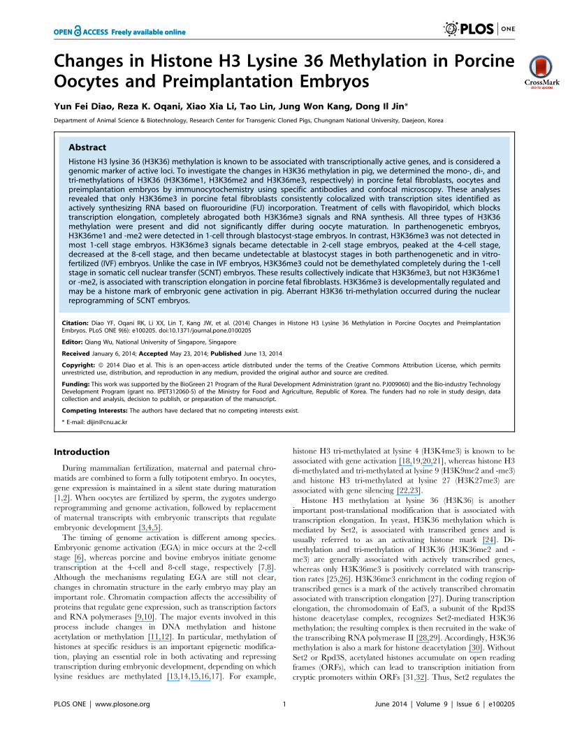
H3K36 Methylation Status and Association withTranscriptional Activity in Porcine Fetal Fibroblasts
It has been reported that H3K36 methylation is associated with
transcription in yeast [25]. Accordingly, to determine whether
H3K36 methylation is related to transcription in mammals, we
investigated H3K36 methylation status in porcine fetal fibroblasts
using H3K36 methylation status-specific antibodies and employed
5-fluorouridine (FU), a cell-permeable, modified RNA precursor,
to label active transcription sites. The cells were incubated with
2.5 mM FU in culture medium for 30 min to allow incorporation
of FU into nascent RNAs. Cells were then washed briefly with
PBS, fixed, and analyzed immunohistochemically using an
antibody specific for 5-bromo-deoxyuridine (BrdU) to detect FU
labeling, and antibodies against H3K36me1, -me2 or -me3, to
detect mono-, di- and tri-methylated H3K36, respectively. As
shown in Figure 1, all three types of methylations were observed in
interphase cells. H3K36me1 and H3K36me2 labeling was evenly
distributed throughout the nucleoplasm, but not in the nucleolus,
whereas H3K36me3 exhibited a dotted staining pattern in the
nucleoplasm. FU labeling corresponding to nascent transcription
site was distributed as dotted patten throughout the nucleus and
nucleolus. To analyze the association between H3K36 methyla-
tion and nascent RNA synthesis, we assayed one transcriptionally
active site (indicated by thick arrow) in each methylation group
indicated by a dot where nascent FU-labeled RNA had
accumulated. In both H3K36me1 and H3K36me2 groups,
transcription sites were not colocalized with methylation sites,
indicating that these two types of histone modifications are not
marks of transcriptional activity. In contrast, staining for
H3K36me3 always exhibited a very clear colocalization with
FU-labeled sites, suggesting that this modification may be
associated with transcriptional activity. To analyze the overall
overlap between FU and H3K36 methylation, we chose 30 dots
with strong FU labeling in the nucleoplasm for each methylation
group (H3K36me1, -me2 and -me3). We found that 80% of the
FU labeling dots were well colocalized with H3K36me3, while
only 13.3% and 6.7% of the FU labeling dots overlap with
H3K36me1 and -me2, respectively (Fig. 1B).
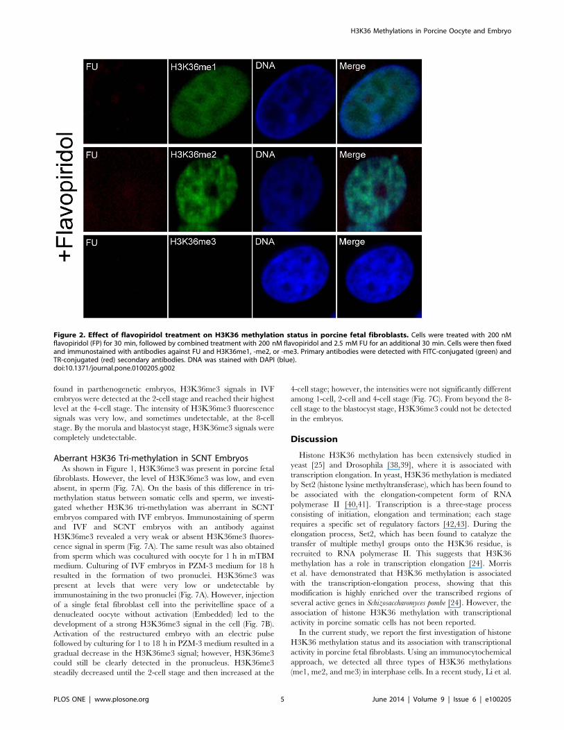
Effects of Flavopiridol on H3K36 Methylation andInactivation of Transcription in Porcine Fetal Fibroblasts
Flavopiridol is a cyclin-dependent kinase 9 (CDK9) inhibitor,
which inhibits transcription by RNA polymerase II in vitro and
in vivo by blocking the transition to productive elongation [36,37].
In preliminary experiments, we confirmed that 200 nM flavopir-
idol inhibited global transcription more efficiently than 100 nM in
porcine fetal fibroblast cells; the transcription could be completely
H3K36 Methylations in Porcine Oocyte and Embryo
PLOS ONE | www.plosone.org 3 June 2014 | Volume 9 | Issue 6 | e100205
abrogated. Thus, to further investigate the association of H3K36
methylations with transcription, we treated fetal fibroblasts with
200 nM flavopiridol to inhibit transcriptional activity. Cells were
treated with flavopiridol and FU and subsequently immunostained
with antibodies against BrdU (detecting FU labeling) and
H3K36me1, -me2, or -me3. The untreated cells were shown in
Figure 1. As shown in Figure 2, after treatment with flavopiridol,
transcriptional signals were undetectable. H3K36me3 staining was
also absent in these cells, whereas H3K36me1 and -me2 signals
still remained intact.
Changes in H3K36 Methylation Status during PorcineOocyte Maturation and Preimplantation Development ofParthenogenetic Embryos
H3K36 methylation status was examined in different stage
oocytes using antibodies directed against mono-, di-, or tri-
methylated lysines 36 on histone H3 (H3K36me1, -me2, -me3).
All three types of methylation on H3K36 were present in germinal
vesicle (GV) oocytes, including those with non-surrounded
nucleolus (NSN) and surrounded nucleolus (SN), as well as
metaphase I (MI) and metaphase II (MII) stage oocytes. The
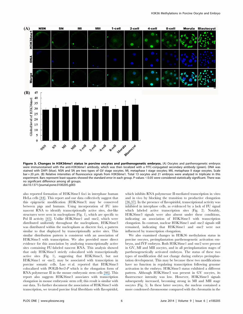
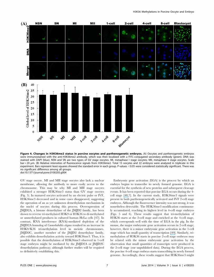
fluorescence intensities of H3K36me1 and -me2 were similar
among all oocyte stages (Figs. 3 and 4). In contrast, the
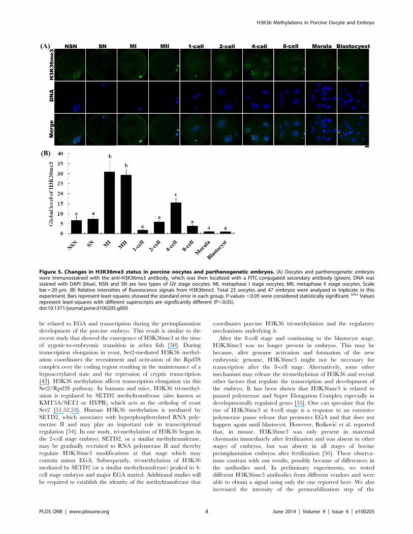
fluorescence intensity of H3K36me3 in NSN and SN oocytes
was lower than that in MI and MII oocytes (Fig. 5).
H3K36 methylation status was also examined in parthenoge-
netic embryos using antibodies against H3K36me1, -me2, or -
me3. H3K36me1 and -me2 signals were observed throughout all
preimplantation development stages, and showed no change in
methylation levels between developmental stages (Figs. 3 and 4).
However, H3K36me3 was only present in 2-cell to 8-cell stage
embryos (Fig. 5). H3K36me3 fluorescence signals rapidly
decreased after activation and were detected again at the 2-cell
stage. H3K36me3 signals increased gradually with the develop-
ment of the embryo until reaching a peak at the 4-cell stage.
H3K36me3 signals decreased to a very low level by the time the
embryo reached the 8-cell stage, and became undetectable in
morula and blastocyst stage embryos.
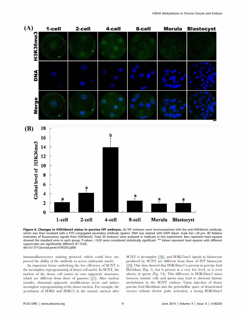
H3K36me3 Status in Porcine IVF EmbryosTo identify the dynamic changes in H3K36 tri-methylation in
porcine IVF embryos, we immunostained IVF embryos with
antibodies against H3K36me3. The results were similar to those
obtained with parthenogenetic embryos. Tri-methylation on
H3K36 was decreased soon after fertilization, during which the
zygotes underwent demethylation (Fig. 6). Similar to the pattern
Figure 1. H3K36 methylation status and association with transcriptional activity in porcine fetal fibroblasts. (A) Cells were cultured inmedium containing 2.5 mM FU for 30 min, fixed, and immunostained with antibodies against FU and H3K36me1, -me2 or -me3. Primary antibodieswere detected with FITC-conjugated (green) and TR-conjugated (red) secondary antibodies. DNA was stained with DAPI (blue). Enlargements(bottom insets) were the area pointed by thick arrows in merge insets showing transcriptional active sites (labeled with FU), H3K36 methylations andtheir overlap. The asterisk indicates the nucleolus. (B) The overall overlap rates between H3K36 methylations and transcriptional active sites wereanalyzed using total 30 dots indicated by the thin and thick arrows in (A).doi:10.1371/journal.pone.0100205.g001
H3K36 Methylations in Porcine Oocyte and Embryo
PLOS ONE | www.plosone.org 4 June 2014 | Volume 9 | Issue 6 | e100205
found in parthenogenetic embryos, H3K36me3 signals in IVF
embryos were detected at the 2-cell stage and reached their highest
level at the 4-cell stage. The intensity of H3K36me3 fluorescence
signals was very low, and sometimes undetectable, at the 8-cell
stage. By the morula and blastocyst stage, H3K36me3 signals were
completely undetectable.
Aberrant H3K36 Tri-methylation in SCNT EmbryosAs shown in Figure 1, H3K36me3 was present in porcine fetal
fibroblasts. However, the level of H3K36me3 was low, and even
absent, in sperm (Fig. 7A). On the basis of this difference in tri-
methylation status between somatic cells and sperm, we investi-
gated whether H3K36 tri-methylation was aberrant in SCNT
embryos compared with IVF embryos. Immunostaining of sperm
and IVF and SCNT embryos with an antibody against
H3K36me3 revealed a very weak or absent H3K36me3 fluores-
cence signal in sperm (Fig. 7A). The same result was also obtained
from sperm which was cocultured with oocyte for 1 h in mTBM
medium. Culturing of IVF embryos in PZM-3 medium for 18 h
resulted in the formation of two pronuclei. H3K36me3 was
present at levels that were very low or undetectable by
immunostaining in the two pronuclei (Fig. 7A). However, injection
of a single fetal fibroblast cell into the perivitelline space of a
denucleated oocyte without activation (Embedded) led to the
development of a strong H3K36me3 signal in the cell (Fig. 7B).
Activation of the restructured embryo with an electric pulse
followed by culturing for 1 to 18 h in PZM-3 medium resulted in a
gradual decrease in the H3K36me3 signal; however, H3K36me3
could still be clearly detected in the pronucleus. H3K36me3
steadily decreased until the 2-cell stage and then increased at the
4-cell stage; however, the intensities were not significantly different
among 1-cell, 2-cell and 4-cell stage (Fig. 7C). From beyond the 8-
cell stage to the blastocyst stage, H3K36me3 could not be detected
in the embryos.
Discussion
Histone H3K36 methylation has been extensively studied in
yeast [25] and Drosophila [38,39], where it is associated with
transcription elongation. In yeast, H3K36 methylation is mediated
by Set2 (histone lysine methyltransferase), which has been found to
be associated with the elongation-competent form of RNA
polymerase II [40,41]. Transcription is a three-stage process
consisting of initiation, elongation and termination; each stage
requires a specific set of regulatory factors [42,43]. During the
elongation process, Set2, which has been found to catalyze the
transfer of multiple methyl groups onto the H3K36 residue, is
recruited to RNA polymerase II. This suggests that H3K36
methylation has a role in transcription elongation [24]. Morris
et al. have demonstrated that H3K36 methylation is associated
with the transcription-elongation process, showing that this
modification is highly enriched over the transcribed regions of
several active genes in Schizosaccharomyces pombe [24]. However, the
association of histone H3K36 methylation with transcriptional
activity in porcine somatic cells has not been reported.
In the current study, we report the first investigation of histone
H3K36 methylation status and its association with transcriptional
activity in porcine fetal fibroblasts. Using an immunocytochemical
approach, we detected all three types of H3K36 methylations
(me1, me2, and me3) in interphase cells. In a recent study, Li et al.
Figure 2. Effect of flavopiridol treatment on H3K36 methylation status in porcine fetal fibroblasts. Cells were treated with 200 nMflavopiridol (FP) for 30 min, followed by combined treatment with 200 nM flavopiridol and 2.5 mM FU for an additional 30 min. Cells were then fixedand immunostained with antibodies against FU and H3K36me1, -me2, or -me3. Primary antibodies were detected with FITC-conjugated (green) andTR-conjugated (red) secondary antibodies. DNA was stained with DAPI (blue).doi:10.1371/journal.pone.0100205.g002
H3K36 Methylations in Porcine Oocyte and Embryo
PLOS ONE | www.plosone.org 5 June 2014 | Volume 9 | Issue 6 | e100205
also reported formation of H3K36me3 foci in interphase human
HeLa cells [44]. This report and our data collectively suggest that
this epigenetic modification (H3K36me3) may be conserved
between pigs and humans. Using incorporation of FU into
nascent RNA to identify transcriptionally active sites, dot-like
structures were seen in nucleoplasm (Fig. 1), which are specific to
Pol II activity [45]. Unlike H3K36me1 and -me2, which were
distributed uniformly throughout the nucleoplasm, H3K36me3
was distributed within the nucleoplasm as discrete foci, a pattern
similar to that displayed by transcriptionally active sites. This
similar distribution pattern is consistent with an association of
H3K36me3 with transcription. We also provided more direct
evidence for this association by analyzing transcriptionally active
sites containing FU-labeled nascent RNA. This analysis showed
that only H3K36me3 strictly colocalized with transcriptionally
active sites (Fig. 1), suggesting that H3K36me3, but not
H3K36me1 or -me2, may be associated with transcription in
porcine somatic cells. Luo et al. reported that H3K36me3
colocalized with POLII-Ser2-P which is the elongation form of
RNA polymerase II in the mouse embryonic stem cells [46]. This
report also suggests H3K36me3 associates with transcription
elongation in mouse embryonic stem cell; this result is similar with
our data. To further document the association of H3K36me3 with
transcription, we treated porcine fetal fibroblasts with flavopiridol,
which inhibits RNA polymerase II-mediated transcription in vitro
and in vivo by blocking the transition to productive elongation
[36,37]. In the presence of flavopiridol, transcriptional activity was
inhibited in interphase cells, as evidenced by a lack of FU signal
which labeled active transcription sites (Fig. 2). Notably,
H3K36me3 signals were also absent under these conditions,
indicating an association of H3K36me3 with transcription
elongation. In contrast, nuclear H3K36me1 and -me2 signals still
remained, indicating that H3K36me1 and -me2 were not
influenced by transcription elongation.
We also examined changes in H3K36 methylation status in
porcine oocytes, preimplantation parthenogenetic activation em-
bryos, and IVF embryos. Both H3K36me1 and -me2 were present
in GV, MI and MII oocytes, and in all preimplantation stages of
parthenogenetically activated embryos. The status of these two
types of modification did not change during embryo preimplan-
tation development. This may be because these two modifications
have no function in regulating transcription following genome
activation in the embryo. H3K36me3 status exhibited a different
pattern. Although H3K36me3 was present in GV oocytes, its
fluorescence intensity was low. However, H3K36me3 signals
subsequently increased, becoming strong in MI and MII stage
oocytes (Fig. 5). In these latter oocytes, the nucleus contained a
more condensed chromosome compared with the chromatin in the
Figure 3. Changes in H3K36me1 status in porcine oocytes and parthenogenetic embryos. (A) Oocytes and parthenogenetic embryoswere immunostained with the anti-H3K36me1 antibody, which was then localized with a FITC-conjugated secondary antibody (green). DNA wasstained with DAPI (blue). NSN and SN are two types of GV stage oocytes. MI, metaphase I stage oocytes; MII, metaphase II stage oocytes. Scalebar = 20 mm. (B) Relative intensities of fluorescence signals from H3K36me1. Total 13 oocytes and 21 embryos were analyzed in triplicate in thisexperiment. Bars represent least-squares showed the standard error in each group. P-values,0.05 were considered statistically significant. There wasno significant difference among all groups.doi:10.1371/journal.pone.0100205.g003
H3K36 Methylations in Porcine Oocyte and Embryo
PLOS ONE | www.plosone.org 6 June 2014 | Volume 9 | Issue 6 | e100205
GV stage oocyte. MI and MII stage oocytes also lack a nuclear
membrane, allowing the antibody to more easily access to the
chromosome. This may be why MI and MII stage oocytes
exhibited a stronger H3K36me3 status than GV stage oocytes
(Fig. 5). In matured oocytes activated by an electric pulse or IVF,
H3K36me3 decreased and in some cases disappeared, suggesting
the operation of an as yet unknown demethylation mechanism in
the nuclei of oocytes during this process. Overexpression of
JMJD2A, a histone demethylase of the JMJD2 family, has been
shown to reverse tri-methylated H3K9 or H3K36 to di-methylated
or unmethylated products in cultured human HeLa cells [47]. In
contrast, RNA interference (RNAi)-mediated depletion of the
JMJD2A homolog of Caenorhabditis elegans resulted in an increase in
H3K9/K36 tri-methylation level in meiotic chromosomes.
JMJD2C, another member of the JMJD2 demethylase family,
also exhibits demethylation activity toward H3K36me3. Thus, it is
possible that the demethylation of H3K36me3 observed in 1-cell
stage embryos might be mediated by the JMJD2A or JMJD2C
demethylation pathway; although further studies will be required
to definitively establishing this.
Embryonic gene activation (EGA) is the process by which an
embryo begins to transcribe its newly formed genome. EGA is
essential for the synthesis of new proteins and subsequent cleavage
events. It has been reported that porcine EGA occurs during the 4-
cell stage [48,7]. In the current study, H3K36me3 signals were
present in both parthenogenetically activated and IVF 2-cell stage
embryos. Although the fluorescence intensity was not strong, it was
nonetheless detectable. The H3K36me3 modification continuous-
ly accumulated, reaching its highest level in 4-cell stage embryos
(Figs. 5 and 6). These results suggest that tri-methylation of
H3K36 starts at the 2-cell stage and enriched at the 4-cell stage,
which corresponds well with the time of EGA in the pig. In the
mouse, the major embryonic gene activation occurs in 2-cell stage;
however, there is a minor embryonic gene activation in the 1-cell
stage which has small quantity of transcription [49]. Similarly, tri-
methylation of H3K36 starts in porcine 2-cell stage embryos may
be related with the minor embryonic gene activation by the
observation that small quantities of transcripts were produced in
the 2-cell stage (our unpublished data). During the EGA process,
the porcine 4-cell stage embryo starts transcribing its newly formed
genome. Accordingly, these results suggest that H3K36me3 might
Figure 4. Changes in H3K36me2 status in porcine oocytes and parthenogenetic embryos. (A) Oocytes and parthenogenetic embryoswere immunostained with the anti-H3K36me2 antibody, which was then localized with a FITC-conjugated secondary antibody (green). DNA wasstained with DAPI (blue). NSN and SN are two types of GV stage oocytes. MI, metaphase I stage oocytes; MII, metaphase II stage oocytes. Scalebar = 20 mm. (B) Relative intensities of fluorescence signals from H3K36me2. Total 15 oocytes and 22 embryos were analyzed in triplicate in thisexperiment. Bars represent least-squares showed the standard error in each group. P-values,0.05 were considered statistically significant. There wasno significant difference among all groups.doi:10.1371/journal.pone.0100205.g004
H3K36 Methylations in Porcine Oocyte and Embryo
PLOS ONE | www.plosone.org 7 June 2014 | Volume 9 | Issue 6 | e100205
be related to EGA and transcription during the preimplantation
development of the porcine embryo. This result is similar to the
recent study that showed the emergence of H3K36me3 at the time
of zygotic-to-embryonic transition in zebra fish [50]. During
transcription elongation in yeast, Set2-mediated H3K36 methyl-
ation coordinates the recruitment and activation of the Rpd3S
complex over the coding region resulting in the maintenance of a
hypoacetylated state and the repression of cryptic transcription
[42]. H3K36 methylation affects transcription elongation via this
Set2/Rpd3S pathway. In humans and mice, H3K36 tri-methyl-
ation is regulated by SETD2 methyltransferase (also known as
KMT3A/SET2 or HYPB), which acts as the ortholog of yeast
Set2 [51,52,53]. Human H3K36 methylation is mediated by
SETD2, which associates with hyperphosphorylated RNA poly-
merase II and may play an important role in transcriptional
regulation [54]. In our study, tri-methylation of H3K36 began in
the 2-cell stage embryo; SETD2, or a similar methyltransferase,
may be gradually recruited to RNA polymerase II and thereby
regulate H3K36me3 modifications at that stage which may
contain minor EGA. Subsequently, tri-methylation of H3K36
mediated by SETD2 (or a similar methyltransferase) peaked in 4-
cell stage embryos and major EGA started. Additional studies will
be required to establish the identity of the methyltransferase that
coordinates porcine H3K36 tri-methylation and the regulatory
mechanisms underlying it.
After the 8-cell stage and continuing to the blastocyst stage,
H3K36me3 was no longer present in embryos. This may be
because, after genome activation and formation of the new
embryonic genome, H3K36me3 might not be necessary for
transcription after the 8-cell stage. Alternatively, some other
mechanism may release the tri-methylation of H3K36 and recruit
other factors that regulate the transcription and development of
the embryo. It has been shown that H3K36me3 is related to
paused polymerase and Super Elongation Complex especially in
developmentally regulated genes [55]. One can speculate that the
rise of H3K36me3 at 4-cell stage is a response to an extensive
polymerase pause release that promotes EGA and that does not
happen again until blastocyst. However, Boskovic et al. reported
that, in mouse, H3K36me3 was only present in maternal
chromatin immediately after fertilization and was absent in other
stages of embryos, but was absent in all stages of bovine
preimplantation embryos after fertilization [56]. These observa-
tions contrast with our results, possibly because of differences in
the antibodies used. In preliminary experiments, we tested
different H3K36me3 antibodies from different vendors and were
able to obtain a signal using only the one reported here. We also
increased the intensity of the permeabilization step of the
Figure 5. Changes in H3K36me3 status in porcine oocytes and parthenogenetic embryos. (A) Oocytes and parthenogenetic embryoswere immunostained with the anti-H3K36me3 antibody, which was then localized with a FITC-conjugated secondary antibody (green). DNA wasstained with DAPI (blue). NSN and SN are two types of GV stage oocytes. MI, metaphase I stage oocytes; MII, metaphase II stage oocytes. Scalebar = 20 mm. (B) Relative intensities of fluorescence signals from H3K36me3. Total 23 oocytes and 47 embryos were analyzed in triplicate in thisexperiment. Bars represent least-squares showed the standard error in each group. P-values,0.05 were considered statistically significant. a,b,c Valuesrepresent least-squares with different superscripts are significantly different (P,0.05).doi:10.1371/journal.pone.0100205.g005
H3K36 Methylations in Porcine Oocyte and Embryo
PLOS ONE | www.plosone.org 8 June 2014 | Volume 9 | Issue 6 | e100205
immunofluorescence staining protocol, which could have im-
proved the ability of the antibody to access embryonic nuclei.
An important factor underlying the low efficiency of SCNT is
the incomplete reprogramming of donor cell nuclei. In SCNT, the
nucleus of the donor cell carries its own epigenetic structures,
which are different from those of gametes [57]. After nuclear
transfer, abnormal epigenetic modifications occur and induce
incomplete reprogramming of the donor nucleus. For example, the
acetylation of H4K8 and H4K12 in the somatic nucleus after
SCNT is incomplete [58], and H3K27me3 signals in blastocysts
produced by SCNT are different from those of IVF blastocysts
[59]. Our data showed that H3K36me3 is present in porcine fetal
fibroblasts (Fig. 1), but is present at a very low level, or is even
absent, in sperm (Fig. 7A). This difference in H3K36me3 status
between somatic cells and sperm may lead to aberrant histone
methylation in the SCNT embryo. Upon injection of donor
porcine fetal fibroblasts into the perivitelline space of denucleated
oocytes without electric pulse activation, a strong H3K36me3
Figure 6. Changes in H3K36me3 status in porcine IVF embryos. (A) IVF embryos were immunostained with the anti-H3K36me3 antibody,which was then localized with a FITC-conjugated secondary antibody (green). DNA was stained with DAPI (blue). Scale bar = 20 mm. (B) Relativeintensities of fluorescence signals from H3K36me3. Total 30 embryos were analyzed in triplicate in this experiment. Bars represent least-squaresshowed the standard error in each group. P-values ,0.05 were considered statistically significant. a,b Values represent least-squares with differentsuperscripts are significantly different (P,0.05).doi:10.1371/journal.pone.0100205.g006
H3K36 Methylations in Porcine Oocyte and Embryo
PLOS ONE | www.plosone.org 9 June 2014 | Volume 9 | Issue 6 | e100205
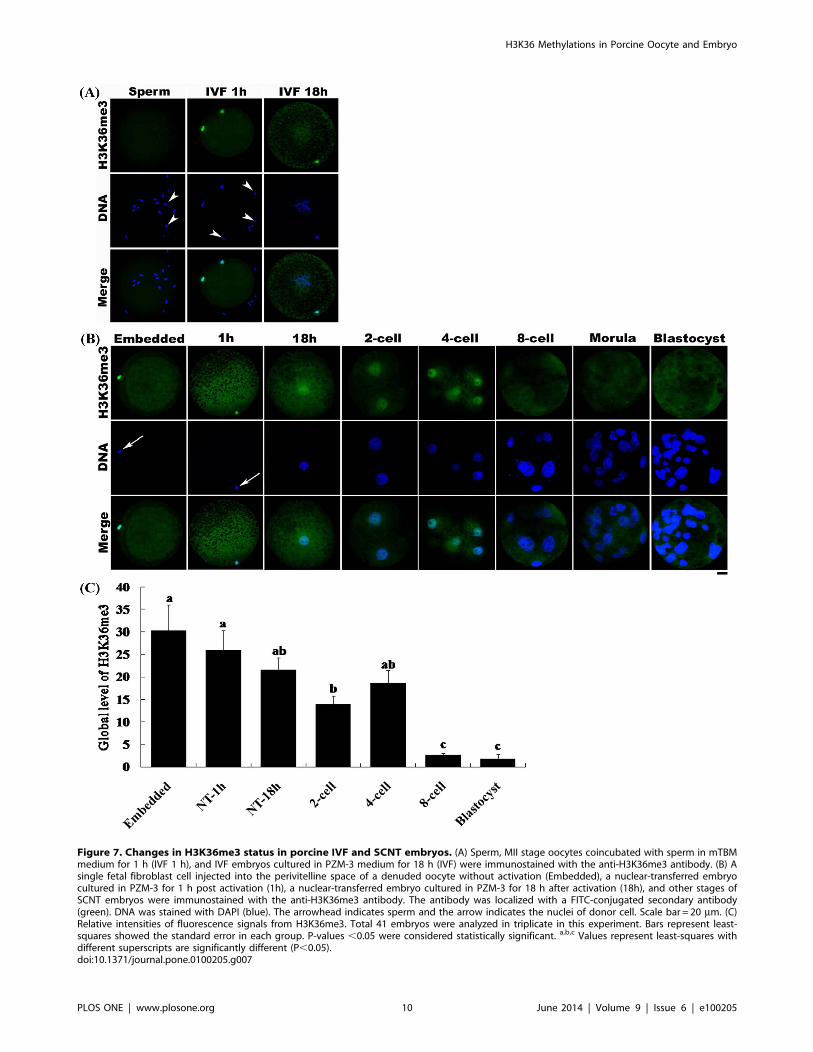
Figure 7. Changes in H3K36me3 status in porcine IVF and SCNT embryos. (A) Sperm, MII stage oocytes coincubated with sperm in mTBMmedium for 1 h (IVF 1 h), and IVF embryos cultured in PZM-3 medium for 18 h (IVF) were immunostained with the anti-H3K36me3 antibody. (B) Asingle fetal fibroblast cell injected into the perivitelline space of a denuded oocyte without activation (Embedded), a nuclear-transferred embryocultured in PZM-3 for 1 h post activation (1h), a nuclear-transferred embryo cultured in PZM-3 for 18 h after activation (18h), and other stages ofSCNT embryos were immunostained with the anti-H3K36me3 antibody. The antibody was localized with a FITC-conjugated secondary antibody(green). DNA was stained with DAPI (blue). The arrowhead indicates sperm and the arrow indicates the nuclei of donor cell. Scale bar = 20 mm. (C)Relative intensities of fluorescence signals from H3K36me3. Total 41 embryos were analyzed in triplicate in this experiment. Bars represent least-squares showed the standard error in each group. P-values ,0.05 were considered statistically significant. a,b,c Values represent least-squares withdifferent superscripts are significantly different (P,0.05).doi:10.1371/journal.pone.0100205.g007
H3K36 Methylations in Porcine Oocyte and Embryo
PLOS ONE | www.plosone.org 10 June 2014 | Volume 9 | Issue 6 | e100205
signal was observed (Fig. 7B). After activation and culture for 1 h,
H3K36me3 was still present in the restructured embryo. After
culturing for 18 h, a pronucleus formed in the restructured
embryo. H3K36me3 decreased to some extent; however, it still
could be clearly detected. During this stage, IVF embryos
underwent demethylation of H3K36me3 (Fig. 7A), whereas
aberrant tri-methylation of H3K36 occurred in SCNT embryos.
H3K36me3 could not be demethylated completely during the
early 1-cell stage in SCNT embryos. Interestingly, the H3K36me3
signal became weak at the 2-cell stage, indicating that the nucleus
of the restructured embryo was gradually demethylated by the
denucleated cytoplasm prior to this point. However, there were no
significant differences of the intensities of H3K36me3 among 1-
cell, 2-cell and 4-cell stage NT embryos (Fig. 7C); this result
indicates the demethylation of H3K36me3 in 1-cell stage of NT
embryo could not be sufficient compared with IVF or partheno-
genetic embryos. When the embryo reached the 4-cell stage,
H3K36me3 signals increased again and then decreased after the
8-cell stage, indicating demethylation. The changes in H3K36me3
status between the 4-cell and blastocyst stages were similar
between SCNT and IVF embryo.
Collectively, our data indicate that H3K36me3 is associated
with the elongation phase of transcription in porcine fetal
fibroblasts. Developmentally regulated H3K36me3, but not -
me1 or -me2, may be a histone mark of EGA in the pig. Aberrant
H3K36 tri-methylation occurs during the 1-cell stage in porcine
SCNT embryos. The mechanisms underlying the interaction of
H3K36me3 with transcription elongation and the effects of
abnormal H3K36 tri-methylation on the development of SCNT
embryos require further investigation.
Author Contributions
Conceived and designed the experiments: DIJ. Performed the experiments:
YFD RKO XXL. Analyzed the data: YFD RKO. Contributed reagents/
materials/analysis tools: TL JWK. Wrote the paper: YFD DIJ.
References
1. Worrad DM, Ram PT, Schultz RM (1994) Regulation of gene expression in themouse oocyte and early preimpantation embryo: developmental changes in Sp1
and TATA box-binding protein, TBP. Development 120: 2347–2357.
2. Latham KE, Schultz RM (2001) Embryonic genome activation. Front Biosci 6:
748–759.
3. Kanka J (2003) Gene expression and chromatin structure in the pre-
implantation embryo. Theriogenology 59: 3–19.
4. Hamatani T, Carter MG, Sharov AA, Ko MS (2004) Dynamics of global gene
expression changes during mouse preimplantation development. Dev Cell 6:
117–131.
5. Wang QT, Piotrowska K, Ciemerych MA, Milenkovic L, Scott MP, et al. (2004)
A genome-wide study of gene activity reveals developmental signaling pathwaysin the preimplantation mouse embryo. Dev Cell 6: 133–44.
6. Schultz M (1993) Regulation of zygotic gene activation in the mouse. Bioessays15: 531–538.
7. Bjerregaard B, Pedersen HG, Jakobsen AS, Rickords LF, Lai L, et al. (2007)Activation of ribosomal RNA genes in porcine embryos produced in vitro or by
somatic cell nuclear transfer. Mol Reprod Dev 74: 35–41.
8. Barnes FL, First NL. (1991) Embryonic transcription in vitro cultured bovine
embryos. Mol Reprod Dev 29: 117–123.
9. Mason K, Liu Z, Aguirre-Lavin T, Beaujean N (2012) Chromatin and epigenetic
modifications during early mammalian development. Anim Reprod Sci 134: 45–55.
10. Dillon N, Festenstein R (2002) Unravelling heterochromatin: competitionbetween positive and negative factors regulates accessibility. Trends Genet 18:
252–258.
11. Kouzarides T (2007) Chromatin modifications and their function. Cell 128:
693–705.
12. Li B, Carey M, Workman JL (2007) The role of chromatin during transcription.Cell 128: 707–719.
13. Zhang Y, Reinberg D (2001) Transcription regulation by histone methylation:interplay between different covalent modifications of the core histone tails.
Genes Dev 15: 2343–2360.
14. Martin C, Zhang Y (2005) The diverse functions of histone lysine methylation.
Nat Rev Mol Cell Biol 6: 838–849.
15. Ng HH, Robert F, Young RA, Struhl K (2003) Targeted recruitment of Set1
histone methylase by elongating Pol II provides a localized mark and memory ofrecent transcriptional activity. Mol Cell 11: 709–719.
16. Surani MA, Hayashi K, Hajkova P (2007) Genetic and epigenetic regulators ofpluripotency. Cell 128: 747–762.
17. Santos F, Peters AH, Otte AP, Reik W, Dean W (2005) Dynamic chromatinmodifications characterise the first cell cycle in mouse embryos. Dev Biol 280:
225–236.
18. Schneider R, Bannister AJ, Myers FA, Thorne AW, Crane-Robinson C, et al.
(2004) Histone H3 lysine 4 methylation patterns in higher eukaryotic genes. Nat
Cell Biol 6: 73–77.
19. Schubeler D, MacAlpine DM, Scalzo D, Wirbelauer C, Kooperberg C, et al.
(2004) The histone modification pattern of active genes revealed throughgenome-wide chromatin analysis of a higher eukaryote. Genes Dev 18: 1263–
1271.
20. Miao F, Natarajan R (2005) Mapping global histone methylation patterns in the
coding regions of human genes. Mol Cell Biol 25: 4650–4661.
21. Zhao XD, Han X, Chew JL, Liu J, Chiu KP, et al. (2007) Whole-genome
mapping of histone H3 Lys4 and 27 trimethylations reveals distinct genomiccompartments in human embryonic stem cells. Cell Stem Cell 1: 286–298.
22. Sims RJ III, Nishioka K, Reinberg D (2003) Histone lysine methylation: asignature for chromatin function. Trends Genet 19: 629–639.
23. Pan G, Tian S, Nie J, Yang C, Ruotti V, et al. (2007) Whole-genome analysis of
histone H3 lysine 4 and lysine 27 methylation in human embryonic stem cells.Cell Stem Cell 1: 299–312.
24. Morris SA, Shibata Y, Noma K, Tsukamoto Y, Warren E, et al. (2005) HistoneH3 K36 methylation is associated with transcription elongation in Schizosac-
charomyces pombe, Eukaryot Cell 4: 1446–1454.
25. Pokholok DK, Harbison CT, Levine S, Cole M, Hannett NM, et al. (2005)Genome-wide map of nucleosome acetylation and methylation in yeast. Cell
122: 517–527.
26. Rao B, Shibata Y, Strahl BD, Lieb JD (2005) Dimethylation of histone H3 atlysine 36 demarcates regulatory and nonregulatory chromatin genome-wide.
Mol Cell boil 25: 9447–9459.
27. Barski A, Cuddapah S, Cui K, Roh TY, Schones DE, et al. (2007) High-resolution profiling of histone methylation in the human genome. Cell 129: 823–
837.
28. Joshi AA, Struhl K (2005) Eaf3 chromodomain interaction with methylated H3–
K36 links histone deacetylation to Pol II elongation. Mol Cell 20: 971–978.
29. Keogh MC, Kurdistani SK, Morris SA, Ahn SH, Podolny V, et al. (2005)Cotranscriptional set2 methylation of histone H3 lysine 36 recruits a repressive
Rpd3 complex. Cell 123: 593–605.
30. Lee JS, Shilatifard A (2007) A site to remember: H3K36 methylation a mark forhistone deacetylation. Mutation Research 618: 130–134.
31. Carrozza MJ, Li B, Florens L, Suganuma T, Swanson SK, et al. (2005) Histone
H3 methylation by Set2 directs deacetylation of coding regions by Rpd3S tosuppress spurious intragenic transcription. Cell 123: 581–592.
32. Li B, Gogol M, Carey M, Pattenden SG, Seidel C, et al. (2007) Infrequentlytranscribed long genes depend on the Set2/Rpd3S pathway for accurate
transcription. Genes Dev 21: 1422–1430.
33. Venkatesh S, Smolle M, Li H, Gogol MM, Saint M, et al. (2012) Set2methylation of histone H3 lysine 36 suppresses histone exchange on transcribed
genes. Nature 489: 52–455.
34. Li XX, Lee DS, Kim KJ, Lee JH, Kim EY, et al. (2013) Leptin and nonessentialamino acids enhance porcine preimplantation embryo development in vitro by
intracytoplasmic sperm injection. Theriogenology 79: 291–298.
35. Diao YF, Naruse KJ, Han RX, Li XX, Oqani RK, et al. (2013) Treatment offetal fibroblasts with DNA methylation inhibitors and/or histone deacetylase
inhibitors improves the development of porcine nuclear transfer-derivedembryos. Anim Reprod Sci 141: 164–171.
36. Chao SH, Fujinaga K, Marion JE, Taube R, Sausville EA, et al. (2000)
Flavopiridol inhibits P-TEFb and blocks HIV-1 replication. J Biol Chem 275:28345–28348.
37. Chao SH, Price DH (2001) Flavopiridol inactivates P-TEFb and blocks most
RNA polymerase II transcription in vivo. J Biol Chem 276: 31793–31799.
38. Stabell M, Larsson J, Aalen RB, Lambertsson A (2007) Drosophila dSet2
functions in H3-K36 methylation and is required for development. BiochemBiophys Res Commun 359: 784–789.
39. Bell O, Wirbelauer C, Hild M, Scharf AN, Schwaiger M, et al. (2007) Localized
H3K36 methylation states define histone H4K16 acetylation during transcrip-tional elongation in Drosophila. EMBO J 26: 4974–4984.
40. Gerber M, Shilatifard A (2003) Transcriptional elongation by RNA polymerase
II and histone methylation. J Biol Chem 278: 26303–26306.
41. Krogan NJ, Kim M, Tong A, Golshani A, Cagney G, et al. (2003) Methylation
of histone H3 by Set2 in Saccharomyces cerevisiae is linked to transcriptional
elongation by RNA polymerase II. Mol Cell Biol 23: 4207–4218.
42. Venkatesh S, Workman JL (2013) Set2 mediated H3 lysine 36 methylation:
regulation of transcription elongation and implications in organismal develop-
ment. WIREs Dev Biol doi: 10.1002/wdev.109.
H3K36 Methylations in Porcine Oocyte and Embryo
PLOS ONE | www.plosone.org 11 June 2014 | Volume 9 | Issue 6 | e100205
43. Sims RJ 3rd, Belotserkovskaya R, Reinberg D (2004) Elongation by RNA
polymerase II: the short and long of it. Genes Dev 18: 2437–2468.44. Li F, Mao GG, Tong D, Huang J, Gu LY, et al. (2013) The Histone Mark
H3K36me3 Regulates Human DNA Mismatch Repair through Its Interaction
with MutSa. Cell 153: 590–600.45. Xie SQ, Martin S, Guillot PV, Bentley DL, Pombo A (2006) Splicing speckles
are not reservoirs of RNA polymerase II, but contain an inactive form,phosphorylated on serine2 residues of the C-terminal domain. Mol Biol Cell 17:
1723–1733.
46. Luo L, Gassman KL, Petell LM, Wilson CL, Bewersdorf J, et al. (2009) Thenuclear periphery of embryonic stem cells is a transcriptionally permissive and
repressive compartment. J Cell Sci 122: 3729–3737.47. Whetstine JR, Nottke A, Lan F, Huarte M, Smolikov S, et al. (2006) Reversal of
Histone Lysine Trimethylation by the JMJD2 Family of Histone Demethylases.Cell 125: 467–481.
48. Hyttel P, Laurincik J, Rosenkranz Ch, Rath D, Niemann H, et al. (2000)
Nucleolar proteins and ultrastructure in preimplantation porcine embryosdeveloped in vivo. Biol Reprod 63: 1848–1856.
49. Bellier S, Chastant S, Adenot P, Vincent M, Renard JP, et al. (1997) Nucleartranslocation and carboxyl-terminal domain phosphorylation of RNA polymer-
ase II delineate the two phases of zygotic gene activation in mammalian
embryos. EMBO J 16: 6250–6262.50. Vastenhouw NL, Zhang Y, Woods IG, Imam F, Regev A, et al. (2010)
Chromatin signature of embryonic pluripotency is established during genomeactivation. Nature 464: 922–926.
51. Yuan W, Xie J, Long C, Erdjument-Bromage H, Ding X, et al. (2009)Heterogeneous nuclear ribonucleoprotein L is a subunit of human KMT3a/Set2
complex required for H3 Lys-36 trimethylation activity in vivo. J Biol Chem
284: 15701–15707.52. Hu M, Sun XJ, Zhang YL, Kuang Y, Hu CQ, et al. (2010) Histone H3 lysine 36
methyltransferase Hypb/Setd2 is required for embryonic vascular remodeling.
Proc Natl Acad Sci USA 107: 2956–2961.53. Edmunds JW, Mahadevan LC, Clayton AL (2008) Dynamic histone H3
methylation during gene induction: HYPB/Setd2 mediates all H3K36trimethylation. EMBO J 27: 406–420.
54. Sun XJ, Wei J, Wu XY, Hu M, Wang L, et al. (2005) Identification and
Characterization of a Novel Human Histone H3 Lysine 36-specific Methyl-transferase. J Biol Chem 280: 35261–35271.
55. Lin C, Garrett AS, De Kumar B, Smith ER, Gogol M, et al. (2011) Dynamictranscriptional events in embryonic stem cells mediated by the super elongation
complex (SEC). Gene Dev 25: 1486–1498.56. Boskovic A, Bender A, Gall L, Ziegler-Birling C, Beaujean N, et al. (2012)
Analysis of active chromatin modifications in early mammalian embryos reveals
uncoupling of H2A.Z acetylation and H3K36 trimethylation from embryonicgenome activation. Epigenetics 7: 747–757.
57. Breton A, LE Bourhis D, Audouard C, Vignon X, Lelievre JM (2010) Nuclearprofiles of H3 histones trimethylated on Lys27 in bovine (Bos taurus) embryos
obtained after in vitro fertilization or somatic cell nuclear transfer. J Reprod Dev
56: 379–388.58. Wang F, Kou Z, Zhang Y, Gao S (2007) Dynamic reprogramming of histone
acetylation and methylation in the first cell cycle of cloned mouse embryos. BiolReprod 77: 1007–1016.
59. Zhang M, Wang F, Kou Z, Zhang Y, Gao S (2009) Defective chromatinstructure in somatic cell cloned mouse embryos. J Biol Chem 284: 24981–24987.
H3K36 Methylations in Porcine Oocyte and Embryo
PLOS ONE | www.plosone.org 12 June 2014 | Volume 9 | Issue 6 | e100205


